EP0945815A2 - Verfahren und Anlage zum Bestimmung von Risiken und Prognosen für einen gegebenen Kurs von medizinischer Behandlung - Google Patents
Verfahren und Anlage zum Bestimmung von Risiken und Prognosen für einen gegebenen Kurs von medizinischer Behandlung Download PDFInfo
- Publication number
- EP0945815A2 EP0945815A2 EP99301862A EP99301862A EP0945815A2 EP 0945815 A2 EP0945815 A2 EP 0945815A2 EP 99301862 A EP99301862 A EP 99301862A EP 99301862 A EP99301862 A EP 99301862A EP 0945815 A2 EP0945815 A2 EP 0945815A2
- Authority
- EP
- European Patent Office
- Prior art keywords
- risk
- course
- path
- brain
- tumour
- Prior art date
- Legal status (The legal status is an assumption and is not a legal conclusion. Google has not performed a legal analysis and makes no representation as to the accuracy of the status listed.)
- Withdrawn
Links
Images




Classifications
-
- G—PHYSICS
- G16—INFORMATION AND COMMUNICATION TECHNOLOGY [ICT] SPECIALLY ADAPTED FOR SPECIFIC APPLICATION FIELDS
- G16H—HEALTHCARE INFORMATICS, i.e. INFORMATION AND COMMUNICATION TECHNOLOGY [ICT] SPECIALLY ADAPTED FOR THE HANDLING OR PROCESSING OF MEDICAL OR HEALTHCARE DATA
- G16H50/00—ICT specially adapted for medical diagnosis, medical simulation or medical data mining; ICT specially adapted for detecting, monitoring or modelling epidemics or pandemics
- G16H50/30—ICT specially adapted for medical diagnosis, medical simulation or medical data mining; ICT specially adapted for detecting, monitoring or modelling epidemics or pandemics for calculating health indices; for individual health risk assessment
-
- A—HUMAN NECESSITIES
- A61—MEDICAL OR VETERINARY SCIENCE; HYGIENE
- A61N—ELECTROTHERAPY; MAGNETOTHERAPY; RADIATION THERAPY; ULTRASOUND THERAPY
- A61N5/00—Radiation therapy
- A61N5/10—X-ray therapy; Gamma-ray therapy; Particle-irradiation therapy
-
- G—PHYSICS
- G16—INFORMATION AND COMMUNICATION TECHNOLOGY [ICT] SPECIALLY ADAPTED FOR SPECIFIC APPLICATION FIELDS
- G16H—HEALTHCARE INFORMATICS, i.e. INFORMATION AND COMMUNICATION TECHNOLOGY [ICT] SPECIALLY ADAPTED FOR THE HANDLING OR PROCESSING OF MEDICAL OR HEALTHCARE DATA
- G16H20/00—ICT specially adapted for therapies or health-improving plans, e.g. for handling prescriptions, for steering therapy or for monitoring patient compliance
- G16H20/40—ICT specially adapted for therapies or health-improving plans, e.g. for handling prescriptions, for steering therapy or for monitoring patient compliance relating to mechanical, radiation or invasive therapies, e.g. surgery, laser therapy, dialysis or acupuncture
-
- G—PHYSICS
- G16—INFORMATION AND COMMUNICATION TECHNOLOGY [ICT] SPECIALLY ADAPTED FOR SPECIFIC APPLICATION FIELDS
- G16H—HEALTHCARE INFORMATICS, i.e. INFORMATION AND COMMUNICATION TECHNOLOGY [ICT] SPECIALLY ADAPTED FOR THE HANDLING OR PROCESSING OF MEDICAL OR HEALTHCARE DATA
- G16H50/00—ICT specially adapted for medical diagnosis, medical simulation or medical data mining; ICT specially adapted for detecting, monitoring or modelling epidemics or pandemics
- G16H50/20—ICT specially adapted for medical diagnosis, medical simulation or medical data mining; ICT specially adapted for detecting, monitoring or modelling epidemics or pandemics for computer-aided diagnosis, e.g. based on medical expert systems
-
- G—PHYSICS
- G16—INFORMATION AND COMMUNICATION TECHNOLOGY [ICT] SPECIALLY ADAPTED FOR SPECIFIC APPLICATION FIELDS
- G16H—HEALTHCARE INFORMATICS, i.e. INFORMATION AND COMMUNICATION TECHNOLOGY [ICT] SPECIALLY ADAPTED FOR THE HANDLING OR PROCESSING OF MEDICAL OR HEALTHCARE DATA
- G16H50/00—ICT specially adapted for medical diagnosis, medical simulation or medical data mining; ICT specially adapted for detecting, monitoring or modelling epidemics or pandemics
- G16H50/50—ICT specially adapted for medical diagnosis, medical simulation or medical data mining; ICT specially adapted for detecting, monitoring or modelling epidemics or pandemics for simulation or modelling of medical disorders
-
- A—HUMAN NECESSITIES
- A61—MEDICAL OR VETERINARY SCIENCE; HYGIENE
- A61N—ELECTROTHERAPY; MAGNETOTHERAPY; RADIATION THERAPY; ULTRASOUND THERAPY
- A61N5/00—Radiation therapy
- A61N5/10—X-ray therapy; Gamma-ray therapy; Particle-irradiation therapy
- A61N5/103—Treatment planning systems
Definitions
- the present invention relates, in general, to a method and system for assessing the risks and prognoses associated with a given course of medical treatment of neurological tumours, such as brain tumours.
- the present invention relates to a method and system for assessing the risks and prognoses associated with a given course of medical treatment of neurological tumours, such as brain tumours, wherein that given course of medical treatment spans multiple medical subspecialties.
- the brain consists of three main structures: the central brain stem, the cerebrum, and the cerebellum. Each such structure contains within it many other defined regions and/or substructures associated with specific brain function(s).
- the brain stem is divided into substructures. Such substructures include the thalamus, hypothalamus, and medulla oblongata.
- the thalamus is the relay station for incoming sensory signals and outgoing motor signals passing to and from the brain stem and cerebrum.
- the hypothalamus regulates or is involved directly in the control of eating, drinking, temperature regulation, sleep, emotional behaviour, sexual activity, and visceral functions.
- the medulla oblongata regulates and controls cardiac, vasoconstrictor, and respiratory functions, as well as other reflex activities, including vomiting.
- the cerebrum is the largest part of the human brain and is divided into several substructures. These substructures include, among others, the right and left cerebral hemispheres, each of which is divided by fissures and gyri (convolutions) into five lobes: the frontal, parietal, temporal, occipital, and insula lobes.
- the somatomotor area located just in front of what is known as the central fissure of one cerebral hemisphere, is responsible for nearly all voluntary movement of body muscles.
- the somatosensory area which is responsible for touch and taste, which is located just behind what is known as the central fissure of one cerebral hemisphere.
- the region of the cortex responsible for hearing is located in the upper, or superior, convolution of the temporal lobe of one cerebral hemisphere.
- the visual cortex the region responsible for seeing, is located in the occipital lobe of one cerebral hemisphere.
- the olfactory area the region responsible for smell, is located in the front, internal portion of the temporal lobe. Broca's area responsible for the muscle movements of the throat and mouth used in speaking, is located just beneath the motor area. The understanding of speech and reading has been associated with areas between the auditory and visual areas.
- the frontal area of the human cortex is responsible for awareness, intelligence, and memory.
- the cerebellum is essential to the control of movement of the human body in space. It acts as a reflex center for the coordination and precise maintenance of equilibrium. Voluntary muscle tone -- as related to posture, balance, and equilibrium -- is similarly controlled by the cerebellum. All motor activity depends on the cerebellum.
- a brain tumour is an abnormal growth, swelling, or enlargement in the brain.
- brain tumours such as those arising from the brain itself (e.g., astrocytoma, glioblastoma, oligodendroglioma, ependymoma), those arising from the brains coverings, or meninges, (e.g., meningiomas, pituitary tumours, pineal tumours), or those arising from nerves at the base of the brain (e.g., acoustic neuromas, schwannomas), and even tumours arising from outside the brain (metastatic brain tumours). This last case occurs when cancer cells travel through the bloodstream and lodge in the brain.
- Brain tumours can be malignant or benign.
- a malignant tumour is one that is actively destroying surrounding brain cells.
- a benign tumour is a mass or swelling that is growing, but is not destroying the surrounding brain cells. While a benign tumour in other organs is not ordinarily cause for alarm, a benign brain tumour is cause for alarm.
- the brain is encased in the cranium.
- the cranium is a dome-like vault of bone and cartilage that is essentially unyielding.
- cerebrospinal fluid Surrounding the brain is cerebrospinal fluid under a pressure, which supports the brain and protects it from injury. Cerebrospinal fluid is essentially incompressible, and thus the introduction of a tumour, even a benign one, into the cranial vault will require compression of the structures which can be compressed: the cells of the brain. Such compression ultimately will result in brain injury, and thus even benign tumours must be removed as quickly as possible. Thus, even a benign tumour can cause severe damage and must be removed quickly and in its entirety. Thus, irrespective of whether a brain tumour is malignant or benign, an active course of treatment must be engaged in to remove the tumour.
- tumours are significantly different from other types of tumours, and hence are uniquely difficult to remove.
- brain tumours are in/on the brain, and thus reaching and removing the tumour with surgical instruments gives rise to the risk that eloquent brain areas will be damaged in either the process of reaching the tumour or removing it.
- a second reason is that brain tumours are not like ordinary tumours: brain tumours are polyclonal, which means that what appears to be one tumour is actually many (sometimes over a thousand) tumour clones co-located in one area. Consequently, true tumour margins do not exist and consequently total removal by local therapy (surgery, radiation, heat, cold, etc.) is not possible.
- a third reason is that the brain is separated from the blood-stream by the blood-brain barrier, and consequently many blood-born chemotherapeutic agents cannot reach the brain via the blood-stream.
- a fourth reason is that many brain tumour cells live in a low oxygen (hypoxic) environment (hypoxic), and it has been found empirically that these hypoxic cells are: (1) radio-resistant; (2) often chemotherapy resistant; and (3) far from the blood supply.
- hypoxic cells live in a low oxygen (hypoxic) environment (hypoxic)
- tumour weighs about 100 grams.
- 100 gm of tumour typically has approximately 100 billion cells.
- a typical tumour can double in size and volume in a matter of weeks, from a course of treatment standpoint it makes sense to decrease the size of the mass of the tumour right away.
- Surgery is the preferred way of radically reducing the volume of a tumour, removing anywhere from 80-90% of the tumour mass.
- Recent advances in surgical technologies have aided in the removal of brain tumour tissue with a newer, higher net percentage tumour reduction of 90-99%.
- brain tumour treatment typically consists of following up the surgical therapy with radiotherapy and/or chemotherapy.
- any given course of treatment of brain tumours usually involves, at a minimum, some form of surgical intervention, plus some form of chemical therapy, plus some form of radiation therapy.
- one of the most significant risks is that of damage to the above-described eloquent brain areas.
- These risks are often closely related to the path taken by the surgical instruments from the outside of the cranium to the tumour within the brain, the location, the size of the tumour at the time of surgery, and the percentage of the tumour that is ultimately removed.
- one path to a tumour might have associated with it the risk of damage to the brain centers controlling feeling in one area of the body, while another path might have the risk damage to the brain centers wherein are contained the patient's individual identity.
- one path might have associated with it a risk of loss to a major eloquent area, while another path might have associated with it damage associated with a number of more minor surgical areas.
- chemotherapeutic agents With respect to chemotherapy, an extraordinary compendium of chemotherapeutic agents is under constant development at present, but as has been discussed, such agents have limited use due to the blood-brain barrier, and thus one risk associated with chemotherapy is the risk of not reaching the tumour. Another risk is that such therapies themselves tend to make the patient very sick, often times wiping out the immune system of the patient in to course of killing the tumour. Furthermore, selectivity (killing tumour cells while sparing healthy cells) is also a risk with chemotherapy. It is possible to surgically place such chemotherapeutic agents, but such placement moves one right back into the risks associated with therapy.
- a given course of treatment for a brain tumour has associated with it typically at least three major components: surgery, radiation, and chemotherapy. Furthermore, as has been discussed, each component associated with a given course of treatment has associated with it several risk factors. Consequently, the risks and prognoses associated with a given course of medical treatment are dependent upon the individual risks associated with each component of the treatment.
- possible courses of medical treatments of brain tumours have associated with them a dizzying array of variables, such as, to name just a few, the possible surgical paths to be taken to the tumour and the risks associated with same, the possible radiation paths to be travelled to the tumour and the risks associated with same, and the effectiveness of various radiation and chemotherapy associated with the tumour type and its locations, as well as side effects associated with such radiation therapy and chemotherapy.
- the method and system achieve their objects related to assessing a course of medical treatment by providing a data-processing system programmed to do at least the following: define a course of medical treatment, and calculate an assessment of the defined course of medical treatment.
- the defined course of medical treatment can include surgical intervention.
- Calculating the assessment of the defined course of medical treatment can include calculating the risk or risks associated with surgical intervention.
- Calculating the risk or risks associated with the surgical intervention can include defining one or more surgical paths to a brain tumour and calculating the risk or risks associated with the one or more defined surgical paths to the brain tumour.
- Calculating the risk or risks associated with the one or more surgical paths to the brain tumour can include associating a probability with at least one brain region and determining whether the at least one brain region, with which the at least one probability has been associated, is intersected by the at least one surgical path, and thereafter calculating a risk based upon the probability associated with the at least one brain region intersected.
- Associating at least one probability with at least one brain region can include defining at least one functional region within the brain, subdividing the at least one functional region into one or more subregions, and associating a risk-of-damage probability with each of the one or more subregions such that a higher probability indicates a concomitant loss of function of the at least one functional area within the brain.
- the present invention provides a method for providing artificial intelligence for assessing a course of medical treatment, the artificial intelligence to be provided by a data-processing system programmed to achieve the method, and the method comprising the steps of: defining a course of medical treatment; and in response to the step of defining, calculating an assessment of the course of medical treatment.
- the step of defining a course of medical treatment further includes the step of defining at least one course of surgical intervention, and preferably the step of calculating an assessment further includes the step of calculating at least one risk associated with the at least one course of surgical intervention.
- the step of calculating at least one risk associated with the at least one course of surgical intervention may suitably further include the steps of: defining at least one surgical path to a brain tumour; and calculating a risk associated with the at least one surgical path to the brain tumour.
- the step of calculating at least one risk associated with the at least one surgical path to the brain tumour preferably further includes the steps of: associating at least one probability with at least one brain region; determining whether the at least one brain region, with which the at least one probability has been associated, is intersected by the at least one surgical path; and in response to the step of determining, calculating a risk based upon the probability of the at least one brain region intersected.
- the step of associating at least one probability with at least one brain region preferably further comprises the steps of: defining at least one functional region within the brain, subdividing the at least one functional region into one or more subregions; and associating a risk-of-damage probability with each of the one or more subregions such that a higher probability indicates a concomitant loss of function of the at least one functional area within the brain.
- the step of defining a course of medical treatment aptly further includes the step of defining at least one course of radiation therapy, or at least one course of chemotherapy.
- the step of calculating an assessment of the course of medical treatment preferably further comprises the steps of: separating the course of medical treatment into one or more component parts; associating at least one risk with each component part; in response to the step of associating, calculating a risk for the course of medical treatment as a whole. More preferably the step of separating the course of medical treatment into one or more component parts further includes the step of separating the course of medical treatment into at least one component part selected from the group including a surgical component part, a chemical therapy component part, a radiation therapy component part, and an immunotherapy component part.
- the step of associating at least one risk with each component part preferably further includes the step of associating at least one risk selected from the group including tumour type, tumour size, tumour removal percentage, total path, eloquence removal percentage, eloquent path, closeness to eloquence, surgery time, chemotherapy factor, radiotherapy factor, radiation factor, patient age, neurosurgeon experience, medical staff skill, and medical facility goodness.
- the step carried out in response to the step of associating is that of calculating a prognosis, and this step preferably further includes the step of calculating a pre-operative prognosis or the step of calculating a post-operative prognosis.
- a system for providing artificial intelligence for assessing a course of medical treatment the artificial intelligence to be provided by a data-processing system programmed to achieve the assessing, the system comprising: means for defining a course of medical treatment; and means, responsive to the means for defining, for calculating an assessment of the course of medical treatment.
- the means for defining a course of medical treatment further include means for defining at least one course of surgical intervention, and preferably the means for calculating an assessment further includes means for calculating at least one risk associated with the at least one course of surgical intervention.
- the means for calculating at least one risk associated with the at least one course of surgical intervention may suitably further include: means for defining at least one surgical path to a brain tumour; and means for calculating a risk associated with the at least one surgical path to the brain tumour.
- the means for calculating at least one risk associated with the at least one surgical path to the brain tumour preferably further includes: means for associating at least one probability with at least one eloquent brain region; means for determining whether the at least one brain region, with which at least one probability has been associated, is intersected by the at least one surgical path; and means, responsive to the step of determining, for calculating a risk based upon the probability of the at least one brain region intersected.
- the means for associating at least one probability with at least one brain region preferably further comprises: means for defining at least one functional region within the brain, means for subdividing the at least one functional region into one or more subregions; and means for associating a risk-of-damage probability with each of the one or more subregions such that a higher probability indicates a concomitant loss of function of the at least one functional area within the brain.
- the means for defining a course of medical treatment further includes means for defining at least one course of radiation therapy, or at least one course of chemotherapy.
- the means for calculating an assessment of the course of medical treatment preferably further comprises: means for separating the course of medical treatment into component parts; means for associating at least one risk with each component part; and means, responsive to the step of associating, for calculating a risk for the course of medical treatment as a whole. More preferably the means for separating the course of medical treatment into one or more component parts further includes means for separating the course of medical treatment into at least one component part selected from the group including a surgical component part, a chemical therapy component part, a radiation therapy component part, and an immunotherapy component part.
- the means for associating at least one risk with each component part further includes means for associating at least one risk selected from the group including tumour type, tumour size, tumour removal percentage, total path, eloquence removal percentage, eloquent path, closeness to eloquence, surgery time, chemotherapy factor, radiotherapy factor, radiation factor, patient age, neurosurgeon experience, medical staff skill, and medical facility goodness.
- the means responsive to the means for associating further includes means for calculating a prognosis, and this means preferably includes means for calculating a pre-operative prognosis or a means for calculating a post-operative prognosis.
- the term artificially intelligent means simulation by a computer system of functions that are normally associated with human intelligence, such as a computer system programmed (by hardware, software, firmware, etc.) to perform functions that are normally associated with human intelligence; furthermore, as used herein the term artificially intelligent, and analogues thereof, also include expert systems, which are computational systems that provide for solving problems in a particular application area by drawing inferences from a knowledge base (a computer database that contains information about human experience in a particular field of knowledge and data resulting from solution of problems that have been encountered) by utilizing a computer system programmed to process information pertaining to a particular application.
- a knowledge base a computer database that contains information about human experience in a particular field of knowledge and data resulting from solution of problems that have been encountered
- the intelligent neurosurgery framework provides services during presurgery, surgery, and postsurgery phases. In what follows the basic principles of this framework will be described.
- Figure 1 shows an overall intelligent neurosurgery framework 100 .
- the user (which can be a human user or a machine user, such as a computer supplying the input) inputs data 102 into this framework.
- the outputs from framework 100 are presurgical plans 104 , surgical paths 106 , and postsurgery prognosis 108.
- the major components of the framework shown in Figure 1 are Static Planning Mechanism 110, Dynamic Planning Mechanism 112 , Rule Base 114 , Risk Assessment Mechanism 116, Reasoning Fact Base 118, and User Interface 120 .
- Static planning mechanism 110 coordinates with risk assessment mechanism 116 and rule base 114 and is used during presurgery planning.
- this tool can be used to both assess the risk of surgery, and also to find the least risky or optimum path to the tumour.
- Dynamic planning mechanism 112 coordinates with risk assessment mechanism 116 and rule base 114 and is used during surgery. During surgery this mechanism (dynamic planning mechanism 112 ) can be used to provide real-time risk assessment. This risk assessment can be used with existing neurosurgery guidance systems to reduce the risk involved with neurosurgery.
- Rule base 114 has inbuilt rules to guide static planning reasoning mechanism 111 , dynamic planning reasoning mechanism 113 , and risk assessment reasoning mechanism 117, included within static planning mechanism 110, dynamic planning mechanism 112, and risk assessment mechanism 116 respectively, intelligently.
- Risk assessment mechanism 116 uses probability spaces, in the fashion described below, derived from brain maps obtained by techniques such as functional MRIs. Risk assessment mechanism 116 can also be used for post surgery assessment of the prognosis and also for providing recommendations.
- Reasoning fact base 118 contains the various variables used to reason with. The tool uses the variables with inbuilt rules to reason intelligently.
- User interface 102 is used to communicate with the user.
- one or more component parts of frame work 100 are to be embedded in a neurosurgery guidance system, and in such embodiment user interface 120 will merely amount to communications between program entities, as is well understood by those in the art, and in such embodiment user interface 120 is expected to be developed by the overall neurosurgery guidance system.
- user interface 120 is implemented by means of standard computer entry mechanisms, such as keyboard entry, touch screen entry, voice recognition systems, etc.
- Reasoning factors are used by risk assessment mechanism 116 for uncertainty reasoning to calculate the risk of neurosurgery during presurgical planning, surgery, or post surgery prognosis.
- the following risk factors are used for uncertainty reasoning: tumour type, tumour size, tumour removal percentage, total path, eloquence removal percentage, eloquent path, closeness to eloquence, surgery time, chemotherapy factor, radiotherapy factor, radiation factor, patient age, neurosurgeon experience, medical staff skill, medical facility.
- the tumour type risk factor is a risk factor associated with the type and grade of a tumour (astrocytoma, glioblastoma).
- the tumour size risk factor is a risk factor associated with the size of a tumour.
- the tumour removal percentage risk factor is a risk factor associated with the percentage of the tumour that can/will be removed.
- the total path risk factor is determined, after a good path is found by the static planning mechanism 110 , by associating a risk factor with the total surgical path.
- the eloquence removal percentage risk factor is a risk factor associated with the percentage of an eloquent area that has to, or will, be removed.
- the eloquent path risk factor is a risk factor that is associated with the fact that a surgical path through an eloquent region cannot be avoided.
- the closeness to eloquence risk factor is risk factor that is associated with a path's closeness to eloquent regions, after such a path has been found by a planning mechanism.
- the surgery time risk factor is a risk factor associated with the total estimated surgery time.
- the chemotherapy risk factor is a risk factor associated with a given tumour's predicted non-responsiveness to chemotherapy.
- the radiotherapy risk factor is a risk factor associated with a given tumour's non-responsiveness to radiotherapy.
- the radiation risk factor is a risk factor associated with a given tumour's non-responsiveness to radiation.
- the patient age risk factor is a risk factor associated with a given patient's age.
- the neurosurgeon experience risk factor is risk factor associated with a neurosurgeon's inexperience either from a global perspective or with respect to a given procedure.
- the medical staff skill risk factor is a risk factor associated with medical staff skills.
- the medical facility risk factor is a risk factor associated with the goodness of the medical facility.
- Risk assessment reasoning is engaged in by static planning reasoning mechanism 111 , dynamic planning reasoning mechanism 113, and risk assessment reasoning mechanism 117 , included within static planning mechanism 110 , dynamic planning mechanism 112 , and risk assessment mechanism 116 to determine various and sundry numeric risks.
- the numeric risk associated with presurgery operations, surgery operations, and post surgery prognosis are based on the 15 risk variables described above.
- the risk variables are partitioned into two groups of risk variables: (1) Peripheral Variables, and (2) Path Variables.
- peripheral variables relate to those factors which make up the totality of the 'environment' in which a given medical procedure is performed, while path variables constitute, in the main, those variables which can be directly associated with specific factors directly related to a given course of medical treatment.
- the aggregate risk factor associated with the peripheral variables is based on the rules in the rule base.
- the peripheral variables are deemed to be tumour type, tumour size, chemotherapy factor, radiotherapy factor, radiation factor, neurosurgeon experience, medical facility, medical staff skill, and patient age.
- the risk factor score associated with each of these variables can be one of: 0, 10, 20, 50, 100.
- the assignment of these scores is based on the rule base which is built in conjunction with medical experts. As an example, if a patients age is 42 a risk score of 50 is assigned. The building of this rule base in conjunction with medical experts can be achieved via standard knowledge engineering techniques well known to those within the art.
- the aggregate risk factor associated with the path variables is based on the planning surgical paths algorithm shown below. In one embodiment, the path variables are deemed to be tumour removal factor, total path, eloquent path, closeness to eloquence, eloquent removal factor, and surgery time.
- the risk factor score associated with each of these variables can be one of. 0, 10, 20, 50, 100.
- the assignment of these scores is based on the planning surgical paths algorithm. Some of the data to this algorithm is provided by medical experts and incorporated via knowledge engineering techniques well known to those within the art.
- both the peripheral and path variables are further subdivided into three groups. These groups are (1) Presurgery Risk Group, (2) Surgical Operation Risk Group, and (3) Post Surgery Risk Group.
- the peripheral variables used to calculate the presurgery risk are tumour type, tumour size, neurosurgeon experience, medical facility, medical staff skill, and patient age. All these variables have a weight of 1. As a result the peripheral variables can contribute anywhere between 0 and 600 towards the presurgery risk. All the path variables are used to calculate the presurgery risk. Tumour removal factor, total path, and surgery time have a weight of 2. Eloquent path, and closeness to eloquence have a weight of 3. Eloquent removal factor has a weight of 4. These weights can be adjusted in conjunction with medical experts. Path variables can contribute from 0 to 1600 towards the presurgery risk. The total presurgery risk score can vary from 0 to 2200.
- the peripheral variables used to calculate the surgical operation risk are tumour type, tumour size, neurosurgeon experience, medical facility, medical staff skill, and patient age. All these variables have a weight of 1. As a result the peripheral variables can contribute anywhere between 0 and 600 towards the surgical operation risk. All the path variables are used to calculate the surgical operation risk. Tumour removal factor, total path, and surgery time have a weight of 2. Eloquent path, and closeness to eloquence have a weight of 3. Eloquent removal factor has a weight of 4. These weights can be adjusted in conjunction with medical experts. Path variables can contribute from 0 to 1600 towards the surgical operation risk. The total surgical operation risk score can vary from 0 to 2200.
- the peripheral variables used to calculate the postsurgery risk are tumour type, tumour size, chemotherapy factor, radiotherapy factor, radiation factor, and patient age. All these variables have a weight of 1. As a result, the peripheral variables can contribute anywhere between 0 and 600 towards the post surgery risk. All the path variables except surgery time are used to calculate the post surgery risk. All these variables have a weight of 1. Path variables can contribute from 0 to 500 towards the post surgery risk. The total post surgery risk score can vary from 0 to 1100.
- an overall risk factor is calculated.
- the overall risk factor is the combination of the presurgery, surgery, and post surgery risk factors.
- the overall risk score can vary between 0 and 5500.
- static planning reasoning mechanism 111 In an embodiment of present invention wherein risk assessment and/or static or dynamic planning are to be engaged in for course of medical treatment involving brain surgery, static planning reasoning mechanism 111 , dynamic planning reasoning mechanism 113, and risk assessment reasoning mechanism 117 all utilize an algorithm for uncertainty reasoning.
- the algorithm for uncertainty reasoning is central to reasoning with brain maps to provide risk assessment.
- This risk assessment can be static or dynamic and is based on the probability spaces concept. The concept of probability spaces is critical to the surgical navigation algorithm.
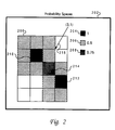
- each grid point of a brain map is defined to have a damage risk associated with it. This damage risk is estimated by how close the grid point is to an eloquence region. Each center of eloquence is assumed to be a source of very high damage risk which outwardly radiates damage risk. Also, a probability space can have a combined risk of two eloquence centers.
- Figure 2 depicts a two-dimensional illustration of the probability space concept. Shown in Figure 2 is a 4*5 grid 200 of a brain map with a key 202 depicting that black shading is representative of 1.0 probability 204, light shading is representative of .5 probability 206 , and darker shading is representative of .75 probability 208 . Depicted are two eloquence centers (dark shaded) 210 , 212 having a probability space (damage risk) of 1, and neighbouring cells having a probability space of 0.5. One grid cell 214 has a combined probability space of 0.75 (0.5 or 0.5). Also, illustrated in Figure 2, are rules for combining probabilities.
- a DOWN, DOWN step sequence has a damage risk of 1 (1 or 0.75); a RIGHT, DOWN step sequence starting at point (3,1) 216 has a damage risk of 0.5 (O or 0.5).
- Such calculations are used by the 2D-Step-Score procedure described below. While the risk calculations herein have been done using probabilistic combination rules for independent variables, embodiments are also envisioned where such risk calculations are based upon heuristically determined formulas when empirical experience dictates that the probabilistic combination rules do not produce acceptable risk assessments.
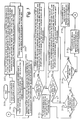
- Method step 300 depicts the start of the process.
- Method step 301 illustrates the location of a point indicative of the center of a tumour as that tumour appears within a brain map.
- Method step 303 shows that a line is drawn from the tumour to a body surface where the entry into the body is to be made such that the line is perpendicular to that body surface.
- a line is then posited as running parallel to the body surface and is associated with the x-axis, and the point where the line drawn from the tumour intersects the body surface is thereafter referred to as the "center” or "middle" of the x-axis.
- Method step 305 depicts that thereafter, utilizing tick marks, a minimum x-value and a maximum x-value are defined relative to the center, or middle, point.
- method step 301, 303 , and method step 305 define an x-y coordinate system relative to the location of the tumour and at least one body surface.
- Method step 302 illustrates setting a FLAG variable to Start and an x-coordinate variable equal to "middle" which equates to the point directly "over" the tumour in the defined x-y coordinate system.
- Method step 304 shows that a 2D path is determined by a 2D path plan process (described below) with the entry point into the body being the defined x-coordinate, and that after the 2D path plan is obtained it is given a numerical "path-score-center" by 2D score process (described below).
- method step 308 illustrates that if either FLAG is set to "start” or "positive” then the process proceeds to method step 309 wherein it is shown that a rightwards-x-coordinate value is obtained by incrementing x-coordinate one tick mark, or moving one tick mark to the right of x-coordinate, and a rightwards 2D path is determined by a 2D path plan process (described below) with the entry point into the body being the defined rightwards-x-coordinate, and that after the rightwards 2D path plan is obtained it is given a numerical score, "path-score-plus,” by 2D score process (described below).
- method step 310 shows that if either FLAG is set to "start” or “negative” then the process proceeds to method step 311 wherein it is shown that a leftwards-x-coordinate value is obtained by decrementing x-coordinate one tick mark, or moving one tick mark to the left of x-coordinate, and a leftwards 2D path is determined by a 2D path plan process (described below) with the entry point into the body being the defined leftwards-x-coordinate, and that after the leftwards 2D path plan is obtained it is given a numerical score, "path-score-minus,” by 2D score process (described below).
- method step 312 depicts that if "path-score-center” is less than both “path-score-plus” and “path-score_minus” then the process proceeds to method step 313 and stops with the path originating at the point where the x-coordinate was the middle or center is deemed the best path to the tumour. However, in the event that "path-score-center” was not less than either "path-score-plus” or "path-score-minus” then the process proceeds to method step 314 which illustrates the inquiry as to whether "path-score-minus” was less than "path-score-plus.”
- method step 316 shows that the x-coordinate value is reset to be equal to leftwards-x-coordinate and that FLAG is set to Negative; thereafter, the process returns to method step 304 and executes from that point.
- method step 318 depicts that the x-coordinate value is reset to be equal to rightwards-x-coordinate and that FLAG is set to Positive; thereafter, the process returns to method step 304 and executes from that point.
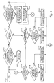
- Figure 4 depicts a high-level logic flow chart for the 2D path plan process referenced above.
- Method step 400 illustrates the start of the process.
- Method step 402 shows the inquiry of whether x-coordinate is not equal to that of the tumour (i.e., center or middle) or whether the y-coordinate is not equal to the location of the tumour (i.e., the x-y point chosen within the tumour from which a line was drawn to reach a body surface and thus establish an x-y coordinate system relative to the tumour).
- method step 404 shows that inquiry is made as to whether y-coordinate is still above the y-coordinate of the tumour.
- method step 406 shows the inquiry of whether a region having a high probability of risk of damage to eloquence is present directly below the current x-y coordinate.
- method step 408 that the process engages in the 2D reroute path process (described below), and reroutes around the high probability of damage to eloquence area.
- method step 409 shows that the y-coordinate is decremented by one tick; that is, is adjusted so that the y-coordinate is now one step nearer the y-coordinate of the tumour. Thereafter, the process returns to method step 402 and re-executes.
- method step 404 If the inquiry of method step 404 shows that y-coordinate is no longer above the y-coordinate of the tumour, the process proceeds to method step 410 which shows the inquiry as to whether x-coordinate is to the left of the x-coordinate of the tumour, in which case the process proceeds to method step 412 which shows the inquiry as to whether the next space to the right has a high probability of risk of damage to eloquence.
- method step 413 shows that the process engages in the 2D reroute path process (described below), and reroutes around the high probability of damage to eloquence area; however if the next space to the right does not have a high probability of damage to eloquence, method step 414 shows that a move one space to the right is added to the path. Thereafter, the process returns to method step 402 and re-executes.
- method step 416 depicts that the inquiry is made as to whether x-coordinate is to the right of the x-coordinate of the tumour, in which case the process proceeds to method step 418 which shows the inquiry as to whether the next space to the left has a high probability of risk of damage to eloquence.
- method step 419 shows that the process engages in the 2D reroute path process (described below), and reroutes around the high probability of eloquence area; however if the next space to the left does not have a high probability of damage to eloquence, method step 420 shows that a move one space to the left is added to the path. Thereafter, the process proceeds to method step 402 and re executes from that point.
- Method step 500 shows the start of the process.
- Method step 502 depicts receipt of a "path" or series of movements through a given brain from a surface location to a point defined to be the location of a tumour.
- Method step 504 illustrates that assessment of various risks (e.g, one or more risks of the type defined above) associated with the received path is performed by the risk assessment of path process (described below).
- Method step 506 shows that one or more of the risks assessed by the risk assessment of path process are then utilized to determine the path "score" (or a "raw risk factor,” a numerical value associated with risk consonant with the structure of numerical risk assessment discussed above) ; in one embodiment, this is achieved by setting “score” equal to “total path risk” + “closeness risk” + “eloquent path risk” (1 - “removal factor risk”) + “eloquent removal risk factor” * 10 + “surgery time risk” * 10), but those skilled in the art will recognize that this is just one among many possible score assessments that could be defined via processes well understood in the art by the system programmer.
- Method step 508 shows that the score for the received path is then returned as being associated with the received path.
- Method step 510 shows the end of the process.
- Figure 6 illustrates the risk assessment of path process referenced above.
- the risk assessment of path process which is part of risk assessment mechanism 116 calculates the risk score for each path variable being utilized. This is done by a simple table lookup in a manner well understood by those within the art.
- Method step 600 shows the start of the process.
- Method step 602 depicts the receipt of a "path" or series of movements through a given brain from a surface location to a point defined to be the location of a tumour.
- Method step 604 depicts initiation of modules which assess various risk via use of the path and table look up of the variable under consideration which are indicated as the path moves toward the tumour from the defined surface point; in one embodiment there are subroutines for total path risk, eloquent path risk, tumour removal risk, eloquent removal risk, eloquence closeness risk, and surgery time risk, but those skilled in the art will recognize that such risks are just exemplary and that the systems programmer can use/define more, less, or different risks utilizing the framework of the present invention.
- Method step 606 shows the return of risks associated with the risk variables in use for the defined path.
- Method step 608 shows the end of the process.
- Figure 7 depicts the 2D reroute path process referenced above.
- the 2D reroute path procedure calculates the next two least risky steps using probability spaces using the 2D step score procedure (described below) and adds them to the path. It first calculates the step-score exhaustively for all valid possible step sequences.
- An example of a valid 2-step sequence is DOWN, LEFT.
- the procedure decides what is a valid permutation. A permutation which has no net result movement like LEFT, RIGHT is not considered a valid permutation.
- Method step 700 depicts the start of the process.
- Method step 702 depicts the receipt of a partial "path" or series of movements through a given brain from a surface location to a point which has been reached and near which a region with an unacceptably high risk of damage is located.
- Method step 704 shows that all valid permutations from the defined location are tried, and for each valid permutation tried, a "2D step score" is associated with that permutation via the use of a "2D step score process” (described below); in one embodiment, these valid permutations are based upon two incremental movements from a present location, but those skilled in the art will recognize that any number of incremental movements can be utilized.
- Method step 706 depicts that upon determining the 2D step scores for all valid permutations of moves, the incremental movement (or combination of movements, such as when two movements are utilized) with the lowest “2D step score” is chosen as the "best move” or “best path step” to be taken next to reroute around the problem area, and that "best move” is thus returned as the appropriate reroute.
- Method step 708 shows the end of the process.
- Method step 800 shows the start of the process.
- Method step 802 depicts the receipt of one or more incremental movements to be taken from a given defined location; in one embodiment, the number of steps is equal to two, but any number of incremental movements could be proposed.
- Method step 804 illustrates that the probability of risk associated with each of the proposed incremental movement(s) is determined; in one embodiment, this is the probability associated with each of two incremental movements.
- Method step 806 depicts that a probability for the incremental movement(s) is calculated; in one embodiment, that probability is a "score" equal to "probability of risk of damage for first move” + “probability of risk of damage for second move” - “probability of risk of damage for first move” * “probability of risk of damage for second move.”
- Method step 808 illustrates that the calculated score for the proposed move(s) is then returned.
- Method step 810 depicts the end of the process.
- the static planning mechanism during presurgery planning to find an optimum or least risky path for tumour removal.
- the path specified starts at the skull and ends at the tumour face. The determination of this path depends upon the risk assessment using probability spaces. Once a path is chosen a presurgery risk factor is calculated by adding up all the risk factors associated with both path and peripheral variables.
- Table 1 shows an example of presurgery planning path variable risk factors.
- the total score is 520, given a peripheral variable score of 340.
- Path variables have at least a weight of 2.
- the closeness to eloquence and eloquent paths have a weight of 3, while the eloquent removal factor has a weight of 4.
- the foregoing specified weights and values are exemplary and those skilled in the art will recognize that other values and weights can be chosen in accordance with the spirit of the inventions disclosed herein.
- the processes disclosed in the above and following flowcharts can also be utilized to produce an artificially intelligent surgical aid to be used in real time during surgery.
- the primary use during surgery is to calculate the next least risky step given a surgeon request. It can also calculate the next two least risky steps given a surgeon request.
- risky and next risky steps can be predicated on certain defined “resolutions” or specified “path perspectives.”
- a local path perspective would utilize the standard spaces with the defined risks of damage probability
- the intermediate path perspective would create "groups” of spaces having defined risks of damage and assign an overall risk of damage to the group
- the global path perspective would define "groups” even larger than the intermediate groups, and define an overall probability to such defined groups dependent upon the probability of the spaces contained within the larger defined groups.
- groups would be utilized "as if" they were the smaller groups, meaning risk assessment, navigation, etc. would be done as normal except that the larger defined groups will be treated and utilized as the smaller regions are normally utilized.
- the surgeon can get a next-best-move assessment of at least three different levels of resolution. The calculation of risk is based on the probability space concept.
- Figure 9 shows a 2D dynamic path plan procedure which, in one embodiment returns the least risky next step or the least risky two-step sequence.
- the 2D dynamic path plan procedure uses the 2D reroute path process to get the 2-step sequence.
- the risk score associated with surgery is usually the same or similar to the presurgery score.
- the risk score associated with surgery changes if manual path changes or overrides are made by the surgeon using the procedure below. As an example, if no changes to the presurgery path were made during surgery the surgery risk score from Table 1 would be 520 .
- Method step 900 depicts the start of the process.
- Method step 902 illustrates the receipt of a current x-coordinate and y-coordinate and desired number of incremental movements; while the desired number of incremental movements could be any size, in one embodiment the number of incremental movements can be specified to be one or two incremental movements.
- Method step 904 depicts the inquiry as to whether the desired number of incremental movements is to be one. In the event that the desired number of incremental movements is to be one, then the process proceeds to method step 906 which depicts that the risks of damage of moves to the left, right, and down are checked, and that the move with the minimum risk of damage is returned.
- Method step 908 shows that the 2D reroute process, described above, is utilized to determine the next two least risky incremental movements, which are then returned.
- Method step 910 depicts the end of the process.
- the post surgery prognosis can be provided by using risk assessment mechanism 116.
- the postsurgery risk factor is calculated by adding up all the risk factors associated with the relevant peripheral and path variables.
- These relevant peripheral variables are tumour type, tumour size, chemotherapy factor, radiotherapy factor, radiation factor, and patient age.
- the relevant path variables are tumour removal factor, total path, eloquent path, closeness to eloquence, and eloquent removal factor.
- Table 2 below shows an example post surgery risk score and its contributing factors as would be calculated in one embodiment of the present invention.
- the total score in this example is 460.
- Example Post Surgery Risk Score Total Score 460 Variable Value Risk-Score Tumour Type Aristocytoma Grade II 50 Tumour Size Medium 50 Chemotherapy Non Responsive 100 Radiotherapy Mid Responsive 50 Radiation Responsive 20 Patient Age 55 100 Tumour Removal 80% 50 Total Path Short 20 Eloquent Path 0% 0 Eloquence Closeness 20% 20 Eloquent Removal 0% 0
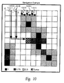
- Figure 10 shows an example of a 10 x 10 grid where the tumour is located at coordinate (4,5).
- the eloquence regions are marked by the light and dark grey shaded areas.
- the 2D static plan path process tries paths starting from (4,0) down to (1,0). The path from (5,0) and higher are not pursued. These five paths 1001 , 1002, 1003, 1004, 1005 are shown in Figure 10.
- Table 3 shows the raw risk factors associated with all five paths 1001, 1002, 1003, 1004, 1005 and the total raw risk factor score used to choose the least risky path. Based on the scores shown in Table 3, the path from (2,0) is chosen. Once this path is chosen the risk assessment path procedure, in the fashion discussed above, calculates the risk factors using lookup tables associated with the path variables of this path. Table 4 shows these risk factor scores for path variables.
- the artificially intelligent style mechanisms disclosed above can be used during presurgery planning, surgery, and post surgery prognosis.
- the tools disclosed herein can be used in conjunction with neurosurgery guidance tools.
- the development of such intelligent risk assessment and navigation tools should prove to be beneficial for the improvement of neurosurgery.
- These tools should enhance a neurosurgery guidance tool and reduce the risk of neurosurgery.
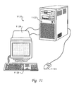
- FIG. 11 there is depicted a pictorial representation of a data-processing system which can be utilized in accordance with the method and system of an illustrative embodiment of the present invention.
- the method and system provided by an illustrative embodiment of the present invention can be implemented with the data-processing system depicted in Figure 11 .
- a computer 1120 is depicted which includes a system unit 1122 , a video display terminal 1124, a keyboard 1126, and a mouse 1128.
- Computer 1120 may be implemented utilizing any suitably powerful computer, such as commercially available mainframe computers, minicomputers, or microcomputers.
- FIG. 12 is an illustration of a representative hardware environment which can be utilized in accordance with the method and system of an illustrative embodiment of the present invention.
- Figure 12 depicts selected components in computer 1120 in which an illustrative embodiment of the present invention may be implemented.
- System unit 1122 includes a Central Processing Unit (“CPU”) 1231 , such as a conventional microprocessor, and a number of other units interconnected via system bus 1232.
- Computer 1120 includes random-access memory (“RAM”) 1234, read-only memory (“ROM”) 1236 , display adapter 1237 for connecting system bus 1232 to video display terminal 1124 , and I/O adapter 1239 for connecting peripheral devices (e.g., disk and tape drives 1233 ) to system bus 1232 .
- RAM random-access memory
- ROM read-only memory
- display adapter 1237 for connecting system bus 1232 to video display terminal 1124
- I/O adapter 1239 for connecting peripheral devices (e.g., disk and tape drives 1233 ) to system bus 12
- Video display terminal 1124 is the visual output of computer 1120 , which can be a CRT-based video display well-known in the art of computer hardware. However, with a portable or notebook-based computer, video display terminal 1124 can be replaced with an LCD-based or a gas plasma-based flat-panel display.
- Computer 1120 further includes user interface adapter 1240 for connecting keyboard 1126, mouse 1128, speaker 1246 , microphone 1248 , and/or other user interface devices, such as a touch screen device (not shown), to system bus 1232.
- Communications adapter 1249 connects computer 1120 to a data-processing network.
- Any suitable machine-readable media may retain the method and system of an illustrative embodiment of the present invention, such as RAM 1234 , ROM 1236 , a magnetic diskette, magnetic tape, or optical disk (the last three being located in disk and tape drives 1233 ).
- Any suitable operating system and associated graphical user interface e.g., Microsoft Windows
- the AIX operating system and AIXwindows windowing system i.e., graphical user interface
- the AIX operating system is IBM's implementation of the UNIX operating system.
- UNIX is a trademark of UNIX Systems Laboratories, Inc.
- the RISC System/6000 system among others, can run on the AIX operating system.
- control program 1151 contains instructions that when executed on CPU 1231 carries out one or more of the operations depicted in the logic flowcharts of Figures 3, 4, 5, 6, 7, 8, 9, and one or more of the associated operations depicted in schematic diagrams of Figures 1, 2, and 10 or any other illustrative example as described herein.
- FIG. 12 may vary for specific applications.
- peripheral devices such as optical disk media, audio adapters, or chip programming devices, such as PAL or EPROM programming devices well-known in the art of computer hardware, and the like may be utilized in addition to or in place of the hardware already depicted.
Landscapes
- Health & Medical Sciences (AREA)
- Engineering & Computer Science (AREA)
- Public Health (AREA)
- Medical Informatics (AREA)
- Biomedical Technology (AREA)
- General Health & Medical Sciences (AREA)
- Epidemiology (AREA)
- Primary Health Care (AREA)
- Pathology (AREA)
- Data Mining & Analysis (AREA)
- Databases & Information Systems (AREA)
- Nuclear Medicine, Radiotherapy & Molecular Imaging (AREA)
- Urology & Nephrology (AREA)
- Surgery (AREA)
- Radiology & Medical Imaging (AREA)
- Life Sciences & Earth Sciences (AREA)
- Animal Behavior & Ethology (AREA)
- Veterinary Medicine (AREA)
- Radiation-Therapy Devices (AREA)
- Medical Treatment And Welfare Office Work (AREA)
- Magnetic Resonance Imaging Apparatus (AREA)
Applications Claiming Priority (2)
| Application Number | Priority Date | Filing Date | Title |
|---|---|---|---|
| US09/046,875 US6112750A (en) | 1998-03-24 | 1998-03-24 | Method and system for assessing risks and prognoses of a given course of medical treatment |
| US46875 | 1998-03-24 |
Publications (2)
| Publication Number | Publication Date |
|---|---|
| EP0945815A2 true EP0945815A2 (de) | 1999-09-29 |
| EP0945815A3 EP0945815A3 (de) | 2001-02-14 |
Family
ID=21945868
Family Applications (1)
| Application Number | Title | Priority Date | Filing Date |
|---|---|---|---|
| EP99301862A Withdrawn EP0945815A3 (de) | 1998-03-24 | 1999-03-11 | Verfahren und Anlage zum Bestimmung von Risiken und Prognosen für einen gegebenen Kurs von medizinischer Behandlung |
Country Status (2)
| Country | Link |
|---|---|
| US (1) | US6112750A (de) |
| EP (1) | EP0945815A3 (de) |
Cited By (6)
| Publication number | Priority date | Publication date | Assignee | Title |
|---|---|---|---|---|
| WO2001056491A3 (en) * | 2000-02-04 | 2002-02-21 | Univ New York State Res Found | Computer aided treatment planning |
| EP1844725A1 (de) * | 2006-04-07 | 2007-10-17 | BrainLAB AG | Risikobewertung von geplanten Trajektorien |
| WO2009031073A3 (en) * | 2007-09-04 | 2009-08-20 | Koninkl Philips Electronics Nv | Multi-treatment planning apparatus and method |
| US8060181B2 (en) | 2006-04-07 | 2011-11-15 | Brainlab Ag | Risk assessment for planned trajectories |
| WO2015067299A1 (en) | 2013-11-05 | 2015-05-14 | Brainlab Ag | Quantification of brain vulnerability |
| CN110970133A (zh) * | 2019-12-10 | 2020-04-07 | 中国医学科学院肿瘤医院 | 一种crt风险评估方法和风险预测系统 |
Families Citing this family (14)
| Publication number | Priority date | Publication date | Assignee | Title |
|---|---|---|---|---|
| ITTO980593A1 (it) * | 1998-07-06 | 2000-01-06 | Olivetti Lexicon S P A | Dispositivo elettronico per il controllo di elementi in movimento di apparecchiature per il trattamento di testi e/o immagini |
| US6718342B1 (en) * | 2000-09-01 | 2004-04-06 | Real Time Boss, Inc. | Method and apparatus for network-enabled risk assessment |
| US20030171657A1 (en) * | 2002-01-22 | 2003-09-11 | Ralph Leonard | Selection of optimal medication methodology (SOOMM) |
| WO2003094114A1 (en) * | 2002-05-03 | 2003-11-13 | Koninklijke Philips Electronics N.V. | Method of producing and displaying an image of a 3 dimensional volume |
| US20090112538A1 (en) * | 2007-10-26 | 2009-04-30 | Joel Anderson | Virtual reality simulations for health care customer management |
| JP2012505007A (ja) * | 2008-10-10 | 2012-03-01 | コーニンクレッカ フィリップス エレクトロニクス エヌ ヴィ | 健康リスク値の決定及び/又は提示 |
| BRPI0918308A2 (pt) * | 2008-12-29 | 2020-08-04 | Koninklijke Philips Electronics N.V. | método para planejar um trajeto de acordo com uma aplicação cirúrgica, e, sistema para planejar um trajeto de acordo com a aplicação cirúrgica |
| US8708906B1 (en) * | 2011-09-07 | 2014-04-29 | Allen J. Orehek | Method for the prevention of dementia and Alzheimer's disease |
| WO2014139024A1 (en) * | 2013-03-15 | 2014-09-18 | Synaptive Medical (Barbados) Inc. | Planning, navigation and simulation systems and methods for minimally invasive therapy |
| SG10201707562PA (en) | 2013-03-15 | 2017-11-29 | Synaptive Medical Barbados Inc | Intramodal synchronization of surgical data |
| GB201616283D0 (en) * | 2016-09-26 | 2016-11-09 | Ucl Business Plc | A system and method for computer assisted planning of a trajectory for a surgical insertion into a skull |
| US12164547B2 (en) | 2019-04-02 | 2024-12-10 | Kpn Innovations, Llc. | Artificial intelligence advisory systems and methods for constitutional guidance |
| US11275985B2 (en) | 2019-04-02 | 2022-03-15 | Kpn Innovations, Llc. | Artificial intelligence advisory systems and methods for providing health guidance |
| CN112494053B (zh) * | 2020-12-23 | 2023-10-03 | 深圳市德力凯医疗设备股份有限公司 | 大脑的缺氧危险程度监控方法、系统、设备及存储介质 |
Family Cites Families (5)
| Publication number | Priority date | Publication date | Assignee | Title |
|---|---|---|---|---|
| US5205289A (en) * | 1988-12-23 | 1993-04-27 | Medical Instrumentation And Diagnostics Corporation | Three-dimensional computer graphics simulation and computerized numerical optimization for dose delivery and treatment planning |
| US5279309A (en) * | 1991-06-13 | 1994-01-18 | International Business Machines Corporation | Signaling device and method for monitoring positions in a surgical operation |
| US5417210A (en) * | 1992-05-27 | 1995-05-23 | International Business Machines Corporation | System and method for augmentation of endoscopic surgery |
| DE4304571A1 (de) * | 1993-02-16 | 1994-08-18 | Mdc Med Diagnostic Computing | Verfahren zur Planung und Kontrolle eines chirurgischen Eingriffs |
| US5343385A (en) * | 1993-08-17 | 1994-08-30 | International Business Machines Corporation | Interference-free insertion of a solid body into a cavity |
-
1998
- 1998-03-24 US US09/046,875 patent/US6112750A/en not_active Expired - Fee Related
-
1999
- 1999-03-11 EP EP99301862A patent/EP0945815A3/de not_active Withdrawn
Cited By (9)
| Publication number | Priority date | Publication date | Assignee | Title |
|---|---|---|---|---|
| WO2001056491A3 (en) * | 2000-02-04 | 2002-02-21 | Univ New York State Res Found | Computer aided treatment planning |
| EP1844725A1 (de) * | 2006-04-07 | 2007-10-17 | BrainLAB AG | Risikobewertung von geplanten Trajektorien |
| US8060181B2 (en) | 2006-04-07 | 2011-11-15 | Brainlab Ag | Risk assessment for planned trajectories |
| WO2009031073A3 (en) * | 2007-09-04 | 2009-08-20 | Koninkl Philips Electronics Nv | Multi-treatment planning apparatus and method |
| US8660800B2 (en) | 2007-09-04 | 2014-02-25 | Koninklijke Philips N.V. | Multi-treatment planning apparatus and method |
| US10521563B2 (en) | 2007-09-04 | 2019-12-31 | Koninklijke Philips N.V. | Multi-treatment planning apparatus and method |
| WO2015067299A1 (en) | 2013-11-05 | 2015-05-14 | Brainlab Ag | Quantification of brain vulnerability |
| CN110970133A (zh) * | 2019-12-10 | 2020-04-07 | 中国医学科学院肿瘤医院 | 一种crt风险评估方法和风险预测系统 |
| CN110970133B (zh) * | 2019-12-10 | 2023-03-21 | 中国医学科学院肿瘤医院 | 一种crt风险评估方法和风险预测系统 |
Also Published As
| Publication number | Publication date |
|---|---|
| US6112750A (en) | 2000-09-05 |
| EP0945815A3 (de) | 2001-02-14 |
Similar Documents
| Publication | Publication Date | Title |
|---|---|---|
| US6390097B1 (en) | Method and system for providing artificial intelligence for planning and risk assessment of surgical paths | |
| US6112750A (en) | Method and system for assessing risks and prognoses of a given course of medical treatment | |
| CN111615372B (zh) | 用于计划颅骨安装的医疗设备的植入的系统 | |
| JP6096167B2 (ja) | 脳内刺激のための標的体積を規定するシステムおよび方法 | |
| JP2021519120A (ja) | Ct/linacコンソールにおける適応放射線療法のための決定支援ツール | |
| Katsoulakis et al. | A detailed dosimetric analysis of spinal cord tolerance in high-dose spine radiosurgery | |
| KR101398881B1 (ko) | 방사선 치료 계획 장치 및 그 방법 | |
| US12491377B2 (en) | Apparatus comprising a memory, a control circuit, a user interface, and a radiation treatment platform to facilitate an administration of a knowledge-based radiation treatment plan | |
| Vakharia et al. | Computer-assisted planning for stereoelectroencephalography (SEEG) | |
| KR102849936B1 (ko) | 환자를 위한 상처 제품을 선택하는 방법 | |
| KR101401340B1 (ko) | 방사선 치료 계획 장치 및 그 방법 | |
| Picton et al. | Introduction of radiation therapist‐led adaptive treatments on a 1.5 T MR‐Linac | |
| Colombo et al. | Surgery versus radiosurgery for vestibular schwannoma: Shared decision making in a multidisciplinary clinic | |
| Khussainova et al. | Retrieval with clustering in a case-based reasoning system for radiotherapy treatment planning | |
| US20120041773A1 (en) | Computerized system for adaptive radiation therapy | |
| CN106687962B (zh) | 用于仿真大脑刺激的方法、对应的计算机程序和设备 | |
| CN119345620A (zh) | 一种用于提升放疗效果的辅助决策模型 | |
| CN117558408A (zh) | 一种基于数据拟合的消融仿真方法 | |
| CN117116421A (zh) | 放疗计划的确定方法和装置 | |
| CN115938587A (zh) | 放疗计划的验证方法、装置、计算机设备和存储介质 | |
| KR102885260B1 (ko) | 인지중재치료 수행 방법 | |
| Bacon | Towards Offline Adaptive Therapy for Prostate Patients Using Velocity | |
| Hays | The role of Addington v Texas on involuntary civil commitment | |
| CN118384428A (zh) | 自适应放疗方法、装置、医用加速器及可读存储介质 | |
| CN121221258A (zh) | 规划seeg电极置入路径的方法、装置及系统 |
Legal Events
| Date | Code | Title | Description |
|---|---|---|---|
| PUAI | Public reference made under article 153(3) epc to a published international application that has entered the european phase |
Free format text: ORIGINAL CODE: 0009012 |
|
| AK | Designated contracting states |
Kind code of ref document: A2 Designated state(s): DE FR GB IE |
|
| AX | Request for extension of the european patent |
Free format text: AL;LT;LV;MK;RO;SI |
|
| PUAL | Search report despatched |
Free format text: ORIGINAL CODE: 0009013 |
|
| AK | Designated contracting states |
Kind code of ref document: A3 Designated state(s): AT BE CH CY DE DK ES FI FR GB GR IE IT LI LU MC NL PT SE |
|
| AX | Request for extension of the european patent |
Free format text: AL;LT;LV;MK;RO;SI |
|
| 17P | Request for examination filed |
Effective date: 20010705 |
|
| AKX | Designation fees paid |
Free format text: DE FR GB IE |
|
| 17Q | First examination report despatched |
Effective date: 20011022 |
|
| GRAG | Despatch of communication of intention to grant |
Free format text: ORIGINAL CODE: EPIDOS AGRA |
|
| GRAG | Despatch of communication of intention to grant |
Free format text: ORIGINAL CODE: EPIDOS AGRA |
|
| STAA | Information on the status of an ep patent application or granted ep patent |
Free format text: STATUS: THE APPLICATION HAS BEEN WITHDRAWN |
|
| 18W | Application withdrawn |
Effective date: 20031218 |