EP0955882B1 - Vorrichtung zum positionieren eines chirurgischen roboters - Google Patents
Vorrichtung zum positionieren eines chirurgischen roboters Download PDFInfo
- Publication number
- EP0955882B1 EP0955882B1 EP96932222A EP96932222A EP0955882B1 EP 0955882 B1 EP0955882 B1 EP 0955882B1 EP 96932222 A EP96932222 A EP 96932222A EP 96932222 A EP96932222 A EP 96932222A EP 0955882 B1 EP0955882 B1 EP 0955882B1
- Authority
- EP
- European Patent Office
- Prior art keywords
- bone
- robotic
- probe
- data set
- image data
- Prior art date
- Legal status (The legal status is an assumption and is not a legal conclusion. Google has not performed a legal analysis and makes no representation as to the accuracy of the status listed.)
- Expired - Lifetime
Links
- 210000000988 bone and bone Anatomy 0.000 claims description 98
- 239000000523 sample Substances 0.000 claims description 46
- 210000000689 upper leg Anatomy 0.000 claims description 29
- 239000003550 marker Substances 0.000 claims description 13
- 239000012636 effector Substances 0.000 claims description 9
- 238000000034 method Methods 0.000 description 48
- 238000001356 surgical procedure Methods 0.000 description 34
- 239000007943 implant Substances 0.000 description 24
- 238000003384 imaging method Methods 0.000 description 10
- 238000011540 hip replacement Methods 0.000 description 9
- 238000012546 transfer Methods 0.000 description 7
- 230000001131 transforming effect Effects 0.000 description 7
- 230000009466 transformation Effects 0.000 description 6
- 210000004394 hip joint Anatomy 0.000 description 5
- 238000011541 total hip replacement Methods 0.000 description 5
- 238000000611 regression analysis Methods 0.000 description 4
- 230000006872 improvement Effects 0.000 description 3
- 238000013150 knee replacement Methods 0.000 description 3
- 238000012544 monitoring process Methods 0.000 description 3
- 230000003287 optical effect Effects 0.000 description 3
- 238000003325 tomography Methods 0.000 description 3
- 210000003484 anatomy Anatomy 0.000 description 2
- 230000001054 cortical effect Effects 0.000 description 2
- 238000013461 design Methods 0.000 description 2
- 238000011161 development Methods 0.000 description 2
- 210000001624 hip Anatomy 0.000 description 2
- 238000002513 implantation Methods 0.000 description 2
- 238000005259 measurement Methods 0.000 description 2
- 238000012986 modification Methods 0.000 description 2
- 230000004048 modification Effects 0.000 description 2
- 238000012545 processing Methods 0.000 description 2
- 238000002601 radiography Methods 0.000 description 2
- 206010028980 Neoplasm Diseases 0.000 description 1
- 210000000588 acetabulum Anatomy 0.000 description 1
- 238000013459 approach Methods 0.000 description 1
- 238000011882 arthroplasty Methods 0.000 description 1
- 230000009286 beneficial effect Effects 0.000 description 1
- 230000008901 benefit Effects 0.000 description 1
- 210000004556 brain Anatomy 0.000 description 1
- 238000012790 confirmation Methods 0.000 description 1
- 238000002059 diagnostic imaging Methods 0.000 description 1
- 206010015037 epilepsy Diseases 0.000 description 1
- 210000002436 femur neck Anatomy 0.000 description 1
- 210000002758 humerus Anatomy 0.000 description 1
- 210000000629 knee joint Anatomy 0.000 description 1
- 238000002595 magnetic resonance imaging Methods 0.000 description 1
- 239000000463 material Substances 0.000 description 1
- 238000012829 orthopaedic surgery Methods 0.000 description 1
- 230000000399 orthopedic effect Effects 0.000 description 1
- 230000008569 process Effects 0.000 description 1
- 238000001959 radiotherapy Methods 0.000 description 1
- 210000002320 radius Anatomy 0.000 description 1
- 238000002432 robotic surgery Methods 0.000 description 1
- 238000002603 single-photon emission computed tomography Methods 0.000 description 1
- 210000002303 tibia Anatomy 0.000 description 1
- 210000001519 tissue Anatomy 0.000 description 1
- 210000000623 ulna Anatomy 0.000 description 1
- 238000012800 visualization Methods 0.000 description 1
Images


Classifications
-
- A—HUMAN NECESSITIES
- A61—MEDICAL OR VETERINARY SCIENCE; HYGIENE
- A61F—FILTERS IMPLANTABLE INTO BLOOD VESSELS; PROSTHESES; DEVICES PROVIDING PATENCY TO, OR PREVENTING COLLAPSING OF, TUBULAR STRUCTURES OF THE BODY, e.g. STENTS; ORTHOPAEDIC, NURSING OR CONTRACEPTIVE DEVICES; FOMENTATION; TREATMENT OR PROTECTION OF EYES OR EARS; BANDAGES, DRESSINGS OR ABSORBENT PADS; FIRST-AID KITS
- A61F2/00—Filters implantable into blood vessels; Prostheses, i.e. artificial substitutes or replacements for parts of the body; Appliances for connecting them with the body; Devices providing patency to, or preventing collapsing of, tubular structures of the body, e.g. stents
- A61F2/02—Prostheses implantable into the body
- A61F2/30—Joints
- A61F2/46—Special tools for implanting artificial joints
-
- A—HUMAN NECESSITIES
- A61—MEDICAL OR VETERINARY SCIENCE; HYGIENE
- A61B—DIAGNOSIS; SURGERY; IDENTIFICATION
- A61B34/00—Computer-aided surgery; Manipulators or robots specially adapted for use in surgery
- A61B34/70—Manipulators specially adapted for use in surgery
-
- A—HUMAN NECESSITIES
- A61—MEDICAL OR VETERINARY SCIENCE; HYGIENE
- A61F—FILTERS IMPLANTABLE INTO BLOOD VESSELS; PROSTHESES; DEVICES PROVIDING PATENCY TO, OR PREVENTING COLLAPSING OF, TUBULAR STRUCTURES OF THE BODY, e.g. STENTS; ORTHOPAEDIC, NURSING OR CONTRACEPTIVE DEVICES; FOMENTATION; TREATMENT OR PROTECTION OF EYES OR EARS; BANDAGES, DRESSINGS OR ABSORBENT PADS; FIRST-AID KITS
- A61F2/00—Filters implantable into blood vessels; Prostheses, i.e. artificial substitutes or replacements for parts of the body; Appliances for connecting them with the body; Devices providing patency to, or preventing collapsing of, tubular structures of the body, e.g. stents
- A61F2/02—Prostheses implantable into the body
- A61F2/30—Joints
- A61F2/3094—Designing or manufacturing processes
- A61F2/30942—Designing or manufacturing processes for designing or making customized prostheses, e.g. using templates, CT or NMR scans, finite-element analysis or CAD-CAM techniques
-
- G—PHYSICS
- G16—INFORMATION AND COMMUNICATION TECHNOLOGY [ICT] SPECIALLY ADAPTED FOR SPECIFIC APPLICATION FIELDS
- G16H—HEALTHCARE INFORMATICS, i.e. INFORMATION AND COMMUNICATION TECHNOLOGY [ICT] SPECIALLY ADAPTED FOR THE HANDLING OR PROCESSING OF MEDICAL OR HEALTHCARE DATA
- G16H20/00—ICT specially adapted for therapies or health-improving plans, e.g. for handling prescriptions, for steering therapy or for monitoring patient compliance
- G16H20/40—ICT specially adapted for therapies or health-improving plans, e.g. for handling prescriptions, for steering therapy or for monitoring patient compliance relating to mechanical, radiation or invasive therapies, e.g. surgery, laser therapy, dialysis or acupuncture
-
- G—PHYSICS
- G16—INFORMATION AND COMMUNICATION TECHNOLOGY [ICT] SPECIALLY ADAPTED FOR SPECIFIC APPLICATION FIELDS
- G16H—HEALTHCARE INFORMATICS, i.e. INFORMATION AND COMMUNICATION TECHNOLOGY [ICT] SPECIALLY ADAPTED FOR THE HANDLING OR PROCESSING OF MEDICAL OR HEALTHCARE DATA
- G16H30/00—ICT specially adapted for the handling or processing of medical images
- G16H30/40—ICT specially adapted for the handling or processing of medical images for processing medical images, e.g. editing
-
- A—HUMAN NECESSITIES
- A61—MEDICAL OR VETERINARY SCIENCE; HYGIENE
- A61B—DIAGNOSIS; SURGERY; IDENTIFICATION
- A61B34/00—Computer-aided surgery; Manipulators or robots specially adapted for use in surgery
- A61B34/10—Computer-aided planning, simulation or modelling of surgical operations
-
- A—HUMAN NECESSITIES
- A61—MEDICAL OR VETERINARY SCIENCE; HYGIENE
- A61B—DIAGNOSIS; SURGERY; IDENTIFICATION
- A61B34/00—Computer-aided surgery; Manipulators or robots specially adapted for use in surgery
- A61B34/30—Surgical robots
-
- A—HUMAN NECESSITIES
- A61—MEDICAL OR VETERINARY SCIENCE; HYGIENE
- A61F—FILTERS IMPLANTABLE INTO BLOOD VESSELS; PROSTHESES; DEVICES PROVIDING PATENCY TO, OR PREVENTING COLLAPSING OF, TUBULAR STRUCTURES OF THE BODY, e.g. STENTS; ORTHOPAEDIC, NURSING OR CONTRACEPTIVE DEVICES; FOMENTATION; TREATMENT OR PROTECTION OF EYES OR EARS; BANDAGES, DRESSINGS OR ABSORBENT PADS; FIRST-AID KITS
- A61F2/00—Filters implantable into blood vessels; Prostheses, i.e. artificial substitutes or replacements for parts of the body; Appliances for connecting them with the body; Devices providing patency to, or preventing collapsing of, tubular structures of the body, e.g. stents
- A61F2/02—Prostheses implantable into the body
- A61F2/30—Joints
- A61F2/32—Joints for the hip
- A61F2/36—Femoral heads ; Femoral endoprostheses
- A61F2/3662—Femoral shafts
-
- A—HUMAN NECESSITIES
- A61—MEDICAL OR VETERINARY SCIENCE; HYGIENE
- A61F—FILTERS IMPLANTABLE INTO BLOOD VESSELS; PROSTHESES; DEVICES PROVIDING PATENCY TO, OR PREVENTING COLLAPSING OF, TUBULAR STRUCTURES OF THE BODY, e.g. STENTS; ORTHOPAEDIC, NURSING OR CONTRACEPTIVE DEVICES; FOMENTATION; TREATMENT OR PROTECTION OF EYES OR EARS; BANDAGES, DRESSINGS OR ABSORBENT PADS; FIRST-AID KITS
- A61F2/00—Filters implantable into blood vessels; Prostheses, i.e. artificial substitutes or replacements for parts of the body; Appliances for connecting them with the body; Devices providing patency to, or preventing collapsing of, tubular structures of the body, e.g. stents
- A61F2/02—Prostheses implantable into the body
- A61F2/30—Joints
- A61F2/46—Special tools for implanting artificial joints
- A61F2/4657—Measuring instruments used for implanting artificial joints
-
- A—HUMAN NECESSITIES
- A61—MEDICAL OR VETERINARY SCIENCE; HYGIENE
- A61F—FILTERS IMPLANTABLE INTO BLOOD VESSELS; PROSTHESES; DEVICES PROVIDING PATENCY TO, OR PREVENTING COLLAPSING OF, TUBULAR STRUCTURES OF THE BODY, e.g. STENTS; ORTHOPAEDIC, NURSING OR CONTRACEPTIVE DEVICES; FOMENTATION; TREATMENT OR PROTECTION OF EYES OR EARS; BANDAGES, DRESSINGS OR ABSORBENT PADS; FIRST-AID KITS
- A61F2/00—Filters implantable into blood vessels; Prostheses, i.e. artificial substitutes or replacements for parts of the body; Appliances for connecting them with the body; Devices providing patency to, or preventing collapsing of, tubular structures of the body, e.g. stents
- A61F2/02—Prostheses implantable into the body
- A61F2/28—Bones
- A61F2002/2825—Femur
-
- A—HUMAN NECESSITIES
- A61—MEDICAL OR VETERINARY SCIENCE; HYGIENE
- A61F—FILTERS IMPLANTABLE INTO BLOOD VESSELS; PROSTHESES; DEVICES PROVIDING PATENCY TO, OR PREVENTING COLLAPSING OF, TUBULAR STRUCTURES OF THE BODY, e.g. STENTS; ORTHOPAEDIC, NURSING OR CONTRACEPTIVE DEVICES; FOMENTATION; TREATMENT OR PROTECTION OF EYES OR EARS; BANDAGES, DRESSINGS OR ABSORBENT PADS; FIRST-AID KITS
- A61F2/00—Filters implantable into blood vessels; Prostheses, i.e. artificial substitutes or replacements for parts of the body; Appliances for connecting them with the body; Devices providing patency to, or preventing collapsing of, tubular structures of the body, e.g. stents
- A61F2/02—Prostheses implantable into the body
- A61F2/30—Joints
- A61F2002/30001—Additional features of subject-matter classified in A61F2/28, A61F2/30 and subgroups thereof
- A61F2002/30316—The prosthesis having different structural features at different locations within the same prosthesis; Connections between prosthetic parts; Special structural features of bone or joint prostheses not otherwise provided for
- A61F2002/30535—Special structural features of bone or joint prostheses not otherwise provided for
- A61F2002/30604—Special structural features of bone or joint prostheses not otherwise provided for modular
- A61F2002/30616—Sets comprising a plurality of prosthetic parts of different sizes or orientations
-
- A—HUMAN NECESSITIES
- A61—MEDICAL OR VETERINARY SCIENCE; HYGIENE
- A61F—FILTERS IMPLANTABLE INTO BLOOD VESSELS; PROSTHESES; DEVICES PROVIDING PATENCY TO, OR PREVENTING COLLAPSING OF, TUBULAR STRUCTURES OF THE BODY, e.g. STENTS; ORTHOPAEDIC, NURSING OR CONTRACEPTIVE DEVICES; FOMENTATION; TREATMENT OR PROTECTION OF EYES OR EARS; BANDAGES, DRESSINGS OR ABSORBENT PADS; FIRST-AID KITS
- A61F2/00—Filters implantable into blood vessels; Prostheses, i.e. artificial substitutes or replacements for parts of the body; Appliances for connecting them with the body; Devices providing patency to, or preventing collapsing of, tubular structures of the body, e.g. stents
- A61F2/02—Prostheses implantable into the body
- A61F2/30—Joints
- A61F2/3094—Designing or manufacturing processes
- A61F2/30942—Designing or manufacturing processes for designing or making customized prostheses, e.g. using templates, CT or NMR scans, finite-element analysis or CAD-CAM techniques
- A61F2002/30943—Designing or manufacturing processes for designing or making customized prostheses, e.g. using templates, CT or NMR scans, finite-element analysis or CAD-CAM techniques using mathematical models
-
- A—HUMAN NECESSITIES
- A61—MEDICAL OR VETERINARY SCIENCE; HYGIENE
- A61F—FILTERS IMPLANTABLE INTO BLOOD VESSELS; PROSTHESES; DEVICES PROVIDING PATENCY TO, OR PREVENTING COLLAPSING OF, TUBULAR STRUCTURES OF THE BODY, e.g. STENTS; ORTHOPAEDIC, NURSING OR CONTRACEPTIVE DEVICES; FOMENTATION; TREATMENT OR PROTECTION OF EYES OR EARS; BANDAGES, DRESSINGS OR ABSORBENT PADS; FIRST-AID KITS
- A61F2/00—Filters implantable into blood vessels; Prostheses, i.e. artificial substitutes or replacements for parts of the body; Appliances for connecting them with the body; Devices providing patency to, or preventing collapsing of, tubular structures of the body, e.g. stents
- A61F2/02—Prostheses implantable into the body
- A61F2/30—Joints
- A61F2/3094—Designing or manufacturing processes
- A61F2/30942—Designing or manufacturing processes for designing or making customized prostheses, e.g. using templates, CT or NMR scans, finite-element analysis or CAD-CAM techniques
- A61F2002/30948—Designing or manufacturing processes for designing or making customized prostheses, e.g. using templates, CT or NMR scans, finite-element analysis or CAD-CAM techniques using computerized tomography, i.e. CT scans
-
- A—HUMAN NECESSITIES
- A61—MEDICAL OR VETERINARY SCIENCE; HYGIENE
- A61F—FILTERS IMPLANTABLE INTO BLOOD VESSELS; PROSTHESES; DEVICES PROVIDING PATENCY TO, OR PREVENTING COLLAPSING OF, TUBULAR STRUCTURES OF THE BODY, e.g. STENTS; ORTHOPAEDIC, NURSING OR CONTRACEPTIVE DEVICES; FOMENTATION; TREATMENT OR PROTECTION OF EYES OR EARS; BANDAGES, DRESSINGS OR ABSORBENT PADS; FIRST-AID KITS
- A61F2/00—Filters implantable into blood vessels; Prostheses, i.e. artificial substitutes or replacements for parts of the body; Appliances for connecting them with the body; Devices providing patency to, or preventing collapsing of, tubular structures of the body, e.g. stents
- A61F2/02—Prostheses implantable into the body
- A61F2/30—Joints
- A61F2/3094—Designing or manufacturing processes
- A61F2/30942—Designing or manufacturing processes for designing or making customized prostheses, e.g. using templates, CT or NMR scans, finite-element analysis or CAD-CAM techniques
- A61F2002/30952—Designing or manufacturing processes for designing or making customized prostheses, e.g. using templates, CT or NMR scans, finite-element analysis or CAD-CAM techniques using CAD-CAM techniques or NC-techniques
-
- A—HUMAN NECESSITIES
- A61—MEDICAL OR VETERINARY SCIENCE; HYGIENE
- A61F—FILTERS IMPLANTABLE INTO BLOOD VESSELS; PROSTHESES; DEVICES PROVIDING PATENCY TO, OR PREVENTING COLLAPSING OF, TUBULAR STRUCTURES OF THE BODY, e.g. STENTS; ORTHOPAEDIC, NURSING OR CONTRACEPTIVE DEVICES; FOMENTATION; TREATMENT OR PROTECTION OF EYES OR EARS; BANDAGES, DRESSINGS OR ABSORBENT PADS; FIRST-AID KITS
- A61F2/00—Filters implantable into blood vessels; Prostheses, i.e. artificial substitutes or replacements for parts of the body; Appliances for connecting them with the body; Devices providing patency to, or preventing collapsing of, tubular structures of the body, e.g. stents
- A61F2/02—Prostheses implantable into the body
- A61F2/30—Joints
- A61F2/46—Special tools for implanting artificial joints
- A61F2002/4632—Special tools for implanting artificial joints using computer-controlled surgery, e.g. robotic surgery
- A61F2002/4633—Special tools for implanting artificial joints using computer-controlled surgery, e.g. robotic surgery for selection of endoprosthetic joints or for pre-operative planning
-
- Y—GENERAL TAGGING OF NEW TECHNOLOGICAL DEVELOPMENTS; GENERAL TAGGING OF CROSS-SECTIONAL TECHNOLOGIES SPANNING OVER SEVERAL SECTIONS OF THE IPC; TECHNICAL SUBJECTS COVERED BY FORMER USPC CROSS-REFERENCE ART COLLECTIONS [XRACs] AND DIGESTS
- Y10—TECHNICAL SUBJECTS COVERED BY FORMER USPC
- Y10S—TECHNICAL SUBJECTS COVERED BY FORMER USPC CROSS-REFERENCE ART COLLECTIONS [XRACs] AND DIGESTS
- Y10S128/00—Surgery
- Y10S128/92—Computer assisted medical diagnostics
- Y10S128/922—Computer assisted medical diagnostics including image analysis
Definitions
- the present invention is intended for registering the image of the long bone with the long bone itself immobilized within a system coordinate space.
- Long bones which may be imaged and registered include the femur, tibia, humerus, ulna, and radius. Image registration of such long bones will be particularly useful in conjunction with robotic surgical procedures, such as joint replacement, with specific procedures including total hip joint replacement, knee joint replacement, long bone osteotomy, and the like. Exemplary methods, systems, and apparatus for transforming an image data set of the femur within a robotic system intended for performing total hip replacement surgery are described hereinafter, but such descriptions are not intended to be limiting to the scope of the present invention.
- Methods, systems, apparatus are provided for transforming the image data set of the long bone to a system coordinate space, typically a robotic system intended to perform,or assist in any of the procedures described above.
- a system embodying the present invention is not limited to such robotic procedures and will be equally useful in manual surgical, diagnostic, and other medical procedures where it is necessary to align a pre-obtained image of a long bone within an actual coordinate space, such as an operative space.
- Such manual systems and procedures include computer-assisted surgical procedures that employ optical surgical measurement tools, passive electromechanical devices, and the like.
- the use of a system embodying the present invention is advantageous in that it will provide highly accurate image registration with an immobilized long bone without the need to preimplant multiple markers along the bone and/or surgically access the bone at multiple points along its length.
- a system embodying the present invention relies on obtaining an image of the bone using a conventional medical imaging technique, such as computerized tomography (CT), radiography (digitized X-ray images), magnetic resonance imaging (MRI), and the like.
- CT and radiographic imaging will be preferred since they provide particularly accurate imaging information of bone material.
- the image will be obtained in or converted to a digital form to produce an image data set which is suitable for digital manipulation using conventional computerized image processing equipment and software.
- the image processing equipment will be in the form of specially programmed computers, which are generally referred to as controllers and processors hereinafter.
- the present invention will utilize a preoperative planning work station (computer) for analyzing and manipulating raw image data which is obtained directly from the image itself.
- the raw image data set will be processed to include specific marker points or locations which are subsequently relied on to transform the image data set into the system coordinate space, as described in detail hereinafter.
- the marker locations may be identified by the user who views the image on the screen and marks particular locations on the image which are intended for alignment with the actual bone when the bone is immobilized in the system coordinate space.
- the preoperative planning workstation could be programmed to identify suitable marker locations without specific user intervention. In both cases, the marker locations will become part of the image data set which is subsequently transferred to the operative or other system in which the bone is to be immobilized.
- a system embodying the present invention relies particularly on obtaining axial and surface positional information on the bone and registering such information between the image data set and the system data set (representing the actual bone) as part of the image transformation process.
- center point data taken along the medullary canal in the image data set are obtained and compared to corresponding center points of the actual bone immobilized in the operative space.
- the center point data in both the image data set and the system data set will thus be non-linear and will require alignment by non-linear techniques, such as regression analysis.
- the center point data will be aligned by the robotic system at the same time as the surface data are aligned.
- Surface positional information will comprise one or more points on the exterior surface of the bone, typically near the proximal end so that the distal end need not be surgically exposed.
- the surface information will be a surface model generated from the image data set, and the surface model will be aligned with at least one, and preferably at least three points determined by the robotic system within the robotic field.
- the surface data in the image data set will be aligned with the point(s) by regression analysis.
- a particular advantage of a system embodying the present invention is the ability to transform the image data set without the need to surgically implant locating markers onto the bone.
- the ability to eliminate the markers derives largely from the use of the axial positional information which is obtained without the use of markers by the methods described in more detail below.
- the surface locational information of the bone will also be obtained without the use of markers.
- one or more surface markers may be attached to the bone and used to provide surface information in combination with the axial information obtained without the use of surface markers.
- use of single surface marker at one end of the long bone, typically at the head of the femur in hip replacement or the bottom of the femur in knee replacement will provide sufficient surface information for performing the transformation of the present invention.
- surface information is obtained in a plurality of positions over a surface region of the bone and the use of implanted markers is eliminated
- FIG. 1 An exemplary system 10 capable of implementing an example method for hip replacement surgery is illustrated in Fig. 1 .
- the system 10 includes both a presurgical planning workstation 12 and a library of implant designs 14 in the form of CAD models which are available from manufacturers on disks 15.
- a raw image data set 16, typically CT data, of the bone is obtained and transferred into the presurgical planning workstation 12.
- a single pin may be implanted in the proximal femur for determining a surface data point.
- the user typically the treating physician or an assistant working with the treating physician, is able to work at the presurgical planning workstation to . select and position a suitable implant design within the patient femur.
- the system 10 further comprises a robotic operative system 20 which includes a robotic controller 22 (typically a digital processor in the form of a programmable computer), an online display screen 24, and a robot 26. Details of the robotic operating system 20 are shown in Fig. 2 .
- the robot can be any conventional industrial robot having a manipulatable arm 28 preferably having at least 5 axes and capable of high precision placement.
- a suitable robotic is available from Sankyo Robotics with the model designation SR-5427-ISS.
- a force sensor 30 is mounted at the distal end of arm 28, and an effector in the form of a probe 32 or a surgical cutting tool (not illustrated) may be attached to the force sensor.
- the robotic system 20 further includes a safety processor 44, and a real time monitoring computer 46, as illustrated in Fig. 1 .
- the force sensor 30, the safety processor 44, the real time monitor 46, and a bone motion monitor 50 each help monitor the position, slippage, and blockage of the effector end of the manipulatable arm 28 while the femur 60 is held in place in a fixator assembly 52. Real time monitoring of these parameters helps assure that the robotic system is operating as planned. Details of these monitoring systems are described in the literature cited above which describes the ROBODOCTM robotic surgical system.
- system 10 architecture and the preoperative planning work station 12 and robotic operative system 20 are generally conventional. In order to practice the present invention, these systems must be modified as described hereinafter.
- a femur 60 comprises a head region 62 and a lower region 64.
- the trabecular bone 65 that is located adjacent the femoral head 62 and the cortical bone is located generally between the two ends of the bone.
- a neck region 68 is located just below the femoral head above the trabecular bone.
- the medullary canal 70 runs generally axially through the cortical bone region of the femur, as shown in broken line.
- the preoperative planning workstation 12 will produce a plurality of cross-sectional images on the viewing screen.
- the image data will delineate the periphery of the medullary canal of the long bone. Typically, from two to eight cross-sections will be produced, but in some cases it may be desirable to produce 12, 16, or even more images.
- the axial distances of each cross-section from the selected position of the implant tip are known.
- the user then identifies a center point in each of the cross-sectional images.
- Such selection could be done subjectively, i.e., by positioning a screen marker visually within the periphery of the medullary canal and marking the position in the image data set when it is selected.
- the system will produce elliptical templates which the user may position and size within each cross-sectional image.
- the elliptical templates can be rotated about their elliptical center and sized in both the major and minor diameters in order to match the periphery of the medullary canal as closely as possible.
- the system can then mark and store the center of the ellipse as the center of the medullary canal at that cross-sectional location.
- the system could be programmed to generate such cross-sectional information automatically, without specific user intervention.
- the center points are made part of the image data set and transferred to the robotic system 20.
- the image center points will then be aligned with actual canal center points (collected as described below) to provide for axial alignment of the image and the immobilized bone.
- At least one surface locational point will be identified within the image data set within the preoperative planning workstation 12. This could be done in a variety of ways. For example, the user could identify one or more specific locations on the exterior surface of the bone and mark them for storage within the image data set. Alternatively, a single data point could be relied on if a marker had been surgically preimplanted in the bone prior to imaging. In that case, the preoperative planning workstation could automatically identify the marker without intervention by the user. In the exemplary embodiment, the workstation 12 will generate a surface model of a portion of the exterior of the bone, usually representing the outer cortex and the proximal calcar region.
- the particular boundaries for the region may be determined by the user or may be calculated by the workstation 12 based on the implant placement which in turn determines the level of which the femoral head will be excised. While it would be possible to generate a surface model of the entire bone, it has been found that use of a small portion of the proximal calcar region is sufficient to provide accurate image registration without excessive computational time.
- the image data set including the identified positional coordinates, is then transferred to the robotic operative system 20 as part of a data transfer file 70 including the image information, implant shape data, and implant placement data. Transfer is conveniently accomplished via a transfer tape 71, but could be done using any conventional data transfer methodology. Additionally, the three-dimensional models of the bone and implant (implant files 14) are also transferred to the online display 24 of the robotic system 20 via the tape 71.
- the robotic operative system 20 is then operated to obtain positional information on the bone when the bone is immobilized within the robotic system.
- the patient will be prepared for hip replacement surgery in a conventional manner, and will be immobilized within the robotic operative system 20 generally as described in the literature related to the ROBODOCTM robotic operative system set forth above.
- the only unique aspects of the method relate to the acquisition of positional information which is to be used for registration with the positional information acquired as described above and incorporated into the image data set transferred to the robotic operative system 20.
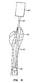
- the probe 32 as utilized to obtain canal center line information through the medullary canal 70.
- the surgeon performs a femoral head osteotomy and prepares the acetabulum in a conventional manner, except that the depth of the osteotomy may be at a higher level in order to retain more of the neck which includes the surface-model region of the bone.
- An access hole 80 is opened in the trabecular bone and the bone is then installed in the femoral fixator of the robotic operative system 20.
- the probe 32 then introduced into the medullary canal 70 by manually guiding the probe.
- the ability to manually guide the probe and cause the manipulator arm 28 to follow is well described in the literature describing the ROBODOCTM robotic operative system.
- the probe 32 is positioned so that an expansible distal end, illustrated as an inflatable balloon, is sequentially aligned at least two axially spaced-apart positions within the canal corresponding to the axial positions which have been cross-sectionally imaged in the preoperative planning session.
- an expansible distal end illustrated as an inflatable balloon
- the balloon is then deflated (or other expansible end reduced in size) and moved to the at least second position, as shown Fig.
- the probe will be located and centered at each of the cross-sectional locations which have been established during the preoperative planning procedure. Positional measurement of the probe will occur while the balloon is inflated and the robotic system is in a force-control mode that will move the probe tip in order to balance forces on the probe as sensed by the force sensor 30. The probe will be moved about the bone entry point, when the forces are balanced, the user will verify that the probe shaft is not contacting the proximal femur. Upon confirmation, the system will record the center point of the probe. This procedure is repeated at each level at which the center point location is determined.
- the probe 32 is used to collect surface locational information in the proximal calcar region 90 of the femur 60 as follows.
- the probe tip 33 will be engaged against at least one point on the anterior surface of the calcar region and one point on the posterior surface of the calcar region.
- the system will assure that the probe tip is force-balanced prior to recording the position in the robot controller.
- a greater number of points e.g., from 10 to 15 point, may be used in order to increase the accuracy of the transformation function which is produced.
- the robot controller 22 now has sufficient information to generate a transformation function which can be used to transform the image data set into robotic coordinates.
- the image data set can be used to control the manipulator arm 28 of the robot for performing the desired surgical procedure, e.g., creation of an implant cavity for receiving the prosthetic hip implant, as generally described in the earlier ROBODOCTM publications.
- the transfer file 72 received from the presurgical planning workstation 12 will include the implant data, canal center point data, surface model of the femoral neck region in a suitable file format, and all other planning information necessary to operate the robotic system 20.
- transformation of the image data set to the robotic coordinate system can be achieved by conventional mathematical techniques.
- the canal center points are fit to the robotically determined center points by conventional regression analysis.
- a plurality of robotically determined surface points are fit with the surface model, also by regression analysis.
Landscapes
- Health & Medical Sciences (AREA)
- Engineering & Computer Science (AREA)
- General Health & Medical Sciences (AREA)
- Public Health (AREA)
- Life Sciences & Earth Sciences (AREA)
- Medical Informatics (AREA)
- Animal Behavior & Ethology (AREA)
- Orthopedic Medicine & Surgery (AREA)
- Transplantation (AREA)
- Veterinary Medicine (AREA)
- Surgery (AREA)
- Nuclear Medicine, Radiotherapy & Molecular Imaging (AREA)
- Biomedical Technology (AREA)
- Heart & Thoracic Surgery (AREA)
- Cardiology (AREA)
- Vascular Medicine (AREA)
- Oral & Maxillofacial Surgery (AREA)
- Epidemiology (AREA)
- Primary Health Care (AREA)
- Manufacturing & Machinery (AREA)
- Geometry (AREA)
- Physics & Mathematics (AREA)
- Robotics (AREA)
- Radiology & Medical Imaging (AREA)
- Molecular Biology (AREA)
- Physical Education & Sports Medicine (AREA)
- Urology & Nephrology (AREA)
- Prostheses (AREA)
Claims (7)
- Robotersystem mit einem manipulativen Arm, der eine chirurgische Schneideeinrichtung aufweist, und einer programmierbaren Steuereinheit, welche die Schneideeinrichtung innehalb eines Roboter-Koordinatensystems positioniert, wobei ein Bilddatensatz, der eine Abbildung eines langen, eine Achse definierenden Knochens repräsentiert, in das Roboter-Koordinatensystem transformiert wird, und mit Mitteln zum Lokalisierten von Positionen in dem Knochen, wobei die Steuereinheit dazu eingerichtet ist, durch das Erfassen (1) von direktionalen, die Achse des Knochens repräsentierenden Koordinaten und (2) von mindestens einer positionellen Koordinate an der Oberfläche des Knochens den Bilddatensatz in das Roboter-Koordinatensystem zu transformieren.
- Robotersystem nach Anspruch 1, wobei die direktionalen Koordinaten eine Linie durch den medullären Kanal des Knochens repräsentieren und die positionellen Koordinate sich an der Oberfläche des Knochens befindet.
- Robotersystem nach Anspruch 1 oder 2, wobei die Steuereinheit zum Erfassen einer Vielzahl von positionellen Koordinaten an der Oberfläche des Knochens eingerichtet ist.
- Robotersystem nach einem der Ansprüche 1 bis 3, das für einen Einsatz angepasst ist, bei dem der lange Knochen ein Femur ist und die Oberfläche in einem Bereich des proximalen Kalkars liegt.
- Robotersystem nach einem der Ansprüche 1 bis 4, wobei die positionelle Koordinate durch einen implantierten Marker definiert ist.
- Robotersystem nach einem der vorangehenden Ansprüche mit einem manipulierbaren Effektor und einer Sonde zum Positionieren, die abnehmbar an dem Effektor angeordnet ist, weiterhin umfassend eine Sonde mit einer aufblasbaren Manschette zum Zentrieren der Sonde in einem luminalen Raum und eine Pumpe zum Aufblasen der Manschette beim Positionieren der Sonde in dem luminalen Raum.
- Sonde nach einem der vorangehenden Ansprüche, wobei die definierte Achse mit einem Knochenkanalzentrum übereinstimmt.
Applications Claiming Priority (3)
| Application Number | Priority Date | Filing Date | Title |
|---|---|---|---|
| US08/526,826 US5806518A (en) | 1995-09-11 | 1995-09-11 | Method and system for positioning surgical robot |
| US526826 | 1995-09-11 | ||
| PCT/US1996/014698 WO1997009929A1 (en) | 1995-09-11 | 1996-09-10 | Method and system for positioning surgical robot |
Publications (3)
| Publication Number | Publication Date |
|---|---|
| EP0955882A1 EP0955882A1 (de) | 1999-11-17 |
| EP0955882A4 EP0955882A4 (de) | 2000-08-02 |
| EP0955882B1 true EP0955882B1 (de) | 2009-08-12 |
Family
ID=24098961
Family Applications (1)
| Application Number | Title | Priority Date | Filing Date |
|---|---|---|---|
| EP96932222A Expired - Lifetime EP0955882B1 (de) | 1995-09-11 | 1996-09-10 | Vorrichtung zum positionieren eines chirurgischen roboters |
Country Status (5)
| Country | Link |
|---|---|
| US (1) | US5806518A (de) |
| EP (1) | EP0955882B1 (de) |
| AU (1) | AU7109496A (de) |
| DE (1) | DE69637996D1 (de) |
| WO (1) | WO1997009929A1 (de) |
Cited By (1)
| Publication number | Priority date | Publication date | Assignee | Title |
|---|---|---|---|---|
| US20230020760A1 (en) * | 2020-03-17 | 2023-01-19 | Icahn School Of Medicine At Mount Sinai | Registration and/or tracking of a patient's bone employing a patient specific bone jig |
Families Citing this family (125)
| Publication number | Priority date | Publication date | Assignee | Title |
|---|---|---|---|---|
| US5718717A (en) | 1996-08-19 | 1998-02-17 | Bonutti; Peter M. | Suture anchor |
| DE19709960A1 (de) | 1997-03-11 | 1998-09-24 | Aesculap Ag & Co Kg | Verfahren und Vorrichtung zur präoperativen Bestimmung der Positionsdaten von Endoprothesenteilen |
| NL1005565C2 (nl) * | 1997-03-18 | 1998-09-24 | Franciscus Pieter Bernoski | Inrichting en werkwijze voor het meten van de positie van een met tenminste één bot in een lichaam verbonden implantaat. |
| US6434507B1 (en) * | 1997-09-05 | 2002-08-13 | Surgical Navigation Technologies, Inc. | Medical instrument and method for use with computer-assisted image guided surgery |
| US6228089B1 (en) * | 1997-12-19 | 2001-05-08 | Depuy International Limited | Device for positioning and guiding a surgical instrument during orthopaedic interventions |
| US6045551A (en) | 1998-02-06 | 2000-04-04 | Bonutti; Peter M. | Bone suture |
| US6033415A (en) * | 1998-09-14 | 2000-03-07 | Integrated Surgical Systems | System and method for performing image directed robotic orthopaedic procedures without a fiducial reference system |
| AU1525400A (en) | 1998-11-18 | 2000-06-05 | Microdexterity Systems, Inc. | Medical manipulator for use with an imaging device |
| WO2000030557A1 (en) | 1998-11-23 | 2000-06-02 | Microdexterity Systems, Inc. | Surgical manipulator |
| US6322567B1 (en) | 1998-12-14 | 2001-11-27 | Integrated Surgical Systems, Inc. | Bone motion tracking system |
| US6430434B1 (en) | 1998-12-14 | 2002-08-06 | Integrated Surgical Systems, Inc. | Method for determining the location and orientation of a bone for computer-assisted orthopedic procedures using intraoperatively attached markers |
| US6368343B1 (en) | 2000-03-13 | 2002-04-09 | Peter M. Bonutti | Method of using ultrasonic vibration to secure body tissue |
| US6447516B1 (en) | 1999-08-09 | 2002-09-10 | Peter M. Bonutti | Method of securing tissue |
| US6702805B1 (en) * | 1999-11-12 | 2004-03-09 | Microdexterity Systems, Inc. | Manipulator |
| US6702821B2 (en) | 2000-01-14 | 2004-03-09 | The Bonutti 2003 Trust A | Instrumentation for minimally invasive joint replacement and methods for using same |
| US6635073B2 (en) | 2000-05-03 | 2003-10-21 | Peter M. Bonutti | Method of securing body tissue |
| US7635390B1 (en) | 2000-01-14 | 2009-12-22 | Marctec, Llc | Joint replacement component having a modular articulating surface |
| US20040068187A1 (en) * | 2000-04-07 | 2004-04-08 | Krause Norman M. | Computer-aided orthopedic surgery |
| US6701174B1 (en) | 2000-04-07 | 2004-03-02 | Carnegie Mellon University | Computer-aided bone distraction |
| US6711432B1 (en) | 2000-10-23 | 2004-03-23 | Carnegie Mellon University | Computer-aided orthopedic surgery |
| US6676706B1 (en) | 2000-04-26 | 2004-01-13 | Zimmer Technology, Inc. | Method and apparatus for performing a minimally invasive total hip arthroplasty |
| US20050043810A1 (en) * | 2000-04-26 | 2005-02-24 | Dana Mears | Method and apparatus for performing a minimally invasive total hip arthroplasty |
| US6991656B2 (en) * | 2000-04-26 | 2006-01-31 | Dana Mears | Method and apparatus for performing a minimally invasive total hip arthroplasty |
| US6837892B2 (en) * | 2000-07-24 | 2005-01-04 | Mazor Surgical Technologies Ltd. | Miniature bone-mounted surgical robot |
| JP4726032B2 (ja) * | 2000-08-31 | 2011-07-20 | スミス アンド ネフュー オーソペディックス アーゲー | 体肢の力学軸の位置を検出する方法及び装置 |
| DE60109541T2 (de) * | 2000-09-18 | 2006-02-16 | Fuji Photo Film Co., Ltd., Minami-Ashigara | System zum Auswählen, Anzeigen und Speichern von Kunstknochenschablonen und Aufzeichnungsträger dafür |
| EP1351619A4 (de) * | 2001-01-16 | 2011-01-05 | Microdexterity Systems Inc | Chirurgischer manipulator |
| US7892243B2 (en) * | 2001-01-16 | 2011-02-22 | Microdexterity Systems, Inc. | Surgical manipulator |
| WO2002089902A2 (en) * | 2001-05-04 | 2002-11-14 | Board Of Regents, The University Of Texas System | Apparatus and methods for delivery of transcranial magnetic stimulation |
| US7708741B1 (en) | 2001-08-28 | 2010-05-04 | Marctec, Llc | Method of preparing bones for knee replacement surgery |
| EP1450683A4 (de) * | 2001-11-08 | 2009-11-11 | Univ Johns Hopkins Med | System und verfahren zur roboterzielsteuerung unter bild-servosteuerung auf fluoroskopiebasis |
| US6719765B2 (en) | 2001-12-03 | 2004-04-13 | Bonutti 2003 Trust-A | Magnetic suturing system and method |
| US7715602B2 (en) * | 2002-01-18 | 2010-05-11 | Orthosoft Inc. | Method and apparatus for reconstructing bone surfaces during surgery |
| WO2003070120A1 (en) * | 2002-02-15 | 2003-08-28 | The John Hopkins University | System and method for laser based computed tomography and magnetic resonance registration |
| US7831292B2 (en) * | 2002-03-06 | 2010-11-09 | Mako Surgical Corp. | Guidance system and method for surgical procedures with improved feedback |
| US8996169B2 (en) | 2011-12-29 | 2015-03-31 | Mako Surgical Corp. | Neural monitor-based dynamic haptics |
| US8010180B2 (en) | 2002-03-06 | 2011-08-30 | Mako Surgical Corp. | Haptic guidance system and method |
| US11202676B2 (en) | 2002-03-06 | 2021-12-21 | Mako Surgical Corp. | Neural monitor-based dynamic haptics |
| US7747311B2 (en) | 2002-03-06 | 2010-06-29 | Mako Surgical Corp. | System and method for interactive haptic positioning of a medical device |
| US9155544B2 (en) * | 2002-03-20 | 2015-10-13 | P Tech, Llc | Robotic systems and methods |
| DE50209767D1 (de) * | 2002-03-27 | 2007-05-03 | Brainlab Ag | Medizinische Navigation bzw. prä-operative Behandlungsplanung mit Unterstützung durch generische Patientendaten |
| EP1501406A4 (de) * | 2002-04-16 | 2006-08-30 | Philip C Noble | Computergestützte schulungsverfahren für chirurgische eingriffe |
| AU2003247338A1 (en) * | 2002-04-25 | 2003-11-10 | The John Hopkins University | Robot for computed tomography interventions |
| DE10306793A1 (de) * | 2002-05-21 | 2003-12-04 | Plus Endoprothetik Ag Rotkreuz | Anordnung und Verfahren zur intraoperativen Festlegung der Lage eines Gelenkersatzimplantats |
| AU2003215562A1 (en) * | 2002-05-21 | 2003-12-02 | Plus Endoprothetik Ag | Arrangement for determining function-determined geometric variables of a joint of a vertebrate |
| US7720522B2 (en) | 2003-02-25 | 2010-05-18 | Medtronic, Inc. | Fiducial marker devices, tools, and methods |
| AU2003273680A1 (en) | 2002-10-04 | 2004-04-23 | Orthosoft Inc. | Computer-assisted hip replacement surgery |
| AU2003287190A1 (en) | 2002-10-23 | 2004-05-13 | Alastair J. T. Clemow | Modular femoral component for a total knee joint replacement for minimally invasive implantation |
| US8518051B2 (en) * | 2003-05-16 | 2013-08-27 | Mazor Robotics Ltd. | Robotic total/partial knee arthroplastics |
| WO2005032390A1 (fr) | 2003-10-09 | 2005-04-14 | Ap Technologies Sa | Dispositif pour traitement medical assiste par robot |
| US8109942B2 (en) * | 2004-04-21 | 2012-02-07 | Smith & Nephew, Inc. | Computer-aided methods, systems, and apparatuses for shoulder arthroplasty |
| FR2871363B1 (fr) * | 2004-06-15 | 2006-09-01 | Medtech Sa | Dispositif robotise de guidage pour outil chirurgical |
| WO2006106419A2 (en) * | 2005-04-07 | 2006-10-12 | Perception Raisonnement Action En Medecine | Robotic guide assembly for use in computer-aided surgery |
| WO2007010330A1 (fr) | 2005-07-15 | 2007-01-25 | Gulhivair Holding Sa | Dispositif et procede de numerisation interne de l'os pour la chirurgie orthopedique et traumatologique assistee par ordinateur |
| EP1857070A1 (de) * | 2006-05-18 | 2007-11-21 | BrainLAB AG | Kontaktfreie medizintechnische Registrierung mit Distanzmessung |
| AU2007254159B2 (en) | 2006-05-19 | 2013-07-04 | Mako Surgical Corp. | System and method for verifying calibration of a surgical device |
| US8560047B2 (en) | 2006-06-16 | 2013-10-15 | Board Of Regents Of The University Of Nebraska | Method and apparatus for computer aided surgery |
| US20080163118A1 (en) * | 2006-12-29 | 2008-07-03 | Jason Wolf | Representation of file relationships |
| US7950306B2 (en) | 2007-02-23 | 2011-05-31 | Microdexterity Systems, Inc. | Manipulator |
| US9522046B2 (en) * | 2010-08-23 | 2016-12-20 | Gip | Robotic surgery system |
| JP4475339B2 (ja) * | 2008-02-26 | 2010-06-09 | トヨタ自動車株式会社 | パワーアシスト装置およびその制御方法 |
| KR100873014B1 (ko) * | 2008-05-16 | 2008-12-09 | 의료법인 장산의료재단 | 로봇을 이용한 관절 절삭시스템 |
| DE102008052680A1 (de) * | 2008-10-22 | 2010-04-29 | Surgitaix Ag | Vorrichtung zur kontrollierten Einstellung einer chirurgischen Positioniereinheit |
| US9033958B2 (en) * | 2008-11-11 | 2015-05-19 | Perception Raisonnement Action En Medecine | Surgical robotic system |
| US9168106B2 (en) * | 2009-05-05 | 2015-10-27 | Blue Ortho | Device and method for instrument adjustment in computer assisted surgery |
| US9655628B2 (en) | 2009-05-06 | 2017-05-23 | Blue Ortho | Reduced invasivity fixation system for trackers in computer assisted surgery |
| ES2545398T3 (es) * | 2009-06-30 | 2015-09-10 | Blue Ortho | Guía ajustable para cirugía ortopédica asistida por ordenador |
| KR101671825B1 (ko) | 2009-10-01 | 2016-11-02 | 마코 서지컬 코포레이션 | 보철 부품의 위치 선정 및/또는 수술 도구의 이동 제한용 수술 시스템 |
| JP5311294B2 (ja) * | 2010-04-28 | 2013-10-09 | 株式会社安川電機 | ロボットの接触位置検出装置 |
| KR101194288B1 (ko) * | 2010-09-14 | 2012-10-29 | 삼성메디슨 주식회사 | 이미지의 시야를 확장하는 3차원 초음파 검사기 및 3차원 초음파 검사기의 동작 방법 |
| US9921712B2 (en) | 2010-12-29 | 2018-03-20 | Mako Surgical Corp. | System and method for providing substantially stable control of a surgical tool |
| US9119655B2 (en) | 2012-08-03 | 2015-09-01 | Stryker Corporation | Surgical manipulator capable of controlling a surgical instrument in multiple modes |
| US9220510B2 (en) | 2011-06-15 | 2015-12-29 | Perception Raisonnement Action En Medecine | System and method for bone preparation for an implant |
| US11911117B2 (en) | 2011-06-27 | 2024-02-27 | Board Of Regents Of The University Of Nebraska | On-board tool tracking system and methods of computer assisted surgery |
| US9498231B2 (en) | 2011-06-27 | 2016-11-22 | Board Of Regents Of The University Of Nebraska | On-board tool tracking system and methods of computer assisted surgery |
| WO2013052187A2 (en) | 2011-06-27 | 2013-04-11 | Board Of Regents Of The University Of Nebraska | On-board tool tracking system and methods of computer assisted surgery |
| US9265965B2 (en) | 2011-09-30 | 2016-02-23 | Board Of Regents, The University Of Texas System | Apparatus and method for delivery of transcranial magnetic stimulation using biological feedback to a robotic arm |
| CN104169739B (zh) | 2011-10-28 | 2017-04-12 | 决策科学国际公司 | 超声成像中的扩频编码波形 |
| KR101362252B1 (ko) | 2012-06-28 | 2014-02-14 | 서울대학교산학협력단 | 환자-맞춤형 정합 가이드 시스템 및 그 방법 |
| US9226796B2 (en) | 2012-08-03 | 2016-01-05 | Stryker Corporation | Method for detecting a disturbance as an energy applicator of a surgical instrument traverses a cutting path |
| US9820818B2 (en) | 2012-08-03 | 2017-11-21 | Stryker Corporation | System and method for controlling a surgical manipulator based on implant parameters |
| CA2879414A1 (en) | 2012-08-03 | 2014-02-06 | Stryker Corporation | Systems and methods for robotic surgery |
| CN108175503B (zh) | 2013-03-13 | 2022-03-18 | 史赛克公司 | 用于在外科程序的准备中布置手术室中的对象的系统 |
| US9545288B2 (en) | 2013-03-14 | 2017-01-17 | Think Surgical, Inc. | Systems and devices for a counter balanced surgical robot |
| KR20230098715A9 (ko) | 2013-03-14 | 2024-11-13 | 씽크 써지컬, 인크. | 크리티컬 리전을 가지는 수술 과정을 모니터링하기 위한 시스템 및 방법 |
| US10105149B2 (en) | 2013-03-15 | 2018-10-23 | Board Of Regents Of The University Of Nebraska | On-board tool tracking system and methods of computer assisted surgery |
| US10368878B2 (en) | 2013-06-11 | 2019-08-06 | Orthotaxy | System for positioning a surgical device |
| US9844359B2 (en) | 2013-09-13 | 2017-12-19 | Decision Sciences Medical Company, LLC | Coherent spread-spectrum coded waveforms in synthetic aperture image formation |
| CN103720514B (zh) * | 2013-12-13 | 2016-03-23 | 北京柏惠维康科技有限公司 | 针对手术空间的医疗外科机器人的参数优化方法 |
| WO2015087335A1 (en) | 2013-12-15 | 2015-06-18 | Mazor Robotics Ltd. | Semi-rigid bone attachment robotic surgery system |
| EP3256811B1 (de) | 2015-02-13 | 2021-09-15 | Think Surgical, Inc. | Lasermessgerät zur robotischen kalibrierung und überwachung |
| US10743838B2 (en) | 2015-02-25 | 2020-08-18 | Decision Sciences Medical Company, LLC | Acoustic signal transmission couplants and coupling mediums |
| CA3001315C (en) | 2015-10-08 | 2023-12-19 | Decision Sciences Medical Company, LLC | Acoustic orthopedic tracking system and methods |
| US10058393B2 (en) | 2015-10-21 | 2018-08-28 | P Tech, Llc | Systems and methods for navigation and visualization |
| US11375975B2 (en) | 2016-01-11 | 2022-07-05 | Kambiz Behzadi | Quantitative assessment of implant installation |
| US10426540B2 (en) * | 2016-01-11 | 2019-10-01 | Kambiz Behzadi | Prosthesis installation |
| KR102650270B1 (ko) | 2016-01-11 | 2024-03-21 | 메이저 로보틱스 엘티디. | 수술용 로봇 시스템 |
| US11751807B2 (en) | 2016-01-11 | 2023-09-12 | Kambiz Behzadi | Invasive sense measurement in prosthesis installation and bone preparation |
| US11399946B2 (en) | 2016-01-11 | 2022-08-02 | Kambiz Behzadi | Prosthesis installation and assembly |
| US20170196707A1 (en) | 2016-01-11 | 2017-07-13 | Kambiz Behzadi | Surgical impaction centering apparatus and method |
| US10849766B2 (en) | 2016-01-11 | 2020-12-01 | Kambiz Behzadi | Implant evaluation in prosthesis installation |
| US10441244B2 (en) | 2016-01-11 | 2019-10-15 | Kambiz Behzadi | Invasive sense measurement in prosthesis installation |
| US11026809B2 (en) | 2016-01-11 | 2021-06-08 | Kambiz Behzadi | Prosthesis installation and assembly |
| US11298102B2 (en) | 2016-01-11 | 2022-04-12 | Kambiz Behzadi | Quantitative assessment of prosthesis press-fit fixation |
| US11331069B2 (en) | 2016-01-11 | 2022-05-17 | Kambiz Behzadi | Invasive sense measurement in prosthesis installation |
| US11241248B2 (en) | 2016-01-11 | 2022-02-08 | Kambiz Behzadi | Bone preparation apparatus and method |
| US11458028B2 (en) | 2016-01-11 | 2022-10-04 | Kambiz Behzadi | Prosthesis installation and assembly |
| US11234840B2 (en) | 2016-01-11 | 2022-02-01 | Kambiz Behzadi | Bone preparation apparatus and method |
| US11109802B2 (en) | 2016-01-11 | 2021-09-07 | Kambiz Behzadi | Invasive sense measurement in prosthesis installation and bone preparation |
| US10251663B2 (en) | 2016-01-11 | 2019-04-09 | Kambiz Behzadi | Bone preparation apparatus and method |
| US11534314B2 (en) | 2016-01-11 | 2022-12-27 | Kambiz Behzadi | Quantitative assessment of prosthesis press-fit fixation |
| US12193951B2 (en) | 2016-01-11 | 2025-01-14 | Kambiz Behzadi | Quantitative assessment of prosthesis press-fit fixation |
| US11291426B2 (en) | 2016-01-11 | 2022-04-05 | Kambiz Behzadi | Quantitative assessment of implant bone preparation |
| WO2018112025A1 (en) | 2016-12-16 | 2018-06-21 | Mako Surgical Corp. | Techniques for modifying tool operation in a surgical robotic system based on comparing actual and commanded states of the tool relative to a surgical site |
| US10806529B2 (en) | 2017-07-20 | 2020-10-20 | Mako Surgical Corp. | System and method for robotically assisting a surgical procedure |
| US10987175B2 (en) * | 2017-12-06 | 2021-04-27 | Medtech S.A. | Robotic shoulder repair and reconstruction |
| WO2020018749A1 (en) * | 2018-07-18 | 2020-01-23 | Obma Padraic | Comparison of varus and valgus information |
| US11969336B2 (en) | 2018-10-08 | 2024-04-30 | Kambiz Behzadi | Connective tissue grafting |
| CN118697470A (zh) * | 2019-03-05 | 2024-09-27 | 马科外科公司 | 用于手术配准的系统和方法 |
| US12017389B2 (en) | 2019-03-06 | 2024-06-25 | Decision Sciences Medical Company, LLC | Methods for manufacturing and distributing semi-rigid acoustic coupling articles and packaging for ultrasound imaging |
| US11154274B2 (en) | 2019-04-23 | 2021-10-26 | Decision Sciences Medical Company, LLC | Semi-rigid acoustic coupling articles for ultrasound diagnostic and treatment applications |
| JP2023549818A (ja) | 2020-11-13 | 2023-11-29 | ディスィジョン サイエンシズ メディカル カンパニー,エルエルシー | 物体の合成開口超音波撮像のためのシステムおよび方法 |
| CN113749651B (zh) * | 2021-10-18 | 2023-05-26 | 长春理工大学 | 一种基于人体姿势识别的压力评估方法及压力评估系统 |
| CN114404047B (zh) * | 2021-12-24 | 2024-06-14 | 苏州微创畅行机器人有限公司 | 定位方法、系统、装置、计算机设备和存储介质 |
| KR20240086002A (ko) * | 2022-12-09 | 2024-06-18 | 주식회사 에어스 | 골절된 뼈의 좌표계의 정보를 처리하는 전자 장치와 그 동작 방법 |
Family Cites Families (16)
| Publication number | Priority date | Publication date | Assignee | Title |
|---|---|---|---|---|
| US4784132A (en) * | 1983-03-25 | 1988-11-15 | Fox Kenneth R | Method of and apparatus for laser treatment of body lumens |
| US4979949A (en) * | 1988-04-26 | 1990-12-25 | The Board Of Regents Of The University Of Washington | Robot-aided system for surgery |
| CA2260688A1 (en) * | 1989-11-21 | 1991-05-21 | I.S.G. Technologies, Inc. | Probe-correlated viewing of anatomical image data |
| US5086401A (en) * | 1990-05-11 | 1992-02-04 | International Business Machines Corporation | Image-directed robotic system for precise robotic surgery including redundant consistency checking |
| US5198877A (en) * | 1990-10-15 | 1993-03-30 | Pixsys, Inc. | Method and apparatus for three-dimensional non-contact shape sensing |
| EP1690511B1 (de) * | 1990-10-19 | 2010-07-14 | St. Louis University | System zur Lokalisierung einer chirurgischen Sonde relativ zum Kopf |
| US5320115A (en) * | 1991-01-16 | 1994-06-14 | Applied Biological Concepts | Method and apparatus for arthroscopic knee surgery |
| US5306306A (en) * | 1991-02-13 | 1994-04-26 | Lunar Corporation | Method for periprosthetic bone mineral density measurement |
| US5249581A (en) * | 1991-07-15 | 1993-10-05 | Horbal Mark T | Precision bone alignment |
| US5524180A (en) * | 1992-08-10 | 1996-06-04 | Computer Motion, Inc. | Automated endoscope system for optimal positioning |
| US5343877A (en) * | 1992-09-09 | 1994-09-06 | University Of Iowa Research Foundation | Orthopedic implant and method |
| FR2699271B1 (fr) * | 1992-12-15 | 1995-03-17 | Univ Joseph Fourier | Procédé de détermination du point d'ancrage fémoral d'un ligament croisé de genou. |
| US5411503A (en) * | 1993-06-18 | 1995-05-02 | Hollstien; Steven B. | Instrumentation for distal targeting of locking screws in intramedullary nails |
| US5423850A (en) * | 1993-10-01 | 1995-06-13 | Berger; J. Lee | Balloon compressor for internal fixation of bone fractures |
| US5480400A (en) * | 1993-10-01 | 1996-01-02 | Berger; J. Lee | Method and device for internal fixation of bone fractures |
| US5546942A (en) * | 1994-06-10 | 1996-08-20 | Zhang; Zhongman | Orthopedic robot and method for reduction of long-bone fractures |
-
1995
- 1995-09-11 US US08/526,826 patent/US5806518A/en not_active Expired - Lifetime
-
1996
- 1996-09-10 AU AU71094/96A patent/AU7109496A/en not_active Abandoned
- 1996-09-10 DE DE69637996T patent/DE69637996D1/de not_active Expired - Lifetime
- 1996-09-10 WO PCT/US1996/014698 patent/WO1997009929A1/en not_active Ceased
- 1996-09-10 EP EP96932222A patent/EP0955882B1/de not_active Expired - Lifetime
Cited By (1)
| Publication number | Priority date | Publication date | Assignee | Title |
|---|---|---|---|---|
| US20230020760A1 (en) * | 2020-03-17 | 2023-01-19 | Icahn School Of Medicine At Mount Sinai | Registration and/or tracking of a patient's bone employing a patient specific bone jig |
Also Published As
| Publication number | Publication date |
|---|---|
| US5806518A (en) | 1998-09-15 |
| WO1997009929A1 (en) | 1997-03-20 |
| EP0955882A1 (de) | 1999-11-17 |
| DE69637996D1 (de) | 2009-09-24 |
| EP0955882A4 (de) | 2000-08-02 |
| AU7109496A (en) | 1997-04-01 |
Similar Documents
| Publication | Publication Date | Title |
|---|---|---|
| EP0955882B1 (de) | Vorrichtung zum positionieren eines chirurgischen roboters | |
| EP1113760B1 (de) | System von bildgesteuerten orthopädischen roboter-operationen ohne marken-referenzsystem | |
| US5824085A (en) | System and method for cavity generation for surgical planning and initial placement of a bone prosthesis | |
| EP4265214B1 (de) | Navigationssystem für gelenkersatzchirurgie | |
| US5776136A (en) | Method and system for finish cutting bone cavities | |
| CN113993445B (zh) | 用于手术配准的系统和方法 | |
| Howe et al. | Robotics for surgery | |
| US5769092A (en) | Computer-aided system for revision total hip replacement surgery | |
| DiGioia III et al. | Computer Assisted Orthopaedic Surgery: Image Guided and Robotic Assistive Technologies. | |
| Lavallee et al. | Computer assisted medical interventions | |
| Lea et al. | Registration and immobilization in robot-assisted surgery | |
| Joskowicz et al. | Computers in imaging and guided surgery | |
| US12440276B2 (en) | Systems and methods for presurgical planning | |
| CN117064557B (zh) | 用于骨科手术的手术机器人 | |
| Taylor et al. | A model-based optimal planning and execution system with active sensing and passive manipulation for augmentation of human precision in computer-integrated surgery | |
| Simon et al. | Development and validation of a navigational guidance system for acetabular implant placement | |
| WO1998036371A1 (en) | Method and system for registering the position of a surgical system with a preoperative bone image | |
| US12303219B2 (en) | Miniature bone-mounted robot for IN-SITU three-dimensional bioprinting | |
| Joskowicz et al. | Computer integrated revision total hip replacement surgery: Preliminary report | |
| Tockus et al. | Computer-aided image-guided bone fracture surgery: Modeling, visualization, and preoperative planning | |
| Wahrburg et al. | Using robots to increase the accuracy of surgical interventions | |
| Zixiang et al. | Robot-assisted orthopedic surgery | |
| O’Toole et al. | Hipnav: pre-operative planning and intra-operative navigational guidance for acetabular implant placement in total hip replacement surgery | |
| Iakovidis et al. | SURGETICA at Grenoble: from computer assisted medical interventions to quality inspired surgery | |
| Musits et al. | Image‐Driven Robot Assists Surgeons with Total Hip Replacements |
Legal Events
| Date | Code | Title | Description |
|---|---|---|---|
| PUAI | Public reference made under article 153(3) epc to a published international application that has entered the european phase |
Free format text: ORIGINAL CODE: 0009012 |
|
| 17P | Request for examination filed |
Effective date: 19980414 |
|
| AK | Designated contracting states |
Kind code of ref document: A1 Designated state(s): DE FR GB IT NL |
|
| A4 | Supplementary search report drawn up and despatched |
Effective date: 20000620 |
|
| AK | Designated contracting states |
Kind code of ref document: A4 Designated state(s): DE FR GB IT NL |
|
| 17Q | First examination report despatched |
Effective date: 20030710 |
|
| 17Q | First examination report despatched |
Effective date: 20030710 |
|
| RTI1 | Title (correction) |
Free format text: SYSTEM FOR POSITIONING SURGICAL ROBOT |
|
| GRAP | Despatch of communication of intention to grant a patent |
Free format text: ORIGINAL CODE: EPIDOSNIGR1 |
|
| GRAS | Grant fee paid |
Free format text: ORIGINAL CODE: EPIDOSNIGR3 |
|
| RAP1 | Party data changed (applicant data changed or rights of an application transferred) |
Owner name: CUREXO TECHNOLOGY CORPORATION |
|
| GRAA | (expected) grant |
Free format text: ORIGINAL CODE: 0009210 |
|
| AK | Designated contracting states |
Kind code of ref document: B1 Designated state(s): DE FR GB IT NL |
|
| REG | Reference to a national code |
Ref country code: GB Ref legal event code: FG4D |
|
| REF | Corresponds to: |
Ref document number: 69637996 Country of ref document: DE Date of ref document: 20090924 Kind code of ref document: P |
|
| NLV1 | Nl: lapsed or annulled due to failure to fulfill the requirements of art. 29p and 29m of the patents act | ||
| PG25 | Lapsed in a contracting state [announced via postgrant information from national office to epo] |
Ref country code: NL Free format text: LAPSE BECAUSE OF FAILURE TO SUBMIT A TRANSLATION OF THE DESCRIPTION OR TO PAY THE FEE WITHIN THE PRESCRIBED TIME-LIMIT Effective date: 20090812 |
|
| PLBE | No opposition filed within time limit |
Free format text: ORIGINAL CODE: 0009261 |
|
| STAA | Information on the status of an ep patent application or granted ep patent |
Free format text: STATUS: NO OPPOSITION FILED WITHIN TIME LIMIT |
|
| 26N | No opposition filed |
Effective date: 20100517 |
|
| PG25 | Lapsed in a contracting state [announced via postgrant information from national office to epo] |
Ref country code: IT Free format text: LAPSE BECAUSE OF FAILURE TO SUBMIT A TRANSLATION OF THE DESCRIPTION OR TO PAY THE FEE WITHIN THE PRESCRIBED TIME-LIMIT Effective date: 20090812 |
|
| REG | Reference to a national code |
Ref country code: FR Ref legal event code: PLFP Year of fee payment: 20 |
|
| PGFP | Annual fee paid to national office [announced via postgrant information from national office to epo] |
Ref country code: GB Payment date: 20150925 Year of fee payment: 20 |
|
| PGFP | Annual fee paid to national office [announced via postgrant information from national office to epo] |
Ref country code: DE Payment date: 20150929 Year of fee payment: 20 |
|
| PGFP | Annual fee paid to national office [announced via postgrant information from national office to epo] |
Ref country code: FR Payment date: 20150929 Year of fee payment: 20 |
|
| REG | Reference to a national code |
Ref country code: DE Ref legal event code: R071 Ref document number: 69637996 Country of ref document: DE |
|
| REG | Reference to a national code |
Ref country code: GB Ref legal event code: PE20 Expiry date: 20160909 |
|
| PG25 | Lapsed in a contracting state [announced via postgrant information from national office to epo] |
Ref country code: GB Free format text: LAPSE BECAUSE OF EXPIRATION OF PROTECTION Effective date: 20160909 |