EP0960938A1 - Nouveau gene de serine-threonine kinase - Google Patents
Nouveau gene de serine-threonine kinase Download PDFInfo
- Publication number
- EP0960938A1 EP0960938A1 EP97950408A EP97950408A EP0960938A1 EP 0960938 A1 EP0960938 A1 EP 0960938A1 EP 97950408 A EP97950408 A EP 97950408A EP 97950408 A EP97950408 A EP 97950408A EP 0960938 A1 EP0960938 A1 EP 0960938A1
- Authority
- EP
- European Patent Office
- Prior art keywords
- protein
- vrk1
- seq
- dna
- cells
- Prior art date
- Legal status (The legal status is an assumption and is not a legal conclusion. Google has not performed a legal analysis and makes no representation as to the accuracy of the status listed.)
- Withdrawn
Links
Images




Classifications
-
- C—CHEMISTRY; METALLURGY
- C12—BIOCHEMISTRY; BEER; SPIRITS; WINE; VINEGAR; MICROBIOLOGY; ENZYMOLOGY; MUTATION OR GENETIC ENGINEERING
- C12N—MICROORGANISMS OR ENZYMES; COMPOSITIONS THEREOF; PROPAGATING, PRESERVING, OR MAINTAINING MICROORGANISMS; MUTATION OR GENETIC ENGINEERING; CULTURE MEDIA
- C12N9/00—Enzymes; Proenzymes; Compositions thereof; Processes for preparing, activating, inhibiting, separating or purifying enzymes
- C12N9/10—Transferases (2.)
- C12N9/12—Transferases (2.) transferring phosphorus containing groups, e.g. kinases (2.7)
- C12N9/1205—Phosphotransferases with an alcohol group as acceptor (2.7.1), e.g. protein kinases
-
- C—CHEMISTRY; METALLURGY
- C12—BIOCHEMISTRY; BEER; SPIRITS; WINE; VINEGAR; MICROBIOLOGY; ENZYMOLOGY; MUTATION OR GENETIC ENGINEERING
- C12Y—ENZYMES
- C12Y207/00—Transferases transferring phosphorus-containing groups (2.7)
- C12Y207/01—Phosphotransferases with an alcohol group as acceptor (2.7.1)
- C12Y207/01037—Protein kinase (2.7.1.37)
Definitions
- the present invention relates to a novel serine-threonine kinase gene.
- Fetal tissues are comprised of many undifferentiated cells that proliferate actively, highly activated cells, nascent vascular endothelial cells, and so on. Although the activity of these cells in fetal tissues is stringently regulated and inhibited as individuals mature, the state of fetal tissues can be considered similar to that of a solid tumor except that the activity is regulated. Therefore, some of the genes expressed specifically or more strongly in fetal tissues (fetal genes) can be genes involved in the phenomena characteristic of solid tumors such as abnormal growth, immortalization, infiltration, metastasis, and angiogenesis. In addition, some diseases other than tumors are also supposed to arise because fetal genes, which are repressed in a normal living body, are abnormally activated. Therefore, genes involved in various diseases such as tumors can be screened by isolating and analyzing fetal genes.
- An objective of this invention is to isolate genes expressed specifically in fetal tissues and to screen genes related to diseases.
- the present inventors thought that fetal tissue cells could be a model for solid tumor cells and that genes involved in diseases such as tumors could be screened by isolating and analyzing fetal genes. Furthermore, the present inventors thought it possible to develop a medicine with a novel action mechanism by designing drugs targeting the genes. Based on these thoughts, the present inventors have tried to isolate fetal genes.
- the present inventors prepared a subtraction library with many genes expressed specifically in fetal livers (or more strongly than adult livers) by the suppression subtractive hybridization method, extracted clones from this library at random, and analyzed their structure. By doing so, the present inventors succeeded in isolating a novel gene, VRK1, having the consensus sequence of a serine-threonine kinase active site. The present inventors also performed a data base search based on the amino acid sequence deduced from the isolated gene. The present inventors thus have found this gene product exhibits a significant homology with B1R kinase, which is presumably involved in DNA replication of vaccinia virus.
- the present inventors found human EST having a very high homology with this gene in the database and isolated its full-length cDNA, VRK2. Analyzing the expression of the two isolated genes in various cells by northern blot analysis showed that these genes are strongly expressed, especially in actively growing cells such as human fetal livers, testes, and various tumor cell lines. Furthermore, the present inventors have found that the VRK1 protein actually has protein kinase activity.
- the present invention relates to novel serine-threonine kinase genus, VRK1 and VRK2. More specifically, the present invention relate to:
- the present invention relates to novel serine-threonine kinases, "VRK1" and “VRK2.”
- the nucleotide sequence of the "VRK1” cDNA and the amino acid sequence of the protein are shown in SEQ ID NO: 1 and 2, respectively.
- the nucleotide sequence of the "VRK2" cDNA and the amino acid sequence of the protein are shown in SEQ ID NO: 3 and 4, respectively.
- "VRK1" cDNA has a significant homology with B1R kinase, which is presumably involved in DNA replication of vaccinia virus.
- the gene is also characterized by its strong expression in actively growing cells such as fetal livers, testes, and various tumor cell lines.
- VRK1 farnesoid protein
- VRK1 protein kinase activity
- VRK2 has a high homology with “VRK1,” especially in the serine-threonine kinase site.
- VRK2 like “VRK1,” has a significant homology with B1R kinase, and the gene is characterized by its strong expression in actively growing cells such as fetal livers, testes, and various tumor cell lines.
- VRK1 and “VRK2" proteins can be prepared as recombinant proteins with recombinant DNA techniques or as natural proteins.
- the recombinant proteins can be prepared, for example, by cultivating cells transformed with the DNAs encoding these proteins, as will be described later.
- Natural proteins can be isolated from fetal livers, testes, or tumor cell strains such as HeLa S3, in which these proteins are highly expressed, by a method well-known to one skilled in the art, such as affinity chromatography with the antibodies of the present invention as described later. Either polyclonal or monoclonal antibodies can be used.
- the polyclonal antibodies can be prepared from, for example, serum from small animals such as rabbits immunized with these proteins by, for example, ammonium sulfate precipitation, protein A- or protein G-column chromatography, DEAE ion exchange chromatography, affinity chromatography using a column coupled with these proteins, etc.
- the monoclonal antibodies can be prepared as follows. First, a small animal such as a mouse is immunized with these proteins. The spleen is extracted from the mouse and dissociated to cells. The resulting cells are fused to mouse myeloma cells using a reagent such as polyethylene glycol, and the clone that produces antibodies against these proteins is screened from the fusion cells (hybridoma) thus generated.
- the hybridoma thus obtained is then transplanted into a mouse abdominal cavity. Ascites is collected from the mouse and purified by, for example, ammonium sulfate precipitation, protein A- or protein G-column chromatography, DEAE ion exchange chromatography, affinity chromatography using a column coupled with "VRK1" or "VRK2" protein, etc. If the antibodies obtained are to be used for administering to a human body (for antibody therapy or the like), humanized antibodies or human antibodies should be used to decrease immunogenicity.
- An example of methods for humanizing antibodies is the CDR graft method, in which an antibody gene is cloned from monoclonal antibody-producing cells and its antigenic determinant is transplanted to an existing human antibody.
- human antibodies can be directly prepared just like usual monoclonal antibodies by immunizing a mouse whose immune system is replaced with a human immune system.
- Methods for amino acid alteration for example, a site-directed mutagenesis system using PCR (GIBCO-BRL, Gaithersburg, Maryland), the oligonucleotide-mediated site-directed mutagenesis method (Kramer, W. and Fritz, HJ (1987) Methods in Enzymol. 154:350-367), and the Kunkel method (Methods Enzymol. 85, 2763-2766 (1988)), are well-known to one skilled in the art. Furthermore, usually ten or less, preferably six or less, and more preferably three or less amino acids are substituted. The site where substitution, deletion, or addition is introduced is not particularly limited as long as the serine-threonine kinase activity is maintained.
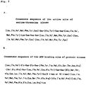
- the addition, deletion, or substitution of amino acids should be performed in a region other than the region corresponding to the consensus sequence of a serine-threonine kinase active site and to the consensus sequence of a protein kinase ATP binding site.
- serine-threonine kinase activity of a protein can be detected, for example, by the method described in Example 9, mentioned later.
- one skilled in the art can usually isolate DNAs having a high homology with the DNA encoding "VRK1" or "VRK2" protein (SEQ ID NO: 1 or 3, respectively) based an the DNA or the part of it using a hybridization technique (Sambrook, J. et al., Molecular Cloning 2nd ed. 9.47-9.58, Cold Spring Harbor Lab. Press, 1989) and obtain proteins having substantially the same function as VRK1 or VRK2 protein (SEQ ID NO: 2 or 4, respectively) from the DNA.
- proteins with serine-threonine kinase activity that are encoded by DNAs hybridizing with DNA encoding "VRK1" or "VRK2" protein are also included in the proteins of the present invention.
- Hybridizing DNAS are isolated from other organisms including, for example, mice, rats, rabbits, bovines, and so on. Tissues such as fetal livers and testes are especially suitable for isolating.
- isolated DNAs encoding proteins having substantially the same function as that of "VRK1" or “VRK2" proteins usually have a high homology with the DNA (SEQ ID NO: 1 or 3) encoding "VRK1" or "VRK2" protein, respectively.
- the term "high homology” used herein means at least 40% or more, preferably 60% or more, and more preferably 80% or more of sequence identity at the amino acid level. From the viewpoint of the protein activity, a high homology should be found in the regions corresponding to the consensus sequence of a serine-threonine kinase active site and to the consensus sequence of a protein kinase ATP binding site.
- Examples of conditions for hybridization to isolate these DNAs are as follows. After prehybridization at 55°C for 30 minutes or longer, hybridization is performed by adding labeled probes and incubating at 37 to 55°C for an hour or longer using "ExpressHyb Hybridization Solution” (CLONTECH). After that, the resulting hybridized product is washed three times for 20 minutes each at room temperature in 2 x SSC and 0.1% SDS then once at 37°C in 1 x SSC and 0.1% SDS. More preferably, after prehybridization at 60°C for 30 minutes or longer, hybridization is performed by adding labeled probes and incubating at 60°C for an hour or longer using "ExpressHyb Hybridization Solution” (CLONTECH).
- hybridized product is washed three times for 20 minutes each at room temperature in 2 x SSC and 0.1% SDS then twice at 50°C in 1 x SSC and 0.1% SDS. Still more preferably, after prehybridization at 68°C for 30 minutes or longer, hybridization is performed by adding labeled probes and incubating at 68 °C for an hour or longer using "ExpressHyb Hybridization Solution" (CLONTECH). Thereafter, the hybridized product is washed three times for 20 minutes each at room temperature in 2 x SSC and 0.1% SDS then twice at 50°C in 0.1 x SSC and 0.1% SDS.
- the present invention also relates to the DNAs encoding the above-described proteins of the present invention.
- the DNAs of the present invention include CDNAs, genomic DNAs, and synthetic DNAs as long as they encode the proteins of the present invention.
- the DNAs of the present invention can be used to produce the recombinant proteins.
- the recombinant proteins can be prepared by inserting the DNA (for example, the DNA of SEQ ID NO: 1 or 3) of the present invention into a suitable expression vector, cultivating the transformant obtained by introducing the vector into suitable cells, and purifying the expressed proteins.
- mammalian cells such as COS, CHO, or NIH3T3 cells
- insect cells such as Sf9 cells
- yeast cells and E.
- coli cells can be used for producing the recombinant proteins.
- Vectors for expressing recombinant proteins in these cells vary depending on the host cells. For example, pcDNA3 (Invitrogen) or pEF-BOS (Nucleic Acids. Res. 1990, 18(17), p5322) is used for mammalian cells; "BAC-to-BAC baculovirus expression system” (GIBCO BRL), for insect cells; "Pichia Expression Kit” (Invitrogen), for yeast cells; and pGEX-5X-1 (Pharmacia) or "QIAexpress system” (Qiagen), for E. coli cells.
- Vectors can be introduced into host cells by, for example, the method using calcium phosphate, DEAE dextran, or cationic liposome DOTAP (Boehringer Mannheim); electroporation; the calcium chloride method; etc.
- the recombinant proteins can be purified from the obtained transformants by the usual methods such as the method described in "The Qiaexpressionist handbook, Qiagen, Hilden, Germany.”
- the DNAs of the present invention can be used for gene therapy of diseases caused by mutations in genomic DNAs.
- the DNAs of the present invention are administered to a living body inserted into adenovirus vectors (e.g., pAdexLcw), retrovirus vectors (e.g., pZIPneo) and so on. They can be administered by either ex vivo methods or in vivo methods.
- adenovirus vectors e.g., pAdexLcw
- retrovirus vectors e.g., pZIPneo
- antisense DNAs against the DNAs of the present invention or part of them can be used as inhibitors for developing cell growth or as antitumor agents.
- the antisense DNAs are administered to a living body directly or in the form of the vectors into which they have been inserted.
- the antisense DNAs can be synthesized by methods well known to one skilled in the art.
- the present invention also relates to a method of screening compounds having inhibitory activity of serine-threonine kinase activity of the proteins of the present invention.
- This screening method comprises two steps. First, the protein of the present invention is caused to contact a substrate to be phosphorylated by this protein in the presence of a test compound to detect the kinase activity of the protein of the present invention. Second, the kinase activity detected in step (a) is compared with that detected in the absence of the test compound, and a compound that lowers the kinase activity of the protein of the present invention is selected.
- Test compounds used for this screening method are not particularly limited and are generally low-molecular-weight compounds, proteins (including the above-described antibodies of the present invention), peptides, etc. Test compounds are either artificially synthesized or natural. Substrates are, for example, casein, IkB ⁇ protein, etc.
- the kinase activity of the protein of the present invention can be detected, for example, by adding ATP having radioactively labeled phosphate to the reaction system containing the protein of the present invention and the substrate and measuring the radioactivity of the phosphate attached to the substrate. Specifically, the activity is detected by the method described in Example 9.
- the compounds thus isolated can be used as cell growth inhibitors or antitumor agents.
- a subtraction library was prepared using the PCR-SelectTM cDNA Subtraction kit (CLONTECH) basically according to the method described by Luda Diatchenko et al. (Proc. Natl. Acad. Sci. USA, Vol.93, 6025-6030, 1996).
- double-stranded cDNAs were synthesized from polyA + RNA prepared from human fetal and adult livers by the standard method using MMLV reverse transcriptase.
- the respective CDNAs were blunt-ended with T4 DNA polymerase, then cleaved by Rsa I.
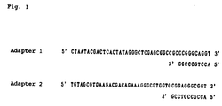
- a part of the cDNA originating from fetal liver (tester) was split in two; one of which was ligated with the adapter-1 and the other with the adapter-2 ( Figure 1). Each aliquot was mixed with an excess amount of the adult liver cDNA (driver), denatured by heat, and subjected to the first hybridization at 68°C for 8 hours.
- PCR primer-1 SEQ ID NO: 5
- PCR primer-2 SEQ ID NO: 6
- PCR primer-1 SEQ ID NO: 7
- SEQ ID NO: 8 primers "Nested PCR primer-2”
- PCR primer-1 SEQ ID NO: 5
- PCR primer-2 SEQ ID NO: 6
- the products were purified using the "QIAquick PCR Purification kit” (QIAGEN), and cloned into the pT7Blue-T vector (Novagen) by the TA cloning method to create a subtraction library.
- Sequence reaction was performed by the cycle-sequencing method using the ABI PRISMTM Dye Terminator Cycle Sequencing Ready Reaction Kit With AmplyTaq DNA Polymerase, FS, and the result was analyzed by the ABI 377 DNA Sequencer.
- Colony PCR was performed as follows. Colonies carrying recombinant vectors were directly suspended into PCR reaction mixtures that contain vector primers, "M13 P4-22 primer” (SEQ ID NO: 9) and “M13 P5-22 primer” (SEQ ID NO: 10). After PCR reaction, amplified insert DNA was separated from unreacted primers and nucleotides by gel filtration or the like, and used as a template for sequencing.
- cDNAs were synthesized by the standard method with SUPERSCRIPTTM II RNase H - Reverse Transcriptase (GIBCO BRL). Some of the cDNAs were used as a template for RT-PCR analysis of fls223. PCR was performed using TaKaRa Taq (TaKaRa) as Taq polymerase by the hot-start method, where the reaction was started by adding TaqStartTM Antibody (CLONTECH). Primers "FLS223 S1 primer” (SEQ ID NO: 11) and "FLS223 A1 primer” (SEQ ID NO: 12) were used to amplifyfls223.
- the G3PDH (glyceraldehyde 3-phosphate dehydrogenase) gene which is a housekeeping gene equally expressed in various tissues and known to be influenced only slightly by various inducers on its expression, was used as a control.
- G3PDH was amplified using the primers "hG3PDH5' primer” (SEQ ID NO: 13) and "hG3PDH3' primer” (SEQ ID NO: 14).
- RT-PCR analysis confirmed that the clone fls223 is strongly expressed in fetal liver, and its expression was also detected in adult liver ( Figure 4). The full-length cDNA was then cloned for more detailed analysis of the gene.
- the reaction performed basically consisted of a reaction at 94°C for 2 minutes; five cycles of reactions at 94°C for 30 seconds and at 68°C 4 minutes; and 30 cycles of reactions at 94°C for 30 seconds, 62°C for 1 minute, and 72°C for 3 minutes; and a reaction at 72°C for 10 minutes.
- TaKaRa Ex Taq was used for PCR, and the reaction was started by the hot-start method by adding TaqStartTM Antibody (CLONTECH). After reaction, detected bands were recovered using the QIAquick Gel Extraction Kit (QIAGEN), and subcloned into the pT7Blue-T vector (Novagen).
- the B1R gene encodes a protein composed of 300 amino acids and is assumed to be a serine-threonine kinase because the gene contains the consensus sequences analogous to that of the ATP binding site of protein kinase and of the active site of serine-threonine kinase.
- B1R kinase is an early gene whose expression is observed in early stages. It appears several hours after vaccinia virus infection and is then repressed. It has been shown that in a mutant strain containing a point mutation on the gene, virus replication stops during DNA replication. Thus, it has been hypothesized that B1R kinase regulates virus DNA replication (J. Biol. Chem., 264, 21458-21461, 1989).
- VRK1 also exhibits an obvious homology to B1R kinase in the region outside of the serine-threonine kinase domain.
- VRK1 may participate in the regulation of cellular DNA replication or, more widely, in cell growth control, as is the case for B1R kinase in virus. This notion is supported by the fact that the VRK1 genes are more strongly expressed in tissues such as fetal liver and the testis, where numerous actively growing cells exist.
- this gene may also encode a novel serine-threonine kinase.
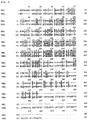
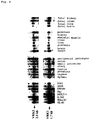
- the amino acid sequence has an extremely high homology to VRK1, especially near the kinase domain ( Figure 6), and a high homology to the vaccinia virus B1R kinase ( Figure 7). These suggest a close relationship between this kinase and B1R kinase. Thus, it was named "Vaccinia virus B1R kinase related Kinase 2" (VRK2).
- RT-PCR confirmed that VRK2 is also expressed more strongly in fetal liver than in adult liver ( Figure 4).
- the primers "RK S2 primer” (SEQ ID NO: 18) and “RK A2 primer” (SEQ ID NO: 16) were used for RT-PCR.
- PCR was performed with a combination of primers "VRK2 A primer” (SEQ ID NO: 19) and “VRK2 B primer” (SEQ ID NO: 20) by a reaction at 94°C for 3 minutes; 30 cycles of reactions at 94°C for 30 seconds, 60°C for 1 minute, and 72°C for 2 minutes; and a reaction at 72°C for 5 minutes.
- the resulting pattern was analyzed on a database on Internet (http://www-genome.wi.mit.edu/cgi-bin/contig/rhmapper.pl), and maps were obtained.
- the VRK1 gene was thus mapped to the position between the STS markers “D14S265" and “APM063XE7” on chromosome 14.
- the VRK2 gene was mapped to the position between the STS markers “CHLC.GATA23H01” and “D2S357” on chromosome 2.
- Hybridization was performed at 68°C in ExpressHyb Hybridization Solution (CLONTECH) using Multiple Tissue Northern (MTN) Blot - Human, Human II, Human Fetal II, and Human Cell line (CLONTECH) according to the method recommended by the manufacturer. Final wash was done at 50°C in 0.1 x SSC, 0.1% SDS, and the image on the filter processed through hybridization was analyzed with the BAS-2000II bioimaging analyzer (Fuji Photo Film).
- tumor cell lines used are malignant melanoma cells "G361,” lung carcinoma cells “A549,” colorectal adenocarcinoma cells “SW480,” Burkitt's lymphoma cells “Raji,” acute lymphoblastic leukemia cells (T cell) "MOLT-4,” chronic myelogenous leukemia cells “K-562,” uterocervical carcinoma cells “HeLaS3,” and promyelocytic leukemia cells "HL60”.
- VRK1 was expressed relatively highly in fetal tissues, and extremely highly in fetal liver. VRK1 was expressed weakly in almost all adult tissues, but it was expressed strongly in the testis and the thymus. In tumor cell lines, a very strong expression of VRK1 was observed in six out of eight cell lines.
- the expression pattern at VRK2 was basically similar to that of VRK1; VRK2 was expressed strongly in fetal liver and the testis. Similarly, it was expressed strongly in tumor cell lines. However, VRK2 was not expressed in MOLT-4 cells, in contrast to the pattern of VRK1 mRNA.
- cDNA containing the entire coding region of VRK1 or VRK2 was amplified by PCR using a combination of primers, VRK1 S1 primer (SEQ ID NO: 21) and VRK1 A1 primer (SEQ ID NO: 22), or VRK2 S1 primer (SEQ ID NO: 23) and VRK2 A1 primer (SEQ ID NO: 24) from cDNA synthesized from polyA + RNA extracted from human fetal liver.
- the amplified product was cleaved at the Not I site that is attached to the end of the primers and purified by agarose gel electrophoresis to obtain DNA fragments of the correct size.
- Plasmids for expressing proteins in which the anti c-Myc antibody epitope sequence (SEQ ID NO: 25) is attached to the C-terminus were constructed as follows. Using about 50 nanograms of the plasmid DNA and with pCOS/VRK1w or pCOS/VRK2w as a temptate, PCR was performed with a combination of primers.
- VRK1 MYC1 primer SEQ ID NO: 26
- VRK1 MYC2 primer SEQ ID NO: 27
- VRK2 MYC1 primer SEQ ID NO: 28
- VRK2 MYC2 primer SEQ ID NO: 29
- cDNA with the anti c-Myc antibody epitope attached to the C-terminus of the coding sequence was amplified.
- KOD DNA polymerase TOYOBO
- the amplified product was cleaved at the Bam HI site that is attached to the end of the primers and purified by agarose gel electrophoresis to obtain DNA fragments of the correct size.
- Expression plasmid DNAs for glutathione-S-transferase (GST) fusion proteins in E. coli were constructed as follows. Using the plasmid DNA pCOS/VRK1w or pCOS/VRK2w as a template, the coding region was amplified by PCR with a combination of primers, VRK1 H3 primer (SEQ ID NO: 30) and VRK1 H4 primer (SEQ ID NO: 31), or VRK2 H3 primer (SEQ ID NO: 32) and VRK2 H4 primer (SEQ ID NO: 33).
- the amplified product was cleaved at the Bam HI site that is attached to the end of the primers purified by agarose gel electrophoresis to obtain DNA fragments of the correct size. These were then subcloned into the pGEX-5X-1 vector (Pharmacia), which was digested with Bam HI and dephosphorylated on its ends with alkaline-phosphatase/CIAP (TaKaRa). By sequencing the thus-obtained subclones, clones (pGEX/VRK1w, pGEX/VRK2w) without mutation such as PCR error were selected and used for later experiments.
- a clone with a mutation introduced to the predicted ATP binding site within the kinase catalytic domain was constructed using the ChameleonTM Double-Stranded Site-Directed Mutagenesis Kit (STRATAGENE) as follows. About one microgram of the pGEX/VRK1w plasmid DNA was mixed with primers VRK1 KW1 primer (SEQ ID NO: 34) and a selection primer, Select1 primer (SEQ ID NO: 35), and heat denatured by boiling for 5 minutes. Plasmid DNA and both primers containing mutation were then annealed by incubating at room temperature for 30 minutes.
- STRATAGENE ChameleonTM Double-Stranded Site-Directed Mutagenesis Kit
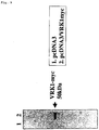
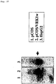
- plasmid DNA About 10 micrograms of plasmid DNA, pcDNA3/VRK1myc or pcDNA3, was introduced (transfected) into COS7 cells using SuperFect (QIAGEN). Specifically, about 10 6 COS7 cells were plated in a 10-cm dish, and cultured overnight. A mixture of 10 micrograms of plasmid DNA and 60 microliters of SuperFect was then added to the culture, and the culture was incubated for about 3 hours. Thereafter, the culture medium was replaced with fresh medium. The cells were then cultured for two more days and collected by detaching in a trypsin-EDTA solution.
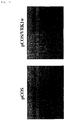
- plasmid DNA pCOS/VRK1w or pCOS
- DNAP cationic phospholipid DOTAP
- transformants were selected by adding G418 (GIBCO-BRL) to the culture medium to a final concentration of 500 micrograms/ml.
- G418 G418
- Total RNA was prepared from each pool of transformants by the method using ISOGEN (Wajo Junyaku). The total RNA was then subjected to northern blot analysis using the VRK1 cDNA as a probe.
- Both wild VRK1 protein and mutant VRK1 protein were expressed in E.coli as a fusion protein with GST protein and purified.
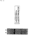
- the E.coli DH5 ⁇ strain cells carrying the above-described plasmid DNA, pGEX/VRK1W, or pGEX/VRK1K71W were cultured overnight at 37°C in 10 ml 2xYT medium. Some of the culture was diluted 100-fold with fresh 2xYT medium and cultured at 37°C until the OD value at 600 nm reached 0.6. IPTG (isopropyl- ⁇ -D(-)-thiogalactopyranoside) was then added to the culture to a final concentration of 0.1 mM, and the culture was incubated further for several hours.
- IPTG isopropyl- ⁇ -D(-)-thiogalactopyranoside
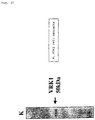
- coli cells were collected by centrifugation, resuspended in PBS containing 1% Triton X-100 and 1% Tween 20, and subsequently disrupted by sonication to solubilize proteins. From the solubilized samples, wild VRK1 protein and mutant VRK1, which were expressed as a fusion protein with GST, were purified by affinity chromatography using glutathione Sepharose4B (Pharmacia). These proteins were subjected to SDS-PAGE and stained with Coomassie Brilliant Blue (CBB) to confirm their purity ( Figure 12). GST protein and GST-IkB ⁇ protein were prepared in the same manner.
- CBB Coomassie Brilliant Blue
- Kinase assay was performed on a total of 50 microliters of a reaction mixture containing 0.2 micrograms of wild or mutant VRK1 protein, 50 mM Tris-HCl (pH 7.2), 1 mM dithiothreitol (DTT), 2 mM or 10 mM of divalent cation (Mg, Mn, Zn, Ca), a maximum of 5 micrograms of substrate protein, and 1 microliter of [ ⁇ - 32 P]-ATP (3000 Ci/mM, 10 mCi/ml [Amersham]).
- another buffer system containing 40 mM Hepes (pH 7.4), 1 mM DTT, and 2.5 mM EGTA was used.
- the reaction was first performed in the presence of 10 mM Mg at 37°C for 30 minutes using, as a protein substrate, histone (Nakalai), casein (Sigma), myelin basic protein/MBP (Sigma), GST, GST-IkB ⁇ , or IkB ⁇ C-terminal peptide (SEQ ID NO: 36).
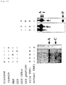
- the reaction mixture was subjected to SDS-PAGE, and the radioactivity of phosphorylated proteins was analyzed with a BAS2000II bioimaging analyzer (Fuji Photo Film). The result indicates that wild VRK1 protein phosphorylates casein and GST-IkB ⁇ ( Figure 13). In contrast, no phosphorylation was observed in reactions with mutant VRK1 carrying mutation in the predicted ATP binding site.
- VRK1 is a protein kinase that contains a typical catalytic domain.
- GST protein was not phosphorylated by VRK1, suggesting that the phosphorylation of GST-IkB ⁇ protein by VRK1 occurs within IkB ⁇ protein but not in GST moiety.
- IkB ⁇ is said to negatively regulate the function of transcription factor NF-kB by forming a complex with it.
- IkB ⁇ is inactivated by self-phosphorylation, immediately thereafter undergoes proteolysis, thereby liberating and activating NF-kB.
- NF-kB is supposed to be a central transcriptional regulator that induces a broad range of immune reactians and inflammatory reactions. Therefore, a kinase that phosphorylates IkB ⁇ is important as a target molecule of anti-inflammatory drugs. Since VRK1 strongly phosphorylates IkB ⁇ in vitro , VRK1 probably participates in the activation of NF-kB by phosphorylating IkB ⁇ in vivo as well. Therefore, it is possible to anticipate anti-inflammatory effects or immunosuppressive effects by inhibiting VRK1 kinase activity, or by reducing its protein amount.
- a peptide (SEQ ID NO: 37) corresponding to the C-terminal sequence of the deduced VRK1 amino acid sequence was synthesized (Sawady Technology) and conjugated to Keyhole limpet hemocyanin (KLH) at its amino-terminal cysteine mediated by m-maleimidobenzoyl-N-hydroxy-succinimide ester (MBS). This was used as an antigen to immunize rabbits, and antisera were obtained. Antibodies that specifically react with the peptide were purified from the antisera by affinity chromatography using Cellulofine (Seikagaku Corporation) conjugated with the peptide.
- the serine-threonine kinase genes isolated by the present inventors show significant homology to a vaccinia virus gene that is thought to be involved in DNA replication and are strongly expressed in actively growing cells. Furthermore, overexpression of proteins encoded by the genes remarkably enhances cell proliferation activity. Thus, the isolated serine-threonine kinase genes are assumed to participate in the regulation of cell growth. Therefore, it is possible to develop cell growth inhibitors or antitumor agents based on a novel mechanism by screening drugs targeted on the genes (such as antisense DNA), or drugs which can regulate either the expression of the genes or the activity of the proteins encoded by the genes of the present invention.
Landscapes
- Life Sciences & Earth Sciences (AREA)
- Chemical & Material Sciences (AREA)
- Health & Medical Sciences (AREA)
- Organic Chemistry (AREA)
- Wood Science & Technology (AREA)
- Zoology (AREA)
- Bioinformatics & Cheminformatics (AREA)
- Engineering & Computer Science (AREA)
- Genetics & Genomics (AREA)
- Biochemistry (AREA)
- General Health & Medical Sciences (AREA)
- General Engineering & Computer Science (AREA)
- Molecular Biology (AREA)
- Medicinal Chemistry (AREA)
- Biomedical Technology (AREA)
- Biotechnology (AREA)
- Microbiology (AREA)
- Enzymes And Modification Thereof (AREA)
- Micro-Organisms Or Cultivation Processes Thereof (AREA)
- Medicines Containing Material From Animals Or Micro-Organisms (AREA)
Applications Claiming Priority (3)
| Application Number | Priority Date | Filing Date | Title |
|---|---|---|---|
| JP35786496 | 1996-12-27 | ||
| JP35786496 | 1996-12-27 | ||
| PCT/JP1997/004855 WO1998029552A1 (fr) | 1996-12-27 | 1997-12-25 | Nouveau gene de serine-threonine kinase |
Publications (2)
| Publication Number | Publication Date |
|---|---|
| EP0960938A1 true EP0960938A1 (fr) | 1999-12-01 |
| EP0960938A4 EP0960938A4 (fr) | 2001-12-05 |
Family
ID=18456329
Family Applications (1)
| Application Number | Title | Priority Date | Filing Date |
|---|---|---|---|
| EP97950408A Withdrawn EP0960938A4 (fr) | 1996-12-27 | 1997-12-25 | Nouveau gene de serine-threonine kinase |
Country Status (5)
| Country | Link |
|---|---|
| US (1) | US6265194B1 (fr) |
| EP (1) | EP0960938A4 (fr) |
| JP (1) | JP4121155B2 (fr) |
| AU (1) | AU5340698A (fr) |
| WO (1) | WO1998029552A1 (fr) |
Cited By (4)
| Publication number | Priority date | Publication date | Assignee | Title |
|---|---|---|---|---|
| WO2001011086A3 (fr) * | 1999-08-11 | 2002-03-07 | Eos Biotechnology Inc | Nouvelles techniques de diagnostic de l'angiogenese, compositions et techniques de criblage pour modulateurs d'angiogenese |
| WO2001040503A3 (fr) * | 1999-12-06 | 2002-07-18 | Axxima Pharmaceuticals Ag | Methode d"identification et de quantification des inhibiteurs de kinase |
| WO2002055713A3 (fr) * | 2000-12-08 | 2002-10-31 | Millennium Pharmaceuticals, Inc. | 58848, un membre de la famille des proteine kinases humaines et utilisations de celui-ci |
| ES2255811A1 (es) * | 2004-05-12 | 2006-07-01 | Laboratorios Indas, S.A. | Metodo para el diagnostico de cancer y/o pronostico de tratamientos oncologicos. |
Families Citing this family (6)
| Publication number | Priority date | Publication date | Assignee | Title |
|---|---|---|---|---|
| US6156523A (en) * | 1996-11-15 | 2000-12-05 | Incyte Pharmaceuticals, Inc. | Serine/threonine protein kinases |
| WO1999011794A2 (fr) * | 1997-09-04 | 1999-03-11 | Incyte Pharmaceuticals, Inc. | Nouvelle kinase vrk1 |
| JP2002539777A (ja) * | 1999-03-19 | 2002-11-26 | ヒューマン ジノーム サイエンシーズ, インコーポレイテッド | 48個のヒト分泌タンパク質 |
| DE10011530A1 (de) * | 2000-03-13 | 2001-09-27 | Robert Elez | Hochwirksame Antisense-Oligodesoxynucleotide gegen Polio-like Kinasel |
| JP2008000003A (ja) * | 2004-10-01 | 2008-01-10 | Oncorex Inc | Pim−1活性/蛋白阻害医薬品 |
| CN117106090B (zh) * | 2023-08-15 | 2024-02-09 | 十堰市太和医院(湖北医药学院附属医院) | 一种靶向GSK3β的纳米抗体及其制备方法 |
Family Cites Families (4)
| Publication number | Priority date | Publication date | Assignee | Title |
|---|---|---|---|---|
| US5925557A (en) * | 1994-09-29 | 1999-07-20 | The Regents Of The University Of California | DNA encoding mitogen activated protein kinase, FRK |
| US5827726A (en) * | 1995-03-16 | 1998-10-27 | Chugai Seiyaku Kabushiki Kaisha | DNA coding protein kinase |
| US5885803A (en) * | 1997-06-19 | 1999-03-23 | Incyte Pharmaceuticals, Inc. | Disease associated protein kinases |
| WO1999011794A2 (fr) * | 1997-09-04 | 1999-03-11 | Incyte Pharmaceuticals, Inc. | Nouvelle kinase vrk1 |
-
1997
- 1997-12-25 AU AU53406/98A patent/AU5340698A/en not_active Abandoned
- 1997-12-25 JP JP52985598A patent/JP4121155B2/ja not_active Expired - Fee Related
- 1997-12-25 WO PCT/JP1997/004855 patent/WO1998029552A1/fr not_active Ceased
- 1997-12-25 EP EP97950408A patent/EP0960938A4/fr not_active Withdrawn
-
1999
- 1999-06-25 US US09/344,700 patent/US6265194B1/en not_active Expired - Lifetime
Cited By (5)
| Publication number | Priority date | Publication date | Assignee | Title |
|---|---|---|---|---|
| WO2001011086A3 (fr) * | 1999-08-11 | 2002-03-07 | Eos Biotechnology Inc | Nouvelles techniques de diagnostic de l'angiogenese, compositions et techniques de criblage pour modulateurs d'angiogenese |
| WO2001040503A3 (fr) * | 1999-12-06 | 2002-07-18 | Axxima Pharmaceuticals Ag | Methode d"identification et de quantification des inhibiteurs de kinase |
| WO2002055713A3 (fr) * | 2000-12-08 | 2002-10-31 | Millennium Pharmaceuticals, Inc. | 58848, un membre de la famille des proteine kinases humaines et utilisations de celui-ci |
| ES2255811A1 (es) * | 2004-05-12 | 2006-07-01 | Laboratorios Indas, S.A. | Metodo para el diagnostico de cancer y/o pronostico de tratamientos oncologicos. |
| ES2255811B1 (es) * | 2004-05-12 | 2007-07-16 | Laboratorios Indas, S.A. | Metodo para el diagnostico de cancer y/o pronostico de tratamientos oncologicos. |
Also Published As
| Publication number | Publication date |
|---|---|
| JP4121155B2 (ja) | 2008-07-23 |
| US6265194B1 (en) | 2001-07-24 |
| WO1998029552A1 (fr) | 1998-07-09 |
| AU5340698A (en) | 1998-07-31 |
| EP0960938A4 (fr) | 2001-12-05 |
Similar Documents
| Publication | Publication Date | Title |
|---|---|---|
| Leung et al. | The p160 RhoA-binding kinase ROKα is a member of a kinase family and is involved in the reorganization of the cytoskeleton | |
| Vasseur et al. | Cloning and expression of the human p8, a nuclear protein with mitogenic activity | |
| JP2004222728A (ja) | ヒトサイクリンe | |
| US20080063638A1 (en) | Mitotic kinesin-like protein-1, MKLP1, and uses therof priority | |
| US6300131B1 (en) | Telomerase-associated proteins | |
| US6265194B1 (en) | Serine-threonine kinase gene | |
| US5989885A (en) | Specific mutations of map kinase 4 (MKK4) in human tumor cell lines identify it as a tumor suppressor in various types of cancer | |
| JP4633259B2 (ja) | NF−κBの活性化を調節する化合物および方法 | |
| CA2208237C (fr) | Proteine serine/threonine kinases nucleaires | |
| US6586577B2 (en) | Telomere repeat binding factors and diagnostic and therapeutic use thereof | |
| JPWO1998029552A1 (ja) | 新規なセリン/スレオニンキナーゼ遺伝子 | |
| Iseki et al. | Molecular cloning of the mouse APS as a member of the Lnk family adaptor proteins | |
| US20040038248A1 (en) | Novel polypeptide-human heterogeneous nuclear ribonucleoprotein 32.01 and the polynucleotide encoding said polypeptide | |
| WO1999003489A2 (fr) | Procedes et compositions permettant de reguler le transport des proteines dans le noyau | |
| US7186536B2 (en) | Serine-threonine kinase gene | |
| JP4270548B2 (ja) | Cdc7−ASKキナーゼ複合体、該キナーゼ複合体の基質、及び該基質に特異的な抗体、並びにこれらを用いたCdc7−ASKキナーゼ阻害能を有する化合物のスクリーニング方法 | |
| US20030073120A1 (en) | Protein tyrosine phosphatase PTP20 and related products and methods | |
| JP4472179B2 (ja) | 新規なリボゾームs6プロテインキナーゼの同定および機能的なキャラクタライゼーション | |
| EP1148125B1 (fr) | Nucleoproteine humaine possedant un domaine ww et polynucleotide codant cette nucleoproteine | |
| US20030073111A1 (en) | Biologically active alternative form of the IKKalpha IkappaB kinase | |
| EP1251178A1 (fr) | Nouveau polypeptide, proteine humaine shc 43, et polynucleotide codant pour ce polypeptide | |
| EP1262487A1 (fr) | Promoteur de squalene epoxydase codant l'adn | |
| JPH10262680A (ja) | Rho標的タンパク質ヒトmDiaおよびその遺伝子 | |
| JPWO2002099110A1 (ja) | 細胞周期調節因子 | |
| JPWO2006054770A1 (ja) | 新規mapキナーゼキナーゼキナーゼ(mapkkk) |
Legal Events
| Date | Code | Title | Description |
|---|---|---|---|
| PUAI | Public reference made under article 153(3) epc to a published international application that has entered the european phase |
Free format text: ORIGINAL CODE: 0009012 |
|
| 17P | Request for examination filed |
Effective date: 19990727 |
|
| AK | Designated contracting states |
Kind code of ref document: A1 Designated state(s): AT BE CH DE DK ES FI FR GB GR IE IT LI LU MC NL PT SE |
|
| RIN1 | Information on inventor provided before grant (corrected) |
Inventor name: OKU, A., CHUGAI RES.INST.F.MOL.MED.,INC. Inventor name: NEZU, JUN-ICHI, C/O CHUGAI RES.INST.F.MOL.MED.,INC |
|
| A4 | Supplementary search report drawn up and despatched |
Effective date: 20011023 |
|
| AK | Designated contracting states |
Kind code of ref document: A4 Designated state(s): AT BE CH DE DK ES FI FR GB GR IE IT LI LU MC NL PT SE |
|
| RAP1 | Party data changed (applicant data changed or rights of an application transferred) |
Owner name: CHUGAI SEIYAKU KABUSHIKI KAISHA |
|
| 17Q | First examination report despatched |
Effective date: 20030604 |
|
| 17Q | First examination report despatched |
Effective date: 20030604 |
|
| STAA | Information on the status of an ep patent application or granted ep patent |
Free format text: STATUS: THE APPLICATION IS DEEMED TO BE WITHDRAWN |
|
| 18D | Application deemed to be withdrawn |
Effective date: 20070412 |