EP0973381B1 - Model animal aux fins d'une appréciation de vaccins - Google Patents
Model animal aux fins d'une appréciation de vaccins Download PDFInfo
- Publication number
- EP0973381B1 EP0973381B1 EP98914591A EP98914591A EP0973381B1 EP 0973381 B1 EP0973381 B1 EP 0973381B1 EP 98914591 A EP98914591 A EP 98914591A EP 98914591 A EP98914591 A EP 98914591A EP 0973381 B1 EP0973381 B1 EP 0973381B1
- Authority
- EP
- European Patent Office
- Prior art keywords
- cells
- hiv
- human
- mice
- tumor
- Prior art date
- Legal status (The legal status is an assumption and is not a legal conclusion. Google has not performed a legal analysis and makes no representation as to the accuracy of the status listed.)
- Expired - Lifetime
Links
- 0 CC(*)(C*(*)C(*)(*1)*(*)*)C1(*)C(*)* Chemical compound CC(*)(C*(*)C(*)(*1)*(*)*)C1(*)C(*)* 0.000 description 1
Images









Classifications
-
- C—CHEMISTRY; METALLURGY
- C07—ORGANIC CHEMISTRY
- C07K—PEPTIDES
- C07K14/00—Peptides having more than 20 amino acids; Gastrins; Somatostatins; Melanotropins; Derivatives thereof
- C07K14/005—Peptides having more than 20 amino acids; Gastrins; Somatostatins; Melanotropins; Derivatives thereof from viruses
-
- A—HUMAN NECESSITIES
- A01—AGRICULTURE; FORESTRY; ANIMAL HUSBANDRY; HUNTING; TRAPPING; FISHING
- A01K—ANIMAL HUSBANDRY; AVICULTURE; APICULTURE; PISCICULTURE; FISHING; REARING OR BREEDING ANIMALS, NOT OTHERWISE PROVIDED FOR; NEW BREEDS OF ANIMALS
- A01K67/00—Rearing or breeding animals, not otherwise provided for; New or modified breeds of animals
- A01K67/027—New or modified breeds of vertebrates
- A01K67/0271—Chimeric vertebrates, e.g. comprising exogenous cells
-
- A—HUMAN NECESSITIES
- A61—MEDICAL OR VETERINARY SCIENCE; HYGIENE
- A61P—SPECIFIC THERAPEUTIC ACTIVITY OF CHEMICAL COMPOUNDS OR MEDICINAL PREPARATIONS
- A61P35/00—Antineoplastic agents
-
- C—CHEMISTRY; METALLURGY
- C12—BIOCHEMISTRY; BEER; SPIRITS; WINE; VINEGAR; MICROBIOLOGY; ENZYMOLOGY; MUTATION OR GENETIC ENGINEERING
- C12N—MICROORGANISMS OR ENZYMES; COMPOSITIONS THEREOF; PROPAGATING, PRESERVING, OR MAINTAINING MICROORGANISMS; MUTATION OR GENETIC ENGINEERING; CULTURE MEDIA
- C12N7/00—Viruses; Bacteriophages; Compositions thereof; Preparation or purification thereof
-
- A—HUMAN NECESSITIES
- A61—MEDICAL OR VETERINARY SCIENCE; HYGIENE
- A61K—PREPARATIONS FOR MEDICAL, DENTAL OR TOILETRY PURPOSES
- A61K39/00—Medicinal preparations containing antigens or antibodies
-
- C—CHEMISTRY; METALLURGY
- C12—BIOCHEMISTRY; BEER; SPIRITS; WINE; VINEGAR; MICROBIOLOGY; ENZYMOLOGY; MUTATION OR GENETIC ENGINEERING
- C12N—MICROORGANISMS OR ENZYMES; COMPOSITIONS THEREOF; PROPAGATING, PRESERVING, OR MAINTAINING MICROORGANISMS; MUTATION OR GENETIC ENGINEERING; CULTURE MEDIA
- C12N2740/00—Reverse transcribing RNA viruses
- C12N2740/00011—Details
- C12N2740/10011—Retroviridae
- C12N2740/16011—Human Immunodeficiency Virus, HIV
- C12N2740/16061—Methods of inactivation or attenuation
-
- C—CHEMISTRY; METALLURGY
- C12—BIOCHEMISTRY; BEER; SPIRITS; WINE; VINEGAR; MICROBIOLOGY; ENZYMOLOGY; MUTATION OR GENETIC ENGINEERING
- C12N—MICROORGANISMS OR ENZYMES; COMPOSITIONS THEREOF; PROPAGATING, PRESERVING, OR MAINTAINING MICROORGANISMS; MUTATION OR GENETIC ENGINEERING; CULTURE MEDIA
- C12N2740/00—Reverse transcribing RNA viruses
- C12N2740/00011—Details
- C12N2740/10011—Retroviridae
- C12N2740/16011—Human Immunodeficiency Virus, HIV
- C12N2740/16111—Human Immunodeficiency Virus, HIV concerning HIV env
- C12N2740/16122—New viral proteins or individual genes, new structural or functional aspects of known viral proteins or genes
-
- Y—GENERAL TAGGING OF NEW TECHNOLOGICAL DEVELOPMENTS; GENERAL TAGGING OF CROSS-SECTIONAL TECHNOLOGIES SPANNING OVER SEVERAL SECTIONS OF THE IPC; TECHNICAL SUBJECTS COVERED BY FORMER USPC CROSS-REFERENCE ART COLLECTIONS [XRACs] AND DIGESTS
- Y02—TECHNOLOGIES OR APPLICATIONS FOR MITIGATION OR ADAPTATION AGAINST CLIMATE CHANGE
- Y02A—TECHNOLOGIES FOR ADAPTATION TO CLIMATE CHANGE
- Y02A50/00—TECHNOLOGIES FOR ADAPTATION TO CLIMATE CHANGE in human health protection, e.g. against extreme weather
- Y02A50/30—Against vector-borne diseases, e.g. mosquito-borne, fly-borne, tick-borne or waterborne diseases whose impact is exacerbated by climate change
Definitions
- the present invention relates to the evaluation of vaccines.
- the present invention provides humanized animal models for the evaluation of vaccines designed to confer immunity against human pathogens, including vaccines directed against the human immunodeficiency virus.
- the present invention further relates to HIV vaccines.
- the present invention provides attenuated replication- competent HIV vaccines and replication-defective HIV vaccines.
- the present invention relates to immunotherapy for the treatment of tumors.
- the present invention provides combinations of immune-modulating proteins that induce systemic immunity against tumors and provides humanized animal models for immunogene therapy.
- chemotherapeutic agents Conventional treatment of cancer typically involves the use of chemotherapeutic agents.
- This target specificity is sought in all types of chemotherapeutics, including anticancer agents.
- anticancer agents have negative hematological affects (e.g ., cessation of mitosis and disintegration of formed elements in marrow and lymphoid tissues) and immunosuppressive action as well as a severe impact on epithelial tissues (e.g ., intestinal mucosa), reproductive tissues and the nervous system [Calabresi and Chabner, In: Goodman and Gilman, The Pharmacological Basis of Therapeutics (Pergamon Press, 8th Edition) pp. 1209-1216 ].
- chemotherapeutics as anticancer agents has also been hampered by the phenomenon of multiple drug resistance, resistance to a wide range of structurally unrelated cytotoxic anticancer compounds [ Gerlach et al. (1986) Cancer Surveys 5:25 ].
- certain cancers are non-responsive to known chemotherapeutics agents and patients with these cancers invariably die within a short period following diagnosis (e.g ., glioblastoma multiforme, recurrent metastatic melanoma, breast, lung and pancreatic cancers).
- T lymphocytes T cells
- T cells play a crucial role in a number of immune responses including the recognition of foreign antigens, destruction of virally infected cells and providing help to B cells to permit the production of antibodies that neutralize foreign antigens.
- the antigen In order for a T cell to recognize its target antigen, the antigen must be presented to the T cell by an antigen-presenting cell (APC) such as dendritic cells, macrophages, Langerhans cells and B cells.
- APC antigen-presenting cell
- the APC presents the target antigen as part of a complex containing immune molecules termed major histocompatibility complex (MHC) in mice and human leukocyte antigens (HLA) in humans.
- MHC major histocompatibility complex
- HLA human leukocyte antigens
- MHC class I molecules which are expressed on all nucleated cells and MHC class II molecules which are expressed only on APCs.
- Class I molecules present endogenous protein fragments (not recognized as foreign) and viral antigens (recognized as foreign) while class II molecules present protein fragments derived from proteins that entered the cell by endocytosis or phagocytosis (i.e., proteins which are mainly derived from infectious agents such as parasites and bacteria).
- T cells recognize MHC-antigen complexes on APCs via their T cell receptor (TCR)/CD3 complex; the TCR complex together with the CD4 or CD8 coreceptors bind to MHC class II or I, respectively. Occupancy of the TCR alone is not sufficient to active the T cell to respond; activation also requires antigen-independent signals provided by the engagement of costimulatory molecules present on the surface of the T cell with their cognate ligands present on the surface of the APC.
- the costimulatory proteins serve to stabilize the interaction of the T cell with the APC and to transduce costimulatory signals that lead to the secretion of cytokines, proliferation of the T cell and induction of the T cell's effector function.
- T cells require growth factors (i.e ., cytokines such as interleukin-2) in order to cause proliferation of antigen-reactive T cells.
- growth factors i.e ., cytokines such as interleukin-2
- CD28/CTLA-4 family of proteins present on the surface of T cell, has been shown to be an important costimulator required for interleukin-2 (IL-2) driven proliferation of T cells.
- the ligands for the CD28/CTLA-4 proteins are members of the B7 family ( e.g ., B7-1, B7-2 and B7-3).
- Cancer immunotherapy aims to induce tumor-specific T cell responses that are effective in the rejection of tumors.
- the notion that the immune system is naturally involved in identifying and suppressing tumors is supported by the fact that immunocompromised patients have an increased incidence of tumors [ Frei et al. (1993) Transplant. Proc. 25:1394 ].
- the immune system fails to recognize and destroy all tumor cells. Indeed animal studies have shown that the majority of tumors fail to provoke an immune response even when these tumors express potentially recognizable tumor-specific antigens [ Boon et al. (1994) Annu. Rev. Immunol. 12:337 and Houghton (1994) J. Exp. Med. 180:1 ].
- genes encoding costimulatory proteins and/or cytokines have been introduced into tumor cells.
- the modified tumor cells are then examined for their ability to induce an anti-tumor response ( in vivo or in vitro ).
- these experiments have been carried out in animal models ( e.g ., in mice and rats) and the results have been conflicting leading some in the field to question the utility of existing animal models for the development of immunogene therapy protocols aimed at the treatment of humans.
- mice and rats immunocompetent syngeneic animals
- established tumor cell lines e.g ., mice and rats
- one immunogene e.g ., mouse IL-2
- the tumor cells are selected in culture to identify cells expressing the transferred immunogene and the modified tumor cells are injected into recipient mice and the mice are examined for the presence of tumors.
- the mice receive irradiated modified tumor cells as a vaccine followed by a challenge with unmodified tumor cells (vaccination/challenge experiment).
- the expression of certain immunogenes has been shown to increase the immunogenicity of the tumor cell (i.e., to reduce the tumorigenicity of the tumor cell).
- An experimentally induced animal tumor is said to be immunogenic if the tumor is rejected following transplantation into syngeneic animals previously immunized or vaccinated with irradiated cells of the same tumor. Nonimmunogenic tumors are not rejected under these conditions.
- B7-1 expression was found to increase the immunogenicity of B-16 cells, the authors reported that animals that rejected the modified B-16 cells did not develop an enhanced systemic immunity against unmodified or wild type B-16 cells [Wu et al., supra ].
- the expression of B7-1 or B7-2 in mouse colorectal tumor or melanoma cells was found to confer a local anti-tumor response in immunocompetent mice [ Chong et al. (1996) Human Gene Ther. 7:1771 ].
- B7-1 expressing tumor cell lines is limited to highly immunogenic cell lines as B7-1 expression in poorly immunogenic fibrosarcomas (e.g ., MAC101, MCA102 and Ag104) and the B-16 melanoma cell line does not reduce the tumorigenicity of these lines.
- expression of B7-1 in the B-16 melanoma cell line was found by one group to reduce the tumorigenicity of these cells (Wu et al., supra). This discrepancy may be explained by differences in the level of B7-1 expression achieved by different groups.
- tumor cells expressing only B7-1 have been found to be ineffective in inducing the rejection of established tumors.
- mice immunized with A20 B cell lymphoma cells mixed with IL-2 and the chemokine lymphotactin developed a potent anti-tumor response while mice immunized with B cell lymphoma cells mixed with GM-CSF developed a much reduced anti-tumor response [ Dilloo et al. (1996) Nature Med. 2:1090 ].
- HIV human immunodeficiency virus
- HIV vaccines The ultimate system for the study of HIV vaccines is, of course, the human. However, given the fact that HIV infection is almost invariably fatal in humans, ethical considerations limit the use of human volunteers in HIV vaccine evaluation. The art needs animal models in which anti-HIV immunity can be assessed to permit the evaluation of HIV vaccines.
- the present invention provides novel humanized animal models that permit the identification of immune-modulating genes and combinations thereof useful for the treatment of human tumors.
- the present invention provides vaccines comprising cells modified to express at least one immune-modulating gene.
- the present invention also provides methods of treating subjects having a tumor with a tumor cell vaccine modified to express one or more immune-modulating genes.
- the novel animals of the present invention provide a means of evaluating vaccines, including cancer vaccines and vaccines directed against human pathogens ( e.g ., HIV, malaria, Leishmania, etc .).
- the subject invention provides HIV vaccines including live attenuated HIV vaccines and HIV DNA vaccines.
- the present invention also concerns an immunodeficient mouse comprising a sufficient number of human immune cells capable of producing a sustained immune response.
- the present invention provides an immunodeficient mouse comprising human T lymphocytes expressing the CD45 antigen wherein at least about 10% of the human T lymphocytes expressing the CD45 antigen represent immature naive T lymphocytes.
- the present invention provides an immunodeficient mouse comprising human T lymphocytes expressing the CD45 antigen wherein at least about 5% of these T lymphocytes represent immature naive T lymphocytes.
- at least about 1% of the human T lymphocytes expressing the CD45 antigen represent immature naive T lymphocytes.
- the invention is not limited by the nature of the immunodeficient mouse strain employed.
- the immunodeficient mouse is a SCID/beige mouse.
- the immunodeficient mouse comprising human T lymphocytes further comprises human tumor cells.
- the invention is not limited by the nature of the human tumor cells employed.
- the human tumor cells may be established tumor cells, primary tumors cells or tumor cells (established or primary) modified to express one or more immune-modulating genes, genes encoding cell cycle regulators and genes encoding inducers of apoptosis.
- the present invention provides a SCID/beige mouse comprising human cells, such as, for example, immune cells.
- human cells such as, for example, immune cells.
- the invention is not limited by the nature of the human immune cells, these cells may be, for example, human PBLs, splenocytes, cells isolated from lymph nodes and/or peritoneal lavage.
- the SCID/beige mouse comprising human immune cells further comprises human tumor cells.
- the tumor cells are derived from central nervous system cells, most preferably glioblastoma cells.
- the tumor cells are malignant melanoma cells.
- the present invention further provides a method comprising: (a) providing: (i) a SCID/beige mouse; (ii) human tumor cells; and (iii) human peripheral blood lymphocytes; (b) introducing a first dose of the tumor cells into said mouse; (c) reconstituting the mouse containing said tumor cells with the lymphocytes; and (d) monitoring the reconstituted mouse for growth of the tumor cells.
- the method of the subject invention further comprises identifying at least one immune modulating gene, or a gene encoding a cell cycle regulator, or a gene that encodes an inducer of apoptosis, whose expression in a tumor cell prevents the growth of the introduced tumor cells in a reconstituted mouse.
- the method comprises, following the reconstitution, the additional step of vaccinating the reconstituted mouse with a second dose of tumor cells.
- the first dose of tumor cells comprises unmodified tumor cells and the second dose of tumor cells comprises irradiated tumor cells.
- the irradiated tumor cells express at least one immune-modulating gene, or a gene encoding a cell cycle regulator, or a gene that encodes an inducer of apoptosis.
- the tumor cells and the lymphocytes come from the same donor. In another embodiment, the tumor cells and the lymphocytes come from different donors.
- the present invention further provides a method comprising: (a) providing: (i) a SCID/beige mouse; (ii) irradiated and unirradiated human tumor cells; and (iii) human peripheral blood lymphocytes; (b) reconstituting said mouse with the lymphocytes; (c) vaccinating the mouse with the irradiated tumor cells; (d) introducing the unirradiated tumor cells into the vaccinated mouse; and (e) monitoring the vaccinated mouse for growth of the unirradiated tumor cells.
- the method further comprises identifying at least one immune modulating gene, or a gene encoding a cell cycle regulator, or a gene that encodes an inducer of apoptosis, whose expression prevents the growth of said unirradiated tumor cells in said vaccinated mouse.
- the present invention also provides vaccines comprising a cell expressing an antigen and an immune-modulating protein.
- the cell can be, for example, a tumor cell or microorganism such as Leishmania, Mycobacterium, Listeria and Plasmodium.
- the immune-modulating protein is a cell cycle regulator, inducer of apoptosis, a viral factor, a tumor suppressor, etc.
- Viral factors include viral proteins and viral growth regulators.
- the vaccines of the present invention are not limited by the nature of the immune modulating protein employed.
- the additional immune modulator is a cytokine.
- the cytokine is selected from the group consisting of interleukin 2, interleukin 4, interleukin 6, interleukin 7, interleukin 12, granulocyte-macrophage colony stimulating factor (GM-CSF), granulocyte colony stimulating factor, interferon-gamma, tumor necrosis factor-alpha.
- GM-CSF granulocyte-macrophage colony stimulating factor
- a tumor cell expressing a gene encoding an immune-modulating protein B7-2 and a gene encoding the cytokine GM-CSF is provided.
- the present invention further provides a method of treating a tumor comprising: (a) providing: (i) a subject having a tumor of the central nervous system; (ii) an expression vector encoding an immune-modulating protein, such as the human B7-2 protein, and at least one additional immune modulator or a cell cycle regulator or inducer of apoptosis; (b) transferring the expression vector into the tumor under conditions such that the immune-modulating protein and the immune-modulator (and/or a cell cycle regulator or inducer of apoptosis) are expressed by at least a portion of the tumor.
- an immune-modulating protein such as the human B7-2 protein
- the method further comprises, prior to transfer of the expression vector, the step of removing at least a portion of the tumor from the subject and following the transfer of said expression vector, irradiating the tumor cells expressing the immune-modulating protein and the immune-modulator (and/or a cell cycle regulator or inducer of apoptosis) and introducing the irradiated tumor cells back into the subject to create an immunized subject.
- the method further comprises introducing at least one additional dose of irradiated tumor cells expressing the immune-modulating protein and an additional immune-modulator (and/or a cell cycle regulator or inducer of apoptosis) into the immunized subject.
- the invention further provides a method comprising: (a) providing: (i) an immunodeficient mouse, such as a SCID/beige or SCID/nod mouse, comprising human cells; (ii) an injectable preparation comprising at least one component derived from a human pathogen; and (iii) a composition comprising an infectious human pathogen; (b) injecting the mouse with the injectable preparation to produce an injected mouse; (c) exposing the injected mouse to the composition; and (d) monitoring the exposed mouse for the presence of infection of the human cells by the infectious human pathogen.
- the human cells are immune cells.
- the injecting of step (b) is repeated at least once prior to exposing the mouse to the composition.
- the injectable preparation comprises a candidate vaccine; a candidate vaccine may comprise proteins or peptides or nucleic acids and may be immunogenic.
- a candidate vaccine may comprise proteins or peptides or nucleic acids and may be immunogenic.
- some candidate vaccines are expected to be immunogenic (i.e ., capable of invoking an immune response directed against the pathogen from which the protein, peptide or nucleic acid was derived) and some are expected to not be immunogenic (and would therefore be rejected as a vaccine for use in a subject such as a human).
- the injectable preparation is immunogenic.
- the injectable preparation may comprise pharmacological carriers and excipients such as aqueous solutions, preferably in physiologically compatible buffers such as Hanks' solution, Ringer's solution, or physiologically buffered saline.
- Aqueous injection suspensions may contain substances which increase the viscosity of the suspension, such as sodium carboxymethyl cellulose, sorbitol, or dextran.
- suspensions of the active compounds may be prepared as appropriate oily injection suspensions.
- Suitable lipophilic solvents or vehicles include fatty oils such as sesame oil, or synthetic fatty acid esters, such as ethyl oleate or triglycerides, or liposomes.
- the suspension may also contain suitable stabilizers or agents which increase the solubility of the compounds to allow for the preparation of highly concentrated solutions.
- the injectable preparation may comprise as adjuvant (e.g ., alum, complete or incomplete Freund's, SAF-1).
- the human pathogen is selected from the group comprising human immunodeficiency virus, Leishmania, Mycobacterium (e.g., M. tuberculosis ), and Plasmodium (the causative agent of malaria).
- the component of the human pathogen comprises at least a portion of a protein derived from the pathogen; the component may be a complete protein molecule or a peptide derived from the complete protein (and may be produced by peptide synthesis, recombinant DNA methodologies or protease digestion of the intact protein).
- the component of the human pathogen comprises DNA.
- the DNA comprises proviral DNA encoding a human immunodeficiency virus genome capable of expressing viral structural genes but incapable of being packaged into viral particles.
- the DNA comprises plasmid DNA selected from the group consisting of pHP-1, pHP-2 and pHP-3.
- the injectable preparation comprises an attenuated replication-competent human immunodeficiency virus (i.e ., capable of producing only a limited infection and preferably incapable of producing the pathological changes associated with infection by a non-attenuated HIV).
- the invention is not limited by the nature of the change which renders the virus attenuated.
- the attenuated virus comprises a mutated tat gene; preferably, the genome of the attenuated virus cannot express a functional Tat protein.
- the genome of the attenuated virus genome further comprises a mutated nef gene; preferably, the genome of the attenuated virus cannot express either a functional Tat protein or Nef protein.
- the attenuated virus is selected from the group consisting of HIV CMV/tat-B , HIV CMV/AD8/nef-B/tat - B , HIV NLA43/AD8/tat-B/nef-B and HIV NL43/nef-B/tat-B.
- the injectable preparation comprises attenuated Leishmania cells.
- the invention is not limited by the nature of the change which renders the Leishmania cell attenuated.
- the attenuated Leishmania cell comprises heterologous DNA encoding a cysteine protease gene.
- the cysteine protease gene may be a leishmanial cysteine protease gene or a cysteine protease gene from another organism. When a leishmanial cysteine protease gene is employed it is preferably placed in operable combination with promoters and, optionally, enhancers functional in a Leishmania cell.
- the leishmanial cysteine protease gene is not present in its native configuration (i.e ., chromosomal configuration), it comprises heterologous DNA. Most preferably, the leishmanial cysteine protease gene is present on an expression vector such as pALT-Neo or pX. As shown herein, the overexpression of cysteine protease in Leishmania cells attenuates the Leishmania cell and provides an effective vaccine for the prevention of leishmaniasis.
- the heterologous DNA further comprises DNA encoding a human immunodeficiency virus gene selected from the group consisting of the env gene, the gag gene, the pol gene, the tat gene, the rev gene, the vif gene, the vpu gene, the vpr gene and the nef gene .
- Leishmania cells infect macrophages and, therefore, Leishmania cells expressing HIV proteins provide a means to present HIV antigens to a host in the context of an APC ( i.e ., provides an HIV vaccine).
- the invention is not limited by the nature of the monitoring employed to detect the presence (or absence) of infection of the human immune cells in the SCID/bg mouse comprising human immune cells (e.g., a hu-PBL-SCID/bg mouse).
- Serum collected from the mouse following vaccination and challenge with the pathogen may be examined for the presence of human immunoglobulin directed against the pathogen (evidence of humoral immunity).
- Cell mediated immunity directed against the pathogen may be examined by a variety of means known to the art, including but not limited to, in vitro ELISPOT analysis of ⁇ -IFN production by lymphocytes (e.g ., splenocytes) isolated from the vaccinated and challenged mice (using pathogen-infected PBLs as stimulators).
- the mouse may be examined for the presence of infection in the sites expected to be infected by the pathogen employed for the challenge (e.g ., immunostaining of lymphocytes collected from mice challenged with HIV to detect the presence of HIV proteins in and/or on lymphocytes and/or macrophages).
- the pathogen employed for the challenge e.g ., immunostaining of lymphocytes collected from mice challenged with HIV to detect the presence of HIV proteins in and/or on lymphocytes and/or macrophages.
- the present invention also provides a method comprising: (a) providing: (i) an immunodeficient mouse, such as a SCID/beige mouse, comprising human immune cells; (ii) an injectable preparation comprising one or more components derived from a human immunodeficiency virus (HIV); and (iii) a composition comprising non-attenuated human immunodeficiency virus; (b) injecting the mouse with the injectable preparation to produce an injected mouse; (c) exposing the injected mouse to the composition; and (d) monitoring the exposed mouse for the presence of infection of the human immune cells by the non-attenuated human immunodeficiency virus.
- an immunodeficient mouse such as a SCID/beige mouse
- an injectable preparation comprising one or more components derived from a human immunodeficiency virus (HIV)
- HIV human immunodeficiency virus
- a composition comprising non-attenuated human immunodeficiency virus
- the invention is not limited by the non-attenuated virus employed; any virus capable of causing a non-attenuated infection may be employed.
- the non-attenuated virus is a virus isolated from a patient with AIDS (i.e ., a clinical isolate) or is a virulent laboratory strain.
- Particularly preferred non-attenuated HIV strains include the NL4-3 strain, the ADA strain and HIV-1 primary isolates covering the different HIV clades (e.g ., 92RW008, 92BR003, 92HT593, etc. available from the NIH AIDS Research and Reference Reagent Program of National Institutes of Health, Bethesda, MD).
- the injecting of step (b) is repeated at least once prior to exposing the mouse to the composition.
- the injectable preparation comprises a candidate vaccine; a candidate vaccine may comprise proteins or peptides or nucleic acids and may be immunogenic.
- some candidate vaccines are expected to be immunogenic (i.e ., capable of invoking an immune response directed against the pathogen from which the protein, peptide or nucleic acid was derived) and some are expected to not be immunogenic (and would therefore be rejected as a vaccine for use in subject such as a human).
- the injectable preparation is immunogenic.
- the injectable preparation may comprise pharmacological carriers, excipients and adjuvants as discussed above.
- the component of the HIV comprises at least a portion of a protein derived from a HIV; the component may be a complete protein molecule or a peptide derived from the complete protein (and may be produced by peptide synthesis, recombinant DNA methodologies or protease digestion of the intact protein).
- the component of the HIV comprises DNA derived from HIV.
- the DNA comprises proviral DNA encoding a HIV genome capable of expressing viral structural genes but incapable of being packaged into viral particles.
- the DNA comprises plasmid DNA selected from the group consisting of pHP-1, pHP-2 and pHP-3.
- the invention also provides an attenuated human immunodeficiency virus wherein the genome of the virus comprises a mutated tat gene and a mutated nef gene.
- the invention is not limited by the nature of the mutation in the tat and nef genes ; insertions, deletions, substitutions may be employed to mutate the tat and nef genes.
- the genome of the attenuated virus comprises a mutated tat gene and a mutated nef gene that cannot express a functional Tat protein or a functional Nef protein.
- the attenuated virus is selected from the group consisting of HIV CMV/tat-B , HIV CMV/AD8/nef-B/tat-B , HIV NL43/AD8/tat-B/nef-B and HIV NL43/nef-B/tat-B .
- the invention further provides a DNA construct comprising the provirus of a replication-defective human immunodeficiency virus, wherein the DNA construct is selected from the group consisting of pHP-1, pHP-2 and pHP-3.
- the invention also provides a method comprising: (a) providing: (i) a SCID/beige mouse; (ii) human fetal hematopoietic tissue comprising human immune cells (e.g ., fetal liver, bone marrow, thymus, lymph node); (iii) an injectable preparation comprising one or more components derived from a human immunodeficiency virus vaccine; (iv) a composition comprising non-attenuated human immunodeficiency virus; (b) inserting the hematopoietic tissue under the kidney capsule of the mouse to provide a transplanted mouse; (c) injecting the transplanted mouse with the vaccine to produce an injected mouse; (d) exposing the injected mouse to the composition; and (e) monitoring the exposed mouse for the presence of infection of the human immune cells by the non-attenuated human immunodeficiency virus.
- a SCID/beige mouse comprising human immune cells (e.g ., fetal liver, bone marrow
- the invention is not limited by the non-attenuated virus employed; any virus capable of causing an non-attenuated infection may be employed.
- the non-attenuated virus is a virus isolated from a patient with AIDS (i.e ., a clinical isolate) or is a virulent laboratory strain.
- Particularly preferred non-attenuated HIV strains include the NL4-3 strain, the ADA strain and HIV-1 primary isolates covering the different HIV clades (e.g ., 92RW008, 92BR003, 92HT593, etc. available from the NIH AIDS Research and Reference Reagent Program of National Institutes of Health, Bethesda, MD).
- the injecting of step b) is repeated at least once prior to exposing the mouse to the composition.
- the injectable preparation comprises a candidate vaccine; a candidate vaccine may comprise proteins or peptides or nucleic acids and may be immunogenic.
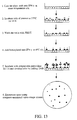
- the subject invention also concerns methods for enumeration and analysis of results obtained from an immunospot assay, such as an enzyme-linked immunospot (ELISPOT) assay.
- the methods comprise performing an immunospot assay wherein a detectable signal produced by the assay is converted to a digital image using computer-assisted video imaging means.
- the detectable signal is produced by means of an enzyme substrate reaction that produces a detectable colorimetric signal.
- the image can be analyzed using analysis software, such as, for example, NIH Image Version 1.55 shareware.
- analysis software such as, for example, NIH Image Version 1.55 shareware.
- the methods of the invention can be used with one- or two-color immunospot assays. Using the methods of the present invention eliminates manual microscopic counting of immunospot assay results and provides objective and reproducible immunospot assay analyses.
- immunodeficient mouse refers to a mouse or mouse strain which is deficient in immune system function, including at least a deficiency in function or presence of mature B and T lymphocytes.
- immunodeficient mouse strains include, but are not limited to the C.B-17-SCID-nod, C.B-17scid/scid and C.B-17-SCID-beige strains.
- Particularly preferred immunodeficient mice have a severe combined immunodeficiency characterized by a lack of mature T, B and natural killer (NK) lymphocytes (e.g ., the C.B-17-SCID-beige mouse strain).
- human T lymphocytes refers to T lymphocytes of human origin. When present in a mouse reconstituted with human blood cells, the human T lymphocytes may be obtained from a variety of sources in the reconstituted mouse including blood (i.e ., peripheral blood lymphocytes or PBLs), lymph nodes, spleen, peritoneal lavage, etc.
- blood i.e ., peripheral blood lymphocytes or PBLs
- lymph nodes i.e ., peripheral blood lymphocytes or PBLs
- spleen spleen
- peritoneal lavage etc.
- Human T lymphocytes are identified by the presence of certain markers or cell surface proteins including CD3, CD4, CD8, T cell antigen receptor (TCR) and CD45.
- the presence of these markers on a lymphocyte may be determined by standard immunocytological means such as incubation (or staining) of a cell suspension containing lymphocytes with antibodies specific for these markers; the antibodies may be directly labeled ( e.g . with a fluorophore such as fluorescein, phycoerythrin, Texas Red, etc.) or the presence of the antibody bound to the surface of a lymphocyte may be detected using a secondary antibody (i.e ., an antibody directed at the first antibody or a component thereof) that is labeled.
- a secondary antibody i.e ., an antibody directed at the first antibody or a component thereof
- the stained lymphocytes may then be analyzed using a fluorescence microscope or a FACS (fluorescence-activated cell sorter) analysis.
- “Mature human T lymphocytes” express either CD4 or CD8, CD3 and a TCR.
- “Immature human T lymphocytes” express both CD4 and CD8 ( i.e ., they are CD4+8+); these cells are also referred to as progenitor T cells.
- “Immature naive human T lymphocytes” are immature T lymphocytes that have not been activated ( i.e ., they have not engaged antigen specific for their TCR or been stimulated by a nonspecific mitogen) and are said to be naive.
- “Immature naive T lymphocytes” includes CD4+8+ T cells as well as CD45RA+ T cells.
- a mouse “comprising CD45+ T lymphocytes wherein at least about 5% of the human CD45+ T cells represent immature naive T lymphocytes” is a mouse in which about 5% or more of the CD45+ T cells are either CD4+8+ or CD45RA+ or the sum of the percentage of CD45+ T cells that are CD4+8+ and CD45RA+ is at least about 5%.
- CD45 proteins are found on the surface of all hematopoietic cells, except for erythrocytes [ The Leukocyte Antigen Facts Book, Barclay et al. (1993), Academic Press, London, UK, pp.202-204 ]. Different isoforms of CD45 are found on different lymphoid cell types; CD45RO is found on activated and memory T cells, whereas CD45RA is found on naive T cells.
- SCID/beige mouse refers to the C.B-17-SCID-beige mouse strain.
- SCID/beige, SCID-beige and SCID/bg are used interchangeably herein.
- human tumor cells refers to tumor cells of human origin; a tumor cell is a neoplastic or cancerous cell.
- Tumor cells may be "established tumor cells,” i.e ., those which can be maintained indefinitely in tissue culture or may be primary tumor cells," i.e ., tumor cells freshly isolated or explanted from a patient.
- primary tumor cells encompasses primary tumor cells maintained in tissue culture for less than or equal to 5 passages.
- human immune cell refers to cells of the immune system (e.g ., T, B and NK lymphocytes, antigen presenting cells) that are of human origin.
- human peripheral blood lymphocytes refers to nucleated, non-erythroid cells derived from the blood of a human.
- PBLs peripheral blood lymphocytes
- PBMCs peripheral blood mononuclear cells
- central nervous system cells refers to cells derived from the central nervous system (i.e ., cells derived from the brain and spinal cord).
- a mouse "reconstituted with human peripheral blood lymphocytes” is a mouse in which human PBLs have been introduced ( e.g ., by intraperitoneal injection) and persist for a period of at least 4 weeks.
- a "SCID/beige mouse comprising human immune cells” is a SCID/bg mouse that has been reconstituted with human PBLs or another source of human immune cells (e.g ., human fetal hematopoietic tissues) and thus, contains human immune cells.
- SCID/beige mouse comprising human immune cells whose serum contains at least 100 ⁇ g/ml of human immunoglobulin (Ig) are preferentially employed for the evaluation of vaccines.
- an "injectable preparation” is a preparation suitable for injection into an animal (e.g ., a mouse).
- immune-modulating gene is a gene encoding a protein that modulates an immune response.
- immune-modulating genes include, but are not limited to, cytokines, costimulatory molecules and chemotactins.
- the product of an immune-modulating gene is said to be an "immune modulator.”
- a “cytokine” is a hormone-like protein, typically of low molecular weight, that regulates the intensity and/or duration of an immune response and is involved in cell to cell communication.
- cytokines include but are not limited to the interleukins (e.g ., interleukin 2, interleukin 4, interleukin 6, interleukin 7, interleukin 12), granulocyte-macrophage colony stimulating factor, granulocyte colony stimulating factor, interferon-gamma (IFN- ⁇ ) and tumor necrosis factor-alpha (TNF- ⁇ ).
- interleukins e.g ., interleukin 2, interleukin 4, interleukin 6, interleukin 7, interleukin 12
- granulocyte-macrophage colony stimulating factor granulocyte colony stimulating factor
- IFN- ⁇ interferon-gamma
- TNF- ⁇ tumor necrosis factor-alpha
- cell cycle regulator refers to any protein whose activity modulates progression of the cell cycle. Particularly preferred cell cycle regulators are those that block cell cycle progression. Examples include but are not limited to the HIV vpr gene product, p21, inhibitors of mammalian cyclins, etc.
- inducers of apoptosis refers to any protein whose activity induces apoptosis in a cell.
- Inducers of apoptosis include but are not limited to apoptin (the product of the chicken anemia virus VP3 gene), BAX, BAD, a BCL-X derivative and the HIV vpr gene product.
- vaccine refers to any preparation that gives rise to an antigen or antigens that is or can be presented to a cell of an animal immune system in order to elicit a prophylactic and/or therapeutic immune response to the antigen.
- expression vector refers to a recombinant DNA molecule containing a desired coding sequence and appropriate nucleic acid sequences necessary for the expression of an operably linked coding sequence in a particular host organism.
- Nucleic acid sequences necessary for expression in prokaryotes include a promoter, optionally an operator sequence, a ribosome binding site and possibly other sequences.
- Eukaryotic cells are known to utilize promoters, enhancers, and termination and polyadenylation signals.
- Promoters and enhancers consist of short arrays of DNA sequences that interact specifically with cellular proteins involved in transcription [ Maniatis, et al., Science 236:1237 (1987 )]. Promoter and enhancer elements have been isolated from a variety of eukaryotic sources including genes in yeast, insect and mammalian cells and viruses (analogous control elements, i.e ., promoters, are also found in prokaryotes). The selection of a particular promoter and enhancer depends on what cell type is to be used to express the protein of interest.
- eukaryotic promoters and enhancers have a broad host range while others are functional in a limited subset of cell types [for review see Voss, et al., Trends Biochem. Sci., 11:287 (1986 ) and Maniatis, et al., supra (1987)].
- the SV40 early gene enhancer is very active in a wide variety of cell types from many mammalian species and has been widely used for the expression of proteins in mammalian cells [ Dijkema, et al., EMBO J. 4:761 (1985 )].
- promoter/enhancer elements active in a broad range of mammalian cell types are those from the human elongation factor 1 ⁇ gene [ Uetsuki et al., J. Biol. Chem., 264:5791 (1989 ); Kim et al., Gene 91:217 (1990 ); and Mizushima and Nagata, Nuc. Acids. Res., 18:5322 (1990 )] and the long terminal repeats of the Rous sarcoma virus [ Gorman et al., Proc. Natl. Acad. Sci. USA 79:6777 (1982 )] and the human cytomegalovirus [ Boshart et al., Cell 41:521 (1985 )].
- promoter/enhancer denotes a segment of DNA which contains sequences capable of providing both promoter and enhancer functions (for example, the long terminal repeats of retroviruses contain both promoter and enhancer functions).
- the enhancer/promoter may be "endogenous” or “exogenous” or “heterologous.”
- An endogenous enhancer/promoter is one which is naturally linked with a given gene in the genome.
- An exogenous (heterologous) enhancer/promoter is one which is placed in juxtaposition to a gene by means of genetic manipulation ( i.e ., molecular biological techniques).
- Splicing signals mediate the removal of introns from the primary RNA transcript and consist of a splice donor and acceptor site [ Sambrook et al., Molecular Cloning: A Laboratory Manual, 2nd ed., Cold Spring Harbor Laboratory Press, New York (1989) pp. 16.7-16.8 ].
- a commonly used splice donor and acceptor site is the splice junction from the 16S RNA of SV40.
- Efficient expression of recombinant DNA sequences in eukaryotic cells requires signals directing the efficient termination and polyadenylation of the resulting transcript. Transcription termination signals are generally found downstream of the polyadenylation signal and are a few hundred nucleotides in length.
- the term "poly A site” or "poly A sequence” as used herein denotes a DNA sequence which directs both the termination and polyadenylation of the nascent RNA transcript. Efficient polyadenylation of the recombinant transcript is desirable as transcripts lacking a poly A tail are unstable and are rapidly degraded.
- the poly A signal utilized in an expression vector may be "heterologous” or "endogenous.”
- An endogenous poly A signal is one that is found naturally at the 3' end of the coding region of a given gene in the genome.
- a heterologous poly A signal is one which is isolated from one gene and placed 3' of another gene.
- a commonly used heterologous poly A signal is the SV40 poly A signal.
- the SV40 poly A signal is contained on a 237 bp Bam HI lBcl I restriction fragment and directs both termination and polyadenylation [Sambrook, supra, at 16.6-16.7]. This 237 bp fragment is contained within a 671 bp Bam HI/ Pst I restriction fragment.
- stable transfection or "stably transfected” refers to the introduction and integration of foreign DNA into the genome of the transfected cell.
- stable transfectant refers to a cell which has stably integrated foreign or exogenous DNA into the genomic DNA of the transfected cell.
- selectable marker or “selectable gene product” as used herein refer to the use of a gene which encodes an enzymatic activity that confers resistance to an antibiotic or drug upon the cell in which the selectable marker is expressed.
- Selectable markers may be "dominant”; a dominant selectable marker encodes an enzymatic activity which can be detected in any mammalian cell line.
- dominant selectable markers examples include the bacterial aminoglycoside 3' phosphotransferase gene (also referred to as the neo gene) which confers resistance to the drug G418 in mammalian cells, the bacterial hygromycin G phosphotransferase ( hyg ) gene which confers resistance to the antibiotic hygromycin and the bacterial xanthine-guanine phosphoribosyl transferase gene (also referred to as the gpt gene) which confers the ability to grow in the presence of mycophenolic acid.
- Other selectable markers are not dominant in that their use must be in conjunction with a cell line that lacks the relevant enzyme activity.
- non-dominant selectable markers include the thymidine kinase (tk ) gene which is used in conjunction with TK- cell lines, the carbamoyl-phosphate synthetase-aspartate transcarbamoylase-dihydroorotase (CAD) gene which is used in conjunction with CAD-deficient cells and the mammalian hypoxanthine-guanine phosphoribosyl transferase ( hprt ) gene which is used in conjunction with HPRT cell lines.
- tk thymidine kinase
- CAD carbamoyl-phosphate synthetase-aspartate transcarbamoylase-dihydroorotase
- hprt mammalian hypoxanthine-guanine phosphoribosyl transferase
- the present invention provides humanized animal models suitable for the evaluation of anti-human tumor immunity. These animal models permit the identification of combinations of immune-modulating genes (IMGs) which when delivered to human tumor cells induce an effective anti-tumor response, including a systemic anti-tumor response.
- IMGs immune-modulating genes
- the Description of the Invention is divided into the following sections: I. Immunogene Therapy; II. The Hu-SCID/beige Model For Immunotherapy; III. Combination Immunogene Therapy; IV. Evaluation Of Candidate Vaccines Using The Hu-PBL-SCID/beige Model; V. HIV Vaccines; VI. Alternative Strategies For Cancer Therapy And Vaccine Production; and VII. Computer-Assisted Immunospot Analysis.
- Immunogene therapy involves the introduction of genes encoding proteins that regulate or modulate the immune response. Particularly useful in the present invention are those genes that encode proteins that are involved in the activation of T cells. As discussed above, the majority of tumors are not immunogenic, i.e ., they fail to provoke an immune response in an animal.
- SCID/bg The C.B-17 SCID/beige (SCID/bg) mouse, an immunocompromised mouse, was employed for the humanized mouse model as this strain of mice lacks T cell, B cell and natural killer (NK) cell function [ Froidevaux and Loor (1991) J. Immunol. Methods 137:275 ].
- the SCID/bg mouse supports the growth of a variety of established human tumor cell lines as well as primary human tumor cells.
- SCID/bg mice were efficiently reconstituted with human peripheral blood lymphocytes (PBLs) with CD45+ human cells constituting up to 60% of the splenocytes and 2-7% of the peripheral blood mononuclear cells in the reconstituted mice.
- PBLs peripheral blood lymphocytes
- CD45+ human cells constituting up to 60% of the splenocytes and 2-7% of the peripheral blood mononuclear cells in the reconstituted mice.
- the peripheral blood of the Hu-PBL-SCID/bg mice were found to contain high numbers of immature or progenitor T cells (i.e ., CD4+8+ cells and CD45RA+ cells). These results are in contrast to the results obtained by reconstitution of C.B-17scid/scid mice (Hu-PBL-SCID).
- the Hu-PBL-SCID/bg mice show evident levels of CD45RA+ and CD4+8+ cells 4-6 weeks after reconstitution.
- the Hu-PBL-SCID/bg mice of the present invention provide a suitable model for the evaluation of anti-tumor immunity.
- Hu-PBL-SCID/bg mice as a model for human immunogene therapy is illustrated herein for the identification of a combination of immune-modulating genes (IMGs) effective in the treatment of human glioblastoma multiforme.
- IMGs immune-modulating genes
- Glioblastoma multiforme is the most common primary central nervous system neoplasm in humans. Despite improvements in diagnosis and treatment of glioblastoma multiforme, mean survival from time of diagnosis remains less than one year [ Chang et al. (1983) Cancer 52:997 ; McDonald and Rosenblum (1994) In: Principles of Neurosurgery, Regachary and Wilkins, eds., Wolfe Publishing, Toronto, pp. 26.21-26.32 ].
- lymphocytes Although glioblastomas in situ are normally infiltrated to varying degrees by lymphocytes [ Kuppner et al. (1989) J. Neurosurgery 71:211 ; Black et al. (1992) J. Neurosurgery 77:120 ], evidence indicates these lymphocytes are unactivated. This may be in part due to secretion of immunosuppressive factors such as prostaglandin E 2 , transforming growth factor ⁇ 2 , and interleukin-10, all of which inhibit lymphocyte activation [ Fontana et al. (1982) J. Immunol. 129:2413 ; Siepl et al. (1988) Eur. J. Immunol.
- immunosuppressive factors such as prostaglandin E 2 , transforming growth factor ⁇ 2 , and interleukin-10
- Immunogene therapy strategies were designed to overcome such local immunosuppression by promoting tumor antigen presentation and/or anti-tumor lymphocyte activation.
- the present invention provides a novel humanized mouse model utilizing a human glioblastoma cell line, human immunogenes and human lymphocytes.
- genes encoding human GM-CSF and/or B7-2 were efficiently transferred in vitro into a human glioblastoma cell line. Thereafter, the effect of GM-CSF and/or B7-2 expression on glioblastoma growth in vivo was examined in a human tumor/human PBL /severe combined immunodeficiency mouse (hu-PBL-SCID) model. Human glioblastoma cells (with or without therapeutic gene transfer) and human PBLs were grafted into SCID/bg or SCID/nod mice and the effect on tumor growth locally and at distant sites was observed. SCID/bg and SCID/nod mice accept human tumor and lymphocyte grafts without rejection as they lack mature T, B, and NK lymphocytes.
- the human glioblastoma-Hu-PBL-SCID/bg model employed herein represents an allogeneic system that comprises an established human glioblastoma cell line and lymphocytes from unrelated donors. This model has more similarities to autologous systems than may be immediately apparent. Unlike the classical immune-mediated rejection seen in allogeneic organ transplantation, this system is free of graft-origin "passenger lymphocytes.” These lymphocytes are important in initiating allogeneic organ rejection responses by presenting antigen to host lymphocytes in the context of allogeneic Class II Major Histocompatibility Complex (MHC) [ Larsen et al. (1990) Annals Surgery 212:308 ; Chandler and Passaro (1993) Archives Surg.
- MHC Allogeneic Class II Major Histocompatibility Complex
- the D54MG glioblastoma cell line employed in this model expresses only Class I MHC and not Class II MHC in vitro. Therefore, the only Class II MHC molecules available for tumor antigen presentation in this model are those present on engrafted human PBLs. In other words, this model glioblastoma system is allogeneic for Class I MHC but autologous for Class II MHC.
- the human tumor/Hu-PBL-SCID/bg model can be used as an autologous model system comprising human tumor cells and PBLs from the same patient.
- Such an autologous model is preferred to an allogeneic model system; however, for certain rapidly progressing tumors it may be difficult to obtain PBLs from the same patient once the patient's tumor has been established in the Hu-PBL-SCID mice.
- an allogeneic model using PBLs from a donor unrelated to the tumor donor is employed.
- the human tumor/Hu-PBL-SCID/bg model of the present invention provides a simple and powerful method to analyze human lymphocyte responses to human tumors in vivo and thus provides a means to determine which combination of IMGs are best suited for the treatment of specific tumors.
- the human tumor/Hu-PBL-SCID/bg model of the present invention provides a simple and powerful method to determine which combination of IMGs or IMGs and/or cell cycle regulators, inducers of apoptosis and tumor suppressor genes (e.g ., the wild type p53 gene) are best suited for the treatment of specific tumors.
- immunomodulatory genes are potentially useful and more than one may be necessary for overcoming tumor immunosuppression. These include genes encoding cytokines, major histocompatibility complex molecules, and T cell costimulatory molecules [ Dranoff et al. (1993) Proc. Natl. Acad. Sci. USA 90:3539 ; Gajewski et al. (1995) J. Immunol. 154:5637 ].
- An ideal tumor vaccine should coexpress a combination of immunostimulatory genes from distinct immunomodulatory pathways (e.g ., costimulators, cytokines, and chemoattractive adjuvants) and may also express cell cycle regulators, inducers of apoptosis and tumor suppressor genes.
- a combination of the T cell costimulatory molecule B7-2 and the therapeutic cytokine granulocyte-macrophage colony stimulating factor (GM-CSF) is employed to increase the immunogenicity of a human glioblastoma cell line.
- Genes encoding the B7-2 and GM-CSF proteins were introduced into and co-expressed in the tumor cells.
- Tumor cells modified to express at least one immune-modulating gene and another immune-modulating gene, a cell cycle regulator, an inducer of apoptosis, and/or a tumor suppressor gene can be used as a tumor vaccine according to the methods of the present invention.
- GM-CSF stimulates growth and differentiation of granulocytes, monocytes/macrophages, microglia, and other antigen presenting cells. It has recently come into widespread clinical use as a treatment of neutropenia due to its hematopoietic effects [ Lieshcke and Burgess (1992) N. Engl. J. Med. 327:28 ; Aglietta et al. (1994) Seminars Oncol. 21:5 ; Engelhard and Brittinger (1994) Seminars Oncol. 21:1 ]. The importance of this cytokine in tumor immunogene therapy was recently demonstrated by Dranoff , et al.
- GM-CSF resultsed in the greatest tumor growth inhibition in this mouse model.
- B7-2 is one of a family (B7-1, B7-2, and B7-3) of lymphocyte cell surface molecules that have recently been identified as costimulatory molecules necessary for T-cell activation in conjunction with antigen presentation in the context of a major histocompatibility complex molecule.
- the absence of costimulatory molecules on tumor cells may contribute to their failure to be detected and eliminated by the immune system [ Galea-Lauri et al. (1996) Cancer Gene Ther. 3:202 ].
- the specific roles of the various costimulatory molecules are yet to be clearly defined.
- B7-1 expression promotes differentiation of intermediate T H cell precursors into T H 1 effector cells (cellular immune responses) while B7-2 expression leads to T H 2 differentiation (humoral immune responses)
- B7-1 and B7-2 have been shown to promote cell mediated immune responses in animal models [ Hodge et al. (1994) Cancer Res. 54:5552 ; Lanier et al. (1995) J. Immunol. 154:97 ; Plumas et al. (1995) Eur. J. Immunol. 25:3332 ].
- IMGs to be examined for their effectiveness in treating human tumors in the novel animal models of the present invention include, but are not limited to, APO-1 (Fas), APO-1 ligand (FasL) [ Hahne et al. (1996) Science 274:1363 ; Seino et al. (1997) Nature Med. 3:165 ; Strand et al. (1996) Nature Med. 2:1361 ], IL-12A and IL-12B, IL-2, IL-4, IL-6, IL-7, IL-10, GM-CSF, G-CSF, IFN- ⁇ , CD40 and TNF- ⁇ .
- genes encoding cell cycle regulators or inducers of apoptosis can be employed in combination with IMGs for the modification of tumor cells.
- Expression vectors including retroviral vectors, containing one or more IMG (and/or cell cycle regulators or inducers of apoptosis) are constructed as described herein.
- each IMG is preferably separated from the other(s) using an IRES as described herein.
- a particularly preferred IRES is the poliovirus IRES.
- those IMGs are delivered to a patient's tumor cells in vivo or in vitro followed by a return of the modified tumor cells (typically the modified cells will be irradiated prior to introduction) to the patient.
- a variety of means may be employed for the delivery of IMGs to human tumor cells (e.g ., biolistic transformation of tumor cells in situ, cationic liposomes, retroviral infection, etc.).
- the human tumor/Hu-PBL-SCID/bg model of the present invention was employed to determine that tumor growth was markedly inhibited when these animals were vaccinated with glioblastoma cells transduced with genes encoding the T cell costimulatory molecule B7-2 and the proinflammatory cytokine GM-CSF.
- Treatment of patients having glioblastoma multiforme tumors can be conducted as follows. One to three grams of tumor are harvested when patients originally present and undergo surgery. The tissue is harvested and treated as described in Ex. 11. Tumor cells are grown until sufficient numbers are present to allow retroviral gene transfer and selection.
- the primary tumor cells are transfected with pLSNBG9 (encodes both B7-2 and GM-CSF) and selected by growth in the presence of G418 as described below. Briefly, virus stock is thawed from -80°C at 37°C. Polybrene is added to the thawed virus solution at a final concentration of 4 ⁇ g/ml. Culture medium is added to the virus solution to bring the final volume to 1.5 ml. Logarithmic growth phase tumor cells in T25 flasks are incubated in the virus supernatant at 37°C, 5% CO 2 for 3 hours. The same volume of medium containing polybrene but lacking virus is used as a control.
- Transduced tumor cells are split into a new T25 culture flask at a density of 1 x 10 6 cells.
- the amount of GM-CSF secreted into the culture medium is determined by ELISA (Quantikine, R&D Systems) 24 hr later.
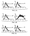
- B7-2 expression is evaluated by flow cytometry using a monoclonal antibody specific for the human B70 antigen (i.e ., B7-2).
- Aliquots of transduced cells to be used as vaccines are tested for the presence of bacteria, fungi, mycoplasma, HIV, Hepatitis B and Hepatitis C and replication-competent retrovirus (using the standard S+L- assay; Bassin et al. (1971) Nature 229:564 ).
- Tumor cells that have been transduced with GM-CSF and B7-2 genes, selected and expanded are cyropreserved for future use. Briefly, cells are aliquoted in small volumes into cryopreservation tubes at 1 x 10 6 cells/tube. Total volume is made up to 0.5 ml with a mixture of DMEM/F12, FCS and DMSO to make a final concentration of 10% FCS and 20% DMSO. Cells are placed in an insulated styrofoam rack and placed at -80°C for 24 hr, then placed in liquid nitrogen for long term storage. When cells are needed, they are thawed from liquid nitrogen at 37°C, washed twice in fresh medium and plated.
- Transduced tumor cells are irradiated prior to their reinjection into patients to render the cells replication incompetent.
- Cells are irradiated with 20,000 Rad in a 60 Cobalt machine.
- Each vaccination comprises 2 x 10 6 irradiated autologous tumor cells modified to express B7-2 and GM-CSF. Injections are given on alternating lumbar flank regions which are marked immediately above the injection site with India ink to allow accurate localization later in the event that a local reaction is not apparent.
- Patients receive 0.1 ml of vaccine injected SC at each site using a 1 ml syringe fitted with a 23 gauge needle. All vaccinations are prepared by resuspension of cells in sterile Ringer's lactate solution.
- Patients having recurrent glioblastomas the first injection is given on the first or second post-operative day. For patients with treatment resistant melanomas (discussed below), the timing of the first injection is not as critical as these patients are not undergoing any further surgical resection of their tumors. All patients receive a second and third vaccination 14 and 28 days, respectively after the first vaccination.
- DTH delayed type hypersensitivity
- punch biopsies are performed at injection sites 2 weeks after each vaccination (i.e ., days 14, 28 and 42). These biopsies are compared to a biopsy taken from normal lumbar flank skin on day 1 (prior to initiation of therapy). Biopsies undergo standard pathologic examination for evidence of tumor cells and inflammation. In addition, immunohistochemical staining for CD45, CD4, CD8 and NK cell markers is performed.
- PBMC peripheral blood mononuclear cells
- Hystopaque density gradient
- the PBMC are then stimulated in vitro by co-incubation for 5 days with irradiated (20,000 Rad) autologous tumor cells.
- the stimulated PBMC are then used in the following two assays.
- a standard 51 Chromium release cytotoxic T lymphocyte (CTL) assay is performed vs. autologous tumor cells.
- CTL cytotoxic T lymphocyte
- ELISPOT assay for interferon- ⁇ production after exposure to autologous tumor cells is performed [ Zhang et al.
- the 51 Chromium release CTL assay is a standard assay for cell mediated immunity. This assay gives direct information concerning the ability of stimulated PBMC to kill tumor cells; however it has a relatively low sensitivity. To overcome this, the much more sensitive ELISPOT assay for interferon- ⁇ production is also used.
- the ELISPOT assay determines the concentration of PBMC present that produce interferon- ⁇ in response to exposure to autologous tumor cells. Since interferon- ⁇ production is closely associated with T H 1 (cell mediated) immune responses, the number of PBMC producing interferon- ⁇ in response to exposure to autologous tumor provides a measure of cell mediated immunity.
- malignant melanoma is rapidly rising in North America. In contrast to other forms of skin cancers which are usually curable with surgery, malignant melanoma is often fatal due to its aggressive nature and tendency of early spread to distant organs.
- Treatment of patients having malignant melanoma can be conducted using tumor cells modified to express B7-2 and GM-CSF as described above. Tumor cells are harvested from patients when patients originally present and undergo surgery and the cells are treated as described in Example 11.
- Combinations of other IMGs (and/or genes encoding cell cycle regulators or inducers of apoptosis) shown to be effective at reducing tumorigenicity and or at inducing local or systemic immunity in the human tumor/Hu-PBL-SCID/bg model of the present invention are employed to treat human tumors, including glioblastoma and malignant melanoma.
- the patient's tumor cells are transduced with retroviruses encoding immune-modulators (e.g ., B7-2 and GM-CSF) (and/or cell cycle regulators or inducers of apoptosis) and the tumor cells are selected in culture prior to reintroduction into the patient
- the patient's tumor cells may be modified by introduction of DNA encoding the desired gene(s) (e.g ., plasmid DNA transferred by biolistics or other physical means, recombinant adenoviruses, liposomes, direct injection of naked DNA).
- the cells are then allowed to express the transduced genes for a few days (or less), irradiated and used to immunize the patient.
- Subsequent boost immunizations may employ retrovirally transduced and selected tumor cells.
- plasmid DNA livered by liposomes, biolistics, adenovirus vectors or as naked DNA
- recombinant retroviruses in those cases where the patient's tumor cells take a lengthy period to grow in cell culture as retroviral transduction and selection takes a period of several weeks and for certain tumors (e.g ., glioblastomas) it is desirable to vaccinate the patient with modified tumor cells within a few days (e.g., ⁇ 3) of the initial surgery.
- Vaccination within ⁇ 3 days of tumor debulking may permit the capture of the "alarm signal" required for costimulator activation on APCs and enhancement of activated macrophage and T cell traffic across the blood-brain barrier [ Fuchs and Matzinger (1992) Science 258:1156 and Matzinger (1994) Annu. Rev. Immunol. 12:991 ].
- the present invention provides humanized animal models that are useful for the evaluation of tumor cell vaccines.
- the hu-PBL-SCID/beige mice of the present invention also provide animal models for the evaluation of vaccines designed to confer immunity against a variety of human pathogens.
- the examples below provide detailed protocols for the establishment of hu-PBL-SCID/bg mice and humanized SCID/bg mice ( i.e ., SCID/bg mice comprising transplanted human fetal hematopoietic tissues).
- SCID/bg mice i.e ., SCID/bg mice comprising transplanted human fetal hematopoietic tissues.
- These animals contain human immune cells and are used to evaluate candidate vaccines (e.g ., HIV vaccines, malaria vaccines, Leishmania vaccines, M. tuberculosis vaccines, etc.). Both cellular anti-pathogen immunity and humoral anti-pathogen immunity is assessed in the reconstituted and vaccinated animals.
- the immunization efficacies of heterologous vaccines of human pathogens can be evaluated in SCID/beige mice intraperitoneally injected with human peripheral blood mononuclear cells (PBMCs) or receiving human fetal thymus/liver tissue transplants.
- PBMCs peripheral blood mononuclear cells
- In vivo human immune cell reconstitution in these animals has been demonstrated by analyzing the expression of different human leukocyte markers, including markers for naive T cells (CD45RA), memory T cells (CD45RO), activation (HLA-DR) and CD4 and CD8 cells.
- CD45RA markers for naive T cells
- CD45RO memory T cells
- HLA-DR activation
- CD4 and CD8 cells CD4 and CD8 cells.
- the in vivo immune functions of the human cells in the reconstituted animals have been demonstrated using an HIV-1 protective immunity model ( see e.g., Ex. 16), and an attenuated Leishmania cell vaccine model ( see Ex. 19).
- mice are injected with freshly isolated PBMCs as described and one week later, human Ig levels are determined by ELISA.
- Hu-PBL-SCID/bg mice whose serum contains greater than 100 microgram/ml of human Igs are immunized via intramuscular injection with naked DNA vaccines or with dendritic cells (DCs) transfected with naked DNA vaccines which express appropriate pathogen antigens. The immunized mice are re-immunized 1-2 weeks later.
- the immunized mice are challenged with wild type pathogens such as HIV-1, malaria, Mycobacterium tuberculosis or Leishmania major one week after the last dose of vaccine.
- the control mice receive DNA vaccines lacking genes encoding the pathogen antigens.
- wild type pathogen challenge infection and/or disease development is examined according to standard methodology.
- the human immune cell-originated anti-pathogen immunity is analyzed, for cell-mediated immune (CMI) responses using reconstituted mouse splenocytes, and for humoral immune responses using antisera.
- CMI cell-mediated immune
- HIV vaccines including live attenuated HIV vaccines, HIV proviral DNA vaccines and modified Leishmania cells expressing HIV proteins.
- the ability of these HIV vaccines to induce cellular anti-HIV immunity and humoral anti-HIV immunity is assessed in hu-PBL-SCID/bg mice (reconstituted with PBLs or transplanted with fetal hematopoietic tissue) as described above and in the examples below.
- replication-incompetent MoMLV-based vectors can be employed to deliver therapeutic genes to tumor cells (section IV).
- Another approach to delivering genes to tumor cells for the purpose of cancer therapy is the use of replication-competent retroviral vectors (e.g ., MoMLV-based vectors) carrying "suicide" genes [ e.g ., the Herpes simplex virus thymidine kinase (HSV tk ) gene].
- a suicide gene is a negative selectable marker whose expression can be selected against. That is under certain conditions, cells expressing the suicide gene are selectively killed. For example, cells expressing the HSV tk gene are killed when they are grown in the presence of gancyclovir.
- the HSV tk gene has been employed for cancer gene therapy.
- Expression vectors containing the HSV tk gene were injected into the brains of patients having brain tumors and the patients were given gancyclovir. These experiments have met with little success as expression of the HSV tk gene was limited to cells along the injection track only; that is the HSV tk gene could not be delivered to enough of the tumor cells using this approach to kill enough tumor cells to make a difference in the outcome of the disease.
- Retroviruses such as MoMLV, preferentially infect rapidly dividing cells; as tumor cells are generally the most rapidly dividing cells in a patient, retroviruses may be employed to deliver genes to the tumor cells.
- the use of a replication competent retroviral vector permits the spread of the therapeutic gene to tumor cells outside of cells located at the site of injection of the vector.
- Replication-competent MoMLV vectors suitable for the delivery of HSV tk include, but are not limited to, Moloney leukemia virus (MLV) vectors [ RNA Tumor Viruses (1995) Molecular Biology of Tumor Viruses, Cold Spring Harbor Laboratory Press, Cold Spring Harbor, NY ].
- MMV Moloney leukemia virus
- the HSV tk gene is inserted into the MoMLV vector using standard recombinant DNA techniques to create a suicide vector and DNA comprising the suicide vector is prepared.
- the suicide vector DNA is injected into the patient's tumor (intratumor injection) and the patient is then given gancyclovir (i.v. injection) to permit the killing of tumor cells expressing the HSV tk gene.
- the present invention contemplates the injection of DNA constructs encoding the components necessary for the packaging of a retroviral vector along with a therapeutic vector into the tumor cells of a patient.
- Three separate DNA constructs are injected: 1) a DNA expression vector encoding the gag-pol genes of a retrovirus (e.g., MoMLV); 2) a DNA expression vector encoding the envelope gene of a retrovirus or a vesicular stomatitis virus G protein; and 3) a replication-defective retroviral vector (e.g ., a vector lacking packaging signals and gag, pol and/or env genes) comprising a therapeutic gene(s) (e.g ., GM-CSF, B7-2, etc.).
- a retrovirus e.g., MoMLV
- a DNA expression vector encoding the envelope gene of a retrovirus or a vesicular stomatitis virus G protein e.g., MoMLV
- a replication-defective retroviral vector e.g ., a vector lacking packaging signals and gag, pol and/or env genes
- a therapeutic gene(s) e.g .
- packaging cell lines are created in situ in the patient's tumor.
- Such an approach is superior to the injection of packaging cell lines into a patient.
- packaging cell lines typically mouse fibroblast cell lines shedding a replication defective virus
- the patient's immune response is often directed against the foreign cellular proteins rather than against the desired target (the tumor cell).
- the injection of DNA comprising the components of the packaging cell line, rather than the cell line itself, permits the immune response to be directed at the desired target.
- the present invention also concerns immunospot assay methods, such as, for example, an enzyme-linked immunospot (ELISPOT) assay [ Cui and Chang (1997) BioTechniques 22:1146-1149 ], that utilize computer-assisted, video-imaging analysis.
- ELISPOT enzyme-linked immunospot
- the immunospot assay has also been used to assess cytokine production from various immune effector cells.
- the immunospot assay can detect antibody or cytokine production at the single cell level, the visual counting of spots in a 96-well plate under microscope makes this method unsuitable for handling large sample sizes.
- the computer-assisted image analysis system and methods used with the immunospot assay according to the subject invention overcomes this problem.
- This system makes the data analysis step of the immunospot assay convenient, objective, reproducible and suitable for handling large sample pools.
- Studies requiring lymphocyte proliferation assay, cytotoxic lymphocyte assay, and precursor frequency assay can be conducted using the immunospot assay of the invention. This is demonstrated using examples such as mixed lymphocyte allogeneic reactions and the human immunodeficiency virus antigen-specific, cell-mediated immune responses.
- the methods of the subject invention comprise performing an immunospot assay wherein a detectable signal produced by the assay is converted to a digital image or signal using computer-assisted video imaging means.
- the detectable signal is produced by means of an enzyme substrate reaction that produces a detectable colorimetric signal.
- the image can be analyzed using analysis software, such as, for example, NIH Image Version 1.55 shareware.
- analysis software such as, for example, NIH Image Version 1.55 shareware.
- the methods of the invention can be used with one- or two-color immunospot assays. Using the methods of the present invention eliminates manual microscopic counting of immunospot assay results and provides objective and reproducible immunospot assay analyses.
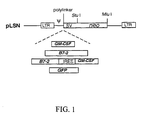
- Retroviral gene therapy vectors were constructed that contained the genes for GM-CSF, B7-2, GM-CSF and B7-2, or GFP gene as shown schematically in Figure 1.
- Figure 1 provides a schematic showing the map of the parental vector (pLSN), pLSNB70 (encodes human B7-2), pLSNGM1 (encodes human GM-CSF), pLSN-BG9 (encodes both B7-2 and GM-CSF) and pLSN-GFP (encodes GFP).
- pLSN is a derivation of pLNL6, a retroviral vector approved for clinical use in the United States of America.
- pLLL an intermediate vector
- pLLL was constructed using pLNL6 (SEQ ID NO: 1) as a starting point.
- pLNL6 contains the MoMuLV promoter in the 3'LTR and the murine sarcoma virus (MSV) promoter in the 5' LTR.
- MSV murine sarcoma virus
- pLNL6 was digested with ClaI and Bcl I and the vector fragment was gel purified using GeneClean (Bio-101) according to the manufacturer's instructions.
- a doublestranded insert containing the polylinker site was constructed using the following two oligonucleotides: 5'-GATCTAAGCTTGCGGCCGCAGATCT CGAGCCATGGATCCTAGGCCTGATCACGCGTCGACTCGCGAT-3' (SEQ ID NO:2) and 5'-CGATCGCGAGTCGACGCGTGATCAGGCCTAGGATCCATGGCTCG AGATCTGCGGCCGCAAGCTTA-3' (SEQ ID NO:3).
- oligonucleotides were annealed, kinased and ligated to the gel purified pLNL6 vector fragment and the ligation mixture was used to transform competent DH5 ⁇ cells (BRL).
- BBL competent DH5 ⁇ cells
- Proper construction of pLLL was confirmed by restriction enzyme digestion of plasmid DNA prepared from ampicillin-resistance bacterial colonies as well as by DNA sequencing near the site of insertion of the polylinker.
- pLSN contains the neo gene under the transcriptional control of the SV40 enhancer/promoter.
- pLSN a Bam HI/ Stu I fragment containing SV40 enhancer/promoter was isolated from pLNSX [ Miller, A.D.and Rosman, G.J. (1989) BioTechniques 7:980 ].
- pLNSX and pLLL were digested with Bam HI and Stu I and the digestion products were gel purified.
- the neo gene was isolated from pLNSX using PCR. Pfu polymerase (Stratagene) was used to amplify the gene. This amplification was conducted in 5 ⁇ l of lOx Pfu reaction buffer, 0.5 ⁇ l of dNTP (15 mM), 0.5 mM of each of the following primers: 5'-AAGCTTGATCACCACCATGATTGAACAAGATGG-3' (SEQ ID NO:4) and 5'-CCGGATCCGTCGACCCCAGAGTCCCGCTCAGAAG-3' (SEQ ID NO:5), 0.5 ⁇ l of pLNSX (0.01 ⁇ g) and 38 ⁇ l of ddH 2 O. These primers contain the modified translation initiation control sequence (-CCACC ATG -), as this modification was found to greatly increase the strength of the neo gene in tissue culture cells [ Kozak, M. (1986) Cell 44
- the mixture was heated at 95 °C for 5 min and 1 ⁇ l of Pfu polymerase was added. This reaction mixture was cycled through 30 cycles at 94°C for 1 min, 55°C for 1 min, and 72°C for 3 min. After amplification, the DNA comprising the neo gene was gel purified and ligated with the Bcl I -digested pLLL/SV40 vector to create pLSN. Confirmation of proper construction was made by restriction enzyme digestion as well as DNA sequencing.
- GM-CSF and B7-2 cDNAs were amplified from normal human lymphocyte RNA by RT-PCR as follows.
- the oligonucleotides used for PCR amplification comprised an optimized eukaryotic initiation sequence.
- the following primer pair was used: 5' primer: 5'-CCCGGG AAGCTT CCACCATG TGGCTGCAGAGCCTG-3' (SEQ ID NO:6) and 3' primer: 5'-AATGGATCCTATCACTCCTGGACTGGCTC-3' (SEQ ID NO:7).
- the sequence of the human GM-CSF cDNA is available in GenBank accession no. M11220 and in SEQ ID NO:8.
- RNA sequence of the human B7-2 cDNA is available in GenBank accession no. U04343 and in SEQ ID NO:11.
- RT-PCR was carried out using an RT-PCR kit (BRL and Promega) according to the manufacturer's instructions.
- the PCR amplified B7-2 gene was cloned directly (i.e ., blunt-end ligation) into HincII digested pT7T318U (Pharmacia) to generate pT7T318U-B7-2.
- the PCR amplified GM-CSF gene was digested with Hind III and Bam HI and cloned into Hind III and Bam HI digested pBluescript KS (-) (Stratagene) to generate pBS-GM-CSF.
- Proper amplification and cloning of the B7-2 and GM-CSF open reading frames was confirmed by partial DNA sequencing and by restriction enzyme digestion.
- the B7-2 and GM-CSF cDNAs were subcloned into the retroviral vector pLSN to generate pLSNB70 and pLSNGM1, respectively.
- pLSNB70 pT7T318U-B7-2 was digested with Bam HI and the B7-2 fragment was gel purified and cloned into Bgl II-digested pLSN.
- pBS-GM-CSF was digested with Hind III and Bam HI and the GM-CSF fragment was gel purified and cloned into Hind III- and Bam HI-digested pLSN.
- cDNA for the reporter gene green fluorescent protein was also inserted into the retroviral vector pLSN to generate pLSN-GFP.
- the GFP gene was PCR amplified using Pfu polymerase (Stratagene) from pGFP (Clontech) using the following primer pair: 5' primer: 5'-AAAAGCTTGGATCCACCATGAGTAAA GGA-3' (SEQ ID NO: 12) and 3' primer: 5'-AATCTAGATTACTATTTGTATAGTT CATCC-3' (SEQ ID NO: 13).
- the PCR amplified GFP gene was cloned directly into EcoRV-digested pBluescript KS(-) to generate pBS-GFP#1.
- pBS-GFP#1 was digested with N o t I and XhoI and the GFP fragment was gel purified and inserted into Not I - and XhoI-digested pLSN to generate pLSN-GFP.
- Each of the resulting vectors contained a neomycin resistance gene driven by an internal SV40 promoter.
- a bi-cistronic retroviral vector, pLSN-BG9, containing the B7-2 and GM-CSF genes separated by an IRES was constructed as follows. Three DNA fragments were gel purified: the EMCV IRES from XhoI- and Hind III-digested pGEM-IRES8 (described below); the GM-CSF gene from Hind III- and Bam HI-digested PCR product (section b); and the B7-2 gene from Not I-and XhoI-digested pLSNB70. The three purified fragments were mixed together and ligated into Not I - and Bam HI-digested pLSN to generate pLSN-BG9.
- pGEM-IRES8 was constructed by isolating the EMCV IRES fragment from Eco RI - and MscI-digested pCITE-1 (Novagen) and inserting this fragment into Eco RI- and Sma I-digested pGEM-7Zf+ (Promega).
- Retroviral plasmid DNA was transfected into the packaging cell line PA317 [ Miller, A.D. and Buttimore, C. (1986) Mol. Cell. Biol. 6:2895 and Miller, A.D. (1990) Hum. Gene Ther. 1:5 ] by lipofection as described (Robinson et al., supra). Briefly, PA317 cells were transfected with pLSNGM1, pLSNB70, pLSN-BG9, or pLSN-GFP using lipofectamine (Gibco/BRL). Lipofection was carried out according to the manufacturer's protocol.
- PA317 cells were grown in DMEM containing 10% FBS and penicillin and streptomycin in an atmosphere containing 10% CO 2 at 37°C. Twenty hours prior to lipofection, PA317 cells were placed into a T25 flask (Falcon) at 50% confluency (approximately 1 x 10 6 cells/flask). To transfect the cells, DNA (4 ⁇ g) was added to 300 ⁇ l serum-free DMEM lacking antibiotics in a microcentrifuge tube (Eppendorf), and mixed gently. In a 15 ml polycarbonate tube (Falcon), 300 ⁇ l serum-free DMEM and 12 ⁇ l of lipofectamine were mixed gently.
- Falcon T25 flask
- the two solutions were combined by adding the DNA-containing solution dropwise into the lipofectamine tube, and the mixture was incubated at RT for 45 min. Following this incubation, 2 ml of serum-free DMEM was added and mixed gently. The cells were washed with serum-free DMEM and the DNA/lipofectamine mixture was gently added to the cells. The cells were incubated at 37°C in a 10% CO 2 incubator for 5 hr. After the 5 hr incubation, 2.5 ml of DMEM containing 20% FBS and antibiotics was added to the T25 flask and the cells were incubated overnight. Twenty hours after the 5 hr incubation, the medium was replaced with fresh DMEM containing 20% FBS and antibiotics.
- the medium was changed at 24 hr after the medium was replaced with fresh DMEM and virus was harvested 24 hr later.
- the transfected PA317 cells were split at a 1:10 ratio into selective medium (i.e ., DMEM containing 500 ⁇ g/ml G418 and 10% FBS).
- Replication incompetent virus was harvested from supernatant of the transfected PA317 cultures 48 hrs after transfection of the PA317 cells and immediately frozen at -80°C for later use.
- Viral particles containing recombinant retroviral genomes encoding either GM-CSF, B7-2, B7-2/GM-CSF or GFP were used to transduce the human glioblastoma cell line D54MG.
- the human glioblastoma cell line D54MG [obtained from Dr. D. Bigner, Duke University, Durham, NC; Bigner et al. (1981) J. Neuropathol. Exp. Neurol. 40:201] was cultured in Dulbucco's Modified Eagle's Media (DMEM) with 10% fetal bovine serum (Gibco), 0.2 units/ml penicillin-streptomycin solution (Sigma), and 0.2 mM glutamate at 37°C in a humidified atmosphere containing 5% CO 2 .
- DMEM Dulbucco's Modified Eagle's Media
- Frozen virus stock was thawed at 37°C.
- Polybrene was added to the thawed virus solution at a final concentration of 4 ⁇ g/ml.
- Culture media was added to the virus solution to bring the final volume to 1.5 ml.
- D54MG cells in logarithmic growth phase in T25 flasks (approximately 1 x 10 6 cells) were incubated in the virus supernatant at 37°C, 5% CO 2 for 3 hours. The same volume of media and polybrene without virus was used as control. After 3 hours, a further 3 ml of culture media was added to each flask and they were incubated overnight. Media was changed the next morning.
- neomycin analog G418 was added to the culture media. Media was changed every two to three days until complete selection had taken place (i.e ., all cells in the control flasks were dead). After selection, the cells were cultured in growth media containing reduced concentrations of G418 (250 ⁇ g/ml) and allowed to grow to confluence.
- Transduced D54MG cells at approximately 70% confluence were harvested by scraping after room temperature incubation for 15 minutes in 0.02% EDTA in Phosphate-Buffered Saline (PBS).
- PBS Phosphate-Buffered Saline
- IF immunofluorescence
- samples were incubated on ice for 1 hour with 1 ⁇ g of RPE-conjugated monoclonal anti-human B7-2 antibody (Ancell) or 1 ⁇ g of RPE-conjugated isotype-control murine IgG 1 antibody (Pharmingen).
- IF immunofluorescence
- Amcell RPE-conjugated monoclonal anti-human B7-2 antibody
- IgG 1 antibody Pharmingen
- Transduced D54MG cells at approximately 70% confluence in T75 flasks were incubated in fresh media for 24 hours. After this period of incubation, media was harvested and centrifuged briefly to remove cells and debris. The cell number per flask was determined. GM-CSF levels in the harvested media was tested using a commercially available kit (Quantikine, R & D Systems) as per the manufacturer's instructions. The level of GM-CSF was converted to ⁇ g/10 6 cells/24 hours based on the cell number in the flasks from which the media originated.
- Figs 2A-E provide flow cytometry histograms for: wild type D54MG (2A), B7-2-transduced D54MG (2B), GM-CSF-transduced D54MG (2C), B7-2 and GM-CSF-transduced D54MG (2D), and GFP-transduced D54MG (2E).
- the histograms on the left represent D54MG cells stained with isotype matched control antibodies while the histograms on the right represent staining with monoclonal anti-human B7-2 antibodies.
- the histogram on the left represents unstained wild type D54MG cells while the histogram on the right represents unstained GFP-transduced D54MG.
- GM-CSF production was significant for GM-CSF-transduced (30 ng/10 6 cells/day) and B7-2/GM-CSF-transduced (5 ng/10 6 cells/day) cells but not for wild type or B7-2-transduced cells.
- Gene expression in the transduced D54MG cells is summarized in Table 1.
- B7-2 molecules were clearly present on the surface of D54MG cells transduced with retroviral vectors containing the B7-2 gene and absent on wild type or GM-CSF-transduced cells. These results demonstrate that the above-described retroviral vectors provide simple, reliable tools for transferring GM-CSF and/or B7-2 genes into human glioblastoma cells in vitro.
- This example describes the reconstitution of SCID/nod and SCID/beige mice with human PBL and the engraftment of human tumor cells in these mouse strains.
- mice For most experiments, four to five week old female C.B-17-SCID-beige mice were purchased from Taconic (Germantown, New York). For the first vaccination/challenge experiment, four to five week old female SCID/nod mice were obtained from Dr. L. Pilarski (Cross Cancer Institute, University of Alberta, Edmonton, Alberta). The mice were maintained in filtered cages in a virus free environment and received cotrimoxazole in their drinking water twice per week.
- Hu-PBL-SCID mouse reconstitution was carried out as previously described [ Zhang et al. (1996) Proc. Natl. Acad. Sci. USA 93:14720 ]. Briefly, each mouse was intraperitoneally (IP) injected with 2-3 x 10 7 PBLs resuspended in 0.5 ml of Hanks' balanced salt solution. A near 100% success rate in reconstitution of SCID/bg mice was obtained when fresh PBLs were used. Five days to three weeks after reconstitution, mice were bled from the tail and the human Ig level was assessed by enzyme-linked immunosorbent assay (ELISA) using a monoclonal rabbit anti-human IgG/IgM antibody (Jackson Labs) and control human IgG (Sigma).
- ELISA enzyme-linked immunosorbent assay
- SCID/nod mice were not as reliably reconstituted with human peripheral blood lymphocytes as were SCID/bg mice.
- reconstitution of SCID/nod mice with human PBLs appeared to vary both from PBL donor to PBL donor and from mouse to mouse.
- many hu-PBL-SCID/nod mice developed a disease characterized by cachexia, alopecia, and facial edema 3 to 6 weeks after reconstitution. This was not seen in unreconstituted SCID/nod mice or in SCID/bg mice with or without reconstitution.
- the etiology was unclear, though the possibilities of Graft vs. Host Disease and diabetes melitis were considered. Because of these difficulties, reconstituted SCID/bg mice are preferred for tumor growth and vaccination/challenge experiments.
- mice Five to seven week old SCID/bg mice were injected subcutaneously (SC) on the right flank with 2 x 10 6 retrovirally-transduced and selected D54MG cells. Control mice were injected SC on the right flank with wild type D54MG cells.
- mice Six days post injection, mice were reconstituted via IP injection as described above with 2 x 10 7 human peripheral blood lymphocytes isolated on a Histopaque gradient (Sigma) from the buffy coat layer of whole blood from healthy donors. Reconstitution was monitored by examination of sera (tail bleeds) for the presence of human Ig by ELISA. In all but one experiment, half of the mice from each group (transduced and untransduced) were left unreconstituted as controls. Tumor size was measured in three directions by calipers every 3 to 5 days. Comparison was made between transduced and untransduced tumor cells and between reconstituted and unreconstituted mice.
- SCID/bg mice receiving human PBL via IP injection supported the growth of a human melanoma cell line with nearly 100% success and these animals demonstrated significant levels of human lymphocytes by flow cytometric analysis in spleen and peripheral blood 38 days post reconstitution.
- This example shows that both SCID/nod and SCID/bg mice supported the growth of wild type D54MG cells (human glioblastoma) subcutaneously with 100% efficiency (35/35) regardless of human lymphocyte reconstitution.
- D54MG tumors transduced with B7-2, GM-CSF, or both B7-2 and GM-CSF also grew with 100% efficiency in unreconstituted SCID/bg mice (4/4, 8/8, and 4/4 respectively). Growth of D54MG tumors transduced with B7-2 and GM-CSF was inhibited in human lymphocyte-reconstituted mice as detailed below.
- Fig. 3 For the results shown in Fig. 3 , all mice received 2 x 10 6 tumor cells (either D54MG or D54MG-B7-2) subcutaneously on the right flank on Day 0. Mice were injected IP with 2 x 10 7 human PBL six days after tumor cell injection. Reconstitution was confirmed by detection of serum human Ig levels >100 ⁇ g/ml.
- 3A In the first experiment (3A), all mice were reconstituted with human PBLs.
- tumor volume (mm 3 ) is plotted over time (days) for mice receiving untransduced D54MG cells (open circles; "D54MG”) and D54MG cells transduced with pLSNB70 (solid squares; "D54MG-B7-2").
- Fig. 3B tumor volume (mm 3 ) is plotted over time (days) for mice receiving untransduced D54MG cells (solid squares) and D54MG cells transduced with pLSNB70 (open triangles).
- arrows are used to indicate the times at which the tumor cells and lymphocytes were injected.
- mice received 2 x 10 6 tumor cells (either D54MG or D54MG-GM-CSF) subcutaneously on the right flank on Day 0. Mice were injected IP with 2 x 10 7 human PBL six days after tumor cell injection. Reconstitution was confirmed by detection of serum human Ig levels >100 ⁇ g/ml in ELISAs. In both experiments shown in Figs. 4A and 4B , half the mice from both groups (D54MG and D54MG-GM-CSF) were left unreconstituted (hatched lines). In Fig.
- tumor volume (mm 3 ) is plotted over time (days) for mice receiving untransduced D54MG cells (solid squares; "D54MG”) and DM54MG cells transduced with pLSNGM1 (open triangles; "D54MG-GM-CSF").
- Fig. 4B tumor volume (mm 3 ) is plotted over time (days) for mice receiving untransduced D54MG cells (solid squares) and D54MG cells transduced with pLSNGM1 (open triangles).
- arrows are used to indicate the times at which the tumor cells and lymphocytes were injected.
- mice were reconstituted with 2 x 10 7 PBL from healthy donors via IP injection. Reconstitution was monitored by examination of sera (tail bleeds) for the presence of human Ig by ELISA. Five days after reconstitution, mice were vaccinated via IP injection of 1 x 10 5 irradiated (20,000 rad) D54MG cells (either wild type, GFP-transduced, or B7-2 and GM-CSF-transduced). Five days post-vaccination, all mice received SC injections of 1 x 10 6 unirradiated wild type D54MG cells on the right flank. Tumor growth was measured as described above.
- mice (Taconic) were used.
- the order of injections was slightly different. These mice first received SC injections of 1 x 10 6 wild type D54MG cells on their right flanks. Ten days later, they were reconstituted with 2 x 10 7 human PBL. Ten days after reconstitution, the mice were vaccinated with 1 x 10 5 irradiated tumor cells (either wild type, GFP-transduced, or B7-2/GM-CSF-transduced).
- SCID/nod and SCID/bg mice were reconstituted, vaccinated and challenged with wild type D54MG cells as described above.
- growth of wild type D54MG tumors in human PBL-reconstituted SCID/nod mice which had been vaccinated with irradiated B7-2/GM-CSF-transduced D54MG was markedly inhibited compared to mice vaccinated with wild type or GFP-transduced D54MG.
- Fig. 5A growth of wild type D54MG tumors in human PBL-reconstituted SCID/nod mice which had been vaccinated with irradiated B7-2/GM-CSF-transduced D54MG was markedly inhibited compared to mice vaccinated with wild type or GFP-transduced D54MG.
- tumor volume (mm 3 ) is plotted over time (days) for mice vaccinated with either D54 cells transduced with pLSN-BG9 (open squares; "D54MG-B7-2-GM-CSF"), D54MG cells transduced with pLSN-GFP (solid circles; "D54MG-GFP”) or untransduced D54MG cells (solid squares; "DM54MG”).
- Fig. 5A an arrow indicates the time at which the tumor cells were injected.
- tumor volume (mm 3 ) is plotted over time (days) for mice vaccinated with either D54 cells transduced with pLSN-BG9 (open squares; "D54MG-B7-2-GM-CSF"), D54MG cells transduced with pLSN-GFP (solid circles; "D54MG-GFP”) or untransduced D54MG cells (solid squares; "DM54MG”).
- arrows are used to indicate the times at which the tumor cells and lymphocytes were injected as well as the time at which the vaccination was given.
- mice vaccinated with wild type or GFP-transduced D54MG tumor growth continued exponentially in mice vaccinated with wild type or GFP-transduced D54MG.
- mice vaccinated with GM-CSFB7-2 transduced D54MG tumor size increased slightly after vaccination (peaking at a mean of 8 mm 3 ) and then regressed. Differences between the experimental and control groups achieved statistical significance by ANOVA (p ⁇ 0.001) by day 42.
- mice vaccinated with irradiated tumor cells transduced with retroviral vectors containing the B7-2 and GM-CSF genes demonstrate that expression of these genes by glioblastoma cells induces a systemic immune response that inhibits tumor growth at distant sites.
- lymphocytes are important in initiating allogeneic organ rejection responses by presenting antigen to host lymphocytes in the context of allogeneic Class II major histocompatibility complex (MHC) [ Larsen et al. (1990) Annals of Surgery 212:308 ; Chandler and Passaro (1993) Archives of Surgery 128:279 ; Moller (1995) Transplantation Proc. 27:24 ].
- MHC major histocompatibility complex
- the D54MG cell line expresses only Class I MHC and not Class II MHC in vitro. Although allogeneic Class I MHC molecules are expressed by the D54MG cells in this model, the absence of graft-origin Class II MHC-positive cells in the tumor renders this model more like an autologous system.
- the use of the hu-PBL-SCID mouse model and a human glioblastoma tumor cell line has demonstrated the therapeutic utility and feasibility of in vivo immunogene therapy using GM-CSF or B7-2 genes (alone or in combination).
- the hu-PBL-SCID model provides a simple and powerful method to analyze human lymphocyte responses to human tumors in vivo.
- Early cancer immunotherapy studies using hu-PBL-SCID mice were limited by the influence of residual murine NK lymphocyte activity [ Reddy et al. (1987) Cancer Res. 47:2456 ; Hill et al. (1991) FASEB J. 5:A965 ; Mueller et al. (1991) Cancer Res.
- the retroviral gene therapy vectors provided herein demonstrated efficient bi-cistronic gene transfer (B7-2 and GM-CSF) to human glioblastoma cells in vitro.
- B7-2 and GM-CSF bi-cistronic gene transfer
- glioblastoma cells that express these genes result in lymphocyte-mediated responses that inhibit tumor growth locally and at distant sites.
- DMEM +++++ / 1000 SK-N-MC neuroblastoma MEM++NaPy+NEa.a +++ / 600 SK-N-SH / neuroblastoma ⁇ MEM+NaPy+NEa.a.
- Table 2 demonstrate that most of the established human tumor cells were efficiently transduced by the pLSNB70 virus. Some of the tumor cells are more resistant to G418 than others. The transduction rate was determined by counting the number of G418-resistant cell colonies. Expression of the B7-2 gene was demonstrated by immunohistochemical staining using an anti-B7-2 monoclonal antibody. Except for the few cell lines that were sensitive to low levels of G418 ( e.g ., PC3), most of the tumor cell lines tested were efficiently transduced with the B7-2 gene, and selected by G418 within two weeks.
- Retroviral transduction of established cell lines is generally more efficient than transduction of primary tumor cells.
- the target cells are fresh (i.e ., primary) tissues or tumors. It is thus important to demonstrate B7-2 transduction in freshly isolated primary tumor cells.
- several tumor specimens from surgery or biopsy were propagated in culture for less than 5 passages and transduced with the pLSNB70 virus as described in Ex. 2. After transduction, the cultures were selected with G418. The results are summarized in Table 3.
- GM-CSF expression has been shown to be related to its therapeutic efficacy [ Jaffee et al. (1993) Cancer Res. 53:2221 and Dranoff et al. (1993) Proc. Natl. Acad. Sci. USA 90:3539 ].
- Two different retroviral vectors were constructed that contained the human GM-CSF cDNA to examine the level of GM-CSF expressed in transduced tumor cells.
- the first vector pLSN which contains the wild type MoMLV LTR, was described above in Ex. 1 as was the construction of pLSNGM1.
- the second vector, pLGCTSN (ATCC 97803; Robinson et al., supra), contains a modified LTR in which the MoMLV U3 region is replaced by the CMV-IE enhancer/promoter and the HIV TATA and TAR elements which leads to an increased level of expression in human cells; in addition, pLGCTSN contains an extended packaging signal and a 3' splice acceptor sequence from MoMLV.
- the human GM-CSF cDNA was inserted into pLGCTSN to create pLGCTSN-GM1 as follows.
- the GM-CSF gene was released from pBS-GM-CSF (Ex. 1) by digestion with Hin dIII and Bam HI and this fragment was inserted into Hin dIII- and Bam HI-digested pLGCTSN to generate pLGCTSN-GM1.
- a human melanoma cell line (SK-MEL-1; ATCC HTB 67) and the primary adenocarcinoma (LJ) culture ["AD 1 (LJ)"] were transduced with retroviruses derived from either pLSNGM1, pLGCTSN-GM, pLSNBG9 or pLSNB70, and selected with G418.
- the expression of GM-CSF was determined by ELISA using culture supernatants collected from the G418-selected cultures. Table 4 summarizes the amount of GM-CSF (ng/10 6 cells/24 hr) present in the culture supernatants.
- the virus derived from pLSNB70 (encodes B7-2) was used as a negative control for the expression of GM-CSF in the transduced cells; pLSNB70-transduced melanoma cells expressed GM-CSF at ⁇ 0.1 ng/ml/10 6 cells in 24 h which was close to the background level of the ELISA.
- the pLSNGM1- and the pLGCTSN-GM-transduced cells expressed 9 ng/ml/10 6 cells of GM-CSF in 24 hr.
- This study used an established human melanoma cell line (SK-MEL-1) and the level of GM-CSF expression was determined 3 months after cells were selected by G418.
- the primary culture was prepared as follows. The tumor tissue was surgically removed, placed in HBSS on ice immediately after surgery and transferred to a biosafety hood. Ten milliliters of HBSS was placed in a sterile petri dish and the tumor tissue was added. Using forceps and a scalpel, normal tissue surrounding the tumor was removed and the tumor was transferred to a second petri dish containing 5-10 ml HBSS and the tumor was minced. Ten ml of a suspension containing the minced tumor was transferred to a 15 ml tube and the cells were collected by centrifugation at 100xg for 8 min. at room temp.
- the cells were resuspended in 2 ml of DMEM containing 20% FCS.
- the resuspended cells (2-3 ml) were transferred to a 50 ml tube and the following enzymes were added: 1 mg/ml collagenase type V, 10 ⁇ g/ml hyaluronidase type V, 300 U/ml DNase type IV and 10 ⁇ g/ml gentamycin sulfate.
- the cells suspension was then incubated at 37°C with shaking for 60 min.
- the cells were then plated in tissue culture flasks containing DMEM containing 10% FCS, 10 ⁇ g/ml gentamycin sulfate, penicillin/streptomycin/glutamine solution (Gibco/BRL) and 1.25 ⁇ g/ml amphotericin (growth medium) and grown overnight in an incubator containing 5%CO2 at 37°C.
- DMEM tissue culture flasks containing 10% FCS, 10 ⁇ g/ml gentamycin sulfate, penicillin/streptomycin/glutamine solution (Gibco/BRL) and 1.25 ⁇ g/ml amphotericin (growth medium) and grown overnight in an incubator containing 5%CO2 at 37°C.
- non-adherent cells were removed by washing the flasks with 3 ml growth medium. The medium was changed every 3-7 days depending upon the growth rate of the cells.
- the cells were transduced with the pLSNB70 virus and grown in the presence of
- the transduced and selected primary human tumor cells [AD1(LJ)] produced GM-CSF in the range of 1-8 ng/ml/10 6 cells/24 hr (GM-CSF expression was determined 2-3 weeks after the cells were selected using G418).
- the un-modified retroviral vector pLSN is capable of producing high levels of GM-CSF in primary human tumor cells.
- the level of GM-CSF expression may be cell-type dependent, and further modification of the vector (e.g ., use of cell-type- or tissue-specific enhancers and/or promoters) may further increase the level of expression in specific tumor cells.
- the bi-cistronic retroviral vector pLSNBG9 (Ex. 1) was employed.
- the plasmid DNA was transfected into PA317 cells and virus was harvested and used for transduction as described in Ex. 1.
- Both the established melanoma cell line SK-MEL-1 and the primary adenocarcinoma cell culture AD1(LJ) were transduced and selected by G418. After selection (typically 2 weeks after applying G418), the expression of GM-CSF and B7-2 in these two cell types was determined by ELISA (Table 4) and flow cytometry, respectively.
- Table 4 shows that the level of GM-CSF expression for the bi-cistronic vector was similar to the mono-cistronic vector pLSN-GM in SK-MEL-1 cells but was ten times less than the mono-cistronic vector in the primary AD1(LJ) culture.
- the FACS analysis indicated that the mono-cistronic (pLSNB70) and bi-cistronic (pLSNBG9) vectors expressed B7-2 in both cell types.
- the promoter strength of the retroviral vector determines the expression level of the transduced gene in eukaryotic cells. To increase the level of gene expression, we have modified the MLV vector and generated a series of vectors with enhanced promoter strength (Robinson et al., supra ) . To further improve the promoter strength, the strength of several promoters was examined in the human tumor cell line, HeLa (ATCC CCL 2), using a reporter CAT assay.
- the plasmids were transfected into HeLa cells and 48 hr later cell lysates were prepared and assayed for CAT activity as described (Robinson et al., supra).
- EF1 ⁇ +intron the 1.442 kb EF1 ⁇ enhancer/promoter element comprising intron 1
- EF1 ⁇ -intron a 475 bp fragment containing the EF1 ⁇ enhancer/promoter corresponding to map units 125 to 600 of the human EF1 ⁇ gene
- HIV+Tat the pHIV-1/LTR-cat construct co-transfected with the tat expression construct
- MCT pMCT-cat
- MCT+Tat pMCT-cat co-transfected with the tat expression construct.
- the results shown in Table 5 indicate that the human EFI ⁇ promoter plus its intron has the strongest activity compared with all other promoters tested.
- the EF1 ⁇ promoter may be used to drive the expression of therapeutic genes in tumor cells. Retroviral and plasmid constructs in which the EF1 ⁇ +intron promoter/enhancer is used to drive the expression of therapeutic genes is described below.
- the human EF1 ⁇ enhancer/promoter is abundantly transcribed in a very broad range of cell types including L929, HeLa, CHU-2 and COS cells [ Uetsuki, T. et al., J. Biol. Chem., 264:5791 (1989 ) and Mizushima, S. and Nagata, S., Nuc. Acids. Res., 18:5322 (1990 )].
- a 1.442 kb fragment containing the human EF1 ⁇ enhancer/promoter and a splice donor and acceptor from the human EF1 ⁇ gene was isolated from human genomic DNA as follows. The 1.442 kb fragment corresponds to map units 125 to 1567 in the human elongation factor 1 ⁇ gene (SEQ ID NO:14).
- Genomic DNA was isolated from the MOU cell line (GM 08605, NIGMS Human Genetic Mutant Cell Repository, Camden, NJ) using standard techniques [ Sambrook, J. et al., Molecular Cloning: A Laboratory Manual, 2nd. ed., Cold Spring Harbor Laboratory Press, (1989) pp. 9.16-9.23 ]. Two synthetic oligonucleotide primers were used to prime the polymerase chain reaction (PCR) for the isolation of the 1.442 kb fragment containing the human EF1 ⁇ enhancer/promoter.
- PCR polymerase chain reaction
- the 5' primer designated HEF1 ⁇ L5
- the HEF1 ⁇ L5 primer contains the following sequence: 5'-AAGCTTTGGAGCTAAGCCAGCAAT-3' (SEQ ID NO:15).
- the HEF1 ⁇ L5 primer generates a Hin dIII site at the 5' end of the 1.442 kb fragment.
- the 3' primer designated HEF1 ⁇ L3B, contains the following sequence: 5'-TCTAGAGTTTTCACGA CACCTGA-3' (SEQ ID NO:16).
- the HEF1 ⁇ L3B primer generates a Xba I site at the 3' end of the 1.442 kb fragment. PCR conditions were as reported in Saiki, R.K. et al., Science 239:487 (1988 ).
- pSSD5 was constructed by digestion of the plasmid pLI [ Okayama, H. and Berg, P., Mol. Cell. Biol., 3:280 (1983 )] with Pst I and Eco RI. Synthetic oligonucleotides (Operon) were ligated onto the Pst I and Ec oRI ends of pLI to generate the polylinker of pSSD5 (the SD5 polylinker).
- sequences of the oligonucleotide pair used to create the polylinker are: SD5A 5'-TCTAGAGCGGCCG CGGAGGCCGAATTCG-3' (SEQ ID NO:17) and SD5B 5'- GATCCGAATTCGGCC TCCGCGGCCGCTCTAGATGCA-3' (SEQ ID NO:18).
- the ligation of this oligonucleotide pair into pLI destroyed the Pst I site.
- the plasmid was digested with Hin dIII and partially digested with Bam HI.
- the 572 bp Hin dIII lBam HI fragment containing the SV40 enhancer/promoter, the 16S splice junction and the SD5 polylinker was isolated by electrophoresis of the digestion products on a low melting temperature agarose gel (SeaPlaque, FMC BioProducts, Rockland, ME).
- the 572 bp fragment was cut out of the gel and the agarose was removed by digestion with ⁇ -Agarase I (New England Biolabs) followed by isopropanol precipitation according to the manufacturer's directions.
- the 572 bp fragment was inserted into the plasmid pcDV1 [Okayama and Berg, supra ] as follows: pcDV1 was digested with Hin dIII and Bam HI and the 2.57 kb fragment containing the SV40 poly A sequences and the pBR322 backbone was ligated to the 572 bp fragment containing the SV40 enhancer/promoter, 16S splice junction and polylinker. The resulting plasmid was named pSSD.
- the 671 bp Bam HI lPst I fragment containing the SV40 poly A sequences (SV40 map units 2533 to 3204) was removed from SV40 DNA and cloned into pUC19 digested with Bam HI and Pst I. The resulting plasmid was then digested with Bc /I (corresponds to SV40 map unit 2770). The ends were treated with the Klenow enzyme and dNTPs to create blunt ends. Unphosphorylated Pvu II linkers (New England Biolabs) were ligated to the blunted ends and the plasmid was circularized to create pUCSSD. The SV40 poly A sequences can be removed from pUCSSD as a Bam HI lPvu II fragment.
- pSSD5 was constructed by ligating together the following three fragments: 1) the 1873 bp Ssp I/ Pvu II fragment from pUC19; this provides the plasmid backbone; 2) the 796 bp fragment containing the SV40 enhancer/promoter and 16S splice junction and the polylinker from pSSD; this fragment was obtained by digestion of pSSD with Ssp I and partial digestion with Bam HI followed by isolation on low melting agarose and recovery as described above; and 3) the 245 bp Bam HI lPvu II fragment from pUCSSD (this fragment contains the SV40 poly A sequences).
- the three fragments were mixed together and ligated using T4 DNA ligase to create pSSD5.
- the polylinker of pSSD5 contains the following restriction sites: Xba I, Not I, Sfi I, Sac II and Eco RI .
- a plasmid, pHEF-B70, in which the EF1 ⁇ enhancer/promoter is used to drive the expression of the human B7-2 gene was constructed as follows.
- the B7-2 gene was removed from pLCTSNB70 (described below) by digestion with Bam HI and the isolated B7-2 fragment was ligated into Bam HI-digested pHEF1 ⁇ BSD5 to generate pHEF-B70.
- pLCTSNB70 was constructed by ligating the B7-2 gene released from pT7T318U-B7-2 (Ex. 1) by Bam HI digestion into Bam HI-digested pLCTSN (ATCC No. 97802; Robinson et al., supra).
- the IL-12A and IL-12B genes are under the transcriptional control of the EF1 ⁇ enhancer/promoter.
- the PCR product was cloned directly into Eco RV-digested pBluescript KS(-) to generate pKS-IL-12A.
- the IL-12A coding region is provided in SEQ ID NO:21.
- the following primers were employed for the amplification of the IL-12B cDNA: 5'-AAAGAGCT CCACCATG TGTCACCAGCAGTTGGTC-3' (SEQ ID NO:22; optimized initiation sequence underlined) and 5'-AAGGATCCTAACTGCAGG GCACAGATGC-3' (SEQ ID NO:23).
- the PCR product was cloned directly into EcoRV-digested pBluescript KS(-) to generate pKS-IL-12B; the IL-12B coding region is provided in SEQ ID NO:24. Proper construction of both IL-12 constructs was verified by restriction enzyme digestion.
- pLSN-12 is a retroviral vector containing a IL-12A-IRES-IL-12B bi-cistron.
- the following four fragments were gel purified: the IL-12A gene released from pKS-IL-12A by digestion with Cla I and Psp AI; the IL-12B gene released from pKS-IL-12B by digestion with Not I and Xho I ; the EMCV IRES was released from pGEM-IRES8 by digestion with Xho I and Cla I; and a pLSN vector backbone containing a Psp AI site generated by removing the HIV vpr gene from pLSNvpr (described below) by digestion with Mlu I and Psp AI . These four fragments were combined and ligated with Not I-and Mlu I-digested pLSNvpr to generate pLSN-IL-12.
- pLSNvpr was constructed by digestion of pBS-vpr with Not I and Xho I and the vpr gene fragment was ligated with Not I- and Xho I-digested pLSN.
- the vpr gene was PCR amplified from pNL4-3 [ Adachi et al. (1986) J. Virol. 59:284 ] and inserted into pBluescript KS(-) to generate pBS-vpr.
- pHEF-IL-12 To construct pHEF-IL-12, the following three fragments were isolated: the IL-12A gene was released from pKS-12A by digestion with Cla I and Psp AI; the IL-12B gene released from pKS-IL-12B by digestion with Not I and Xho I ; the EMCV IRES was released from pGEM-IRES8 by digestion with Xho I and Cla I. These fragments were cloned into Not I - and Psp AI-digested pHEFvpr. pHEFvpr was constructed by ligation of a 360 bp Bam HI fragment isolated from pLSNvpr with pHEF1 ⁇ BSD5 that had been digested with Bam HI and treated with alkaline phosphatase.
- pHEF-IL-12-GM-CSF contains a IL-12A-EMCV IRES-IL-12B- poliovirus IRES-GM-CSF tri-cistron under the transcriptional control of the EF1 ⁇ enhancer/promoter.
- the following four fragments were gel purified: a 2373 bp Nde I -Psp AI fragment containing the IL-12B-EMCV IRES-IL-12A cistron isolated from pHEF-IL12; a 1320 bp Nde I -Sph I fragment containing the EF1 ⁇ intron isolated from pHEF-IL-12; a 735 bp Psp AI -Xho I fragment containing the poliovirus IRES from pKS-P2 (an equivalent DNA fragment is generated by PCR amplification of the IRES located within the first ⁇ 730 bp of the poliovirus genome [ LaMonica et al. (1986) J. Virol. 57:51515
- pHEF-GM-CSF was derived from pHEF-IL-12-GM-CSF by complete digestion with Sma I and partial digestion with Hinc II. The resulting approximately 4409 bp fragment was gel purified and self-ligated to produce pHEF-GM-CSF.
- a plasmid, pHEF-BG, in which the 1.442 kb EF1 ⁇ enhancer/promoter is used to drive the expression of both the human B7-2 and GM-CSF cDNAs (a bi-cistronic construct) is constructed as follows.
- the B7-2-IRES-GM-CSF bi-cistronic fragment was removed from pLSN-BG9 (Ex. 1) by digestion with Not I and Bam HI and the gel purified fragment was inserted into Not I - and Bam HI-digested pHEF1 ⁇ BSD5 to generate pHEF-BG.
- pLSN-IL-12-GM-CSF provides a retroviral vector in which the MoMLV LTR is used to drive the expression of both the human IL-12 gene and GM-CSF cDNAs.
- pLSN-IL-12-GM-CSF was constructed by ligation of the following four fragments into Bam HI-digested pLSN-GFP: the 1489 bp MoMLV vector fragment derived from Sac II -Xba I-digested pLSN-GFP; the 1680 bp IL-12B-IRES containing fragment from Xba I -Not I-digested pHEF-IL-12; the 760 bp IL-12A containing fragment from Not I -PspA I digested pHEF-IL-12; and the 1252 bp GM-CSF containing fragment from Psp AI -Bam HI digested pHEF-IL-12-GM-CSF.
- pLCTSN-pA-BG-EF contains a cistron comprising the EF1 ⁇ enhancer/promoter, B7-2 and GM-CSF cDNAs and SV40 polyadenylation signal inserted in an inverted orientation (relative to transcription from the 5' LTR).
- This vector was constructed by ligation of the following three fragments with Hin dIII and Sal I digested pLCTSN vector: EFI ⁇ enhancer/promoter from Sal I -Not I digested pHEF, B7-2/IRES/GM-CSF cistron from Sal I -Not I digested pLCTSN-BG, and SV40 polyA signal from Bgl II- Hin dIII digested pSP72SVpA.
- pLCTSN-BG was constructed by gel-purifying the following three DNA fragments: the B7-2 cDNA from pLSNB70 cut with Not I -Xho I , the EMCV IRES from pGEM-IRES8 cut with Xho I -Hin dIII ( Hin dIII partial), and the GM-CSF cDNA from PCR amplified and Hin dIII -Bam HI digested DNA (Ex. 1). The purified fragment were then ligated with Not I -Bam HI digested pLCTSN to create pLCTSN-BG.
- pLCT-pA-BG-EF contains a cistron comprising the EF1 ⁇ enhancer/promoter, B7-2 and GM-CSF cDNAs and SV40 polyadenylation signal inserted in an inverted orientation (relative to transcription from the 5' LTR) but lacks the the neo gene found in pLCTSN-pA-BG-EF vector.
- pLCT-pA-BG-EF was constructed by ligation of the following three fragments with Hin dIII -Sal I digested pLCTSN: EF1 ⁇ enhancer/promoter from Sal I -Not I digested pHEF, the B7-2/IRES/GM-CSF cistron from Not I -Bam HI digested pLCTSN-BG, and the SV40 polyA signal from Not I -Hin dIII digested pSP72SVpA.
- pSP72SVpA was made by PCR amplification of the SV40 polyA signal from pBlueBac-His B (Invitrogen) with primers containing Hin dIII and Eco RI sites and cloned into Hin dIII -Eco RI digested pSP72 (Promega).
- the plasmids described above are introduced into tumor cells, including freshly explanted tumor cells as well as tumor cells in a patient, using injection of naked plasmid DNA, liposome mediated gene transfer or through the use of a gene gun (biolistics).
- the retroviral vectors described above are used to generate recombinant virus which is used to transduce the encoded IMGs into tumor cells (primary and established) in culture as described in Exs. 1 and 2.
- GM-CSF cDNA sequences isolated by PCR amplification and used to generate mono- and multi-cistronic constructs are capable of expressing active GM-CSF
- the following GM-CSF bioactivity assay was performed using the GM-CSF-dependent Mo7e cell line [Genetics Institute, Cambridge, MA; Avanzi et al. (1990) Cellular Phyisol. 145:458 ].
- Mo7e cells were maintained in DMEM supplemented with 10% FBS, 2 mM L-glutamine, 100 U/ml penicillin-streptomycin, and 100 U/ml GM-CSF at 37°C, 10% CO 2 . When the cells were at exponential growth phase (viability > 90%), they were washed twice with DMEM, and cultured for 48 hours in the absence of GM-CSF.
- the Mo7e cells After two days of GM-CSF starvation, the Mo7e cells, with around 20% viability, were washed once with DMEM and seeded into V-bottomed 96-well plates at 1 x 10 4 cells/well in a total volume of 200 ⁇ l with serial dilutions of a standard GM-CSF solution of known concentration or supernatant harvested from HeLa cells transfected with either pHEF-GM-CSF or pHEF-IL-12-GM-CSF plasmid DNA (a 1:2000 dilution of the culture supernatant was employed).
- GM-CSF concentration in the harvested supernatant was obtained by comparing its OD with the standard curve. The results are summarized in Table 6. TABLE 6 Determination Of GM-CSF Concentration In Cell Culture Supernatants Of Transfected HeLa Cells Samples GM-CSF Bioactivity (U/ml) GM-CSF Protein Content (pg/ml) OD 570 Standard 1 0 0 0.2 Standard 2 0.01 1 0.203 Standard 3 0.05 5 0.233 Standard 4 0.2 20 0.343 Standard 5 0.5 50 0.60 Standard 6 1 100 0.65 pHEF-GM-CSF (1:2000) 0.29 29 0.365 pHEF-IL-12-GM-CSF (1:2000) 0.03 3 0.235
- GM-CSF GM-CSF
- pLSNBG9 bi-cistronic retrovirus
- C.B-17 scid/scid mice [ Boussiotis et al. (1993) Proc. Natl. Acad. Sci. USA 90:11059 ] lack both T- and B-cell function but do maintain normal myeloid cell, natural killer (NK) cell, macrophage and dendritic cell functions [ Shpitz et al. (1994) J. Immunol. Meth. 169:1 ].
- SCID/bg which has the NK cell function deleted was employed (C.B-17-scid-beige Inbred, Taconic).
- SCID/bg mice To investigate the ability of SCID/bg mice to support the growth of human tumor cells, several established human tumor cell lines and 5 primary tumors were transplanted into the SCID/bg mice and the mice were monitored for tumor growth for up to 5 months. Primary tumors were treated as follows. Tumor samples were obtained in the operating room at the time of resection under sterile conditions. One to three grams of tumor were harvested for immediate processing. Fresh tumor biopsies were dissected to remove necrotic debris and connective tissue and were minced into 1-2 mm pieces. The pieces were then treated with the following enzymatic solutions in HBSS to provide cell suspensions:
- the resulting cell suspensions were then filtered through a fine mesh and layered onto a Ficoll-hypaque density gradient medium (Sigma) and centrifuged at 400xg for 30 min at room temp.
- the cells at the interface were removed and washed twice with HBSS (centrifuged at 100xg for 10 min) and counted.
- the cells were then seeded into the appropriate medium [ e.g ., RPMI 1640, DMEM, DMEM/F12 (all available from Sigma), etc. containing 10% FCS and antibiotics if desired] for culturing, resuspended into freezing medium for storage or used directly for injection into mice or for gene transfection applications.
- the appropriate medium e.g ., RPMI 1640, DMEM, DMEM/F12 (all available from Sigma), etc. containing 10% FCS and antibiotics if desired
- mice can support growth of the established human tumor lines very efficiently and they support the growth of some primary tumor cells (e.g ., melanoma cells), albeit with a lower efficiency relative to the use of established tumor cell lines.
- Injection of 3 x 10 6 primary glioblastoma cells (Ed 86 and Ed 120) as well as injection of primary heptatoma cells (single donor) did not give rise to palpable tumors within 3 months in SCID/bg mice.
- mice were sacrificed and cells harvested from the peritoneal cavity, spleen, and peripheral blood were analyzed by FACS using antibodies against mouse or human cell markers. Isotype-matched mouse Igs were used for control staining. The results are summarized in Table 8. The following antibodies were used in these experiments: human markers: anti-CD45 (anti-HLe-1, Becton Dickinson), anti-CD3 (Leu-4, Becton Dickinson), anti-CD4 (Becton Dickinson), anti-CD8 (Becton Dickinson) and mouse marker: anti-mouse-H-2K° (Pharmingen). TABLE 8 % Human CD45 Spleen 1-59% PBL 0.1-17% Peritoneal Cavity 5-69%
- mice were efficiently reconstituted with human lymphocytes, with CD45+ human cells constituting up to ⁇ 60 % of splenocytes, up to 17% of PBLs and up to ⁇ 70% of cells obtained by peritoneal lavage in the reconstituted mice.
- the percentage of different human lymphocyte subsets in the CD45+ population present in the reconstituted SCID/bg mice was examined by FACS as follows. SCID/bg mice were reconstituted with PBLs isolated from three healthy human donors as described in Ex. 3. After 4-8 weeks, the PBL-injected mice were sacrificed and lymphoid organs, PBLs and peritoneal exudates were harvested and were analyzed by FACS. The percentage ofT lymphocyte subsets was also determined within the CD45+ population in the peripheral blood of human donors A and B.
- CD45RO anti-CD45RO, Becton Dickinson
- CD45RA anti-CD45RA, Becton Dickinson
- HLA-DR HLA-DR
- CD4 and CD8 The results are summarized in Table 9.
- Table 9 the following abbreviations are used: RA+ (CD45RA positive); RO+ (CD45RO positive); RO+DR+ (CD45RO and HLA-DR positive).
- the Hu-PBL-SCID/bg mice show evident levels of CD45RA+ and CD4+8+ cells 4-6 weeks after reconstitution.
- the Hu-PBL-SCID/bg mice of the present invention provide a suitable model for the evaluation of anti-tumor immunity.
- primary human tumor cells are prepared and injected SC into the flanks of SCID/bg mice as described in Ex. 11.
- the primary tumor cells are first transduced with vectors encoding one or more therapeutic proteins (e.g. , B7-2, GM-CSF, IL-12A, IL-12B, etc.) and mice are injected with the transduced tumor cells (after selection) as well as non-transduced tumor cells.
- therapeutic proteins e.g. , B7-2, GM-CSF, IL-12A, IL-12B, etc.
- mice are reconstituted with autologous PBLs (i.e ., PBLs isolated from the same patient that provided the tumor cells).
- mice For each tumor, a minimum of 10 SCID/bg mice are injected with tumor cells and the rate of establishment of palpable tumors is determined. Tumors that grow with a success rate of >40% in the injected mice will receive autologous PBLs and 1 week later will receive IMG-transduced tumor cells. The tumor size (mm 3 ) is measured every 3-4 days and the mice are monitored for survival. These mice are used to determine which combinations of therapeutic or immune-modulating genes (IMGs) result in the regression of tumor size and prolonged mouse survival.
- IMGs immune-modulating genes
- mice from each group are sacrificed at various intervals and spleen cells and draining lymph node cells are assayed for immune reactivity (i.e ., anti-tumor cellular immunity) (e.g ., 51 Cr release assays, proliferation and cytokine production) to determine whether any of the assays of immune reactivity correlate with observed tumor rejection.
- immune reactivity i.e ., anti-tumor cellular immunity
- Splenocytes and lymph node cells isolated from the reconstituted mice are co-cultured with lethally irradiated tumor cells transduced with IMGs for various lengths of time (minimum of 5 days) and assayed for proliferation by pulsing the cultures with 3 H-thymidine, e.g ., on day 4 of culture and determining 3 H-thymidine incorporation into cellular DNA 18-24 hr later.
- Responding lymphocytes may be restimulated repeatedly (weekly) with untransduced or transduced tumor cells to increase the frequency of T cell precursors specific to the tumor.
- the tumor-responsive immune cells are then transferred into mice carrying the parental tumor to determine if in vitro priming generates cells which can cause tumor regression.
- Interferon ⁇ IFN ⁇
- tumor necrosis factor ⁇ TNF ⁇
- IL-2 Interferon ⁇
- NK cells NK cells
- LAK lymphokine activated killer
- Lymphocytes isolated from the PBL and tumor reconstituted mice are co-cultured with autologous untransduced or mock-transduced and IMG expressing tumor cells are assayed for the production of cytokines associated with cell-mediated immunity. Secretion of IFN ⁇ , TNF ⁇ and IL-2 into the culture supernatants at various times during co-culture are assayed by sandwich ELISA [ Sad et al. (1995) Immunity 2:271 ].
- Viable cells are harvested and enumerated from co-cultures of lymphocytes with autologous tumor cells (mock transduced or expressing IMGs) after 5 days of culture or after multiple weekly stimulations (as described above). Untransduced autologous tumor cells are labelled with 51 NaChromate and standard 5 hour 51 Cr-release assays are performed. Lysis of autologous and allogenic tumor targets are compared to the original stimulating cells. Lysis of tumor cells matching those used as the original stimulus, but not of other tumor cells, indicates the lysis is likely antigen specific. Proof of antigen specific lysis is made by the ability to block lysis using antibodies to class I MHC molecules and the ability to eliminate the effector cell responsible for lysis through the use of anti-CD8 and complement. non-MHC restricted lysis may indicate the induction of LAK cells.
- NK/LAK cells may therefore serve to eliminate cells expressing altered or reduced levels of class I molecules that normally escape CD8+ T cell surveillance.
- NK/LAK non MHC restricted cytolytic activity that includes the stimulating tumor target if initially observed, or the induction of substantial NK/LAK activity when none was present (against the tumor target) in the initial assay.
- Immune-modulating genes may be delivered to human tumor cells in vivo and ex vivo by a variety of means.
- Retroviral vectors encoding immune-modulating proteins may be used to introduce IMGs into established or primary tumor cells as described in Exs. 2, 6 and 7.
- the transfer of IMGs using retroviruses may be made more efficient by increasing the titer of the virus encoding the IMG(s) and increasing the transduction efficiency.
- single cell clones from the producer PA317 cells are be selected by growth in the presence of G418 (or selective medium suitable for the selectable marker carried on the retroviral construct) and clones producing the highest titers of virus are be expanded.
- the PA317 cells can be reinfected with ecotropic virus [e.g ., virus produced in GPE-86 packaging cells] and the best producer cell clones can be selected.
- ecotropic virus e.g ., virus produced in GPE-86 packaging cells
- retrovirus are used in combination with liposomes or poly-L-omithine or polylysine to enhance virus uptake.
- Another way to improve gene transfer efficiency using retroviruses is to increase the targeting efficiency.
- Many tumor cells including glioblastomas and melanomas express excess levels of the transferrin receptor.
- Transferrin has been used to increase the transduction efficiency of adenovirus in combination with polylysine [ Lozier et al. (1994) Human Gene Ther. 5:313 ].
- the human transferrin gene is 2097 bp long (coding region provided in SEQ ID NO:25). Insertion of such a long sequence into the SU domain of the env gene of MLV vector may not produce a stable Env product. However, earlier studies have suggested that the modified Env fusion protein requires the native Env for stable assembly and efficient entry. Thus, the transferrin-env fusion gene is co-transfected with the native env gene to produce retrovirus particle bearing a mixture of wild type and recombinant Env. The gene transfer efficiency of the new vector is examined by transducing glioblastomas or melanomas expressing high levels of transferrin receptor.
- Recombinant adenoviruses can accommodate relatively large segments of foreign DNA ( ⁇ 7 kb), and have the advantage of a broad host cell range and high titer virus production [ Graham and Prevec (1991) Meth. Mol. Biol. 7:109-128 ].
- Adenoviruses have been used in vivo in rats to efficiently deliver genes to the liver and the pancreatic islets [reviewed in Becker et al. (1994) In Protein Expression in Animal Cells, Roth et al. eds .] and to the central nervous system [ Davidson et al. (1993) Nature Genet. 3:219 ].
- Rat livers have also been efficiently transduced ex vivo and then re-implanted [ Shaked et al. (1994) Transplantation 57:1508 ].
- the replication defective recombinant adenoviruses are employed; these viruses contain a deletion of the key immediate early genes E1a and E1b (Graham and Prevec, supra ) .
- a packaging cell line such as 293 cells which supply the E1a and E2a proteins in trans is employed.
- Recombinant adenoviruses are created by making use of intracellular recombination between a much larger plasmid encoding most of the viral genome and a small plasmid containing the gene of interest (e.g ., a cytokine) bracketed by regions of homology with the viral integration site.
- Recombinant adenoviruses expressing mouse IL-4 and IL-10, both under the control of a CMV immediate early enhancer promoter, have been constructed.
- Recombinant adenoviruses expressing human B7-2 and GM-CSF as well as a second virus expressing both the IL-12A and IL-12B subunits are constructed.
- the DNA constructs are cloned as bi-cistronic units with IRES as described in Ex. 1.
- each plasmid is co-transfected together with pJM 17 (Microbix Systems, Toronto) into sub-confluent monolayers of 293 cells (ATCC CRL 1573) using calcium phosphate precipitation and a glycerol shock.
- pJM 17 Movable Markup Language
- ATCC CRL 1573 sub-confluent monolayers of 293 cells
- glycerol shock initial recombinant viral stocks are titered on monolayers of 293 cells, and isolated single plaques are obtained and tested for cytokine expression using an ELISA.
- Viral stocks are amplified and titered on 293 cells, and stored in aliquots at -70°C; if necessary, stocks are concentrated by centrifugation on density gradients.
- To infect tumor cells with recombinant adenoviruses freshly isolated tumor cells are mixed with adenoviral stocks in a minimal volume. Titers of stocks are typically 10 5 -10 8 /ml. Medium is replaced after several hours and the cells are followed for expression of the recombinant adenoviral-encoded IMGs and/or reporter genes.
- a potential drawback of using a adenoviral delivery system is that the transduced cells may retain or express small quantities of adenoviral antigens on their surface [ Yang et al. (1994) Nature Genet. 7:362 ].
- "Second generation" adenoviral vectors which contain deletions in the E2a gene are available and are associated with less inflammation in the recipient and a longer period of expression of the gene of interest [Yang et al., supra and Engelhardt et al. (1994) Proc. Natl. Acad. Sci. USA 91:6196 ]. If necessary, IMGs are inserted into second generation adenoviral vectors.
- the transduced tumor cells are lethally irradiated before injection into the recipient and since other manipulations are undertaken to induce the tumor cells to secrete immune-stimulating cytokines and to express surface signalling molecules, the expression of small quantities of adenoviral proteins (when first generation vectors are employed) may provide a desirable adjuvant effect.
- the recipient is subsequently boosted with retrovirally transduced tumor cells (which express the cytokines and tumor antigens, but not the adenoviral antigens).
- the recipient is monitored for tumor-specific immune responses at a secondary distant site where non-transduced tumor is implanted; the generation of such a response indicates that the desired tumor-specific immunity has been achieved.
- Cationic liposomes have proven to be a safe and effective means for inducing the transient expression of DNA in target cells [ Ledley (1995) Human Gene Ther. 6:1129 ; Feigner (1990) Adv. Drug Delivery Rev. 5:167 ; Felgner et al. (1987) Proc. Natl. Acad. Sci. USA 84:7413 ; and Smith et al. (1993) Biochim. Biophys. Acta 1154:327 ]. Clinical trails are underway using cationic liposomes to introduce the CFTR gene into the lungs of cystic fibrosis patients [ Caplen et al. (1994) Gene Ther. 1:139 and Alton et al. (1993) Nature Genet.
- Cationic liposomes e.g ., DOTAP/DOPE
- ligand-targeted cationic liposomes are employed for the delivery of IMGs to tumor cells.
- Ligand-targeted liposomes are made by covalently attaching ligands or antibodies to the surface of the cationic liposome.
- transferrin is used as the ligand as glioblastoma cells express high levels of the transferrin receptor on their surface.
- monoclonal antibodies directed against melanoma-specific surface antigens e.g ., mAb HMSA5 are employed as the ligand.
- Plasmid DNA encoding IMGs (e.g ., B7-2, GM-CSF and IL-12) is formed into a complex with preformed cationic liposomes using standard methodology or alternatively the DNA is encapsulated into the liposome interior.
- the DNA-containing liposomes are then used to transfer the DNA to tumor cells in vivo by direct intra-tumor injection or in vitro (using freshly explanted tumor cells) followed by return of the transduced cells to the recipient ( e.g ., a patient).
- Biolistics is a method of delivery DNA into cells by projection of DNA-coated particles into cells or tissues.
- DNA is coated onto the surface of tiny ( ⁇ 1-3 ⁇ m diameter) gold or tungsten microparticles and these particles are accelerated to high velocity and are impacted onto the target cells.
- the particles burst through the cell membrane and lodge within the target cell.
- the cell membrane quickly reseals and the passenger DNA elutes off of the particle and is expressed.
- the biolistic method has been used to transfect mammalian cells [ Williams et al. (1991) Proc. Natl. Acad. Sci. USA 88:2726 ; Tang et al. (1992) Nature 356:152 ; Sanford et al. (1993) Methods Enzymol. 217:483 ].
- a hand-held biolistic apparatus (BioRad) is used to transfer DNA into tumor cells or isolated tumor fragments.
- This device uses compressed helium to drive a disc-shaped macroprojectile which carries on its surface microparticles of gold (1-5 ⁇ m) of gold which have been coated with purified plasmid DNA (coprecipitated with spermine) (Williams et al., supra ).
- This apparatus has been used to successfully transfect primary tissues.
- Plasmid DNA encoding the IMGs is coated onto the surface of gold microparticles according to the manufacturer's instructions (BioRad) and the biolistic apparatus is used to transfer the DNA into freshly explanted tumor cells or directly into exposed tumors (e.g ., metastatic nodules on the surface of the liver, melanoma lesions on the skin).
- This example provides additional characterization of the reconstituted SCID/nod and SCID/beige mice described in Examples 3 and 11 above.
- SCID/nod and SCID/beige mice were reconstituted with PBLs of different donors as described in Ex. 3. Analyses of human Ig levels at 2, or 4-7 weeks after reconstitution showed that both of these strains of mice were capable of producing human Igs at levels averaging 500-600 ⁇ g/ml; Ig levels were assessed by ELISA (Ex. 3). Approximately half of the SCID/nod mice used in the reconstitution study did not produce detectable amount of human Igs.
- mice The percentages of human lymphocytes present in different organ compartments of the reconstituted mice were determined two months after reconstitution. As described in Ex. 11, cells were harvested from peritoneal cavity (P.W. or peritoneal wash), spleen (Splenocytes or S.P.), and peripheral blood (PBL), and analyzed by flow cytometry using antibodies against the human leukocyte surface marker CD45. The results are summarized in Figures 6A and 6B .
- Figs. 6A The results shown in Figs. 6A indicated that SCID/beige mice were consistently reconstituted with human PBLs in all three organ compartments with little variation among donors (donors A-I: the numbers in the brackets by the donor designation indicates the number of animals reconstituted with donor PBLs).
- the overall reconstitution efficiency in the SCId/nod mice was low, i.e., ⁇ 10% in the peritoneal cavity, ⁇ 5% in the spleen, and almost undetectable in the peripheral blood ( Fig. 6B ; donors X-Z).
- Donor-to-donor variation was also more pronounced in the SCID/nod mice. For this reason, as well as the reasons discussed in Ex. 3, reconstituted SCID/bg mice are preferred for vaccination/challenge experiments ( i.e ., evaluation of candidate vaccines).
- Both host genetic factors and the infection-immunization process following HIV exposure may play roles in the development of protective immunity.
- the mechanism of resistance to HIV infection in the HIV-exposed but uninfected individuals may be associated with HIV-specific cytotoxic T cell activity, a switch from Th2 to Th1 cytokine response, genetic loci linked to HLA, transporter associated with antigen processing (TAP), and HIV coreceptors, or development of anti-HLA antibodies. While strong indirect evidence supports the hypothesis that these exposed but uninfected individuals have protective immunity against HIV infection, direct in vivo experiments attesting to this hypothesis are still lacking. Understanding the mechanism of such protective immunity is necessary for to the design and testing of effective prophylactic vaccines and immunotherapeutic regimens.
- lymphocytes from high risk (HR) individuals were susceptible to HIV-1 infection their PBLs were collected, activated with PHA in culture and then incubated with T cell or macrophage tropic HIV-1. Infection was scored on the basis of HIV-1 RT activity and immunohistochemical staining with AIDS patients' sera.
- High-risk (HR) donor 1 is the heterosexual partner of a bisexual individual who had a CD4 count of 400-500/ ⁇ l at the time of positive IDV diagnosis s in July 1992. An estimated 144 episodes of vaginal intercourse between the HR donor 1 and the partner took place before his diagnosis, and the HIV-positive partner's CD4 count has decreased to 30/ ⁇ l at the time of study.
- HR donor 2 is the homosexual partner of an individual known to be HIV-1 positive since 1986 and whose CD4 count remained in the 300-400/ ⁇ l range. It was estimated that unprotected anal sex took place ⁇ 225 times before this study, and unprotected sexual contact has "continued” since diagnosis. Infectious virus had been isolated from phytohemagglutinin (PRA)-blasted PBLs of both HIV-positive partners.
- PRA phytohemagglutinin
- HIV-1 strains used in this study include laboratory-established T cell tropic (HIV NL4-3 ) and macrophage tropic HN-1 (HIV NLAD8 also referred to as HIV ADA ) [ Adachi et al. (1986) J. Virol. 59:284 ].
- HIV NL4-3 and HIV NLAD8 are available as catalog no. 78 and no. 416, respectively from the NIH AIDS Research and Reference Reagent Program of National Institutes of Health, Bethesda, MD and the sequence of pNL4-3, a plasmid containing a proviral form of HIV NL4-3 is available in the GenBank database under accession number M19921.
- Virus stocks were prepared from infected peripheral blood mononuclear cells or macrophages as described [ Chang and Zhang (1995) Virol. 211:157 ]. Briefly, macrophage cultures were prepared from HIV-seronegative donors by adherence of PBLs to plastic flasks as described previously with minor modifications [ Hassan et al. (1986) J. Immunol. Methods 95:273 ]. PBLs were prepared by density gradient separation using lymphocyte separation medium (Organon Teknika Corp., Durham, NC). The PBLs were resuspended in RPMI 1640 medium supplemented with 20% heat-inactivated human serum. Approximately 5x10 7 PBLs were attached to a T-75 flask and incubated overnight at 37°C.
- the next day cells were washed three times with PBS and the attached cells were incubated with 0.02% EDTA in PBS for 5-10 min.
- the cells were collected with a cell scraper and plated onto a 48-well plate at 5 x 10 4 cells per well.
- the viability approached 100% as determined by trypan blue staining.
- the initial monocytes were characterized by Wright's staining and the mature macrophages by both Wright's staining and microscopic examination.
- HIV virus stocks were prepared by transfecting HeLa cells or by infecting human PBLs, CD4+ lymphocytes (T cell or macrophage tropic viruses) or macrophages (macrophage tropic viruses); infections may be performed using an moi of about 0.2. After transfection, culture media was collected 48 hr later and frozen in aliquots at -80°C. After infection, HIV RT kinetics were followed and virus was harvested at the peak ofRT production. Virus aliquots were examined by RT assay and TCID50 assay.
- the same virus stocks were used for both in vitro and in vivo experiments.
- the multiplicity of infection (moi) or tissue culture infectious dose (TCID 50 ) was determined by serial dilution of virus stock on HIV-1 negative, PHA-stimulated PBLs as described previously [Chang and Zhang, supra and Ho et al. (1989) N. Engl. J. Med. 321:1621 ]. Briefly, PBLs from HIV-seronegative donors were stimulated with 0.25 ⁇ g/ml PHA for 3 days prior to infection and then maintained in RPMI 1640 supplemented with 10% FBS and 10% interleukin-2 (Pharmacia).
- PBLs were plated in 48 well plates at 0.5 million per well and infected with serially diluted virus supernatants in duplicate. Culture supernatants were harvested every 3-4 days. At approximately 3-4 weeks of time, infection kinetics was determined. The dilution point that gave 50% of positive infection (one out of two wells) was considered to be the TCID50 point.
- PBLs Heparinized peripheral blood was collected from HR and low-risk (LR) HIV-negative donors.
- PBLs were separated by gradient centrifugation with Histopaque (Sigma). Infection was performed as described previously (Chang and Zhang, supra ). Briefly, PBLs were treated with PHA (5 ⁇ g/ml, Sigma) and infected with T cell or macrophage tropic HIV-1 at 0.2 moi. The culture was split at a 1:3 ratio every 3-4 days. The supernatant was harvested for reverse transcriptase (RT) assay, and infections were also confirmed by immunohistochemical staining as described (Chang and Zhang, supra ).
- RT reverse transcriptase
- adherent cells were washed with PBS three times, fixed in cold acetone and methanol (1:1) for 2 min, washed three times in PBS, and incubated in blocking solution (20% FBS, 0.1 % TritonX100 in PBS) for 30 min.
- blocking solution (20% FBS, 0.1 % TritonX100 in PBS) for 30 min.
- Non-adherent cells were attached to the surface of a 24-well plate which had been pretreated with poly-D-lysine (1 mg/ml, Sigma) at room temperature for 10 min.
- An HIV patient serum which was diluted at 1:2000 in a blocking solution containing 20% FBS, 0.1% TritonX100 and 2% dry milk in PBS was used as the first antibody and the incubation was done at room temperature for 1 hr or at 4°C overnight with constant shaking.
- the cells After being washed in PBS for 5 min 4 times, the cells were incubated with a 1:200 dilution of normal sheep antisera at room temperature for 30 min to block non-specific signals.
- the secondary antibody was a biotinylated sheep anti-human antibody (Amersham) which was used at 1:2000 dilution and incubated at room temperature for 1 hr.
- the cells were washed four times in PBS-Tween 20 (0.3%) and incubated in the ultra-sensitive ABC staining solution (containing avidin and biotinylated horseradish peroxidase, Pierce Chemical Co.) at room temperature for 30 min.
- Reverse transcriptase levels were measured as follows. A 10 ⁇ l sample of culture medium (supernatant) was incubated with 50 ⁇ l of a reaction cocktail containing 50 mM Tris-HCl, pH 8.3, 20 mM DTT, 0.6 mM MnCl 2 , 60 mM NaCl, 0.05% NP40, 5 ⁇ g/ml of oligodeoxythymidilic acid, 10 ⁇ g/ml of polyriboadenylic acid and 10 ⁇ M of [ ⁇ - 32 P]dTTP (DuPont NEN, specific activity 800 Ci/mmol). The reaction was incubated at 37°C for 1 hr.
- a reaction cocktail containing 50 mM Tris-HCl, pH 8.3, 20 mM DTT, 0.6 mM MnCl 2 , 60 mM NaCl, 0.05% NP40, 5 ⁇ g/ml of oligodeoxythymidilic acid, 10 ⁇ g/ml of poly
- DE-81 paper (Whatman), and air dried.
- the DE-81 paper was washed 3 times in 2X SSC (20X SSC comprises: 3 M NaCl, 0.3 M sodium citrate, pH 7.0) and autoradiographed.
- the cells were stained with PE-anti-mouse-H-2K d (Pharmingen), fluorescein isothiocyanate (FITC)-anti-human-CD45 (anti-HLe-1, Becton Dickinson), phycoerythrin-anti-human-CD4 or FITC-anti-human CD8 (Becton Dickinson) for 30 min on ice followed by three washes with immunostaining buffer (PBS containing 0.1% FBS, 0.02% NaN 3 )- Isotype-matched PE- and FITC-mouse Igs were used for control staining. The samples were analyzed using the LYSIS II program on a FACScan (Becton Dickinson).
- Fig. 7a the RT assay demonstrated that PBLs from these two HR, HN-1-seronegative individuals were as susceptible as PBLs from normal LR donors to HIV-1 infection in tissue culture ( FIG. 7a ).
- the RT activity (CPM/ ⁇ l) is plotted against days post infection.
- the RT activity in PBLs from two LR individuals (LR-1 and LR-2) infected with the macrophage tropic HIV NLAD8 ("HIV-1-M”) at 0.2 moi is shown using open squares and triangles, respectively; RT activity in uninfected PBLs from LR-1 is shown using the open circles ("LR-1-control").
- Fig. 7b the tissue culture infection was performed using a series of diluted virus preparations (from moi of 10 -1 to 10 -4 ). The results are summarized in Fig. 7b .
- the RT activity (CPM/ ⁇ l) is plotted against days post infection.
- the open circles show RT activity in uninfected PBLs from LR-1; the open squares and triangles show RT activity in PBLs from LR-1 infected with HIV-1 NL4-3 at a moi of 0.001 and 0.0001, respectively.
- mice Two weeks after reconstitution, the hu-PBL-SCID/bg mice were challenged with T cell or macrophage tropic HIV-1 using a TCID 50 of 100 by IP injection under metofane-induced anesthesia. Two weeks after HIV-1 challenge, mice were killed, and single cell suspensions were prepared from peritoneal lavage, spleen, and peripheral blood and processed for immununostaining, flow cytometry analysis, and PCR. All experiments were performed in a biosafety level 3 facility and protocols were approved by the Biosafety Committee, Research Ethics Board, and Health Sciences Animal Welfare Committee at the University of Alberta.
- mice The cells harvested from the infected reconstituted mice were analyzed by flow cytometry using anti-mouse major histocompatibility complex class I antibody (H-2K d ) and anti-human CD45, CD3, CD4, and CD8 antibodies as described in Ex.11.
- H-2K d anti-mouse major histocompatibility complex class I antibody
- CD45, CD3, CD4, and CD8 antibodies anti-human CD45, CD3, CD4, and CD8 antibodies as described in Ex.11.
- HIV-1 infection was examined by a sensitive single-cell immunohistochemical staining method using either HIV-positive patients' sera or a monoclonal anti-p24 antibody as described in Ex.15. Representative immunostaining results are shown in Fig. 8 .
- Fig. 8 For the results shown in Fig. 8 , all mice were killed 4 weeks after reconstitution and single cell suspensions were prepared from the three different organ compartments for immunostaining.
- FIGS. 8a and 8b show HIV-1-positive immunostaining of peritoneal lavage and splenocytes, respectively, of mice reconstituted from a LR individual.
- Figs. 8c and 8d show HIV-1-negative immunostaining of peritoneal lavage and splenocytes, respectively, of mice reconstituted from a HR individual.
- Fig. 8e the solid, hatched and shaded bars represent cells from peritoneal lavage, spleen and peripheral blood, respectively. Reconstitution of mice with PBLs from the LR-1 donor (#1-7), the LR-2 donor (#8-14), the HR-1 donor (#15-21) and the HR-2 donor (#22-27) were confirmed by ELISA for human Ig and FACS analysis for the human CD45 leuckocyte marker.
- Fig. 8e the solid, hatched and shaded bars represent cells from peritoneal lavage, spleen and peripheral blood, respectively.
- IFN- ⁇ is produced by T cells and natural killer cells activated by antigens and is the key mediator of CD4 + Th1 cell development [ Wenner et al. (1996) J. Immunol. 156:1442 ].
- ELISPOT was performed as described [ Miyahira et al. (1995) J. Immunol. Methods 181:45 ] with the following modifications.
- a 96-well nitrocellulose-bottomed plate (Multiscreen-HA, Millipore) was coated with 75 ⁇ l per well mouse anti-human IFN- ⁇ (10 ⁇ g/ml), Pharmingen) at room temperature overnight.
- HIV-1-infected PBLs were used as stimulators in the ELISPOT assay. Infection was confirmed by RT assay and by immunohistochemical staining (ranged 3-5% of HIV-1-positive). No difference was observed between HR and LR individuals in either RT or HIV antigen expression in the infected PBLs.
- HIV-1-infected PBLs were treated with mitomycin C (5 ⁇ g/ml) for 2.5 hr, washed, and resuspended in RPMI medium 1640 containing 10% FBS and 20 units/ml interleukin 2 (Boehringer Mannheim), and seeded in the anti-IFN- ⁇ -coated 96-well plate at 1 x 10 5 per well as target cells (T).
- Frozen unstimulated autologous PBLs which were thawed and used as effector cells (E), were added to the wells at E/T ratio of 0.4:1, 2:1, and 10:1 1 in triplicates, and incubated in RPMI medium 1640 containing 10% FBS and 20 units/ml IL-2 at 37°C, 95% O 2 /5% CO 2 for 24 hr.
- Fig. 9 the number of IFN- ⁇ ELISPOT/well is plotted against the E/T ratio.
- the Xs, open diamonds, open circles and open triangles show the results using PBLs from donors LR-1, LR-2, LR-3 and LR-4, respectively.
- the solid squares, solid diamonds and solid circles show the results using PBLs from donors HR-1, HR-2 and HR-3, respectively.
- HIV-1-specific-ELISPOT are shown as the mean ⁇ SE in triplicate wells after subtracting the background from controls.
- CD4, CD8, or CD56 cells from PBLs of two of these HR donors prior to the in vitro ELISPOT assay.
- In vitro lymphocyte depletion was conducted as follows. For the depletion of CD4, CDB, and CD56 cells, thawed PBLs were incubated with mouse anti-human CD4-, CD8-, CD56-, or mouse IgG 1 -labeled BioMag magnetic beads (PerSeptive Diagnostics, Cambridge, MA) at 50 beads per cell on ice for 30 min. The depletion was carried out by sorting the cells on a magnet for 5 min two times using mouse IgG 1 sorting as control.
- SCID/bg mice were reconstituted with PBLs from LR and HR donors which were either depleted of CD8 T cells or untreated. The reconstituted mice were then inoculated with the macrophage-tropic HIV-1 NLAD8 and the ability to infect the human PBLs in vivo was examined. PBLs from HR donor 1 were employed in this study. As CD4 cells are the targets of HIV-1 infection, they were not depleted in this study.
- CD8 T cells were depleted as described [ Baskar et al. (1993) Proc. Natl. Acad. Sci. USA 90:5687 ] with the following modification.
- Anti-human CD8 antibody was purified from ascites of OKT8 hybridoma (ATCC CRL 8014) using protein A affinity column (ImmunoPure Plus, Pierce). Mice were injected IP with 20 ⁇ g of the purified antibody five times in total: 2 days before PBL reconstitution, at the time of PBL injection, and at 1, 2, and 3 weeks after PBL injection.
- hu-PBL-SCID/bg mice were killed, and the lymphocytes were analyzed by FACS analysis as described in Ex. 11.
- the cells were stained with PE-anti-mouse-H-2K d (Pharmingen) and FITC-anti-human-CD45 (Anti-HLe-1, Becton Dickinson) or with PE-anti-human-CD4 and FTTC-anti-human CD8 (Becton Dickinson) for 30 min on ice followed by three washes with immunostaining buffer (PBS containing 0.1% FBS and 0.02% NaN 3 ). Isotype-matched PE-mouse-Ig and FITC-mouse Ig were used as controls.
- FIG. 10 Figs. 10a and 10c show FACS analysis of splenocytes from the reconstituted hu-PBL-SCID/bg mouse #11 immunostained with anti-mouse H-2Kd, anti-human CD45, anti-human CD4 or anti-human CD8 as depicted.
- Figs. 10b and 10d show FACS analysis of splenocytes derived from the reconstituted hu-PBL-SCID/bg mouse #14, after anti-CD8 antibody treatment.
- mice receiving no antibody the following percentages of human lymphocyte subsets were observed: 14% human CD45 cells ( Fig. 10a ), 7% human CD4 cells, and 8% human CD8 cells ( Fig. 10c ) in spleens 4 weeks after reconstitution.
- mice treated with anti-CD8 antibody there were similar numbers of CD45 cells in the spleen (12%, Fig. 10b ), but the CD8 cells were virtually absent (0.1%, Fig. 10d ).
- These mice were inoculated with the macrophage-tropic HIV-1 NLAD8 on day 14 after reconstitution and killed 14 days later for analysis as described above. Results of this study are summarized in Fig. 10e .
- Fig. 10e the percentage of HIV-1 infected cells in the spleen (open bars) and in the peritoneal lavage (solid bars) 2 weeks after virus challenge is shown.
- mice reconstituted with PBLs from HR donor 1 continued to be resistant to HIV-1 infection as long as CD8 cells were present.
- HR-1 HR donor 1
- similarly reconstituted mice became susceptible to HIV-1 infection ( Fig.10e , hu-PBL-SC1D/bgmice #14, #15, and #16).
- the hu-PBL-SCID/bg model of the present invention provides a suitable model for the assessment of anti-HIV-1 protective immunity.
- Results of the in vivo HIV-1 challenge suggest that the cell-mediated immunity developed in these HR subjects also protects against alternative strains of HIV-1 since the challenge strains used in this study, T cell and macrophage tropic HIV-1, are unrelated to the endogenous HIV-1 strains in the corresponding partners of these HR individuals under investigation.
- HLA haplotype may also contribute to the development of resistance to HIV-1 infection [Haynes et al., supra; Kaslow et al. (1996) Nat. Med. 2:405 and Detels et al. (1996) AIDS 10:102 ].
- the two HR subjects who participated in the above hu-PBL-SCID/bg mouse challenge study do not share common HLA haplotypes.
- CD8 T cells that secret anti-HIV chemokines [ Cocchi et al. (1995) Science 270:1811 ].
- CD4 T cells from 2 of 25 frequently exposed but uninfected individuals have recently been shown to have relative resistance to HIV-1 infection [ Paxton et al. (1996) Nat. Med. 2:412 ]; subsequent genotype analysis of these two individuals by PCR and sequencing indicates that they have homozygous defects in the CCR-5 loci that encode the macrophage-tropic HIV-1 coreceptor [ Liu et al. (1996) Cell 86:367 ].
- the above results demonstrate the usefulness of the in vivo hu-PBL-SCID/bg mouse model for evaluating the protective immunity of individuals exposed to HIV-1.
- the SCID/bg mice can be reconstituted with human PBLs and infected with HIV-1 at a near 100% success rate.
- this model is useful for the assessment of the immune status of HIV-1 vaccinees.
- the first alternative is the exogenous pathway through which antigen-presenting cells take up extracellular proteins by either endocytosis or phagocytosis.
- MHC class II molecules in the endoplasmic reticulum pass through the Golgi apparatus and enter acidified endosomes in which the foreign protein has been fragmented into peptides. The MHC-peptide complex is then brought to the cell surface, where it can be recognized by helper T cells (CD4 + ).
- the second alternative is the endogenous pathway by which intracellular proteins in the cytosol are cleaved into short peptides (composing 8 to 10 amino acids). The peptides enter the endoplasmic reticulum with transport-associated proteins (TAP1 and TAP2) and bind there to MHC class I molecules.
- TAP1 and TAP2 transport-associated proteins
- cysteine protease and HIV proteins were expressed in Leishmania cells to generate an attenuated Leishmania cell capable of acting a vector to transfer HIV proteins to macrophages.
- Biologically active cysteine protease was produced using this system.
- Other investigators have reported the production of biologically active human p53, murine INF- ⁇ , Leishmania gp63 and Plasmodium yoelii circumsporozoite proteins in Leishmania [Zhang, et al., supra; Tobin et al. (1993) J. Immunol. 150:5059 ; Liu and Chang (1992) Proc. Natl. Acad. Sci. 89:4991 ; and Wang et al. (1995) Mol. Biochem. Parasitol. 69:139 ]. Therefore, it is possible to produce eukaryotic proteins which assume natural configuration in this system.
- cysteine proteases are powerful immunogens which provide protection against a variety of species of Leishmania. Since Leishmania infects macrophages only, this system provides a unique opportunity to generate proteins of interest (i.e ., from pathogens) within the macrophages (APC) without causing disease.
- This system is used to provide a vector for delivering vaccines against HIV so that immunogenic proteins whose configuration is identical with that of the expected pathogens (e.g ., wild type HIV strains) can be delivered and be delivered within the APC to humans.
- immunogenic proteins whose configuration is identical with that of the expected pathogens (e.g ., wild type HIV strains) can be delivered and be delivered within the APC to humans.
- pathogens e.g ., wild type HIV strains
- Such a protein would be processed and presented to the immune system through the endogenous pathway resulting in the production of cytotoxic T lymphocytes which are protective against HIV.
- sequences of HIV gag-pol / env, env and/or gag genes are cloned downstream of the Leishmania cysteine protease gene as bi- or tri-cistronic constructs using pX or pALT-Neo vectors (kindly provided by Drs. Steven Beverely and Dyann Wirth, respectively; both located at Harvard Medical School, Boston, MA).
- pALT-Neo contains the neo gene inserted between two ⁇ -tubulin intergenic sequences and permits the stable expression of the neo gene in Leishmania cells. Expression of such bi- or tri-cistronic constructs have been successfully demonstrated (see Exs. 2, 9 and 10).
- gag-pol and env proteins from a replication defective gag-pol-env construct e.g ., pHP-1
- Plasmids comprising the gene(s) of interest are used to transfect Leishmania major cells as described [ Laban et al. (1990) Nature 343:572 ]. Briefly, plasmid DNA is linearized and introduced into the promastigote forms of Leishmania cells by electroporation. The electroporated cells are then grown in selective medium (e.g ., in medium containing 200 ⁇ g/ml G418 when the plasmid employed contains the neo gene).
- the transformed Leishmania major cells are examined for their ability to confer protection against HIV.
- Scid/bg mice reconstituted with human PBLs i.e ., hu-PBL-SCID/bg mice
- Leishmania cells transformed with a cysteine protease-HIV-1 gag-pol/env construct.
- mice Two to four weeks later, mice are challenged with HIV-1. Evaluation of protection against infection is conducted two weeks after HIV-1 challenge.
- Splenocytes from the reconstituted mice are isolated and HIV-specific CMI (cell-mediated immunity) is analyzed using a modified ELISPOT method Ex.17).
- the cysteine protease gene will provide protection against Leishmania while the gag-pol/env gene will provide protection against HIV.
- Attenuated Leishmania vaccines which demonstrate the ability to reduce and/or prevent HIV infection and/or which induce CMI directed against HIV in the animal model of the present invention are then employed for the vaccination of humans.
- Attenuated viruses have shown promise in AIDS research; the most potent protection of an AIDS-like virus (SIV) in a primate has been observed using attenuated SIV in macaques [ Daniels et al. (1992) Science 258:1938 ]. Therefore, attenuated HIV vaccines may provide effective HIV immunity in humans.
- DNA constructs capable of generating viral particles containing an attenuated HIV genome are provided.
- DNA constructs comprising a replication-defective HIV provirus which are employed as DNA vaccines are provided.
- HIV constructs may be employed as proviral DNA vaccines (i.e ., naked DNA) or employed as a vaccine comprising viral particles.
- Figures 11A-E provide schematics showing the organization of the HIV-1 genome (11A) and the organization of the HIV NL4-3 , (11B) HIV NL43/AD8/tat-B/nef - B (11C), HIV NL43/nef-B/tat-B (11D), HIV CMV/nef-B/tat-B (11E) and HIV CMV/AD8/nef-B/tat-B (11F) clones.
- the open boxes indicate sequences derived from HIV NL4-3
- the solid boxes indicate sequences derived from HIV CMV/tat-B'
- the shaded boxes indicate sequences derived from HIV NL43/AD8
- the striped boxes indicate sequences derived from HIV NL43/nef-B .
- HIV ADA is a virus that shows macrophage tropism which localizes to the gp120 domain of the envelope protein. Sequences encoding the envelope protein of HIV ADA were cloned into HIV NL4-3 to yield HIV AD8 , a virus which has demonstrated macrophage tropism (the sequence of the env gene from HIV ADA is available from the GenBank database under accession number M60472). HIV CMV/tatB is an HIV NL4-3 derivative containing two alterations (Chang and Zhang, supra ).
- the first alteration consists of the insertion of the CMV-IE enhancer into the 3' and 5' LTR regions of HIV and a deletion of the Sp1 element from the HIV LTR.
- the second mutation consists of the integration of three stop codons between nt 5851 and 5851 of the tat gene [numbered according to the system of Myers et al. (1992) Human Retroviruses and AIDS, Los Alamos National Laboratory, Los Alamos, NM ]. This tat- HIV clone has been shown to be efficiently expressed in several cell lines (Chang and Zhang, supra ).
- HIV NL43/nef-B is an HIV NL4-3 derivative in which three stop codons were introduced in the nef gene between nt 8781 and 9031. In addition to the introduction of the three stop codons, this clone contains an engineered Hind III site at nt 8784 and two engineered Xba I sites at nt 9019 and nt 9025.
- Figures 12A-B provide schematics showing a portion of the wild type HIV-1 sequence as well as the tat-B ( Fig. 12A ; wild-type sequence provided in SEQ ID NO:26) and nef-B mutations ( Fig. 12B ; wild-type sequence provided in SEQ ID NOS:27 and 28).
- HIV NL43/AD8/tat-B a Tat-virus
- HIV NL43/AD8/tat-B a Tat-virus
- HIV NL43/AD8/tat-B/nef-B was constructed by ligation of the following two fragments: 1) a -5.7 kb Bam HI -Eco RI fragment isolated from HIV NL43/nef-B which provides the env -vector backbone-gag-pol region (nt 8465 to 5743) and 2) a ⁇ 2.7 kb Eco RI -Bam HI fragment isolated from HIV NL43/AD8/tat-B which provides the tat-env region. HIV NL43/AD8/tat-B/nef-B does not produce functional Tat or Nef proteins and is macrophage tropic (AD8 env gene).
- HIVNL43/nef-B/tat-B was constructed by ligation of the following two fragments: 1) a ⁇ 5.7 kb BamHI EcoRI fragment isolated from HIV NLA3ref-B which provides the env -vector backbone- gag-pol region (nt 8465 to 5743) and 2) a ⁇ 2.7 kb Eco RI -Bam HI fragment isolated from HIV CMV/tat-B which provides the tat-env region. HIV NL43/nef - B/tat-B does not produce functional Tat or Nef proteins and is T cell tropic (NL4-3 env gene).
- HIV CMV/nef-B/tat-D was constructed by ligation of the following three fragments: 1) a ⁇ 5.7kb Ngo MI-SalI fragment isolated from HIV CMV/tat-B which provides the vector backbone- U3-gag-pol-tat region (nt 10349 to 5785), 2) a ⁇ 3.6 kb Sal I -Bbr PI fragment isolated from HIV NL43/nef-B/tat-B which provides the tat-env-nef region (nt 5785 to 9365) and 3) a ⁇ 0.98 kb Bbr PI -Ngo MI fragment isolated from HIV CMV/tat-B which provides U5 (nt 9365 to 10349). HIV CMV/nef-B/tat-B does not produce functional Tat or Nef proteins and is T cell tropic (NL4-3 env gene).
- HIV CMV/AD8/nef-B/tat-B was constructed by ligation of the following three fragments: 1) a ⁇ 5.7kd Ngo MI- Sal I fragment isolated from HIV CMV/tat-B wich provides the vector backbone- U3-gag-pol-tat region (nt 10349 to 5785), 2) a ⁇ 3.6 kb Sal I -Bbr PI fragment isolated from HIV CMV/tat-B which provides the tat-env-nef region (nt 5785 to 9365) and 3) a ⁇ 0.98 kb Bbr PI -Ngo MI fragment isolated from HIV CMV/tat-B which provides U5 (nt 9365 to 10349). HIV CMV/AD8/nef-B/tat-B does not produce functional Tat or Nef proteins and is macrophage tropic (AD8 env gene).
- tat- nef- HIV clones described in sections i-iv above may be employed as naked DNA vaccines or alternatively, these DNA constructs may be transfected into either PBLs (HIV NL43/nef-B/tat-B and HIV CMV/nef-B/tat-B ) or macrophages (HIV NL43/AD8/tat-B/nef-B and HIV CMV/AD8/nef-B/tat-B ) and virus may be isolated as described in Ex. 15 and the attenuated viruses may be employed as vaccines.
- PBLs HIV NL43/nef-B/tat-B and HIV CMV/nef-B/tat-B
- macrophages HIV NL43/AD8/tat-B/nef-B and HIV CMV/AD8/nef-B/tat-B
- virus may be isolated as described in Ex. 15 and the attenuated viruses may be employed as vaccines
- tat- nef- HIV clones described above will establish an infection in the host and induce both humoral and cell-mediated immune responses which will then control or limit the infection induced by these viruses.
- Ex. 21 below provide protocols for the evaluation of these attenuated viruses and proviral DNA vaccines in hu-PBL-SCID/bg mice.
- Expression vectors useful for the packaging of HIV-derived vectors are provide useful HIV-1 DNA vaccines as these constructs express viral structural genes but lack the ability to generate viral RNA capable of being packaged into viral particles.
- pHP-1 lacks the primer binding site (PBS), polypurine tract (PPT), 3' LTR and most of the untranslated 5' leader sequences including the conventional retroviral packaging signal ( ⁇ ) and the major HIV-1 splice donor (SD) site.
- pHP-1 contains all HIV structural genes except for the nef gene and thus is capable of expressing the vast majority of the viral proteins.
- pHP-1 provides a provirus capable of mimicking HIV-1 infection in term of the viral proteins expressed yet this virus cannot be packaged into viral particles. Further the mutations introduced into the pHP-1 provirus greatly reduce the possibility that wild-type HIV virus will be produced by recombination. Thus, pHP-1 provides an excellent HIV DNA vaccine.
- pHP-1 was constructed as follows. First, the Tat-responsive enhancer promoter CMV-TATA-TAR fragment (approximately 400 bp) was isolated from dl.kB/Spl-CMV-TATA-TAR HIV [ Chang et al. (1993) J. Virol. 67:743 ] by Brp I- Hind III digestion, and cloned into Eco RV- Bam HI digested pSP72 (Promega) via a linker providing HindIII and Bam HI cohesive sites which contains a modified gag AUG with Kozak translation initiation context and a major splice donor site of Rous sarcoma virus.
- This linker was formed by annealing the following oligonucleotides: 5'AGCTTGGTCGCCCGGTGGATCAAGACOGGTAGCCGTCATAAAGGTGATTTCGTCG-3' (SEQ ID NO:29) and 5'-GATCCGACGA AATCACCTTT ATGACGGCT ACCGGTCTTG ATCCACCGGGCGACCA-3' (SEQ ID NO:30). This first subclone was called pSP-CMV-TAR-SD.
- gag coding sequence was obtained by PCR from pNL4-3 (a full-length HIV-1 plasmid) using a 5' primer [5'-CGGGATCCAC CATGGGTGCG AGAGCGTC-3' (SEQ ID NO:31)] and a 3' primer downstream of the Sph I site in the gag gene [5'-ATCCTATTTG TTCCTGAAGG-3' (SEQ ID NO:32)].
- the PCR product was digested with Bam HI- Sph I ( ⁇ 660 bp) and this fragment was ligated with Bam HI- Sph I digested pSP-CMV-TAR-SD to obtain pSP-CMV-TAR-SD-dl.gag.
- poly-A minus subclone pHP-dl.pA was constructed by ligating the following three fragments: a 1112 bp Hpa I- Sph I fragment isolated from pSP-CMV-TAR-SD-dl.gag (contains the promoter-TAR-SD-dl.gag), a 7922 bp Sph I- Xho I fragment (dl.gag-pol-env-gpt) of pNLgpt, and a plasmid vector backbone provided by Eco RV- Xho I digested pBS-KS(-) (Stratagene).
- pHP-1 was made by the following ligation: Not I- Xho I (9059 bp) of pHP-dl.pA containing dl.CMV-TATA-TAR-SD-gag-pol-env-gpt, a 422 bp poly-A site from Xho I- Pst I digested pREP9 (Invitrogen), and Not I- Pst I digested pBS-KS(-).
- the sequence of pHP-1 (12.494 kb) is provided in SEQ ID NO:33; this sequence begins at the promoter at the half- Bbr PI site.
- human TE671 cells ATCC CRL 8805
- MT4 cells NIH AIDS Research and Reference Reagent Program of National Institutes of Health, Bethesda, MD
- MT4 cells are an HTLV-1 transformed human CD4+ lymphoma cell line, which are very sensitive to HIV-1 infection. No infectious HIV-1 was detected after a 2 month coculture indicating the absence of RC-HIV production in cells containing pHP-1.
- pHP-1 contains the bacterial gpt gene and lacks the HIV nef gene.
- the gpt gene is removed from pHP-1 to create pHP-2 as follows.
- pHP-1 is digested with Bam HI and Sph I and the ⁇ 7 kb Bam HI (nt 8870 of pHP-1)- Sph I (nt 1842 of pHP-1) fragment is isolated.
- pHP-1 is digested with BamHI and Hind III and the 322 bp Bam HI (nt 8870 of pHP-1)- Hind III (nt 9192 of pHP-1) fragment is isolated.
- pHP-1 is digested with Xho I and Eco RI and the 431 bpXhoI (nt 9764 of pHP-1)-EcoRI (nt 10195 of pHP-1) fragment is isolated.
- pHP-1 is digested with Eco RI and Sph I and the ⁇ 1.8 kb Eco RI (nt 10195 of pHP-1) -Sph I (nt 1842 of pHP-1) fragment is isolated. These four fragments are ligated together along with an Hind III -Xho I adapter to generate pHP-2.
- pHP-2 lacks the gpt gene of pHP-1 and also lacks the HIV nef gene.
- the nef gene is inserted into pHP-2 to generate pHP-3.
- pHP-3 contains all HIV structural genes and like pHP-1 and pHP-2 is replication-defective.
- the nef gene is isolated by PCR amplification using primers having a Hind III (5' primer) or a Xho I site (3' primer) in addition to sequences complementary to the nef gene.
- the amplified PCR product is digested with Hind III and Xho I and ligated with the four fragments derived from pHP-1 used to generate pHP-2 (described above) to generate pHP-3.
- the nef PCR product is thus used in place of the Hind III- Xho I adapter used to generate pHP-2 in the construction of pHP-3.
- the proviruses contained on pHP-1, pHP-2 and pHP-3 can be placed on vector backbone having a kanamycin-resistance gene rather than an ampicillin-resistance gene using standard techniques in order to satisfy requirements for FDA approval of DNA vaccines.
- pHP-1, pHP-2 and/orpHP-3 DNA or kanamycin-resistant derivatives
- This example describes the use of hu-PBL-SCID/bg mice for the evaluation of vaccines designed to induce immunity against human pathogens.
- extreme caution is taken to avoid exposure to HIV.
- Double gloves are worn and mice are handled (prior to anaesthesia) using long forceps. All procedures related to HIV are conducted in a Bio-safety level 3 facility; all blood and tissue samples transferred out of the level 3 facility are kept in a bio-safety container.
- Hu-PBL-SCID/bg mice are prepared as described in Ex. 3 or as follows. Human PBLs are isolated by density gradient centrifugation on Ficoll-Hypaque (Ex. 15). The separated PBL are resuspended in PBS at a concentration of 4 x 10 7 /ml. Freshly isolated PBLs ( i.e ., within 2 hr of isolation) are preferred for the reconstitution of SCID/bg mice. Frozen PBLs and buffy coat preparations (as provided by blood banks) permitted to stand at room temperature for ⁇ 24 hr demonstrate a reduced efficiency of reconstitution compared to freshly isolated PBLs.
- SCID/bg mice are injected i.p. with 2 x 10 7 human PBLs in 0.5 ml PBS.
- the injected animals are placed in clean cages on a folded piece of paper towel (on the cage bottom) to avoid inhalation of the bedding and are checked daily.
- mice Three to four weeks after the injection of human PBLs, blood is collected from the mouse tail using a microtainer (Beckton Dickinson). The blood sample is tested for human immunoglobulin (hu-Ig) by ELISA. Animals with hu-Ig levels > 100 ⁇ g/ml are considered successfully reconstituted. Animals with hu-Ig levels > 100 ⁇ g/ml at the time they are to be vaccinated ( i.e ., injected with a preparation comprising a candidate vaccine such as an attenuated HIV or a HIV DNA preparation) may be employed for the evaluation of vaccines. Typically, animals receive the first dose of vaccine 1-2 weeks after injection of human PBLs.
- a candidate vaccine such as an attenuated HIV or a HIV DNA preparation
- Hu-PBL-SCID/beige mice are generated as described in section a) above.
- the reconstituted mice are anesthetized with metofane.
- blood samples (0.1 ml) are collected weekly from the mouse tail to monitor HIV-1 infection by immunohistochemical staining using an anti-p24 monoclonal antibody (prepared from hybridoma 183-H12-5C; available from the NIH AIDS Research and Reference Reagent Program of National Institutes of Health); the staining is carried out as described in Ex. 15.
- the animals are sacrificed by cervical dislocation after induction of anesthesia.
- the blood, peritoneal lavage and spleen are collected for examination of HIV-1 infection.
- the splenocytes are also used to investigate HIV-1-specific CMI responses by ELISPOT (Ex. 17).
- Human fetal tissue is prepared for transplantation as follows. Human fetal tissue (HIV-negative), 17-24 gestational weeks, is obtained from Advanced Bioscience Resources, Inc. (Alameda, CA), and placed in a sterile 50 ml tube containing RPMI 1640 medium supplemented with 10% fetal calf serum (FCS), 50 units/ml penicillin and 50 ⁇ g/ml streptomycin. The samples are shipped on wet ice and received within 36 hours. Tissue and blood is tested for the presence of HIV using a PCR assay. In addition, for future use, the organ fragments or dissociated cells are frozen in 10% DMSO and 40% FCS.
- FCS fetal calf serum
- SCID/bg mice are anesthetized with metofane. A 1.5 cm 2 area on the back of each mouse is shaved using a # 22 blade and this area is swabbed with povidone iodine. A 1-cm flank incision is made to expose the right or/and left kidneys.
- Hematopoietic stem cells are transduced (i.e ., infected) using Moloney murine leukemia virus (MoMLV)-based vectors or HIV-based vectors comprising anti-HIV genes (e.g ., anti-HIV ribozymes and intracellular single chain antibody genes).
- MoMLV Moloney murine leukemia virus
- HIV-based vectors comprising anti-HIV genes
- Examples of MoMLV-based vectors comprising anti-HIV genes are provided in co-pending application Serial No. 08/336,132 the disclosure of which is herein incorporated by reference.
- the animals are sacrificed by cervical dislocation post-anesthesia.
- the blood, peritoneal lavage and spleen are collected for the examination of HIV infection and in vitro CMI by ELISPOT as described in Ex. 17.
- the present invention provides animal models suitable for the evaluation of HIV vaccines and anti-HIV immunity.
- the present invention provides attenuated live HIV vaccines and DNA vaccines for the induction of anti-HIV immunity.
- AA-2 cell line is derived from the WIL-2 human splenic EBV+ B-lymphoblastoid line which expresses high levels of CD4 [ Chaffee et al. (1988) J. Exp. Med. 168:605-621 ].
- AA-2 was obtained from AIDS Research and Reference Reagent Program (NIH, Bethesda, Maryland) and maintained at 37°C, 5% CO 2 in 1640 RPMI supplemented with 10% fetal calf serum (FCS), 0.1 mg/ml Penicillin-Streptomycin, 2 mM L-glutamine, 1 mM non-essential amino acids, and sodium pyruvate.
- FCS fetal calf serum
- PBLs Human peripheral blood lymphocytes
- PBLs were thawed at 37°C and used only if the viability was higher than 90%. They were incubated with mouse anti-human CD4-, CD8-, CD56- or mouse IgG 1 -labeled BIOMEG magnetic beads (PerSeptive Biosystems, Framingham, MA) at 50 beads per cell on ice for 30 min. Cell depletion was carried out by sorting the cells on a magnet for 5 min twice using mouse IgG, sorting as a control. After sorting, cells were washed with medium and seeded with either stimulator or target cells in each experiment as described below.
- AA-2 cells were used as stimulator or target cells in the allogeneic reaction.
- AA-2 was treated with 5 ⁇ g/ml of mitomycin for 2.5 h , washed with phosphate-buffered saline (PBS) three times, and seeded at 5 X 10 4 cells per well in a round-bottomed 96-well plate together with various subsets of T cells prepared as described above from 2 X 10 2 to 5 X 10 3 per well in triplicates in RPMI 1640 containing 10% FCS and 20 U/ml of IL-2 (Boehringer Mannheim Canada) in a total volume of 200 ⁇ l.
- PBS phosphate-buffered saline
- the cells were incubated at 37°C, 5% CO 2 for 3 days before being used in the ELISPOT assay.
- PBLs from either healthy or HIV-1-exposed but uninfected donors were treated with PHA (5 ⁇ g/ml) for 2 days and infected with wild-type HIV-1 at a multiplicity of infection doses of 0.2 for 3 h. After infection, PBLs were incubated in RPMI 1640 containing 10% FCS in the presence of IL-2 (20 U/ml) for 5 days. HIV-1 infection was confirmed by reverse transcriptase assay as reported previously [ Chang et al. (1995) Virology 211:157-169 ]. The infected PBLs were frozen in liquid nitrogen for later use. PHA-stimulated but uninfected PBLs of the same individual were used as a negative control.
- the ELISPOT assay several steps which were carried out over four consecutive days as shown in Figure 13 : coating the plates with antibody, capturing the cells of interest, binding of secondary antibody, enzyme conjugation, and color development [ Czerkinsky et al. (1988) J. Immunol. Methods 110:29-36 ] with the following modifications.
- 1X10 5 mitomycin C-treated AA-2 cells in 100 ⁇ l of RPMI 1640 containing 10% FCS and IL-2 (20 U/ml) were added to each well.
- PBLs which had been incubated for 3 days with stimulators were also transferred from the round-bottomed 96-well plate to the nitrocellulose-well plate in 100 ⁇ l and incubated at 37°C, 5% CO 2 for 24 h, using AA-2 or PBLs alone as controls.
- HIV-1-specific immune responses were studied using unstimulated, frozen PBLs (4 X 10 4 to 1 X 10 6 ) which were obtained from seronegative HTV-1-exposed, high-risk or un-exposed, low-risk donors.
- the thawed effector PBLs were incubated with 1 X 10 5 mitomycin C-treated autologous HIV-infected PBLs in a total volume of 200 ⁇ l for 24 h.
- the uninfected, autologous PBLs were used as controls.
- the dried nitrocellulose 96-well plates were placed on top of an illumination box and images were captured using a Cohu 4910 series monochrome CCD camera which was connected to an Appligene computer using program "The Imager” version 2.03 (Appligene, Plesonton, Georgia).
- the image was optimized by adjusting contrast, greyscale, and exposure time and saved in a picture file format (TIFF) before it was analyzed using the shareware "NIH Image Version 1.55.”
- TIFF picture file format
- the readings of 6-24 wells can be captured and saved in one file and processed together. When retrieved, the image was sharpened and/or smoothed by adjusting the greyscale to obtain the best possible resolution.
- the image was then converted to a binary (black and white) format by setting up a proper threshold.
- the individual well was selected and the spots were counted using the "analysis particles" option in the program. The size of particles can be pre-defined so that background spots of different size will be automatically excluded.
- the allogeneic antigen stimulation induces a strong cell-mediated immune response, accompanied with marked IFN- ⁇ production.
- This reaction serves as a good model for the ELISPOT assay.
- unmatched donors' PBLs were mixed with irradiated AA-2 as target cells for three days, and the frequency of the IFN- ⁇ producing cells in the PBL population was determined by ELISPOT assay as described in the Materials and Methods.
- the spots formed by IFN- ⁇ secreting cells in the nitrocellulose-bottomed 96-wells when viewed under the stereomicroscope were dark purple in the center and a little lighter in the surrounding region, similar to previous observation [ Czerkinsky et al. (1988) J.
- FIG 14A shows a print-out of a fine-tuned computer ELISPOT image processed by the free shareware "NIH Image.”
- the well on the left had the target AA-2 alone.
- the wells in the center and on the right contained 1,000 and 200 PBLs respectively, along with the target AA-2.
- Figure 14B the image of ELISPOTs is easily viewed as black spots and can be counted automatically ( Figure 14C ).
- IPC IFN- ⁇ producing cells
- FIG. 15A A representative ELISPOT assay for non-specific allogeneic response using mixed lymphocytes is shown in Figure 15A .
- the PBLs of a healthy donor reacted to AA-2 showing over 1,000 IPCs in 1X10 4 PBLs after a 3 day-stimulation and the number of IPCs doubled after a 6-day-stimulation ( Fig. 15A ).
- the allogeneic immune response of a cancer patient, who had been under chemotherapy two weeks before the test was 50 times less than that of the healthy donor.
- ELISPOT is a simple, fast and yet sensitive protocol for assessing the frequency of a strong, non-specific immune response.
- the frequency of HIV-1 specific IPCs in an HIV-1 repeatedly exposed (high-risk), but uninfected individual, compared to that in a control, low-risk individual, using the autologous HIV-1 -antigen presenting cells as the target cells was studied.
- the secretion of IFN- ⁇ upon encountering HIV-1 antigen presenting cells would be an indication of prior exposure to HIV-1 [ Jassoy et al. (1993) J. Virology 67:2844-2852 ].
- the target cells were prepared by infecting autologous PBLs from the corresponding individuals as described in the Materials and Methods section above.
- HIV-1 specific CTLs can be detected in HIV-1-infected individuals without in vitro stimulation [ Walker et al. (1987) Nature 328:345-348 ], HIV-1-specific CTLs and the precursor frequencies in high-risk individuals were demonstrated only after in vitro stimulation [ Aldhous et al. (1994) Clin. Exp. Immunol. 97-61-67 , Clerici et al. (1992) J. Inf Dis. 165:1012-1019 and Rowland-Jones et al. (1995) Nature Medicine 1:59-64 ]. This result thus demonstrates that the ELISPOT assay is sensitive enough to detect a weak antigen-specific immune response and to determine the precursor frequency without an additional step of in vitro stimulation.
- CD4, CD8, CD56 CD56
- NK CD56
- all three cell types contributed, albeit to different extents, to IFN- ⁇ production in both the allogeneic and HIV-1-specific reactions.
- NK cells accounted for about 30% , CD8 26%, and CD416% of the IFN- ⁇ production.
- CD8 cells contributed the most, up to 71%, to the HIV-1 specific IFN- ⁇ secretion and CD4 and NK cells only accounted for 14% and 10% of total IFN- ⁇ production respectively. Together these studies demonstrated a quick analysis for the quantitation of IFN- ⁇ producing cells in antigen-specific and non-specific immune reactions.
- Immunospot assays can also be used more conveniently in many immunological studies, including new applications such as establishing bioassays for miscellaneous interleukin functions [Chang and Cui, in preparation].
- the methods of the present invention can also be used with two-color ELISPOT methods [ Czerkinsky et al. (1988) J. Immunol. Methods 115:31-37 ] using a color image capture system or a pair of appropriate filters with the monochrome image system.
- GENERAL INFORMATION (i) APPLICANT INFORMATION: Applicant Name(s): Chang, Lung-Ji Street Address: 3102 N.W. 57 th Terrace City: Gainesville State/Province: Florida Country: US Postal Code/Zip: 32606-6939 Phone number: (352) 392-3315 (ii) TITLE OF INVENTION: Animal Model For Evaluation Of Vaccines (iii) NUMBER OF SEQUENCES: 33 (iv) CORRESPONDENCE ADDRESS:
Landscapes
- Life Sciences & Earth Sciences (AREA)
- Health & Medical Sciences (AREA)
- Chemical & Material Sciences (AREA)
- Organic Chemistry (AREA)
- General Health & Medical Sciences (AREA)
- Medicinal Chemistry (AREA)
- Zoology (AREA)
- Genetics & Genomics (AREA)
- Environmental Sciences (AREA)
- Animal Behavior & Ethology (AREA)
- Bioinformatics & Cheminformatics (AREA)
- Engineering & Computer Science (AREA)
- Wood Science & Technology (AREA)
- Virology (AREA)
- Biochemistry (AREA)
- Cell Biology (AREA)
- Proteomics, Peptides & Aminoacids (AREA)
- Microbiology (AREA)
- Biotechnology (AREA)
- Biomedical Technology (AREA)
- Animal Husbandry (AREA)
- Biodiversity & Conservation Biology (AREA)
- Immunology (AREA)
- Gastroenterology & Hepatology (AREA)
- Biophysics (AREA)
- Molecular Biology (AREA)
- General Engineering & Computer Science (AREA)
- Chemical Kinetics & Catalysis (AREA)
- General Chemical & Material Sciences (AREA)
- Nuclear Medicine, Radiotherapy & Molecular Imaging (AREA)
- Pharmacology & Pharmacy (AREA)
- Public Health (AREA)
- Veterinary Medicine (AREA)
- Medicines Containing Antibodies Or Antigens For Use As Internal Diagnostic Agents (AREA)
- Micro-Organisms Or Cultivation Processes Thereof (AREA)
- Investigating Or Analysing Biological Materials (AREA)
- Medicines That Contain Protein Lipid Enzymes And Other Medicines (AREA)
Claims (5)
- Méthode d'évaluation de la réponse de cellules immunitaires humaines à un virus de l'immunodéficience humaine, ladite méthode comprenant :(a) l'utilisation :i) d'une souris SCID/beige qui a reçu des lymphocytes du sang périphérique humain, où lesdits lymphocytes du sang périphérique sont utilisés pour la reconstitution dans les 2 heures qui suivent l'isolation, ladite souris ayant été injectée avec une préparation injectable comprenant un ou plusieurs composants dudit virus de l'immunodéficience humaine ;ii) d'une composition comprenant un virus de l'immunodéficience humaine non atténué ;(b) l'exposition de ladite souris injectée à ladite composition ; et(c) la surveillance de ladite souris exposée pour détecter une réponse immunitaire desdites cellules immunitaires humaines au dit virus de l'immunodéficience humaine non atténué ;où ledit composant dudit virus de l'immunodéficience humaine comprend un ADN, ledit ADN comprenant un ADN proviral qui code pour un génome du virus de l'immunodéficience humaine capable d'exprimer les gènes viraux structuraux mais qui ne peut pas être empaqueté dans des particules virales.
- Méthode de la revendication 1, où l'injection à laquelle il est fait référence à l'étape (a) a été répétée au moins une fois avant l'exposition de ladite souris à ladite composition.
- Méthode de la revendication 1, où ladite préparation injectable comprend un virus de l'immunodéficience humaine atténué compétent pour la réplication.
- Méthode de la revendication 3, où le génome dudit virus atténué comprend un gène tat portant une mutation telle qu'une protéine Tat fonctionnelle n'est pas exprimée.
- Méthode de la revendication 4, où le génome dudit virus atténué comprend également un gène nef portant une mutation telle qu'une protéine Nef fonctionnelle n'est pas exprimée.
Applications Claiming Priority (7)
| Application Number | Priority Date | Filing Date | Title |
|---|---|---|---|
| US83870297A | 1997-04-09 | 1997-04-09 | |
| US838702 | 1997-04-09 | ||
| US08/848,760 US6248721B1 (en) | 1997-04-09 | 1997-05-01 | Method of using mouse model for evaluation of HIV vaccines |
| US848760 | 1997-05-01 | ||
| US6916397P | 1997-12-09 | 1997-12-09 | |
| US69163P | 1997-12-09 | ||
| PCT/US1998/006944 WO1998044788A2 (fr) | 1997-04-09 | 1998-04-09 | Modele animal aux fins d'une appreciation de vaccins |
Publications (2)
| Publication Number | Publication Date |
|---|---|
| EP0973381A2 EP0973381A2 (fr) | 2000-01-26 |
| EP0973381B1 true EP0973381B1 (fr) | 2010-03-03 |
Family
ID=27371491
Family Applications (1)
| Application Number | Title | Priority Date | Filing Date |
|---|---|---|---|
| EP98914591A Expired - Lifetime EP0973381B1 (fr) | 1997-04-09 | 1998-04-09 | Model animal aux fins d'une appréciation de vaccins |
Country Status (6)
| Country | Link |
|---|---|
| EP (1) | EP0973381B1 (fr) |
| JP (1) | JP4312265B2 (fr) |
| AU (1) | AU752753B2 (fr) |
| CA (1) | CA2284689C (fr) |
| IL (1) | IL132164A0 (fr) |
| WO (1) | WO1998044788A2 (fr) |
Families Citing this family (13)
| Publication number | Priority date | Publication date | Assignee | Title |
|---|---|---|---|---|
| US7135191B2 (en) * | 1997-09-04 | 2006-11-14 | Zsolt Istvan Hertelendy | Urogenital or anorectal transmucosal vaccine delivery system |
| US6121021A (en) * | 1997-12-16 | 2000-09-19 | Connaught Laboratories Limited | Constitutive expression of non-infectious HIV-like particles |
| CA2401544A1 (fr) * | 2000-02-28 | 2001-09-07 | Stratagene | Proteine verte fluorescente de renilla reniformis et mutants |
| JP2004535151A (ja) * | 2000-10-10 | 2004-11-25 | インスティトゥート フューア ヴィロロギー タイルレヒツフェヒゲス インスティトゥート アン デア ヴェテリネーアメディツィニーシェン ウニヴェルズィテート ヴィーン | ウイルス性成分の活性および/または特異性の評価に対する新システム |
| US7135339B2 (en) | 2003-11-20 | 2006-11-14 | University Of Iowa Research Foundation | Methods for producing and using in vivo pseudotyped retroviruses using envelope glycoproteins from lymphocytic choriomeningitis virus (LCMV) |
| US8541646B2 (en) | 2009-10-06 | 2013-09-24 | Regeneron Pharmaceuticals, Inc. | Genetically modified mice and engraftment |
| EP2422618A1 (fr) * | 2010-08-27 | 2012-02-29 | Technologie Integrale Ltd. | Modèle animal pour l'évaluation de l'efficacité d'un vaccin contre le VIH |
| PT4233537T (pt) | 2011-02-15 | 2025-09-08 | Regeneron Pharma | Ratinhos humanizados para m-csf e utilizações dos mesmos |
| IN2015DN02032A (fr) | 2012-09-07 | 2015-08-14 | Univ Yale | |
| ES2884598T3 (es) | 2012-11-05 | 2021-12-10 | Regeneron Pharma | Animales no humanos modificados genéticamente y métodos de uso de los mismos |
| PT3482629T (pt) | 2014-05-19 | 2021-02-18 | Univ Yale | Animais não humanos geneticamente modificados que expressam epo humana |
| PT3282835T (pt) | 2015-04-13 | 2023-07-25 | Univ Yale | Ratinhos knockin de sirpa-il15 humanizadas e métodos de utilização dos mesmos |
| CN113749052A (zh) * | 2021-09-24 | 2021-12-07 | 北京艾德摩生物技术有限公司 | 用于消化道肿瘤药物筛选的腹水肿瘤模型、构建方法、应用 |
Family Cites Families (9)
| Publication number | Priority date | Publication date | Assignee | Title |
|---|---|---|---|---|
| DE3853201T2 (de) * | 1987-12-23 | 1995-09-14 | Univ Leland Stanford Junior | Schimäre immunkompromittierende Säugetiere und ihre Verwendung. |
| JP2981486B2 (ja) * | 1988-06-14 | 1999-11-22 | メディカル・バイオロジー・インスティチュート | 哺乳動物の免疫系研究方法 |
| DK0481058T3 (da) * | 1990-05-03 | 1996-12-02 | Systemix Inc | Humant, lymfatisk væv i en immunokompromitteret vært |
| FR2683725B1 (fr) * | 1991-11-15 | 1995-07-07 | Pasteur Institut | Composition cellulaire pour le traitement des organismes humains ou animaux. |
| WO1996011279A2 (fr) * | 1994-10-03 | 1996-04-18 | The Government Of The United States Of America, Represented By The Secretary, Department Of Health And Human Services | Reponse immunitaire amelioree par l'introduction par recombinaison du gene de la cytokine et/ou du gene de la proteine co-stimulatrice b7 dans un systeme d'expression virale |
| US6190657B1 (en) * | 1995-06-07 | 2001-02-20 | Yale University | Vectors for the diagnosis and treatment of solid tumors including melanoma |
| AU1865297A (en) * | 1996-03-08 | 1997-09-22 | University Of Toronto, The | Methods and nucleic immunogenic compositions encoding antigens and co-stimulatory molecules for immunization |
| GB2325003B (en) * | 1996-10-17 | 2000-05-10 | Oxford Biomedica Ltd | Rectroviral vectors |
| WO1998039463A2 (fr) * | 1997-03-06 | 1998-09-11 | Ueberla Klaus | Vecteur a base de lentivirus et systeme de vecteur |
-
1998
- 1998-04-09 EP EP98914591A patent/EP0973381B1/fr not_active Expired - Lifetime
- 1998-04-09 CA CA002284689A patent/CA2284689C/fr not_active Expired - Fee Related
- 1998-04-09 WO PCT/US1998/006944 patent/WO1998044788A2/fr not_active Ceased
- 1998-04-09 AU AU68909/98A patent/AU752753B2/en not_active Ceased
- 1998-04-09 IL IL13216498A patent/IL132164A0/xx active IP Right Grant
- 1998-04-09 JP JP54306298A patent/JP4312265B2/ja not_active Expired - Fee Related
Also Published As
| Publication number | Publication date |
|---|---|
| JP4312265B2 (ja) | 2009-08-12 |
| AU752753B2 (en) | 2002-09-26 |
| EP0973381A2 (fr) | 2000-01-26 |
| AU6890998A (en) | 1998-10-30 |
| IL132164A0 (en) | 2001-03-19 |
| CA2284689A1 (fr) | 1998-10-15 |
| WO1998044788A2 (fr) | 1998-10-15 |
| WO1998044788A3 (fr) | 1999-03-11 |
| CA2284689C (fr) | 2009-10-06 |
| WO1998044788A9 (fr) | 1999-04-22 |
| JP2002501375A (ja) | 2002-01-15 |
Similar Documents
| Publication | Publication Date | Title |
|---|---|---|
| US20100316603A1 (en) | Combination Immunogene Therapy | |
| Chen et al. | Tumor immunogenicity determines the effect of B7 costimulation on T cell-mediated tumor immunity. | |
| JP4041535B2 (ja) | 標的細胞にベクター構造体を運搬する組換レトロウィルス | |
| US5856185A (en) | Method for making reflection defective retroviral vectors for infecting human cells | |
| JP5033303B2 (ja) | 抗原性c型hivポリペプチドをコードするポリヌクレオチド、ポリペプチドおよびそれらの使用 | |
| AU651311B2 (en) | Recombinant retroviruses | |
| JP2012147792A (ja) | 抗原性hivc型ポリペプチドをコードするポリヌクレオチド、ポリペプチドおよびその使用 | |
| CA2284689C (fr) | Modele animal aux fins d'une appreciation de vaccins | |
| US20110136208A1 (en) | Antigen-specific regulatory t-cell induction | |
| JP2009082136A (ja) | 抗原性b型hivポリペプチドおよび/または抗原性c型hivポリペプチドをコードするポリヌクレオチド、それらのポリペプチドおよびそれらの使用 | |
| JP2009254383A (ja) | 抗原性b型hivポリペプチドをコードするポリヌクレオチド、そのポリペプチドおよびそれらの使用 | |
| US6187307B1 (en) | Cancer immunotherapy with semi-allogeneic cells | |
| US6248721B1 (en) | Method of using mouse model for evaluation of HIV vaccines | |
| WO2000072686A1 (fr) | Regulation de la reponse immunitaire systemique a l'aide de cytokines et d'antigenes | |
| Clay et al. | Potential use of T cell receptor genes to modify hematopoietic stem cells for the gene therapy of cancer | |
| JP2008237216A (ja) | ワクチン評価のための動物モデル | |
| AU2002323895B2 (en) | Combination immunogene compositions | |
| AU689799B2 (en) | Recombinant retroviruses | |
| Johnston et al. | Tumor Irrmaunogenicity Determines the Effect of B7 Costlmulation on T Cell-mediated Tumor Immunity By Lieping Chen, Patrick McGowan, Stephanie Ashe, Janet Johnston, Yiwen Li, Ingegerd Hellstr6m, and Karl Erik Hellstr6m | |
| Zhang | Novel approaches to identify T cell-recognized tumor antigens and to redirect T cells for adoptive immunotherapy |
Legal Events
| Date | Code | Title | Description |
|---|---|---|---|
| PUAI | Public reference made under article 153(3) epc to a published international application that has entered the european phase |
Free format text: ORIGINAL CODE: 0009012 |
|
| 17P | Request for examination filed |
Effective date: 19991008 |
|
| AK | Designated contracting states |
Kind code of ref document: A2 Designated state(s): AT BE CH CY DE DK ES FI FR GB GR IE IT LI LU MC NL PT SE |
|
| 17Q | First examination report despatched |
Effective date: 20050512 |
|
| RTI1 | Title (correction) |
Free format text: ANIMAL MODEL FOR EVALUATION OF VACCINES |
|
| RAP1 | Party data changed (applicant data changed or rights of an application transferred) |
Owner name: AMDL, INC. |
|
| RIN1 | Information on inventor provided before grant (corrected) |
Inventor name: CHANG, LUNG-JI |
|
| GRAP | Despatch of communication of intention to grant a patent |
Free format text: ORIGINAL CODE: EPIDOSNIGR1 |
|
| GRAS | Grant fee paid |
Free format text: ORIGINAL CODE: EPIDOSNIGR3 |
|
| GRAA | (expected) grant |
Free format text: ORIGINAL CODE: 0009210 |
|
| AK | Designated contracting states |
Kind code of ref document: B1 Designated state(s): AT BE CH CY DE DK ES FI FR GB GR IE IT LI LU MC NL PT SE |
|
| REG | Reference to a national code |
Ref country code: GB Ref legal event code: FG4D |
|
| REG | Reference to a national code |
Ref country code: CH Ref legal event code: EP |
|
| REG | Reference to a national code |
Ref country code: IE Ref legal event code: FG4D |
|
| REF | Corresponds to: |
Ref document number: 69841529 Country of ref document: DE Date of ref document: 20100415 Kind code of ref document: P |
|
| REG | Reference to a national code |
Ref country code: NL Ref legal event code: VDEP Effective date: 20100303 |
|
| REG | Reference to a national code |
Ref country code: CH Ref legal event code: PFA Owner name: RADIENT PHARMACEUTICALS CORPORATION Free format text: AMDL, INC.#2492 WALNUT AVENUE, SUITE 100#TUSTIN CA 92780 (US) -TRANSFER TO- RADIENT PHARMACEUTICALS CORPORATION#2492 WALNUT AVENUE, SUITE 100#TUSTIN CA 92780 (US) Ref country code: CH Ref legal event code: NV Representative=s name: ISLER & PEDRAZZINI AG |
|
| PG25 | Lapsed in a contracting state [announced via postgrant information from national office to epo] |
Ref country code: FI Free format text: LAPSE BECAUSE OF FAILURE TO SUBMIT A TRANSLATION OF THE DESCRIPTION OR TO PAY THE FEE WITHIN THE PRESCRIBED TIME-LIMIT Effective date: 20100303 Ref country code: AT Free format text: LAPSE BECAUSE OF FAILURE TO SUBMIT A TRANSLATION OF THE DESCRIPTION OR TO PAY THE FEE WITHIN THE PRESCRIBED TIME-LIMIT Effective date: 20100303 |
|
| PG25 | Lapsed in a contracting state [announced via postgrant information from national office to epo] |
Ref country code: SE Free format text: LAPSE BECAUSE OF FAILURE TO SUBMIT A TRANSLATION OF THE DESCRIPTION OR TO PAY THE FEE WITHIN THE PRESCRIBED TIME-LIMIT Effective date: 20100303 Ref country code: NL Free format text: LAPSE BECAUSE OF FAILURE TO SUBMIT A TRANSLATION OF THE DESCRIPTION OR TO PAY THE FEE WITHIN THE PRESCRIBED TIME-LIMIT Effective date: 20100303 Ref country code: GR Free format text: LAPSE BECAUSE OF FAILURE TO SUBMIT A TRANSLATION OF THE DESCRIPTION OR TO PAY THE FEE WITHIN THE PRESCRIBED TIME-LIMIT Effective date: 20100604 Ref country code: ES Free format text: LAPSE BECAUSE OF FAILURE TO SUBMIT A TRANSLATION OF THE DESCRIPTION OR TO PAY THE FEE WITHIN THE PRESCRIBED TIME-LIMIT Effective date: 20100614 Ref country code: CY Free format text: LAPSE BECAUSE OF FAILURE TO SUBMIT A TRANSLATION OF THE DESCRIPTION OR TO PAY THE FEE WITHIN THE PRESCRIBED TIME-LIMIT Effective date: 20100303 Ref country code: BE Free format text: LAPSE BECAUSE OF FAILURE TO SUBMIT A TRANSLATION OF THE DESCRIPTION OR TO PAY THE FEE WITHIN THE PRESCRIBED TIME-LIMIT Effective date: 20100303 |
|
| PG25 | Lapsed in a contracting state [announced via postgrant information from national office to epo] |
Ref country code: MC Free format text: LAPSE BECAUSE OF NON-PAYMENT OF DUE FEES Effective date: 20100430 |
|
| PLBE | No opposition filed within time limit |
Free format text: ORIGINAL CODE: 0009261 |
|
| STAA | Information on the status of an ep patent application or granted ep patent |
Free format text: STATUS: NO OPPOSITION FILED WITHIN TIME LIMIT |
|
| PG25 | Lapsed in a contracting state [announced via postgrant information from national office to epo] |
Ref country code: PT Free format text: LAPSE BECAUSE OF FAILURE TO SUBMIT A TRANSLATION OF THE DESCRIPTION OR TO PAY THE FEE WITHIN THE PRESCRIBED TIME-LIMIT Effective date: 20100705 Ref country code: IE Free format text: LAPSE BECAUSE OF NON-PAYMENT OF DUE FEES Effective date: 20100409 Ref country code: DK Free format text: LAPSE BECAUSE OF FAILURE TO SUBMIT A TRANSLATION OF THE DESCRIPTION OR TO PAY THE FEE WITHIN THE PRESCRIBED TIME-LIMIT Effective date: 20100303 |
|
| 26N | No opposition filed |
Effective date: 20101206 |
|
| PG25 | Lapsed in a contracting state [announced via postgrant information from national office to epo] |
Ref country code: IT Free format text: LAPSE BECAUSE OF FAILURE TO SUBMIT A TRANSLATION OF THE DESCRIPTION OR TO PAY THE FEE WITHIN THE PRESCRIBED TIME-LIMIT Effective date: 20100303 |
|
| PGFP | Annual fee paid to national office [announced via postgrant information from national office to epo] |
Ref country code: FR Payment date: 20110504 Year of fee payment: 14 Ref country code: DE Payment date: 20110427 Year of fee payment: 14 Ref country code: CH Payment date: 20110425 Year of fee payment: 14 |
|
| PGFP | Annual fee paid to national office [announced via postgrant information from national office to epo] |
Ref country code: GB Payment date: 20110426 Year of fee payment: 14 |
|
| PG25 | Lapsed in a contracting state [announced via postgrant information from national office to epo] |
Ref country code: LU Free format text: LAPSE BECAUSE OF NON-PAYMENT OF DUE FEES Effective date: 20100409 |
|
| REG | Reference to a national code |
Ref country code: CH Ref legal event code: PL |
|
| GBPC | Gb: european patent ceased through non-payment of renewal fee |
Effective date: 20120409 |
|
| REG | Reference to a national code |
Ref country code: FR Ref legal event code: ST Effective date: 20121228 |
|
| PG25 | Lapsed in a contracting state [announced via postgrant information from national office to epo] |
Ref country code: CH Free format text: LAPSE BECAUSE OF NON-PAYMENT OF DUE FEES Effective date: 20120430 Ref country code: GB Free format text: LAPSE BECAUSE OF NON-PAYMENT OF DUE FEES Effective date: 20120409 Ref country code: LI Free format text: LAPSE BECAUSE OF NON-PAYMENT OF DUE FEES Effective date: 20120430 |
|
| REG | Reference to a national code |
Ref country code: DE Ref legal event code: R119 Ref document number: 69841529 Country of ref document: DE Effective date: 20121101 |
|
| PG25 | Lapsed in a contracting state [announced via postgrant information from national office to epo] |
Ref country code: FR Free format text: LAPSE BECAUSE OF NON-PAYMENT OF DUE FEES Effective date: 20120430 |
|
| PG25 | Lapsed in a contracting state [announced via postgrant information from national office to epo] |
Ref country code: DE Free format text: LAPSE BECAUSE OF NON-PAYMENT OF DUE FEES Effective date: 20121101 |