EP0981999A2 - Filtrage de rayons x pour la réalisation d'une qualité de faisceau dépendante de la position - Google Patents
Filtrage de rayons x pour la réalisation d'une qualité de faisceau dépendante de la position Download PDFInfo
- Publication number
- EP0981999A2 EP0981999A2 EP99306634A EP99306634A EP0981999A2 EP 0981999 A2 EP0981999 A2 EP 0981999A2 EP 99306634 A EP99306634 A EP 99306634A EP 99306634 A EP99306634 A EP 99306634A EP 0981999 A2 EP0981999 A2 EP 0981999A2
- Authority
- EP
- European Patent Office
- Prior art keywords
- filter
- ray beam
- ray
- movable
- scan
- Prior art date
- Legal status (The legal status is an assumption and is not a legal conclusion. Google has not performed a legal analysis and makes no representation as to the accuracy of the status listed.)
- Ceased
Links
Images

Classifications
-
- A—HUMAN NECESSITIES
- A61—MEDICAL OR VETERINARY SCIENCE; HYGIENE
- A61B—DIAGNOSIS; SURGERY; IDENTIFICATION
- A61B6/00—Apparatus or devices for radiation diagnosis; Apparatus or devices for radiation diagnosis combined with radiation therapy equipment
- A61B6/02—Arrangements for diagnosis sequentially in different planes; Stereoscopic radiation diagnosis
- A61B6/03—Computed tomography [CT]
- A61B6/032—Transmission computed tomography [CT]
-
- A—HUMAN NECESSITIES
- A61—MEDICAL OR VETERINARY SCIENCE; HYGIENE
- A61B—DIAGNOSIS; SURGERY; IDENTIFICATION
- A61B6/00—Apparatus or devices for radiation diagnosis; Apparatus or devices for radiation diagnosis combined with radiation therapy equipment
- A61B6/40—Arrangements for generating radiation specially adapted for radiation diagnosis
- A61B6/4035—Arrangements for generating radiation specially adapted for radiation diagnosis the source being combined with a filter or grating
-
- G—PHYSICS
- G21—NUCLEAR PHYSICS; NUCLEAR ENGINEERING
- G21K—HANDLING OF PARTICLES OR IONISING RADIATION NOT OTHERWISE PROVIDED FOR; IRRADIATION DEVICES; GAMMA RAY OR X-RAY MICROSCOPES
- G21K1/00—Arrangements for handling particles or ionising radiation, e.g. focusing or moderating
- G21K1/10—Scattering devices; Absorbing devices; Ionising radiation filters
Definitions
- This invention relates generally to computed tomography (CT) imaging and more particularly, to filtration of an x-ray beam in an imaging system.
- CT computed tomography
- an x-ray source projects a fan-shaped beam which is collimated to lie within an X-Y plane of a Cartesian coordinate system and generally referred to as the "imaging plane".
- the x-ray beam passes through the object being imaged, such as a patient.
- the beam after being attenuated by the object, impinges upon an array of radiation detectors.
- the intensity of the attenuated beam radiation received at the detector array is dependent upon the attenuation of the x-ray beam by the object.
- Each detector element of the array produces a separate electrical signal that is a measurement of the beam attenuation at the detector location.
- the attenuation measurements from all the detectors are acquired separately to produce a transmission profile.
- the x-ray source and the detector array are rotated with a gantry within the imaging plane and around the object to be imaged so that the angle at which the x-ray beam intersects the object constantly changes.
- a group of x-ray attenuation measurements, i.e., projection data, from the detector array at one gantry angle is referred to as a "view”.
- a "scan" of the object comprises a set of views made at different gantry angles during one revolution of the x-ray source and detector.
- the projection data is processed to construct an image that corresponds to a two dimensional slice taken through the object.
- One method for reconstructing an image from a set of projection data is referred to in the art as the filtered back projection technique. This process converts that attenuation measurements from a scan into integers called “CT numbers” or “Hounsfield units”, which are used to control the brightness of a corresponding pixel on a cathode ray tube display.
- a "helical” scan may be performed.
- the patient is moved while the data for the prescribed number of slices is acquired.
- Such a system generates a single helix from a one fan beam helical scan.
- the helix mapped out by the fan beam yields projection data from which images in each prescribed slice may be reconstructed.
- the x-ray source is typically comprised of an evacuated glass x-ray tube containing an anode and a cathode. X-rays are produced when electrons from the cathode are accelerated against a focal spot on the anode by means of a high voltage across the anode and cathode.
- the spectrum of the x-rays produced encompasses a band of radiation of differential frequencies having different energies.
- the short wavelength radiation of higher energy is referred to as “hard” x-ray radiation and the longer wavelength radiation of lower radiation is referred to as “soft” x-ray radiation.
- the very lowest energy x-rays are almost entirely absorbed by the body and therefore provide little contribution to the x-ray image. Nevertheless, these soft x-rays contribute to the total exposure of the patient to harmful ionizing radiation.
- a filter is used to remove or reduce the amount of "soft" x-rays.
- Filters are typically of a "fixed” type or a "shaped” type.
- the fixed filters are used to improve beam quality by removing soft x-rays which contribute to patient dose but do not contribute to image data measurement.
- Shaped filters are used to modify the x-ray intensity as a function of fan angle to obtain a more uniform x-ray intensity when a patient is present.
- the shaped filters are used to reduce x-ray intensity toward a patient extremity where less x-ray beam penetration is required.
- selection of the ideal filter is difficult, if not impossible. As a result, the selected filter typically compromises either patient dose or beam quality.
- the filter may be selectably configured to provide proper filtration for suitable x-ray beam quality and intensity for various types of scans.
- an x-ray beam filter assembly for an imaging system, the imaging stem including a detector array and an x-ray source for radiating an x-ray beam toward the detector array, said filter assembly comprising a fixed filter portion; and a z-axis movable filter comprising a plurality of portions, wherein each portion configured to alter the x-ray beam intensity and quality.
- the movable filter may comprise a first portion configured to alter the x-ray beam into a first beam and a second portion configured to alter the x-ray beam into a second beam.
- the first portion may comprise at least one filter material and said second portion comprises at least a first filter material, preferably a first filter material, a second filter material and a third filter material.
- the third material may be positioned between said first portion first filter material and said first portion second filter material.
- the first portion first filter material may comprise graphite, said first portion second filter material may comprise aluminium, and said first portion third material may comprise copper.
- the first portion third filter material may have a thickness of about 75 micrometers.
- the second portion may comprise a first filter material and a second filter material.
- the second portion first filter material may comprise graphite and said second portion second filter material may comprise aluminium.
- the first portion may be configured to generate a harder x-ray beam quality and may be configured to perform a body scan.
- the second portion may be configured to generate a softer x-ray beam quality and may be configured to perform a head scan.
- the assembly may further comprise a drive assembly coupled to said movable filter and be configured to alter the z-axis position of said movable filter.
- a method for altering an x-ray beam in an imaging system comprising the steps of:
- the movable filter may comprise a first portion configured to alter the x-ray beam into a first beam and a second portion configured to alter the x-ray beam into a second beam, and wherein positioning the movable filter may comprise the step of positioning the movable filter so that the x-ray beam is filtered by the movable filter first portion.
- the first portion may comprise a first filter material, a second filter material and a third filter material positioned between the first filter material and the second filter material.
- Positioning the movable filter may further comprise the step of positioning the movable filter so that the x-ray beam is filtered by the movable filter second portion.
- the filter assembly may further include a drive assembly coupled to the movable filter, and wherein positioning the movable filter so that the x-ray beam is filtered by the movable filter first portion may comprise the step of altering the position of the movable filter with the drive assembly.
- Positioning the movable filter so that the x-ray beam is filtered by the movable filter second portion may comprise the step of altering the position of the movable filter with the drive assembly.
- Selecting a scan type may comprise the step of selecting a body scan or a head scan.
- an imaging system including a detector array, an x-ray source for radiating an x-ray beam toward the detector array and a filter assembly including a movable filter having a plurality of portions, said imaging system configured to:
- the movable filter may include a first portion configured to alter the x-ray beam into a first beam and a second portion configured to alter the x-ray beam into a second beam, and wherein, to position said movable filter, said system may be configured to position the movable filter so that the x-ray beam is filtered by said first portion.
- the first portion may comprise a first filter material, a second filter material and a third filter material positioned between said first filter material and said second filter material.
- said system may be further configured to position said movable filter so that the x-ray beam is filtered by said second portion.
- the imaging system may further include a drive assembly coupled to said movable filter and wherein, to position the movable filter so that the x-ray beam is filtered by said first portion, said system may be configured to alter position of said movable filter with said drive assembly.
- said system may be configured to alter the position of the movable filter with the drive assembly.
- said system may be configured to obtain scan type from an operator.
- the obtained scan type may be a body scan or a head scan.
- the detector array may be a multislice detector array and the scan may be helical scan.
- the filter assembly includes a movable filter having a plurality of filter portions for altering the quality and intensity of the x-ray beam.
- the filter material and physical shape of each filter portion is configured so that a different quality and intensity x-ray beam is generated from the filtered x-ray beam radiated from an x-ray source.
- the amount of soft x-rays and the intensity of the x-ray beam is altered to perform a selected type of scan, i.e., a body portion scan.
- a selected type of scan i.e., a body portion scan.
- the filter assembly By positioning the filter assembly to the second portion, the shape of the x-ray beam is altered to perform a different type of scan, i.e., a head scan.
- the x-ray beam quality and shape is alterable depending upon the scan to be completed. More specifically, the filtration characteristics of the imaging system maybe selected to provide proper filtration for suitable x-ray beam quality and intensity for various types of scans.
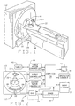
- Figure 1 is a pictorial view of a CT imaging system.
- Figure 2 is a block schematic diagram of the system illustrated in Figure 1.
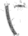
- Figure 3 is a perspective view of a CT system detector array.
- Figure 4 is a perspective view of a detector module.
- Figure 5 is an x-axis schematic view of the CT imaging system shown in Figure 1.
- Figure 6 is a z-axis schematic view of the CT imaging system shown in Figure 5.
- a computed tomography (CT) imaging system 10 is shown as including a gantry 12 representative of a "third generation" CT scanner.
- Gantry 12 has an x-ray source 14 that projects a beam of x-rays 16 toward a detector array 18 on the opposite side of gantry 12.
- Detector array 18 is formed by detector elements 20 which together sense the projected x-rays that pass through a medical patient 22.
- Each detector element 20 produces an electrical signal that represents the intensity of an impinging x-ray beam and hence the attenuation of the beam as it passes through-patient 22.
- gantry 12 and the components mounted thereon rotate about a center of rotation 24.
- Control mechanism 26 includes an x-ray controller 28 that provides power and timing signals to x-ray source 14 and a gantry motor controller 30 that controls the rotational speed and position of gantry 12.
- a data acquisition system (DAS) 32 in control mechanism 26 samples analog data from detector elements 20 and converts the data to digital signals for subsequent processing.
- An image reconstructor 34 receives sampled and digitized x-ray data from DAS 32 and performs high speed image reconstruction. The reconstructed image is applied as an input to a computer 36 which stores the image in a mass storage device 38.
- DAS data acquisition system
- Computer 36 also receives and supplies signals via a user interface, or graphical user interface (GUI). Specifically, computer receives commands and scanning parameters from an operator via console 40 that has a keyboard and a mouse (not shown). An associated cathode ray tube display 42 allows the operator to observe the reconstructed image and other data from computer 36. The operator supplied commands and parameters are used by computer 36 to provide control signals and information to x-ray controller 28, gantry motor controller 30, DAS 32, and table motor controller 44.
- GUI graphical user interface
- detector array 18 includes a plurality of detector modules 58. Each detector module 58 is secured to a detector housing 60. Each module 58 includes a multidimensional scintillator array 62 and a high density semiconductor array (not visible). A post patient collimator (not shown) is positioned over and adjacent scintillator array 62 to collimate x-ray beams before such beams impinge upon scintillator array 62. Scintillator array 62 includes a plurality of scintillation elements arranged in an array, and the semiconductor array includes a plurality of photodiodes (not visible) arranged in an identical array. The photodiodes are deposited, or formed on a substrate 64, and scintillator array 62 is positioned over and secured to substrate 64.
- Detector module 58 also includes a switch apparatus 66 electrically coupled to a decoder 68.
- Switch apparatus 66 is a multidimensional semiconductor switch array of similar size as the photodiode array.
- switch apparatus 66 includes an array of field effect transistors (not shown) with each field effect transistor (FET) having an input, an output, and a control line (not shown).
- Switch apparatus 66 is coupled between the photodiode array and DAS 32. Particularly, each switch apparatus FET input is electrically connected to a photodiode array output and each switch apparatus FET output is electrically connected to DAS 32, for example, using flexible electrical cable 70.
- Decoder 68 controls the operation of switch apparatus 66 to enable, disable, or combine the outputs of the photodiode array in accordance with a desired number of slices and slice resolutions for each slice.
- Decoder 68 in one embodiment, is a decoder chip or a FET controller as known in the art. Decoder 68 includes a plurality of output and control lines coupled to switch apparatus 66 and computer 36. Particularly, the decoder outputs are electrically connected to the switch apparatus control lines to enable switch apparatus 66 to transmit the proper data from the switch apparatus inputs to the switch apparatus outputs. The decoder control lines are electrically connected to the switch apparatus control lines and determine which of the decoder outputs will be enabled.
- decoder 68 Utilizing decoder 68, specific FETs within switch apparatus 66 are enabled, disable, or combined so that specific outputs of the photodiode array are electrically connected to CT system DAS 32.
- decoder 68 enables switch apparatus 66 so that all rows of the photodiode array are electrically connected to DAS 32, resulting in 16 separate, simultaneous slices of data being sent to DAS 32.
- many other slice combinations are possible.
- detector 18 includes fifty-seven detector modules 58.
- the semiconductor array and scintillator array 62 each have an array size of 16 x 16.
- detector 18 has 16 rows and 912 columns (16 x 57 modules), which enables 16 simultaneous slices of data to be collected with each rotation of gantry 12.
- the present invention is not limited to any specific array size, and it is contemplated that the array can be larger or smaller depending upon the specific operator needs.
- detector 18 may be operated in many different slice thickness and number modes, e.g., one, two, and four slice modes.
- the FETs can be configured in the four slice mode, so that data is collected for four slices from one or more rows of the photodiode array.
- various combinations of outputs of the photodiode array can be enabled, disabled, or combined so that the slice thickness may, for example, be 1.25 mm, 2.5 mm, 3.75 mm, or 5 mm. Additional examples include a single slice mode including one slice with slices ranging from 1.25 mm thick to 20 mm thick, and a two slice mode including two slices with slices ranging from 1.25 mm thick to 10 mm thick. Additional modes beyond those described are possible.
- FIGS. 5 and 6 are schematic views of one embodiment of system 10 in accordance with the present invention.
- X-ray beam 16 emanates from a focal spot 90 of x-ray source 14.
- the intensity and quality of x-ray beam 16 is altered by filter assembly 92, and filtered beam 16 is projected toward detector array 18.
- filter assembly 92 includes a fixed filter portion 94, a z-axis movable filter 98 having a first portion 100 and a second portion 102.
- Respective portions 100 and 102 are configured to alter the intensity and quality of x-ray beam 16. More specifically, the shape and material composition of respective portions 100 and 102 are configured so that unique, or different, quality and intensity beams are created by filter assembly 92 based upon the position the movable filter 98.
- first portion 100 includes a first filter material 110, a second filter material 112 and a third filter material 114 positioned between, or interposed, materials 110 and 112.
- first portion 100 is configured as a bowtie filter and respective materials 110, 112, and 114 are graphite, aluminum, and copper.
- material 110 may be 54.0 mm thick
- material 112 may be 6.0mm thick
- material 114 is about 75 micrometers thick so that first portion 100 is configured to generate a harder x-ray beam quality, for example to perform a body scan.
- third layer 114 is positioned between layers 110 and 112 so that third layer 114 is protected from damaged during operation and handling.
- materials 110, 112, and 114 may selected from other attenuating materials of various thicknesses. Further, one skilled in the art could select materials 110, 112, 114 from elements, compounds, or epoxy mixtures with approximately similar mass attenuation coefficients and adjust the thickness to compensate for material density differences. For example, material 110 (54 mm of graphite) could be substituted with 95mm of polyethylene or material 114 (75 micrometers of copper) could be substituted with 325 micrometers of titanium.
- Second portion 102 in one embodiment, includes a first filter material 120 and a second filter material 122.
- the physical configuration and selection for respective materials 120 and 122 are selected so that an x-ray beam radiating from second portion 102 has an intensity and quality unique from an x-ray beam radiating from first portion 100.
- second portion 102 is configured to generate a softer x-ray beam quality and materials 120 and 122 are selected from the same materials as respective materials 110 and 120, however the physical shape of material 120 and 122 are altered.
- second portion 102 is fabricated as a bowtie filter for generating a narrower x-ray beam and second portion filter material 120 is graphite and second filter material 122 is aluminum.
- respective shape and filter materials 120 and 122 may be selected from other shapes and materials other than materials 110 and 112.
- second portion 102 may include any number of materials.
- filter assembly 92 further includes a drive assembly 116 coupled to movable filter 92.
- Drive assembly 116 is configured to alter the z-axis position of movable filter 92 so that the intensity and quality of x-ray beam 16 can be altered.
- Drive assembly in one embodiment, is coupled to computer 36. In alternative embodiments drive assembly may also be coupled to a filter drive controller (not shown).
- movable filter 92 is positioned so that the proper x-ray beam is radiated toward patient 22, or an object.
- a reconstructed image is generated. More specifically, the scan type is initially determined using known selection criteria, or is prescribed by the operator, for example, a body scan.
- movable filter 92 is positioned so that x-ray beam 16 is filtered using the appropriate portion of movable filter 92. More particularly and in one embodiment, an appropriate quality and intensity x-ray beam is generated by positioning movable filter 92 so that x-ray beam 16 is radiated into first portion 100, for example for a body scan.
- the z-axis position of movable filter 92 is adjusted by drive assembly 116.
- the position of movable filter 92 is adjusted, or repositioned. More specifically, using drive assembly 116, the z-axis position of movable filter 92 is adjusted so that x-ray beam 16 is filtered by second portion 102. The filtering by second portion 102 alters the x-ray beam so that, for example for the head scan, x-ray beam 16 radiated toward detector array 18 is narrower and the beam quality is softer.
- x-ray beam 16 is filtered so that the proper x-ray beam intensity and quality is directed toward detector array 18.
- movable filter 92 may include any number of portions so that any number of unique quality and intensity beams may be radiated toward patient 22 and detector array 18.
- the above described filter assembly allows selection of filtration characteristics depending upon the scan to be completed. More specifically, the filter assembly includes a plurality of filters so that the proper filtration is provided for various specific types of scans, i.e., head portion scans or body portion scans.
- the CT system described herein is a "third generation” system in which both the x-ray source and detector rotate with the gantry.
- Many other CT systems including "fourth generation” systems wherein the detector is a full-ring stationary detector and only the x-ray source rotates with the gantry, may be used.
- the systems described herein have been two-slice and four-slice, any multi-slice system may be used.
Landscapes
- Health & Medical Sciences (AREA)
- Life Sciences & Earth Sciences (AREA)
- Engineering & Computer Science (AREA)
- Medical Informatics (AREA)
- Physics & Mathematics (AREA)
- High Energy & Nuclear Physics (AREA)
- Heart & Thoracic Surgery (AREA)
- Animal Behavior & Ethology (AREA)
- Optics & Photonics (AREA)
- Pathology (AREA)
- Radiology & Medical Imaging (AREA)
- Biomedical Technology (AREA)
- Biophysics (AREA)
- Molecular Biology (AREA)
- Surgery (AREA)
- Nuclear Medicine, Radiotherapy & Molecular Imaging (AREA)
- General Health & Medical Sciences (AREA)
- Public Health (AREA)
- Veterinary Medicine (AREA)
- Pulmonology (AREA)
- Theoretical Computer Science (AREA)
- Spectroscopy & Molecular Physics (AREA)
- General Engineering & Computer Science (AREA)
- Apparatus For Radiation Diagnosis (AREA)
Applications Claiming Priority (2)
| Application Number | Priority Date | Filing Date | Title |
|---|---|---|---|
| US09/140,112 US6307918B1 (en) | 1998-08-25 | 1998-08-25 | Position dependent beam quality x-ray filtration |
| US140112 | 1998-08-25 |
Publications (2)
| Publication Number | Publication Date |
|---|---|
| EP0981999A2 true EP0981999A2 (fr) | 2000-03-01 |
| EP0981999A3 EP0981999A3 (fr) | 2003-02-05 |
Family
ID=22489814
Family Applications (1)
| Application Number | Title | Priority Date | Filing Date |
|---|---|---|---|
| EP99306634A Ceased EP0981999A3 (fr) | 1998-08-25 | 1999-08-20 | Filtrage de rayons x pour la réalisation d'une qualité de faisceau dépendante de la position |
Country Status (4)
| Country | Link |
|---|---|
| US (1) | US6307918B1 (fr) |
| EP (1) | EP0981999A3 (fr) |
| JP (1) | JP2000079112A (fr) |
| IL (1) | IL131563A (fr) |
Cited By (10)
| Publication number | Priority date | Publication date | Assignee | Title |
|---|---|---|---|---|
| US6275562B1 (en) | 1998-04-28 | 2001-08-14 | General Electric Company | Apparatus and methods for performing scalable multislice computed tomography scan |
| EP1111624A3 (fr) * | 1999-12-23 | 2001-11-14 | Philips Patentverwaltung GmbH | Appareil d'examen radiologique |
| EP1192901A1 (fr) * | 2000-09-28 | 2002-04-03 | GE Medical Systems Global Technology Company LLC | Filtration de rayons X dépendante de la position |
| WO2003012414A1 (fr) * | 2001-07-30 | 2003-02-13 | American Science And Engineering, Inc. | Inspection par rayons x a l'aide de faisceaux spatialement et spectralement personnalises |
| US7538325B2 (en) | 2000-02-10 | 2009-05-26 | American Science And Engineering, Inc. | Single-pulse-switched multiple energy X-ray source applications |
| CN104127198A (zh) * | 2014-07-10 | 2014-11-05 | 中国科学院苏州生物医学工程技术研究所 | 一种ct前置滤线器 |
| CN104127199A (zh) * | 2014-07-14 | 2014-11-05 | 沈阳东软医疗系统有限公司 | 一种形状过滤器的设定方法和装置 |
| WO2015173750A1 (fr) * | 2014-05-15 | 2015-11-19 | Koninklijke Philips N.V. | Système d'imagerie à points focaux multiples |
| EP2662021A4 (fr) * | 2011-01-07 | 2016-05-04 | Toshiba Kk | Scanner tdm à rayons x |
| CN108814562A (zh) * | 2018-07-12 | 2018-11-16 | 孙际燕 | 一种光波检验诊断装置 |
Families Citing this family (29)
| Publication number | Priority date | Publication date | Assignee | Title |
|---|---|---|---|---|
| US6647095B2 (en) * | 2002-04-02 | 2003-11-11 | Ge Medical Systems Global Technology Co., Llc | Method and apparatus for optimizing dosage to scan subject |
| US6836535B2 (en) | 2002-04-22 | 2004-12-28 | General Electric Company | Method and apparatus of modulating the filtering of radiation during radiographic imaging |
| US20030199757A1 (en) * | 2002-04-22 | 2003-10-23 | Toth Thomas L. | Method and apparatus of modulating radiation filtering during radiographic imaging |
| JP3910501B2 (ja) * | 2002-07-17 | 2007-04-25 | 浜松ホトニクス株式会社 | エアロゾル粒子荷電装置 |
| US7273479B2 (en) * | 2003-01-15 | 2007-09-25 | Cryodynamics, Llc | Methods and systems for cryogenic cooling |
| US6954516B2 (en) * | 2003-03-14 | 2005-10-11 | Ge Medical Systems Global Technology Company, Llc | Imaging systems and methods |
| US7046756B2 (en) * | 2003-05-20 | 2006-05-16 | General Electric Company | Rotatable filter for a pre-subject CT collimator having multiple filtering profiles |
| US6968030B2 (en) * | 2003-05-20 | 2005-11-22 | General Electric Company | Method and apparatus for presenting multiple pre-subject filtering profiles during CT data acquisition |
| US7120222B2 (en) * | 2003-06-05 | 2006-10-10 | General Electric Company | CT imaging system with multiple peak x-ray source |
| JP2005006772A (ja) * | 2003-06-17 | 2005-01-13 | Ge Medical Systems Global Technology Co Llc | X線診断装置及びct画像の生成方法 |
| JP4041025B2 (ja) * | 2003-07-15 | 2008-01-30 | ジーイー・メディカル・システムズ・グローバル・テクノロジー・カンパニー・エルエルシー | X線分布調整フィルタ装置およびそれを用いたx線ct装置 |
| US6990171B2 (en) * | 2003-10-27 | 2006-01-24 | General Electric Company | System and method of determining a user-defined region-of-interest of an imaging subject for x-ray flux management control |
| US7076029B2 (en) * | 2003-10-27 | 2006-07-11 | General Electric Company | Method and apparatus of radiographic imaging with an energy beam tailored for a subject to be scanned |
| US7082183B2 (en) * | 2004-07-21 | 2006-07-25 | General Electric Company | Computed tomography dose indexing phantom selection for dose reporting |
| US7474736B2 (en) * | 2004-10-01 | 2009-01-06 | Varian Medical Systems, Inc. | Devices and methods for providing spatially variable x-ray beam intensity |
| US7054407B1 (en) * | 2005-02-08 | 2006-05-30 | General Electric Company | Methods and apparatus to facilitate reconstruction of images |
| CN100582757C (zh) * | 2005-07-22 | 2010-01-20 | 同方威视技术股份有限公司 | 一种用于集装箱检查系统的准直与校正一体装置 |
| US7254216B2 (en) * | 2005-07-29 | 2007-08-07 | General Electric Company | Methods and apparatus for filtering a radiation beam and CT imaging systems using same |
| US7330535B2 (en) * | 2005-11-10 | 2008-02-12 | General Electric Company | X-ray flux management device |
| US8270569B2 (en) * | 2010-07-11 | 2012-09-18 | Moshe Ein-Gal | Cascaded modulation system |
| US9101272B2 (en) * | 2011-03-24 | 2015-08-11 | Jefferson Radiology, P.C. | Fixed anterior gantry CT shielding |
| JP6266284B2 (ja) * | 2013-09-19 | 2018-01-24 | 東芝メディカルシステムズ株式会社 | X線診断装置 |
| JP2015136390A (ja) * | 2014-01-20 | 2015-07-30 | キヤノン株式会社 | 制御装置、断層撮影装置 |
| CN103932729A (zh) * | 2014-04-08 | 2014-07-23 | 江苏中惠医疗科技股份有限公司 | 一种计算机断层扫描系统的x光射线过滤器 |
| US10667767B2 (en) | 2014-05-02 | 2020-06-02 | General Electric Company | Systems and methods for selecting bowtie filter configuration |
| CN105675042B (zh) * | 2015-12-28 | 2018-08-10 | 同方威视技术股份有限公司 | 射线标定装置及其操作方法、辐射成像系统及其操作方法 |
| CN105433973B (zh) * | 2015-12-30 | 2018-09-18 | 沈阳东软医疗系统有限公司 | Ct扫描设备、ct系统和控制过滤器组件的方法及装置 |
| WO2020097800A1 (fr) * | 2018-11-13 | 2020-05-22 | 西安大医集团有限公司 | Filtre optique, appareil de balayage de rayonnement et procédé de balayage de rayonnement |
| US11160518B2 (en) * | 2019-08-16 | 2021-11-02 | GE Precision Healthcare LLC | Methods and systems for integrated filter system |
Family Cites Families (10)
| Publication number | Priority date | Publication date | Assignee | Title |
|---|---|---|---|---|
| US3860817A (en) * | 1973-08-10 | 1975-01-14 | Gen Electric | Reducing patient X-ray dose during fluoroscopy with an image system |
| NL8304398A (nl) * | 1983-12-22 | 1985-07-16 | Philips Nv | Roentgenonderzoekapparaat met selectief filter. |
| US5054048A (en) * | 1985-11-14 | 1991-10-01 | Hologic, Inc. | X-ray radiography method and system |
| US4868843A (en) * | 1986-09-10 | 1989-09-19 | Varian Associates, Inc. | Multileaf collimator and compensator for radiotherapy machines |
| US4975933A (en) * | 1990-03-26 | 1990-12-04 | General Electric Company | Bow-tie X-ray filter assembly for dual energy tomography |
| US5430783A (en) | 1992-08-07 | 1995-07-04 | General Electric Company | Reconstruction method for helical scanning computed tomography apparatus with multi-row detector array employing overlapping beams |
| FI103176B1 (fi) * | 1993-06-15 | 1999-05-14 | Planmeca Oy | Pehmytkudossuodinlaite kefalostaattiin |
| US5400379A (en) | 1994-02-25 | 1995-03-21 | General Electric Company | Multi-slice x-ray CT using a detector mask |
| US5644614A (en) | 1995-12-21 | 1997-07-01 | General Electric Company | Collimator for reducing patient x-ray dose |
| US5828719A (en) * | 1996-12-23 | 1998-10-27 | General Electric Company | Methods and apparatus for modulating data acquisition system gain |
-
1998
- 1998-08-25 US US09/140,112 patent/US6307918B1/en not_active Expired - Lifetime
-
1999
- 1999-08-18 JP JP11230995A patent/JP2000079112A/ja active Pending
- 1999-08-20 EP EP99306634A patent/EP0981999A3/fr not_active Ceased
- 1999-08-24 IL IL13156399A patent/IL131563A/xx not_active IP Right Cessation
Cited By (15)
| Publication number | Priority date | Publication date | Assignee | Title |
|---|---|---|---|---|
| US6275562B1 (en) | 1998-04-28 | 2001-08-14 | General Electric Company | Apparatus and methods for performing scalable multislice computed tomography scan |
| EP1111624A3 (fr) * | 1999-12-23 | 2001-11-14 | Philips Patentverwaltung GmbH | Appareil d'examen radiologique |
| US6563909B2 (en) | 1999-12-23 | 2003-05-13 | Koninklijke Philips Electronics N.V. | X-ray examination apparatus |
| US7538325B2 (en) | 2000-02-10 | 2009-05-26 | American Science And Engineering, Inc. | Single-pulse-switched multiple energy X-ray source applications |
| US7010094B2 (en) | 2000-02-10 | 2006-03-07 | American Science And Engineering, Inc. | X-ray inspection using spatially and spectrally tailored beams |
| EP1192901A1 (fr) * | 2000-09-28 | 2002-04-03 | GE Medical Systems Global Technology Company LLC | Filtration de rayons X dépendante de la position |
| US6633627B2 (en) | 2000-09-28 | 2003-10-14 | Ge Medical Systems Global Technology Company, Llc | X-ray CT system, gantry apparatus, console terminal, method of controlling them, and storage medium |
| WO2003012414A1 (fr) * | 2001-07-30 | 2003-02-13 | American Science And Engineering, Inc. | Inspection par rayons x a l'aide de faisceaux spatialement et spectralement personnalises |
| EP2662021A4 (fr) * | 2011-01-07 | 2016-05-04 | Toshiba Kk | Scanner tdm à rayons x |
| US9724053B2 (en) | 2011-01-07 | 2017-08-08 | Toshiba Medical Systems Corporation | X-ray CT apparatus |
| WO2015173750A1 (fr) * | 2014-05-15 | 2015-11-19 | Koninklijke Philips N.V. | Système d'imagerie à points focaux multiples |
| US10568593B2 (en) | 2014-05-15 | 2020-02-25 | Koninklijke Philips N.V. | Multi-focal spot imaging system |
| CN104127198A (zh) * | 2014-07-10 | 2014-11-05 | 中国科学院苏州生物医学工程技术研究所 | 一种ct前置滤线器 |
| CN104127199A (zh) * | 2014-07-14 | 2014-11-05 | 沈阳东软医疗系统有限公司 | 一种形状过滤器的设定方法和装置 |
| CN108814562A (zh) * | 2018-07-12 | 2018-11-16 | 孙际燕 | 一种光波检验诊断装置 |
Also Published As
| Publication number | Publication date |
|---|---|
| JP2000079112A (ja) | 2000-03-21 |
| US6307918B1 (en) | 2001-10-23 |
| IL131563A0 (en) | 2001-01-28 |
| EP0981999A3 (fr) | 2003-02-05 |
| IL131563A (en) | 2003-04-10 |
Similar Documents
| Publication | Publication Date | Title |
|---|---|---|
| US6307918B1 (en) | Position dependent beam quality x-ray filtration | |
| US6061419A (en) | Methods and apparatus for noise compensation in an imaging system | |
| US5982846A (en) | Methods and apparatus for dose reduction in a computed tomograph | |
| US6298117B1 (en) | Variable aperture z-axis tracking collimator for a computed tomograph system | |
| US6056437A (en) | Methods and apparatus for imaging system detector alignment | |
| EP0981998B1 (fr) | Procédé et dispositif de réduction du bruit dans une image | |
| US7088799B2 (en) | Method and apparatus for presenting multiple pre-subject filtering profiles during CT data acquisition | |
| EP1071044B1 (fr) | Procédé et dispositif de compensation de bruit dans des systèmes d'imagerie | |
| JP4478335B2 (ja) | X線ビームの動きを補正するための方法および装置 | |
| US6280084B1 (en) | Methods and apparatus for indirect high voltage verification in an imaging system | |
| JPH08168484A (ja) | Ctイメージングシステム | |
| EP1959835B1 (fr) | Systemes et procedes pour balayage et acquisition de donnees dans des applications de tomographie informatisee (ct) | |
| JP2002533150A (ja) | マルチスライス型イメージング・システム用の画像厚選択法 | |
| EP0982000B1 (fr) | Appareil de vérification de dosage dans un système d'imagerie | |
| US6325539B1 (en) | Calibration simplification for a computed tomograph system | |
| US6118840A (en) | Methods and apparatus to desensitize incident angle errors on a multi-slice computed tomograph detector | |
| US6343110B1 (en) | Methods and apparatus for submillimeter CT slices with increased coverage | |
| US7102137B2 (en) | Method and apparatus for improving slice to slice resolution by staggering cells in the Z-axis | |
| US6366637B1 (en) | Methods and apparatus for generating thin-slice imaging data on a multi-slice imaging system | |
| JP2009028110A (ja) | X線ct装置及びそれに使用するフィルタ板 | |
| EP0990419A1 (fr) | Méthodes et appareil pour la génération de scout-scans |
Legal Events
| Date | Code | Title | Description |
|---|---|---|---|
| PUAI | Public reference made under article 153(3) epc to a published international application that has entered the european phase |
Free format text: ORIGINAL CODE: 0009012 |
|
| AK | Designated contracting states |
Kind code of ref document: A2 Designated state(s): AT BE CH CY DE DK ES FI FR GB GR IE IT LI LU MC NL PT SE |
|
| AX | Request for extension of the european patent |
Free format text: AL;LT;LV;MK;RO;SI |
|
| PUAL | Search report despatched |
Free format text: ORIGINAL CODE: 0009013 |
|
| AK | Designated contracting states |
Designated state(s): AT BE CH CY DE DK ES FI FR GB GR IE IT LI LU MC NL PT SE |
|
| AX | Request for extension of the european patent |
Extension state: AL LT LV MK RO SI |
|
| RIC1 | Information provided on ipc code assigned before grant |
Ipc: 7G 21K 1/10 B Ipc: 7A 61B 6/03 A |
|
| 17P | Request for examination filed |
Effective date: 20030805 |
|
| AKX | Designation fees paid |
Designated state(s): DE NL |
|
| 17Q | First examination report despatched |
Effective date: 20040521 |
|
| 17Q | First examination report despatched |
Effective date: 20040521 |
|
| STAA | Information on the status of an ep patent application or granted ep patent |
Free format text: STATUS: THE APPLICATION HAS BEEN REFUSED |
|
| 18R | Application refused |
Effective date: 20071223 |