EP1006362A1 - Méthode et appareil pour la séparation de composants d'un matériau biologique - Google Patents
Méthode et appareil pour la séparation de composants d'un matériau biologique Download PDFInfo
- Publication number
- EP1006362A1 EP1006362A1 EP98122848A EP98122848A EP1006362A1 EP 1006362 A1 EP1006362 A1 EP 1006362A1 EP 98122848 A EP98122848 A EP 98122848A EP 98122848 A EP98122848 A EP 98122848A EP 1006362 A1 EP1006362 A1 EP 1006362A1
- Authority
- EP
- European Patent Office
- Prior art keywords
- components
- proteins
- gel
- mpd
- electrophoresis
- Prior art date
- Legal status (The legal status is an assumption and is not a legal conclusion. Google has not performed a legal analysis and makes no representation as to the accuracy of the status listed.)
- Withdrawn
Links
- 238000000034 method Methods 0.000 title claims abstract description 86
- 238000000926 separation method Methods 0.000 title claims description 30
- 239000012620 biological material Substances 0.000 title claims 2
- 108090000623 proteins and genes Proteins 0.000 claims abstract description 183
- 102000004169 proteins and genes Human genes 0.000 claims abstract description 181
- 150000007523 nucleic acids Chemical class 0.000 claims abstract description 10
- 108020004707 nucleic acids Proteins 0.000 claims abstract description 9
- 102000039446 nucleic acids Human genes 0.000 claims abstract description 9
- 108090000765 processed proteins & peptides Proteins 0.000 claims abstract description 4
- 102000004196 processed proteins & peptides Human genes 0.000 claims abstract description 4
- 150000002632 lipids Chemical class 0.000 claims abstract 2
- 238000005251 capillar electrophoresis Methods 0.000 claims description 27
- 238000001514 detection method Methods 0.000 claims description 21
- 108091023037 Aptamer Proteins 0.000 claims description 16
- 239000000523 sample Substances 0.000 claims description 14
- 239000000463 material Substances 0.000 claims description 13
- 239000003153 chemical reaction reagent Substances 0.000 claims description 11
- 238000005516 engineering process Methods 0.000 claims description 10
- 238000001502 gel electrophoresis Methods 0.000 claims description 9
- 238000001962 electrophoresis Methods 0.000 claims description 8
- 230000007246 mechanism Effects 0.000 claims description 6
- 238000000539 two dimensional gel electrophoresis Methods 0.000 claims description 5
- 238000001704 evaporation Methods 0.000 claims description 4
- 230000008020 evaporation Effects 0.000 claims description 4
- 238000004949 mass spectrometry Methods 0.000 claims description 4
- 238000003491 array Methods 0.000 claims description 2
- 238000005192 partition Methods 0.000 claims 9
- 230000004807 localization Effects 0.000 claims 2
- 238000000638 solvent extraction Methods 0.000 claims 1
- 239000000203 mixture Substances 0.000 abstract description 16
- 238000004458 analytical method Methods 0.000 abstract description 12
- 230000006872 improvement Effects 0.000 abstract description 8
- 239000004215 Carbon black (E152) Substances 0.000 abstract 1
- 229930195733 hydrocarbon Natural products 0.000 abstract 1
- 150000002430 hydrocarbons Chemical class 0.000 abstract 1
- 239000000499 gel Substances 0.000 description 113
- 210000004027 cell Anatomy 0.000 description 26
- XSQUKJJJFZCRTK-UHFFFAOYSA-N Urea Chemical compound NC(N)=O XSQUKJJJFZCRTK-UHFFFAOYSA-N 0.000 description 16
- 230000035945 sensitivity Effects 0.000 description 16
- 230000008569 process Effects 0.000 description 14
- 108010026552 Proteome Proteins 0.000 description 12
- 239000000872 buffer Substances 0.000 description 9
- 239000012530 fluid Substances 0.000 description 8
- 238000003384 imaging method Methods 0.000 description 8
- 238000001155 isoelectric focusing Methods 0.000 description 8
- 240000004808 Saccharomyces cerevisiae Species 0.000 description 7
- 230000004888 barrier function Effects 0.000 description 7
- 239000004202 carbamide Substances 0.000 description 7
- 238000001212 derivatisation Methods 0.000 description 7
- 235000014680 Saccharomyces cerevisiae Nutrition 0.000 description 6
- 239000012491 analyte Substances 0.000 description 6
- 238000005520 cutting process Methods 0.000 description 6
- 230000000694 effects Effects 0.000 description 6
- 238000011068 loading method Methods 0.000 description 6
- 238000002360 preparation method Methods 0.000 description 6
- 102000055501 telomere Human genes 0.000 description 6
- 108091035539 telomere Proteins 0.000 description 6
- 210000003411 telomere Anatomy 0.000 description 6
- 210000001519 tissue Anatomy 0.000 description 6
- 239000013078 crystal Substances 0.000 description 5
- 238000004128 high performance liquid chromatography Methods 0.000 description 5
- 230000001900 immune effect Effects 0.000 description 5
- 238000013021 overheating Methods 0.000 description 5
- 238000004809 thin layer chromatography Methods 0.000 description 5
- UMCMPZBLKLEWAF-BCTGSCMUSA-N 3-[(3-cholamidopropyl)dimethylammonio]propane-1-sulfonate Chemical compound C([C@H]1C[C@H]2O)[C@H](O)CC[C@]1(C)[C@@H]1[C@@H]2[C@@H]2CC[C@H]([C@@H](CCC(=O)NCCC[N+](C)(C)CCCS([O-])(=O)=O)C)[C@@]2(C)[C@@H](O)C1 UMCMPZBLKLEWAF-BCTGSCMUSA-N 0.000 description 4
- 230000008901 benefit Effects 0.000 description 4
- 238000011161 development Methods 0.000 description 4
- 230000018109 developmental process Effects 0.000 description 4
- 230000006870 function Effects 0.000 description 4
- 229920002521 macromolecule Polymers 0.000 description 4
- 238000011160 research Methods 0.000 description 4
- 241000894007 species Species 0.000 description 4
- UMGDCJDMYOKAJW-UHFFFAOYSA-N thiourea Chemical compound NC(N)=S UMGDCJDMYOKAJW-UHFFFAOYSA-N 0.000 description 4
- 230000003321 amplification Effects 0.000 description 3
- 230000001413 cellular effect Effects 0.000 description 3
- 210000000349 chromosome Anatomy 0.000 description 3
- 238000009792 diffusion process Methods 0.000 description 3
- 238000002372 labelling Methods 0.000 description 3
- 238000005259 measurement Methods 0.000 description 3
- 238000003199 nucleic acid amplification method Methods 0.000 description 3
- 238000002823 phage display Methods 0.000 description 3
- 238000012800 visualization Methods 0.000 description 3
- QFVHZQCOUORWEI-UHFFFAOYSA-N 4-[(4-anilino-5-sulfonaphthalen-1-yl)diazenyl]-5-hydroxynaphthalene-2,7-disulfonic acid Chemical compound C=12C(O)=CC(S(O)(=O)=O)=CC2=CC(S(O)(=O)=O)=CC=1N=NC(C1=CC=CC(=C11)S(O)(=O)=O)=CC=C1NC1=CC=CC=C1 QFVHZQCOUORWEI-UHFFFAOYSA-N 0.000 description 2
- 208000002109 Argyria Diseases 0.000 description 2
- PVNIIMVLHYAWGP-UHFFFAOYSA-N Niacin Chemical compound OC(=O)C1=CC=CN=C1 PVNIIMVLHYAWGP-UHFFFAOYSA-N 0.000 description 2
- 108700026244 Open Reading Frames Proteins 0.000 description 2
- BQCADISMDOOEFD-UHFFFAOYSA-N Silver Chemical compound [Ag] BQCADISMDOOEFD-UHFFFAOYSA-N 0.000 description 2
- 108010017842 Telomerase Proteins 0.000 description 2
- 238000001261 affinity purification Methods 0.000 description 2
- 150000001413 amino acids Chemical class 0.000 description 2
- 230000032823 cell division Effects 0.000 description 2
- 230000003196 chaotropic effect Effects 0.000 description 2
- 239000003795 chemical substances by application Substances 0.000 description 2
- 238000004140 cleaning Methods 0.000 description 2
- 238000011109 contamination Methods 0.000 description 2
- 238000006731 degradation reaction Methods 0.000 description 2
- 230000001419 dependent effect Effects 0.000 description 2
- 230000002349 favourable effect Effects 0.000 description 2
- 238000005194 fractionation Methods 0.000 description 2
- 238000004519 manufacturing process Methods 0.000 description 2
- 239000012528 membrane Substances 0.000 description 2
- 229910052751 metal Inorganic materials 0.000 description 2
- 239000002184 metal Substances 0.000 description 2
- 150000003141 primary amines Chemical class 0.000 description 2
- 238000012545 processing Methods 0.000 description 2
- 229910052709 silver Inorganic materials 0.000 description 2
- 239000004332 silver Substances 0.000 description 2
- 239000000126 substance Substances 0.000 description 2
- 239000006228 supernatant Substances 0.000 description 2
- 230000009897 systematic effect Effects 0.000 description 2
- 108010088751 Albumins Proteins 0.000 description 1
- 102000009027 Albumins Human genes 0.000 description 1
- 241001146702 Candidatus Entotheonella factor Species 0.000 description 1
- 241000252212 Danio rerio Species 0.000 description 1
- 241000255581 Drosophila <fruit fly, genus> Species 0.000 description 1
- 241000588724 Escherichia coli Species 0.000 description 1
- 241000282412 Homo Species 0.000 description 1
- 230000005374 Kerr effect Effects 0.000 description 1
- QIVBCDIJIAJPQS-VIFPVBQESA-N L-tryptophane Chemical compound C1=CC=C2C(C[C@H](N)C(O)=O)=CNC2=C1 QIVBCDIJIAJPQS-VIFPVBQESA-N 0.000 description 1
- OUYCCCASQSFEME-QMMMGPOBSA-N L-tyrosine Chemical compound OC(=O)[C@@H](N)CC1=CC=C(O)C=C1 OUYCCCASQSFEME-QMMMGPOBSA-N 0.000 description 1
- 241000124008 Mammalia Species 0.000 description 1
- 108010085220 Multiprotein Complexes Proteins 0.000 description 1
- 102000007474 Multiprotein Complexes Human genes 0.000 description 1
- 241000341511 Nematodes Species 0.000 description 1
- 206010028980 Neoplasm Diseases 0.000 description 1
- 239000000020 Nitrocellulose Substances 0.000 description 1
- 108091005461 Nucleic proteins Proteins 0.000 description 1
- 241000283973 Oryctolagus cuniculus Species 0.000 description 1
- 241000255588 Tephritidae Species 0.000 description 1
- 102000040945 Transcription factor Human genes 0.000 description 1
- 108091023040 Transcription factor Proteins 0.000 description 1
- QIVBCDIJIAJPQS-UHFFFAOYSA-N Tryptophan Natural products C1=CC=C2C(CC(N)C(O)=O)=CNC2=C1 QIVBCDIJIAJPQS-UHFFFAOYSA-N 0.000 description 1
- 230000001594 aberrant effect Effects 0.000 description 1
- 238000001042 affinity chromatography Methods 0.000 description 1
- 238000013459 approach Methods 0.000 description 1
- 238000010420 art technique Methods 0.000 description 1
- 238000003556 assay Methods 0.000 description 1
- 239000012298 atmosphere Substances 0.000 description 1
- QYTOONVFPBUIJG-UHFFFAOYSA-N azane;cyanic acid Chemical compound [NH4+].[O-]C#N QYTOONVFPBUIJG-UHFFFAOYSA-N 0.000 description 1
- 210000003719 b-lymphocyte Anatomy 0.000 description 1
- 230000008827 biological function Effects 0.000 description 1
- 230000033228 biological regulation Effects 0.000 description 1
- 238000001574 biopsy Methods 0.000 description 1
- 230000015572 biosynthetic process Effects 0.000 description 1
- 230000006287 biotinylation Effects 0.000 description 1
- 238000007413 biotinylation Methods 0.000 description 1
- 210000000988 bone and bone Anatomy 0.000 description 1
- 210000001185 bone marrow Anatomy 0.000 description 1
- 238000004364 calculation method Methods 0.000 description 1
- 201000011510 cancer Diseases 0.000 description 1
- 238000005515 capillary zone electrophoresis Methods 0.000 description 1
- 230000021235 carbamoylation Effects 0.000 description 1
- 210000000845 cartilage Anatomy 0.000 description 1
- 230000015556 catabolic process Effects 0.000 description 1
- 230000006037 cell lysis Effects 0.000 description 1
- 230000004663 cell proliferation Effects 0.000 description 1
- 230000008859 change Effects 0.000 description 1
- 239000012568 clinical material Substances 0.000 description 1
- 230000000295 complement effect Effects 0.000 description 1
- 238000001816 cooling Methods 0.000 description 1
- 230000002596 correlated effect Effects 0.000 description 1
- 230000036425 denaturation Effects 0.000 description 1
- 238000004925 denaturation Methods 0.000 description 1
- 230000008021 deposition Effects 0.000 description 1
- 239000003599 detergent Substances 0.000 description 1
- 238000006073 displacement reaction Methods 0.000 description 1
- 230000005684 electric field Effects 0.000 description 1
- 230000008030 elimination Effects 0.000 description 1
- 238000003379 elimination reaction Methods 0.000 description 1
- 238000010828 elution Methods 0.000 description 1
- 230000002255 enzymatic effect Effects 0.000 description 1
- 238000011156 evaluation Methods 0.000 description 1
- 230000007717 exclusion Effects 0.000 description 1
- 238000002474 experimental method Methods 0.000 description 1
- 239000002360 explosive Substances 0.000 description 1
- 239000007850 fluorescent dye Substances 0.000 description 1
- 230000006127 geranylation Effects 0.000 description 1
- 239000011521 glass Substances 0.000 description 1
- 230000013595 glycosylation Effects 0.000 description 1
- 238000006206 glycosylation reaction Methods 0.000 description 1
- 239000008241 heterogeneous mixture Substances 0.000 description 1
- 210000005260 human cell Anatomy 0.000 description 1
- 230000002209 hydrophobic effect Effects 0.000 description 1
- 238000000338 in vitro Methods 0.000 description 1
- 230000006698 induction Effects 0.000 description 1
- 230000010354 integration Effects 0.000 description 1
- 230000003993 interaction Effects 0.000 description 1
- 230000003834 intracellular effect Effects 0.000 description 1
- ZCYVEMRRCGMTRW-YPZZEJLDSA-N iodine-125 Chemical compound [125I] ZCYVEMRRCGMTRW-YPZZEJLDSA-N 0.000 description 1
- 229940044173 iodine-125 Drugs 0.000 description 1
- 210000004185 liver Anatomy 0.000 description 1
- 210000004698 lymphocyte Anatomy 0.000 description 1
- 238000012423 maintenance Methods 0.000 description 1
- 238000013507 mapping Methods 0.000 description 1
- 108020004999 messenger RNA Proteins 0.000 description 1
- 230000002503 metabolic effect Effects 0.000 description 1
- 230000003278 mimic effect Effects 0.000 description 1
- 230000004048 modification Effects 0.000 description 1
- 238000012986 modification Methods 0.000 description 1
- 238000002534 molecular mass spectrometry Methods 0.000 description 1
- 230000035772 mutation Effects 0.000 description 1
- 229960003512 nicotinic acid Drugs 0.000 description 1
- 235000001968 nicotinic acid Nutrition 0.000 description 1
- 239000011664 nicotinic acid Substances 0.000 description 1
- 229920001220 nitrocellulos Polymers 0.000 description 1
- 230000003287 optical effect Effects 0.000 description 1
- 238000005457 optimization Methods 0.000 description 1
- 210000000056 organ Anatomy 0.000 description 1
- 210000003463 organelle Anatomy 0.000 description 1
- 239000008188 pellet Substances 0.000 description 1
- 230000026731 phosphorylation Effects 0.000 description 1
- 238000006366 phosphorylation reaction Methods 0.000 description 1
- 229920001184 polypeptide Polymers 0.000 description 1
- 230000004481 post-translational protein modification Effects 0.000 description 1
- 239000000047 product Substances 0.000 description 1
- 230000004952 protein activity Effects 0.000 description 1
- 235000019624 protein content Nutrition 0.000 description 1
- 238000002331 protein detection Methods 0.000 description 1
- 230000006916 protein interaction Effects 0.000 description 1
- 238000001799 protein solubilization Methods 0.000 description 1
- 230000007925 protein solubilization Effects 0.000 description 1
- 230000006337 proteolytic cleavage Effects 0.000 description 1
- 238000004445 quantitative analysis Methods 0.000 description 1
- 230000005855 radiation Effects 0.000 description 1
- 230000005258 radioactive decay Effects 0.000 description 1
- 238000000163 radioactive labelling Methods 0.000 description 1
- 238000011896 sensitive detection Methods 0.000 description 1
- 210000003491 skin Anatomy 0.000 description 1
- 238000002415 sodium dodecyl sulfate polyacrylamide gel electrophoresis Methods 0.000 description 1
- 230000007928 solubilization Effects 0.000 description 1
- 238000005063 solubilization Methods 0.000 description 1
- 230000003381 solubilizing effect Effects 0.000 description 1
- 238000010186 staining Methods 0.000 description 1
- 238000003860 storage Methods 0.000 description 1
- 239000000758 substrate Substances 0.000 description 1
- 239000004094 surface-active agent Substances 0.000 description 1
- 230000008685 targeting Effects 0.000 description 1
- OUYCCCASQSFEME-UHFFFAOYSA-N tyrosine Natural products OC(=O)C(N)CC1=CC=C(O)C=C1 OUYCCCASQSFEME-UHFFFAOYSA-N 0.000 description 1
- 238000000825 ultraviolet detection Methods 0.000 description 1
- 238000001262 western blot Methods 0.000 description 1
Images

Classifications
-
- G—PHYSICS
- G01—MEASURING; TESTING
- G01N—INVESTIGATING OR ANALYSING MATERIALS BY DETERMINING THEIR CHEMICAL OR PHYSICAL PROPERTIES
- G01N27/00—Investigating or analysing materials by the use of electric, electrochemical, or magnetic means
- G01N27/26—Investigating or analysing materials by the use of electric, electrochemical, or magnetic means by investigating electrochemical variables; by using electrolysis or electrophoresis
- G01N27/416—Systems
- G01N27/447—Systems using electrophoresis
- G01N27/44756—Apparatus specially adapted therefor
- G01N27/44773—Multi-stage electrophoresis, e.g. two-dimensional electrophoresis
-
- G—PHYSICS
- G01—MEASURING; TESTING
- G01N—INVESTIGATING OR ANALYSING MATERIALS BY DETERMINING THEIR CHEMICAL OR PHYSICAL PROPERTIES
- G01N33/00—Investigating or analysing materials by specific methods not covered by groups G01N1/00 - G01N31/00
- G01N33/48—Biological material, e.g. blood, urine; Haemocytometers
- G01N33/50—Chemical analysis of biological material, e.g. blood, urine; Testing involving biospecific ligand binding methods; Immunological testing
- G01N33/68—Chemical analysis of biological material, e.g. blood, urine; Testing involving biospecific ligand binding methods; Immunological testing involving proteins, peptides or amino acids
- G01N33/6803—General methods of protein analysis not limited to specific proteins or families of proteins
Definitions
- the invention relates to a method for separation of components from a material to be analysed, in particular of complex mixtures of biological molecules.
- the mixtures of a very large number of analyte molecules are present.
- a good example can be provided by a collection of all of the proteins from a given cellular line, a given tissue, or a given organism. It is important to understand that in such collections one observes not only a very large number of clearly definable species, but also large differences in abundance between these said species. Alas, contrary to intuition, in many biomedical situations the economical importance of biological function does not necessarily correlate with abundance. Actually, it is currently believed that a very large proportion of switch molecules which regulate the networks of protein activity within cells have a very low abundance.
- telomeres are cap-like structures to be found at the extreme termini of chromosomes.
- the number of telomeres possessed by a chromosome within a cell determines the number of times the cell may divide, as one telomere is lost per cell division.
- cancerous cells must escape from the telomere copy number control. This is possible if the cell contains an enzymatic activity, telomerase, which can add new telomeres to a chromosome.
- the aberrant presence of a single or a few telomerase enzyme complexes per cell could carry a pre-cancer cell through a crisis period, enabling continued cell division until a compensating mutation alleviated telomere dependance, creating a vigorous and clinically important cancerous cell.
- proteomics is the study of all proteins expressed by a genome of the cells under study.
- the acronym "PROTEOME” PROTEins within a genOME
- Proteomics may eventually reveal the identity and regulation of most proteins expressed within a particular cell-type [Wilkins, M. R., et al. eds (1997).
- Proteome Research New Frontiers in Functional Genomics (Berlin: Springer) 243 pp].
- the sensitivity and resolution currently attainable require improvement before this can be accomplished.
- proteomics requires detection and quantitation of a very large number of different species, it is necessary to use the available separation methods.
- the preferred tools of proteomics are currently 2D electrophoretic gels for detection and quantitation, followed by a plurality of methods for protein identification, including amino acid analysis, Edman degradation, isoelectric point, apparent molecular mass, and mass spectrometry.
- An intrinsic strength of 2D gels is that they can reveal molecules which undergo post translational modifications, such as phosphorylation, geranylation, glycosylation, proteolytic cleavage, translational induction, alternative splicing, or any modification that affects molecular weight or isoelectric point.
- Current proteome technology essentially corresponds to a differential display on the protein level and complements the nucleic acid-based method. An overview of the recent state of the field has been recently published [Wilkins, M. R., et al. eds (1997). Proteome Research: New Frontiers in Functional Genomics (Berlin: Springer) 243 pp].
- yeast As the human proteome can only be partially estimated from genome sequence, it may be appropriate to remember that the functional protein complement is largely evolutionarily conserved. Thus, it is possible to argue from model organisms, such as yeast or fruit fly, about the dynamic range which might be expected in human situation.
- the entire genome sequence of the yeast Saccharomyces cerevisiae has been determined. About one half of predicted proteins have been identified, and their quantity measured.
- the yeast data suggest that the first 100 most frequent proteins account for 50% of total protein, and a dynamic range of 100,000 is observed between the most frequent (high abundant or in the following abundant) and the least frequent (low abundant) protein detected. Detection of low abundance proteins is currently limited by instrumental background, so there is no estimate of the actual true dynamic range possible.
- This invention facilitates the task to provide solutions for an improvement of analysis of complex mixtures of molecules.
- the problem is solved by a method and apparatus comprising the features of claim 1 or claim 25 to 28, respectively.
- Preferred embodiments of the inventive method are shown in claims 2 to 24. The wording of all claims is hereby made to the content of the specification by reference.
- the different embodiments of the invention mainly concern macromolecules such as proteins, but the invention is not limited to that kind of biological molecules.
- the key enabling capability of this invention is a removal of abundant proteins, resultant alleviation of local gel overheating and associated diffusional loss of resolution, and/or by using the most sensitive imaging and quantitation methods possible, e.g. use of the MPD detectors.
- the relative frequency (amount) of a component in a mixture is meant, wherein in the following “abundant molecules” is meant as molecules, which are comparatively or very frequent and “low abundant molecules” is meant as molecules, which are comparatively or very rare in the mixture.
- the boundary between “abundance” i.e. "high abundance” and “low abundance” has to be defined according to the circumstances, e.g. the most frequent molecules of a mixture, which account for e.g. 50% of total molecule mass, could be defined as high abundant, i.e. abundant.
- the boundary between abundance and low abundance could be defined as an absolute amount of a component, e.g. 500 fmole.
- MPD Multiple Photon Detector technology
- the high degree of sensitivity of MPD is obtained by considering only signals with coincident X- and gamma-rays, and where the energy profile of the measured photons correspond to those expected for the isotope in question.
- the isotopes compatible with MPD come from two broad families of radioisotopes which together include over 100 members appropriate for use as tags. In particular, they include the isotope iodine-125 ( 125 I), one of the most commonly used isotopes in biomedical diagnostics. By carefully comparing each detected event with the highly specific decay signature of the used radioisotope, MPD is able to reduce the number of background events to less than one per week.
- MPD permits multi-labeling (up to 16 co-resident labels), good spatial resolution (about 0.3 mm) and very high dynamic range (linearity over nine orders of magnitude in label down to 10 -21 mole).
- Two significant advantages which are derived from this capability are that substances labeled with appropriate isotopes can be reliably detected at four orders of magnitude lower concentrations, and the amount of isotope which must be used is reduced 1,000-fold to a level much below the naturally occurring background radiation.
- the fractionation device will be able to divert peaks to one or many storage receptacles, when peak intensity is detected over a predetermined threshold value.
- the load volumes for CE are in the order of 0.1-0.4 microlitres ( ⁇ l), such that a detector to discriminate between peaks has to be extremely sensitive.
- the present possibilities, without modifying the proteins, is UV detection at 260/280 nm for tryptophan and tyrosine, or 215 nm for the peptide bond, but for very low protein concentrations neither of the UV methods is sensitive enough. Also, 35 S labelling of cells is not sensitive enough for such low protein concentrations due to the short passage time through the detector.
- present detection of proteins requires the loading of protein amounts large enough to extend peak breadth, reducing the number of resolution windows available.
- the lower loading amounts permitted by the preferred MPD detection will increase the number of windows of resolution analogous to peak narrowing in either HPLC or CE when less material is loaded.
- This patent discloses a method which will permit at least partial solutions to the complexity issue and thus permits simplifications and improvements in subsequent separation and protein detection, such as that currently performed using 2D gel.
- this method is independent of 2D gels and could be applicable to other technological developments. Note that a subset of the concepts disclosed here are not meant to replace two dimensional gel technology at this stage, however such a development is possible.
- the disclosed method will be used for the reproducible detection of analytes, and their reliable dispensation to an appropriate fraction collection space.
- the ultimate case of the second class are studies of protein contents of single cells or organelles.
- the notion of a global overloading of the system of separation e.g. a 2D gel is important.
- current detectors can reliably detect and quantitate a spot at about a femtomole level.
- dynamic ranges of protein abundance may be as large as million, this would lead to a loading requirement of up to a few hundred nmol of proteins to a gel to allow detection of low abundance proteins.
- This would lead to a total overload of a broad pH range IPG (immobilized pH gradient) isoelectric focussing gel, and often to very serious streaking artifacts in both dimensions. Loading the sample to narrow range IPGs would alleviate these problems for large domains of the gel, however the high abundance components would still occupy their preferred pH values, introducing the above mentioned local artifacts to these gel regions.
- MPD technology permits imaging at sub-attomole per spot level, which may lead to a few hundred picomol (or less) per gel total load requirements for unfractionated cell proteins, which considerably removes global overloading.
- This principle applies not only to electrophoretic, but also to chromatographic and other separation methods.
- the local temperature of a gel may limit both diffusion and mobility of a given protein in a gel.
- any local overloading which may happen at nanomol per mm 2 level is a latent risk for reliable high spatial resolution 2D gels.
- a partial solution to this problem is obtainable by optimization of the gel running condition.
- the optimal condition, the condition at which spot size is minimized may be different for high abundance and low abundance spots.
- Even a small improvement in resolution, e.g. factor two leads to a quite large improvement in number of spaces resolved, i.e.
- the new strategy proposed comprises analyzing separately subsets of proteins, each selected according to their relative abundance. Each of these subsets will be run separately, with optimized gel running conditions. This allows maintenance within each subset of a number of analytes which are within the resolution capacity of the chosen separation system. For example for 2D gels, within each subset the dynamic range would be kept to 2 or 3 logs so as to diminish artifacts due to overload and overheating.
- the precedures proposed here provide initial progress towards identifying very low abundance cellular proteins.
- the invention is the approach to the solution of the problems of numerical complexity and linear dynamics by providing a step of prefractionation by abundance, preferably with subsequent separate analysis of abundance classes with suitable levels of sensitivity.
- the removal of abundant biological molecules is performed using immunological means, i.e. affinity reagents.
- immunological means i.e. affinity reagents.
- Another preferred possibility is a fractionation in appropriate separation media, but one advantage of the immunological method is the potential for very high specificity.
- the immunological method comprises the following steps:
- any other high resolution protein separation method can be used instead of 2D gel electrophoresis.
- the molecules with affinity to abundant proteins are the prior art aptamer short nucleic acid molecules. These can be amplified and selected with high efficiency at low cost.
- a low sensitivity 2D gel is used to identify the proteins in the sample. The abundant spots are then removed and are used in the subsequent steps of antibody/aptamer selection. Each of the proteins could be identified by Mass Spectrometry or other methods. This information could be used for the selection of the appropriate antibody/aptamer from a catalogue or the proteins themselves could be used to generate antibodies, e.g. in the mouse or in the rabbit.
- a very large library of antibodies or aptamers exists, e.g. a few thousand antibodies or molecules with affinity to the proteins of interest, and this library is challenged with the protein mixture of interest.
- the library is organized randomly or preferred into an array of spatially distinguishable pixels, e.g. on a chip. Fluid containing the protein analytes is incubated with or passed over said chip. This is in result a type of micro affinity chromatography, where according to the invention low abundant molecules of interest remain in the fluid. Abundant targeted molecules could be recovered from the affinity array.
- such an affinity chip is challenged with the collection of abundant proteins cut from the gel and marked isotopically. The protein interactions with specific antibodies create a spatial pattern which is read-out with an appropriate detector.
- the use of the MPD technique is preferred, which permits sensitive detection with antibodies which do interact strongly with the protein mix of interest. Additionally, the number of proteins which were not trapped (e.g. in the supernatant) can be estimated from the mix (e.g. from the supernatant).
- the said antibodies or the said collection of antibodies consist of monoclonal antibodies.
- the probability that two proteins will bind to the same antibody is very small.
- the probability that a given protein will bind to two antibodies is also small, and is a function of the number of antibodies employed.
- this binding process is quantitative even at an attomole level. It can be used for selecting the antibodies to proteins present in the gel at sub-femtomole level. Herein, e.g. it is assumed that only about 10% of total amount of protein is loaded in the "trial 2-D gel".
- a very efficient strategy to create a large collection of affinity reagents according to this invention is using a library of high abundance proteins to evolve a collection of highly specific affinity reagents.
- Two implementations of this concept are disclosed which involve evolution of a specific set of either aptamers, or polypetides through the phage display.
- Aptamers are the nucleic acid constructs evolved through the SELEX process as described in a plurality of prior art patents and papers (e.g. US Patent No. 05567588; US Patent No. 05637459; Tuerk, C., et al. (1992) PNAS 89: 6988-6992).
- nucleic target is amplified a billion fold. By repeating this process a few times, very specific aptamers can be evolved in a short time. If challenged by a dominant protein in the spot this selection process would lead to only a few candidate nucleic acids, and the probability of binding to a low abundance protein in the same peak would be very small.
- aptamers specific to a given molecular target Due to the possibility of efficient amplification of nucleic acid structures the production of aptamers specific to a given molecular target is relatively low cost as compared with classical immunological methods.
- a series of libraries of aptamers specific to the dominant proteins for a given model organism, organ, type of tissue, or cellular line can be efficiently and economically created. These arrays could be used as a general tool for affinity purification and removal for almost all the most abundant proteins in a given application.
- the device proposed should separate abundant analytes, such as proteins, from less abundant ones in a complex mixture, such as proteins extracted from primary human clinical biopsies. By differentially examining the abundant and non abundant components with appropriate sensitivity, additional biological information should be harvested which is unobtainable using current technology.
- a preferred embodiment of the subject invention is an innovative procedure for separating proteins by abundance, wherein two subsequent 2D-gels are obtained.
- the first, preparative, gel is used to obtain the realistic estimation of the number of abundant proteins, i.e. proteins above a certain abundance threshold. In the following it is assumed that this threshold is above 1 picogram of protein per spot (1 pg/spot). However, even lower thresholds, say down to 100 fg/spot (100 femtogram of protein per spot), can be useful in certain applications.
- the said 2D gel image is then analyzed either manually or with means of computer generated representation.
- the first preparative 2D gel can be of lower spatial resolution and hence can be run much more quickly than an analytical 2D gel.
- a main innovative step is to localize and physically remove a large fraction, typically 90% of each abundant protein, from the preparative gel. This can be achieved by either mechanically cutting a part of the gel or by using local electroelution to remove the selected proteins. Also, evaporation by laser beam is disclosed. After this abundant protein removal step, all "below the threshold" proteins are removed from the gel, either by dissolving the gel or by electroelution. The collection of such "below the threshold” proteins is then analyzed by means of another 2D gel.
- a preferred embodiment of the invention consists of the following steps:
- Typical pre-gel derivatization processes are either radiolabeling or biotinylation of proteins.
- the post-gel derivatizations are currently more popular and include staining with Coomassie Blue, silver staining or use of fluorescent dyes. It is noted that the above said stains lead to a large change of mobility of the derivatized proteins. Thus, it could be difficult to use them in the proposed method of abundant proteins elimination. Both Coommassie Blue and silver stain binds to proteins reversibly and could be removed, but this could lead to irreproducible losses of proteins and hence be difficult to practice.
- Another disclosed implementation uses an automatic X-Y-Z (coordinates) computer based mover.
- the automatic arm is moved into position under control of a computer and is placed above the spot identified as a high protein abundance spot.
- the spot diameter is evaluated, and a cylindrical cutting tool of appropriate diameter is selected.
- the cutter diameter is selected after the computer analyzes the density within a spot and calculates the diameter which will remove the preset fraction of the total amount of the proteins in the spot.
- vacuum can be applied for a short period of time while the cutting tool is in contact with the 2D gel.
- the thickness of the gel is sufficiently uniform so that a single Z displacement mechanism can be used.
- the use of special sensors to establish when the cutting tool touches the gel is disclosed.
- the said sensor can be either optical or electrical, i.e. taking account of the electrical conductivity of the gel or in the case of capacitive sensors, the dielectric constant of gel medium.
- the problem of contamination can be a challenge in the case of repetitively used mechanical cutter tools.
- the tool can be carefully washed and dried.
- a cutter tool with different diameter could be selected.
- the computer analyzes the 2D gel and classifies all high protein abundance spots according to their diameter. Then a cutter tool with, say, the largest diameter is selected and mounted on X-Y-Z arm. This cutter tool is sequentially displaced over the surface of the 2D gel until all high abundance protein spots are excised.
- the other cutting tool with a smaller diameter is selected.
- the process continues until the desired set of cutting tools is used to excise all high abundance proteins spots.
- the order of use of cutter tools of different diameter is immaterial. One could e.g. as well start with the cutter tools of smallest diameter.
- variable diameter cutter tool connected to the disclosed X-Y-Z mover steered by the computer analyzing the preparative 2D gel.
- the mechanical excision using a set of the mechanical cutters can be replaced by the electroelution of the proteins from high abundance spots.
- an appropriate semi-permeable electrolution probe typically a wick, is used. When it touches the gel surface and is placed under electric voltage, it will move the proteins from the gel through the electroelution probe towards an appropriate receptacle.
- the amount of mobilized protein is roughly proportional to elution time, and can be well controlled electronically using feedback from a computer.
- Yet another process uses the X-Y(-Z) mover and a high intensity pulsed laser beam.
- the gel is placed in the vacuum chamber.
- a laser beam is directed towards the high abundance protein spot.
- a very short but intense laser beam is used to explosively evaporate the gel.
- the evaporated gel material takes with itself the proteins.
- the remaining low abundance proteins are liberated from the gel and the high spatial resolution 2D gel is run and analyzed.
- an appropriate light deflector is used instead of an X-Y(-Z) mover.
- the laser beam is moved by means of a mechanical moving mirror or by means of an electro-optical deflector, e.g. using Kerr effect, towards any spot on the surface of the 2D gel.
- the proteins preferably are transferred onto a membrane.
- a nitrocellulose membrane with an admixture of nicotinic acid is preferred and an infrared laser is used.
- Another preferred embodiment of the subject invention uses electroelution and a special preferably computer generated impermeable mask.
- a thin plastic or rubber with appropriate hole patterns can be used. This pattern is generated by laser beam or simple mechanical cutter to mimic both the positions and diameters of the high abundance proteins spots. In the following it is called multi-aperture barrier.
- the 2D gel is placed horizontally on an appropriate smooth substrate, e.g. metal plate or metal coated glass or plastic. It is covered by the multi-aperture barrier so that barrier holes are alligned with the high abundance protein spots.
- an electrophoretic gel is placed which in the following is called covering gel.
- a transverse electrophoresis is initiated leading to removal of the proteins from "preparative 2D gel" to the covering gel in a direction perpendicular to both dimensions of previous electrophoresis in the 20 gel.
- the multi-aperture barrier and covering gel are removed.
- the preparative 20 gel is processed to liberate all remaining low abundance proteins, and the covering gel is used to liberate all high abundance proteins.
- the mobility of proteins under transverse electrophoresis depends on their size.
- the high abundance proteins of low mass will tend to be removed more efficiently than high abundance proteins of high mass.
- a method is disclosed in which in the step of multi-aperture barrier production, the aperture diameter is calculated taking in account both the spot diameter and expected protein mobility. For spots of the same diameter, i.e. the same abundance, the diameter of aperture in multi-aperture barrier will be smaller for low mass proteins than for high mass proteins.
- CE capillary electrophoresis
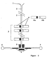
- Figures 1 and 2 show two embodiments of the inventive CE separating device.
- the device according to Figure 1 comprises a fluid inlet 1, a capillary 2, a capillary region 3, where (e.g. a number of 16) detectors 4 each consisting of parts 4a and 4b are arranged, and a microchip 5 with fluidic channels where the fluid flow is gated into one of two channels (not designated by reference numbers).
- 4a and 4b could be a MPD detector which uses the same type, or different crystal types to detect photons.
- Electro-osmotic pumps or microvalves 8 are responsible for gating of fluid flow to either 6 or 7.
- An appropriate device consists of a capillary, which could be kept at a certain temperature in a controlled manner, in particular the capillary could be cooled. Cooling is e.g. necessary to prevent carbamylation of primary amines in the urea buffer. Alternatively, if primary amines have been suitably derivatized, it is conceivable that increased heat may permit better analyte solubility. Ammonium cyanate produced under these conditions may be subsequently electrophoretically removed. Urea/CHAPS is suggested as the buffer since it is used in the isoelectric focusing step which follows. However other buffers could be employed. Proteins would be loaded in a buffer of, e.g.
- the detector 4 is a MPD Imager. After a suitable separation, e.g. by capillary zone electrophoresis, the analytes pass the detector 4 or its parts 4a and 4b, respectively.
- the flow rate and detection window should be optimized to allow maximum possible time in the detection window.
- a gating mechanism (comprising 5 and 8) subsequent to the detector region 3 gates fractions toward alternative receptacles 6 and 7 based on the abundance of proteins.
- Said gating mechanism may operate by means of one or more microvalves, or by means of pumps causing fluidic flow into alternative channels, and thereby providing the gating function, which permits controlled deposition of e.g. nanoliter amounts of fluids into an array of receptacles.
- the gating mechanism is isolated from the atmosphere to prevent evaporation of the small volumes involved.
- the flow velocity of a typical peak is about 30 seconds per mm. Assuming a peak size of few mm, and similar detector size, it is possible to establish reliably that the peak content is larger than about 20 attomol. At the level of about 0.1 femtomol a reasonable quantitation, say 10%, will be possible.
- a linear array of MPD detectors is considered, each 5 mm in size, say a linear array of 16 detectors, as indicated in Fig. 1.
- peak formation during CE is achieved at a relatively long distance from the capillary exit. For example for a typical 1 m capillary the peaks are well formed at about 20 cm from the exit.
- the velocity of peak propagation is known, and in any case can be calibrated to within a few percent.
- characteristic integration time about 30 seconds, the same peak will be seen by subsequent detectors.
- Threshold detection can then be achieved for about one attomol per peak. This will also allow the reliable quantitation of peak height for peaks with about 10 attomol.
- the exit section of the capillary is wound in an appropriate spiral pattern, as embodied in Fig. 2.
- the detectors on a surface of the SR-PMT are placed with the same geometrical pattern as the above-said geometrical spiral.
- a full exit section of the capillary, say 20 cm, is concurrently measured by the MPD detectors.
- the capillary 2 (connected to a fluid inlet 1) in form of a spiral has in a capillary (detection) region 10 an array of crystals 12 for MPD coupled to a single SR-PMT 11 and on the other (opposite) side one single large crystal 13 coupled to a non spatially resolved photomultiplier 14. All other parts of the device are designated as shown in Figure 1.
- FIG. 2A shows a side view of the device
- Fig. 2B shows the arrangement of crystals 12 relative to capillary 2 in a top view.
- the arrangement of parts 11, 12, 13 and 14 forms a spatially resolving MPD (SR-MPD).
- SR-MPD spatially resolving MPD
- a plurality of capillaries is coupled to a spatially resolved MPD with the detector pattern featuring n x m detectors.
- n capillaries are measured concurrently, each with m detectors.
- 13 capillaries For example with a 13 x 13 array of detectors one can use 13 capillaries and detect about 10 attomole peaks.
- Such a configuration solves the throughput issue.
- a preferred embodiment of the invention is an array of electrophoresis capillaries, which is coupled to a SR-MPD detector, consisting of a plurality of scintillators placed and read-out by a single SR-PMT, wherein the said scintillators are placed in an essentially quadratic grid pattern, i.e. each capillary is seen by the same row of detectors and the information from each row of detectors is used to activate a miniaturized fluidic valve.
- the procedures for removal of high abundance proteins preferably comprise the use of highest possible sensitivity read-out methods, e.g. the use of sub-attomole sensitivity of MPD instrumentation.
- the proteinic material from each spot is available and proteins are essentially not destroyed.
- the proteinic material from each of the high abundance protein spots is subsequently analyzed by any of a plurality of separation techniques such as CE, HPLC (high performance liquid chromatography), TLC (thin layer chromatography) or size exclusion filters.
- CE high performance liquid chromatography
- TLC thin layer chromatography
- size exclusion filters size exclusion filters
- the high abundance fractions are eliminated and low abundance proteins returned to a pool of proteins to be re-analyzed by repeat of 2D electrophoresis.
- the low abundance fractions are believed to be sufficiently well analyzed by the preparative 2D gel and CE step.
- the data are placed in an appropriate computer database, and fractions are forwarded for further steps of identification, e.g. by mass spectrometry or Edman degradation process.
Landscapes
- Life Sciences & Earth Sciences (AREA)
- Health & Medical Sciences (AREA)
- Molecular Biology (AREA)
- Engineering & Computer Science (AREA)
- Chemical & Material Sciences (AREA)
- Physics & Mathematics (AREA)
- Immunology (AREA)
- Urology & Nephrology (AREA)
- General Physics & Mathematics (AREA)
- General Health & Medical Sciences (AREA)
- Biomedical Technology (AREA)
- Biochemistry (AREA)
- Analytical Chemistry (AREA)
- Hematology (AREA)
- Pathology (AREA)
- Biophysics (AREA)
- Microbiology (AREA)
- Food Science & Technology (AREA)
- Medicinal Chemistry (AREA)
- Cell Biology (AREA)
- Biotechnology (AREA)
- Proteomics, Peptides & Aminoacids (AREA)
- Bioinformatics & Computational Biology (AREA)
- Bioinformatics & Cheminformatics (AREA)
- Chemical Kinetics & Catalysis (AREA)
- Electrochemistry (AREA)
- Investigating Or Analysing Biological Materials (AREA)
Priority Applications (3)
| Application Number | Priority Date | Filing Date | Title |
|---|---|---|---|
| EP98122848A EP1006362A1 (fr) | 1998-12-02 | 1998-12-02 | Méthode et appareil pour la séparation de composants d'un matériau biologique |
| PCT/EP1999/009325 WO2000033075A1 (fr) | 1998-12-02 | 1999-12-01 | Procede et appareil de separation de composants d'un materiau biologique |
| AU18611/00A AU1861100A (en) | 1998-12-02 | 1999-12-01 | Method and apparatus for the separation of components from biological material |
Applications Claiming Priority (1)
| Application Number | Priority Date | Filing Date | Title |
|---|---|---|---|
| EP98122848A EP1006362A1 (fr) | 1998-12-02 | 1998-12-02 | Méthode et appareil pour la séparation de composants d'un matériau biologique |
Publications (1)
| Publication Number | Publication Date |
|---|---|
| EP1006362A1 true EP1006362A1 (fr) | 2000-06-07 |
Family
ID=8233074
Family Applications (1)
| Application Number | Title | Priority Date | Filing Date |
|---|---|---|---|
| EP98122848A Withdrawn EP1006362A1 (fr) | 1998-12-02 | 1998-12-02 | Méthode et appareil pour la séparation de composants d'un matériau biologique |
Country Status (3)
| Country | Link |
|---|---|
| EP (1) | EP1006362A1 (fr) |
| AU (1) | AU1861100A (fr) |
| WO (1) | WO2000033075A1 (fr) |
Cited By (7)
| Publication number | Priority date | Publication date | Assignee | Title |
|---|---|---|---|---|
| WO2001033230A3 (fr) * | 1999-11-02 | 2002-01-10 | Chiron Corp | Purification et affichage differentiel de composants d'echantillons biologiques |
| WO2001081924A3 (fr) * | 2001-04-23 | 2003-08-28 | Biotraces Inc | Micro-arrangements reutilisables pour quantification de proteines peu abondantes |
| WO2002029094A3 (fr) * | 2000-10-03 | 2003-09-04 | Andreas Kage | Procede pour selectionner des acides nucleiques se liant avec une grande affinite a une cible, par separation en deux dimensions |
| WO2002077016A3 (fr) * | 2001-03-22 | 2003-10-09 | Vlaams Interuniv Inst Biotech | Procedes et appareils destines a une analyse qualitative et quantitative sans gel du proteome et utilisations associees |
| US6783929B1 (en) | 1999-11-02 | 2004-08-31 | Chiron Corporation | Biological sample component purification and differential display |
| EP1580559A1 (fr) * | 2004-03-23 | 2005-09-28 | Ciphergen Biosystems, Inc. | Procedes visant a reduire la variance de concentration d'analytes des melanges d'echantillons complexes |
| US7754861B2 (en) | 2005-03-23 | 2010-07-13 | Bio-Rad Laboratories, Inc. | Method for purifying proteins |
Families Citing this family (2)
| Publication number | Priority date | Publication date | Assignee | Title |
|---|---|---|---|---|
| US7691645B2 (en) | 2001-01-09 | 2010-04-06 | Agilent Technologies, Inc. | Immunosubtraction method |
| US6800449B1 (en) | 2001-07-13 | 2004-10-05 | Syngenta Participations Ag | High throughput functional proteomics |
Citations (3)
| Publication number | Priority date | Publication date | Assignee | Title |
|---|---|---|---|---|
| US5582981A (en) * | 1991-08-14 | 1996-12-10 | Gilead Sciences, Inc. | Method for identifying an oligonucleotide aptamer specific for a target |
| WO1997009068A2 (fr) * | 1995-09-01 | 1997-03-13 | University Of Washington | Conjugues moleculaires interactifs |
| WO1998023950A1 (fr) * | 1996-11-29 | 1998-06-04 | Oxford Glycosciences (Uk) Ltd. | Gels, procedes et appareil d'identification et de caracterisation de biomolecules |
-
1998
- 1998-12-02 EP EP98122848A patent/EP1006362A1/fr not_active Withdrawn
-
1999
- 1999-12-01 WO PCT/EP1999/009325 patent/WO2000033075A1/fr not_active Ceased
- 1999-12-01 AU AU18611/00A patent/AU1861100A/en not_active Abandoned
Patent Citations (3)
| Publication number | Priority date | Publication date | Assignee | Title |
|---|---|---|---|---|
| US5582981A (en) * | 1991-08-14 | 1996-12-10 | Gilead Sciences, Inc. | Method for identifying an oligonucleotide aptamer specific for a target |
| WO1997009068A2 (fr) * | 1995-09-01 | 1997-03-13 | University Of Washington | Conjugues moleculaires interactifs |
| WO1998023950A1 (fr) * | 1996-11-29 | 1998-06-04 | Oxford Glycosciences (Uk) Ltd. | Gels, procedes et appareil d'identification et de caracterisation de biomolecules |
Cited By (10)
| Publication number | Priority date | Publication date | Assignee | Title |
|---|---|---|---|---|
| WO2001033230A3 (fr) * | 1999-11-02 | 2002-01-10 | Chiron Corp | Purification et affichage differentiel de composants d'echantillons biologiques |
| US6783929B1 (en) | 1999-11-02 | 2004-08-31 | Chiron Corporation | Biological sample component purification and differential display |
| WO2002029094A3 (fr) * | 2000-10-03 | 2003-09-04 | Andreas Kage | Procede pour selectionner des acides nucleiques se liant avec une grande affinite a une cible, par separation en deux dimensions |
| WO2002077016A3 (fr) * | 2001-03-22 | 2003-10-09 | Vlaams Interuniv Inst Biotech | Procedes et appareils destines a une analyse qualitative et quantitative sans gel du proteome et utilisations associees |
| WO2001081924A3 (fr) * | 2001-04-23 | 2003-08-28 | Biotraces Inc | Micro-arrangements reutilisables pour quantification de proteines peu abondantes |
| EP1580559A1 (fr) * | 2004-03-23 | 2005-09-28 | Ciphergen Biosystems, Inc. | Procedes visant a reduire la variance de concentration d'analytes des melanges d'echantillons complexes |
| AU2005227916B2 (en) * | 2004-03-23 | 2010-09-16 | American National Red Cross | Methods for reducing the range in concentrations of analyte species in a sample |
| EP1672984A4 (fr) * | 2004-03-23 | 2011-11-30 | Ciphergen Biosystems Inc | Procedes de reduction de l'ecart de concentrations entre des especes d'analytes dans un echantillon |
| US8906608B2 (en) | 2004-03-23 | 2014-12-09 | Bio-Rad Laboratories, Inc. | Methods for reducing the range in concentrations of analyte species in a sample |
| US7754861B2 (en) | 2005-03-23 | 2010-07-13 | Bio-Rad Laboratories, Inc. | Method for purifying proteins |
Also Published As
| Publication number | Publication date |
|---|---|
| WO2000033075A1 (fr) | 2000-06-08 |
| AU1861100A (en) | 2000-06-19 |
Similar Documents
| Publication | Publication Date | Title |
|---|---|---|
| Vistain et al. | Single-cell proteomics | |
| Petrosius et al. | Recent advances in the field of single-cell proteomics | |
| US6613210B1 (en) | Molecular imaging | |
| Kellner | Proteomics. Concepts and perspectives | |
| Chambers et al. | Proteomics: a new approach to the study of disease | |
| Fey et al. | 2D or not 2D | |
| Beranova-Giorgianni | Proteome analysis by two-dimensional gel electrophoresis and mass spectrometry: strengths and limitations | |
| Sadowski et al. | Quantitative proteomic approach to study subcellular localization of membrane proteins | |
| Govorun et al. | Proteomic technologies in modern biomedical science | |
| US6677114B1 (en) | Polypeptide fingerprinting methods and bioinformatics database system | |
| Minakshi et al. | Single-cell proteomics: technology and applications | |
| WO2001035089A1 (fr) | Appareil d'electrophorese multidimensionnelle | |
| Ong et al. | Mass spectrometry-based characterization of endogenous peptides and metabolites in small volume samples | |
| Klepárník et al. | Electrophoresis today and tomorrow: Helping biologists' dreams come true | |
| EP1006362A1 (fr) | Méthode et appareil pour la séparation de composants d'un matériau biologique | |
| Liu et al. | Micro-separation toward systems biology | |
| Lin et al. | Review and Practical Guide for Getting Started With Single‐Cell Proteomics | |
| Panisko et al. | The postgenomic age: characterization of proteomes | |
| Witzmann et al. | II. Proteomics: core technologies and applications in physiology | |
| Resing | Analysis of signaling pathways using functional proteomics | |
| Unwin et al. | How will haematologists use proteomics? | |
| Mitchell | In the pursuit of industrial proteomics | |
| Reddy et al. | Proteomics in transfusion medicine. | |
| Page et al. | Proteomics: applications relevant to transfusion medicine | |
| Scobioala et al. | Proteomics: state of the art and its application in cardiovascular research |
Legal Events
| Date | Code | Title | Description |
|---|---|---|---|
| PUAI | Public reference made under article 153(3) epc to a published international application that has entered the european phase |
Free format text: ORIGINAL CODE: 0009012 |
|
| AK | Designated contracting states |
Kind code of ref document: A1 Designated state(s): AT BE CH CY DE DK ES FI FR GB GR IE IT LI LU MC NL PT SE |
|
| AX | Request for extension of the european patent |
Free format text: AL;LT;LV;MK;RO;SI |
|
| 17P | Request for examination filed |
Effective date: 20000831 |
|
| AKX | Designation fees paid |
Free format text: AT BE CH CY DE DK ES FI FR GB IT LI NL PT SE |
|
| RBV | Designated contracting states (corrected) |
Designated state(s): AT BE CH CY DE DK ES FI FR GB GR IE IT LI LU MC NL PT SE |
|
| RAP1 | Party data changed (applicant data changed or rights of an application transferred) |
Owner name: PROTEOSYS AG |
|
| RIN1 | Information on inventor provided before grant (corrected) |
Inventor name: NORDHEIM, ALFRED, PROF. DR. Inventor name: DRUKIER, ANDRZEY, DR. Inventor name: CAHILL, MICHAEL, DR. |
|
| RAP1 | Party data changed (applicant data changed or rights of an application transferred) |
Owner name: PROTEOSYS AG |
|
| STAA | Information on the status of an ep patent application or granted ep patent |
Free format text: STATUS: THE APPLICATION IS DEEMED TO BE WITHDRAWN |
|
| 18D | Application deemed to be withdrawn |
Effective date: 20020702 |