EP1006876B1 - Procede de mesure de l'histologie cutanee - Google Patents
Procede de mesure de l'histologie cutanee Download PDFInfo
- Publication number
- EP1006876B1 EP1006876B1 EP97912388A EP97912388A EP1006876B1 EP 1006876 B1 EP1006876 B1 EP 1006876B1 EP 97912388 A EP97912388 A EP 97912388A EP 97912388 A EP97912388 A EP 97912388A EP 1006876 B1 EP1006876 B1 EP 1006876B1
- Authority
- EP
- European Patent Office
- Prior art keywords
- skin
- colour
- ordinates
- area
- melanin
- Prior art date
- Legal status (The legal status is an assumption and is not a legal conclusion. Google has not performed a legal analysis and makes no representation as to the accuracy of the status listed.)
- Expired - Lifetime
Links
- 238000000034 method Methods 0.000 title claims abstract description 33
- 238000005259 measurement Methods 0.000 title claims description 17
- XUMBMVFBXHLACL-UHFFFAOYSA-N Melanin Chemical compound O=C1C(=O)C(C2=CNC3=C(C(C(=O)C4=C32)=O)C)=C2C4=CNC2=C1C XUMBMVFBXHLACL-UHFFFAOYSA-N 0.000 claims abstract description 160
- 210000003491 skin Anatomy 0.000 claims abstract description 98
- 210000004207 dermis Anatomy 0.000 claims abstract description 65
- 230000002500 effect on skin Effects 0.000 claims abstract description 33
- 230000002159 abnormal effect Effects 0.000 claims abstract description 19
- 230000005855 radiation Effects 0.000 claims abstract description 16
- 230000036074 healthy skin Effects 0.000 claims abstract description 15
- 238000011835 investigation Methods 0.000 claims abstract description 9
- 239000008280 blood Substances 0.000 claims description 17
- 210000004369 blood Anatomy 0.000 claims description 17
- 230000037311 normal skin Effects 0.000 claims description 15
- 210000002615 epidermis Anatomy 0.000 claims description 12
- 238000004364 calculation method Methods 0.000 claims description 6
- 230000000694 effects Effects 0.000 claims description 5
- 238000012937 correction Methods 0.000 claims description 3
- 238000001228 spectrum Methods 0.000 claims description 3
- 230000005856 abnormality Effects 0.000 claims description 2
- 238000010183 spectrum analysis Methods 0.000 claims description 2
- 230000035515 penetration Effects 0.000 abstract description 4
- 230000009545 invasion Effects 0.000 description 15
- 102000008186 Collagen Human genes 0.000 description 8
- 108010035532 Collagen Proteins 0.000 description 8
- 229920001436 collagen Polymers 0.000 description 8
- 238000010521 absorption reaction Methods 0.000 description 5
- 238000010586 diagram Methods 0.000 description 4
- 201000001441 melanoma Diseases 0.000 description 4
- 208000000453 Skin Neoplasms Diseases 0.000 description 3
- 201000000849 skin cancer Diseases 0.000 description 3
- 230000009466 transformation Effects 0.000 description 3
- 206010064390 Tumour invasion Diseases 0.000 description 2
- 230000001419 dependent effect Effects 0.000 description 2
- 238000003745 diagnosis Methods 0.000 description 2
- 239000000463 material Substances 0.000 description 2
- 230000036564 melanin content Effects 0.000 description 2
- 238000004393 prognosis Methods 0.000 description 2
- 231100000444 skin lesion Toxicity 0.000 description 2
- 206010040882 skin lesion Diseases 0.000 description 2
- 210000004003 subcutaneous fat Anatomy 0.000 description 2
- 208000006981 Skin Abnormalities Diseases 0.000 description 1
- 238000004458 analytical method Methods 0.000 description 1
- 230000005540 biological transmission Effects 0.000 description 1
- 239000003086 colorant Substances 0.000 description 1
- 229940079593 drug Drugs 0.000 description 1
- 239000003814 drug Substances 0.000 description 1
- 230000001747 exhibiting effect Effects 0.000 description 1
- 238000013178 mathematical model Methods 0.000 description 1
- 239000000203 mixture Substances 0.000 description 1
- 238000011160 research Methods 0.000 description 1
- 238000012216 screening Methods 0.000 description 1
- 210000002265 sensory receptor cell Anatomy 0.000 description 1
- 102000027509 sensory receptors Human genes 0.000 description 1
- 108091008691 sensory receptors Proteins 0.000 description 1
- 230000003595 spectral effect Effects 0.000 description 1
- 238000004611 spectroscopical analysis Methods 0.000 description 1
- 210000001519 tissue Anatomy 0.000 description 1
- 230000025366 tissue development Effects 0.000 description 1
- 238000002604 ultrasonography Methods 0.000 description 1
- 230000002792 vascular Effects 0.000 description 1
Images


Classifications
-
- A—HUMAN NECESSITIES
- A61—MEDICAL OR VETERINARY SCIENCE; HYGIENE
- A61B—DIAGNOSIS; SURGERY; IDENTIFICATION
- A61B5/00—Measuring for diagnostic purposes; Identification of persons
- A61B5/44—Detecting, measuring or recording for evaluating the integumentary system, e.g. skin, hair or nails
- A61B5/441—Skin evaluation, e.g. for skin disorder diagnosis
- A61B5/444—Evaluating skin marks, e.g. mole, nevi, tumour, scar
-
- A—HUMAN NECESSITIES
- A61—MEDICAL OR VETERINARY SCIENCE; HYGIENE
- A61B—DIAGNOSIS; SURGERY; IDENTIFICATION
- A61B5/00—Measuring for diagnostic purposes; Identification of persons
- A61B5/0059—Measuring for diagnostic purposes; Identification of persons using light, e.g. diagnosis by transillumination, diascopy, fluorescence
Definitions
- This invention relates to a method and apparatus for the non-invasive measurement of skin histology and is particularly, but not exclusively, concerned with a method for identifying and measuring the presence and depth of dermal invasion of melanin.
- the presence and extent of dermal invasion within a skin cancer is considered to be the most important factor governing a patient's prognosis.
- the present invention is considered to be potentially useful for the preliminary screening of patients to identify those who should be referred to an appropriate clinician for diagnosis and further to assist the clinician in diagnosis.
- the present invention is based on the findings reported by Symon D'O Cotton in "Do all human skin colours lie on a defined surface within LMS space?", University of Birmingham Technical Report, 30 December 1995.
- this Technical Report the relation between healthy skin and the colour of the skin represented in LMS, a particular colour space, is reported, and it discloses that, for healthy skin, the coloration, regardless of race or amount of tanning, lies on a defined curved surface within a three-dimensional colour space.
- This if used with a correct colour measurement system, can measure and quantify the amount of melanin and blood at any particular point at which this measurement is made. If the skin is sampled as an image, then corresponding images showing the variation of blood and melanin across the skin can be obtained.
- a method of non-invasively analysing skin structure comprising the steps of: (i) measuring visible radiation having a wavelength of at least 600 nm and/or infrared radiation from at least one location in an area of skin under investigation so as to give an indication of papillary dermis thickness over said area; (it) measuring skin colour co-ordinates at said at least one location in said area of skin; (iii) using data obtained in measuring steps (i) and (ii) to calculate corrected skin colour co-ordinates over said area which correspond to a predetermined papillary dermis thickness, and; (iv) comparing the corrected skin colour co-ordinates obtained in step (iii) with a reference colour co-ordinate range for healthy skin of said predetermined papillary dermis thickness.
- the method can be used for locating and measuring the properties of a skin abnormality, in which case the method further comprises the steps of; (v) identifying an abnormal location (i.e. a region where melanin exists within the dermis) within said area of skin where the corrected skin colour co-ordinates lie outside the reference colour co-ordinate range; (vi) calibrating the corrected skin colour co-ordinates of said abnormal location with the corrected skin colour co-ordinates of at least one skin location having colour co-ordinates lying within said reference colour co-ordinate range for normal skin, and; (vii) using the skin colour co-ordinates to assess the degree of abnormality of said abnormal skin location.
- an abnormal location i.e. a region where melanin exists within the dermis
- the corrected skin colour co-ordinates lie outside the reference colour co-ordinate range
- calibrating the corrected skin colour co-ordinates of said abnormal location with the corrected skin colour co-ordinates of at least one skin location having colour co-ordinates lying within said reference colour co-ordinate range for normal skin, and; (vii
- the invention includes apparatus for non-invasively analysing skin structure, comprising: a projector for projecting visible radiation having a wavelength of at least 600 nm and/or infrared radiation onto an area of skin under investigation; measuring means for measuring remitted radiation from at least one location over an area of skin under investigation so as to give an indication of papillary dermis thickness over said area; skin colour co-ordinate measuring means for measuring skin colour co-ordinates at said at least one location over said area of skin; calculating means for using data obtained in measuring steps (i) and (ii) to calculate corrected skin colour co-ordinates over said area which correspond to a predetermined papillary dermis thickness, and; colour comparison means for comparing corrected skin colour co-ordinates obtained in step (iii) with a reference colour co-ordinate range for healthy skin of said predetermined papillary dermis thickness.
- papillary dermal skin thickness can change markedly with some skin lesions which are not otherwise of concern. This throws the coloration of the skin off the surface of predicted coloration and so can give rise to false measurements of the histology of such skin lesions. It is for this reason that papillary dermis thickness is measured first, and subsequent calculations are based on the skin colour co-ordinates corrected to a predetermined papillary dermis thickness. Any arbitrary value for this thickness may be chosen, such as 2.0 ⁇ 10 -4 m which is the average value for healthy human skin.
- the thickness of the papillary dermis may be obtained by utilising the property of human skin to vary its absorption of infrared radiation with varying papillary dermis thickness.
- absorption and thickness there is an inverse relationship between absorption and thickness.
- infrared radiation is also absorbed by other materials within the skin, particularly melanin and blood, is a complicating factor.
- the effect on absorption of varying blood and melanin content is far smaller than the effect of papillary dermis thickness, and so the layer may still be measured. This can be done by obtaining two infrared images, each at a different wavelength. The chosen wavelengths are not important, but one should be further into the infrared (i.e. at longer wavelength) than the other.
- Suitable wavelength bands are 800-1000nm and 600-800nm, in that readily available infrared films and filters may be used.
- the brightness of points within the image obtained at the longer wavelength is affected to a greater extent by variations in the papillary dermis thickness. Conversely, the image obtained at shorter wavelength will be affected to a greater extent by other materials such as melanin and blood.
- a reference graph (Fig 1) can be obtained which consists of lines of constant papillary dermis thickness, wherein Primary 1 is the measurement made at the longer (800-1000nm) wavelength and Primary 2 is the measurement made at the shorter (600-800nm) wavelength.
- Primary 1 is the measurement made at the longer (800-1000nm) wavelength
- Primary 2 is the measurement made at the shorter (600-800nm) wavelength.
- the absorption of blood within these wavelengths is very small (a hundredth of its peak value for visible wavelengths at 600-800nm and even less for 800-1000nm) and to a first approximation may be ignored.
- the reference colour coordinate range for normal skin at the predetermined papillary dermis thickness is obtained as disclosed in the above-mentioned Technical Report as a curved surface lying within a three-dimensional colour space, with one of the bounding axes relating to the amount of melanin within the epidermis and the other relating to the amount of blood within the dermis.
- the epidermal melanin value within this area is estimated by either reference to the reference colour coordinate range for normal skin within regions identified as normal, or by reference to the epidermal melanin levels calculated within normal regions adjacent to said area containing dermal melanin.
- the corrected skin colour coordinates for the area of skin under investigation may be calibrated to values equivalent to zero epidermal melanin.
- LMS colour space it is possible to use any other colour space, for example, the RGB colour space or a UV G IR colour space.
- the dermis contrasts strongly in structure to that of the epidermis, being highly vascular, containing many sensory receptors and being made largely from collagen fibres to provide the essential structure of the skin. Between the epidermis and the dermis, the junction presents an extremely uneven boundary with finger-like dermal protrusions called dermal papillae projecting towards the skin surface.
- the dermis can be split into two further histologically distinct layers, the papillary dermis and the reticular dermis within which the structure of the collagen fibres differs significantly.
- the papillary dermis is situated directly below the epidermis and within which the collagen exists as a fine network of fibres.
- Fig 2 there are now four distinct layers within the dermis which can combine to construct a simple model, 1) a layer within the upper papillary dermis containing no melanin, 2) a layer within the upper papillary dermis containing melanin, 3) a layer within the lower papillary dermis containing melanin, 4) a layer within the lower papillary dermis containing no melanin.
- melanin existing up to the dermo-epidermal junction is facilitated by allowing the thickness of layer 1 to be zero and likewise melanin can exist up to the papillary-reticular dermis boundary by setting the thickness of layer 4 to be zero.
- ⁇ ( ⁇ ) scattering coefficient
- ⁇ fraction of radiation absorbed per unit path length
- ⁇ the density of dermal melanin within that layer.
- ⁇ ( ⁇ , ⁇ , ⁇ ) can be shown to be the sum of ⁇ iv ( ⁇ ), ⁇ b ( ⁇ ) and ⁇ m ( ⁇ ), where ⁇ m ( ⁇ ) is the absorption coefficient of melanin. From the above it is possible to calculate R and T (diffuse radiation and transmission respectively).
- This system of equations can therefore compute the total remitted and transmitted light from an n layered system of arbitrary complexity provided that the thickness and composition of the layers is specified.
- This equation can then be used to generate the expected coloration of human skin exhibiting dermal descent of melanin.
- the depth of melanin penetration within the dermis is large enough for such saturation to have occurred, an estimate of the deviation from the reference colour co-ordinate range for normal skin can be made.
- This estimate allows a calculation to be made of the skin coloration assuming no dermal melanin, and therefore by reference to the colour co-ordinate range for normal skin, of the level of epidermal melanin.
- the epidermal melanin levels are measured directly, for example using polarised light, and such measurements are incorporated in the measuring step (ii) above.
- the values of those three parameters can be obtained for every point in the image. Since the papillary dermis thickness and epidermal melanin content are known, it is possible to compute a detailed three-dimensional reconstruction of the top layers of human skin. This is of great potential interest to the medical profession and enables routine examination of the internal structure of living skin just as X-rays, NMR and ultrasound are used for examining other parts of the body. It is also within the scope of the invention to acquire the infra-red and/ or visible images using lasers of different wavelengths or by using spectral analysis.
- an image of the area of skin under investigation must be represented in the same colour space as for the normal skin reference colour coordinate range.
- the skin colour coordinates are acquired from an image using the same lighting conditions and a CCD camera calibrated in the same way as that used to produce the healthy skin reference colour coordinate range.
- a white standard or other appropriate correction factor can be used to allow calibration of the image within the software.
- a colour image can be acquired using a colour photographic film which is then digitised.
- a schematic skin section is shown wherein melanin (indicated by the black circles in Fig 3) in normal healthy skin are present in the lower part of epidermis 10 adjacent but above the dermo-epidermal junction 12 between the epidermis and the papillary dermis 14.
- the Breslow thickness referred to above is the depth of melanin invasion in millimetres measured from granular layer 16 which is a layer in the epidermis 10 where the skin goes scaly and forms the tough outer cornified layer 18.
- the melanin is shown as having descended not only into the papillary dermis 14, but also into the underlying reticular dermis 20 lying above the subcutaneous fat layer 22. It is to be appreciated that, in other cases, melanin decent can be into any layer of the skin and may even be into the subcutaneous fat layer 22.
- M ethod step (i) above is exemplified by the determination of papillary dermis thickness by shining infrared light at two wavelengths on an area of skin being subjected to measurement and measuring the amount of light reflected from a plurality of points within that area.
- Block 40 exemplifies method step (ii) above the acquisition of an image at visible wavelengths of the same skin area. This can be by CCD camera, digitised film or any other convenient means.
- Block 42 exemplifies method step (iii) above the transformation of the image into corrected colour space of the skin model at a predetermined papillary dermis thickness.
- Block 44 exemplifies method steps (iv and v) above- the identification of regions containing dermal melanin, by comparing the corrected skin colour coordinates with the reference colour coordinate range.
- Block 46 exemplifies method step (vi) above- use of the corrected colour space to calculate the amounts of epidermal melanin within normal regions adjacent to the regions containing dermal melanin and use thereof to give an indication of the amounts thereof which exist in the regions containing dermal melanin.
- Block 48 exemplifies a first part of method step (vii) above- calculation of dermal invasion using the measured coloration of the abnormal regions and the calculated amount of epidermal melanin from 46.
- Block 50 exemplifies a second part of method step (vii) above- transformation of the calculated dermal invasion of melanin into either the Breslow thickness or the Clark's level of invasion. This can be reported as either representing the maximum invasion or as an image showing invasion over the skin.
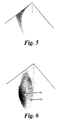
- the shaded surface indicates the range of colorations which can exist in normal healthy skin corrected to the predetermined papillary dermis thickness. Skin colorations which depart from this surface are indicative of dermal melanin.
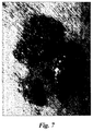
- a region of the skin which is shown in Fig 7 and which is indicated by arrow H in Fig 6 lies at a position corresponding to part of the shaded surface illustrated in Fig 5 and is indicative of normal healthy skin, whereas an adjacent region indicated by arrow U in Fig 6 lies outside such surface and is indicative of skin containing dermal melanin. Comparison of the coloration of these two adjacent regions H and U enables the depth of melanin invasion in the abnormal region of the skin in Fig 7 to be computed.
Landscapes
- Health & Medical Sciences (AREA)
- Life Sciences & Earth Sciences (AREA)
- Molecular Biology (AREA)
- Surgery (AREA)
- Biophysics (AREA)
- Pathology (AREA)
- Engineering & Computer Science (AREA)
- Biomedical Technology (AREA)
- Heart & Thoracic Surgery (AREA)
- Medical Informatics (AREA)
- Veterinary Medicine (AREA)
- Physics & Mathematics (AREA)
- Animal Behavior & Ethology (AREA)
- General Health & Medical Sciences (AREA)
- Public Health (AREA)
- Dermatology (AREA)
- Measuring And Recording Apparatus For Diagnosis (AREA)
- Measurement Of The Respiration, Hearing Ability, Form, And Blood Characteristics Of Living Organisms (AREA)
- Investigating Or Analysing Biological Materials (AREA)
- Steroid Compounds (AREA)
- Medicines Containing Plant Substances (AREA)
Claims (15)
- Procédé d'analyse de façon non invasive de la structure de la peau, comprenant les étapes consistant à :(i) mesurer un rayonnement visible présentant une longueur d'onde d'au moins 600 nm et/ou un rayonnement infrarouge provenant d'au moins un emplacement dans une zone de la peau en cours d'examen de façon à procurer une indication de l'épaisseur du derme papillaire au-dessus de ladite zone,(ii) mesurer les coordonnées de couleur de la peau au niveau dudit au moins un emplacement dans ladite zone de peau,(iii) utiliser les données obtenues au cours des étapes de mesure (i) et (ii) afin de calculer des coordonnées corrigées de couleur de la peau au-dessus de ladite zone qui correspondent à une épaisseur prédéterminée du derme papillaire, et(iv) comparer les coordonnées corrigées de couleur de la peau obtenues à l'étape (iii) à une plage de coordonnées de couleur de référence pour une peau en bonne santé de ladite épaisseur prédéterminée du derme papillaire.
- Procédé selon la revendication 1, comprenant les étapes supplémentaires consistant à : (v) identifier un emplacement anormal à l'intérieur de ladite zone de la peau où les coordonnées corrigées de couleur de peau se situent à l'extérieur de la plage de coordonnées de couleur de référence, (vi) étalonner les coordonnées corrigées de couleur de la peau dudit emplacement anormal avec les coordonnées corrigées de couleur de la peau d'au moins un emplacement de peau ayant les coordonnées de couleur se situant à l'intérieur de ladite plage de coordonnées de couleur de référence pour une peau normale, et
(vii) utiliser les coordonnées de couleur de la peau pour évaluer un degré d'anomalie dudit emplacement de peau anormale. - Procédé selon la revendication 1 ou 2, dans lequel une mesure indépendante du niveau de mélanine épidermique est réalisée.
- Procédé selon la revendication 3, dans lequel ladite mesure indépendante du niveau de mélanine épidermique se fait par lumière polarisée.
- Procédé selon la revendication 2, dans lequel ledit étalonnage au cours de l'étape (vi) comprend l'estimation du niveau de mélanine épidermique dans ledit emplacement anormal par référence aux niveaux de mélanine épidermique calculés à l'intérieur d'au moins un emplacement de la peau normale adjacent à ladite région anormale.
- Procédé selon la revendication 2, dans lequel ledit étalonnage au cours de l'étape (vi) comprend une mesure des niveaux de mélanine épidermique audit emplacement anormal en évaluant l'écart au niveau de l'extrémité bleue du spectre audit emplacement anormal par rapport à la place de coordonnées de couleur de référence pour une peau normale.
- Procédé selon l'une quelconque des revendications précédentes, dans lequel au cours de l'étape (i) une seule image infrarouge à une longueur d'onde supérieure à environ 1100 nm est obtenue pour chacun desdits emplacements.
- Procédé selon l'une quelconque des revendications 1 à 6, dans lequel au cours de l'étape (i) deux images infrarouges, chacune à une longueur d'onde différente, sont obtenues pour chacun desdits emplacements, afin de permettre ainsi que l'effet de la présence de mélanine épidermique et de sang dermique soit pris en compte dans le calcul de l'étape (iii).
- Procédé selon la revendication 8, dans lequel lesdites longueurs d'ondes s'inscrivent à l'intérieur des bandes de 800 à 1000 nm et de 600 à 800 nm respectivement.
- Procédé selon l'une quelconque des revendications 7 à 9, dans lequel la ou les dite(s) image(s) infrarouge(s) est/sont obtenue(s) en utilisant un film photographique infrarouge, ou un ou des lasers, ou par analyse spectrale.
- Procédé selon l'une quelconque des revendications précédentes, dans lequel la plage de coordonnées de couleur de référence pour une peau normale à l'épaisseur prédéterminée du derme papillaire citée dans l'étape (iv), est obtenue sous forme d'une surface incurvée se situant à l'intérieur d'un espace de couleur tridimensionnel, un premier axe limite se rapportant à la quantité de mélanine à l'intérieur de l'épiderme et un second axe limite se rapportant à la quantité de sang à l'intérieur du derme.
- Procédé selon la revendication 11, dans lequel ledit espace de couleur tridimensionnel est choisi parmi les espaces de couleur LMS, RGB et UV G IR.
- Procédé selon l'une quelconque des revendications précédentes, dans lequel les coordonnées de couleur de la peau de l'étape (ii) sont acquises à partir d'une image en utilisant les conditions d'éclairage et le même montage d'étalonnage que ceux utilisés pour produire la plage de coordonnées de couleur de référence d'une peau en bonne santé.
- Procédé selon l'une quelconque des revendications 1 à 12, dans lequel les coordonnées de couleur de la peau de l'étape (ii) sont acquises à partir d'une image en utilisant des conditions d'éclairage différentes de celles utilisées en vue d'obtenir la plage de coordonnées de couleur de référence d'une peau en bonne santé, et un étalon blanc ou un autre facteur de correction sont utilisés afin de permettre l'étalonnage de l'image avec la plage de coordonnées de couleur de référence.
- Dispositif destiné à analyser de façon non invasive la structure de la peau, comprenant :un projecteur destiné à projeter un rayonnement visible présentant une longueur d'onde d'au moins 600 nm et/ou un rayonnement infrarouge sur une zone de la peau en cours d'examen,un moyen de mesure destiné à mesurer le rayonnement réémis depuis au moins un emplacement au-dessus d'une zone de la peau en cours d'examen de façon à procurer une indication de l'épaisseur du derme papillaire au-dessus de ladite zone,un moyen de mesure des coordonnées de couleur de la peau destiné à mesurer les coordonnées de couleur de la peau au niveau dudit au moins un emplacement au-dessus de ladite zone de peau,un moyen de calcul destiné à utiliser les données obtenues au cours des étapes de mesure (i) et (ii) en vue de calculer des coordonnées corrigées de couleur de la peau au-dessus de ladite zone qui correspondent à une épaisseur prédéterminée du derme papillaire, etun moyen de comparaison de couleur destiné à comparer les coordonnées corrigées de couleur de la peau obtenues au cours de l'étape (iii) à une plage de coordonnées de couleur de référence pour une peau en bonne santé de ladite épaisseur prédéterminée du derme papillaire.
Applications Claiming Priority (3)
| Application Number | Priority Date | Filing Date | Title |
|---|---|---|---|
| GB9624003 | 1996-11-19 | ||
| GBGB9624003.1A GB9624003D0 (en) | 1996-11-19 | 1996-11-19 | Method and apparatus for measurement of skin histology |
| PCT/GB1997/003177 WO1998022023A1 (fr) | 1996-11-19 | 1997-11-19 | Procede de mesure de l'histologie cutanee |
Publications (2)
| Publication Number | Publication Date |
|---|---|
| EP1006876A1 EP1006876A1 (fr) | 2000-06-14 |
| EP1006876B1 true EP1006876B1 (fr) | 2002-05-08 |
Family
ID=10803145
Family Applications (1)
| Application Number | Title | Priority Date | Filing Date |
|---|---|---|---|
| EP97912388A Expired - Lifetime EP1006876B1 (fr) | 1996-11-19 | 1997-11-19 | Procede de mesure de l'histologie cutanee |
Country Status (12)
| Country | Link |
|---|---|
| US (1) | US6324417B1 (fr) |
| EP (1) | EP1006876B1 (fr) |
| JP (1) | JP2001504020A (fr) |
| AT (1) | ATE217172T1 (fr) |
| AU (1) | AU725766B2 (fr) |
| BR (1) | BR9713097A (fr) |
| CA (1) | CA2272290C (fr) |
| DE (1) | DE69712533T2 (fr) |
| ES (1) | ES2175373T3 (fr) |
| GB (2) | GB9624003D0 (fr) |
| IL (1) | IL130008A (fr) |
| WO (1) | WO1998022023A1 (fr) |
Families Citing this family (56)
| Publication number | Priority date | Publication date | Assignee | Title |
|---|---|---|---|---|
| GB2367125B (en) * | 1999-06-04 | 2004-07-14 | Astron Clinica Ltd | Method of and apparatus for investigating tissue histology |
| GB2396007A (en) * | 1999-06-04 | 2004-06-09 | Astron Clinica Ltd | Method of and apparatus for investigating tissue histology |
| US7280866B1 (en) | 1999-10-06 | 2007-10-09 | National Research Council Of Canada | Non-invasive screening of skin diseases by visible/near-infrared spectroscopy |
| US20010041829A1 (en) * | 2000-01-12 | 2001-11-15 | Suresh Thennadil | Non-invasive method of determining skin thickness and characterizing layers of skin tissue in vivo |
| WO2001054580A1 (fr) | 2000-01-27 | 2001-08-02 | National Research Council Of Canada | Spectroscopie proche infrarouge visible dans l'evaluation des brulures |
| GB2361994B (en) * | 2000-05-06 | 2004-12-08 | Astron Clinica Ltd | Apparatus and methods for analysing skin histology |
| GB2364376A (en) * | 2000-07-05 | 2002-01-23 | Astron Clinica Ltd | Skin illumination and examination apparatus |
| AU2001270762A1 (en) * | 2000-07-05 | 2002-01-14 | Astron Clinica Limited | Epithelial diagnostic aid |
| GB2364567A (en) * | 2000-07-07 | 2002-01-30 | Astron Clinica Ltd | Determining distribution of chromophores in skin or other epithelial tissue |
| NO325061B1 (no) * | 2001-03-06 | 2008-01-28 | Photosense As | Fremgangsmate og arrangement for bestemmelse av den optiske egenskap av et multisjiktvev |
| US7024037B2 (en) * | 2002-03-22 | 2006-04-04 | Unilever Home & Personal Care Usa, A Division Of Conopco, Inc. | Cross-polarized imaging method for measuring skin ashing |
| US20060089553A1 (en) * | 2002-07-19 | 2006-04-27 | Astron Clinica Limited | Method and apparatus for investigating histology of epithelial tissue |
| GB0217570D0 (en) | 2002-07-30 | 2002-09-11 | Univ Birmingham | Method and apparatus for quantifying material or object properties |
| AU2003255745B2 (en) * | 2002-07-30 | 2007-03-22 | Medx Health Corporation | Method and apparatus for quantifying tissue histology |
| GB0225060D0 (en) * | 2002-10-29 | 2002-12-04 | Astron Clinica Ltd | Methods and apparatus for measuring tissue histology |
| SE0401004D0 (sv) | 2004-04-19 | 2004-04-19 | Gert Nilsson | Non-invasive method to monitor microcalculation |
| US20060241495A1 (en) * | 2005-03-23 | 2006-10-26 | Eastman Kodak Company | Wound healing monitoring and treatment |
| US20080218732A1 (en) * | 2005-07-27 | 2008-09-11 | University Of Massachusetts Lowell | Infrared Scanner for Biological Applications |
| GB2429385C (en) | 2005-09-23 | 2008-04-24 | Astron Clinica Ltd | Image processing method and apparatus. |
| KR20080064155A (ko) | 2005-10-14 | 2008-07-08 | 어플라이드 리써치 어쏘시에이츠 뉴질랜드 리미티드 | 표면 특징을 모니터링하는 방법 및 장치 |
| GB2429523B (en) | 2005-12-23 | 2008-03-26 | Astron Clinica Ltd | Method and apparatus for detecting the presence of dermal melanin in epithelial tissue |
| WO2007081385A2 (fr) | 2006-01-11 | 2007-07-19 | Raindance Technologies, Inc. | Dispositifs microfluidiques et leurs procédés d'utilisation dans la formation et le contrôle de nanoréacteurs |
| EP2530167A1 (fr) | 2006-05-11 | 2012-12-05 | Raindance Technologies, Inc. | Dispositifs microfluidiques |
| US9562837B2 (en) | 2006-05-11 | 2017-02-07 | Raindance Technologies, Inc. | Systems for handling microfludic droplets |
| US8000777B2 (en) * | 2006-09-19 | 2011-08-16 | Kci Licensing, Inc. | System and method for tracking healing progress of tissue |
| GB2443389A (en) | 2006-11-03 | 2008-05-07 | Astron Clinica Ltd | Method and apparatus for obtaining a measurement of sun damage |
| WO2008097559A2 (fr) | 2007-02-06 | 2008-08-14 | Brandeis University | Manipulation de fluides et de réactions dans des systèmes microfluidiques |
| US8592221B2 (en) | 2007-04-19 | 2013-11-26 | Brandeis University | Manipulation of fluids, fluid components and reactions in microfluidic systems |
| EP2005886B1 (fr) * | 2007-06-19 | 2011-09-14 | Biocompatibles UK Limited | Procédé et appareil de mesure de la texture de la peau |
| ATE498355T1 (de) * | 2007-09-20 | 2011-03-15 | Biocompatibles Uk Ltd | Vorrichtung und verfahren zur messung der kollagendicke |
| WO2009094623A2 (fr) * | 2008-01-24 | 2009-07-30 | Balter, Inc. | Procédé permettant de faire la distinction entre des lésions tissulaires malignes et bénignes |
| US20110160563A1 (en) * | 2008-02-26 | 2011-06-30 | Glogau Richard G | Diagnostic skin mapping by mrs, mri and other methods |
| NZ588545A (en) * | 2008-03-18 | 2013-05-31 | Balter Inc | Optical method for determining morphological parameters and physiological properties of a lesion |
| EP4047367A1 (fr) | 2008-07-18 | 2022-08-24 | Bio-Rad Laboratories, Inc. | Procedé de détection d'analytes cibles au moyens des bibliothèques de gouttelettes |
| US12038438B2 (en) | 2008-07-18 | 2024-07-16 | Bio-Rad Laboratories, Inc. | Enzyme quantification |
| GB0904080D0 (en) | 2009-03-09 | 2009-04-22 | Mologic Ltd | Imaging method |
| WO2010111231A1 (fr) | 2009-03-23 | 2010-09-30 | Raindance Technologies, Inc. | Manipulation de gouttelettes microfluidiques |
| JP5323600B2 (ja) * | 2009-07-17 | 2013-10-23 | 花王株式会社 | 角層厚の計測方法 |
| JP5635762B2 (ja) * | 2009-11-11 | 2014-12-03 | ポーラ化成工業株式会社 | 乳頭形状又はコラーゲン様構造の算出方法 |
| EP4484577A3 (fr) | 2010-02-12 | 2025-03-26 | Bio-Rad Laboratories, Inc. | Analyse numérique d'analyte |
| US9399797B2 (en) | 2010-02-12 | 2016-07-26 | Raindance Technologies, Inc. | Digital analyte analysis |
| WO2012112804A1 (fr) | 2011-02-18 | 2012-08-23 | Raindance Technoligies, Inc. | Compositions et méthodes de marquage moléculaire |
| EP3216872B1 (fr) | 2011-06-02 | 2020-04-01 | Bio-Rad Laboratories, Inc. | Quantification d'enzyme |
| US8658430B2 (en) | 2011-07-20 | 2014-02-25 | Raindance Technologies, Inc. | Manipulating droplet size |
| US9179844B2 (en) | 2011-11-28 | 2015-11-10 | Aranz Healthcare Limited | Handheld skin measuring or monitoring device |
| US9001326B2 (en) | 2011-12-13 | 2015-04-07 | Welch Allyn, Inc. | Method and apparatus for observing subsurfaces of a target material |
| JP6131597B2 (ja) * | 2012-12-28 | 2017-05-24 | 花王株式会社 | 皮膚の評価方法及び皮膚の評価システム |
| EP2951301A4 (fr) * | 2013-02-01 | 2016-11-02 | Daniel Farkas | Procédé et système pour caractériser un tissu en trois dimensions à l'aide de mesures optiques multimodales |
| US11901041B2 (en) | 2013-10-04 | 2024-02-13 | Bio-Rad Laboratories, Inc. | Digital analysis of nucleic acid modification |
| US9944977B2 (en) | 2013-12-12 | 2018-04-17 | Raindance Technologies, Inc. | Distinguishing rare variations in a nucleic acid sequence from a sample |
| RU2601678C2 (ru) * | 2014-12-15 | 2016-11-10 | Самсунг Электроникс Ко., Лтд. | Портативное устройство для измерения хромофоров в коже и способ применения устройства |
| WO2016099099A1 (fr) | 2014-12-15 | 2016-06-23 | Samsung Electronics Co., Ltd. | Dispositif de capture d'images et dispositif de protection de détection |
| US10013527B2 (en) | 2016-05-02 | 2018-07-03 | Aranz Healthcare Limited | Automatically assessing an anatomical surface feature and securely managing information related to the same |
| US11116407B2 (en) | 2016-11-17 | 2021-09-14 | Aranz Healthcare Limited | Anatomical surface assessment methods, devices and systems |
| EP4183328A1 (fr) | 2017-04-04 | 2023-05-24 | Aranz Healthcare Limited | Procédés, dispositifs et systèmes d'évaluation de surface anatomique |
| US12039726B2 (en) | 2019-05-20 | 2024-07-16 | Aranz Healthcare Limited | Automated or partially automated anatomical surface assessment methods, devices and systems |
Family Cites Families (9)
| Publication number | Priority date | Publication date | Assignee | Title |
|---|---|---|---|---|
| US5139025A (en) * | 1983-10-14 | 1992-08-18 | Somanetics Corporation | Method and apparatus for in vivo optical spectroscopic examination |
| US5016173A (en) * | 1989-04-13 | 1991-05-14 | Vanguard Imaging Ltd. | Apparatus and method for monitoring visually accessible surfaces of the body |
| US5079698A (en) * | 1989-05-03 | 1992-01-07 | Advanced Light Imaging Technologies Ltd. | Transillumination method apparatus for the diagnosis of breast tumors and other breast lesions by normalization of an electronic image of the breast |
| US5845639A (en) * | 1990-08-10 | 1998-12-08 | Board Of Regents Of The University Of Washington | Optical imaging methods |
| US5353790A (en) * | 1992-01-17 | 1994-10-11 | Board Of Regents, The University Of Texas System | Method and apparatus for optical measurement of bilirubin in tissue |
| IL108350A0 (en) * | 1993-01-19 | 1994-04-12 | Computer Aided Medical Inc | Diagnostic imaging method and device |
| US5440388A (en) * | 1993-08-02 | 1995-08-08 | Erickson; Jon W. | Chemical analysis and imaging by discrete fourier transform spectroscopy |
| DE4427101A1 (de) | 1994-07-30 | 1996-02-01 | Boehringer Mannheim Gmbh | Apparat und Methode zur optischen Charakterisierung von Struktur und Zusammensetzung einer streuenden Probe |
| US6081612A (en) * | 1997-02-28 | 2000-06-27 | Electro Optical Sciences Inc. | Systems and methods for the multispectral imaging and characterization of skin tissue |
-
1996
- 1996-11-19 GB GBGB9624003.1A patent/GB9624003D0/en active Pending
-
1997
- 1997-11-19 ES ES97912388T patent/ES2175373T3/es not_active Expired - Lifetime
- 1997-11-19 CA CA002272290A patent/CA2272290C/fr not_active Expired - Fee Related
- 1997-11-19 IL IL13000897A patent/IL130008A/xx not_active IP Right Cessation
- 1997-11-19 BR BR9713097-4A patent/BR9713097A/pt active Search and Examination
- 1997-11-19 GB GB9910980A patent/GB2334099B/en not_active Expired - Fee Related
- 1997-11-19 AT AT97912388T patent/ATE217172T1/de not_active IP Right Cessation
- 1997-11-19 EP EP97912388A patent/EP1006876B1/fr not_active Expired - Lifetime
- 1997-11-19 AU AU49615/97A patent/AU725766B2/en not_active Ceased
- 1997-11-19 JP JP52335798A patent/JP2001504020A/ja active Pending
- 1997-11-19 WO PCT/GB1997/003177 patent/WO1998022023A1/fr not_active Ceased
- 1997-11-19 DE DE69712533T patent/DE69712533T2/de not_active Expired - Lifetime
-
1999
- 1999-05-19 US US09/314,751 patent/US6324417B1/en not_active Expired - Fee Related
Also Published As
| Publication number | Publication date |
|---|---|
| ES2175373T3 (es) | 2002-11-16 |
| GB2334099B (en) | 2000-12-27 |
| DE69712533D1 (de) | 2002-06-13 |
| JP2001504020A (ja) | 2001-03-27 |
| GB2334099A (en) | 1999-08-11 |
| ATE217172T1 (de) | 2002-05-15 |
| AU725766B2 (en) | 2000-10-19 |
| CA2272290C (fr) | 2006-02-07 |
| GB9624003D0 (en) | 1997-01-08 |
| EP1006876A1 (fr) | 2000-06-14 |
| IL130008A0 (en) | 2000-02-29 |
| BR9713097A (pt) | 2000-03-28 |
| DE69712533T2 (de) | 2002-08-29 |
| IL130008A (en) | 2003-11-23 |
| US6324417B1 (en) | 2001-11-27 |
| WO1998022023A1 (fr) | 1998-05-28 |
| CA2272290A1 (fr) | 1998-05-28 |
| GB9910980D0 (en) | 1999-07-14 |
| AU4961597A (en) | 1998-06-10 |
Similar Documents
| Publication | Publication Date | Title |
|---|---|---|
| EP1006876B1 (fr) | Procede de mesure de l'histologie cutanee | |
| US7647085B2 (en) | Method of and apparatus for investigating tissue histology | |
| US7920908B2 (en) | Multispectral imaging for quantitative contrast of functional and structural features of layers inside optically dense media such as tissue | |
| EP2050385B1 (fr) | Procédé et appareil pour la mesure de l'épaisseur du collagène | |
| US11653874B2 (en) | Method and system for characterizing tissue in three dimensions using multimode optical measurements | |
| Heusmann et al. | Characterization of female breasts in vivo by time-resolved and spectroscopic measurements in the near infrared spectroscopy | |
| KR100765250B1 (ko) | 피부의 빌리루빈 레벨의 비침입성 측정 방법 및 장치 | |
| EP1185853B1 (fr) | Procede et appareil d'etude histologique des tissus | |
| CA2900138A1 (fr) | Procede et systeme pour caracteriser un tissu en trois dimensions a l'aide de mesures optiques multimodales | |
| EP1810614A2 (fr) | Dispositif et méthode pour détecter la présence de mélanine dermique dans le tissu épithélial | |
| CN108135477A (zh) | 处理光学相干断层扫描 | |
| EP3351162A1 (fr) | Procédé mis en uvre par ordinateur, système et produits-programmes informatiques pour caractériser une lésion de la peau | |
| Setiadi et al. | A new LED-based multispectral imaging system for blood and melanin content estimation: The validation | |
| MacKinnon et al. | In vivo skin chromophore mapping using a multimode imaging dermoscope (SkinSpect™) | |
| Vasefi et al. | Toward in vivo diagnosis of skin cancer using multimode imaging dermoscopy:(II) molecular mapping of highly pigmented lesions | |
| Yang et al. | An imaging colorimeter for noncontact skin color measurement | |
| EP1567050A1 (fr) | Methodes et dispositif de mesure histologique | |
| CN1240339A (zh) | 用于测量皮肤组织的方法 | |
| MXPA99004657A (en) | Method for measurement of skin histology | |
| US12558000B2 (en) | Estimating the thickness of rigid material in a tooth | |
| GB2396007A (en) | Method of and apparatus for investigating tissue histology | |
| Weissman et al. | Measurement of skin pigmentation in-vivo with multispectral dermoscopy |
Legal Events
| Date | Code | Title | Description |
|---|---|---|---|
| PUAI | Public reference made under article 153(3) epc to a published international application that has entered the european phase |
Free format text: ORIGINAL CODE: 0009012 |
|
| 17P | Request for examination filed |
Effective date: 19990614 |
|
| AK | Designated contracting states |
Kind code of ref document: A1 Designated state(s): AT BE CH DE DK ES FI FR GB GR IE IT LI LU MC NL PT SE |
|
| 17Q | First examination report despatched |
Effective date: 20001116 |
|
| GRAG | Despatch of communication of intention to grant |
Free format text: ORIGINAL CODE: EPIDOS AGRA |
|
| GRAG | Despatch of communication of intention to grant |
Free format text: ORIGINAL CODE: EPIDOS AGRA |
|
| GRAH | Despatch of communication of intention to grant a patent |
Free format text: ORIGINAL CODE: EPIDOS IGRA |
|
| REG | Reference to a national code |
Ref country code: GB Ref legal event code: IF02 |
|
| GRAH | Despatch of communication of intention to grant a patent |
Free format text: ORIGINAL CODE: EPIDOS IGRA |
|
| GRAA | (expected) grant |
Free format text: ORIGINAL CODE: 0009210 |
|
| RAP1 | Party data changed (applicant data changed or rights of an application transferred) |
Owner name: ASTRON CLINICA LIMITED |
|
| AK | Designated contracting states |
Kind code of ref document: B1 Designated state(s): AT BE CH DE DK ES FI FR GB GR IE IT LI LU MC NL PT SE |
|
| PG25 | Lapsed in a contracting state [announced via postgrant information from national office to epo] |
Ref country code: GR Free format text: LAPSE BECAUSE OF FAILURE TO SUBMIT A TRANSLATION OF THE DESCRIPTION OR TO PAY THE FEE WITHIN THE PRESCRIBED TIME-LIMIT Effective date: 20020508 Ref country code: FI Free format text: LAPSE BECAUSE OF FAILURE TO SUBMIT A TRANSLATION OF THE DESCRIPTION OR TO PAY THE FEE WITHIN THE PRESCRIBED TIME-LIMIT Effective date: 20020508 Ref country code: BE Free format text: LAPSE BECAUSE OF FAILURE TO SUBMIT A TRANSLATION OF THE DESCRIPTION OR TO PAY THE FEE WITHIN THE PRESCRIBED TIME-LIMIT Effective date: 20020508 |
|
| REF | Corresponds to: |
Ref document number: 217172 Country of ref document: AT Date of ref document: 20020515 Kind code of ref document: T |
|
| REG | Reference to a national code |
Ref country code: CH Ref legal event code: EP |
|
| REG | Reference to a national code |
Ref country code: IE Ref legal event code: FG4D |
|
| REF | Corresponds to: |
Ref document number: 69712533 Country of ref document: DE Date of ref document: 20020613 |
|
| PG25 | Lapsed in a contracting state [announced via postgrant information from national office to epo] |
Ref country code: PT Free format text: LAPSE BECAUSE OF FAILURE TO SUBMIT A TRANSLATION OF THE DESCRIPTION OR TO PAY THE FEE WITHIN THE PRESCRIBED TIME-LIMIT Effective date: 20020808 Ref country code: DK Free format text: LAPSE BECAUSE OF FAILURE TO SUBMIT A TRANSLATION OF THE DESCRIPTION OR TO PAY THE FEE WITHIN THE PRESCRIBED TIME-LIMIT Effective date: 20020808 |
|
| REG | Reference to a national code |
Ref country code: CH Ref legal event code: NV Representative=s name: BOVARD AG PATENTANWAELTE |
|
| ET | Fr: translation filed | ||
| REG | Reference to a national code |
Ref country code: ES Ref legal event code: FG2A Ref document number: 2175373 Country of ref document: ES Kind code of ref document: T3 |
|
| PG25 | Lapsed in a contracting state [announced via postgrant information from national office to epo] |
Ref country code: LU Free format text: LAPSE BECAUSE OF NON-PAYMENT OF DUE FEES Effective date: 20021119 |
|
| PLBE | No opposition filed within time limit |
Free format text: ORIGINAL CODE: 0009261 |
|
| STAA | Information on the status of an ep patent application or granted ep patent |
Free format text: STATUS: NO OPPOSITION FILED WITHIN TIME LIMIT |
|
| 26N | No opposition filed |
Effective date: 20030211 |
|
| PG25 | Lapsed in a contracting state [announced via postgrant information from national office to epo] |
Ref country code: MC Free format text: LAPSE BECAUSE OF NON-PAYMENT OF DUE FEES Effective date: 20030601 |
|
| GBPC | Gb: european patent ceased through non-payment of renewal fee | ||
| PGFP | Annual fee paid to national office [announced via postgrant information from national office to epo] |
Ref country code: NL Payment date: 20031128 Year of fee payment: 7 Ref country code: AT Payment date: 20031128 Year of fee payment: 7 |
|
| PGFP | Annual fee paid to national office [announced via postgrant information from national office to epo] |
Ref country code: IE Payment date: 20031201 Year of fee payment: 7 Ref country code: SE Payment date: 20031201 Year of fee payment: 7 |
|
| PGFP | Annual fee paid to national office [announced via postgrant information from national office to epo] |
Ref country code: ES Payment date: 20031203 Year of fee payment: 7 |
|
| PGFP | Annual fee paid to national office [announced via postgrant information from national office to epo] |
Ref country code: CH Payment date: 20031229 Year of fee payment: 7 |
|
| REG | Reference to a national code |
Ref country code: GB Ref legal event code: 728V |
|
| REG | Reference to a national code |
Ref country code: GB Ref legal event code: 728Y |
|
| PG25 | Lapsed in a contracting state [announced via postgrant information from national office to epo] |
Ref country code: IE Free format text: LAPSE BECAUSE OF NON-PAYMENT OF DUE FEES Effective date: 20041119 Ref country code: AT Free format text: LAPSE BECAUSE OF NON-PAYMENT OF DUE FEES Effective date: 20041119 |
|
| PG25 | Lapsed in a contracting state [announced via postgrant information from national office to epo] |
Ref country code: SE Free format text: LAPSE BECAUSE OF NON-PAYMENT OF DUE FEES Effective date: 20041120 Ref country code: ES Free format text: LAPSE BECAUSE OF NON-PAYMENT OF DUE FEES Effective date: 20041120 |
|
| PG25 | Lapsed in a contracting state [announced via postgrant information from national office to epo] |
Ref country code: LI Free format text: LAPSE BECAUSE OF NON-PAYMENT OF DUE FEES Effective date: 20041130 Ref country code: CH Free format text: LAPSE BECAUSE OF NON-PAYMENT OF DUE FEES Effective date: 20041130 |
|
| PG25 | Lapsed in a contracting state [announced via postgrant information from national office to epo] |
Ref country code: NL Free format text: LAPSE BECAUSE OF NON-PAYMENT OF DUE FEES Effective date: 20050601 |
|
| EUG | Se: european patent has lapsed | ||
| REG | Reference to a national code |
Ref country code: CH Ref legal event code: PL |
|
| NLV4 | Nl: lapsed or anulled due to non-payment of the annual fee |
Effective date: 20050601 |
|
| REG | Reference to a national code |
Ref country code: IE Ref legal event code: MM4A |
|
| PG25 | Lapsed in a contracting state [announced via postgrant information from national office to epo] |
Ref country code: IT Free format text: LAPSE BECAUSE OF NON-PAYMENT OF DUE FEES;WARNING: LAPSES OF ITALIAN PATENTS WITH EFFECTIVE DATE BEFORE 2007 MAY HAVE OCCURRED AT ANY TIME BEFORE 2007. THE CORRECT EFFECTIVE DATE MAY BE DIFFERENT FROM THE ONE RECORDED. Effective date: 20051119 |
|
| REG | Reference to a national code |
Ref country code: ES Ref legal event code: FD2A Effective date: 20041120 |
|
| REG | Reference to a national code |
Ref country code: GB Ref legal event code: 732E Free format text: REGISTERED BETWEEN 20100225 AND 20100303 |
|
| REG | Reference to a national code |
Ref country code: FR Ref legal event code: TP |
|
| PGFP | Annual fee paid to national office [announced via postgrant information from national office to epo] |
Ref country code: DE Payment date: 20101126 Year of fee payment: 14 |
|
| PGFP | Annual fee paid to national office [announced via postgrant information from national office to epo] |
Ref country code: GB Payment date: 20101124 Year of fee payment: 14 |
|
| REG | Reference to a national code |
Ref country code: GB Ref legal event code: 732E Free format text: REGISTERED BETWEEN 20111124 AND 20111129 |
|
| REG | Reference to a national code |
Ref country code: DE Ref legal event code: R082 Ref document number: 69712533 Country of ref document: DE Representative=s name: GRAPE & SCHWARZENSTEINER, DE Effective date: 20111110 Ref country code: DE Ref legal event code: R081 Ref document number: 69712533 Country of ref document: DE Owner name: MEDX HEALTH CORP, CA Free format text: FORMER OWNER: BIOCOMPATIBLES UK LTD., FARNHAM, GB Effective date: 20111110 |
|
| PGFP | Annual fee paid to national office [announced via postgrant information from national office to epo] |
Ref country code: FR Payment date: 20111118 Year of fee payment: 15 |
|
| REG | Reference to a national code |
Ref country code: FR Ref legal event code: TP Owner name: MEDX HEALTH CORPORATION, CA Effective date: 20120130 |
|
| GBPC | Gb: european patent ceased through non-payment of renewal fee |
Effective date: 20121119 |
|
| REG | Reference to a national code |
Ref country code: FR Ref legal event code: ST Effective date: 20130731 |
|
| REG | Reference to a national code |
Ref country code: DE Ref legal event code: R119 Ref document number: 69712533 Country of ref document: DE Effective date: 20130601 |
|
| PG25 | Lapsed in a contracting state [announced via postgrant information from national office to epo] |
Ref country code: DE Free format text: LAPSE BECAUSE OF NON-PAYMENT OF DUE FEES Effective date: 20130601 |
|
| PG25 | Lapsed in a contracting state [announced via postgrant information from national office to epo] |
Ref country code: GB Free format text: LAPSE BECAUSE OF NON-PAYMENT OF DUE FEES Effective date: 20121119 Ref country code: FR Free format text: LAPSE BECAUSE OF NON-PAYMENT OF DUE FEES Effective date: 20121130 |