EP1006882B1 - Procede et dispositif servant a controler l'evolution du travail pendant l'accouchement - Google Patents
Procede et dispositif servant a controler l'evolution du travail pendant l'accouchement Download PDFInfo
- Publication number
- EP1006882B1 EP1006882B1 EP98919447A EP98919447A EP1006882B1 EP 1006882 B1 EP1006882 B1 EP 1006882B1 EP 98919447 A EP98919447 A EP 98919447A EP 98919447 A EP98919447 A EP 98919447A EP 1006882 B1 EP1006882 B1 EP 1006882B1
- Authority
- EP
- European Patent Office
- Prior art keywords
- mother
- computer
- presenting part
- output device
- cervix
- Prior art date
- Legal status (The legal status is an assumption and is not a legal conclusion. Google has not performed a legal analysis and makes no representation as to the accuracy of the status listed.)
- Expired - Lifetime
Links
- 238000012544 monitoring process Methods 0.000 title claims abstract description 18
- 238000000034 method Methods 0.000 title abstract description 4
- 230000001605 fetal effect Effects 0.000 claims abstract description 42
- 210000003049 pelvic bone Anatomy 0.000 claims abstract description 13
- 210000003679 cervix uteri Anatomy 0.000 claims description 28
- 239000003550 marker Substances 0.000 claims description 4
- 230000010339 dilation Effects 0.000 claims description 2
- 238000003384 imaging method Methods 0.000 abstract description 8
- 230000035606 childbirth Effects 0.000 abstract description 4
- 208000037805 labour Diseases 0.000 description 21
- 208000018672 Dilatation Diseases 0.000 description 12
- 239000000523 sample Substances 0.000 description 12
- 208000036029 Uterine contractions during pregnancy Diseases 0.000 description 6
- 230000008602 contraction Effects 0.000 description 6
- 208000014674 injury Diseases 0.000 description 4
- 208000027418 Wounds and injury Diseases 0.000 description 3
- 230000006378 damage Effects 0.000 description 3
- 238000010586 diagram Methods 0.000 description 3
- 210000002458 fetal heart Anatomy 0.000 description 3
- 238000011109 contamination Methods 0.000 description 2
- 210000003754 fetus Anatomy 0.000 description 2
- 208000015181 infectious disease Diseases 0.000 description 2
- 210000004197 pelvis Anatomy 0.000 description 2
- 230000002250 progressing effect Effects 0.000 description 2
- 230000000750 progressive effect Effects 0.000 description 2
- 238000012285 ultrasound imaging Methods 0.000 description 2
- 210000004291 uterus Anatomy 0.000 description 2
- 230000000007 visual effect Effects 0.000 description 2
- 206010061308 Neonatal infection Diseases 0.000 description 1
- 208000036365 Normal labour Diseases 0.000 description 1
- 206010036872 Prolonged labour Diseases 0.000 description 1
- 230000003466 anti-cipated effect Effects 0.000 description 1
- 230000001419 dependent effect Effects 0.000 description 1
- 238000003780 insertion Methods 0.000 description 1
- 230000037431 insertion Effects 0.000 description 1
- 238000005259 measurement Methods 0.000 description 1
- 238000012986 modification Methods 0.000 description 1
- 230000004048 modification Effects 0.000 description 1
- 230000004962 physiological condition Effects 0.000 description 1
- 210000002826 placenta Anatomy 0.000 description 1
- 238000012545 processing Methods 0.000 description 1
- 210000003689 pubic bone Anatomy 0.000 description 1
- 230000008733 trauma Effects 0.000 description 1
- 210000001215 vagina Anatomy 0.000 description 1
- 238000012795 verification Methods 0.000 description 1
Images




Classifications
-
- A—HUMAN NECESSITIES
- A61—MEDICAL OR VETERINARY SCIENCE; HYGIENE
- A61B—DIAGNOSIS; SURGERY; IDENTIFICATION
- A61B5/00—Measuring for diagnostic purposes; Identification of persons
- A61B5/103—Measuring devices for testing the shape, pattern, colour, size or movement of the body or parts thereof, for diagnostic purposes
- A61B5/107—Measuring physical dimensions, e.g. size of the entire body or parts thereof
- A61B5/1076—Measuring physical dimensions, e.g. size of the entire body or parts thereof for measuring dimensions inside body cavities, e.g. using catheters
-
- A—HUMAN NECESSITIES
- A61—MEDICAL OR VETERINARY SCIENCE; HYGIENE
- A61B—DIAGNOSIS; SURGERY; IDENTIFICATION
- A61B5/00—Measuring for diagnostic purposes; Identification of persons
- A61B5/43—Detecting, measuring or recording for evaluating the reproductive systems
- A61B5/4306—Detecting, measuring or recording for evaluating the reproductive systems for evaluating the female reproductive systems, e.g. gynaecological evaluations
- A61B5/4343—Pregnancy and labour monitoring, e.g. for labour onset detection
- A61B5/435—Assessing cervix alteration or dilation
-
- A—HUMAN NECESSITIES
- A61—MEDICAL OR VETERINARY SCIENCE; HYGIENE
- A61B—DIAGNOSIS; SURGERY; IDENTIFICATION
- A61B8/00—Diagnosis using ultrasonic, sonic or infrasonic waves
- A61B8/08—Clinical applications
- A61B8/0866—Clinical applications involving foetal diagnosis; pre-natal or peri-natal diagnosis of the baby
Definitions
- the present invention relates to an apparatus for monitoring the progress of labor during childbirth.
- the first stage begins with the onset of labor and ends when dilatation of the cervix is complete; the second stage begins at that point and ends with the complete birth of the baby; and this is followed by the third stage which ends with the delivery of the placenta.
- labor it is common to use either an external Ultrasonic system for recording the baby's heart rate, and an external system for detecting the mothers uterine contractions, or an electronic system to sense the baby's heart pulses by an electrode attached to the baby's head and the mother's contractions by a pressure catheter applied to the mother inside the uterus.
- station The more common determination of station is the distance between the tip of the fatal head and the ischial spines which can be palpable by the physician; but a more accurate determination of station is the distance between the bi-parietal diameter (BPD) of the fetal head and the mother's pelvic inlet.
- BPD bi-parietal diameter
- the foregoing conditions are generally determined by a physical examination. i.e., by the insertion of a finger through the mother's vagina.
- a physical examination i.e., by the insertion of a finger through the mother's vagina.
- the accuracy of such a "finger" examination is very subjective and depends to a great extent on the experience, Judgment, and even finger size, of the physician.
- Other drawbacks in such a physical examination are that it can be done only at spaced intervals, it generally produces discomfort to the mother, and it involves a number of risks including contamination, infection, dislodgment of a fetal monitor, injury to the baby, etc. Failure to interpret the precise stage of the labor progress from the physical examination can result in injury or even death of the baby or of the mother.
- US Patent 4,476,871 proposes an elongated tube having electrodes spaced along its length to monitor cervical dilatation during labor
- US Patents 4,942,882 and 5,135,006 propose a fetal monitor probe attached to the fetal head to monitor heart beat, which probe is calibrated to monitor progress of descent
- US Patent 5,222,485 proposes an elongated inflatable sac to monitor the position of the fetus and the configuration of the cervix
- US Patent 5,406,981 proposes a pessary to monitor the configuration of the cervix.
- DE 31 03 367 A1 describes an apparatus for determining cervical dilatation by using a transmitter probe and a reception probe both of which are placed on opposite sides of the cervical opening. By using a transmitter probe and a reception probe, the distance between these two probes can be determined.
- US 5 438 996 discloses an apparatus for monitoring cervical dilatation by using an ultrasonic transmitter and an ultrasonic preceptor which both are secured to tha wall of the cervix uteri of human female at spaced apart positions so that a straight line ultrasonic acoustic path exists between them. Similar to DE 31 03 367 A1 , US 5 438 996 uses a transmitting probe and a reception probe to determine the distance between these two elements.
- a transmitter probe and a reception probe are also used in FR 2 158 767 A for measuring the cervical dilatation. Again, these probes are placed one the cervical opening.
- An object of the present invention is to provide an apparatus having advantages over the conventional "finger" examination technique for monitoring the progress of labor in a mother during childbirth.
- an apparatus can be used for monitoring the progress of labor in a mother during childbirth, by attaching a position sensor to a predetermined point on the mother's pelvic bones: Monitoring the location of the position sensor In three-dimensional space relative to a reference; and monitoring the location of the fetal presenting part with respect to the predetermined point on the mother's pelvic bones to provide an indication of the progress of labor.
- the location of the fetal presenting part, and also of the opposite sides of the end of the mother's uterine cervix are monitored by position sensors attached to these respective elements.
- the latter are monitored by operating an ultrasonic transducer to image the mothers cervix and pelvic bones, and the fetal head, on a screen, and by using a position sensor on the ultrasonic transducer, and a marker for marking the screen, to locate the positions of these elements.
- a third embodiment is described utilizing both the four position sensors applied to the mother and the fetal presenting part, and a fifth position sensor applied to an ultrasonic transducer for imaging and locating the mother's cervix and pelvic bones and the fetal head
- the cervical dilatation of the mother's cervix is continuously indicated by continuously monitoring the positions of the position sensors applied to the opposite sides of the end of the cervix, and continuously displaying the spatial distance between them.
- the position of the fetal presenting part e.g., fetal head
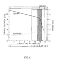
- the above conditions are computed and displayed In the form of units of distance (e.g., cm), and/or in the form of a graph, called a Partogram, showing the interrelation of the cervical dilatation and the descent of the fetal presenting part
- the output device of the apparatus is preferably a display, but could be a plotter, recorder, or other device for displaying, recording, and/or processing the data outputted by the computer.
- such an apparatus permits the progress of labor to be monitored in a manner which is continuous rather than intermittent, which is less dependent for accuracy on the experience, judgment or finger size of the attendant in the conventional "finger examination", which subjects the mother to less discomfort, and which involves less risk of contamination, infection, dislodgment of a fetal monitor, or injury to or death of the baby or mother due to a wrong assessment of the fetal position or of labor progress.
- this technique enables more precise monitoring of the critical condition, namely the changes in the spatial distance of the BPD of the baby's head with respect to the pelvic inlet.
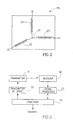
- Fig. 1 schematically illustrates the mother's womb particularly illustrated in Fig. 4 , is of special value since it provides a visual display of the progress of labor and can be recorded if desired.
- Partogram By using the Partogram, a better determination can be made whether labor is progressing normally.
- "Alert” and “action” lines may be printed on the Partogram to provide a visible indication of whether labor is progressing normally or abnormally, and thereby to better alert the attending personnel to take prompt action if necessary.
- Such an "electronic Partogram” can also markedly reduce the number of prolonged labors, the rate of intrapartum, post partum and early neonatal infections, the number of unnecessary interventions, and neonatal trauma due to wrong assessment of the fetal head.
- Fig. 5 illustrates a monitoring system similar to that of Fig. 1 but further equipped with an imaging system for imaging the womb area of the mother and for continuously displaying the mother's cervix, pelvic bones, and fetal head (or other presenting part).
- Fig 5 includes an ultrasonic transducer 40 for imaging the womb area, via the computer 12, on an image display 42. It also includes a position sensor PS5 attached to the ultrasonic transducer 40.
- a marker device 43 such as a mouse or touch screen
- the computer 12 can compute the various relationships displayed in displays 14 and 16.
- the image displayed in display 42 may be used in the same manner for marking the BPD on the fetal head as illustrated in Fig 8 , thereby enabling particularly the spatial distance between the fetal BPD and the pelvic inlet to be computed and monitored.
- reference points other than the BPD or the tip of the fetus head, as well as any other point of the mother's pelvis, may be used as the reference points for monitoring the progress of the labor. This freedom is important because of the variety of preferences among various physicians.
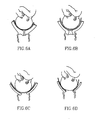
- the imaging system illustrated in Fig. 5 could also be used to provide a visual image of the various stages of labor, e.g., as illustrated in Figs. 6A - 6D showing the progressive dilatation and effacement of the cervix, or as illustrated in Figs. 7 and 8 showing the progressive descent of the fetal head tip through the various stations with respect to the ischial spines 7 ( Fig. 7 ) or mother's pelvic inlet ( Fig. 8 ).
- the ultrasound imaging may be used only to measure the BPD at the beginning of labor or later.
- the computer determines the distance between the BPD and the tip of the fetal head, and thereafter it can use the position of the tip of the fetal head also to determine the BPD position.
- the ultrasound imaging may thereafter be used only for verification if desired. It can also be used to verify cervical dilatation and effacement.
- the system illustrated in Fig. 5 may also be used for sensing contractions in the mother's uterus.
- the fetal head moves slightly, and the dilatation also grows slightly; and after contractions, they both retract to their previous positions.
- the attending physician may discern the occurrence of contractions as well as the duration and strength of such contractions.
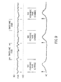
- the physician may observe the relation of the fetal heart rate (FHR) in relation to the uterine contractions (UC), to show the relationship between the two as illustrated in Fig. 9 .
- Computer 12 may be programmed to receive the above information from the various sensors and produce, in a monitor 45, a display corresponding to the fetal heart rate (FHR) in relation to the uterine contractions (UC), as illustrated in Fig. 9 .
- FHR fetal heart rate
- UC uterine contractions
Landscapes
- Health & Medical Sciences (AREA)
- Life Sciences & Earth Sciences (AREA)
- Biomedical Technology (AREA)
- Medical Informatics (AREA)
- Public Health (AREA)
- Biophysics (AREA)
- Veterinary Medicine (AREA)
- General Health & Medical Sciences (AREA)
- Physics & Mathematics (AREA)
- Animal Behavior & Ethology (AREA)
- Pathology (AREA)
- Heart & Thoracic Surgery (AREA)
- Engineering & Computer Science (AREA)
- Molecular Biology (AREA)
- Surgery (AREA)
- Gynecology & Obstetrics (AREA)
- Pregnancy & Childbirth (AREA)
- Radiology & Medical Imaging (AREA)
- Reproductive Health (AREA)
- Dentistry (AREA)
- Oral & Maxillofacial Surgery (AREA)
- Nuclear Medicine, Radiotherapy & Molecular Imaging (AREA)
- Ultra Sonic Daignosis Equipment (AREA)
- Surgical Instruments (AREA)
- Measurement Of The Respiration, Hearing Ability, Form, And Blood Characteristics Of Living Organisms (AREA)
- Investigating Or Analysing Materials By Optical Means (AREA)
- Measuring Pulse, Heart Rate, Blood Pressure Or Blood Flow (AREA)
- Paper (AREA)
- Investigating Or Analyzing Materials By The Use Of Ultrasonic Waves (AREA)
- Measuring And Recording Apparatus For Diagnosis (AREA)
- Spinning Or Twisting Of Yarns (AREA)
- General Factory Administration (AREA)
Claims (13)
- Appareil de contrôle de l'avancement du travail, caractérisé par :un capteur de position (PS1) à rattacher à un point prédéterminé des os du bassin de la mère et servant à produire un signal de sortie identifiant sa localisation dans l'espace ;un moyen (PS2) de détection de la localisation de la partie présentant le foetus (4) par rapport audit point prédéterminé des os du bassin de la mère et servant à produire un signal de sortie identifiant la localisation de ladite partie présentant le foetus (4), ledit moyen de détection de la localisation de la partie présentant le foetus (4) en se référant audit point prédéterminé des os du bassin de la mère comprenant un deuxième capteur de position (PS2) à rattacher à la pointe extérieure de la partie présentant le foetus pour produire vers l'ordinateur (12) un signal de sortie identifiant la localisation dudit deuxième capteur de position (PS2),un ordinateur (12) connecté de manière à recevoir lesdits signaux de sortie ; etun dispositif d'édition (42) commandé par ledit ordinateur (12) pour éditer la position de ladite partie présentant le foetus (4) par rapport audit point prédéterminé des os du bassin de la mère,ledit moyen de détection de la localisation de la partie présentant le foetus (4) et des côtés opposés du col de l'utérus de la mère comprenant :un transducteur ultrasonique (40) pour illustrer en images ladite partie présentant le foetus (4) et ledit col de l'utérus sur un écran (42) ; et un capteur de position (PS5) rattaché audit transducteur ultrasonique (40) pour produire un signal de sortie identifiant sa localisation en direction de l'ordinateur (12) ;et ledit appareil comprenant un outre : un système de marquage pour marquer un point sélectionné de la partie présentant le foetus (4) et les côtés opposés du col de l'utérus de la mère, comme illustré par des images sur l'écran (42) et pour éditer un signal électrique identifiant leurs localisations respectives en direction de l'ordinateur (12).
- Appareil selon la revendication 1, comprenant en outre un moyen (PS3, PS4) de détection des localisations des côtés opposés du col de l'utérus de la mère les uns par rapport aux autres afin de produire des signaux de sortie qui sont reçus par ledit ordinateur (12) et traités dans le but de commander ledit dispositif d'édition (42) pour éditer une indication de la dilatation du col de la mère.
- Appareil selon la revendication 2, comprenant en outre un moyen (PS3, PS4) de détection des localisations des côtés opposés du col de l'utérus de la mère les uns par rapport à la partie présentant le foetus (4) et aux os du bassin de la mère pour produire des signaux de sortie qui sont reçus par ledit ordinateur (12) et traités dans le but de commander ledit dispositif d'édition (42) pour éditer une indication de la position du col de la mère.
- Appareil selon la revendication 2 ou 3, dans lequel ledit moyen de détection des localisations des côtés opposés du col de l'utérus de la mère comprend un troisième (PS3) et un quatrième (PS4) capteurs de position à rattacher aux côtés opposés du col de l'utérus de la mère et servant à produire des signaux de sortie en direction dudit ordinateur (12) identifiant leurs positions respectives.
- Appareil selon la revendication 4, dans lequel ledit ordinateur (12) calcule et ledit dispositif d'édition (42) affiche la position de la partie présentant le foetus (4) sous forme de distance spatiale entre lesdits premier (PS1) et deuxième (PS2) capteurs de position.
- Appareil selon la revendication 5, dans lequel ledit ordinateur (12) calcule et ledit dispositif d'édition (42) affiche la dilatation du col de l'utérus de la femme sous forme de distance spatiale entre lesdits troisième (PS3) et quatrième (PS4) capteurs de position.
- Appareil selon la revendication 6, dans lequel ledit ordinateur (12) calcule et ledit dispositif d'édition (42) affiche ladite dilatation du col et ladite position en centimètres.
- Appareil selon la revendication 6, dans lequel ledit ordinateur (12) calcule et ledit dispositif d'édition (42) affiche ladite dilatation du col et ladite position sous forme d'un partogramme montrant l'interrelation entre la dilatation du col et la descente de la partie présentant le foetus (4).
- Appareil selon l'une quelconque des revendications 4 à 8, dans lequel ledit ordinateur (12) calcule et ledit dispositif d'édition (42) affiche l'effacement de la cavité de la mère sous forme de distance spatiale entre ledit deuxième (PS2) et lesdits troisième (PS3) et quatrième (PS4) capteurs de position.
- Appareil selon l'une quelconque des revendications 4 à 9, dans lequel ledit ordinateur (12) calcule et ledit dispositif d'édition (42) affiche la position du col de l'utérus de la mère sous forme de distances spatiales entre lesdits premier (PS1), deuxième (PS2), troisième (PS3) et quatrième (PS4) capteurs de position.
- Appareil selon la revendication 10, dans lequel ledit effacement et ladite position du col de l'utérus de la mère sont calculés et affichés en centimètres.
- Appareil selon l'une quelconque des revendications 4 à 9, dans lequel ledit ordinateur (12) calcule et ledit dispositif d'édition (42) affiche l'angle de l'axe cervical tel qu'indiqué par lesdits deuxième (PS2), troisième (PS3) et quatrième (PS4) capteurs de position.
- Appareil selon la revendication 12, dans lequel ledit marqueur marque le diamètre bi-pariétal (BPD) de la tête du foetus (4) tel qu'illustré par une image sur l'écran (42) et ledit ordinateur (12) calcule la distance entre ledit BPD et l'entrée du bassin de la femme.
Applications Claiming Priority (3)
| Application Number | Priority Date | Filing Date | Title |
|---|---|---|---|
| US4555697P | 1997-05-05 | 1997-05-05 | |
| US45556P | 1997-05-05 | ||
| PCT/IL1998/000208 WO1998049942A1 (fr) | 1997-05-05 | 1998-05-04 | Procede et dispositif servant a controler l'evolution du travail pendant l'accouchement |
Publications (3)
| Publication Number | Publication Date |
|---|---|
| EP1006882A1 EP1006882A1 (fr) | 2000-06-14 |
| EP1006882A4 EP1006882A4 (fr) | 2000-08-02 |
| EP1006882B1 true EP1006882B1 (fr) | 2009-11-25 |
Family
ID=21938597
Family Applications (1)
| Application Number | Title | Priority Date | Filing Date |
|---|---|---|---|
| EP98919447A Expired - Lifetime EP1006882B1 (fr) | 1997-05-05 | 1998-05-04 | Procede et dispositif servant a controler l'evolution du travail pendant l'accouchement |
Country Status (15)
| Country | Link |
|---|---|
| US (1) | US6200279B1 (fr) |
| EP (1) | EP1006882B1 (fr) |
| JP (1) | JP3474584B2 (fr) |
| KR (1) | KR100555353B1 (fr) |
| CN (2) | CN100482158C (fr) |
| AT (1) | ATE449568T1 (fr) |
| AU (1) | AU733125B2 (fr) |
| BR (1) | BR9808723B1 (fr) |
| CA (1) | CA2289551C (fr) |
| DE (1) | DE69841312D1 (fr) |
| ES (1) | ES2337425T3 (fr) |
| IL (1) | IL132703A (fr) |
| RU (1) | RU2225165C2 (fr) |
| UA (1) | UA65566C2 (fr) |
| WO (1) | WO1998049942A1 (fr) |
Cited By (1)
| Publication number | Priority date | Publication date | Assignee | Title |
|---|---|---|---|---|
| CN104000656A (zh) * | 2014-05-08 | 2014-08-27 | 朱秀红 | 胎儿分娩处理器 |
Families Citing this family (49)
| Publication number | Priority date | Publication date | Assignee | Title |
|---|---|---|---|---|
| US6669653B2 (en) * | 1997-05-05 | 2003-12-30 | Trig Medical Ltd. | Method and apparatus for monitoring the progress of labor |
| US6270458B1 (en) * | 1999-03-05 | 2001-08-07 | Barnev Inc. | Cervix dilation and labor progression monitor |
| EP1292252A1 (fr) * | 2000-05-11 | 2003-03-19 | Medical Clip S.R.L. | Procede de suspension abdominale pour femme enceinte et dispositif de protection dorsale et abdominale de mise en oeuvre du procede |
| US6423016B1 (en) * | 2000-06-08 | 2002-07-23 | Lms Medical Systems Ltd. | System and method for evaluating labor progress during childbirth |
| US20040153008A1 (en) * | 2001-06-05 | 2004-08-05 | Yehuda Sharf | Probe anchor |
| US20040089308A1 (en) * | 2002-11-13 | 2004-05-13 | Welch Robert A. | Cervical ring to deliver medication |
| CA2534837A1 (fr) * | 2003-08-06 | 2005-02-17 | Trig Medical Ltd. | Procede et appareil pour controler des parametres de travail |
| US8636676B2 (en) * | 2005-06-07 | 2014-01-28 | Perigen, Inc | Method and apparatus for providing information related to labor progress for an obstetrics patient |
| US7811239B2 (en) * | 2005-12-29 | 2010-10-12 | Intrapartum, Llc | Cervical dilation measurement apparatus |
| US7527601B2 (en) * | 2005-12-29 | 2009-05-05 | Intrapartum Ventures, Llc | Cervimeter |
| US7713216B2 (en) * | 2006-04-10 | 2010-05-11 | Intrapartum, Llc | Method for cervical dilation and/or measurement |
| US20070213640A1 (en) * | 2006-02-26 | 2007-09-13 | Hebah Noshy Mansour | Measurement aid for digital cervix examination |
| US10134490B2 (en) | 2006-05-01 | 2018-11-20 | Perigen, Inc. | Method and system for monitoring labour progression for an obstetrics patient |
| US9805164B2 (en) * | 2006-05-01 | 2017-10-31 | Perigen, Inc. | Method and apparatus for providing contraction information during labour |
| US9892475B1 (en) | 2006-11-03 | 2018-02-13 | E&C Medical Intelligence, Inc. | System and method for interactive clinical support and compliance with clinical standards and guidelines in real-time |
| US20080167581A1 (en) * | 2007-01-10 | 2008-07-10 | Yoav Paltieli | Determining parameters associated with a female pelvis and cervix |
| US20090093716A1 (en) * | 2007-10-04 | 2009-04-09 | General Electric Company | Method and apparatus for evaluation of labor with ultrasound |
| US20100268124A1 (en) * | 2007-12-10 | 2010-10-21 | Emily Hamilton | Method and apparatus for providing contraction information during labour |
| EP2328475B1 (fr) * | 2008-08-18 | 2016-01-06 | Glenveigh Medical, LLC | Dispositif de mesure de dilatation cervicale |
| IT1391829B1 (it) | 2008-11-21 | 2012-01-27 | C N R Consiglio Naz Delle Ricerche | Apparato basato sugli ultrasuoni per misurare parametri indicatori di avanzamento di un parto |
| US9131860B2 (en) | 2008-12-29 | 2015-09-15 | Mark Evans | Identifying the level of fetal risk during labor |
| WO2011032065A1 (fr) | 2009-09-13 | 2011-03-17 | Trig Medical Ltd. | Dispositif d'accouchement à capteur de position |
| EP2525702A4 (fr) * | 2009-09-28 | 2014-10-29 | Illuminare Holdings Ltd | Dispositif et réseau de surveillance intravaginale |
| BR112013002284A2 (pt) * | 2010-08-02 | 2016-05-24 | Koninkl Philips Electronics Nv | método de monitoramento cardíaco fetal, programa de computador e dispositivo para monitoramento cardíaco fetal ultrassônico |
| US9597055B2 (en) * | 2011-01-07 | 2017-03-21 | General Electric Company | Fetal scalp doppler device and system |
| SG191435A1 (en) * | 2011-12-13 | 2013-07-31 | Smart Communications Inc | System and method for transmitting partograph information and analysing the same |
| CN104272366B (zh) * | 2012-04-30 | 2017-11-28 | 挪度环球医疗股份公司 | 产后子宫模型 |
| US20160278688A1 (en) * | 2012-11-26 | 2016-09-29 | Ferring B.V. | Method and System for Diagnosing Uterine Contraction Levels Using Image Analysis |
| US8597306B1 (en) * | 2013-03-14 | 2013-12-03 | Plexus Biomedical, Inc. | Labor management methods for decreasing the incidence of cesarean childbirth |
| US10478151B2 (en) * | 2014-08-12 | 2019-11-19 | General Electric Company | System and method for automated monitoring of fetal head descent during labor |
| US10368833B2 (en) | 2014-09-12 | 2019-08-06 | General Electric Company | Method and system for fetal visualization by computing and displaying an ultrasound measurement and graphical model |
| US20160081663A1 (en) * | 2014-09-18 | 2016-03-24 | General Electric Company | Method and system for automated detection and measurement of a target structure |
| KR102356719B1 (ko) * | 2014-12-01 | 2022-01-27 | 삼성메디슨 주식회사 | 초음파 영상 장치 및 그 동작 방법 |
| RU2018123737A (ru) * | 2015-11-30 | 2020-01-09 | Конинклейке Филипс Н.В. | Система и способ контроля позиции плода |
| JP7224283B2 (ja) | 2016-11-21 | 2023-02-17 | エバンス,マーク | 分娩中の胎児リスクのレベルをモニタし、評価するためのシステム、装置、および方法 |
| US10595792B2 (en) * | 2017-06-11 | 2020-03-24 | Fetal Life Llc | Tocodynamometer GPS alert system |
| CN110141272A (zh) * | 2018-02-14 | 2019-08-20 | 王幼萍 | 一种骨盆测算装置及其方法 |
| CN109171910A (zh) * | 2018-07-26 | 2019-01-11 | 佟玲 | 一种产科吸盘助产装置 |
| JP7331087B2 (ja) | 2018-08-30 | 2023-08-22 | トリグ メディカル リミテッド | 出産磁気追跡システム |
| EP3880067A4 (fr) * | 2018-11-14 | 2022-08-10 | Mark Evans | Procédé et appareil pour réduire le risque de lésion neurologique néonatale |
| KR102700671B1 (ko) * | 2019-01-30 | 2024-08-30 | 삼성메디슨 주식회사 | 초음파 영상 장치 및 초음파 영상 생성 방법 |
| CN110400510B (zh) * | 2019-07-31 | 2021-12-07 | 中国人民解放军陆军军医大学第一附属医院 | 一种孕产妇产道仿真实训检测结果的评估装置 |
| WO2021022254A2 (fr) | 2019-08-01 | 2021-02-04 | Mark Evans | Procédés et appareil permettant de réduire le risque, et d'identifier l'existence, d'une lésion neurologique à un fœtus humain pendant et avant le travail |
| KR102840187B1 (ko) * | 2020-01-17 | 2025-07-31 | 삼성메디슨 주식회사 | 초음파 영상 장치 및 그 제어 방법 |
| KR102857835B1 (ko) * | 2020-02-10 | 2025-09-10 | 삼성메디슨 주식회사 | 초음파 진단 장치 및 초음파 영상처리방법 |
| WO2022025062A1 (fr) * | 2020-07-28 | 2022-02-03 | 国立大学法人東京大学 | Dispositif d'évaluation d'état d'avancement du travail, procédé d'évaluation d'état d'avancement du travail et programme |
| CN114365996B (zh) * | 2021-07-16 | 2023-01-20 | 张太斌 | 足月产妇子宫环境分析平台 |
| CN116763351A (zh) * | 2022-03-07 | 2023-09-19 | 深圳市理邦精密仪器股份有限公司 | 基于超声图像的胎头方位测量方法以及相关装置 |
| CN117373689B (zh) * | 2023-12-05 | 2024-02-13 | 吉林大学第一医院 | 一种产程心率实时分析方法及系统 |
Family Cites Families (15)
| Publication number | Priority date | Publication date | Assignee | Title |
|---|---|---|---|---|
| US3768459A (en) * | 1971-06-28 | 1973-10-30 | Utah Res & Dev Co Inc | Cervical dilation measuring device |
| FR2158767A5 (fr) | 1971-10-29 | 1973-06-15 | Farre Jean | |
| DE3103367C2 (de) | 1981-01-27 | 1987-02-19 | Uwe Dipl.-Ing. Blücher | Einrichtung zur Messung der Abstandsänderungen zweier Punkte eines biologischen, insbesondere menschlichen Organs |
| US4476871A (en) | 1982-04-23 | 1984-10-16 | American Home Products Corporation | Monitoring of cervical dilatation during labor |
| US4945305A (en) | 1986-10-09 | 1990-07-31 | Ascension Technology Corporation | Device for quantitatively measuring the relative position and orientation of two bodies in the presence of metals utilizing direct current magnetic fields |
| US5069218A (en) | 1987-02-03 | 1991-12-03 | Terumo Kabushiki Kaisha | Fetus monitoring apparatus |
| US5135006A (en) | 1988-03-31 | 1992-08-04 | Susan Bellinson | Method and apparatus for monitoring descent of fetus |
| US4942882A (en) | 1988-03-31 | 1990-07-24 | Susan Bellinson | Method and apparatus for monitoring descent of fetus |
| US5222485A (en) * | 1990-09-17 | 1993-06-29 | Ravinder Jerath | Ultrasound labor monitoring method and apparatus |
| JP3432825B2 (ja) * | 1992-08-14 | 2003-08-04 | ブリテイッシュ・テレコミュニケーションズ・パブリック・リミテッド・カンパニー | 位置決定システム |
| US5301680A (en) * | 1992-12-09 | 1994-04-12 | Hygeia Biomedical Research Inc. | Apparatus and method for the diagnosis of labor |
| US5406961A (en) | 1993-10-29 | 1995-04-18 | Artal; Raul | Monitoring device and method for detection of premature labor |
| US5438996A (en) | 1994-10-12 | 1995-08-08 | Triton Technology, Inc. | Ambulatory, ultrasonic transit time, real-time, cervical effacement and dilatation monitor with disposable probes |
| US5713371A (en) * | 1995-07-07 | 1998-02-03 | Sherman; Dani | Method of monitoring cervical dilatation during labor, and ultrasound transducer particularly useful in such method |
| US5935061A (en) * | 1997-01-03 | 1999-08-10 | Biosense, Inc. | Obstetrical instrument system and method |
-
1998
- 1998-04-05 UA UA99116047A patent/UA65566C2/uk unknown
- 1998-05-04 CN CNB988065525A patent/CN100482158C/zh not_active Expired - Fee Related
- 1998-05-04 DE DE69841312T patent/DE69841312D1/de not_active Expired - Lifetime
- 1998-05-04 CN CNB2004100899837A patent/CN100387191C/zh not_active Expired - Fee Related
- 1998-05-04 ES ES98919447T patent/ES2337425T3/es not_active Expired - Lifetime
- 1998-05-04 RU RU99125611/14A patent/RU2225165C2/ru not_active IP Right Cessation
- 1998-05-04 BR BRPI9808723-1A patent/BR9808723B1/pt not_active IP Right Cessation
- 1998-05-04 JP JP54789398A patent/JP3474584B2/ja not_active Expired - Fee Related
- 1998-05-04 WO PCT/IL1998/000208 patent/WO1998049942A1/fr not_active Ceased
- 1998-05-04 AU AU72306/98A patent/AU733125B2/en not_active Ceased
- 1998-05-04 KR KR1019997010191A patent/KR100555353B1/ko not_active Expired - Fee Related
- 1998-05-04 AT AT98919447T patent/ATE449568T1/de not_active IP Right Cessation
- 1998-05-04 CA CA2289551A patent/CA2289551C/fr not_active Expired - Fee Related
- 1998-05-04 EP EP98919447A patent/EP1006882B1/fr not_active Expired - Lifetime
- 1998-05-04 IL IL13270398A patent/IL132703A/en not_active IP Right Cessation
- 1998-05-05 US US09/072,850 patent/US6200279B1/en not_active Expired - Lifetime
Cited By (1)
| Publication number | Priority date | Publication date | Assignee | Title |
|---|---|---|---|---|
| CN104000656A (zh) * | 2014-05-08 | 2014-08-27 | 朱秀红 | 胎儿分娩处理器 |
Also Published As
| Publication number | Publication date |
|---|---|
| AU7230698A (en) | 1998-11-27 |
| KR100555353B1 (ko) | 2006-02-24 |
| IL132703A (en) | 2004-08-31 |
| CN100482158C (zh) | 2009-04-29 |
| BR9808723B1 (pt) | 2011-10-18 |
| BR9808723A (pt) | 2000-07-11 |
| EP1006882A4 (fr) | 2000-08-02 |
| US6200279B1 (en) | 2001-03-13 |
| UA65566C2 (uk) | 2004-04-15 |
| EP1006882A1 (fr) | 2000-06-14 |
| IL132703A0 (en) | 2001-03-19 |
| CA2289551A1 (fr) | 1998-11-12 |
| CA2289551C (fr) | 2010-12-14 |
| ES2337425T3 (es) | 2010-04-23 |
| WO1998049942A1 (fr) | 1998-11-12 |
| KR20010012241A (ko) | 2001-02-15 |
| CN100387191C (zh) | 2008-05-14 |
| AU733125B2 (en) | 2001-05-10 |
| JP2001504380A (ja) | 2001-04-03 |
| ATE449568T1 (de) | 2009-12-15 |
| JP3474584B2 (ja) | 2003-12-08 |
| RU2225165C2 (ru) | 2004-03-10 |
| CN1631322A (zh) | 2005-06-29 |
| CN1261260A (zh) | 2000-07-26 |
| DE69841312D1 (de) | 2010-01-07 |
Similar Documents
| Publication | Publication Date | Title |
|---|---|---|
| EP1006882B1 (fr) | Procede et dispositif servant a controler l'evolution du travail pendant l'accouchement | |
| US6669653B2 (en) | Method and apparatus for monitoring the progress of labor | |
| US20080167581A1 (en) | Determining parameters associated with a female pelvis and cervix | |
| US5301680A (en) | Apparatus and method for the diagnosis of labor | |
| US7850625B2 (en) | Method and apparatus for monitoring labor parameter | |
| RU99125611A (ru) | Способ и устройство контроля за ходом родов | |
| WO2000051494A1 (fr) | Dispositif de surveillance de la dilatation du col de l'uterus et de la progression du travail | |
| JP2001523507A (ja) | 子宮頚部の拡張、展退およびコンシステンシ監視システム | |
| EP1643900B1 (fr) | Surveillance tridimensionnelle de l'activite myographique | |
| MXPA99010172A (en) | Method and apparatus for monitoring the progress of labor | |
| CN215306081U (zh) | 一种全时段可靠型胎监设备 | |
| Farine | Limitations in current management of labor |
Legal Events
| Date | Code | Title | Description |
|---|---|---|---|
| PUAI | Public reference made under article 153(3) epc to a published international application that has entered the european phase |
Free format text: ORIGINAL CODE: 0009012 |
|
| 17P | Request for examination filed |
Effective date: 19991123 |
|
| AK | Designated contracting states |
Kind code of ref document: A1 Designated state(s): AT BE CH CY DE DK ES FI FR GB GR IE IT LI NL PT SE |
|
| A4 | Supplementary search report drawn up and despatched |
Effective date: 20000621 |
|
| AK | Designated contracting states |
Kind code of ref document: A4 Designated state(s): AT BE CH CY DE DK ES FI FR GB GR IE IT LI NL PT SE |
|
| RIC1 | Information provided on ipc code assigned before grant |
Free format text: 7A 61B 8/02 A, 7A 61B 5/107 B, 7A 61B 8/08 B |
|
| RAP1 | Party data changed (applicant data changed or rights of an application transferred) |
Owner name: TRIG MEDICAL LTD. |
|
| 17Q | First examination report despatched |
Effective date: 20050126 |
|
| 17Q | First examination report despatched |
Effective date: 20050126 |
|
| GRAP | Despatch of communication of intention to grant a patent |
Free format text: ORIGINAL CODE: EPIDOSNIGR1 |
|
| GRAS | Grant fee paid |
Free format text: ORIGINAL CODE: EPIDOSNIGR3 |
|
| GRAA | (expected) grant |
Free format text: ORIGINAL CODE: 0009210 |
|
| AK | Designated contracting states |
Kind code of ref document: B1 Designated state(s): AT BE CH CY DE DK ES FI FR GB GR IE IT LI NL PT SE |
|
| REG | Reference to a national code |
Ref country code: GB Ref legal event code: FG4D |
|
| REG | Reference to a national code |
Ref country code: CH Ref legal event code: EP |
|
| REG | Reference to a national code |
Ref country code: IE Ref legal event code: FG4D |
|
| REF | Corresponds to: |
Ref document number: 69841312 Country of ref document: DE Date of ref document: 20100107 Kind code of ref document: P |
|
| REG | Reference to a national code |
Ref country code: CH Ref legal event code: NV Representative=s name: SERVOPATENT GMBH |
|
| REG | Reference to a national code |
Ref country code: NL Ref legal event code: VDEP Effective date: 20091125 |
|
| REG | Reference to a national code |
Ref country code: ES Ref legal event code: FG2A Ref document number: 2337425 Country of ref document: ES Kind code of ref document: T3 |
|
| PG25 | Lapsed in a contracting state [announced via postgrant information from national office to epo] |
Ref country code: SE Free format text: LAPSE BECAUSE OF FAILURE TO SUBMIT A TRANSLATION OF THE DESCRIPTION OR TO PAY THE FEE WITHIN THE PRESCRIBED TIME-LIMIT Effective date: 20091125 Ref country code: PT Free format text: LAPSE BECAUSE OF FAILURE TO SUBMIT A TRANSLATION OF THE DESCRIPTION OR TO PAY THE FEE WITHIN THE PRESCRIBED TIME-LIMIT Effective date: 20100325 Ref country code: FI Free format text: LAPSE BECAUSE OF FAILURE TO SUBMIT A TRANSLATION OF THE DESCRIPTION OR TO PAY THE FEE WITHIN THE PRESCRIBED TIME-LIMIT Effective date: 20091125 |
|
| PG25 | Lapsed in a contracting state [announced via postgrant information from national office to epo] |
Ref country code: CY Free format text: LAPSE BECAUSE OF FAILURE TO SUBMIT A TRANSLATION OF THE DESCRIPTION OR TO PAY THE FEE WITHIN THE PRESCRIBED TIME-LIMIT Effective date: 20091125 |
|
| PG25 | Lapsed in a contracting state [announced via postgrant information from national office to epo] |
Ref country code: BE Free format text: LAPSE BECAUSE OF FAILURE TO SUBMIT A TRANSLATION OF THE DESCRIPTION OR TO PAY THE FEE WITHIN THE PRESCRIBED TIME-LIMIT Effective date: 20091125 Ref country code: AT Free format text: LAPSE BECAUSE OF FAILURE TO SUBMIT A TRANSLATION OF THE DESCRIPTION OR TO PAY THE FEE WITHIN THE PRESCRIBED TIME-LIMIT Effective date: 20091125 |
|
| PG25 | Lapsed in a contracting state [announced via postgrant information from national office to epo] |
Ref country code: NL Free format text: LAPSE BECAUSE OF FAILURE TO SUBMIT A TRANSLATION OF THE DESCRIPTION OR TO PAY THE FEE WITHIN THE PRESCRIBED TIME-LIMIT Effective date: 20091125 Ref country code: DK Free format text: LAPSE BECAUSE OF FAILURE TO SUBMIT A TRANSLATION OF THE DESCRIPTION OR TO PAY THE FEE WITHIN THE PRESCRIBED TIME-LIMIT Effective date: 20091125 |
|
| PLBE | No opposition filed within time limit |
Free format text: ORIGINAL CODE: 0009261 |
|
| STAA | Information on the status of an ep patent application or granted ep patent |
Free format text: STATUS: NO OPPOSITION FILED WITHIN TIME LIMIT |
|
| PG25 | Lapsed in a contracting state [announced via postgrant information from national office to epo] |
Ref country code: GR Free format text: LAPSE BECAUSE OF FAILURE TO SUBMIT A TRANSLATION OF THE DESCRIPTION OR TO PAY THE FEE WITHIN THE PRESCRIBED TIME-LIMIT Effective date: 20100226 |
|
| 26N | No opposition filed |
Effective date: 20100826 |
|
| PG25 | Lapsed in a contracting state [announced via postgrant information from national office to epo] |
Ref country code: IE Free format text: LAPSE BECAUSE OF NON-PAYMENT OF DUE FEES Effective date: 20100504 |
|
| REG | Reference to a national code |
Ref country code: FR Ref legal event code: PLFP Year of fee payment: 19 |
|
| PGFP | Annual fee paid to national office [announced via postgrant information from national office to epo] |
Ref country code: DE Payment date: 20160421 Year of fee payment: 19 Ref country code: GB Payment date: 20160426 Year of fee payment: 19 Ref country code: ES Payment date: 20160506 Year of fee payment: 19 Ref country code: CH Payment date: 20160421 Year of fee payment: 19 |
|
| PGFP | Annual fee paid to national office [announced via postgrant information from national office to epo] |
Ref country code: IT Payment date: 20160427 Year of fee payment: 19 Ref country code: FR Payment date: 20160422 Year of fee payment: 19 |
|
| REG | Reference to a national code |
Ref country code: DE Ref legal event code: R119 Ref document number: 69841312 Country of ref document: DE |
|
| REG | Reference to a national code |
Ref country code: CH Ref legal event code: PL |
|
| GBPC | Gb: european patent ceased through non-payment of renewal fee |
Effective date: 20170504 |
|
| PG25 | Lapsed in a contracting state [announced via postgrant information from national office to epo] |
Ref country code: CH Free format text: LAPSE BECAUSE OF NON-PAYMENT OF DUE FEES Effective date: 20170531 Ref country code: LI Free format text: LAPSE BECAUSE OF NON-PAYMENT OF DUE FEES Effective date: 20170531 |
|
| REG | Reference to a national code |
Ref country code: FR Ref legal event code: ST Effective date: 20180131 |
|
| PG25 | Lapsed in a contracting state [announced via postgrant information from national office to epo] |
Ref country code: DE Free format text: LAPSE BECAUSE OF NON-PAYMENT OF DUE FEES Effective date: 20171201 Ref country code: GB Free format text: LAPSE BECAUSE OF NON-PAYMENT OF DUE FEES Effective date: 20170504 |
|
| PG25 | Lapsed in a contracting state [announced via postgrant information from national office to epo] |
Ref country code: FR Free format text: LAPSE BECAUSE OF NON-PAYMENT OF DUE FEES Effective date: 20170531 Ref country code: IT Free format text: LAPSE BECAUSE OF NON-PAYMENT OF DUE FEES Effective date: 20170504 |
|
| REG | Reference to a national code |
Ref country code: ES Ref legal event code: FD2A Effective date: 20180625 |
|
| PG25 | Lapsed in a contracting state [announced via postgrant information from national office to epo] |
Ref country code: ES Free format text: LAPSE BECAUSE OF NON-PAYMENT OF DUE FEES Effective date: 20170505 |