EP1008326A2 - Photosonic diffusion wave-based tumor detector - Google Patents
Photosonic diffusion wave-based tumor detector Download PDFInfo
- Publication number
- EP1008326A2 EP1008326A2 EP99309789A EP99309789A EP1008326A2 EP 1008326 A2 EP1008326 A2 EP 1008326A2 EP 99309789 A EP99309789 A EP 99309789A EP 99309789 A EP99309789 A EP 99309789A EP 1008326 A2 EP1008326 A2 EP 1008326A2
- Authority
- EP
- European Patent Office
- Prior art keywords
- diffusion
- waves
- light
- mass
- ultrasound
- Prior art date
- Legal status (The legal status is an assumption and is not a legal conclusion. Google has not performed a legal analysis and makes no representation as to the accuracy of the status listed.)
- Granted
Links
Images

Classifications
-
- G—PHYSICS
- G01—MEASURING; TESTING
- G01N—INVESTIGATING OR ANALYSING MATERIALS BY DETERMINING THEIR CHEMICAL OR PHYSICAL PROPERTIES
- G01N21/00—Investigating or analysing materials by the use of optical means, i.e. using sub-millimetre waves, infrared, visible or ultraviolet light
- G01N21/17—Systems in which incident light is modified in accordance with the properties of the material investigated
- G01N21/47—Scattering, i.e. diffuse reflection
- G01N21/4795—Scattering, i.e. diffuse reflection spatially resolved investigating of object in scattering medium
-
- A—HUMAN NECESSITIES
- A61—MEDICAL OR VETERINARY SCIENCE; HYGIENE
- A61B—DIAGNOSIS; SURGERY; IDENTIFICATION
- A61B5/00—Measuring for diagnostic purposes; Identification of persons
- A61B5/0048—Detecting, measuring or recording by applying mechanical forces or stimuli
-
- A—HUMAN NECESSITIES
- A61—MEDICAL OR VETERINARY SCIENCE; HYGIENE
- A61B—DIAGNOSIS; SURGERY; IDENTIFICATION
- A61B5/00—Measuring for diagnostic purposes; Identification of persons
- A61B5/0059—Measuring for diagnostic purposes; Identification of persons using light, e.g. diagnosis by transillumination, diascopy, fluorescence
- A61B5/0082—Measuring for diagnostic purposes; Identification of persons using light, e.g. diagnosis by transillumination, diascopy, fluorescence adapted for particular medical purposes
- A61B5/0091—Measuring for diagnostic purposes; Identification of persons using light, e.g. diagnosis by transillumination, diascopy, fluorescence adapted for particular medical purposes for mammography
-
- A—HUMAN NECESSITIES
- A61—MEDICAL OR VETERINARY SCIENCE; HYGIENE
- A61B—DIAGNOSIS; SURGERY; IDENTIFICATION
- A61B5/00—Measuring for diagnostic purposes; Identification of persons
- A61B5/43—Detecting, measuring or recording for evaluating the reproductive systems
- A61B5/4306—Detecting, measuring or recording for evaluating the reproductive systems for evaluating the female reproductive systems, e.g. gynaecological evaluations
- A61B5/4312—Breast evaluation or disorder diagnosis
-
- A—HUMAN NECESSITIES
- A61—MEDICAL OR VETERINARY SCIENCE; HYGIENE
- A61B—DIAGNOSIS; SURGERY; IDENTIFICATION
- A61B8/00—Diagnosis using ultrasonic, sonic or infrasonic waves
- A61B8/08—Clinical applications
- A61B8/0825—Clinical applications for diagnosis of the breast, e.g. mammography
-
- G—PHYSICS
- G01—MEASURING; TESTING
- G01N—INVESTIGATING OR ANALYSING MATERIALS BY DETERMINING THEIR CHEMICAL OR PHYSICAL PROPERTIES
- G01N21/00—Investigating or analysing materials by the use of optical means, i.e. using sub-millimetre waves, infrared, visible or ultraviolet light
- G01N21/17—Systems in which incident light is modified in accordance with the properties of the material investigated
- G01N21/1717—Systems in which incident light is modified in accordance with the properties of the material investigated with a modulation of one or more physical properties of the sample during the optical investigation, e.g. electro-reflectance
Definitions
- This invention relates to systems for imaging human tissue using both ultrasound and light waves and, more particularly, to systems for imaging the interior of a highly scattering medium such as the human breast in a non-invasive manner using ultrasound to generate modulated optical intensity waves (diffusion waves).
- Visible and near-infrared light tomography has been used to image the interior of tissue media.
- Diffusion wave tomography has also been used to image tissue.
- Visible and near-infrared light tomography is limited to relatively small depths of penetration, e.g., on the order of a few millimeters in breast tissue, whereas diffusion wave tomography has relatively poor resolution, e.g., on the order of a centimeter in breast tissue. It would be an advance in the state of the art if a system were developed to overcome the foregoing disadvantages of the prior art.
- a system which generates optical diffusion waves inside a localized region in a highly scattering medium such as human breast tissue by simultaneously using visible and near-infrared light and focusing ultrasound waves in localized regions of the breast.
- the sources of visible and near-infrared light each comprise at least one laser.
- the vibrating tissue medium scatters the light impinging thereon to produce intensity-modulated diffusion waves.
- the diffusion waves emanating from the insonified region have a frequency equal to the frequency of the ultrasound waves (or a harmonic thereof).
- the resulting diffusion waves are detected at the boundary of the breast and are processed to acquire data on the absorption and scattering parameters of the insonified region.
- one or more diffusion wave detectors are arranged at the boundary of the breast to detect scattered diffusion waves at respective locations.
- Each diffusion wave detector comprises an optical-to-electrical transducer, i.e., a photodetector, such as a photomultiplier tube or a photodiode.
- the output signal of each diffusion wave detector is in turn fed to a detector which detects the amplitude and phase of the diffusion waves. Then the amplitude and phase signal components are fed to a processor which computes pixel values for display on a monitor.
- the amplitude and phase of the scattered diffusion waves are proportional to the density/composition of the insonified region.
- the amplitude and phase of the scattered diffusion waves are proportional to the density/composition of the insonified region.
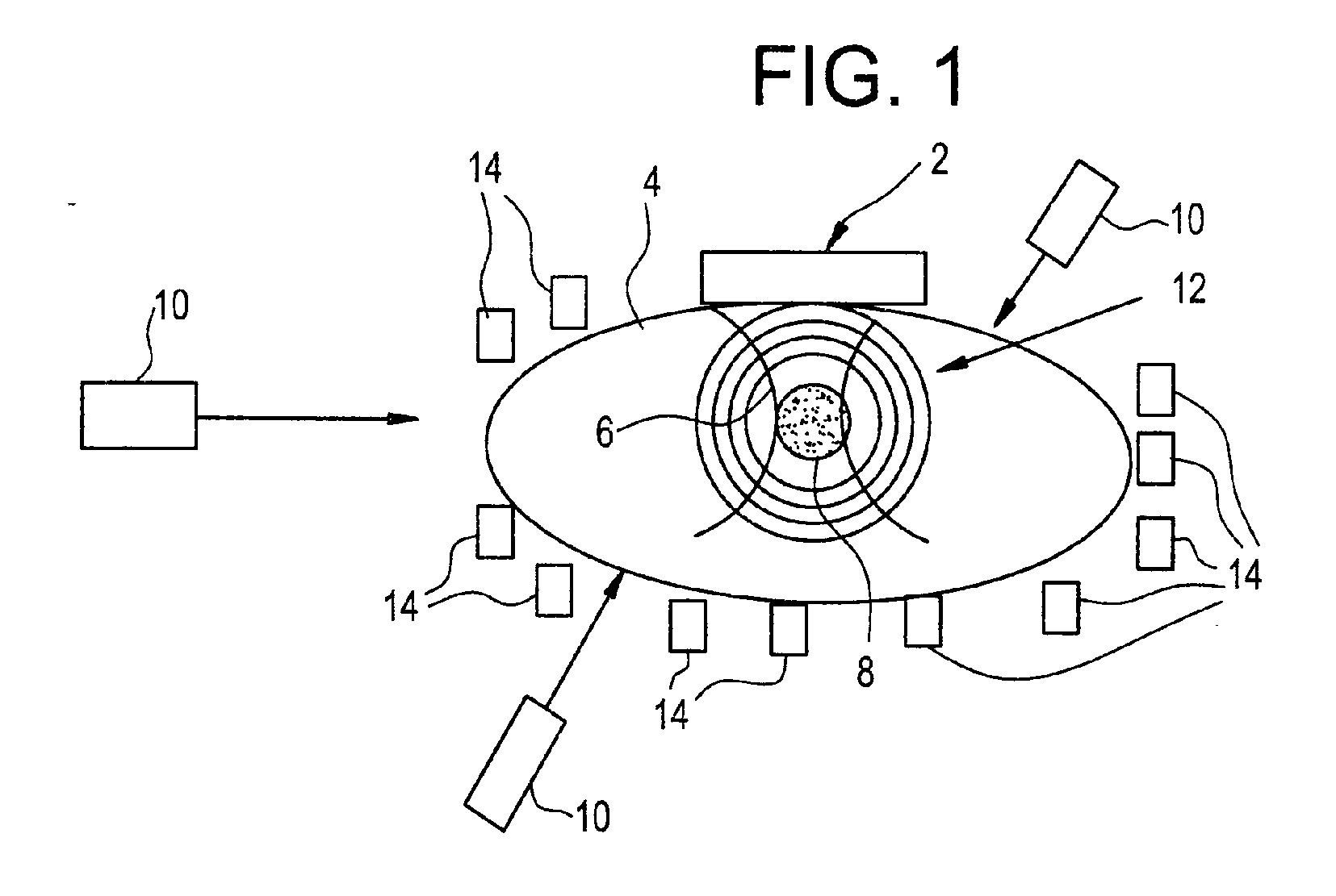
- FIG. 1 is a schematic illustration of a photosonic diffusion wave-based tumor detection system as described herein.
- FIG. 2 is a schematic illustration of a portion of a photosonic diffusion wave-based tumor detection system in accordance with one preferred embodiment of the invention.
- a highly scattering medium such as a human breast is illuminated by light in the visible and near-infrared region.
- the transmitted light will penetrate and distribute inside the breast as if it were diffused into the inside of the tissue.
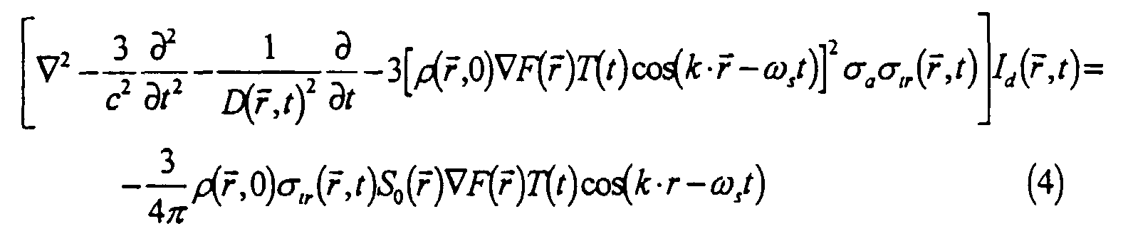
- the intensity of light waves inside the tissue will obey a diffusion-like wave equation, such as where I d ( r , t ) is the light intensity, c is the velocity of light in a vacuum, D is the diffusion constant.
- S 0 ( r , t ) in Eq. (1) is the modulated intensity of the external light source. In this embodiment of the invention, the external light source is not modulated and is a constant. For this situation, the right-hand side of Eq. (1) follows the time dependence of the density and scattering parameters, giving the following equation:
- an ultrasound beam is focused into a localized region inside the breast.
- the ultrasound waves perturb the density of the tissue in that region and also the scattering coefficient by modulating the size of the cells or constituents of the inhomogeneous medium.
- Equation (4) clearly points out the mechanism for the origin of the diffusion waves by means of the ultrasound waves.
- the intensity of the laser light varies as a function of the ultrasound frequency and this variation in light intensity constitutes the diffusion waves emanating from the region at which the ultrasound beam is focused. Because of the self-interaction of these waves, harmonics of the diffusion waves also emanate from the insonified region.
- FIG. 1 The basic structure of a system for carrying out this technique is shown in FIG. 1.
- An ultrasound transducer array 2 is sonically coupled to a highly scattering medium 4, such as breast tissue, and transmits ultrasound waves 6 which are focused in a localized region 8.
- the ultrasound beam frequency is preferably on the order of several megahertz which is selected to minimize attenuation in highly scattering medium 4.
- medium 4 is illuminated by light from a plurality of incoherent sources or from a plurality of laser sources 10. The light sources are placed so that light enters the highly scattering medium 4 from different directions.
- the light which penetrates insonified region 8 is modulated by ultrasound waves 6 and emanates from the insonified region in the form of diffusion waves 12. In contrast, light waves which penetrate regions unperturbed by the ultrasound waves are not modulated to form diffusion waves.
- a multiplicity of diffusion wave detectors 14, placed around the boundary of highly scattering medium 4, are used to detect diffusion waves 12.
- the strength of the diffusion waves is a function of ultrasound intensity and light intensity, but more importantly is a function of the absorption and scattering coefficients of insonified region 8.
- the absorption and scattering properties of the tumor can be used to determine whether the tumor is benign or cancerous since the absorption and scattering coefficients of cancerous tissue are different than those of benign tissue in the near-infrared region.
- reflected and diffracted diffusion waves can be suppressed by collecting those diffusion waves that originate from the region where the ultrasound beam is focused.
- the signal-to-noise ratio will be high because without the ultrasound waves, diffusion waves are not present, whereas in the presence of the ultrasound waves, diffusion waves are created corresponding to the frequency of the ultrasound waves and harmonics thereof.
- FIG. 2 A portion of the tumor detection system in accordance with a preferred embodiment of the invention is depicted in FIG. 2.
- a laser 10 is optically coupled to a first multiplicity 16 of optical fibers by means of a first fiber coupler 18.
- the distal ends of the optical fibers of the first multiplicity 16 are placed in contact with the breast or other highly scattering medium 4 such that laser light is directed toward region 8 where the ultrasound beam transmitted by transducer array 2 is focused.
- a photodetector 20 is optically coupled to the breast tissue through a second multiplicity 16' of optical fibers and a second fiber coupler 18'.
- the distal ends of the optical fibers of the second multiplicity 16' are placed in contact with the breast such that the scattered diffusion waves emanating from insonified region 4 are detected by photodetector 20.
- the photodetector transduces the diffusion waves into electrical signals which are supplied to a detector 22.
- Detector 22 comprises a conventional mixer for forming in-phase ( I ) and quadrature ( Q ) components and circuitry for determining amplitude and phase of the diffusion waves from the I and Q components.
- the amplitude and phase of the diffusion waves are related to the density and the absorption and scattering coefficients of the tissue in the insonified region.
- the amplitude and/or phase information is provided to a display processor 24 which converts the data into pixel information suitable for display on a display monitor 26.
- An amplitude and/or phase image of the interior of the breast can be synthesized or constructed by scanning the ultrasound focal region to cover the interior of the breast.
- FIG. 2 depicts only a single laser light and a single photodetector, it will be appreciated that the system preferably comprises multiple light sources and multiple photodetectors.
- the output signals of the multiple photodetectors can be summed before or after detection of the amplitude and phase of the diffusion waves by applying appropriate time delays as a function of the distance of propagation from the insonified region to the respective photodetector.
- the frequency of the laser light is selected to provide spectroscopic differences in the visible and near-infrared regions (500nm-1,400 nm) between different tissue types.
- the use of different optical wavelengths for the transmitted light enables differentiation of different types of cancerous and non-cancerous tissues because the optical scattering parameters of cancerous and non-cancerous tissues are different. This appears as a color parameter in the final reconstruction, helping to discriminate normal tissue from abnormal and cancerous tissue.
- the resolution obtained by the above-described process can be enhanced by use of an image reconstruction procedure.
- a near-field reconstruction procedure can be employed which incorporates the so-called evanescent waves and window functions to limit the growth of side lobes.
- the approach is as follows: (1) measure the diffusion wave phase and amplitudes at a plane far from the insonified region; (2) take a windowed cosine transform (Fourier transform) to obtain the angular spectrum amplitude; (3) filter out evanescent wave propagation factors; and (4) take a windowed cosine transform. This results in reconstruction of the scattering parameters of the insonified region.
- the ultrasound beam is scanned over different portions and the foregoing procedure is repeated to obtain a picture of the whole interior of the breast.
- finite differential element technique can be used for source reconstruction.
- the system uses scattered light and pulsed ultrasound.
- the technique employed is not based on the phase of the coherent light inside the tissue.
- the system can image tumors deep inside the tissue illuminated by scattered (diffused) light and insonified by pulsed ultrasound waves. By properly choosing the ultrasound frequency, tissue penetration of about 10 cm can be achieved in breast tissue.
- Coherent light sources are also not necessary as the technique uses diffused or scattered light and generates optical diffusion waves at different localized regions inside the breast by focusing pulsed ultrasound in those localized regions. The phase and amplitudes of these coherent optical diffusion waves are measured using detectors situated at the surface of the breast or scattering medium.
Landscapes
- Health & Medical Sciences (AREA)
- Life Sciences & Earth Sciences (AREA)
- Physics & Mathematics (AREA)
- General Health & Medical Sciences (AREA)
- Pathology (AREA)
- Biomedical Technology (AREA)
- Engineering & Computer Science (AREA)
- Heart & Thoracic Surgery (AREA)
- Medical Informatics (AREA)
- Molecular Biology (AREA)
- Surgery (AREA)
- Animal Behavior & Ethology (AREA)
- Biophysics (AREA)
- Public Health (AREA)
- Veterinary Medicine (AREA)
- Chemical & Material Sciences (AREA)
- Analytical Chemistry (AREA)
- Biochemistry (AREA)
- General Physics & Mathematics (AREA)
- Immunology (AREA)
- Optics & Photonics (AREA)
- Nuclear Medicine, Radiotherapy & Molecular Imaging (AREA)
- Radiology & Medical Imaging (AREA)
- Gynecology & Obstetrics (AREA)
- Reproductive Health (AREA)
- Ultra Sonic Daignosis Equipment (AREA)
- Investigating Or Analysing Materials By Optical Means (AREA)
Abstract
Description


Claims (18)
- A method for detecting a property in an interior of a mass of tissue, comprising the steps of:focusing an ultrasound beam having an ultrasound frequency at a localized region in the mass of tissue during one detection cycle;directing a first beam of light of unmodulated intensity and having a first light frequency into the mass of tissue during said one detection cycle; anddetecting diffusion waves of said ultrasound frequency emanating from the mass of tissue during said one detection cycle.
- The method as defined in claim 1, further comprising the step of determining amplitudes of the detected diffusion waves.
- The method as defined in claim 1, further comprising the step of determining phase of the detected diffusion waves.
- The method as defined in claim 2, further comprising the step of displaying as an image a function of said amplitudes of said detected diffusion waves.
- The method as defined in claim 3, further comprising the step of displaying as an image a function of said phase of said detected diffusion waves.
- The method as defined in claim 1, wherein the step of detecting diffusion waves comprises transducing said diffusion waves of said ultrasound frequency into electrical signals.
- The method as defined in claim 1, further comprising the steps of:focusing said ultrasound beam having said ultrasound frequency at said localized region during a second detection cycle;directing a second beam of light unmodulated in intensity and having a second light frequency different than said first light frequency into the mass of tissue during said second detection cycle; anddetecting diffusion waves of said ultrasound frequency emanating from the mass of tissue during said second detection cycle.
- The method as defined in claim 1, further comprising the steps of repeating said focusing, directing and detecting steps for each one of a multiplicity of localized regions in the mass of tissue.
- The method as defined in claim 1 including the step of directing a second beam of light unmodulated in intensity and having a second light frquency different than said first light frequency into the mass of tissue during said one detection cycle.
- A system for detecting a property in an interior of a mass of tissue, comprising:an ultrasound transducer array arranged and controlled to focus an ultrasound beam having an ultrasound frequency at a localized region in the mass of tissue during one detection cycle;a light source having a first light frequency and arranged to direct a first beam of light of unmodulated intensity into the mass of tissue during said one detection cycle; anda diffusion wave detector arranged to detect diffusion waves of said ultrasound frequency emanating from the mass of tissue during said one detection cycle.
- The system as defined in claim 10, further comprising an amplitude detector operatively coupled to said diffusion wave detector for determining amplitudes of the detected diffusion waves.
- The system as defined in claim 10, further comprising a phase detector operatively coupled to said diffusion wave detector for determining phase of the detected diffusion waves.
- The system as defined in claim 11, further comprising a display monitor for displaying as an image a function of said amplitudes of said detected diffusion waves.
- The system as defined in claim 12, further comprising a display monitor for displaying as an image a function of said phase of said detected diffusion waves.
- The system as defined in claim 10, wherein said diffusion wave detector comprises a photodetector.
- The system as defined in claim 10, wherein said light source comprises a source of incoherent light.
- The system as defined in claim 10, wherein said light source comprises a laser.
- The system as defined in claim 10 including a second light source having a second light frequency different than said first frequency and arranged to direct a second beam of light of unmodulated intensity into the mass of tissue during said one detection cycle.
Applications Claiming Priority (2)
| Application Number | Priority Date | Filing Date | Title |
|---|---|---|---|
| US09/207,009 US6245015B1 (en) | 1998-12-07 | 1998-12-07 | Photosonic diffusion wave-based tumor detector |
| US207009 | 1998-12-07 |
Publications (3)
| Publication Number | Publication Date |
|---|---|
| EP1008326A2 true EP1008326A2 (en) | 2000-06-14 |
| EP1008326A3 EP1008326A3 (en) | 2001-05-16 |
| EP1008326B1 EP1008326B1 (en) | 2007-06-20 |
Family
ID=22768843
Family Applications (1)
| Application Number | Title | Priority Date | Filing Date |
|---|---|---|---|
| EP99309789A Expired - Lifetime EP1008326B1 (en) | 1998-12-07 | 1999-12-06 | Photosonic diffusion wave-based tumor detector |
Country Status (4)
| Country | Link |
|---|---|
| US (1) | US6245015B1 (en) |
| EP (1) | EP1008326B1 (en) |
| JP (1) | JP4705707B2 (en) |
| DE (1) | DE69936333T2 (en) |
Cited By (9)
| Publication number | Priority date | Publication date | Assignee | Title |
|---|---|---|---|---|
| EP1709427A1 (en) * | 2003-11-17 | 2006-10-11 | General Electric Company | Method and system for ultrasonic tagging of fluorescence |
| RU2288753C2 (en) * | 2005-01-27 | 2006-12-10 | Анатолий Васильевич Кобзев | Device for making physiotherapeutic effect |
| EP1967129A1 (en) * | 2007-03-08 | 2008-09-10 | Olympus Medical Systems Corp. | Medical apparatus obtaining information indicative of internal state of an object based on interaction between sound waves and light |
| WO2008115089A1 (en) * | 2007-03-16 | 2008-09-25 | Germanov, Evgeny Pavlovich | Diagnosis method using ultrasonic, acoustic and electromagnetic waves |
| EP2260754A1 (en) * | 2009-06-10 | 2010-12-15 | Universiteit Twente | Device and method for photon absorption coefficient measurement |
| US8108022B2 (en) | 2003-09-12 | 2012-01-31 | Or-Nim Medical Ltd. | Method and apparatus for noninvasively monitoring parameters of a region of interest in a human body |
| US8423116B2 (en) | 2005-03-16 | 2013-04-16 | Or-Nim Medical Ltd. | Noninvasive measurements in a human body |
| CN103385734A (en) * | 2012-11-15 | 2013-11-13 | 广州呼研所红外科技有限公司 | Double check comprehensive diagnostic apparatus guiding ultrasound by infrared thermography and detection method of diagnostic apparatus |
| US9237850B2 (en) | 2007-06-04 | 2016-01-19 | Or-Nim Medical Ltd. | System and method for noninvasively monitoring conditions of a subject |
Families Citing this family (24)
| Publication number | Priority date | Publication date | Assignee | Title |
|---|---|---|---|---|
| JP4696079B2 (en) * | 2003-12-17 | 2011-06-08 | ヤンセン アルツハイマー イミュノセラピー | Aβ immunogenic peptide carrier conjugate and method for producing the same |
| US7144370B2 (en) * | 2004-05-12 | 2006-12-05 | General Electric Company | Method and apparatus for imaging of tissue using multi-wavelength ultrasonic tagging of light |
| US7188786B2 (en) * | 2004-10-28 | 2007-03-13 | Meadwestvaco Corporation | Hose-end sprayer assembly |
| JP4619803B2 (en) * | 2005-01-26 | 2011-01-26 | 富士フイルム株式会社 | Fluorescence tomographic image acquisition device |
| DE102005034219A1 (en) * | 2005-07-19 | 2007-02-22 | Fachhochschule Lübeck | Method for in vivo tissue classification |
| JP2008168038A (en) * | 2007-01-15 | 2008-07-24 | Olympus Medical Systems Corp | SUBJECT INFORMATION ANALYSIS DEVICE, ENDOSCOPE DEVICE, AND SUBJECT INFORMATION ANALYSIS METHOD |
| JP4939237B2 (en) * | 2006-01-20 | 2012-05-23 | オリンパスメディカルシステムズ株式会社 | SUBJECT INFORMATION ANALYSIS DEVICE, ENDOSCOPE DEVICE, AND SUBJECT INFORMATION ANALYSIS METHOD |
| EP2275022B8 (en) * | 2006-01-20 | 2016-10-19 | Olympus Corporation | Apparatus for analyzing characteristic information of object with the use of mutual interaction between ultrasound wave and light |
| JP5192846B2 (en) | 2008-02-25 | 2013-05-08 | オリンパスメディカルシステムズ株式会社 | Biological observation apparatus and method of operating biological observation apparatus |
| JP5183381B2 (en) * | 2008-09-16 | 2013-04-17 | キヤノン株式会社 | Measuring apparatus and measuring method |
| JP5183406B2 (en) * | 2008-10-03 | 2013-04-17 | キヤノン株式会社 | Biological information processing apparatus and biological information processing method |
| GB0818775D0 (en) * | 2008-10-13 | 2008-11-19 | Isis Innovation | Investigation of physical properties of an object |
| JP5376910B2 (en) * | 2008-11-14 | 2013-12-25 | キヤノン株式会社 | Biological information measuring device and biological information measuring method |
| CN102307528B (en) * | 2009-02-23 | 2014-12-31 | 奥林巴斯医疗株式会社 | Biomedical imaging apparatus and biomedical tomographic image generation method |
| CN102292627B (en) | 2009-06-08 | 2014-04-30 | 奥林巴斯医疗株式会社 | biological observation device |
| US20120075956A1 (en) * | 2010-09-29 | 2012-03-29 | Lockheed Martin Corporation | Mag-phase process |
| CN102686165B (en) * | 2010-10-21 | 2015-04-01 | 松下电器产业株式会社 | Ultrasonic testing device and ultrasonic testing method |
| WO2012172764A1 (en) * | 2011-06-17 | 2012-12-20 | パナソニック株式会社 | Optoacoustic image pick-up system and optoacoustic image pick-up device |
| GB201116518D0 (en) | 2011-09-23 | 2011-11-09 | Isis Innovation | Investigation of physical properties of an object |
| CN103370010A (en) * | 2011-10-24 | 2013-10-23 | 松下电器产业株式会社 | photoacoustic camera device |
| JPWO2013157228A1 (en) * | 2012-04-19 | 2015-12-21 | パナソニック株式会社 | Photoacoustic imaging device |
| WO2013172020A1 (en) * | 2012-05-15 | 2013-11-21 | パナソニック株式会社 | Photoacoustic vibration meter |
| WO2013183247A1 (en) * | 2012-06-04 | 2013-12-12 | パナソニック株式会社 | Acoustooptic imaging device |
| WO2013183302A1 (en) * | 2012-06-08 | 2013-12-12 | パナソニック株式会社 | Acoustooptic imaging device |
Family Cites Families (9)
| Publication number | Priority date | Publication date | Assignee | Title |
|---|---|---|---|---|
| US4614116A (en) * | 1985-02-04 | 1986-09-30 | International Business Machines Corporation | Phase sensitive ultrasonic modulation method for the detection of strain-sensitive spectral features |
| FR2617602B1 (en) * | 1987-07-03 | 1989-10-20 | Thomson Csf | METHOD AND SYSTEM FOR PHOTON FREQUENCY MARKING TRANSILLUMINATION IMAGING |
| JPH04329936A (en) * | 1991-01-08 | 1992-11-18 | Olympus Optical Co Ltd | Non-destructive light inspection method and system therefor |
| DE4128744C1 (en) * | 1991-08-29 | 1993-04-22 | Siemens Ag, 8000 Muenchen, De | |
| JP3251417B2 (en) * | 1994-03-09 | 2002-01-28 | 株式会社日立製作所 | Biological measurement device |
| DE4419900A1 (en) * | 1994-06-07 | 1995-12-14 | Siemens Ag | Method and arrangement for imaging an object with light |
| GB9619693D0 (en) * | 1996-09-20 | 1996-11-06 | Johnson & Johnson Medical | Apparatus and method for non-invasive measurement of a substance |
| DE19654053A1 (en) * | 1996-12-23 | 1998-06-25 | Schweiger Gustav Prof Dr Techn | Ultrasonic determination of optical characteristics in biological tissue |
| JP2000088742A (en) * | 1998-09-17 | 2000-03-31 | Aloka Co Ltd | Light-measuring device |
-
1998
- 1998-12-07 US US09/207,009 patent/US6245015B1/en not_active Expired - Fee Related
-
1999
- 1999-11-30 JP JP33899899A patent/JP4705707B2/en not_active Expired - Fee Related
- 1999-12-06 DE DE69936333T patent/DE69936333T2/en not_active Expired - Lifetime
- 1999-12-06 EP EP99309789A patent/EP1008326B1/en not_active Expired - Lifetime
Cited By (15)
| Publication number | Priority date | Publication date | Assignee | Title |
|---|---|---|---|---|
| US8644900B2 (en) | 2003-09-12 | 2014-02-04 | Or-Nim Medical Ltd. | Method and apparatus for noninvasively monitoring parameters of a region of interest in a human body |
| US8108022B2 (en) | 2003-09-12 | 2012-01-31 | Or-Nim Medical Ltd. | Method and apparatus for noninvasively monitoring parameters of a region of interest in a human body |
| US8126524B2 (en) | 2003-09-12 | 2012-02-28 | Or-Nim Medical Ltd. | Method and apparatus for noninvasively monitoring parameters of a region of interest in a human body |
| EP1709427A1 (en) * | 2003-11-17 | 2006-10-11 | General Electric Company | Method and system for ultrasonic tagging of fluorescence |
| RU2288753C2 (en) * | 2005-01-27 | 2006-12-10 | Анатолий Васильевич Кобзев | Device for making physiotherapeutic effect |
| US8423116B2 (en) | 2005-03-16 | 2013-04-16 | Or-Nim Medical Ltd. | Noninvasive measurements in a human body |
| US9131880B2 (en) | 2005-03-16 | 2015-09-15 | Or-Nim Medical Ltd. | Noninvasive measurements in a human body |
| EP1967129A1 (en) * | 2007-03-08 | 2008-09-10 | Olympus Medical Systems Corp. | Medical apparatus obtaining information indicative of internal state of an object based on interaction between sound waves and light |
| WO2008115089A1 (en) * | 2007-03-16 | 2008-09-25 | Germanov, Evgeny Pavlovich | Diagnosis method using ultrasonic, acoustic and electromagnetic waves |
| US9237850B2 (en) | 2007-06-04 | 2016-01-19 | Or-Nim Medical Ltd. | System and method for noninvasively monitoring conditions of a subject |
| WO2010142530A1 (en) * | 2009-06-10 | 2010-12-16 | Universiteit Twente | Device and method for photon absorption coefficient measurement |
| EP2260754A1 (en) * | 2009-06-10 | 2010-12-15 | Universiteit Twente | Device and method for photon absorption coefficient measurement |
| US9357962B2 (en) | 2009-06-10 | 2016-06-07 | Universiteit Twente | Device and method for photon absorption coefficient measurement |
| CN103385734A (en) * | 2012-11-15 | 2013-11-13 | 广州呼研所红外科技有限公司 | Double check comprehensive diagnostic apparatus guiding ultrasound by infrared thermography and detection method of diagnostic apparatus |
| CN103385734B (en) * | 2012-11-15 | 2016-08-24 | 广州呼研所红外科技有限公司 | Infrared thermal imagery is utilized to guide ultrasonic duplication check comprehensive diagnostic instrument and the detection method of this diagnostic apparatus |
Also Published As
| Publication number | Publication date |
|---|---|
| EP1008326B1 (en) | 2007-06-20 |
| DE69936333T2 (en) | 2008-02-21 |
| JP4705707B2 (en) | 2011-06-22 |
| EP1008326A3 (en) | 2001-05-16 |
| JP2000197635A (en) | 2000-07-18 |
| US6245015B1 (en) | 2001-06-12 |
| DE69936333D1 (en) | 2007-08-02 |
Similar Documents
| Publication | Publication Date | Title |
|---|---|---|
| EP1008326B1 (en) | Photosonic diffusion wave-based tumor detector | |
| US6738653B1 (en) | Metabolism monitoring of body organs | |
| US8280494B2 (en) | Apparatus and method to measure a spectroscopic characteristic in an object | |
| US5570182A (en) | Method for detection of dental caries and periodontal disease using optical imaging | |
| EP0647120B1 (en) | Examination of subjects using photon migration with high directionality techniques | |
| US5477051A (en) | Apparatus for measuring optical information in scattering medium and method therefor | |
| US7139603B2 (en) | Optical techniques for examination of biological tissue | |
| EP1303756B1 (en) | Apparatus and method for probing light absorbing agents in biological tissues | |
| US7777891B2 (en) | Elasticity and viscosity measuring apparatus | |
| EP1356265B1 (en) | Method and apparatus for imaging absorbing objects in a scattering medium | |
| US7917312B2 (en) | Photoacoustic doppler flow sensing and imaging | |
| US5820558A (en) | Optical techniques for examination of biological tissue | |
| US7023558B2 (en) | Acousto-optic monitoring and imaging in a depth sensitive manner | |
| CN101677765A (en) | Biological information imaging apparatus, biological information analyzing method, and biological information imaging method | |
| JPH10510626A (en) | Optical techniques for examination of biological tissues | |
| WO2007138493A1 (en) | Photoacoustic imaging method | |
| US20170258332A1 (en) | System and method for non-contact ultrasound with enhanced safety | |
| US20210076944A1 (en) | System and method for non-contact ultrasound image reconstruction | |
| US7652773B2 (en) | Enhanced detection of acousto-photonic emissions in optically turbid media using a photo-refractive crystal-based detection system | |
| WO2010143572A1 (en) | Subject information analysis device and subject information analysis method | |
| Sakadžić et al. | Toward very high resolution imaging in ultrasound-modulated optical tomography of biological tissues | |
| Leveque-Fort et al. | Toward simultaneous acousto-optical and acoustical imaging in biological tissues | |
| Schuetz et al. | Imaging of xenofluorescent objects in strongly scattering media: first results | |
| Sakadzic et al. | High-resolution imaging using ultrasound-modulated optical tomography | |
| JPH04329936A (en) | Non-destructive light inspection method and system therefor |
Legal Events
| Date | Code | Title | Description |
|---|---|---|---|
| PUAI | Public reference made under article 153(3) epc to a published international application that has entered the european phase |
Free format text: ORIGINAL CODE: 0009012 |
|
| AK | Designated contracting states |
Kind code of ref document: A2 Designated state(s): DE NL |
|
| AX | Request for extension of the european patent |
Free format text: AL;LT;LV;MK;RO;SI |
|
| PUAL | Search report despatched |
Free format text: ORIGINAL CODE: 0009013 |
|
| AK | Designated contracting states |
Kind code of ref document: A3 Designated state(s): AT BE CH CY DE DK ES FI FR GB GR IE IT LI LU MC NL PT SE |
|
| AX | Request for extension of the european patent |
Free format text: AL;LT;LV;MK;RO;SI |
|
| 17P | Request for examination filed |
Effective date: 20011116 |
|
| AKX | Designation fees paid |
Free format text: DE NL |
|
| 17Q | First examination report despatched |
Effective date: 20030610 |
|
| GRAP | Despatch of communication of intention to grant a patent |
Free format text: ORIGINAL CODE: EPIDOSNIGR1 |
|
| GRAS | Grant fee paid |
Free format text: ORIGINAL CODE: EPIDOSNIGR3 |
|
| GRAA | (expected) grant |
Free format text: ORIGINAL CODE: 0009210 |
|
| AK | Designated contracting states |
Kind code of ref document: B1 Designated state(s): DE NL |
|
| REF | Corresponds to: |
Ref document number: 69936333 Country of ref document: DE Date of ref document: 20070802 Kind code of ref document: P |
|
| PLBE | No opposition filed within time limit |
Free format text: ORIGINAL CODE: 0009261 |
|
| STAA | Information on the status of an ep patent application or granted ep patent |
Free format text: STATUS: NO OPPOSITION FILED WITHIN TIME LIMIT |
|
| 26N | No opposition filed |
Effective date: 20080325 |
|
| PGFP | Annual fee paid to national office [announced via postgrant information from national office to epo] |
Ref country code: DE Payment date: 20111229 Year of fee payment: 13 |
|
| PGFP | Annual fee paid to national office [announced via postgrant information from national office to epo] |
Ref country code: NL Payment date: 20120103 Year of fee payment: 13 |
|
| REG | Reference to a national code |
Ref country code: NL Ref legal event code: V1 Effective date: 20130701 |
|
| REG | Reference to a national code |
Ref country code: DE Ref legal event code: R119 Ref document number: 69936333 Country of ref document: DE Effective date: 20130702 |
|
| PG25 | Lapsed in a contracting state [announced via postgrant information from national office to epo] |
Ref country code: NL Free format text: LAPSE BECAUSE OF NON-PAYMENT OF DUE FEES Effective date: 20130701 Ref country code: DE Free format text: LAPSE BECAUSE OF NON-PAYMENT OF DUE FEES Effective date: 20130702 |