EP1010016B1 - Angiographie par resonance magnetique en 3d a contraste renforce et a resolution temporelle synchronisee - Google Patents
Angiographie par resonance magnetique en 3d a contraste renforce et a resolution temporelle synchronisee Download PDFInfo
- Publication number
- EP1010016B1 EP1010016B1 EP97936143A EP97936143A EP1010016B1 EP 1010016 B1 EP1010016 B1 EP 1010016B1 EP 97936143 A EP97936143 A EP 97936143A EP 97936143 A EP97936143 A EP 97936143A EP 1010016 B1 EP1010016 B1 EP 1010016B1
- Authority
- EP
- European Patent Office
- Prior art keywords
- space
- recited
- image
- peripheral
- subregions
- Prior art date
- Legal status (The legal status is an assumption and is not a legal conclusion. Google has not performed a legal analysis and makes no representation as to the accuracy of the status listed.)
- Expired - Lifetime
Links
Images


Classifications
-
- G—PHYSICS
- G01—MEASURING; TESTING
- G01R—MEASURING ELECTRIC VARIABLES; MEASURING MAGNETIC VARIABLES
- G01R33/00—Arrangements or instruments for measuring magnetic variables
- G01R33/20—Arrangements or instruments for measuring magnetic variables involving magnetic resonance
- G01R33/44—Arrangements or instruments for measuring magnetic variables involving magnetic resonance using nuclear magnetic resonance [NMR]
- G01R33/48—NMR imaging systems
- G01R33/54—Signal processing systems, e.g. using pulse sequences ; Generation or control of pulse sequences; Operator console
- G01R33/56—Image enhancement or correction, e.g. subtraction or averaging techniques, e.g. improvement of signal-to-noise ratio and resolution
- G01R33/561—Image enhancement or correction, e.g. subtraction or averaging techniques, e.g. improvement of signal-to-noise ratio and resolution by reduction of the scanning time, i.e. fast acquiring systems, e.g. using echo-planar pulse sequences
-
- G—PHYSICS
- G01—MEASURING; TESTING
- G01R—MEASURING ELECTRIC VARIABLES; MEASURING MAGNETIC VARIABLES
- G01R33/00—Arrangements or instruments for measuring magnetic variables
- G01R33/20—Arrangements or instruments for measuring magnetic variables involving magnetic resonance
- G01R33/28—Details of apparatus provided for in groups G01R33/44 - G01R33/64
- G01R33/281—Means for the use of in vitro contrast agents
-
- G—PHYSICS
- G01—MEASURING; TESTING
- G01R—MEASURING ELECTRIC VARIABLES; MEASURING MAGNETIC VARIABLES
- G01R33/00—Arrangements or instruments for measuring magnetic variables
- G01R33/20—Arrangements or instruments for measuring magnetic variables involving magnetic resonance
- G01R33/44—Arrangements or instruments for measuring magnetic variables involving magnetic resonance using nuclear magnetic resonance [NMR]
- G01R33/48—NMR imaging systems
- G01R33/54—Signal processing systems, e.g. using pulse sequences ; Generation or control of pulse sequences; Operator console
- G01R33/56—Image enhancement or correction, e.g. subtraction or averaging techniques, e.g. improvement of signal-to-noise ratio and resolution
- G01R33/563—Image enhancement or correction, e.g. subtraction or averaging techniques, e.g. improvement of signal-to-noise ratio and resolution of moving material, e.g. flow contrast angiography
-
- G—PHYSICS
- G01—MEASURING; TESTING
- G01R—MEASURING ELECTRIC VARIABLES; MEASURING MAGNETIC VARIABLES
- G01R33/00—Arrangements or instruments for measuring magnetic variables
- G01R33/20—Arrangements or instruments for measuring magnetic variables involving magnetic resonance
- G01R33/44—Arrangements or instruments for measuring magnetic variables involving magnetic resonance using nuclear magnetic resonance [NMR]
- G01R33/48—NMR imaging systems
- G01R33/54—Signal processing systems, e.g. using pulse sequences ; Generation or control of pulse sequences; Operator console
- G01R33/56—Image enhancement or correction, e.g. subtraction or averaging techniques, e.g. improvement of signal-to-noise ratio and resolution
- G01R33/563—Image enhancement or correction, e.g. subtraction or averaging techniques, e.g. improvement of signal-to-noise ratio and resolution of moving material, e.g. flow contrast angiography
- G01R33/56308—Characterization of motion or flow; Dynamic imaging
-
- G—PHYSICS
- G01—MEASURING; TESTING
- G01R—MEASURING ELECTRIC VARIABLES; MEASURING MAGNETIC VARIABLES
- G01R33/00—Arrangements or instruments for measuring magnetic variables
- G01R33/20—Arrangements or instruments for measuring magnetic variables involving magnetic resonance
- G01R33/44—Arrangements or instruments for measuring magnetic variables involving magnetic resonance using nuclear magnetic resonance [NMR]
- G01R33/48—NMR imaging systems
- G01R33/54—Signal processing systems, e.g. using pulse sequences ; Generation or control of pulse sequences; Operator console
- G01R33/56—Image enhancement or correction, e.g. subtraction or averaging techniques, e.g. improvement of signal-to-noise ratio and resolution
- G01R33/567—Image enhancement or correction, e.g. subtraction or averaging techniques, e.g. improvement of signal-to-noise ratio and resolution gated by physiological signals, i.e. synchronization of acquired MR data with periodical motion of an object of interest, e.g. monitoring or triggering system for cardiac or respiratory gating
Definitions
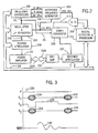
- a non-selective RF excitation pulse 220 having a flip angle of 60° is applied to produce transverse magnetization. This is followed by a phase encoding gradient pulse 224 directed along the z axis and a phase encoding gradient pulse 226 directed along the y axis.
- a readout gradient pulse 228 directed along the x axis follows and a partial echo (60%) NMR signal 230 is acquired and digitized as described above.
- rewinder gradient pulses 232 and 234 rephase the magnetization before the pulse sequence is repeated as taught in U.S. patent No. 4,665,365.
- the k-space to be sampled is divided into regions.
- the 3D k-space is divided into two regions, a central region 235 and a peripheral region indicated at 236 and 237. Each of these are in turn divided into twenty-five subregions.
- the central region 235 contains seven subregions labelled A, B 1 , C 1 , D 1 , B 2 , C 2 , and D 2 , each of which requires 64 separate acquisitions (i.e. 8 k z phase encodings at 8 different k y phase encodings).
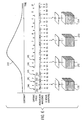
- Fig. 6 depicts a preferred acquisition method to be used during a dynamic study.
- Curve 245 depicts the contrast enhancement that is achieved by injecting the subject with a contrast agent. Twelve cardiac cycles are shown and indicated by the cardiac trigger signals 241. Thus, twelve interleaved systolic acquisition windows 242 and diastolic acquisition windows 243 are depicted.
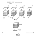
- a "difference" projection image can be produced to provide further diagnostic information. As shown in Fig. 7, this is achieved by selecting two of the 3D image frame data sets 251'-254' and calculating the difference between their corresponding pixel values. A 3D difference image is produced as indicated by data set 270 and this is then used to produce a 2D difference projection image 272 using the same projection method described above. The selection of two 3D image frame data sets 251'-254' is made by the diagnostician to enhance the image contrast in the particular vasculature of interest. Since it is difficult to predict exactly when the peak contrast agent flow will occur through the subject vasculature, the series of 3D image frames provides a time range during which this event should occur.
- matched filters and Eigen filters can be applied.
- a matched filter combines several of the images in the series to obtain an image containing improved signal to noise ratio.
- the images are weighted in the matched filter sum so as to maximize SNR.
- Eigen filters use a particular set of weighing coefficients such that venous signal is removed from the summed image. This technique requires the measurement of signal within a venous ROI to determine the required coefficients.
Landscapes
- Physics & Mathematics (AREA)
- Radiology & Medical Imaging (AREA)
- Nuclear Medicine, Radiotherapy & Molecular Imaging (AREA)
- Health & Medical Sciences (AREA)
- Engineering & Computer Science (AREA)
- Signal Processing (AREA)
- General Health & Medical Sciences (AREA)
- High Energy & Nuclear Physics (AREA)
- Condensed Matter Physics & Semiconductors (AREA)
- General Physics & Mathematics (AREA)
- Magnetic Resonance Imaging Apparatus (AREA)
- Apparatus For Radiation Diagnosis (AREA)
- Medicines Containing Antibodies Or Antigens For Use As Internal Diagnostic Agents (AREA)
- Pharmaceuticals Containing Other Organic And Inorganic Compounds (AREA)
Claims (13)
- Procédé d'acquisition de données RMN d'un sujet pour produire une série d'images matricielles par acquisition répétée d'échantillons à partir d'un espace k sélectionné au cours d'une étude dynamique du sujet, le procédé comprenant les étapes consistant à:échantillonner une région centrale de l'espace k sélectionné à une cadence temporelle donnée;échantillonner des régions périphériques de l'espace k donné à une cadence temporelle inférieure à ladite cadence temporelle donnée;former un ensemble de données pour chaque ensemble d'échantillons de région centrale d'espace k, qui comprend lesdits échantillons de région centrale d'espace k et des échantillons provenant d'échantillonnages temporellement adjacents des régions périphériques d'espace k; etreconstruire une image matricielle à partir de l'ensemble de données,caractérisé en ce que:le procédé comprend en outre le fait de produire un signal représentant le cycle cardiaque du sujet et de définir dans chaque cycle cardiaque une fenêtre d'acquisition systolique et une fenêtre d'acquisition diastolique;ladite région centrale de l'espace k sélectionné est échantillonnée durant la fenêtre d'acquisition diastolique; etlesdites régions périphériques de l'espace k sélectionné sont échantillonnées durant la fenêtre d'acquisition systolique.
- Procédé selon la revendication 1, dans lequel les données RMN sont acquises à partir d'une région tridimensionnelle du sujet.
- Procédé selon la revendication 2, dans lequel l'image matricielle est une image matricielle tridimensionnelle.
- Procédé selon la revendication 3, dans lequel une image bidimensionnelle est produite par projection de l'image matricielle tridimensionnelle.
- Procédé selon la revendication 3, dans lequel une pluralité d'images matricielles tridimensionnelles sont reconstruites à partir desdits ensembles de données formés, et une image tridimensionnelle supplémentaire est produite par soustraction de l'une desdites images matricielles tridimensionnelles à une autre desdites images matricielles tridimensionnelles.
- Procédé selon la revendication 5, dans lequel une image bidimensionnelle est produite par projection de l'image tridimensionnelle supplémentaire.
- Procédé selon la revendication 1, dans lequel les échantillons provenant des échantillonnages temporellement adjacents des régions périphériques d'espace k sont obtenus en sélectionnant les échantillons des régions périphériques d'espace k qui sont temporellement les plus proches de l'échantillonnage de la région centrale.
- Procédé selon la revendication 1, dans lequel les échantillons provenant des échantillonnages temporellement adjacents des régions périphériques d'espace k sont obtenus par interpolation entre les deux échantillons des régions périphériques d'espace k temporellement les plus proches.
- Procédé selon la revendication 8, dans lequel l'interpolation est une interpolation linéaire.
- Procédé selon la revendication 1, dans lequel ladite région centrale de l'espace k sélectionné est divisée en sous-régions, et au moins une desdites sous-régions est échantillonnée durant la fenêtre d'acquisition diastolique de chaque cycle cardiaque successif.
- Procédé selon la revendication 10, dans lequel certaines desdites sous-régions sont échantillonnées à une cadence temporelle différente de celle d'autres desdites sous-régions.
- Procédé selon la revendication 1, dans lequel ladite région périphérique de l'espace k sélectionné est divisée en sous-régions et au moins une desdites sous-régions est échantillonnée durant la fenêtre d'acquisition systolique de chaque cycle cardiaque successif.
- Procédé selon la revendication 12, dans lequel certaines desdites sous-régions sont échantillonnées à une cadence temporelle différente de celle d'autres desdites sous-régions.
Applications Claiming Priority (3)
| Application Number | Priority Date | Filing Date | Title |
|---|---|---|---|
| US787181 | 1997-01-21 | ||
| US08/787,181 US5830143A (en) | 1997-01-21 | 1997-01-21 | Gated time-resolved contrast-enhanced 3D MR angiography |
| PCT/US1997/012769 WO1998032026A1 (fr) | 1997-01-21 | 1997-07-22 | Angiographie par resonance magnetique en 3d a contraste renforce et a resolution temporelle synchronisee |
Publications (2)
| Publication Number | Publication Date |
|---|---|
| EP1010016A1 EP1010016A1 (fr) | 2000-06-21 |
| EP1010016B1 true EP1010016B1 (fr) | 2006-01-18 |
Family
ID=25140654
Family Applications (1)
| Application Number | Title | Priority Date | Filing Date |
|---|---|---|---|
| EP97936143A Expired - Lifetime EP1010016B1 (fr) | 1997-01-21 | 1997-07-22 | Angiographie par resonance magnetique en 3d a contraste renforce et a resolution temporelle synchronisee |
Country Status (8)
| Country | Link |
|---|---|
| US (1) | US5830143A (fr) |
| EP (1) | EP1010016B1 (fr) |
| JP (1) | JP4167233B2 (fr) |
| AT (1) | ATE316248T1 (fr) |
| AU (1) | AU3888397A (fr) |
| DE (1) | DE69735138T2 (fr) |
| IL (1) | IL130991A (fr) |
| WO (1) | WO1998032026A1 (fr) |
Cited By (1)
| Publication number | Priority date | Publication date | Assignee | Title |
|---|---|---|---|---|
| DE102005044336A1 (de) * | 2005-09-16 | 2007-04-05 | Siemens Ag | Verfahren zur Aufnahme von Bilddaten des Herzens |
Families Citing this family (58)
| Publication number | Priority date | Publication date | Assignee | Title |
|---|---|---|---|---|
| US5987348A (en) * | 1997-07-23 | 1999-11-16 | Philips Electronics North America Corporation | ECG triggered MR imaging method and apparatus |
| US6198959B1 (en) * | 1998-03-27 | 2001-03-06 | Cornell Research Foundation Inc. | Coronary magnetic resonance angiography using motion matched acquisition |
| US6381486B1 (en) * | 1999-01-08 | 2002-04-30 | Wisconsin Alumni Research Foundation | Magnetic resonance angiography with vessel segmentation |
| US6144873A (en) * | 1998-04-17 | 2000-11-07 | Board Of Trustees Of The Leland Stanford Junior University | Method of efficient data encoding in dynamic magnetic resonance imaging |
| US6201985B1 (en) * | 1998-08-14 | 2001-03-13 | General Electric Company | Segmented k-space method for three-dimensional MR imaging |
| US6192264B1 (en) * | 1998-12-28 | 2001-02-20 | General Electric Company | Method and system for MRI venography including arterial and venous discrimination |
| US6556856B1 (en) * | 1999-01-08 | 2003-04-29 | Wisconsin Alumni Research Foundation | Dual resolution acquisition of magnetic resonance angiography data with vessel segmentation |
| US6295465B1 (en) * | 1999-02-17 | 2001-09-25 | Siemens Medical Systems, Inc. | Myocardial perfusion studies using magnetic resonance imaging |
| US6760611B1 (en) * | 1999-04-30 | 2004-07-06 | Hitachi Medical Corporation | Magnetic resonance imaging method and device therefor |
| JP4653270B2 (ja) * | 1999-08-25 | 2011-03-16 | 東芝医用システムエンジニアリング株式会社 | 磁気共鳴イメージング装置 |
| US6801800B2 (en) * | 1999-11-29 | 2004-10-05 | Kabushiki Kaisha Toshiba | MR imaging using ECG-prep scan |
| JP4632535B2 (ja) | 2000-12-27 | 2011-02-16 | 株式会社東芝 | Mri装置 |
| JP4451528B2 (ja) * | 1999-12-24 | 2010-04-14 | Geヘルスケア・ジャパン株式会社 | 磁気共鳴撮影装置 |
| DE19962848C2 (de) * | 1999-12-24 | 2003-03-27 | Forschungszentrum Juelich Gmbh | Echo-Planar-Bildgebungsverfahren |
| DE19962845C2 (de) * | 1999-12-24 | 2003-03-27 | Forschungszentrum Juelich Gmbh | Verfahren zur Untersuchung eines Objekts mittels Erfassung des Ortsfrequenzraumes |
| US6535821B2 (en) * | 2000-02-11 | 2003-03-18 | University Of Iowa Research Foundation | System and method of bolus-chasing angiography with adaptive real-time computed tomography (CT) |
| US6505064B1 (en) * | 2000-08-22 | 2003-01-07 | Koninklijke Philips Electronics, N.V. | Diagnostic imaging systems and methods employing temporally resolved intensity tracing |
| US6377835B1 (en) * | 2000-08-30 | 2002-04-23 | Siemens Aktiengesellschaft | Method for separating arteries and veins in 3D MR angiographic images using correlation analysis |
| WO2002045584A1 (fr) * | 2000-12-04 | 2002-06-13 | Hitachi Medical Corporation | Procede de mesure dans un dispositif d'imagerie par resonance magnetique et dispositif d'imagerie par resonance magnetique |
| JP5002099B2 (ja) | 2001-08-31 | 2012-08-15 | 株式会社東芝 | 磁気共鳴イメージング装置 |
| US6728569B2 (en) | 2001-10-25 | 2004-04-27 | Evanston Northwestern Healthcare Corp. | Scoutless whole-body imaging with fast positioning |
| DE10210647A1 (de) * | 2002-03-11 | 2003-10-02 | Siemens Ag | Verfahren zur Bilddarstellung eines in einen Untersuchungsbereich eines Patienten eingebrachten medizinischen Instruments |
| US6683454B2 (en) * | 2002-03-28 | 2004-01-27 | Ge Medical Systems Global Technology Company, Llc | Shifting of artifacts by reordering of k-space |
| US7182083B2 (en) * | 2002-04-03 | 2007-02-27 | Koninklijke Philips Electronics N.V. | CT integrated respiratory monitor |
| US7797031B1 (en) * | 2002-05-17 | 2010-09-14 | General Electric Company | Method and apparatus for breath-held MR data acquisition using interleaved acquisition |
| JP4509932B2 (ja) * | 2003-03-14 | 2010-07-21 | 株式会社日立メディコ | 磁気共鳴イメージング装置 |
| US7343193B2 (en) * | 2003-06-16 | 2008-03-11 | Wisconsin Alumni Research Foundation | Background suppression method for time-resolved magnetic resonance angiography |
| CN100479748C (zh) | 2004-03-26 | 2009-04-22 | 株式会社东芝 | 核磁共振成像装置 |
| US7522744B2 (en) * | 2004-08-31 | 2009-04-21 | University Of Iowa Research Foundation | System and method for adaptive bolus chasing computed tomography (CT) angiography |
| US7023207B1 (en) * | 2005-02-16 | 2006-04-04 | General Electric Company | Method and system of MR imaging with reduced radial ripple artifacts |
| US7999543B2 (en) * | 2005-03-23 | 2011-08-16 | Koninklijke Philips Electronics N.V. | MR method for spatially-resolved determination of relaxation parameters |
| US20070055138A1 (en) * | 2005-08-22 | 2007-03-08 | Edelman Robert R | Accelerated whole body imaging with spatially non-selective radio frequency pulses |
| US7486073B2 (en) * | 2006-04-25 | 2009-02-03 | The Board Of Trustees Of The Leland Stanford Junior University | Sliding window reconstruction and phase/field map updating for dynamic chemical shift imaging |
| US7592810B2 (en) * | 2006-04-25 | 2009-09-22 | The Board Of Trustees Of The Leland Stanford Junior University | MRI methods for combining separate species and quantifying a species |
| US7592807B2 (en) * | 2006-04-25 | 2009-09-22 | The Board Of Trustees Of The Leland Stanford Junior University | Maximum likelihood estimator in the presence of non-identically distributed noise for decomposition of chemical species in MRI |
| US7741842B2 (en) * | 2006-04-25 | 2010-06-22 | The Board Of Trustees Of The Leland Stanford Junior University | Calibration maps for parallel imaging free of chemical shift artifact |
| JP5366370B2 (ja) * | 2006-09-06 | 2013-12-11 | 株式会社東芝 | 磁気共鳴イメージング装置 |
| JP5100181B2 (ja) * | 2006-09-06 | 2012-12-19 | 株式会社東芝 | 磁気共鳴イメージング装置 |
| US8214013B2 (en) * | 2007-01-26 | 2012-07-03 | Mayo Foundation For Medical Education And Research | Accelerated shells trajectory MRI acquisition |
| DE102007057553B4 (de) * | 2007-11-30 | 2012-02-16 | Siemens Ag | Verfahren zur Untersuchung eines menschlichen oder tierischen Körpers sowie medizinische Bildgebungsvorrichtung hierfür |
| US7880465B2 (en) * | 2008-06-04 | 2011-02-01 | General Electric Company | Method and apparatus for contrast inflow dynamic MR angiography |
| US8301224B2 (en) * | 2008-10-09 | 2012-10-30 | Siemens Aktiengesellschaft | System and method for automatic, non-invasive diagnosis of pulmonary hypertension and measurement of mean pulmonary arterial pressure |
| US8185187B2 (en) * | 2009-03-11 | 2012-05-22 | Siemens Medical Solutions Usa, Inc. | Magnetic resonance lmethod and apparatus with gated shimming of the basic magnetic field |
| US8890522B2 (en) | 2010-04-02 | 2014-11-18 | General Electric Company | Accelerated pseudo-random data magnetic resonance imaging system and method |
| US8738113B2 (en) | 2010-09-30 | 2014-05-27 | University Of Utah Research Foundation | Retrospectively correlated turbo spin echo imaging |
| US9159145B2 (en) | 2010-12-21 | 2015-10-13 | Koninklijke Philips N.V. | Fast dual contrast MR imaging |
| DE102011086369B4 (de) * | 2011-11-15 | 2022-08-11 | Siemens Healthcare Gmbh | Verfahren zur Erstellung von MR-Angiographiebildern und entsprechende Magnetresonanzanlage |
| JP5718476B2 (ja) | 2012-06-27 | 2015-05-13 | 古河電気工業株式会社 | リチウムイオン二次電池用電解銅箔、リチウムイオン二次電池の負極電極及びリチウムイオン二次電池 |
| US10197658B2 (en) * | 2013-08-02 | 2019-02-05 | Siemens Healthcare Gmbh | Methods, systems and apparatuses for using flexible triggered segmentation to optimize magnetic resonance imaging |
| JP6088711B2 (ja) | 2013-12-20 | 2017-03-01 | コーニンクレッカ フィリップス エヌ ヴェKoninklijke Philips N.V. | 磁気共鳴イメージングのための息止め検出 |
| DE102015203181A1 (de) * | 2015-02-23 | 2016-08-25 | Karl-Franzens-Universität Graz | Verfahren zur zeitlichen und räumlichen Interpolation von fMRI-Zeitreihen in Bezug auf ein Einzelsubjekt |
| WO2017090161A1 (fr) * | 2015-11-26 | 2017-06-01 | 近藤 和夫 | Solution de cuivrage acide, produit à cuivrage acide, et procédé de fabrication de dispositif à semi-conducteurs |
| US10799196B2 (en) * | 2017-11-20 | 2020-10-13 | General Electric Company | System and method for encouraging patient stillness during imaging |
| CN109917315B (zh) | 2019-04-30 | 2021-09-28 | 上海联影医疗科技股份有限公司 | 磁共振成像扫描方法、装置、计算机设备和存储介质 |
| US11899085B2 (en) * | 2019-10-21 | 2024-02-13 | Shanghai United Imaging Healthcare Co., Ltd. | System and method for magnetic resonance imaging |
| US11852707B2 (en) | 2019-10-21 | 2023-12-26 | Shanghai United Imaging Healthcare Co., Ltd. | Systems and methods for simultaneous multi-slice magnetic resonance imaging |
| US12553970B2 (en) | 2019-10-21 | 2026-02-17 | Shanghai United Imaging Healthcare Co., Ltd. | System and method for magnetic resonance imaging |
| DE102022208726A1 (de) | 2022-08-23 | 2024-02-29 | Siemens Healthcare Gmbh | Computerimplementiertes Verfahren zum Betrieb einer medizinischen Bildgebungseinrichtung, Bildgebungseinrichtung, Computerprogramm und elektronisch lesbarer Datenträger |
Family Cites Families (15)
| Publication number | Priority date | Publication date | Assignee | Title |
|---|---|---|---|---|
| US4204226A (en) * | 1978-05-16 | 1980-05-20 | Wisconsin Alumni Research Foundation | Real-time digital X-ray time interval difference imaging |
| US4204225A (en) * | 1978-05-16 | 1980-05-20 | Wisconsin Alumni Research Foundation | Real-time digital X-ray subtraction imaging |
| US4830012A (en) * | 1987-08-14 | 1989-05-16 | Duke University | High speed NMR imaging method and apparatus |
| JPH02140145A (ja) * | 1988-11-21 | 1990-05-29 | Toshiba Corp | 磁気共鳴イメージング装置 |
| US5125407A (en) * | 1990-02-23 | 1992-06-30 | Baylor Research Foundation | Method for magnetic resonance imaging of an object |
| US5348011A (en) * | 1991-11-14 | 1994-09-20 | Picker International, Inc. | Shared excitation phase encode grouping for improved throughput cardiac gated MRI cine imaging |
| US5522390A (en) * | 1991-11-21 | 1996-06-04 | U.S. Philips Corporation | Magnetic resonance imaging method |
| US5320099A (en) * | 1992-08-07 | 1994-06-14 | Trustees Of The University Of Penna. | MR angiography using steady-state transport-induced adiabatic fast passage |
| DE4317028C3 (de) * | 1993-05-21 | 2000-06-29 | Martin Busch | Verfahren zur Akquisition und Auswertung von Daten in einem Kernspin-Tomographen |
| US5417213A (en) * | 1993-06-07 | 1995-05-23 | Prince; Martin R. | Magnetic resonance arteriography with dynamic intravenous contrast agents |
| US5435303A (en) * | 1993-08-04 | 1995-07-25 | General Electric Company | MRA image produced by temporal flow data sharing |
| US5377680A (en) * | 1993-08-04 | 1995-01-03 | General Electric Company | MRI cardiac image produced by temporal data sharing |
| DE4327325C1 (de) * | 1993-08-13 | 1995-01-12 | Siemens Ag | Verfahren zur zeitaufgelösten MR-Bildgebung |
| US5429134A (en) * | 1994-06-27 | 1995-07-04 | General Electric Company | Multi-phase fat suppressed MRI cardiac imaging |
| WO1996035130A1 (fr) * | 1995-05-02 | 1996-11-07 | Philips Electronics N.V. | Procede et dispositif d'imagerie d'objets par resonance magnetique |
-
1997
- 1997-01-21 US US08/787,181 patent/US5830143A/en not_active Expired - Lifetime
- 1997-07-22 IL IL13099197A patent/IL130991A/en not_active IP Right Cessation
- 1997-07-22 WO PCT/US1997/012769 patent/WO1998032026A1/fr not_active Ceased
- 1997-07-22 DE DE69735138T patent/DE69735138T2/de not_active Expired - Lifetime
- 1997-07-22 EP EP97936143A patent/EP1010016B1/fr not_active Expired - Lifetime
- 1997-07-22 AT AT97936143T patent/ATE316248T1/de not_active IP Right Cessation
- 1997-07-22 AU AU38883/97A patent/AU3888397A/en not_active Abandoned
-
2005
- 2005-02-04 JP JP2005029307A patent/JP4167233B2/ja not_active Expired - Lifetime
Cited By (1)
| Publication number | Priority date | Publication date | Assignee | Title |
|---|---|---|---|---|
| DE102005044336A1 (de) * | 2005-09-16 | 2007-04-05 | Siemens Ag | Verfahren zur Aufnahme von Bilddaten des Herzens |
Also Published As
| Publication number | Publication date |
|---|---|
| ATE316248T1 (de) | 2006-02-15 |
| EP1010016A1 (fr) | 2000-06-21 |
| JP4167233B2 (ja) | 2008-10-15 |
| IL130991A (en) | 2002-12-01 |
| DE69735138D1 (de) | 2006-04-06 |
| WO1998032026A1 (fr) | 1998-07-23 |
| IL130991A0 (en) | 2001-01-28 |
| JP2005185853A (ja) | 2005-07-14 |
| DE69735138T2 (de) | 2006-08-17 |
| AU3888397A (en) | 1998-08-07 |
| US5830143A (en) | 1998-11-03 |
Similar Documents
| Publication | Publication Date | Title |
|---|---|---|
| EP1010016B1 (fr) | Angiographie par resonance magnetique en 3d a contraste renforce et a resolution temporelle synchronisee | |
| JP3742114B2 (ja) | ゲート化され時間解像されたコントラスト増幅3次元mr血管造影法 | |
| US5881728A (en) | Digital subtraction magnetic resonance angiography with image artifact suppression | |
| US5713358A (en) | Method for producing a time-resolved series of 3D magnetic resonance angiograms during the first passage of contrast agent | |
| US5873825A (en) | Three dimensional digital subtraction magnetic resonance angiography with limited k-space mask | |
| JP3964202B2 (ja) | Mrイメージングにおいて造影強調後の定常自由歳差運動を使用する方法及び装置 | |
| EP1145029B1 (fr) | Reconstruction guidee par contraste en angiographie par resonance magnetique a contrastes ameliores | |
| US6556856B1 (en) | Dual resolution acquisition of magnetic resonance angiography data with vessel segmentation | |
| EP1269212B1 (fr) | Angiographie par resonance magnetique avec segmentation automatique de vaisseau | |
| US6381486B1 (en) | Magnetic resonance angiography with vessel segmentation | |
| JP4477176B2 (ja) | Mrアンジオグラフィのための選択的な動脈画像及び静脈画像の取得の方法及びシステム | |
| US20090143666A1 (en) | System And Method For Non-Contrast Agent MR Angiography | |
| US5827187A (en) | Dynamic MR digital subtraction angiography with complex subtraction | |
| US8154287B2 (en) | System and method for ghost magnetic resonance imaging | |
| WO2000040989A1 (fr) | Imagerie de flux a resonance magnetique a contraste de phase au moyen de donnees de projection entrelacees | |
| US6044290A (en) | Time-resolved digital subtraction magnetic resonance angiography using echo-planar imaging | |
| WO2001075469A1 (fr) | Angiographie par resonance magnetique avec segmentation automatique de vaisseau | |
| Pattynama et al. | Magnetic resonance coronary angiography |
Legal Events
| Date | Code | Title | Description |
|---|---|---|---|
| PUAI | Public reference made under article 153(3) epc to a published international application that has entered the european phase |
Free format text: ORIGINAL CODE: 0009012 |
|
| 17P | Request for examination filed |
Effective date: 19990812 |
|
| AK | Designated contracting states |
Kind code of ref document: A1 Designated state(s): AT BE CH DE DK ES FI FR GB GR IE IT LI LU MC NL PT SE |
|
| 17Q | First examination report despatched |
Effective date: 20040813 |
|
| GRAP | Despatch of communication of intention to grant a patent |
Free format text: ORIGINAL CODE: EPIDOSNIGR1 |
|
| GRAS | Grant fee paid |
Free format text: ORIGINAL CODE: EPIDOSNIGR3 |
|
| GRAA | (expected) grant |
Free format text: ORIGINAL CODE: 0009210 |
|
| AK | Designated contracting states |
Kind code of ref document: B1 Designated state(s): AT BE CH DE DK ES FI FR GB GR IE IT LI LU MC NL PT SE |
|
| PG25 | Lapsed in a contracting state [announced via postgrant information from national office to epo] |
Ref country code: LI Free format text: LAPSE BECAUSE OF FAILURE TO SUBMIT A TRANSLATION OF THE DESCRIPTION OR TO PAY THE FEE WITHIN THE PRESCRIBED TIME-LIMIT Effective date: 20060118 Ref country code: IT Free format text: LAPSE BECAUSE OF FAILURE TO SUBMIT A TRANSLATION OF THE DESCRIPTION OR TO PAY THE FEE WITHIN THE PRESCRIBED TIME-LIMIT;WARNING: LAPSES OF ITALIAN PATENTS WITH EFFECTIVE DATE BEFORE 2007 MAY HAVE OCCURRED AT ANY TIME BEFORE 2007. THE CORRECT EFFECTIVE DATE MAY BE DIFFERENT FROM THE ONE RECORDED. Effective date: 20060118 Ref country code: FI Free format text: LAPSE BECAUSE OF FAILURE TO SUBMIT A TRANSLATION OF THE DESCRIPTION OR TO PAY THE FEE WITHIN THE PRESCRIBED TIME-LIMIT Effective date: 20060118 Ref country code: CH Free format text: LAPSE BECAUSE OF FAILURE TO SUBMIT A TRANSLATION OF THE DESCRIPTION OR TO PAY THE FEE WITHIN THE PRESCRIBED TIME-LIMIT Effective date: 20060118 Ref country code: BE Free format text: LAPSE BECAUSE OF FAILURE TO SUBMIT A TRANSLATION OF THE DESCRIPTION OR TO PAY THE FEE WITHIN THE PRESCRIBED TIME-LIMIT Effective date: 20060118 Ref country code: AT Free format text: LAPSE BECAUSE OF FAILURE TO SUBMIT A TRANSLATION OF THE DESCRIPTION OR TO PAY THE FEE WITHIN THE PRESCRIBED TIME-LIMIT Effective date: 20060118 |
|
| REG | Reference to a national code |
Ref country code: GB Ref legal event code: FG4D |
|
| REG | Reference to a national code |
Ref country code: CH Ref legal event code: EP |
|
| REG | Reference to a national code |
Ref country code: IE Ref legal event code: FG4D |
|
| REF | Corresponds to: |
Ref document number: 69735138 Country of ref document: DE Date of ref document: 20060406 Kind code of ref document: P |
|
| PG25 | Lapsed in a contracting state [announced via postgrant information from national office to epo] |
Ref country code: SE Free format text: LAPSE BECAUSE OF FAILURE TO SUBMIT A TRANSLATION OF THE DESCRIPTION OR TO PAY THE FEE WITHIN THE PRESCRIBED TIME-LIMIT Effective date: 20060418 Ref country code: DK Free format text: LAPSE BECAUSE OF FAILURE TO SUBMIT A TRANSLATION OF THE DESCRIPTION OR TO PAY THE FEE WITHIN THE PRESCRIBED TIME-LIMIT Effective date: 20060418 |
|
| PG25 | Lapsed in a contracting state [announced via postgrant information from national office to epo] |
Ref country code: ES Free format text: LAPSE BECAUSE OF FAILURE TO SUBMIT A TRANSLATION OF THE DESCRIPTION OR TO PAY THE FEE WITHIN THE PRESCRIBED TIME-LIMIT Effective date: 20060429 |
|
| PG25 | Lapsed in a contracting state [announced via postgrant information from national office to epo] |
Ref country code: PT Free format text: LAPSE BECAUSE OF FAILURE TO SUBMIT A TRANSLATION OF THE DESCRIPTION OR TO PAY THE FEE WITHIN THE PRESCRIBED TIME-LIMIT Effective date: 20060619 |
|
| PG25 | Lapsed in a contracting state [announced via postgrant information from national office to epo] |
Ref country code: GB Free format text: LAPSE BECAUSE OF NON-PAYMENT OF DUE FEES Effective date: 20060722 |
|
| PG25 | Lapsed in a contracting state [announced via postgrant information from national office to epo] |
Ref country code: IE Free format text: LAPSE BECAUSE OF NON-PAYMENT OF DUE FEES Effective date: 20060724 |
|
| PG25 | Lapsed in a contracting state [announced via postgrant information from national office to epo] |
Ref country code: MC Free format text: LAPSE BECAUSE OF NON-PAYMENT OF DUE FEES Effective date: 20060731 |
|
| REG | Reference to a national code |
Ref country code: CH Ref legal event code: PL |
|
| PLBE | No opposition filed within time limit |
Free format text: ORIGINAL CODE: 0009261 |
|
| STAA | Information on the status of an ep patent application or granted ep patent |
Free format text: STATUS: NO OPPOSITION FILED WITHIN TIME LIMIT |
|
| 26N | No opposition filed |
Effective date: 20061019 |
|
| EN | Fr: translation not filed | ||
| GBPC | Gb: european patent ceased through non-payment of renewal fee |
Effective date: 20060722 |
|
| PG25 | Lapsed in a contracting state [announced via postgrant information from national office to epo] |
Ref country code: GR Free format text: LAPSE BECAUSE OF FAILURE TO SUBMIT A TRANSLATION OF THE DESCRIPTION OR TO PAY THE FEE WITHIN THE PRESCRIBED TIME-LIMIT Effective date: 20060419 Ref country code: FR Free format text: LAPSE BECAUSE OF FAILURE TO SUBMIT A TRANSLATION OF THE DESCRIPTION OR TO PAY THE FEE WITHIN THE PRESCRIBED TIME-LIMIT Effective date: 20070309 |
|
| PG25 | Lapsed in a contracting state [announced via postgrant information from national office to epo] |
Ref country code: LU Free format text: LAPSE BECAUSE OF NON-PAYMENT OF DUE FEES Effective date: 20060722 |
|
| PG25 | Lapsed in a contracting state [announced via postgrant information from national office to epo] |
Ref country code: FR Free format text: LAPSE BECAUSE OF FAILURE TO SUBMIT A TRANSLATION OF THE DESCRIPTION OR TO PAY THE FEE WITHIN THE PRESCRIBED TIME-LIMIT Effective date: 20060118 |
|
| PGFP | Annual fee paid to national office [announced via postgrant information from national office to epo] |
Ref country code: NL Payment date: 20160711 Year of fee payment: 20 |
|
| PGFP | Annual fee paid to national office [announced via postgrant information from national office to epo] |
Ref country code: DE Payment date: 20160720 Year of fee payment: 20 |
|
| REG | Reference to a national code |
Ref country code: DE Ref legal event code: R071 Ref document number: 69735138 Country of ref document: DE |
|
| REG | Reference to a national code |
Ref country code: NL Ref legal event code: MK Effective date: 20170721 |