EP1010764A2 - Hybridisierungsnachweisverfahren unter Verwendung von Biochips - Google Patents
Hybridisierungsnachweisverfahren unter Verwendung von Biochips Download PDFInfo
- Publication number
- EP1010764A2 EP1010764A2 EP99124644A EP99124644A EP1010764A2 EP 1010764 A2 EP1010764 A2 EP 1010764A2 EP 99124644 A EP99124644 A EP 99124644A EP 99124644 A EP99124644 A EP 99124644A EP 1010764 A2 EP1010764 A2 EP 1010764A2
- Authority
- EP
- European Patent Office
- Prior art keywords
- probe
- amount
- sample
- hybridization
- dna
- Prior art date
- Legal status (The legal status is an assumption and is not a legal conclusion. Google has not performed a legal analysis and makes no representation as to the accuracy of the status listed.)
- Granted
Links
- 238000001514 detection method Methods 0.000 title claims abstract description 36
- 238000000018 DNA microarray Methods 0.000 title claims description 14
- 239000000523 sample Substances 0.000 claims abstract description 238
- 238000009396 hybridization Methods 0.000 claims abstract description 67
- 239000000463 material Substances 0.000 claims abstract description 33
- 239000000758 substrate Substances 0.000 claims abstract description 23
- 238000000034 method Methods 0.000 claims abstract description 17
- 238000002372 labelling Methods 0.000 claims abstract description 16
- 229920001222 biopolymer Polymers 0.000 abstract description 32
- 108020004414 DNA Proteins 0.000 description 113
- MHMNJMPURVTYEJ-UHFFFAOYSA-N fluorescein-5-isothiocyanate Chemical compound O1C(=O)C2=CC(N=C=S)=CC=C2C21C1=CC=C(O)C=C1OC1=CC(O)=CC=C21 MHMNJMPURVTYEJ-UHFFFAOYSA-N 0.000 description 17
- 238000011156 evaluation Methods 0.000 description 11
- 230000005284 excitation Effects 0.000 description 11
- 230000003287 optical effect Effects 0.000 description 8
- 239000011521 glass Substances 0.000 description 7
- KCXVZYZYPLLWCC-UHFFFAOYSA-N EDTA Chemical compound OC(=O)CN(CC(O)=O)CCN(CC(O)=O)CC(O)=O KCXVZYZYPLLWCC-UHFFFAOYSA-N 0.000 description 4
- FAPWRFPIFSIZLT-UHFFFAOYSA-M Sodium chloride Chemical compound [Na+].[Cl-] FAPWRFPIFSIZLT-UHFFFAOYSA-M 0.000 description 4
- DBMJMQXJHONAFJ-UHFFFAOYSA-M Sodium laurylsulphate Chemical compound [Na+].CCCCCCCCCCCCOS([O-])(=O)=O DBMJMQXJHONAFJ-UHFFFAOYSA-M 0.000 description 4
- 230000005540 biological transmission Effects 0.000 description 4
- 239000000243 solution Substances 0.000 description 4
- 230000000295 complement effect Effects 0.000 description 3
- 238000012545 processing Methods 0.000 description 3
- 102000004169 proteins and genes Human genes 0.000 description 3
- 108091032973 (ribonucleotides)n+m Proteins 0.000 description 2
- 238000004458 analytical method Methods 0.000 description 2
- 239000012153 distilled water Substances 0.000 description 2
- 238000002474 experimental method Methods 0.000 description 2
- 230000001678 irradiating effect Effects 0.000 description 2
- WSFSSNUMVMOOMR-NJFSPNSNSA-N methanone Chemical compound O=[14CH2] WSFSSNUMVMOOMR-NJFSPNSNSA-N 0.000 description 2
- 239000011259 mixed solution Substances 0.000 description 2
- 238000012986 modification Methods 0.000 description 2
- 230000004048 modification Effects 0.000 description 2
- 108090000623 proteins and genes Proteins 0.000 description 2
- 239000011780 sodium chloride Substances 0.000 description 2
- 239000001509 sodium citrate Substances 0.000 description 2
- HRXKRNGNAMMEHJ-UHFFFAOYSA-K trisodium citrate Chemical compound [Na+].[Na+].[Na+].[O-]C(=O)CC(O)(CC([O-])=O)C([O-])=O HRXKRNGNAMMEHJ-UHFFFAOYSA-K 0.000 description 2
- 229940038773 trisodium citrate Drugs 0.000 description 2
- XLYOFNOQVPJJNP-UHFFFAOYSA-N water Chemical compound O XLYOFNOQVPJJNP-UHFFFAOYSA-N 0.000 description 2
- 102000040650 (ribonucleotides)n+m Human genes 0.000 description 1
- 108091005461 Nucleic proteins Proteins 0.000 description 1
- 230000000694 effects Effects 0.000 description 1
- 238000005194 fractionation Methods 0.000 description 1
- 108020004707 nucleic acids Proteins 0.000 description 1
- 102000039446 nucleic acids Human genes 0.000 description 1
- 150000007523 nucleic acids Chemical class 0.000 description 1
- 229910052724 xenon Inorganic materials 0.000 description 1
- FHNFHKCVQCLJFQ-UHFFFAOYSA-N xenon atom Chemical compound [Xe] FHNFHKCVQCLJFQ-UHFFFAOYSA-N 0.000 description 1
Images




Classifications
-
- C—CHEMISTRY; METALLURGY
- C12—BIOCHEMISTRY; BEER; SPIRITS; WINE; VINEGAR; MICROBIOLOGY; ENZYMOLOGY; MUTATION OR GENETIC ENGINEERING
- C12Q—MEASURING OR TESTING PROCESSES INVOLVING ENZYMES, NUCLEIC ACIDS OR MICROORGANISMS; COMPOSITIONS OR TEST PAPERS THEREFOR; PROCESSES OF PREPARING SUCH COMPOSITIONS; CONDITION-RESPONSIVE CONTROL IN MICROBIOLOGICAL OR ENZYMOLOGICAL PROCESSES
- C12Q1/00—Measuring or testing processes involving enzymes, nucleic acids or microorganisms; Compositions therefor; Processes of preparing such compositions
- C12Q1/68—Measuring or testing processes involving enzymes, nucleic acids or microorganisms; Compositions therefor; Processes of preparing such compositions involving nucleic acids
- C12Q1/6813—Hybridisation assays
- C12Q1/6834—Enzymatic or biochemical coupling of nucleic acids to a solid phase
- C12Q1/6837—Enzymatic or biochemical coupling of nucleic acids to a solid phase using probe arrays or probe chips
Definitions
- the present invention relates to a hybridization detection method for the analysis of the presence or absence of a sequence of interest in a biopolymer sample by utilizing the hybridization between the sample biopolymer and a probe biopolymer, and also relates to a biochip applicable for the method.
- hybridization methods have been often utilized which uses a nucleic acid or protein having a known sequence as a probe.
- a sample DNA labeled with a fluorescent material is hybridized to a probe DNA immobilized onto a substrate.
- the sample DNA is bound to the probe DNA, the sample DNA is in turn immobilized on the substrate together with the probe DNA.
- the fluorescent material attached to the sample DNA is fluorescently excited by irradiation with excitation light from a light source to emit fluorescence, and the fluorescence is then detected. In this manner, the hybridization between the sample DNA and the probe DNA can be detected.
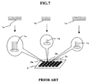
- probe DNA 1a is immobilized on a substrate (e.g., a glass plate) 4 as a spot 3a.
- a substrate e.g., a glass plate
- Another probe DNAs 1b, 1c, ... are also immobilized on the substrate 4 as spots 3b, 3c, ..., respectively. In this case, however, it is impossible to immobilize all of the probe DNAs in equal amounts on the respective spots.
- each of sample DNAs 5a, 5b, 5c, ... is labeled with a fluorescent material 6.
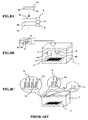
- the substrate 4 spotted with the probe DNAs 5a, 5b, 5c, ... is placed in a hybridization solution 7 and then the fluorescently labeled sample DNAs 5a, 5b, 5c, ... are added thereto to cause the hybridization between the probe DNAs and the sample DNAs.
- the hybridization solution 7 is a mixed solution comprising, for example, formaldehyde, SSC (sodium chloride/trisodium citrate), SDS (sodium dodecyl sulfate), EDTA (ethylenediaminetetraacetic acid), distilled water, and the like, in which the mixing ratio between the components may vary depending on the nature of the DNAs employed.
- SSC sodium chloride/trisodium citrate
- SDS sodium dodecyl sulfate
- EDTA ethylenediaminetetraacetic acid
- the sample DNA is hybridized to the probe DNA to form a double-stranded structure (see the illustrations for the probe DNAs 1a and 1b). If not, the sample DNA remains unbound (see the illustration for the probe DNA 1c).
- the detection of the hybridization can be performed by irradiating the substrate 4 with excitation light from a lamp 9 (i.e., an excitation light source) to excite the fluorescent material 6, cutting off the light having wavelengths out of the emission wavelength range of the fluorescent material 6 with an optical filter 10, and then detecting the light emitted from each spot with a two-dimensional photosensor 8 (e.g., a CCD camera).
- a lamp 9 i.e., an excitation light source
- the fluorescent material 6 is present, and therefore fluorescent emission can be detected by exciting the fluorescent material 6 by irradiation with excitation light from the lamp 9.
- the spots where the hybridization does not take place e.g., spot 3c
- no fluorescent material 6 is present, and therefore no fluorescent emission is observed by irradiation with excitation light from the lamp 9.
- a light or dark spot is observed depending on the presence or absence of the hybridization event.
- the image data detected by the two-dimensional photosensor 8 is transferred to a computer 13 via a controller 12 and indicated on a display.
- the object of the present invention is to provide a detection method which can quantitatively determine the degree of the hybridization between a probe DNA and a sample DNA.
- a probe biopolymer and a sample biopolymer are labeled with different fluorescent materials, so that the probe biopolymer and the sample biopolymer present on spots deposited on a substrate can be detected separately for each spot utilizing the difference of the emission wavelength of the fluorescent materials.
- the emission wavelength of the fluorescent material labeling the probe biopolymer and the emission wavelength of the fluorescent material labeling the sample biopolymer are detected separately, so that it becomes possible to separately detect and, therefore, quantitatively determine the amount of the probe biopolymer and the amount of the sample biopolymer hybridized to the probe biopolymer for each spot.
- a fluorescent material that has labeled a probe biopolymer is caused to emit fluorescence to determine the amount of the probe biopolymer immobilized on a spot deposited on a substrate (e.g., a glass plate).
- a substrate e.g., a glass plate
- Another type of fluorescent material that has labeled a sample biopolymer is also caused to emit fluorescence to determine the amount of the sample biopolymer hybridized to the probe biopolymer.
- the difference between the amount of the probe biopolymer and the amount of the sample biopolymer is normalized with the amount of the probe biopolymer. Based on the normalized value, the amount of the sample biopolymer relative to the amount of the probe biopolymer spotted on the substrate can be determined.
- biopolymer refers to any polymeric material constituting a living body, such as DNA, RNA and a protein.
- one aspect of the present invention is a hybridization detection method for detecting the hybridization between a probe and a sample, which comprising detecting both the amount of the probe and the amount of the sample bound to the probe.
- probe refers to any biopolymer to be immobilized onto a substrate, such as DNA
- sample refers to any biopolymer to be hybridized to the probe, such as DNA.
- Another aspect of the present invention is a hybridization detection method for detecting the hybridization between a probe and a sample, which comprising detecting a value produced by normalizing the difference between the amount of the probe and the amount of the sample bound to the probe with the amount of the probe.
- the amount of the probe may be detected prior to the hybridization, while the amount of the sample bound to the probe may be detected after the completion of the hybridization. Alternatively, both the amounts of the probe and the sample bound to the probe may be detected after the completion of the hybridization.
- the detection of the amounts of the probe and the sample bound to the probe may be performed by labeling the probe and the sample with different labeling materials and then detecting the labeling materials separately.
- a value produced by normalizing the difference between the amount of the probe and the amount of the sample bound to the probe with the amount of the probe may be indicated on a display.
- the probe is immobilized on a support.
- the support comprising the probe is a biochip.
- Still another aspect of the present invention is a biochip comprising a fluorescently labeled probe spotted on a substrate.
- the amount of the probe immobilized onto a substrate may be different for each probe and each substrate.
- a biochip 1 has 10 ng of a probe immobilized thereon, while a biochip 2 has 8 ng of the same type of probe immobilized thereon; the fluorescent intensities of the probe before the hybridization are 100 for the biochip 1 and 80 for the biochip 2 in terms of a 256-level gradation; and when the samples A and B are hybridized to the probe on the biochips 1 and 2, respectively, the fluorescent intensities of the samples are 70 for the biochip 1 (sample A) and 60 for the biochip 1 (sample B). From these result, it would be assessed that the DNA amount of the sample A hybridized to the probe is larger than that of the sample B hybridized to the probe.
- the ratio of the DNA amount of each sample hybridized to the probe can be calculated by determining the difference between the DNA amount of the probe initially immobilized onto each biochip and the DNA amount of the sample bound to the probe and then normalizing the obtained difference with the DNA amount of the probe, as follows:
- the method of the present invention enables a more precise analysis of the hybridization compared with the prior art methods in which the hybridization is analyzed only based on the fluorescent intensity of a sample after the hybridization.
- probe biopolymers and sample biopolymers are DNAs.
- other biopolymers such as RNAs and proteins are also applicable in the present invention.
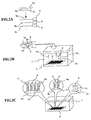
- FIGs. 1 to 3 illustrate the principle of an embodiment of the hybridization detection method in accordance with the present invention.
- each of probe DNAs 1a, 1b, 1c, ... are labeled with the same fluorescent material 2.
- the fluorescent material 2 for example, fluorescein isothiocyanate (FITC) may be used.
- the probe DNA 1a is immobilized onto a glass plate (substrate) 4 as a spot 3a.
- Other probe DNAs 1b, 1c, ... are also immobilized onto the glass plate 4 as spot 3b, 3c, ..., respectively.
- each of sample DNAs 5a, 5b, 5c, ... is labeled with another type of fluorescent material 6.
- the fluorescent material 6 for example, Cy5 may be used.
- the hybridization solution 7 is a mixed solution comprising, for example, formaldehyde, SSC (sodium chloride/trisodium citrate), SDS (sodium dodecyl sulfate), EDTA (ethylenediaminetetraacetic acid) and distilled water, in which the proportion of the components may vary depending on the nature of the probe DNAs and sample DNAs employed.
- SSC sodium chloride/trisodium citrate
- SDS sodium dodecyl sulfate
- EDTA ethylenediaminetetraacetic acid
- the sample DNA is hybridized to the probe DNA to form a double-stranded structure, as illustrated in FIG. 2C (see the illustrations for the sample DNAs 5a and 5b and the probe DNAs 1a and 1b).
- the sample DNA remains unbound, as illustrated in FIG. 2C (see the illustration for the probe DNA 1c). That is, on the spots in which the hybridization takes place (e.g., spots 3a and 3b), both the fluorescent material 2 labeling the probe DNAs and the fluorescent material 6 labeling the sample DNAs individually bound to the probe DNAs are present. In contrast, on the spots in which no hybridization takes place (e.g., 3c), only the fluorescent material 2 labeling the probe DNAs is present.
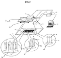
- the detection of the hybridization may be performed in the following manner, as illustrated in FIG. 3.
- the fluorescent material 6 labeling the sample DNAs, and the fluorescent material 2 labeling the probe DNAs are excited by irradiation with excitation light from a lamp 9 to emit fluorescence.
- the lamp 9 used as an excitation light source may be, for example, a xenon lamp having the emission wavelength range from about 300 to about 700 nm. The reason why such lamp is used is that it can cause the fluorescent emission of both FITC (excitation wavelength: 490 nm, emission wavelength: 520 nm) and Cy5 (excitation wavelength: 650 nm, emission wavelength: 667 nm).
- reading of emission from FITC and Cy5 is taken by using a two-dimensional photosensor 8 through an optical filter 10 having transmission wavelength of 520 nm for FITC and through another optical filter 11 having transmission wavelength of 667 nm for Cy5.
- the date from the two-dimensional photosensor is transferred to a computer 13 via a controller 12.
- the two-dimensional photosensor 8 may be, for example, a CCD camera.
- the optical filters 10 and 11 are movable in the direction of the arrow by driving a stage 14, and changeable to each other.
- the amount of each probe DNA is calculated for each spot based on the emission reading from FITC, and then the amount of the sample DNA hybridized to the probe DNA is calculated for each spot based on the emission reading from Cy5.
- Ci (Ai - Bi)/Ai
- a smaller Ci value means that the amount of a sample DNA hybridized to a probe DNA is larger, which also means the sample DNA has higher complementarity to the probe DNA.
- FIG. 4 is a flow chart of the processing steps for providing an evaluation value Ci.
- step 11 the position number of a spot to be calculated for the evaluation value is preset.
- step 12 both the emission quantity Ai of FITC and the emission quantity Bi of Cy5 are determined for a spot i.
- the evaluation value Ci thus obtained represents the amount of the probe DNA not hybridized with any sample DNA. Based on the evaluation value, the degree of the complementarity between the sample DNA and the probe DNA can be assessed.
- step 14 the obtained evaluation value Ci is indicated on a display of the computer 13 as a tone image.
- step 15 it is checked whether processing of all of the spots to be examined is completed. If not, the position number of a spot to be processed in the next step is determined in step 16, and the procedures from step 12 to step 15 are repeated. When the calculation for all of the spots to be examined is completed, the process exits.
- FIGs. 5 and 6 illustrate the principle of another embodiment of the detection method in accordance with the present invention.
- the detection method of this embodiment includes the steps of reading an emission quantity of a fluorescent material (e.g., FITC) labeling a probe DNA, which corresponds to the amount of a probe DNA, prior to the hybridization; reading an emission quantity of another fluorescent material (e.g., Cy5) labeling a sample DNA, which corresponds to the amount of a sample DNA, after the completion of the hybridization; and then determining an evaluation value as described above.
- a fluorescent material e.g., FITC
- another fluorescent material e.g., Cy5
- probe DNAs each labeled with a fluorescent material FITC are immobilized onto a glass plate (a substrate) 4 as spots 3a, 3b, 3c, ....
- the immobilization of the probe DNAs may be performed in the same manner as illustrated in FIG. 1.
- FITC labeling each probe DNA is exited by irradiation with excitation light to emit fluorescence, and the emission quantity of FITC is read with a two-dimensional photosensor 8, thereby determining the amount of each of the probe DNAs present for each spot.
- an optical filter 10 having transmission wavelength of 520 nm is located in the optical path of the photosensor 8.
- the reading of the emission quantity may be performed in the same manner as illustrated for FITC in FIG. 3 above.
- the emission quantity Ai for each spot is stored in a storage medium 14 (e.g., a floppy disk).
- the hybridization between sample DNAs 5a, 5b, ... each labeled with a fluorescent material Cy5 6 and the probe DNAs may be performed in the same manner as illustrated in FIG. 2B above.
- the detection of the hybridization may be performed, as illustrated in FIG. 6B, by irradiating the glass plate 4 with excitation light from the lamp 9 to cause to emit fluorescence from Cy5, and reading the emission quantity Bi of Cy5 with the two-dimensional photosensor 8.
- another optical filter 11 having transmission wavelength of 667 nm is located in the optical path of the photosensor 8.
- the reading of the emission quantity may be performed in the same manner as illustrated for Cy5 in FIG. 3 above.
- the emission quantity Ai of FITC stored in the storage medium 14 is then inserted into the computer 12, and the difference between the emission quantity Ai of FITC and the emission quantity Bi of Cy5 is determined. Each of the determined difference values is divided by the emission quantity Ai of FITC. The determination of the difference may be performed in the same manner as illustrated in FIG. 3 above. The method of this embodiment also enables to quantitatively determine the amounts of a sample DNA hybridized to a probe DNA.
- the method of this embodiment enables to perform a hybridization experiment with a good efficiency due to its capacity to address such problems prior to the experiment.
- the present invention it becomes possible to determine the amount of a probe spotted on a substrate and the amount of a sample hybridized to the probe; to calculate the precise amount of the sample hybridized to the probe (i.e., the degree of the complementarity) by determining a value produced by normalizing the difference between the amount of the probe and the amount of the sample with the amount of the probe; and therefore to determine the amount of the sample bound to the probe with a high precision.
Landscapes
- Chemical & Material Sciences (AREA)
- Organic Chemistry (AREA)
- Life Sciences & Earth Sciences (AREA)
- Zoology (AREA)
- Wood Science & Technology (AREA)
- Proteomics, Peptides & Aminoacids (AREA)
- Health & Medical Sciences (AREA)
- Engineering & Computer Science (AREA)
- Microbiology (AREA)
- Biochemistry (AREA)
- Physics & Mathematics (AREA)
- Molecular Biology (AREA)
- Biotechnology (AREA)
- Biophysics (AREA)
- Analytical Chemistry (AREA)
- Immunology (AREA)
- Bioinformatics & Cheminformatics (AREA)
- General Engineering & Computer Science (AREA)
- General Health & Medical Sciences (AREA)
- Genetics & Genomics (AREA)
- Measuring Or Testing Involving Enzymes Or Micro-Organisms (AREA)
- Investigating Or Analysing Biological Materials (AREA)
- Apparatus Associated With Microorganisms And Enzymes (AREA)
Applications Claiming Priority (4)
| Application Number | Priority Date | Filing Date | Title |
|---|---|---|---|
| JP35595698 | 1998-12-15 | ||
| JP35595698 | 1998-12-15 | ||
| JP32835299A JP3442327B2 (ja) | 1998-12-15 | 1999-11-18 | ハイブリダイゼーション検出方法及びバイオチップ |
| JP32835299 | 1999-11-18 |
Publications (3)
| Publication Number | Publication Date |
|---|---|
| EP1010764A2 true EP1010764A2 (de) | 2000-06-21 |
| EP1010764A3 EP1010764A3 (de) | 2003-01-22 |
| EP1010764B1 EP1010764B1 (de) | 2006-04-12 |
Family
ID=26572838
Family Applications (1)
| Application Number | Title | Priority Date | Filing Date |
|---|---|---|---|
| EP99124644A Expired - Lifetime EP1010764B1 (de) | 1998-12-15 | 1999-12-10 | Hybridisierungsnachweisverfahren unter Verwendung von Biochips |
Country Status (4)
| Country | Link |
|---|---|
| US (2) | US6775621B1 (de) |
| EP (1) | EP1010764B1 (de) |
| JP (1) | JP3442327B2 (de) |
| DE (1) | DE69930826T8 (de) |
Cited By (2)
| Publication number | Priority date | Publication date | Assignee | Title |
|---|---|---|---|---|
| EP1275954A3 (de) * | 2001-07-11 | 2004-01-28 | Hitachi Software Engineering Co., Ltd. | Vorrichtung zum Auslesen von Fluoreszenz |
| US7273704B2 (en) | 2002-08-12 | 2007-09-25 | Hitachi High-Technologies Corporation | Method of detecting nucleic acid by using DNA microarrays and nucleic acid detection apparatus |
Families Citing this family (4)
| Publication number | Priority date | Publication date | Assignee | Title |
|---|---|---|---|---|
| US20050014147A1 (en) * | 2001-08-21 | 2005-01-20 | Hessner Martin J | Method and apparatus for three label microarrays |
| DE10145701A1 (de) * | 2001-09-17 | 2003-04-10 | Infineon Technologies Ag | Fluoreszenz-Biosensorchip und Fluoreszenz-Biosensorchip-Anordnung |
| JP5065913B2 (ja) | 2005-02-16 | 2012-11-07 | 株式会社日立ハイテクノロジーズ | キャピラリ照明における屈折率整合 |
| EP1975247A1 (de) * | 2007-03-30 | 2008-10-01 | Micronas GmbH | Biochip und Verfahren zu dessen Herstellung |
Family Cites Families (7)
| Publication number | Priority date | Publication date | Assignee | Title |
|---|---|---|---|---|
| US5849486A (en) | 1993-11-01 | 1998-12-15 | Nanogen, Inc. | Methods for hybridization analysis utilizing electrically controlled hybridization |
| JP3479100B2 (ja) | 1993-06-02 | 2003-12-15 | 帝国臓器製薬株式会社 | 免疫化学的簡易半定量方法および装置 |
| GB9326451D0 (en) | 1993-12-24 | 1994-02-23 | Multilyte Ltd | Binding assay |
| US5861247A (en) * | 1996-01-26 | 1999-01-19 | University Of Chicago | Rapid method to detect duplex formation in sequencing by hybridization methods |
| US6023540A (en) * | 1997-03-14 | 2000-02-08 | Trustees Of Tufts College | Fiber optic sensor with encoded microspheres |
| US6197503B1 (en) | 1997-11-26 | 2001-03-06 | Ut-Battelle, Llc | Integrated circuit biochip microsystem containing lens |
| US6245518B1 (en) * | 1998-12-11 | 2001-06-12 | Hyseq, Inc. | Polynucleotide arrays and methods of making and using the same |
-
1999
- 1999-11-18 JP JP32835299A patent/JP3442327B2/ja not_active Expired - Fee Related
- 1999-12-10 EP EP99124644A patent/EP1010764B1/de not_active Expired - Lifetime
- 1999-12-10 DE DE69930826T patent/DE69930826T8/de active Active
- 1999-12-13 US US09/459,712 patent/US6775621B1/en not_active Expired - Fee Related
-
2004
- 2004-02-19 US US10/780,862 patent/US20040161801A1/en not_active Abandoned
Cited By (2)
| Publication number | Priority date | Publication date | Assignee | Title |
|---|---|---|---|---|
| EP1275954A3 (de) * | 2001-07-11 | 2004-01-28 | Hitachi Software Engineering Co., Ltd. | Vorrichtung zum Auslesen von Fluoreszenz |
| US7273704B2 (en) | 2002-08-12 | 2007-09-25 | Hitachi High-Technologies Corporation | Method of detecting nucleic acid by using DNA microarrays and nucleic acid detection apparatus |
Also Published As
| Publication number | Publication date |
|---|---|
| DE69930826T8 (de) | 2007-08-23 |
| JP2000235035A (ja) | 2000-08-29 |
| JP3442327B2 (ja) | 2003-09-02 |
| DE69930826T2 (de) | 2006-11-30 |
| US6775621B1 (en) | 2004-08-10 |
| EP1010764B1 (de) | 2006-04-12 |
| DE69930826D1 (de) | 2006-05-24 |
| US20040161801A1 (en) | 2004-08-19 |
| EP1010764A3 (de) | 2003-01-22 |
Similar Documents
| Publication | Publication Date | Title |
|---|---|---|
| US11385179B2 (en) | Target molecule density determination in a fluorescence image | |
| BE1025903B1 (nl) | Overspraakcorrectie bij multiplexing-analyse van biologisch monster | |
| US20050227231A1 (en) | Device for sequencing nucleic acid molecules | |
| US6861251B2 (en) | Translucent solid matrix assay device for microarray analysis | |
| US20020146688A1 (en) | Method of analyzing a target nucleic acid | |
| WO2007118120A1 (en) | Method for multiplex bead-based assays using chemiluminescence and fluorescence | |
| US6255048B1 (en) | Highly sensitive fluoroassay | |
| JP4979516B2 (ja) | 画像読み取り方法および装置 | |
| JP2010511148A (ja) | 生物検定における分子の多変検出 | |
| EP1010764A2 (de) | Hybridisierungsnachweisverfahren unter Verwendung von Biochips | |
| US20030039383A1 (en) | Flat field correction of two-dimensional biochemical assay images | |
| US7818129B2 (en) | Detection of feature boundary pixels during array scanning | |
| Freeman et al. | Use of Nanobarcodes® Particles in Bioassays | |
| JP7841429B2 (ja) | ターゲット計測方法及びターゲット計測装置 | |
| JP3969361B2 (ja) | 固定化された物質の定量方法 | |
| JP2004029009A (ja) | バイオチップ | |
| JP2000235036A (ja) | 試験片と生体由来物質の定量方法及び装置 | |
| US7627435B2 (en) | Filtering of pixel signals during array scanning | |
| JP2002181709A (ja) | 画像解析方法および装置 | |
| JP2003207505A (ja) | 生体分子の分析方法 | |
| JP2011047873A (ja) | ブロッキング剤 | |
| Herron et al. | Planar waveguide biosensors for nucleic acid hybridization reactions | |
| JP2020532306A (ja) | 核酸を分析するための方法及びデバイス | |
| US7876962B2 (en) | Multi-gain photodetection system for array analysis | |
| JP2011047871A (ja) | ブロッキング剤 |
Legal Events
| Date | Code | Title | Description |
|---|---|---|---|
| PUAI | Public reference made under article 153(3) epc to a published international application that has entered the european phase |
Free format text: ORIGINAL CODE: 0009012 |
|
| AK | Designated contracting states |
Kind code of ref document: A2 Designated state(s): AT BE CH CY DE DK ES FI FR GB GR IE IT LI LU MC NL PT SE |
|
| AX | Request for extension of the european patent |
Free format text: AL;LT;LV;MK;RO;SI |
|
| PUAL | Search report despatched |
Free format text: ORIGINAL CODE: 0009013 |
|
| AK | Designated contracting states |
Kind code of ref document: A3 Designated state(s): AT BE CH CY DE DK ES FI FR GB GR IE IT LI LU MC NL PT SE |
|
| AX | Request for extension of the european patent |
Free format text: AL;LT;LV;MK;RO;SI |
|
| 17P | Request for examination filed |
Effective date: 20030306 |
|
| 17Q | First examination report despatched |
Effective date: 20030403 |
|
| AKX | Designation fees paid |
Designated state(s): DE FR GB |
|
| GRAP | Despatch of communication of intention to grant a patent |
Free format text: ORIGINAL CODE: EPIDOSNIGR1 |
|
| GRAS | Grant fee paid |
Free format text: ORIGINAL CODE: EPIDOSNIGR3 |
|
| GRAA | (expected) grant |
Free format text: ORIGINAL CODE: 0009210 |
|
| AK | Designated contracting states |
Kind code of ref document: B1 Designated state(s): DE FR GB |
|
| REG | Reference to a national code |
Ref country code: GB Ref legal event code: FG4D |
|
| REF | Corresponds to: |
Ref document number: 69930826 Country of ref document: DE Date of ref document: 20060524 Kind code of ref document: P |
|
| ET | Fr: translation filed | ||
| PLBE | No opposition filed within time limit |
Free format text: ORIGINAL CODE: 0009261 |
|
| STAA | Information on the status of an ep patent application or granted ep patent |
Free format text: STATUS: NO OPPOSITION FILED WITHIN TIME LIMIT |
|
| 26N | No opposition filed |
Effective date: 20070115 |
|
| PGFP | Annual fee paid to national office [announced via postgrant information from national office to epo] |
Ref country code: DE Payment date: 20131220 Year of fee payment: 15 Ref country code: GB Payment date: 20131219 Year of fee payment: 15 |
|
| PGFP | Annual fee paid to national office [announced via postgrant information from national office to epo] |
Ref country code: FR Payment date: 20131220 Year of fee payment: 15 |
|
| REG | Reference to a national code |
Ref country code: DE Ref legal event code: R119 Ref document number: 69930826 Country of ref document: DE |
|
| GBPC | Gb: european patent ceased through non-payment of renewal fee |
Effective date: 20141210 |
|
| REG | Reference to a national code |
Ref country code: FR Ref legal event code: ST Effective date: 20150831 |
|
| PG25 | Lapsed in a contracting state [announced via postgrant information from national office to epo] |
Ref country code: DE Free format text: LAPSE BECAUSE OF NON-PAYMENT OF DUE FEES Effective date: 20150701 Ref country code: GB Free format text: LAPSE BECAUSE OF NON-PAYMENT OF DUE FEES Effective date: 20141210 |
|
| PG25 | Lapsed in a contracting state [announced via postgrant information from national office to epo] |
Ref country code: FR Free format text: LAPSE BECAUSE OF NON-PAYMENT OF DUE FEES Effective date: 20141231 |