EP1024681A2 - Dispositif de diagnostic à rayons-x avec moyens pour la détermination de dose - Google Patents
Dispositif de diagnostic à rayons-x avec moyens pour la détermination de dose Download PDFInfo
- Publication number
- EP1024681A2 EP1024681A2 EP00200226A EP00200226A EP1024681A2 EP 1024681 A2 EP1024681 A2 EP 1024681A2 EP 00200226 A EP00200226 A EP 00200226A EP 00200226 A EP00200226 A EP 00200226A EP 1024681 A2 EP1024681 A2 EP 1024681A2
- Authority
- EP
- European Patent Office
- Prior art keywords
- ray
- dose
- parameters
- diagnostic device
- weighting factor
- Prior art date
- Legal status (The legal status is an assumption and is not a legal conclusion. Google has not performed a legal analysis and makes no representation as to the accuracy of the status listed.)
- Withdrawn
Links
- 210000000056 organ Anatomy 0.000 claims abstract description 16
- 230000005855 radiation Effects 0.000 claims abstract description 12
- 238000002594 fluoroscopy Methods 0.000 claims description 8
- 238000003384 imaging method Methods 0.000 claims 1
- 230000005540 biological transmission Effects 0.000 abstract 1
- 210000003484 anatomy Anatomy 0.000 description 8
- 238000001914 filtration Methods 0.000 description 2
- 230000006870 function Effects 0.000 description 2
- 210000004072 lung Anatomy 0.000 description 2
- 238000002601 radiography Methods 0.000 description 2
- 230000002123 temporal effect Effects 0.000 description 2
- 230000001419 dependent effect Effects 0.000 description 1
- 230000005611 electricity Effects 0.000 description 1
- 238000005259 measurement Methods 0.000 description 1
- 230000000191 radiation effect Effects 0.000 description 1
- 210000003625 skull Anatomy 0.000 description 1
- 210000001519 tissue Anatomy 0.000 description 1
- 230000007704 transition Effects 0.000 description 1
Images
Classifications
-
- A—HUMAN NECESSITIES
- A61—MEDICAL OR VETERINARY SCIENCE; HYGIENE
- A61B—DIAGNOSIS; SURGERY; IDENTIFICATION
- A61B6/00—Apparatus or devices for radiation diagnosis; Apparatus or devices for radiation diagnosis combined with radiation therapy equipment
- A61B6/54—Control of apparatus or devices for radiation diagnosis
- A61B6/542—Control of apparatus or devices for radiation diagnosis involving control of exposure
-
- A—HUMAN NECESSITIES
- A61—MEDICAL OR VETERINARY SCIENCE; HYGIENE
- A61B—DIAGNOSIS; SURGERY; IDENTIFICATION
- A61B6/00—Apparatus or devices for radiation diagnosis; Apparatus or devices for radiation diagnosis combined with radiation therapy equipment
- A61B6/06—Diaphragms
Definitions
- the invention relates to an X-ray diagnostic device with an X-ray generator for feeding an X-ray source and with means for determining the X-ray examination dose administered to a patient.
- Such an X-ray diagnostic device is known from DE-OS 2 124 035 (page 2, 2nd paragraph).
- the dose is measured by means of an ionization chamber which is attached to an ionization chamber connected to the X-ray emitter.
- This ionization chamber is so large that it detects the entire X-ray radiation emerging from the primary radiation aperture even at the maximum radiation field size that can be set with the primary radiation aperture.
- the output signal of the ionization chamber thus corresponds to the ⁇ dose area product measured in ⁇ Gy * m 2 .
- the dose area product is a purely physical quantity corresponding to the integral of the surface Kerma (k inetic e nergy r eleased in atter m) in air. This physical quantity only says something to a limited extent about the radiation exposure of the examined patient or about the effective dose, which takes into account the different radiation risks of the individual organs or tissues with regard to stochastic radiation effects. With the same dose area product applied, for example, a patient's bladder is significantly more damaged by X-rays than his skull.
- the object of the present invention is therefore to provide an X-ray diagnostic device type mentioned in such a way that the radiation exposure of a patient an X-ray examination can be specified more precisely.
- This object is achieved according to the invention solved in that a memory arrangement is provided in which for a Number of organs each a set of acquisition and / or fluoroscopy parameters it is stored that each set in addition to exposure or fluoroscopy parameters for the X-ray generator one of the biological effectiveness of the radiation for each Organ contains corresponding weighting factor and that a computing unit for calculation the effective dose from the dose and the weighting factor is provided.
- APR anatomicaly programmed radiography
- each organ has a weighting factor can assign a measure of the biological effectiveness of the dose at a X-ray examination of the organ in question is.
- this weighting factor By multiplying this weighting factor with the dose determined during the X-ray examination (i.e. the Kerma in air) can be calculated the effective dose, which is a measure of the biological Efficacy of x-rays in the patient represents.
- the invention is not only applicable to X-ray devices with which one can only X-rays can be taken ⁇ like the so-called Bucky facilities ⁇ also with X-ray devices with which both a recording operation and a X-ray operation is possible.
- the X-ray parameters saved for different organs or anatomical regions be, with a transition to an x-ray, the recording parameters either derived from the fluoroscopic parameters or from a separate one Memory with the recording parameters for the organ concerned removed become.
- the dose or the dose area product can be recorded with a suitable measuring chamber become.
- the development according to claim 2 does not require such a measuring chamber, the dose being derived on the one hand from the exposure or fluoroscopy parameters (voltage at the X-ray emitter or temporal integral of the current through the X-ray emitter, Filtering etc.) and on the other hand from geometric parameters of the X-ray examination (e.g. the opening of the X-ray beam).
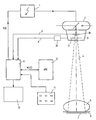
- An x-ray generator 1 supplies the high voltage for an x-ray emitter 2 and the Electricity through the X-ray tube.
- a primary beam aperture attached to the X-ray emitter 2 3 there is a first pair of collimators 31 perpendicular to the plane of the drawing Collimator edges, which is the opening of the beam that is blocked by the X-ray emitter 4 determined in the drawing plane.
- a second pair of collimators, not shown with collimator edges parallel to the plane of the drawing determines the opening of the beam 4 in the direction perpendicular to the plane of the drawing.
- the EP a patient 5, which is located on a patient table 6. That as a result EP of radiography
- the resulting X-ray image is recorded by a suitable EP7 image converter.
- the high voltage generated by the X-ray generator for and the current through the X-ray emitter 2 and the time course of these variables are controlled by a control unit 8 predefined, for example a microprocessor which interacts with a memory 9.
- the control unit 8 is coupled to a display unit or a monitor 10 and also with an input unit 11 (keyboard, mouse or trackball) with which a user can determine the course of the subsequent X-ray examination and with which he can proceed in particular which organ or which anatomical Region to be examined in a subsequent X-ray.
- c is a constant and ⁇ is a factor which depends on the high voltage U at the X-ray tube and on the filter 35 which is effective in the beam path and in which the primary beam diaphragm 3 is located.
- Q corresponds to the temporal integral of the current through the X-ray tube (as a rule, this is the tube current multiplied by the duration of an X-ray exposure), and A represents the opening of the radiation beam or the cross-sectional area of the radiation beam delimited by the collimator pairs 31.
- the product c ⁇ ⁇ ⁇ Q ⁇ A ⁇ corresponds to the dose D.
- w is a weighting factor that describes the biological effectiveness of the X-rays in the anatomical area examined in each case.
- the examiner gives the person to be examined at the input unit 11 Area, such as "Lung p.a.” in front.
- the control unit 8 calls the one Recording of this region, optimal recording parameters stored in the memory arrangement 9, namely the tube voltage U and the mAs product corresponding to the factor Q, and a weighting factor w on that of the anatomical region in question assigned. From the tube voltage called up in this way and that in the beam path effective filtering, which is transmitted by the signal F to the control unit 8, this calculates the factor ⁇ .
- the cross-sectional area A of the beam is from a transmitter arrangement 33 which, for example, with the collimator pairs in the Primary beam diaphragm can include coupled potentiometers. From the given or parameters determined in the control unit 8 can then determine the parameters during the recording calculate the effective dose given to the anatomical region.
- the effective dose D eff can also be determined in an analogous manner in the case of X-ray fluoroscopy, the weighting factor w and the tube voltage U being called up from the memory arrangement 9 as a function of the predetermined anatomical region.
- the quantity Q is then formed by the control unit by forming the time integral over the tube current flowing through the X-ray tube during the fluoroscopy. If the collimator pairs 31 are opened wide, parts of other organs can enter the beam path for which the X-rays have a different biological effectiveness. This can have the consequence that a different weighting factor must be used for a picture with a large radiation field than for a picture with a My radiation field.
- weighting factor w must also be made dependent on the cross-sectional area A of the collimator arrangement. This can be done, for example, by storing several weighting factors for an anatomical region in the memory arrangement 9, one of which is determined as a function of the respective value A and is used as a basis for calculating the effective dose.
- the dose D can also be calculated instead of by a calculation Determine measurement.
- the effective dose is then obtained by multiplying the measured value with a weighting factor w.
Landscapes
- Health & Medical Sciences (AREA)
- Life Sciences & Earth Sciences (AREA)
- Medical Informatics (AREA)
- Engineering & Computer Science (AREA)
- Radiology & Medical Imaging (AREA)
- Molecular Biology (AREA)
- Biophysics (AREA)
- Nuclear Medicine, Radiotherapy & Molecular Imaging (AREA)
- Optics & Photonics (AREA)
- Pathology (AREA)
- Physics & Mathematics (AREA)
- Biomedical Technology (AREA)
- Heart & Thoracic Surgery (AREA)
- High Energy & Nuclear Physics (AREA)
- Surgery (AREA)
- Animal Behavior & Ethology (AREA)
- General Health & Medical Sciences (AREA)
- Public Health (AREA)
- Veterinary Medicine (AREA)
- Apparatus For Radiation Diagnosis (AREA)
- X-Ray Techniques (AREA)
Applications Claiming Priority (2)
| Application Number | Priority Date | Filing Date | Title |
|---|---|---|---|
| DE19903749 | 1999-01-30 | ||
| DE19903749A DE19903749A1 (de) | 1999-01-30 | 1999-01-30 | Röntgen-Diagnostikeinrichtung mit Mitteln zur Bestimmung der Dosis |
Publications (2)
| Publication Number | Publication Date |
|---|---|
| EP1024681A2 true EP1024681A2 (fr) | 2000-08-02 |
| EP1024681A3 EP1024681A3 (fr) | 2003-09-10 |
Family
ID=7895917
Family Applications (1)
| Application Number | Title | Priority Date | Filing Date |
|---|---|---|---|
| EP00200226A Withdrawn EP1024681A3 (fr) | 1999-01-30 | 2000-01-20 | Dispositif de diagnostic à rayons-x avec moyens pour la détermination de dose |
Country Status (4)
| Country | Link |
|---|---|
| US (1) | US6292537B1 (fr) |
| EP (1) | EP1024681A3 (fr) |
| JP (1) | JP2000225113A (fr) |
| DE (1) | DE19903749A1 (fr) |
Families Citing this family (16)
| Publication number | Priority date | Publication date | Assignee | Title |
|---|---|---|---|---|
| DE10161708A1 (de) * | 2001-12-15 | 2003-06-18 | Philips Intellectual Property | Röntgeneinrichtung mit einer Speicheranordnung für Aufnahmeparameter von Röntgenaufnahmen |
| DE10201868C1 (de) * | 2002-01-18 | 2003-07-17 | Siemens Ag | Röntgeneinrichtung |
| WO2004071303A1 (fr) * | 2003-02-11 | 2004-08-26 | Philips Intellectual Property & Standards Gmbh | Dispositif a rayons x possedant un collimateur, et procede de reglage de ce dispositif |
| US20050053199A1 (en) * | 2003-09-04 | 2005-03-10 | Miles Dale A. | Portable x-ray device and method |
| JP4208694B2 (ja) * | 2003-10-21 | 2009-01-14 | キヤノン株式会社 | X線透視撮影装置 |
| US7597476B2 (en) * | 2007-11-07 | 2009-10-06 | Dornier Medtech Systems Gmbh | Apparatus and method for determining air-kerma rate |
| US7835496B2 (en) * | 2008-04-22 | 2010-11-16 | Siemens Aktiengesellschaft | User interface of an X-ray system and method for manufacturing such an user interface |
| JP2010057633A (ja) * | 2008-09-02 | 2010-03-18 | Fujifilm Corp | 放射線撮影装置及び放射線撮影方法 |
| US8755490B2 (en) * | 2009-04-07 | 2014-06-17 | Shimadzu Corporation | X-ray imaging device |
| DE102009021239A1 (de) | 2009-05-14 | 2010-11-18 | Siemens Aktiengesellschaft | Verfahren zur Überwachung der einem Patienten durch eine Strahlungsquelle verabreichten Röntgendosis bei einer Röntgeneinrichtung und Röntgeneinrichtung |
| DE102012205246A1 (de) * | 2012-03-30 | 2013-10-02 | Siemens Aktiengesellschaft | Vorrichtung zur Aufnahme von Strahlungsbildern eines Patienten und Katheter zum Einsatz in der Vorrichtung |
| US9416652B2 (en) | 2013-08-08 | 2016-08-16 | Vetco Gray Inc. | Sensing magnetized portions of a wellhead system to monitor fatigue loading |
| DE102013219249A1 (de) * | 2013-09-25 | 2015-03-26 | Siemens Aktiengesellschaft | Verfahren und System zur automatischen Auswahl eines Scanprotokolls |
| CN105326514A (zh) * | 2014-06-10 | 2016-02-17 | 上海西门子医疗器械有限公司 | X射线设备 |
| TWI606752B (zh) * | 2016-10-28 | 2017-11-21 | Iner Aec | 用於數位x光機的自動曝露控制系統與其方法 |
| KR102730872B1 (ko) * | 2017-01-25 | 2024-11-18 | 삼성전자주식회사 | 엑스선 영상 장치 및 그 제어 방법 |
Citations (1)
| Publication number | Priority date | Publication date | Assignee | Title |
|---|---|---|---|---|
| DE2124035A1 (de) | 1971-05-14 | 1972-11-23 | Siemens AG, 1000 Berlin u. 8000 München | Röntgenuntersuchungsgerät mit einer Meßeinrichtung zur Patientendosimetrie |
Family Cites Families (2)
| Publication number | Priority date | Publication date | Assignee | Title |
|---|---|---|---|---|
| SU1685272A3 (ru) * | 1989-05-04 | 1991-10-15 | С.Г.Жут ев, Б.Я.Мишкинис, Р.В.Ставицкий и И.Ш.Хасидашвили | Рентгенодиагностический аппарат |
| US5625662A (en) * | 1995-11-20 | 1997-04-29 | General Electric Company | Modulating x-ray tube current in a CT system |
-
1999
- 1999-01-30 DE DE19903749A patent/DE19903749A1/de not_active Withdrawn
-
2000
- 2000-01-20 EP EP00200226A patent/EP1024681A3/fr not_active Withdrawn
- 2000-01-28 JP JP2000020576A patent/JP2000225113A/ja active Pending
- 2000-01-28 US US09/493,963 patent/US6292537B1/en not_active Expired - Fee Related
Patent Citations (1)
| Publication number | Priority date | Publication date | Assignee | Title |
|---|---|---|---|---|
| DE2124035A1 (de) | 1971-05-14 | 1972-11-23 | Siemens AG, 1000 Berlin u. 8000 München | Röntgenuntersuchungsgerät mit einer Meßeinrichtung zur Patientendosimetrie |
Also Published As
| Publication number | Publication date |
|---|---|
| JP2000225113A (ja) | 2000-08-15 |
| US6292537B1 (en) | 2001-09-18 |
| DE19903749A1 (de) | 2000-08-03 |
| EP1024681A3 (fr) | 2003-09-10 |
Similar Documents
| Publication | Publication Date | Title |
|---|---|---|
| EP1024681A2 (fr) | Dispositif de diagnostic à rayons-x avec moyens pour la détermination de dose | |
| DE102005006895B4 (de) | Röntgendiagnostikeinrichtung sowie Verfahren zu deren Regelung | |
| DE102007008118B4 (de) | Verfahren zur Erzeugung tomographischer Darstellungen mit einem Röntgen-Computertomographie-System mit Streustrahlungskorrektur | |
| DE102010043712B4 (de) | Verfahren zur Ermittlung des Wertes einer Röhrenspannung, Röntgeneinrichtung, Rechenprogramm und Datenträger | |
| DE102016207437B4 (de) | Spektralunabhängige Ermittlung von Kalkablagerungen in Blutgefäßen | |
| DE102015202999B4 (de) | Automatisierte Justierung von Signalanalyseparametern für Röntgendetektoren | |
| DE69727007T2 (de) | Verfahren und Vorrichtung zur Erkennung und Korrektur von fehlerhaften Belichtungen während der Bilderzeugung mittels Röntgenstrahlen | |
| DE102011076781B4 (de) | Verfahren zur Korrektur einer Zählratendrift bei einem quantenzählenden Detektor, Röntgen-System mit quantenzählendem Detektor und Schaltungsanordnung für einen quantenzählenden Detektor | |
| DE10164170A1 (de) | Automatische Belichtungssteuerung und Belichtungsoptimierung digitaler Röntgenradiographie | |
| DE102005027436B4 (de) | Verfahren zur Berechnung von absorberspezifischen Gewichtungskoeffizienten und Verfahren zur Verbesserung eines von einem Absorber abhängigen Kontrast-zu-Rausch-Verhältnisses in einem von einer Röntgeneinrichtung erzeugten Röntgenbild eines zu untersuchenden Objektes | |
| DE20321723U1 (de) | Röntgendiagnostikeinrichtung mit einem Patientenlagerungstisch | |
| DE102008014738A1 (de) | Verfahren zur medizinischen Bildgebung sowie medizinische Bildgebungsvorrichtung | |
| DE102014203465A1 (de) | Verfahren zur Auswahl eines Strahlungsformfilters und Röntgenbildgebungssystem | |
| DE10322142A1 (de) | Durchleuchtungsgerät und Verfahren zum Ermitteln der Strahlendosis bei Durchleuchtungsuntersuchungen | |
| WO2014048748A1 (fr) | Détermination automatique d'une distribution spectrale de rayons x d'un nombre de sources de rayons x | |
| DE10141346A1 (de) | Verfahren zur Aufnahme von Messdaten mit einem Computertormographen | |
| DE19651722A1 (de) | Belichtungsautomatik für einen Röntgenapparat | |
| DE10310127A1 (de) | Gating-Verfahren, Gating-Gerät sowie Therapieeinrichtung | |
| DE69023167T2 (de) | Verfahren zum Eichen eines Röntgensystems und zur Messung der äquivalenten Dicke eines Gegenstandes. | |
| DE10322143B4 (de) | Durchleuchtungsanlage und Verfahren zum Ermitteln der effektiven Hauteingangsdosis bei Durchleuchtungsuntersuchungen | |
| DE102019217421A1 (de) | Verfahren zur automatischen Regelung von Strahlendosen medizinischer Röntgengeräte | |
| EP3552547B1 (fr) | Procédé de fourniture d'informations de conversion à un ensemble de données d'image, dispositif à rayons x, programme informatique et support de données lisible électroniquement | |
| DE102007023655B4 (de) | Verfahren zur Ermittlung wenigstens eines PET-Parameters | |
| EP0325120B1 (fr) | Appareil de diagnostic par rayons X | |
| DE102005021020A1 (de) | Verfahren zur Berechnung einer Orthogonal-Röntgenabschwächung eines Objekts anhand einer gemessenen Referenz-Röntgenabschwächung sowie zugehörige Vorrichtung |
Legal Events
| Date | Code | Title | Description |
|---|---|---|---|
| PUAI | Public reference made under article 153(3) epc to a published international application that has entered the european phase |
Free format text: ORIGINAL CODE: 0009012 |
|
| AK | Designated contracting states |
Kind code of ref document: A2 Designated state(s): AT BE CH CY DE DK ES FI FR GB GR IE IT LI LU MC NL PT SE |
|
| AX | Request for extension of the european patent |
Free format text: AL;LT;LV;MK;RO;SI |
|
| RAP1 | Party data changed (applicant data changed or rights of an application transferred) |
Owner name: KONINKLIJKE PHILIPS ELECTRONICS N.V. Owner name: PHILIPS CORPORATE INTELLECTUAL PROPERTY GMBH |
|
| RAP1 | Party data changed (applicant data changed or rights of an application transferred) |
Owner name: KONINKLIJKE PHILIPS ELECTRONICS N.V. Owner name: PHILIPS INTELLECTUAL PROPERTY & STANDARDS GMBH |
|
| PUAL | Search report despatched |
Free format text: ORIGINAL CODE: 0009013 |
|
| RIC1 | Information provided on ipc code assigned before grant |
Ipc: 7A 61B 6/00 B Ipc: 7H 05G 1/26 A |
|
| AK | Designated contracting states |
Kind code of ref document: A3 Designated state(s): AT BE CH CY DE DK ES FI FR GB GR IE IT LI LU MC NL PT SE |
|
| AX | Request for extension of the european patent |
Extension state: AL LT LV MK RO SI |
|
| 17P | Request for examination filed |
Effective date: 20040310 |
|
| 17Q | First examination report despatched |
Effective date: 20040420 |
|
| AKX | Designation fees paid |
Designated state(s): DE FR GB NL |
|
| STAA | Information on the status of an ep patent application or granted ep patent |
Free format text: STATUS: THE APPLICATION IS DEEMED TO BE WITHDRAWN |
|
| 18D | Application deemed to be withdrawn |
Effective date: 20041103 |