EP1031316A1 - Kompensation der Konzentrationsmesswerte eines Patienten - Google Patents
Kompensation der Konzentrationsmesswerte eines Patienten Download PDFInfo
- Publication number
- EP1031316A1 EP1031316A1 EP99660037A EP99660037A EP1031316A1 EP 1031316 A1 EP1031316 A1 EP 1031316A1 EP 99660037 A EP99660037 A EP 99660037A EP 99660037 A EP99660037 A EP 99660037A EP 1031316 A1 EP1031316 A1 EP 1031316A1
- Authority
- EP
- European Patent Office
- Prior art keywords
- tract
- chemical compound
- concentration
- measured
- concentration values
- Prior art date
- Legal status (The legal status is an assumption and is not a legal conclusion. Google has not performed a legal analysis and makes no representation as to the accuracy of the status listed.)
- Withdrawn
Links
Images




Classifications
-
- A—HUMAN NECESSITIES
- A61—MEDICAL OR VETERINARY SCIENCE; HYGIENE
- A61B—DIAGNOSIS; SURGERY; IDENTIFICATION
- A61B5/00—Measuring for diagnostic purposes; Identification of persons
- A61B5/145—Measuring characteristics of blood in vivo, e.g. gas concentration or pH-value ; Measuring characteristics of body fluids or tissues, e.g. interstitial fluid or cerebral tissue
- A61B5/14542—Measuring characteristics of blood in vivo, e.g. gas concentration or pH-value ; Measuring characteristics of body fluids or tissues, e.g. interstitial fluid or cerebral tissue for measuring blood gases
-
- A—HUMAN NECESSITIES
- A61—MEDICAL OR VETERINARY SCIENCE; HYGIENE
- A61B—DIAGNOSIS; SURGERY; IDENTIFICATION
- A61B5/00—Measuring for diagnostic purposes; Identification of persons
- A61B5/145—Measuring characteristics of blood in vivo, e.g. gas concentration or pH-value ; Measuring characteristics of body fluids or tissues, e.g. interstitial fluid or cerebral tissue
- A61B5/1468—Measuring characteristics of blood in vivo, e.g. gas concentration or pH-value ; Measuring characteristics of body fluids or tissues, e.g. interstitial fluid or cerebral tissue using chemical or electrochemical methods, e.g. by polarographic means
- A61B5/1473—Measuring characteristics of blood in vivo, e.g. gas concentration or pH-value ; Measuring characteristics of body fluids or tissues, e.g. interstitial fluid or cerebral tissue using chemical or electrochemical methods, e.g. by polarographic means invasive, e.g. introduced into the body by a catheter
-
- A—HUMAN NECESSITIES
- A61—MEDICAL OR VETERINARY SCIENCE; HYGIENE
- A61B—DIAGNOSIS; SURGERY; IDENTIFICATION
- A61B5/00—Measuring for diagnostic purposes; Identification of persons
- A61B5/68—Arrangements of detecting, measuring or recording means, e.g. sensors, in relation to patient
- A61B5/6846—Arrangements of detecting, measuring or recording means, e.g. sensors, in relation to patient specially adapted to be brought in contact with an internal body part, i.e. invasive
- A61B5/6847—Arrangements of detecting, measuring or recording means, e.g. sensors, in relation to patient specially adapted to be brought in contact with an internal body part, i.e. invasive mounted on an invasive device
- A61B5/6852—Catheters
- A61B5/6853—Catheters with a balloon
-
- A—HUMAN NECESSITIES
- A61—MEDICAL OR VETERINARY SCIENCE; HYGIENE
- A61B—DIAGNOSIS; SURGERY; IDENTIFICATION
- A61B5/00—Measuring for diagnostic purposes; Identification of persons
- A61B5/145—Measuring characteristics of blood in vivo, e.g. gas concentration or pH-value ; Measuring characteristics of body fluids or tissues, e.g. interstitial fluid or cerebral tissue
- A61B5/1455—Measuring characteristics of blood in vivo, e.g. gas concentration or pH-value ; Measuring characteristics of body fluids or tissues, e.g. interstitial fluid or cerebral tissue using optical sensors, e.g. spectral photometrical oximeters
- A61B5/1459—Measuring characteristics of blood in vivo, e.g. gas concentration or pH-value ; Measuring characteristics of body fluids or tissues, e.g. interstitial fluid or cerebral tissue using optical sensors, e.g. spectral photometrical oximeters invasive, e.g. introduced into the body by a catheter
Definitions
- This invention relates to a method for compensating measured concentration values of at least a first chemical compound within a first internal tract of a patient, which concentration values as measured are affected by a second chemical compound, which is becoming present in the body of said patient and has a limited transfer speed to mix with the first chemical compound at least in a detection site for said measured concentration values, rendering these measured concentration values to deviate from actual concentration values within said first internal tract.
- Especially invention relates to a use of the method for compensating measured concentration values of carbon dioxide within an gastrointestinal tract of a patient, which concentration values are affected by at least nitrous oxide, which is being dosed into the respiratory gas of said patient and has a limited absorption and/or diffusion speed to a detection site for said carbon dioxide concentration measurement, rendering its measured concentration values to deviate from actual concentration values.
- gastric tonometry provides information on the regional partial pressure of carbon dioxide, which relates to the adequacy of perfusion and rate of metabolism of the patient.
- the tonometric measurement is done in hollow viscus, typically gastrointestinal, by a special catheter, which is inserted nasogastricly into the stomach or gut.
- the catheter tip includes a gas-permeable and liquid-impermeable balloon.
- Carbon dioxide present or developing freely equilibrates between the gastric mucosa, the gastric lumen, and the gas contents of the balloon. After an equilibrium time the catheter gas content corresponds to the gas or liquid composition in the gastric mucosa.
- Gastric tonometric monitoring apparatus may be designed to automatically infuse and sample gas mixture from the catheter balloon at intervals and then automatically provide data of the carbon dioxide (CO 2 ) concentration of the gas sample e.g. with the infrared absorption technique.
- CO 2 carbon dioxide
- Gastric tonometry is currently used mainly for intensive care patients, but it is increasingly used also for surgical patients during longer operations.
- the hepatosplanchic area which is the usual site of tonometric measurement, has an important role in the pathogenesis of the multiple organ dysfunction syndrome.
- Splanchnic tissue perfusion has a low priority in the acute circulatory failure. Blood circulation is redistributed during a hypovolemic or cardiogenic shock so, that the most vital organs, heart and brain, get enough blood. Even though the reduction of splachnic tissue perfusion may initially be vital for survival, the prolonged reduction of splanchnic blood increases the risk of tissue damage and organ dysfunction and failures and even death of the patient.
- Carbon dioxide level in the tissue is also increasing, because it cannot be removed effectively from the tissue. If the increased P CO2 level can be detected early enough, the patient can be recuscitated better.
- This problem is discussed in the publication WO-94/21163 and a method is suggested, according to which the partial pressure of carbon dioxide is measured in hollow viscus and the carbon dioxide level or the pH of arterial blood is measured as well, and a parameter, more detailed called either a pCO 2 -gap or a pH-gap, indicative of condition of the hollow viscus is determined on the basis of these two values.
- the carbon dioxide level of the arterial blood might also be measured through detection of the end tidal partial pressure (pCO 2 ) of carbon dioxide to describe the overall "global" or "systemic” bicarbonate content of the blood.
- the pH-gap is calculated by mathematically subtracting intramucosal pH from arterial pH, but calculating pCO 2 -gap is not clearly described.
- the additional detection and independent display of anaesthetic gases, such as N 2 O, in the aspirated air of the patient using separate techniques is mentioned, which is very elaborate method and requires additional equipment.
- the partial pressure (P CO2 ) of carbon dioxide level in the gastrointestinal area is determined by analysing the carbon dioxide (CO 2 ) concentration sampled from the tonometric catheter balloon.
- the measurement/detection technique for determining this partial pressure (P CO2 ) of carbon dioxide is utilises an infrared absorption sensor.
- An infrared sensor typically comprises an infrared radiation source, gas measuring chamber, at least one optical bandpass filter within an absorption peak of CO 2 and an infrared detector delivering an electrical signal proportional to the amount of carbon dioxide in the measuring chamber.
- the purpose of the optical bandpass filter like interference filter, is to choose the wavelengths, where CO 2 molecules absorb infrared (IR) light.
- Nitrous oxide (N 2 O) is commonly given to surgical patients in very high concentrations (20-80%) as an anaesthetic gas in the respiratory tract.
- a typical inhaled gas mixture includes in the order of 60-70 vol.-% of N 2 O and in the order of 25-30 vol.-% oxygen, and further containing an additional vaporised anaesthetic agent as halothane, desflurane, isoflurane, enflurane and/or sevoflurane usually less than about 12 vol-%.
- a detector "sees" an increased absorption value, due to the fact that this kind of measuring arrangement detects transmission instead of actual absorbance.
- polar gases N 2 O being one of them, have a major effect on spectral line broadening.
- intravenous dosage of drugs or medicinal preparations or additives into the patient may affect the concentration measurements of a chemical compound in an internal tract or organ, either because of spectral line broadening or an overlapping absorption described later.
- One way of compensating the line broadening error caused by N 2 O is to use a double detector combination as a sensor arrangement, which detectors simultaneously measure both the CO 2 and the N 2 O concentrations in the same gas mixture.
- This measurement can be done by positioning two optical filters, which have different radiation transmitting bands, one for CO 2 and one for N 2 O, to detect IR-radiation absorption in a measuring chamber.
- a linear mathematical correction model can be applied to CO 2 concentration.
- This kind of sensor arrangement and correction method is disclosed in publication US-4 423 739 to correct the carbon dioxide measurement result of exhaled air using the nitrous oxide measurement result of the same exhaled air.
- each gas component which might be present and might contribute to collision broadening must be measured separately, which is unpractical, because at least individual optical bandpass filter(s) and measuring channels are required for each gas component rendering the large and too heavy sensor, and unreliable, because the gas mixture may include varying amounts of hard-to-measure gas components.
- EP-0 834 733 discloses a totally different method for correcting the spectral line broadening error, with utilising a measured viscosity or viscosity related quantity of the gas mixture. This method also requires an use of additional detection means, and hence are impractical in many cases, where a small and lightweight detector arrangement is needed or is preferable.
- the main object of this invention is to find a method for correcting a by detection measured concentration value of a first gas component, which measured concentration value is affected by a second gas component in the gas mixture, without several detectors for separate measuring of each of said at least two gas components.
- the second object of this invention is to find a method, which provides applicability to measure the first gas component in any necessary body part of a patient, which means that the method should not as such restrict the site for detection.
- the site of detection, from which the gas mixture to be analysed is gathered, and is then transferred, if necessary, to a measuring chamber of the measuring apparatus, should be selectable on medical or other practical basis not essentially limited by the method.
- the third object of this invention is to find a method, which provides data and/or calculations for correction of concentration value of the first gas component with acceptable accuracy.
- the fourth object of this invention is to find a method, which is also applicable under conditions, where the second gas component is transferred or dosed into the body of the patient in an area or organ(s) separate from said detection site for the first gas component, and/or applicable under conditions, where the second gas component has a limited transfer speed to said detection site for the first gas component. If only possible the method should allow the first gas component to occur mixed with another medium, like liquid or solid or tissue in that body section of the patient, where the site for detection is decided to be.
- a further limited object of the invention is to find this kind of method, which could be applied in the field related to medical diagnostics and/or intensive care and/or other medical or clinical operations.
- Fig. 1 shows generally and schematically a body of a human and some of his/her internal and external tracts, which include the optional detection sites of the invention. Detection site is a point in the patient, wherefrom the required concentration values shall originate. Some of the possible detector arrangements are shown guided in their operation position.
- This figure also shows schematically one possible embodiment for a measuring apparatus with a first detector arrangement for measuring concentration of a first chemical compound within a first internal tract of a patient and a second arrangement for measuring or deriving concentration of a second chemical compound within a second tract of a patient.
- CO 2 concentration value is measured with an gastrointestinal tonometry and at least N 2 O concentration value is from the respiratory gas/air.
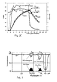
- Fig. 2A illustrates a first example of the relationship between a gastrointestinally measured CO 2 concentration, a N 2 O and CO 2 concentrations measured from exhaled air, and estimated N 2 O concentration defined according to the invention and time during a surgical operation of a patient.
- a gastrointestinally measured N 2 O concentration also shown, which concentration is not measured in practise.
- Fig. 2B illustrates a second example of the relationship between a gastrointestinally measured CO 2 concentration, a N 2 O and CO 2 concentrations measured from inhaled air, and estimated N 2 O concentration defined according to the invention and time during a surgical operation of another patient.
- a gastrointestinally measured N 2 O concentration also shown, which concentration is not measured in practise.
- Fig. 2C illustrates a third example of the relationship between a gastrointestinally measured CO 2 concentration, a N 2 O concentration derived from dosing apparatus and CO 2 concentration measured from respiratory air, and estimated N 2 O concentration defined according to the invention and time during a surgical operation of a patient.
- a gastrointestinally measured N 2 O concentration also shown, which concentration is not measured in practise.
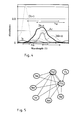
- Fig. 3 shows the approximate absorption peaks of e.g. carbon dioxide at a wavelength area of 4.3 ⁇ m and nitrous oxide at a wavelength area of 3.9 ⁇ m, and the absorption spectra of the respective optical IR passband filters.
- Fig. 4 shows the absorption curves of some chemical compounds Di, Is, Et, Ac and Me being in gaseous state and having overlapping absorption bands in another portion of IR.
- Fig. 5 shows schematically the spectral line broadening interactions between the main gas components of a gas mixture for respiratory air used for anaesthetic purposes during e.g. a surgical operation.
- Fig. 6 illustrates more detailed the detector arrangement according to Fig. 1 for measuring the concentrations of defined gaseous components in respiratory air or gas mixture M of the patient.
- This detector arrangement which is directly positioned in the intubation tube for the patient's inhalation/exhalation air, is one of the several alternative embodiments for measuring at least the concentration of the chemical compound B dosed.
- Fig. 7 illustrates more detailed a detector arrangement according to Fig. 1 for measuring the concentration of a defined gaseous component in the gas mixture from the internal tract of the patient.
- the gas mixture is withdrawn from the tonometric balloon via a catheter tubing, which is one of the several alternative embodiments for procedures to provide the measuring of said gas mixture within the tonometric balloon.
- the detector arrangement shown is also one of the several possible alternative embodiments for measuring the concentration of the chemical compound A in catheter gas mixture.
- Fig. 8 illustrates more detailed another detector arrangement applicable to Fig. 1 for measuring the concentration of a defined gaseous component in the gas mixture from the internal tract of the patient.
- the data from the gas mixture in the tonometric balloon guided via optical fibers which is one of the several alternative embodiments for data transfer procedures from said internal tract.
- Fig. 9 is a flow chart of the invention illustrating the basic steps thereof.
- the measured or derived nitrous oxide concentration from a respiratory tract 2 of a patient is used for correcting an error of measured carbon dioxide concentration in gastrointestinal tract 1.
- a catheter with a tonometric balloon 31, which is gas-permeable, but liquid-impermeable, is guided nasogastrictly into the gastric hollow organs, usually within stomach 10, or possibly within colon 11 or small intestine 12.
- the gaseous content of the balloon 31 is then analysed using infrared absorption technique as described earlier in this text.
- the measured concentration values of carbon dioxide CO 2 within an gastrointestinal tract 1 of a patient are compensated using the inventive method to get an accurate value thereof.
- the initial CO 2 concentration values detected from the gastrointestinal tract 1 are affected by at least nitrous oxide N 2 O, which is generally being dosed in the respiratory gas M of the patient as an anaesthetic medium.
- the N 2 O dosed and inhaled has a limited or low absorption and/or diffusion and/or transport speed from the pulmonary area through tissue to the gastric mucosa, but has a much higher diffusion speed to the catheter balloon that CO 2 .
- Both N 2 O and CO 2 has a limited absorption speed through the wall 45 of catheter balloon 31. After a catheter equilibrium time, the N 2 O partial pressure in gastric area should be close to the partial pressure P N2O in the tonometric balloon.
- the intestinal hollow organ like stomach 10 is the point of interest and so the detection site, wherefrom necessary predefined data shall be available, in order that the medical state of the patient can be evaluated.
- accurate gastrointestinal CO 2 concentration is one required data for evaluation of hepatosplanchic area 9.
- the measurement or detection of CO 2 concentration can be performed in detection site or any other position, as described later, but the valid gas mixture, which is analysed, shall be gathered from said detection site, that is in the point of interest, with the aid of e.g. tonometric balloon.
- the nitrous oxide present in the intestinal tract and so in the gas mixture within the tonometric balloon causes the measured carbon dioxide concentration values P CO2 to deviate from actual concentration values.
- concentration values E N2O of at least nitrous oxide is measured or derived several times in the inhaled or exhaled gas M of said patient, that is in the respiratory tract 2, during a period T of a clinical operation. Further the concentration values C N2O for nitrous oxide within the gastrointestinal tract are estimated without detection therein and the measured initial concentration values P CO2 [n] of carbon dioxide in the gastrointestinal tract 1 are corrected by a compensating factor Fc.
- the basic features of the invention is a determining of e.g. N 2 O using a new method.
- the second chemical component is measured in a sample taken from a second tract 2*, which can be an internal tract or an external area of the patient.
- the lower indices (1) and (2) in the formulas I B and II B refer to the first and second tract respectively.
- the first internal tract 1* can be an intestinal tract 1, or a respiratory tract 2, or an urinary tract 3, or one of the vascular tracts 4, or a genital tract 5
- the second tract 2* can be an intestinal 1 tract, or a respiratory tract 2, or an urinary tract 3, or a nervous tract 8, or one of the vascular tracts 4, or a genital tract 5, other than the first internal tract, or a cutaneous or mucous are 6, 7 of the body of the person or animal.
- said first chemical compound A is arranged to be in the gaseous state using tonometric means 31, 34, 35 within said first internal tract 1*, whereupon this first tract can be either the intestinal tract 1, or the urinary tract 3, or the genital tract 5 or any other accessible internal hollow organ of the patient or a person or an animal.
- the second chemical compound B and the first chemical compound A is arranged to be in the gaseous state using air or gas mixture, like breathing air M, provided to or from said second internal tract 2* with a dosing apparatus 26, whereupon this second tract can be either the intestinal 1 tract, or the respiratory tract 2 or any other accessible hollow organ.
- the second chemical compound B may also be in a liquid state when provided to said second internal tract 2*, whereupon this second tract can be either the intestinal 1 tract, or the urinary tract 3, or the nervous tract 8, or one of the vascular tracts 4, or the genital tract 5, or a cutaneous or mucous are 6, 7 of the body of a patient or a person or an animal.
- this second tract can be either the intestinal 1 tract, or the urinary tract 3, or the nervous tract 8, or one of the vascular tracts 4, or the genital tract 5, or a cutaneous or mucous are 6, 7 of the body of a patient or a person or an animal.
- the second chemical compound B can be dosed either in a hollow organ, or transcutaneously with an pressure unit 27 or through injection unit 28 into other internal organs. It is also possible to draw the first chemical compound A as a part of a liquid, like blood, using injection unit 28 in reverse mode.
- the second internal tract 2* is other portion of a body than the first internal tract 1*.
- the second chemical compound B and/or the third chemical compound G described below can be directly dosed in a controlled manner into the patient, or one or both of them can be basically end up in the patient or person or animal because of an uncontrolled reason, but in every case that amount of these chemical compounds, which is received by said body, shall be measurable. So at least the concentration values C N2O for nitrous oxide are measured or derived, by detection or by using dosing data D directly from a dosing apparatus 26, from that area or channel forming a dosing route of the nitrous oxide N 2 O into the patient.
- K B and K G are empirical spectral line broadening constants, and Fc is a first compensating factor and Fc* is a second compensating factor described later. This is practical when there are one or several of those other anaesthetic gases, which is/are being dosed into the respiratory air of said patient, and the concentration values E G of at least one of them, is/are additionally measured in the second tract 2* or derived as a data D from a dosing apparatus 26 as later described in more detail.
- concentration values C G for specified anaesthetic gas component(s) within gastrointestinal tract 1 are estimated without detection according to the invention, and an altered additional compensating factor Fc* is formulated to provide a further correction for measured initial concentration values P CO2 [n] of carbon dioxide or generally concentration value P A [n] gas component.
- the third or several chemical compound(s) G, which is/are received by said body and further transferred to the detection site can be measured or derived in an third area or tract 3* deviating at least from the first internal tract and possibly from the second tract 2*. Concentration value(s) C G thereof is/are estimated without detection as described later.
- the time between two tonometric measurements is 10 minutes and over a time period T including first traces of N 2 O or said second chemical compound B respectively within or in contact with said patient, and especially from the beginning of dosage of nitrous oxide N 2 O into a patient under said clinical operation.
- the inventive estimated concentration value C N2O is so a predetermined cumulative function of a prevailing concentration value E N2O e.g. in the respiratory tract 2 and previous estimated concentration values C N2O [n-1] in the gastrointestinal tract 1. This estimated concentration value C N2O [n] is used in conjunction with formula II S to get the compensating factor Fc.
- the smoothing parameter ⁇ (n) is dependent on perfusion related parameters, in this case the measured partial pressure P CO2 of carbon dioxide in the gastric organ 10 and the measured partial pressure E CO2 in the end tidal of inhaled or exhaled air, and more detailed form the P CO2 (n-1) and E CO2 (n-1).
- concentration marking C means a corrected or estimated concentration value
- marking P means a measured value from any first tract
- marking E means a measured or derived - e.g. data D transferred from dosing apparatus 26 to display unit 24 - value from any second tract
- marking R means a measured or derived value from a further tract of the patient obtained by detection or data transmitting.
- C B [n] ⁇ (n) ⁇ [E B (n)] + [1- ⁇ (n)] ⁇ [C B (n-1)] (III S ), whereupon the estimated concentration value C B [n] so is a cumulative function of one in succession previous estimated concentration value C B [n-1], and is directly related to both a measured or derived concentration value E B [n] and a previous estimated concentration value C B [n-1] thereof, and is additionally related to a constant or variable parameter ⁇ .
- This estimated concentration value C B [n] is used in conjunction with formula II B to get the compensating factor Fc, and an analogous estimated concentration value C G [n] similarly in conjunction with formula II G to get the compensating factor Fc*.
- the smoothing parameter ⁇ has at least successive actual concentration values of the first chemical compound A from said first and/or second tract 1* and/or 2* as variables.
- the estimated concentration value C is an inverse function of these actual concentration values P, E, R of at least one of the chemical compounds in one of tracts.
- the initial measured concentration PA which is at least partly corrected by a factor Q1 is preferably further corrected -typically by subtraction - by additional terms, which are functions of compensating factors Q2 and Q3 and respective estimated concentration values C B and C G .
- C B C G and possible further concentrations are calculated cumulatively in the same inventive way as described by e.g. referring to formulas 2 to 3 as well as III and IVa to IVe earlier in this text, providing an estimated concentration value to simulate their actual concentration without their detection in the first tract 1*.
- the error caused by a gas component is or can be altered to be approximately linearly related to its concentration, which can be done for example by a proper selection of passband widths and peak transmittances of the optical filters used.
- the third chemical compound G can be transferred to the body trough the same or different route than the second chemical compound B.
- the second and/or third chemical compounds are allowed to have any variable concentration in its area or tract, where the compound is received from to said body with time, but the concentration data in this second tract 2* shall be available.
- the tonometric measuring of concentration P A of the first chemical compound A in the first internal tract 1* is an advantageous and preferred alternative.
- One possible construction of tonometric arrangement is illustrated in Fig. 1. It comprises a tonometric balloon 31 and one or two tubes 32, which connects) the sampling chamber 46, formed by the gas-permeable wall 45 of the balloon 31, and a detector unit 21 for measuring e.g. one gas component in the gas mixture within the sampling chamber 46.
- the balloon and the connecting tube(s) form the tonometric catheter, which is guided into the detection site within the first internal tract, like a hollow organ.
- the circulation of gas mixture trough the tube 32 can be done to-and-from the sampling chamber in a alternate manner, or trough the tubes 32 as an continuos flow as disclosed in the patent publication US-5 479 923.
- Another possible construction of tonometric arrangement is illustrated in Fig. 8. It comprises a tonometric balloon 34 and for example a pair of optical fibers 36, which connect the sampling chamber 46, formed by the gas-permeable wall 45 of the balloon 31, and a detector unit 21 for measuring e.g. one gas component in the gas mixture within the sampling chamber 46.
- the gas mixture to be analysed stays in the sampling chamber 46, and the radiation S is sent to the chamber and received therefrom through the optical fiber or a pair of optical fibers 36 by a detector unit 21.
- a tonometric balloon 35 of this same type but provide the sampling chamber with e.g. one or several chemically responsive transistors, which are connected with electrical conductors 37 to a processor unit, not shown in the figures.
- the wall of the tonometric balloon is usually prepared from dimethyl silicone rubber (35%), the diffusion constants for CO 2 and N 2 O thereof being in the order of 325 cm 2 /s and 435 cm 2 /s respectively.
- other polymers can be used for preparing the wall of the tonometric balloon, some examples thereof being cellulose acetate, polycarbonate, polysulfone, different polyethylenes, butyl rubbers etc. These materials have a different diffusion properties for various gas components as compared those for various types of silicone rubbers.
- the detector unit 21, 22 can be any known or new type, which is capable for measuring in the required wavelength area of the radiation S and with required accuracy. At least the initial concentration value P A or e.g. P CO2 of the gaseous first chemical compound is detected using radiation absorption method to have the measurement data, and preferably though not necessarily also the concentration values E A , E B ,R G of the gaseous first, second, third chemical compounds are detected using radiation absorption method to get the measurement data.
- Fig. 8 it is schematically illustrated a basic detector unit 21, comprising only the basic parts, which are a radiation source 44, an optical passband filter 41b, and a radiation detector 43a, which provides an electrical signal for further processing.
- the sampling chamber 46 in the tonometric balloon 34 forms its measuring chamber.
- a detector unit 21 comprising a radiation source 44, a measuring chamber 40 separate from the sampling chamber 46, a hollow radiation tube 47 with an internal guide section 49 for directing the radiation, an optical passband filter 41a, and a radiation detector 43b, which provides an electrical signal for further processing.
- the measuring cell or chamber 40 also have two connections for tubes 32, which allow flow of the gas mixture from the sampling chamber and trough the measuring chamber.
- a detector unit 21 which is advantageous for analysing the inhaled or exhaled air M of the patient, because it can be installed in the respiration mouth piece provided with a measuring cell or chamber 40 between a dosing/respiration apparatus 26 and the respiratory tract 1 of the patient.
- This detector unit comprising a radiation source 44, a radiation tube 47, which is either partly hollow or filled with an radiation transparent material, two optical passband filters 41a, 41b, and two radiation detectors 43a, 43b with an separation wall 42 therebetween. Combination of two filters with different transmission bands and two detectors permits measuring of the concentrations of two gas components, but they can be also used for other purposes as known.
- the detectors provide electrical signals through electrical cable 29 for further processing unit 23.
- the measuring chamber 40 is connected to an intubation tube 48 on the one side and with a connection tube 33 to the dosing/respiration apparatus 26. All of these measuring chambers are provided with an with permanent absorption length(s) H, which is a general practise. Of course also other types of detector units can be employed. The different detector units and their functions are generally known, so that they not described more in detail.
- the second chemical compound B like nitrous oxide and other anaesthetic gasses or liquids, are being dosed into the patient directly from a dosing apparatus 26, its is not necessary to measure by detection, as described in the preceding chapter, in a conventional manner, but the required concentration values E B of the second chemical compound B are derived or transmitted from the dosage D data already present in the dosing apparatus 26.
- At least one of the initial concentration values P A and/or E A and/or E B and/or R G etc. of the gaseous first and/or second and/or third chemical compounds A, B, G are detected by radiation absorption in a measuring chamber(s) 40 or from a sampling chamber 46, which are/is in connection with the respective one of said tracts 1-8.
- Said radiation is electromagnetic radiation S provided to function with specific wavelength bands ⁇ p each of which is arranged in a predetermined manner within an area of an absorption peak ⁇ b of said gaseous first chemical compound A, e.g. CO 2 as described, and optionally within an area of an absorption peak of said gaseous second chemical compound B, e.g.
- concentration values P CO2 of carbon dioxide and concentration values P N2O of nitrous oxide from the internal tract 1* are detected using two wavelength bands ⁇ b 1 , ⁇ b 2 respectively in the infrared area of said radiation S.
- concentration values P CO2 of carbon dioxide and concentration values P N2O of nitrous oxide from the internal tract 1* are detected using two wavelength bands ⁇ b 1 , ⁇ b 2 respectively in the infrared area of said radiation S.
- concentration value C CO2 [n] is displayed or transmitted to further data processing as final compensated concentration values in a monitoring equipment 25.
- the measured concentration values of at least a first chemical compound A within a first internal tract 1* of a patient, which concentration values PA in initial measured state are affected by a second chemical compound B, are compensated in conditions, where the second chemical compound is becoming present in the body of said patient and render these initial measured concentration values P A to deviate from actual concentration values PR A , if some degree of compensation is not applied.
- the second chemical compound B has in practise a limited transfer speed to mix with the first chemical compound at least in a detection site for said_measured concentration values P A , within said first internal tract 1.
- Concentration values E B of at least said second chemical compound B in a second tract 2* of said patient is measured or respective data D derived several times during a time period T including first chemical compound measurements in said first internal tract 1* or from the beginning of presence for absorption and/or diffusion to the patient, person or animal.
- Estimated concentration values C B of said second chemical compound is determined simulating its non-detected concentrations in said first internal tract 1*.
- the measured/dtected initial concentration values P A of said first chemical compound A the first internal tract 1* are corrected by a compensating factor Fc and optionally Fc* and/or Fc 1 and or Fc 2 etc. Each said successive and corrected concentration value C A is made evident as the respective one of the compensated concentration values.
Landscapes
- Health & Medical Sciences (AREA)
- Life Sciences & Earth Sciences (AREA)
- Physics & Mathematics (AREA)
- Surgery (AREA)
- General Health & Medical Sciences (AREA)
- Engineering & Computer Science (AREA)
- Biomedical Technology (AREA)
- Heart & Thoracic Surgery (AREA)
- Medical Informatics (AREA)
- Molecular Biology (AREA)
- Biophysics (AREA)
- Animal Behavior & Ethology (AREA)
- Pathology (AREA)
- Public Health (AREA)
- Veterinary Medicine (AREA)
- Optics & Photonics (AREA)
- Chemical & Material Sciences (AREA)
- Chemical Kinetics & Catalysis (AREA)
- General Chemical & Material Sciences (AREA)
- Measurement Of The Respiration, Hearing Ability, Form, And Blood Characteristics Of Living Organisms (AREA)
- Investigating Or Analysing Biological Materials (AREA)
Priority Applications (3)
| Application Number | Priority Date | Filing Date | Title |
|---|---|---|---|
| EP99660037A EP1031316A1 (de) | 1999-02-25 | 1999-02-25 | Kompensation der Konzentrationsmesswerte eines Patienten |
| US09/460,868 US6280382B1 (en) | 1999-02-25 | 1999-12-14 | Compensation of concentration values concerning a patient |
| JP2000054430A JP2000245711A (ja) | 1999-02-25 | 2000-02-25 | 測定濃度値に補償を加えるための方法及びその使用方法 |
Applications Claiming Priority (1)
| Application Number | Priority Date | Filing Date | Title |
|---|---|---|---|
| EP99660037A EP1031316A1 (de) | 1999-02-25 | 1999-02-25 | Kompensation der Konzentrationsmesswerte eines Patienten |
Publications (1)
| Publication Number | Publication Date |
|---|---|
| EP1031316A1 true EP1031316A1 (de) | 2000-08-30 |
Family
ID=8242609
Family Applications (1)
| Application Number | Title | Priority Date | Filing Date |
|---|---|---|---|
| EP99660037A Withdrawn EP1031316A1 (de) | 1999-02-25 | 1999-02-25 | Kompensation der Konzentrationsmesswerte eines Patienten |
Country Status (3)
| Country | Link |
|---|---|
| US (1) | US6280382B1 (de) |
| EP (1) | EP1031316A1 (de) |
| JP (1) | JP2000245711A (de) |
Families Citing this family (1)
| Publication number | Priority date | Publication date | Assignee | Title |
|---|---|---|---|---|
| JP5927013B2 (ja) | 2012-04-16 | 2016-05-25 | 日本光電工業株式会社 | 生体情報モニタ装置 |
Citations (7)
| Publication number | Priority date | Publication date | Assignee | Title |
|---|---|---|---|---|
| US4423739A (en) | 1981-08-24 | 1984-01-03 | Andros Analyzers Incorporated | End tidal carbon dioxide gas analyzer |
| US4585007A (en) * | 1982-05-15 | 1986-04-29 | Kabushiki Kaisha Kyoto Daiichi Kagaku | Method for continuously measuring a specified ingredient in a diluted liquid |
| US4914719A (en) | 1989-03-10 | 1990-04-03 | Criticare Systems, Inc. | Multiple component gas analyzer |
| US5186172A (en) | 1982-03-22 | 1993-02-16 | Mountpelier Investments, S.A. | Remote sensing tonometric catheter apparatus |
| WO1994021163A1 (en) | 1993-03-22 | 1994-09-29 | Instrumentarium Corporation | Remote sensing tonometric catheter apparatus and method |
| US5479923A (en) | 1992-10-16 | 1996-01-02 | Instrumentarium Corporation | Method and apparatus for analyzing a sample |
| EP0834733A2 (de) | 1996-09-18 | 1998-04-08 | Instrumentarium Oy | Korrektur der Querempfindlichkeit in Mischgasen bei Abssorptionsmessungen |
Family Cites Families (2)
| Publication number | Priority date | Publication date | Assignee | Title |
|---|---|---|---|---|
| FI101749B (fi) * | 1996-12-30 | 1998-08-14 | Instrumentarium Oy | Kaasukomponentin pitoisuuden tarkka mittaaminen kaasuseoksessa, jossa muut komponentit vaikuttavat pitoisuusmääritykseen |
| US6112576A (en) * | 1998-10-05 | 2000-09-05 | Panametrics, Inc. | Gas analyzer with background gas compensation |
-
1999
- 1999-02-25 EP EP99660037A patent/EP1031316A1/de not_active Withdrawn
- 1999-12-14 US US09/460,868 patent/US6280382B1/en not_active Expired - Fee Related
-
2000
- 2000-02-25 JP JP2000054430A patent/JP2000245711A/ja active Pending
Patent Citations (7)
| Publication number | Priority date | Publication date | Assignee | Title |
|---|---|---|---|---|
| US4423739A (en) | 1981-08-24 | 1984-01-03 | Andros Analyzers Incorporated | End tidal carbon dioxide gas analyzer |
| US5186172A (en) | 1982-03-22 | 1993-02-16 | Mountpelier Investments, S.A. | Remote sensing tonometric catheter apparatus |
| US4585007A (en) * | 1982-05-15 | 1986-04-29 | Kabushiki Kaisha Kyoto Daiichi Kagaku | Method for continuously measuring a specified ingredient in a diluted liquid |
| US4914719A (en) | 1989-03-10 | 1990-04-03 | Criticare Systems, Inc. | Multiple component gas analyzer |
| US5479923A (en) | 1992-10-16 | 1996-01-02 | Instrumentarium Corporation | Method and apparatus for analyzing a sample |
| WO1994021163A1 (en) | 1993-03-22 | 1994-09-29 | Instrumentarium Corporation | Remote sensing tonometric catheter apparatus and method |
| EP0834733A2 (de) | 1996-09-18 | 1998-04-08 | Instrumentarium Oy | Korrektur der Querempfindlichkeit in Mischgasen bei Abssorptionsmessungen |
Non-Patent Citations (1)
| Title |
|---|
| GUILLERMO GUTIERREZ, STEVEN D. BROWN: "Gastric Tonometry : A new Monitoring Modality in the Intensive Care Unit", J.INTENSIVE CARE MED., vol. 10, 1995, pages 34 - 44 |
Also Published As
| Publication number | Publication date |
|---|---|
| US6280382B1 (en) | 2001-08-28 |
| JP2000245711A (ja) | 2000-09-12 |
Similar Documents
| Publication | Publication Date | Title |
|---|---|---|
| US7018340B2 (en) | Apparatus and method for non-invasively measuring cardiac output | |
| US6659961B2 (en) | Method and apparatus for measuring pulmonary blood flow by pulmonary exchange of oxygen and an inert gas with the blood | |
| US6217524B1 (en) | Method of continuously, non-invasively monitoring pulmonary capillary blood flow and cardiac output | |
| US6029076A (en) | Remote sensing tonometric catheter method | |
| US6238339B1 (en) | Remote sensing tonometric catheter apparatus and method | |
| US6200271B1 (en) | Bi-directional partial re-breathing method | |
| US6210342B1 (en) | Bi-directional partial re-breathing method | |
| EP1238631B1 (de) | Verfahren zur nicht-invasiven Bestimmung des Zustandes des Kreislaufes eines Individuums | |
| D'Mello et al. | Capnography | |
| US20120277613A1 (en) | Methods of intravenous drug monitoring | |
| WO1994021164A1 (en) | Remote sensing tonometric catheter apparatus and method | |
| US6280382B1 (en) | Compensation of concentration values concerning a patient | |
| Abramo et al. | Comparison of pediatric end-tidal CO2 measured with nasal/oral cannula circuit and capillary PCO2 | |
| Duke-Novakovski et al. | Comparison of mainstream (Capnostat 5) and two low-flow sidestream capnometers (VM-2500-S and Capnostream) in spontaneously breathing rabbits anesthetized with a Bain coaxial breathing system | |
| US6216023B1 (en) | Method and apparatus for determining differences between regional CO2 and systemic CO2 measurements | |
| LaValle et al. | Capnography: Assessing End-Tidal CO: 2: Levels | |
| Melton | Continuous fiberoptic PCO2 monitoring indicates poorer gastric perfusion during supraceliac aortic clamping than conventional gastric tonometry in humans: a pilot study |
Legal Events
| Date | Code | Title | Description |
|---|---|---|---|
| PUAI | Public reference made under article 153(3) epc to a published international application that has entered the european phase |
Free format text: ORIGINAL CODE: 0009012 |
|
| 17P | Request for examination filed |
Effective date: 19991018 |
|
| AK | Designated contracting states |
Kind code of ref document: A1 Designated state(s): DE FI FR GB IT |
|
| AX | Request for extension of the european patent |
Free format text: AL;LT;LV;MK;RO;SI |
|
| AKX | Designation fees paid |
Free format text: DE FI FR GB IT |
|
| STAA | Information on the status of an ep patent application or granted ep patent |
Free format text: STATUS: THE APPLICATION IS DEEMED TO BE WITHDRAWN |
|
| 18D | Application deemed to be withdrawn |
Effective date: 20050901 |