EP1031835A1 - Diagnostische, prognostische und therapeutische Verfahren für feste, nicht-lymphoide Tumoren und ihre Metastasen - Google Patents
Diagnostische, prognostische und therapeutische Verfahren für feste, nicht-lymphoide Tumoren und ihre Metastasen Download PDFInfo
- Publication number
- EP1031835A1 EP1031835A1 EP00105520A EP00105520A EP1031835A1 EP 1031835 A1 EP1031835 A1 EP 1031835A1 EP 00105520 A EP00105520 A EP 00105520A EP 00105520 A EP00105520 A EP 00105520A EP 1031835 A1 EP1031835 A1 EP 1031835A1
- Authority
- EP
- European Patent Office
- Prior art keywords
- cells
- tcrβ
- tumor
- idiotype
- metastatic
- Prior art date
- Legal status (The legal status is an assumption and is not a legal conclusion. Google has not performed a legal analysis and makes no representation as to the accuracy of the status listed.)
- Withdrawn
Links
Images



Classifications
-
- G—PHYSICS
- G01—MEASURING; TESTING
- G01N—INVESTIGATING OR ANALYSING MATERIALS BY DETERMINING THEIR CHEMICAL OR PHYSICAL PROPERTIES
- G01N33/00—Investigating or analysing materials by specific methods not covered by groups G01N1/00 - G01N31/00
- G01N33/48—Biological material, e.g. blood, urine; Haemocytometers
- G01N33/50—Chemical analysis of biological material, e.g. blood, urine; Testing involving biospecific ligand binding methods; Immunological testing
- G01N33/68—Chemical analysis of biological material, e.g. blood, urine; Testing involving biospecific ligand binding methods; Immunological testing involving proteins, peptides or amino acids
- G01N33/6863—Cytokines, i.e. immune system proteins modifying a biological response such as cell growth proliferation or differentiation, e.g. TNF, CNF, GM-CSF, lymphotoxin, MIF or their receptors
- G01N33/6869—Interleukin
-
- A—HUMAN NECESSITIES
- A61—MEDICAL OR VETERINARY SCIENCE; HYGIENE
- A61K—PREPARATIONS FOR MEDICAL, DENTAL OR TOILETRY PURPOSES
- A61K47/00—Medicinal preparations characterised by the non-active ingredients used, e.g. carriers or inert additives; Targeting or modifying agents chemically bound to the active ingredient
- A61K47/50—Medicinal preparations characterised by the non-active ingredients used, e.g. carriers or inert additives; Targeting or modifying agents chemically bound to the active ingredient the non-active ingredient being chemically bound to the active ingredient, e.g. polymer-drug conjugates
- A61K47/51—Medicinal preparations characterised by the non-active ingredients used, e.g. carriers or inert additives; Targeting or modifying agents chemically bound to the active ingredient the non-active ingredient being chemically bound to the active ingredient, e.g. polymer-drug conjugates the non-active ingredient being a modifying agent
- A61K47/68—Medicinal preparations characterised by the non-active ingredients used, e.g. carriers or inert additives; Targeting or modifying agents chemically bound to the active ingredient the non-active ingredient being chemically bound to the active ingredient, e.g. polymer-drug conjugates the non-active ingredient being a modifying agent the modifying agent being an antibody, an immunoglobulin or a fragment thereof, e.g. an Fc-fragment
- A61K47/6801—Drug-antibody or immunoglobulin conjugates defined by the pharmacologically or therapeutically active agent
- A61K47/6803—Drugs conjugated to an antibody or immunoglobulin, e.g. cisplatin-antibody conjugates
- A61K47/6811—Drugs conjugated to an antibody or immunoglobulin, e.g. cisplatin-antibody conjugates the drug being a protein or peptide, e.g. transferrin or bleomycin
- A61K47/6813—Drugs conjugated to an antibody or immunoglobulin, e.g. cisplatin-antibody conjugates the drug being a protein or peptide, e.g. transferrin or bleomycin the drug being a peptidic cytokine, e.g. an interleukin or interferon
-
- A—HUMAN NECESSITIES
- A61—MEDICAL OR VETERINARY SCIENCE; HYGIENE
- A61P—SPECIFIC THERAPEUTIC ACTIVITY OF CHEMICAL COMPOUNDS OR MEDICINAL PREPARATIONS
- A61P35/00—Antineoplastic agents
-
- C—CHEMISTRY; METALLURGY
- C12—BIOCHEMISTRY; BEER; SPIRITS; WINE; VINEGAR; MICROBIOLOGY; ENZYMOLOGY; MUTATION OR GENETIC ENGINEERING
- C12Q—MEASURING OR TESTING PROCESSES INVOLVING ENZYMES, NUCLEIC ACIDS OR MICROORGANISMS; COMPOSITIONS OR TEST PAPERS THEREFOR; PROCESSES OF PREPARING SUCH COMPOSITIONS; CONDITION-RESPONSIVE CONTROL IN MICROBIOLOGICAL OR ENZYMOLOGICAL PROCESSES
- C12Q1/00—Measuring or testing processes involving enzymes, nucleic acids or microorganisms; Compositions therefor; Processes of preparing such compositions
- C12Q1/68—Measuring or testing processes involving enzymes, nucleic acids or microorganisms; Compositions therefor; Processes of preparing such compositions involving nucleic acids
- C12Q1/6876—Nucleic acid products used in the analysis of nucleic acids, e.g. primers or probes
- C12Q1/6883—Nucleic acid products used in the analysis of nucleic acids, e.g. primers or probes for diseases caused by alterations of genetic material
- C12Q1/6886—Nucleic acid products used in the analysis of nucleic acids, e.g. primers or probes for diseases caused by alterations of genetic material for cancer
-
- G—PHYSICS
- G01—MEASURING; TESTING
- G01N—INVESTIGATING OR ANALYSING MATERIALS BY DETERMINING THEIR CHEMICAL OR PHYSICAL PROPERTIES
- G01N33/00—Investigating or analysing materials by specific methods not covered by groups G01N1/00 - G01N31/00
- G01N33/48—Biological material, e.g. blood, urine; Haemocytometers
- G01N33/50—Chemical analysis of biological material, e.g. blood, urine; Testing involving biospecific ligand binding methods; Immunological testing
- G01N33/5005—Chemical analysis of biological material, e.g. blood, urine; Testing involving biospecific ligand binding methods; Immunological testing involving human or animal cells
- G01N33/5008—Chemical analysis of biological material, e.g. blood, urine; Testing involving biospecific ligand binding methods; Immunological testing involving human or animal cells for testing or evaluating the effect of chemical or biological compounds, e.g. drugs, cosmetics
- G01N33/5011—Chemical analysis of biological material, e.g. blood, urine; Testing involving biospecific ligand binding methods; Immunological testing involving human or animal cells for testing or evaluating the effect of chemical or biological compounds, e.g. drugs, cosmetics for testing antineoplastic activity
-
- G—PHYSICS
- G01—MEASURING; TESTING
- G01N—INVESTIGATING OR ANALYSING MATERIALS BY DETERMINING THEIR CHEMICAL OR PHYSICAL PROPERTIES
- G01N33/00—Investigating or analysing materials by specific methods not covered by groups G01N1/00 - G01N31/00
- G01N33/48—Biological material, e.g. blood, urine; Haemocytometers
- G01N33/50—Chemical analysis of biological material, e.g. blood, urine; Testing involving biospecific ligand binding methods; Immunological testing
- G01N33/53—Immunoassay; Biospecific binding assay; Materials therefor
- G01N33/575—Immunoassay; Biospecific binding assay; Materials therefor for cancer
- G01N33/5758—Immunoassay; Biospecific binding assay; Materials therefor for cancer involving compounds serving as markers for tumours, cancers or neoplasias, e.g. cellular determinants, receptors, heat shock/stress proteins, A-protein, oligosaccharides or metabolites
- G01N33/57585—Immunoassay; Biospecific binding assay; Materials therefor for cancer involving compounds serving as markers for tumours, cancers or neoplasias, e.g. cellular determinants, receptors, heat shock/stress proteins, A-protein, oligosaccharides or metabolites involving compounds identifiable in body fluids
-
- G—PHYSICS
- G01—MEASURING; TESTING
- G01N—INVESTIGATING OR ANALYSING MATERIALS BY DETERMINING THEIR CHEMICAL OR PHYSICAL PROPERTIES
- G01N33/00—Investigating or analysing materials by specific methods not covered by groups G01N1/00 - G01N31/00
- G01N33/48—Biological material, e.g. blood, urine; Haemocytometers
- G01N33/50—Chemical analysis of biological material, e.g. blood, urine; Testing involving biospecific ligand binding methods; Immunological testing
- G01N33/53—Immunoassay; Biospecific binding assay; Materials therefor
- G01N33/575—Immunoassay; Biospecific binding assay; Materials therefor for cancer
- G01N33/5758—Immunoassay; Biospecific binding assay; Materials therefor for cancer involving compounds serving as markers for tumours, cancers or neoplasias, e.g. cellular determinants, receptors, heat shock/stress proteins, A-protein, oligosaccharides or metabolites
- G01N33/5759—Immunoassay; Biospecific binding assay; Materials therefor for cancer involving compounds serving as markers for tumours, cancers or neoplasias, e.g. cellular determinants, receptors, heat shock/stress proteins, A-protein, oligosaccharides or metabolites involving compounds localised on the membrane of tumour or cancer cells
-
- C—CHEMISTRY; METALLURGY
- C12—BIOCHEMISTRY; BEER; SPIRITS; WINE; VINEGAR; MICROBIOLOGY; ENZYMOLOGY; MUTATION OR GENETIC ENGINEERING
- C12Q—MEASURING OR TESTING PROCESSES INVOLVING ENZYMES, NUCLEIC ACIDS OR MICROORGANISMS; COMPOSITIONS OR TEST PAPERS THEREFOR; PROCESSES OF PREPARING SUCH COMPOSITIONS; CONDITION-RESPONSIVE CONTROL IN MICROBIOLOGICAL OR ENZYMOLOGICAL PROCESSES
- C12Q2600/00—Oligonucleotides characterized by their use
- C12Q2600/118—Prognosis of disease development
-
- G—PHYSICS
- G01—MEASURING; TESTING
- G01N—INVESTIGATING OR ANALYSING MATERIALS BY DETERMINING THEIR CHEMICAL OR PHYSICAL PROPERTIES
- G01N2333/00—Assays involving biological materials from specific organisms or of a specific nature
- G01N2333/435—Assays involving biological materials from specific organisms or of a specific nature from animals; from humans
- G01N2333/52—Assays involving cytokines
- G01N2333/54—Interleukins [IL]
- G01N2333/55—IL-2
-
- G—PHYSICS
- G01—MEASURING; TESTING
- G01N—INVESTIGATING OR ANALYSING MATERIALS BY DETERMINING THEIR CHEMICAL OR PHYSICAL PROPERTIES
- G01N2333/00—Assays involving biological materials from specific organisms or of a specific nature
- G01N2333/435—Assays involving biological materials from specific organisms or of a specific nature from animals; from humans
- G01N2333/705—Assays involving receptors, cell surface antigens or cell surface determinants
- G01N2333/70503—Immunoglobulin superfamily, e.g. VCAMs, PECAM, LFA-3
- G01N2333/7051—T-cell receptor (TcR)-CD3 complex
-
- G—PHYSICS
- G01—MEASURING; TESTING
- G01N—INVESTIGATING OR ANALYSING MATERIALS BY DETERMINING THEIR CHEMICAL OR PHYSICAL PROPERTIES
- G01N2333/00—Assays involving biological materials from specific organisms or of a specific nature
- G01N2333/435—Assays involving biological materials from specific organisms or of a specific nature from animals; from humans
- G01N2333/705—Assays involving receptors, cell surface antigens or cell surface determinants
- G01N2333/715—Assays involving receptors, cell surface antigens or cell surface determinants for cytokines; for lymphokines; for interferons
-
- G—PHYSICS
- G01—MEASURING; TESTING
- G01N—INVESTIGATING OR ANALYSING MATERIALS BY DETERMINING THEIR CHEMICAL OR PHYSICAL PROPERTIES
- G01N2800/00—Detection or diagnosis of diseases
- G01N2800/52—Predicting or monitoring the response to treatment, e.g. for selection of therapy based on assay results in personalised medicine; Prognosis
-
- Y—GENERAL TAGGING OF NEW TECHNOLOGICAL DEVELOPMENTS; GENERAL TAGGING OF CROSS-SECTIONAL TECHNOLOGIES SPANNING OVER SEVERAL SECTIONS OF THE IPC; TECHNICAL SUBJECTS COVERED BY FORMER USPC CROSS-REFERENCE ART COLLECTIONS [XRACs] AND DIGESTS
- Y10—TECHNICAL SUBJECTS COVERED BY FORMER USPC
- Y10S—TECHNICAL SUBJECTS COVERED BY FORMER USPC CROSS-REFERENCE ART COLLECTIONS [XRACs] AND DIGESTS
- Y10S436/00—Chemistry: analytical and immunological testing
- Y10S436/811—Test for named disease, body condition or organ function
- Y10S436/813—Cancer
Definitions
- the present invention relates to a novel methods involving the correlation between the probability of metastasis (metastatic potential) of non-lymphoid tumors with the expression of Interleukin-2 receptor alpha (IL-2R ⁇ ), wherein the measurement of IL-2R ⁇ expressed by the tumor cells can be used to establish a metastasis prognosis; aid in diagnosis of metastases localization; target therapeutic treatment in prometastatic territories, and monitor efficacy of therapeutic treatment of tumors, particularly metastatic tumor cells, or tumor cells having a high probability of metastasis.
- the present invention also relates to the expression by solid non-lymphoid tumors of T-cell receptor ⁇ chain (TCR ⁇ ), or tumoral specific variants thereof, and methods for targeting therapeutic treatment, and monitoring efficacy of therapeutic treatment of these tumors.
- TCR ⁇ T-cell receptor ⁇ chain
- Metastasis is the spread of malignant tumors to secondary sites remote from the original or primary tumor. Metastasis presents a cancer clinician with difficulty in diagnosing and treating the malignant tumor because (a) metastases may be comprised of as little as one or a few cells thereby evading clinical diagnosis even with modern techniques; (b) often metastases have already been seeded by the time a patient is diagnosed with a malignant non-lymphoid tumor (Silverberg et al., 1989, CA Cancer J. Clin. 39:3-21); (c) treatment is more complex than simple surgical excision of the primary tumor; (d) systemic therapy for metastatic non-lymphoid tumors, such as renal cell carcinoma (Rosenberg et al., 1985, N.
- Engl. J. Med. 313:1485-1492 remains ineffective with little survival advantage; and (e) not all malignant tumors have the same metastatic potential and no direct relationship has been established in determining whether any particular non-lymphoid tumor will develop metastasis.
- T-Cell Surface Molecules Interleukin-2, and the IL-2 Receptor.
- T cell surface molecules such as the clusters of differentiation (CD) of human leukocyte antigens.
- the T cell receptor (TCR) is an integral membrane protein, expressed on the surface of T lymphocytes, occurring as a disulfide linked heterodimer which is non-covalently associated with CD3.
- TCR has been linked to autoimmune disease as anti-TCR antibodies have shown therapeutic potential for treating autoimmune disease (Basi et al., 1992, J. Immunol. Methods 155:175-191).
- Kung et al. U.S. Patent No. 5,006,459
- the elevated levels of CD8 appear related to the stage of the lymphoma and responsiveness to therapy.
- IL-2 and the IL-2 receptor interact in regulating the T cell immune response.
- the IL-2R is present in three forms classified by their binding affinity for IL-2 and by the different combinations of two binding proteins (the ⁇ and ⁇ chains). High affinity IL-2R contain both ⁇ and ⁇ chains, intermediate affinity contain ⁇ chains, and low affinity contain ⁇ chains (Leonard et al., 1990, Prog. Clin. Biol. Res. , 352:179-187).
- IL-2R ⁇ is only expressed on activated T cells, and it has been shown that IL-2 exerts its T cell growth promoting effects via stimulation of the ⁇ chain. Kung et al.
- IL2R serum IL-2 receptors
- Immunotherapy with lymphokines are being used as a systemic form of anticancer therapy in attempting to stimulate the host immune response against tumor cells.
- Different compositions comprising IL-2 have been described including: U.S. Patent No. 5,229,109 to Grimm et al. disclosing low toxicity IL-2 analogues; U.S. Patent No. 5,089,261 to Nitecki et al., disclosing IL-2 conjugated to a polymer to reduce immunogenicity and to enhance solubility and circulating half-life; U.S. Patent Nos.
- the background and related art does not disclose or correlate the existence of IL-2R ⁇ with metastases or the metastatic potential of solid, non-lymphoid tumors. Further, no known methods exist for utilizing the presence of IL-2R ⁇ in establishing the prognosis of the metastatic potential of non-lymphoid tumors; or in the diagnosis of or targeting anticancer agents to their metastases. Nor does the background and related art disclose methods for administering anticancer therapy against non-lymphoid metastatic cells directly to the areas in organs where metastases develop (prometastatic territories), rather than conventional systemic administration.
- TCR ⁇ is expressed on non-lymphoid tumors and that TCR ⁇ represents a target for methods of anticancer therapy directed against tumors expressing TCR ⁇ .
- the methods of the present invention will greatly facilitate the diagnosis and anticancer therapy of individuals bearing non-lymphoid tumors, metastases thereof, or non-lymphoid tumors having a high probability of metastasis.
- a primary object of the invention is to provide a method for determining the metastatic potential of a non-lymphoid tumor by determining the tumor cell-associated expression of IL-2R ⁇ .
- Another object of the invention is to provide methods for targeting anticancer agents to non-lymphoid tumors, metastases thereof, or non-lymphoid tumors having a high probability of metastasis.
- a related object of the present invention is to provide methods and compounds for targeting anticancer therapy against metastatic cells to the prometastatic territories of the affected organ(s).
- a further object of the present invention is to provide a method for monitoring the effectiveness of anticancer therapy against non-lymphoid tumors, metastases thereof, or tumors having a high probability of metastasis.
- the present invention is directed to the measurement of TCR ⁇ , or tumoral-specific variants thereof, and/or IL-2R ⁇ expressed by non-lymphoid tumor cells.
- the measurement of IL-2R ⁇ can be used to aid in the diagnosis of metastases and to determine the metastatic potential of solid, non-lymphoid tumors.
- the measurement of IL-2R ⁇ or TCR ⁇ can be used in targeting anticancer agents to non-lymphoid tumors expressing the respective receptor; and in monitoring the efficacy of anticancer therapy of such tumors expressing that receptor.
- This present invention consists of the proceeding elements gathered in three clusters:
- the present invention provides methods for measuring IL-2R ⁇ expressed by solid, non-lymphoid tumors.
- the cell-associated expression of IL-2R ⁇ is measured on/in solid, non-lymphoid tumors.
- the experiments showed that human and experimental metastatic cells on non-lymphoid tumors express IL-2R ⁇ in relation to their metastatic potential. Also, the number of metastases obtained are directly proportional to the percent of tumor cells expressing IL-2R ⁇ .
- Sections A through E demonstrate the detection of cell-associated IL-2R ⁇ in human and experimental tumors using one or more methods selected from the group consisting of fluorescence microscopy (measuring surface IL-2R ⁇ ), Western blot analysis (measuring internal and membrane IL-2R ⁇ fractions), mRNA Northern and slot blot analysis (measuring IL-2R ⁇ m-RNA expression), enzymatic amplification and analysis of mRNA (measuring IL-2R ⁇ m-RNA expression), and fluorescence activated cell sorting (FACS, measuring surface IL-2R ⁇ ) analysis.
- the human and experimental tumors were obtained as follows: B16F10 melanoma cells were supplied by Dr. MF Poupon (Paris).
- Colon 51B and colon 51B lim 10 cells were supplied by Dr. R. Bresalier (USA).
- MCA-26 cells were supplied by Dr. I. Fidler (USA).
- MMC-7489 and cells were supplied by Dr. J. Vaageand (USA).
- SW480, SW480E, 5W480R and SW620 were supplied by Dr. I.B. Weinstein.
- Human biopsies were supplied by Dr. Alonso and Dr. Pellin (Spain). All tumor cell lines were grown in plastic T-flasks in a humid atmosphere of 5% CO 2 in air, in adequate media supplemented with 10% FBS and containing penicillin and streptomycin.
- Tumor biopsies or surgical specimens containing non-lymphoid tumor may be disaggregated, and prepared directly for fluorescence analysis.
- the tumor cells can be cultured in 8-well slide plates (Biotek) for 48 hours, then the supernatants removed and the wells washed gently with PBS and fixed in Carnoy's fixative. After three PBS rinses, the cells were incubated for 30 minutes at 37°C in a solution of PBS containing 4 ⁇ l/ml FITC-rat anti-mouse-IL-2R ⁇ IgG 2 monoclonal antibody (clone AMT, Boehringer Manheim). After incubation, the wells were washed twice with PBS. Controls were prepared only with FITC rat anti-mouse-IgG 2 monoclonal antibody (Boehringer Manheim). The cells were observed with a laser-scanning fluorescence microscope.
- the protein was transferred onto nitrocellulose filters by electrophoretic transfer for 2 hours at 40 V.
- the filter was then incubated in avidin D blocking solution with 3% normal serum, following three washes with TTBS.
- the monoclonal antibody anti-mouse interleukin-2 receptor was added at a 1:40 dilution in TTBS and incubated for 2 hours.
- a biotinylated rabbit-anti rat Ig G was added at a 1:200 dilution in TTBS for 1 hour.
- the blot was incubated with avidin DH and biotinylated horseradish peroxidase H for 30 minutes.
- the blot was then incubated with DAB and the sites of antibody binding were revealed by brown precipitate.
- the molecular weight (MW) marker proteins were: beta-galactosidase (116 Kd), Bovine albumin (66 Kd), Egg albumin (45 Kd) and carbonic anhydrase (29 Kd).
- the physical presence of cell-associated IL-2R ⁇ , expressed by non-lymphoid cells, may also be determined from the membrane fraction or cytosolic extract of such cells in assays which utilize IL-2R ⁇ as an antigen.
- assays includes any immunoassay system known in the art including, but not limited to, radioimmunoassays, enzyme-linked immunosorbent assays (ELISA), "sandwich” assays, precipitin reactions, agglutination assays, and fluorescent immunoassays.
- mRNA was obtained from spleen cells, ConA-activated spleen cells, and tumor cells. Tumor cells were examined as untreated, and as pretreated in vitro . Pretreatments of tumor cells was accomplished by incubating the cells for 24 hours either in regular media containing 125 U/ml IL-2 in 5% CO 2 , in media containing 10 U/ml IL-1 ⁇ , or in media containing 5 ⁇ g/ml ConA. Cells were detached by brief exposure to 0.1% trypsin plus 2mM EDTA, then incubated for 48 h. in spinner-flasks in DMEM plus 10% FBS at 5% CO 2 .
- RNA After extraction and quantification of the mRNA (shown to be intact by 0.7% agarose gel electrophoresis), a total of 20 ⁇ g of purified RNA was separated on a 1.2% agarose gel containing 2.2 M formaldehyde. The electrophoresis was performed at 80 V. The tracks containing the molecular size standard was cut off the gels and the migration distances were measured. The gels were rinsed and transferred onto nitrocellulose filter. Or alternatively, samples of RNA were blotted on nitrocellulose filter presoaked in 10xSSC with a slot blot apparatus.
- mRNA Two ⁇ g of mRNA were diluted in a final volume of 100 ⁇ l in 10xSSC-formaldehyde and denatured by heating at 65°C for 10 minutes before blotting. mRNA was cross-linked to the nitrocellulose filter with UV irradiation. The blots were prehybridized 4 hours at 45°C in 6xSSC, 0.5 SDS, 5x Denhardt's, 10 mM EDTA, 50 ⁇ g/ml of sheared salmon sperm DNA, and 50 ⁇ g/ml of E. coli tRNA.
- the blots were exposed to x-ray film with intensifying screens at -70°C for various periods.
- the probes were removed from the filters by incubation in sterile water at 100°C for 10 minutes and the filters were rehybridized with other probes. Quantitative analysis of 10 autoradiograms were performed with video-densitometry.
- Expression of IL-2R ⁇ mRNA can be detected by extracting and purifying mRNA from non-lymphoid tumor cells and then subjecting the purified mRNA to enzymatic amplification to obtain sufficient quantities for analysis and detection.
- Enzymatic amplification techniques which could be employed include those known in the art such as PCRTM (polymerase chain reaction), QB replicase, and NASBA (nucleic acid sequence-based amplification).
- Detection of the amplified nucleic acid includes techniques known in the art including, but not limited to, agarose gel electrophoresis and Northern blotting; fluorescence-based hybridization assays; chemiluminescence-based hybridization assays, and capture hybridization microtiter assays.
- Oligonucleotides primers and probes can be synthesized from the nucleic acid sequence of the IL-2R ⁇ gene (Leonard et al., 1984, Nature 311:626-631). Probes can be synthesized with methods known to those skilled in the art to incorporate either non-isotopic or isotopic label. Alternatively, the label may be incorporated directly into the amplified product.
- single-stranded cDNA obtained by reverse transcription from the extracted tumor cell mRNA, was amplified by PCRTM utilizing primers for IL-2R and Il-2R ⁇ genes from commercial sources. The amplified products were separated on a 1.2% agarose gels and stained with ethidium bromide in parallel with positive and negative controls. Quantification was made with Photoquant SystemTM (BAF, Spain).
- Tumor cells were cultured in 24-wells plates for 48 hours, and then the supernatants were removed and the wells washed gently with PBS, then the cells were gently scraped from tissue culture plates and incubated in a solution of PBS containing 1 ⁇ g/ml rat anti-mouse-IL-2R ⁇ monoclonal antibody for 30 minutes at 4°C. After being washed, the cells were incubated for 30 minutes at 4°C in a solution of PBS containing 20 ⁇ g/ml FITC-rabbit anti-rat Ig antibody, fixed with 1% paraformaldehyde, and analyzed within 48 hours using a FACS (Coulter). In all cases, controls were prepared with second antibody alone. Spleen cells were used as negative control and ConA-activated spleen cells as positive control of antigen expression. Quantitative analysis of IL-2R ⁇ expression on tumor cells was performed using a fluorescence activated cell sorter.
- Table 1 contains human and experimental non-lymphoid tumors in which the metastatic potential was compared with their cell-associated IL-2R ⁇ expression.
- the in vivo metastatic potential of the human tumors was determined by clinical observation.
- the metastatic potential of the experimental tumors was determined by in vivo tumor cell growth and metastasis efficiency analysis (described below in 1.1, Section F).
- Table 1 shows that cell-associated expression of IL-2R ⁇ correlates well with the metastatic potential of experimental and human non-lymphoid tumors, where the metastatic potential has been determined.
- B16F10 melanoma cells were treated under different conditions to contrast the number of metastatic foci with the intensity or percentage of metastatic cells expressing IL-2R ⁇ .
- the in vitro pretreatments consisted of incubating, at 37°C in 5%CO 2 , a different group of cells in of the following solutions: A) for 24 hours in regular growth media (control); B) for 24 hours in media containing 125 U/ml IL-2 (activates growth of metastatic cells); C) for 24 hours in media containing 10 U/ml IL-1 ⁇ (activates cell growth); D) 24 hours in media containing 5 ⁇ g/ml ConA (activates cell growth); or E) detaching the cells by brief exposure to 0.1% trypsin plus 2mM EDTA, then incubation of the cells for 48 hours in spinner flasks in DMEM plus 10% FBS (enhances cell growth).
- the supernatants were removed from some of the plates containing cells representative of each of the 5 groups; the cells were washed gently with PBS; adherent cells were gently scraped from the tissue culture plate; and the cells were incubated in a solution of PBS containing 1 ⁇ g/ml rat anti-mouse IL-2R ⁇ monoclonal antibody for 30 minutes at 4°C. After being washed, the cells were incubated for 30 minutes at 4°C in a solution of PBS containing 20 ⁇ g/ml FITC-rabbit anti-rat Ig antibody. The cells were then fixed with 1% paraformaldehyde and analyzed within 48 hours by FACS for quantitative analysis of IL-2R ⁇ expression. For antigen expression, spleen cells were used as a negative control, and ConA-activated spleen cells as a positive control.
- SDH-staining succinate-dehydrogenase
- Sections were examined with a microscope provided with image analysis facilities. Images were obtained from each field by transmitted light (SDH-staining). Individual foci were clearly delineated as non-SDH-staining regions of liver tissue. The frequency and volumes of individual foci and the total tumor volume, in each liver, were calculated from the image analysis data using specially modified software (DPLAN, Spain) according to described stereologic criteria (Aherne & Dunnill, Morphometry , ed. Edward Arnold, London, 1982).
- Figure 1 showing the metastatic foci frequency plotted against the IL-2R ⁇ amount expressed per cell (average of 10,000 cells) for cells from each different pretreatment group, indicates that an increase in IL-2R ⁇ cell expression correlates with an increase in frequency of metastatic foci. For example, the greatest number of IL-2R ⁇ receptors expressed per cell occurred in those cells exposed to ConA.
- the ConA-activated melanoma cells were also the group of cells wherein the frequency of metastatic foci was found to be the greatest (Fig. 1, circle D).
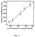
- Figure 2 showing the number of metastatic foci in the liver plotted against the IL-2R ⁇ expression in the whole sample (similar as to what would be measured by ELISA), indicates that a sample showing the average intensity of IL-2R ⁇ expression corresponds to the number of metastatic foci found in the liver.
- the sample of melanoma cells having the highest intensity of IL-2R ⁇ expression, cells exposed to ConA was also the sample of cells that caused the highest number of metastatic foci (Fig. 2, circle D).
- One embodiment for determining the metastasis prognosis comprises the following steps:
- the anti-TCM antibody is coupled with a fluorochrome, chromophore, or enzyme differentiated from the one coupled to the anti-IL-2R ⁇ antibody.
- the metastatis probability two percentages are determined for the non-lymphoid tumor.
- the first percentage is calculated by measuring the percent of the tumor cells or percentage of tumor tissue surface, showing both antigens (IL-2R ⁇ + , TCM + cells).
- the second percentage is calculated by counting the percentage of tumor cells which are identifiable by phenotype (including size, shape traits, flow cytometry forward angle light scatter (FALS)) and which are IL-2R ⁇ positive (Tumor cell phenotype + , IL-2R ⁇ + ).
- the addition of both percentages is the coefficient of metastatic colonies forming cells (Met-CFC coefficient).
- the Met-CFC coefficient is proportional to the metastatic probability. Experimentally estimated as follows: Met-CFC coefficient Metastasis Probability 0% 0% ⁇ 10% ⁇ 30% >45% 100%
- IL-2R ⁇ expression itself can be used experimentally to determine metastasis because T-cells are excluded in the purified tumor.
- anti-TCM and tumor phenotype are used concurrently with IL-2Ra ⁇ .
- B16F10 melanoma cells were treated under different conditions to contrast metastasis efficiency with IL-2R ⁇ expression.
- mice FACS analysis for IL-2 expression, and in vivo tumor cell growth and metastasis efficiency analysis were performed as described above in Section F except that each group of mice consisted of 6 members rather than 20.
- the results, shown in Table 3, indicate that where less than 10% of the cells express IL-2R ⁇ , metastasis occurred in approximately 30% of the mice (B16F10, untreated; and IL-2 treated cells); where no cells express IL-2R ⁇ , metastases did not occur in any of the mice (0% mice, cells treated with IL-4); where 35% of the cells express IL-2R ⁇ , metastasis occurred in greater than 80% of mice (B16F10 grown in suspension); and where greater than 50% of the cells express IL-2R ⁇ , metastasis occurred in 100% of the mice (B16F10 treated with ConA).
- Haematological malignancies including leukemias and lymphomas, have been effectively treated by IL-2 fusion toxins which selectively bind and intoxicate cells bearing high affinity IL-2 receptors (LeMaistre et al., 1992, Immunol-Res. , 11, 42-53; and Waldmann et al., 1992, Ann Intern Med. 116: 148-160); and by humanized antibodies to high affinity IL-2 receptors (hIL-2R), or anti-hIL-2R linked with toxins or radionuclides (Waldmann et al., 1992, supra ).
- the method of the present invention for treating metastases of solid non-lymphoid tumors, and solid non-lymphoid tumors having a high probability of metastasis differs in several important respects.
- the haematological malignancies express hIL-2R (comprised of both ⁇ and ⁇ chains) which is efficiently bound by IL-2 and anti-hIL-2R.
- treatment with the IL-2-toxin or anti-hIL-2R-toxin or anti-hIL-2R comprises infusion systemically.
- one method of the present invention for anticancer therapy against metastases of solid non-lymphoid tumors, and solid non-lymphoid tumors having a high probability of metastasis comprises administering a therapeutically effective amount of a molecule linked to a toxic, radionuclide, or chemotherapeutic agent wherein the molecule is specific for (i.e. has high affinity binding to) IL-2R ⁇ , such as humanized anti-IL-2R ⁇ antibody.
- Another method for treating metastatic colony forming cells and dormant metastatic cells comprises simultaneous delivery of IL-2 and antineoplastic drug to the prometastatic territories.
- the methods of anticancer therapy according to this embodiment of the present invention do not comprise systemic administration, but rather comprise targeting the therapy directly to prometastatic territories.
- the prometastatic territories, targeted by the anticancer therapy of the present invention, are identified by their vasculature, by the terminal sugar concentration, and concentration of extracellular adhesion molecules (ICAMs) expressed on the surface of the cells contained within the territory.
- IAMs extracellular adhesion molecules
- Metastatic cells of solid non-lymphoid tumors only develop metastatic foci ("colonize") in a predictable location comprising unique capillary zones of the target organs such as liver and lung (Barberá-Guillem et al., 1989, Cancer Research 49:4003-4010; Barberá-Guillem et al., 1992, Int. J. Cancer Res. 52:974-977; Barberá-Guillem et al., 1993, Int. J.
- the prometastatic territories are located in the 1st half (primarily in the second quadrant of that half) of the sinusoids extending from the portal vein to the hepatic vein. This area of the sinusoids is also known as the periportal segment of the sinusoids.
- the prometastatic territories are in the terminal pre alveolar venules and the pleural terminal capillaries. Similar regions have been demonstrated in other organs including the adrenal gland.
- Type 1 endothelial cells display an identifying characteristic on their cell surface comprising oligosaccharides having a high concentration of specific terminal sugars (N-acetyl galactosamine; Gal NAC), which is between 6 and 10 times higher than in the rest of the capillary zones (Barberá-Guillem et al., 1991, Hepatology 14:131-139; Vidal-Vanachlocha et al., 1993, supra ).
- the higher expression of these specific sugars results in preferential binding to type 1 endothelial cells by lectins that join specifically to those sugars when the lectin is injected in a circulatory site just prior to the prometastatic territory.
- the injected lectins such as wheat germ agglutinin (WGA)
- WGA wheat germ agglutinin
- Figure 4 wherein the liver zone where tumor cells do not colonize and develop, is free of WGA tagged with rhodamine (dark areas); and the capillary type 1 endothelial cells comprising the prometastatic territories are bound by WGA tagged with rhodamine (bright zones).
- the abduction of the lectin-fluorescent molecules occurs in the target area of the liver where tumor cells colonize and develop metastasis.
- the higher concentration of lectin contained in the capillaries comprising the prometastatic territories reflects the higher terminal sugar (Gal NAC) concentration of capillary type 1 endothelial cells in these territories.
- the endothelial cells of the liver in the prometastatic territories also contain a higher concentration of ICAMs.
- a ligand or antibody with binding specificity for the ICAM can be used as a targeting agent specific for this cell subpopulation.
- Lung prometastatic territories also comprise endothelial cells containing a higher concentration of ICAMs (Zhu et al., 1993, Int. J. Cancer 53:628-633; Zhu et al., 1992, J. Clin. Invest. 89:1718-1724), and ligand or antibody with binding specificity for the ICAM can be used as a targeting agent specific for this cell subpopulation.
- the metastatic cells arrested in the prometastatic territories can go into a dormant state or to a slow proliferative state (pseudo-resting) that is highly resistant to NK cells and to the antitumor drugs. They come out of this state under the influence of low local concentrations of IL-2 and IL-1. When the proliferation of blastic metastatic cell triggers off, they become more sensitive to antineoplastic drugs and to NK cells.
- B16F10 melanoma cells were exposed to different concentrations of Il-L and IL-2, and the resulting growth response was noted.
- 10 4 viable cancer cells were added to each well of a 96 well plate and the cultures were incubated at 5%CO 2 for 48 hours in serum-free or 10% FBS supplemented regular media containing a concentration of mouse recombinant IL-2 ranging from 10-200 U/ml; or 20, 125, or 250 U human recombinant IL-2 (hrIL-2).
- each well was pulsed with 1 ⁇ Ci of [ 3 H]-thymidine, and harvested after 6 hours wherein the radioactivity was subsequently measured.
- the protocol for the measurement of IL-1 effect on B16F10 melanoma cell growth 10 4 viable cancer cells were added to each well of a 96 well plate and the cultures were incubated at 5%CO 2 for 48 hours in serum-free or 10% FBS supplemented regular media containing either 1, 10 or 20 U/ml mouse IL-1 ⁇ . In some cases, anti IL-2R ⁇ antibody was added simultaneously with the IL-1 ⁇ .
- Each well was pulsed with 1 ⁇ Ci of [ 3 H]-thymidine, and harvested after 6 hours wherein the radioactivity was subsequently measured.
- Figure 5 shows that high concentrations (greater than 75 U/ml) of IL-2 induces death or blastic cell cytostatis but does not affect the B16F10 metastatic cells in pseudo-resting state.
- IL-2 concentrations greater than 75 U/ml have a suppressing effect on cell growth of metastatic cells, and that concentrations of between 75 U/ml and 100 U/ml are not toxic for lymphocytes (Fig. 5, represented by CTLL-2) prepared in a similar manner.
- Table 4 shows that IL-2 and IL-1 in low concentrations induces the step from pseudo-resting to blastic and induces the blastic cells proliferation.
- the methods of the present invention for treatment of metastases of solid non-lymphoid tumor comprise anticancer therapy targeted to prometastatic territories.
- the advantages of such a method include that the anticancer therapy is directly targeted to cells in the prometastatic territories in the specific organ, thereby achieving therapeutic concentrations of antineoplastic drug or toxic agent in the area where metastases of solid non-lymphoid tumor cells are localized, with minimal harm to the immune and the hematopoietic systems.
- the anticancer therapy according to the present invention comprises one or more of the following embodiments:
- the compound is administered through a catheter so as to introduce the compound directly into the vascular access of the prometastatic territories.
- the efficacy of treatment may be monitored by measuring the cell-associated expression of IL-2R ⁇ by using any one of the methods, or a combination thereof, described above in 1.1 Sections A-E, wherein a decrease in the number of metastatic cells of non-lymphoid tumor expressing cell-associated IL-2R ⁇ relative to the number of metastatic cells expressing cell-associated IL-2R ⁇ prior to the initiation of treatment is indicative of an anticancer effect from therapy.
- anticancer therapy of non-lymphoid tumors relies on the expression by these tumors of the T-cell receptor ⁇ (TCR ⁇ ) and/or ⁇ chain variants.
- tumor cells may express several variable regions of the ⁇ chain of the TCR (mostly V ⁇ 8), which display a hypervariable region candidate to express a tissular and/or tumoral specificity. Nevertheless, because its functional resemblance with lymphocyte TCR, these sequences, even being specific to the tissue, cannot be immunogenic. Moreover, they can behave like inductor elements of the immunosuppression or of anergy.
- a method according to the present invention is to target anticancer therapy to non-lymphoid tumor cells expressing TCR ⁇ .
- the cell-associated expression of TCR ⁇ was measured on solid, non-lymphoid tumors.
- the experiments showed that human and experimental non-lymphoid tumors express TCR ⁇ .
- the following examples in Sections A through D demonstrate the detection of cell-associated TCR ⁇ in human and experimental tumors using one or more methods selected from the group consisting of fluorescence microscopy (measuring surface TCR ⁇ ), mRNA Northern and slot blot analysis (measuring TCR ⁇ m-RNA expression), enzymatic amplification and analysis of mRNA (measuring TCR ⁇ m-RNA expression), and quantitative fluorescence analysis (measuring surface TCR ⁇ ) analysis.
- the human and experimental tumors were obtained as follows: B16F10 melanoma cells were supplied by Dr. MF Poupon (Paris). Colon 51B and colon 51B lim 10 cells were supplied by Dr. R. Bresalier (USA). MCA-26 cells were supplied by Dr. I. Fidler (USA). MMC-7489 and cells were supplied by Dr. J. Vaageand (USA). SW480, SW480E, SW480R and SW620 were supplied by Dr. I.B. Weinstein. Human biopsies were supplied by Dr. Alonso and Dr. Pellin (Spain). Other tumor cell lines were obtained from the ATCC.
- All tumor cell lines were grown in plastic T-flasks in a humid atmosphere of 5% CO 2 in air, in adequate media supplemented with 10% FBS and containing penicillin and streptomycin.
- the specimens were prepared as follows. Samples of tumors were removed from the patient under surgical conditions; washed in medium containing penicillin and streptomycin; and minced and stirred in 20 ml GBSS containing 0.02% PronaseE (Sigma, USA) and 0.05% Type I Collagenase at 37°C for 10 minutes at a pH of 7.4. The cell suspensions were then filtered through nylon gauze and centrifuged twice at 250 x g for 10 minutes. The isolated cells were then implanted subcutaneously in nude mice. After 21 days of tumor growth, the tumor cells were sampled for investigations of TCR ⁇ expression.
- Tumor biopsies or surgical specimens containing non-lymphoid tumor may be disaggregated, and prepared directly for fluorescence analysis.
- the tumor cells can be cultured in 8-well slide plates (Biotek) for 48 hours, then the supernatants removed and the wells washed gently with PBS and fixed in Carnoy's fixative. After three PBS rinses, the cells were incubated for 30 minutes at 37°C in a solution of PBS containing 4 ⁇ l/ml FITC-rat anti-human-TCR ⁇ (constant and variable regions) monoclonal antibodies (Genzyme, Boehringer Manheim). After incubation, the wells were washed twice with PBS. Controls were prepared only with FITC rat anti-mouse-IgG 2 monoclonal antibody (Boehringer Manheim). The cells were observed with a laser-scanning fluorescence microscope.
- mRNA was obtained from spleen cells, ConA-activated spleen cells, and tumor cells. After extraction and quantification of the mRNA (shown to be intact by 0.7% agarose gel electrophoresis), a total of 20 ⁇ g of purified RNA was separated on a 1.2% agarose gel containing 2.2 M formaldehyde. The electrophoresis was performed at 80 V. The tracks containing the molecular size standard was cut off the gels and the migration distances were measured. The gels were rinsed and transferred onto nitrocellulose filter. Two ⁇ g of mRNA were diluted in a final volume of 100 ⁇ l in 10xSSC-formaldehyde and denatured by heating at 65°C for 10 minutes before blotting.
- mRNA was cross-linked to the nitrocellulose filter with UV irradiation.
- the blots were prehybridized 4 hours at 45°C in 6xSSC, 0.5 SDS, 5x Denhardt's, 10 mM EDTA, 50 ⁇ g/ml of sheared salmon sperm DNA, and 50 ⁇ g/ml of E. coli tRNA.
- human beta-actin DNA probe (Clontech) was labeled by random priming.
- the radiolabeled probes (2x10 9 cpm/ ⁇ l specific radioactivity) were used for hybridization of mRNA blot at 45°C for 24 hours. Then the blots were washed with 5xSSC-0.1 SDS at the hybridization temperature for 20 minutes. The blots were exposed to x-ray film with intensifying screens at -70°C for various periods. The probes were removed from the filters by incubation in sterile water at 100% for 10 minutes and the filters were rehybridized with other probes. Quantitative analyses of 10 autoradiograms were performed with video-densitometry.
- TCR ⁇ mRNA can be detected by extracting and purifying mRNA from non-lymphoid tumor cells and then subjecting the purified mRNA to enzymatic amplification to obtain sufficient quantities for analysis and detection.
- Enzymatic amplification techniques which could be employed include those known in the art such as PCRTM (polymerase chain reaction), QB replicase, and NASBA (nucleic acid sequence-based amplification).
- Detection of the amplified nucleic acid includes techniques known in the art including, but not limited to, agarose gel electrophoresis and Northern blotting; fluorescence-based hybridization assays; chemiluminescence-based hybridization assays, and capture hybridization microtiter assays.
- Oligonucleotides primers and probes can be synthesized from the nucleic acid sequence of the TCR ⁇ gene as evidenced in the art (Waters et al., 1992, Diabetes 41:308-312). Probes can be synthesized with methods known to those skilled in the art to incorporate either non-isotopic or isotopic label. Alternatively, the label may be incorporated directly into the amplified product.
- single-stranded cDNA obtained by reverse transcription from the extracted tumor cell mRNA, was amplified by PCRTM utilizing primers for TCR ⁇ genes from commercial sources. The amplified products were separated on a 1.2% agarose gels and stained with ethidium bromide in parallel with positive and negative controls. Quantification was made with Photoquant SystemTM (BAF, Spain).
- Tumor cells were cultured in 24-wells plates for 48 hours, and then the supernatants were removed and the wells washed gently with PBS, then the cells were gently scraped from tissue culture plates and incubated in a solution of PBS containing 1 ⁇ g/ml rat FITC conjugated anti-mouse-TCR ⁇ monoclonal antibody for 30 minutes at 4°C, then fixed with 1% paraformaldehyde, and analyzed within 48 hours using a microfluorimeter. In all cases, controls were prepared with second antibody alone. Spleen cells were used as negative control and ConA-activated spleen cells as positive control of antigen expression. Quantitative analysis of TCR ⁇ expression on tumor cells was performed using special software (ImageI, USA; and Photoquant, Spain).
- the present invention provides methods for targeting compounds, via TCR ⁇ specificity, directed to metastatic colony forming cells and to dormant metastatic tumor cells.
- the Staphylococcal Enterotoxins (SEA, SEB, etc.) link TCR variable ⁇ chains of tumor cells to CMH II of antigen presenting cells, and consequently a comodulation of immune system and of tumor cells is produced. These reactions are related with the absence of immune response against most of the tumors and with the tumor cells passing to a reserve state capable of being reverted under casual inflammatory circumstances, even years later after the primary tumor eradication.
- TCR ⁇ -targeted methods of the present invention directed to colony former metastatic cells, dormant cells and cancer cells in general, involve immune modulation as described in the following embodiments:
- Immune modulation or tumor proliferation modulation can be achieved by producing anti-TCR ⁇ idiotype monoclonal antibodies against non-lymphoid tumor cells of different groups, based on organ specificity of the tumor, for their diagnostic and pharmacological use.
- antibodies to soluble human TCR ⁇ isolated from the specific non-lymphoid tumor can be made according to the method of Basi et al. (1992, J. of Immunol. Methods 155:175-191), wherein the antibodies generated recognize multiple epitopes on cell-surface TCR expressed by that non-lymphoid tumor.
- a compound comprising a therapeutically effective amount of IL-2 or IL-1 linked to the anti-TCR ⁇ idiotype antibody provide the compound with properties that may effect immune modulation.
- Such a compound may comprise IL-1 or IL-2 contained within carrier liposomes, and anti-TCR ⁇ idiotype antibody incorporated into the surface of liposomes using methods known to those skilled in the art.
- the compound may be administered systemically.
- the TCR ⁇ -targeted antineoplastic drug therapy comprises a compound comprising a therapeutically effective amount of a molecule linked to a toxic, radionuclide, or chemotherapeutic agent wherein the molecule is humanized anti-TCR ⁇ idiotype antibody which is administered systemically.
- a compound may comprise chemotherapeutic agents contained within the carrier liposomes, and the anti-TCR ⁇ idiotype antibody incorporated into the surface of the liposomes, using methods known to those skilled in the art.
- the efficacy of treatment may be monitored by measuring the cell-associated expression of TCR ⁇ by using any one of the methods, or a combination thereof, described above in 3.1 Sections A-D, or by using clinical diagnostic kits comprising immunoassays with anti-TCR ⁇ idiotype antibody, wherein a decrease in the number non-lymphoid tumor cells expressing cell-associated TCR ⁇ relative to the number of non-lymphoid tumor cells expressing TCR ⁇ prior to initiation of treatment is indicative of an anticancer effect from therapy.
- Peptides corresponding to the anti-TCR ⁇ idiotype of tumor cells can be incorporated to MHC II context of the APC for their pharmacologic use.
Landscapes
- Health & Medical Sciences (AREA)
- Life Sciences & Earth Sciences (AREA)
- Engineering & Computer Science (AREA)
- Immunology (AREA)
- Chemical & Material Sciences (AREA)
- Molecular Biology (AREA)
- Biomedical Technology (AREA)
- Hematology (AREA)
- Urology & Nephrology (AREA)
- Bioinformatics & Cheminformatics (AREA)
- General Health & Medical Sciences (AREA)
- Analytical Chemistry (AREA)
- Pathology (AREA)
- Medicinal Chemistry (AREA)
- Physics & Mathematics (AREA)
- Biotechnology (AREA)
- Microbiology (AREA)
- Biochemistry (AREA)
- Cell Biology (AREA)
- General Physics & Mathematics (AREA)
- Food Science & Technology (AREA)
- Proteomics, Peptides & Aminoacids (AREA)
- Organic Chemistry (AREA)
- Wood Science & Technology (AREA)
- Genetics & Genomics (AREA)
- Zoology (AREA)
- Public Health (AREA)
- Veterinary Medicine (AREA)
- Animal Behavior & Ethology (AREA)
- Pharmacology & Pharmacy (AREA)
- Toxicology (AREA)
- General Engineering & Computer Science (AREA)
- Hospice & Palliative Care (AREA)
- Epidemiology (AREA)
- Oncology (AREA)
- Biophysics (AREA)
- Tropical Medicine & Parasitology (AREA)
- General Chemical & Material Sciences (AREA)
- Chemical Kinetics & Catalysis (AREA)
- Nuclear Medicine, Radiotherapy & Molecular Imaging (AREA)
Applications Claiming Priority (3)
| Application Number | Priority Date | Filing Date | Title |
|---|---|---|---|
| US08/118,969 US5536642A (en) | 1993-09-09 | 1993-09-09 | Diagnostic and prognostic methods for solid non-lymphoid tumors and their metastases |
| US118969 | 1993-09-09 | ||
| EP94928028A EP0737311A4 (de) | 1993-09-09 | 1994-09-02 | Diagnostische, prognostische und therapeutische verfahren für feste, nicht-lymphoide tumoren und ihre metastasen |
Related Parent Applications (1)
| Application Number | Title | Priority Date | Filing Date |
|---|---|---|---|
| EP94928028A Division EP0737311A4 (de) | 1993-09-09 | 1994-09-02 | Diagnostische, prognostische und therapeutische verfahren für feste, nicht-lymphoide tumoren und ihre metastasen |
Publications (1)
| Publication Number | Publication Date |
|---|---|
| EP1031835A1 true EP1031835A1 (de) | 2000-08-30 |
Family
ID=22381858
Family Applications (2)
| Application Number | Title | Priority Date | Filing Date |
|---|---|---|---|
| EP94928028A Withdrawn EP0737311A4 (de) | 1993-09-09 | 1994-09-02 | Diagnostische, prognostische und therapeutische verfahren für feste, nicht-lymphoide tumoren und ihre metastasen |
| EP00105520A Withdrawn EP1031835A1 (de) | 1993-09-09 | 1994-09-02 | Diagnostische, prognostische und therapeutische Verfahren für feste, nicht-lymphoide Tumoren und ihre Metastasen |
Family Applications Before (1)
| Application Number | Title | Priority Date | Filing Date |
|---|---|---|---|
| EP94928028A Withdrawn EP0737311A4 (de) | 1993-09-09 | 1994-09-02 | Diagnostische, prognostische und therapeutische verfahren für feste, nicht-lymphoide tumoren und ihre metastasen |
Country Status (10)
| Country | Link |
|---|---|
| US (1) | US5536642A (de) |
| EP (2) | EP0737311A4 (de) |
| JP (1) | JPH09503582A (de) |
| CN (1) | CN1130944A (de) |
| AU (1) | AU686233B2 (de) |
| CA (1) | CA2170623A1 (de) |
| NO (1) | NO960925L (de) |
| PL (1) | PL313349A1 (de) |
| RU (1) | RU2144675C1 (de) |
| WO (1) | WO1995007462A1 (de) |
Families Citing this family (30)
| Publication number | Priority date | Publication date | Assignee | Title |
|---|---|---|---|---|
| SE9602545L (sv) | 1996-06-25 | 1997-12-26 | Michael Mecklenburg | Metod för att diskriminera komplexa biologiska prover |
| EP0878552A1 (de) * | 1997-05-13 | 1998-11-18 | Erasmus Universiteit Rotterdam | Molekularer Nachweis von chromosomalen Veränderungen |
| DE19756782A1 (de) * | 1997-12-19 | 1999-06-24 | Dade Behring Marburg Gmbh | Nachweis und Bestimmung festphasenassoziierter Faktoren |
| CA2322282A1 (en) * | 1998-02-27 | 1999-09-02 | Cli Oncology, Inc. | Method and compositions for differential detection of primary tumor cells and metastatic cells |
| US6790604B1 (en) | 1998-05-12 | 2004-09-14 | Stefan A. Cohen | Methods for identifying or diagnosing carcinoma cells with metastatic potential based on the measurement of lymphoid genes or their products in carcinoma cells |
| AU1442700A (en) * | 1998-10-07 | 2000-04-26 | Biocrystal Limited | Immunotherapy of b cell involvement in progression of solid, nonlymphoid tumors |
| MXPA02006778A (es) * | 2000-01-12 | 2004-04-05 | Ventana Inc | Metodo para determinar la respuesta a una terapia de cancer. |
| RU2166197C1 (ru) * | 2000-01-26 | 2001-04-27 | Научно-исследовательский институт онкологии им. проф. Н.Н. Петрова | Способ комплексной оценки чувствительности опухоли к эндокринотерапии у женщин постменопаузального возраста больных раком молочной железы |
| JP3821636B2 (ja) | 2000-08-21 | 2006-09-13 | 松下電器産業株式会社 | 通信端末装置、基地局装置および無線通信方法 |
| RU2175242C1 (ru) * | 2000-09-27 | 2001-10-27 | Московский НИИ глазных болезней имени Гельмгольца | Способ лечения увеальной меланомы |
| RU2184971C1 (ru) * | 2001-07-24 | 2002-07-10 | Центральный научно-исследовательский рентгено-радиологический институт | Способ прогнозирования рецидива солидных опухолей |
| EP1427810B9 (de) | 2001-08-21 | 2012-04-25 | Ventana Medical Systems, Inc. | Verfahren und quantifizierungsassay zur bestimmung des c-kit/scf/pakt-status |
| RU2202957C1 (ru) * | 2001-08-27 | 2003-04-27 | Новичков Евгений Владимирович | Способ прогнозирования продолжительности жизни больных холангиогенной карциномой печени |
| RU2194992C1 (ru) * | 2002-04-09 | 2002-12-20 | Центральный научно-исследовательский рентгено-радиологический институт | Способ прогнозирования гормонозависимости рака молочной железы |
| RU2234921C1 (ru) * | 2003-09-16 | 2004-08-27 | Уральский научно-исследовательский институт дерматовенерологии и иммунопатологии | Способ лечения грибовидного микоза |
| RU2260799C1 (ru) * | 2004-01-26 | 2005-09-20 | Новокузнецкий государственный институт усовершенствования врачей | Способ прогнозирования предрасположенности к раку легкого у лиц, проживающих в экологически неблагополучных условиях |
| RU2275633C2 (ru) * | 2004-07-12 | 2006-04-27 | Государственное образовательное учреждение высшего профессионального образования Московская медицинская академия им. И.М. Сеченова | Способ диагностики карциномы эрлиха |
| US7989001B2 (en) * | 2004-09-13 | 2011-08-02 | Untae Kim | Method of separating tumor cells with and without lymphotropic metastatic potential in a human carcinoma |
| RU2297632C2 (ru) * | 2005-06-23 | 2007-04-20 | Российский научно-исследовательский нейрохирургический институт им. проф. А.Л. Поленова | Способ отбора нейроонкологических больных для иммунотерапии |
| RU2300109C2 (ru) * | 2005-08-17 | 2007-05-27 | Ростовский научно-исследовательский онкологический институт Федерального агентства по здравоохранению и социальному развитию | Способ прогнозирования церебральных метастатических очагов у больных с метастатическим поражением головного мозга |
| JPWO2007049542A1 (ja) * | 2005-10-24 | 2009-04-30 | 大鵬薬品工業株式会社 | RAR−α作動薬の有効性の予測方法 |
| RU2326601C2 (ru) * | 2006-06-19 | 2008-06-20 | ФГУ Ростовский научно-исследовательский онкологический институт Росздрава | Способ адъювантной иммунотерапии первичных и метастатических злокачественных нерезектабельных опухолей печени |
| JP4943074B2 (ja) * | 2006-07-03 | 2012-05-30 | シスメックス株式会社 | がん転移の判定方法及び装置 |
| CN101970491A (zh) * | 2007-08-30 | 2011-02-09 | 沃尔特及伊莱萨霍尔医学研究院 | 树突状细胞标记物及其用途 |
| RU2422833C2 (ru) * | 2008-06-27 | 2011-06-27 | Институт прикладной механики Российской Академии Наук (Статус государственного учреждения) (ИПРИМ РАН) | Способ одновременного иммунохроматографического определения онкоантигенов psa и сеа |
| GB0908788D0 (en) * | 2009-05-21 | 2009-07-01 | Univ Southampton | Tissue analysis |
| RU2417758C1 (ru) * | 2009-10-21 | 2011-05-10 | Федеральное государственное учреждение "Ростовский научно-исследовательский онкологический институт Федерального агентства по высокотехнологичной медицинской помощи" | Способ оценки эффективности противоопухолевой терапии при нейробластомах у детей |
| RU2550944C2 (ru) * | 2013-08-08 | 2015-05-20 | Федеральное государственное бюджетное учреждение "Научно-исследовательский институт медицинских проблем Севера" Сибирского отделения Российской академии медицинских наук (ФГБУ "НИИМПС" СО РАМН) | Способ прогнозирования развития терминальной стадии у больных хроническим миелолейкозом |
| RU2646787C1 (ru) * | 2017-04-26 | 2018-03-07 | федеральное государственное бюджетное образовательное учреждение высшего образования "Новосибирский государственный медицинский университет" Министерства здравоохранения Российской Федерации (ФГБОУ ВО НГМУ Минздрава России) | Способ определения метастатического потенциала опухоли молочной железы |
| CN118657755B (zh) * | 2024-08-09 | 2024-11-22 | 天津市肿瘤医院(天津医科大学肿瘤医院) | 一种基于深度学习的pd-l1表达的联合阳性分数获取方法及系统 |
Citations (3)
| Publication number | Priority date | Publication date | Assignee | Title |
|---|---|---|---|---|
| EP0202975A2 (de) * | 1985-04-19 | 1986-11-26 | THE UNITED STATES OF AMERICA as represented by the Secretary United States Department of Commerce | Löslicher Interleukin-2-Rezeptor als Anzeiger eines bösartigen Tumors und Verfahren zu dessen Nachweis |
| EP0256714A2 (de) * | 1986-08-01 | 1988-02-24 | Cetus Oncology Corporation | Kombinationstherapie mit Verwendung von Antitumor-Monoklonalen Antikörpern und/oder Immunotoxinen mit Interleukin-2 |
| WO1992000762A1 (en) * | 1990-07-05 | 1992-01-23 | Akzo N.V. | Receptor directed-toxin conjugates |
Family Cites Families (10)
| Publication number | Priority date | Publication date | Assignee | Title |
|---|---|---|---|---|
| JPH0751511B2 (ja) * | 1982-03-15 | 1995-06-05 | 味の素株式会社 | インターロイキン2を含有してなる癌治療剤 |
| US4690915A (en) * | 1985-08-08 | 1987-09-01 | The United States Of America As Represented By The Department Of Health And Human Services | Adoptive immunotherapy as a treatment modality in humans |
| US5006459A (en) * | 1986-03-31 | 1991-04-09 | T Cell Sciences, Inc. | Therapeutic and diagnostic methods using soluble T cell surface molecules |
| US4939093A (en) * | 1987-02-02 | 1990-07-03 | Cetus Corporation | Human IL-2 like polypeptides, DNA sequences and recombinant DNA molecules therefore and methods for the production and use thereof |
| SU1477087A1 (ru) * | 1987-06-30 | 1990-09-23 | Всесоюзный онкологический научный центр АМН СССР | Способ дифференциального определени доброкачественных и злокачественных опухолей молочных желез |
| US5061488A (en) * | 1988-04-15 | 1991-10-29 | The United States Of America As Represented Department Of Health & Human Services | Flavone-8-acetic acid and interleukin-2 for cancer therapy |
| US5126129A (en) * | 1988-05-23 | 1992-06-30 | The Government Of The United States Of America As Represented By The Secretary Of The Department Of Health & Human Services | Cancer therapy using interleukin-2 and flavone compounds |
| DE3821998A1 (de) * | 1988-06-30 | 1990-01-04 | Spectrospin Ag | Icr-ionenfalle |
| US5089261A (en) * | 1989-01-23 | 1992-02-18 | Cetus Corporation | Preparation of a polymer/interleukin-2 conjugate |
| US5229109A (en) * | 1992-04-14 | 1993-07-20 | Board Of Regents, The University Of Texas System | Low toxicity interleukin-2 analogues for use in immunotherapy |
-
1993
- 1993-09-09 US US08/118,969 patent/US5536642A/en not_active Expired - Fee Related
-
1994
- 1994-09-02 RU RU96105073A patent/RU2144675C1/ru active
- 1994-09-02 JP JP7508772A patent/JPH09503582A/ja active Pending
- 1994-09-02 EP EP94928028A patent/EP0737311A4/de not_active Withdrawn
- 1994-09-02 PL PL94313349A patent/PL313349A1/xx unknown
- 1994-09-02 AU AU77220/94A patent/AU686233B2/en not_active Ceased
- 1994-09-02 EP EP00105520A patent/EP1031835A1/de not_active Withdrawn
- 1994-09-02 WO PCT/US1994/010060 patent/WO1995007462A1/en not_active Ceased
- 1994-09-02 CN CN94193358A patent/CN1130944A/zh active Pending
- 1994-09-02 CA CA002170623A patent/CA2170623A1/en not_active Abandoned
-
1996
- 1996-03-07 NO NO960925A patent/NO960925L/no not_active Application Discontinuation
Patent Citations (3)
| Publication number | Priority date | Publication date | Assignee | Title |
|---|---|---|---|---|
| EP0202975A2 (de) * | 1985-04-19 | 1986-11-26 | THE UNITED STATES OF AMERICA as represented by the Secretary United States Department of Commerce | Löslicher Interleukin-2-Rezeptor als Anzeiger eines bösartigen Tumors und Verfahren zu dessen Nachweis |
| EP0256714A2 (de) * | 1986-08-01 | 1988-02-24 | Cetus Oncology Corporation | Kombinationstherapie mit Verwendung von Antitumor-Monoklonalen Antikörpern und/oder Immunotoxinen mit Interleukin-2 |
| WO1992000762A1 (en) * | 1990-07-05 | 1992-01-23 | Akzo N.V. | Receptor directed-toxin conjugates |
Also Published As
| Publication number | Publication date |
|---|---|
| CN1130944A (zh) | 1996-09-11 |
| EP0737311A1 (de) | 1996-10-16 |
| WO1995007462A1 (en) | 1995-03-16 |
| AU686233B2 (en) | 1998-02-05 |
| PL313349A1 (en) | 1996-06-24 |
| EP0737311A4 (de) | 1999-02-24 |
| NO960925L (no) | 1996-05-09 |
| AU7722094A (en) | 1995-03-27 |
| US5536642A (en) | 1996-07-16 |
| JPH09503582A (ja) | 1997-04-08 |
| CA2170623A1 (en) | 1995-03-16 |
| NO960925D0 (no) | 1996-03-07 |
| RU2144675C1 (ru) | 2000-01-20 |
Similar Documents
| Publication | Publication Date | Title |
|---|---|---|
| US5536642A (en) | Diagnostic and prognostic methods for solid non-lymphoid tumors and their metastases | |
| US8568985B2 (en) | Breast endothelial cell expression patterns | |
| Talpur et al. | CD25 expression is correlated with histological grade and response to denileukin diftitox in cutaneous T-cell lymphoma | |
| CN102272599B (zh) | 抗cxcr1组合物和方法 | |
| US7776541B2 (en) | Psoriasin expression by breast epithelial cells | |
| US20120225073A1 (en) | Isolation and Use of Melanoma Cancer Stem Cells | |
| Loberg et al. | Detection and isolation of circulating tumor cells in urologic cancers: a review | |
| Nakano et al. | Expression of tumor necrosis factor‐α and interleukin‐6 in oral squamous cell carcinoma | |
| JP2022522479A5 (de) | ||
| EP3361251A1 (de) | Verfahren zur detektion von krebszellen, reagenz zum einbringen einer substanz in krebszellen und zusammensetzung zur krebsbehandlung | |
| Liang et al. | Proteasome inhibitors restore the STAT1 pathway and enhance the expression of MHC class I on human colon cancer cells | |
| WO1993005804A1 (en) | Activated stromal fibroblast-specific antibody, f19 and methods of using the same | |
| WO1999042140A1 (en) | Detector molecule for use in a method for detecting and surgically removing lymphoid tissue involved in tumor progression | |
| Pilard et al. | RANKL blockade inhibits cancer growth through reversing the tolerogenic profile of tumor-infiltrating (plasmacytoid) dendritic cells | |
| Pentheroudakis et al. | The neutrophil, not the tumor: Serum CA 15‐3 elevation as a result of granulocyte—Colony‐stimulating factor–induced neutrophil MUC1 overexpression and neutrophilia in patients with breast carcinoma receiving adjuvant chemotherapy | |
| Ji et al. | Dual-target CAR-T therapy for ovarian cancer: synergistic targeting of MSLN and B7H3 enhances anti-tumor efficacy and overcomes antigen heterogeneity | |
| CN108324943B (zh) | Cd30+癌症的检测和治疗 | |
| Selli et al. | Serum beta 2 microglobulin levels in patients with renal cell carcinoma | |
| 趙樹康 et al. | IL-32 production from lung adenocarcinoma cells is potentially involved in immunosuppressive microenvironment | |
| OEHLER | Immunomodulatory biomarkers under the influence of neoadjuvant chemotherapy with epirubicin and docetaxel in breast cancer patients | |
| WO2022211620A1 (en) | Cd4/il-2 biomarker. | |
| Miura et al. | Immunohistochemical analysis of mesocolic lymph node cells in human colorectal cancer | |
| GODFREY | SOME RECENT THESES ON THE THYMUS AND RELATED TOPICS | |
| Greenberg et al. | Metastatic pancreatic neuroendocrine tumors manifest elevated T cell infiltration |
Legal Events
| Date | Code | Title | Description |
|---|---|---|---|
| PUAI | Public reference made under article 153(3) epc to a published international application that has entered the european phase |
Free format text: ORIGINAL CODE: 0009012 |
|
| AC | Divisional application: reference to earlier application |
Ref document number: 737311 Country of ref document: EP |
|
| AK | Designated contracting states |
Kind code of ref document: A1 Designated state(s): AT BE CH DE DK ES FR GB GR IE IT LI LU MC NL PT SE |
|
| AKX | Designation fees paid | ||
| STAA | Information on the status of an ep patent application or granted ep patent |
Free format text: STATUS: THE APPLICATION IS DEEMED TO BE WITHDRAWN |
|
| 18D | Application deemed to be withdrawn |
Effective date: 20010301 |