EP1035508B1 - Procédé et système automatisés de segmentation d'images médicales - Google Patents
Procédé et système automatisés de segmentation d'images médicales Download PDFInfo
- Publication number
- EP1035508B1 EP1035508B1 EP00111213A EP00111213A EP1035508B1 EP 1035508 B1 EP1035508 B1 EP 1035508B1 EP 00111213 A EP00111213 A EP 00111213A EP 00111213 A EP00111213 A EP 00111213A EP 1035508 B1 EP1035508 B1 EP 1035508B1
- Authority
- EP
- European Patent Office
- Prior art keywords
- image
- determining
- recited
- pixels
- feature
- Prior art date
- Legal status (The legal status is an assumption and is not a legal conclusion. Google has not performed a legal analysis and makes no representation as to the accuracy of the status listed.)
- Expired - Lifetime
Links
Images






Classifications
-
- G—PHYSICS
- G06—COMPUTING OR CALCULATING; COUNTING
- G06T—IMAGE DATA PROCESSING OR GENERATION, IN GENERAL
- G06T7/00—Image analysis
- G06T7/0002—Inspection of images, e.g. flaw detection
- G06T7/0012—Biomedical image inspection
-
- G—PHYSICS
- G06—COMPUTING OR CALCULATING; COUNTING
- G06T—IMAGE DATA PROCESSING OR GENERATION, IN GENERAL
- G06T7/00—Image analysis
- G06T7/10—Segmentation; Edge detection
- G06T7/11—Region-based segmentation
-
- G—PHYSICS
- G06—COMPUTING OR CALCULATING; COUNTING
- G06T—IMAGE DATA PROCESSING OR GENERATION, IN GENERAL
- G06T7/00—Image analysis
- G06T7/10—Segmentation; Edge detection
- G06T7/12—Edge-based segmentation
-
- G—PHYSICS
- G06—COMPUTING OR CALCULATING; COUNTING
- G06T—IMAGE DATA PROCESSING OR GENERATION, IN GENERAL
- G06T7/00—Image analysis
- G06T7/10—Segmentation; Edge detection
- G06T7/136—Segmentation; Edge detection involving thresholding
-
- G—PHYSICS
- G06—COMPUTING OR CALCULATING; COUNTING
- G06T—IMAGE DATA PROCESSING OR GENERATION, IN GENERAL
- G06T7/00—Image analysis
- G06T7/10—Segmentation; Edge detection
- G06T7/155—Segmentation; Edge detection involving morphological operators
-
- G—PHYSICS
- G06—COMPUTING OR CALCULATING; COUNTING
- G06T—IMAGE DATA PROCESSING OR GENERATION, IN GENERAL
- G06T7/00—Image analysis
- G06T7/10—Segmentation; Edge detection
- G06T7/187—Segmentation; Edge detection involving region growing; involving region merging; involving connected component labelling
-
- G—PHYSICS
- G06—COMPUTING OR CALCULATING; COUNTING
- G06V—IMAGE OR VIDEO RECOGNITION OR UNDERSTANDING
- G06V10/00—Arrangements for image or video recognition or understanding
- G06V10/20—Image preprocessing
- G06V10/26—Segmentation of patterns in the image field; Cutting or merging of image elements to establish the pattern region, e.g. clustering-based techniques; Detection of occlusion
- G06V10/267—Segmentation of patterns in the image field; Cutting or merging of image elements to establish the pattern region, e.g. clustering-based techniques; Detection of occlusion by performing operations on regions, e.g. growing, shrinking or watersheds
-
- G—PHYSICS
- G06—COMPUTING OR CALCULATING; COUNTING
- G06T—IMAGE DATA PROCESSING OR GENERATION, IN GENERAL
- G06T2207/00—Indexing scheme for image analysis or image enhancement
- G06T2207/10—Image acquisition modality
- G06T2207/10116—X-ray image
-
- G—PHYSICS
- G06—COMPUTING OR CALCULATING; COUNTING
- G06T—IMAGE DATA PROCESSING OR GENERATION, IN GENERAL
- G06T2207/00—Indexing scheme for image analysis or image enhancement
- G06T2207/20—Special algorithmic details
- G06T2207/20036—Morphological image processing
-
- G—PHYSICS
- G06—COMPUTING OR CALCULATING; COUNTING
- G06T—IMAGE DATA PROCESSING OR GENERATION, IN GENERAL
- G06T2207/00—Indexing scheme for image analysis or image enhancement
- G06T2207/20—Special algorithmic details
- G06T2207/20036—Morphological image processing
- G06T2207/20041—Distance transform
-
- G—PHYSICS
- G06—COMPUTING OR CALCULATING; COUNTING
- G06T—IMAGE DATA PROCESSING OR GENERATION, IN GENERAL
- G06T2207/00—Indexing scheme for image analysis or image enhancement
- G06T2207/30—Subject of image; Context of image processing
- G06T2207/30004—Biomedical image processing
- G06T2207/30068—Mammography; Breast
-
- G—PHYSICS
- G06—COMPUTING OR CALCULATING; COUNTING
- G06V—IMAGE OR VIDEO RECOGNITION OR UNDERSTANDING
- G06V2201/00—Indexing scheme relating to image or video recognition or understanding
- G06V2201/03—Recognition of patterns in medical or anatomical images
Definitions
- the invention relates generally to a method and system for the computerized automatic segmentation of medical images. Specific applications are given for breast mammograms including the extraction of the skin line as well as correction for non-uniform exposure conditions, for hand radiographs, and for chest radiographs. Techniques include novel developments and implementations including noise filtering, local gray value range determination, modified global histogram analysis, region growing and determination of object contour.
- mammography is currently the best method for the detection of breast cancer, between 10-30% of women who have breast cancer and undergo mammography have negative mammograms. In approximately two-thirds of these false-negative mammograms, the radiologist failed to detect the cancer that was evident retrospectively. The missed detections may be due to the subtle nature of the radiographic findings (i.e., low conspicuity of the lesion), poor image quality, eye fatigue or oversight by the radiologists. In addition, it has been suggested that double reading (by two radiologists) may increase sensitivity. It is apparent that the efficiency and effectiveness of screening procedures could be increased by using a computer system, as a "second opinion or second reading", to aid the radiologist by indicating locations of suspicious abnormalities in mammograms. In addition, mammography is becoming a high volume x-ray procedure routinely interpreted by radiologists.
- a suspicious region is detected by a radiologist, he or she must then visually extract various radiographic characteristics. Using these features, the radiologist then decides if the abnormality is likely to be malignant or benign, and what course of action should be recommended (i.e., return to screening, return for follow-up or return for biopsy).
- Many patients are referred for surgical biopsy on the basis of a radiographically detected mass lesion or cluster of microcalcifications. Although general rules for the differentiation between benign and malignant breast lesions exist, considerable misclassification of lesions occurs with current radiographic techniques. On average, only 10-20% of masses referred for surgical breast biopsy are actually malignant.
- EP-A-0 427 912 describes a medical image segmentation system based on region growing. Growing criteria include local and global density changes.
- an object of this invention is to provide an automated method and system for segmenting medical images.
- Another object of this invention is to provide an automated method and system for the determination of skin line in medical images.
- the invention is directed to an automated system and method for segmentation of an image of an anatomic feature of a subject, said method comprising:
- a further embodiment of the invention is directed to a method wherein said step of segmenting said image produces a segmented image; and said method further comprises: determining an external skinline of said feature using said image segmented image; determining an internal skinline of said feature using said segmented image; and measuring a skin thickness of said feature using said internal and external skinlines.
- a further method comprises also obtaining a gradient image of said segmented image; and determining said internal and external skinlines using said gradient image.
- determining said internal skinline comprises:
- breast mammograms including the extraction of the skin line as well as correction for non-uniform exposure conditions, for hand radiographs, and for chest radiographs.
- Techniques include novel developments and implementations including noise filtering, local gray value range determination, modified global histogram analysis, region growing and determination of object contour.
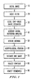
- FIG. 1 a schematic diagram of the automated method for the segmentation of breast images is shown.
- the method includes an initial acquisition of a radiograph of the breast and digitization (step 10).
- Noise filtering is applied to the digital image (step 20) followed by application of the gray-value range operator (step 30).
- step 40 Using information from the local range operator a modified global histogram analysis is performed (step 40).
- Region growing is performed on the threshold image using connectivity (counting pixels) in step 50, followed by a morphological erosion operation (step 60).
- the distance map of the image is determined (step 70) and the boundary of the segmented object in the image is then tracked to yield its contour (step 80).
- the contour can then be output onto the digital image or passed to other computer algorithms (step 90).
- noise filtering using a square median filter 3 by 3 pixels in size, is employed in order to eliminate digitizer line artifacts and spike noise.
- the advantage of using a median filter is that the noise reduction process does not affect the smooth edges.
- Figure 2 shows a schematic diagram illustrating the application of the gray-value range operator.
- a 7 pixel by 7 pixel ring kernel is used to find the local maximum and local minimum pixel values.
- the difference between the local maximum and the center pixel value, and that between the center pixel value and the local minimum are calculated as the range and stored for later reference. Pixels yielding a small local gray-value range are considered as possible "non-object" (non-breast) pixels.
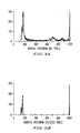
- the range is determined on the basis of the base width of a pixel histogram, as shown in Fig. 3.
- the global gray-value histogram of the image is determined as illustrated in Figs. 3A and 3B.
- the original histogram (Fig. 3A) contains gray values from all pixels in the image.
- the modified histogram (Fig. 3B) contains only contributions from pixels with a small local range (maximum - minimum value), which correspond to possible non-breast pixels.
- the criteria used in classifying a pixel as a non-breast pixel include (1) having a pixel value close to a global histogram peak, (2) having a small local gray value range and (3) being part of a large connected region. This can be thought as obtaining three sequential images.
- Figs. 4A-4C illustrate the effect of these three classification criteria, respectively.
- the direct exposure region (with black corresponding to a gray level of zero) would have pixel values in which the local minimum must be small and the non exposed region (with white being at 1023) would have pixel values in which the local maximum must be small.
- the image is in the form of a 3-gray-level image, where one value corresponds to potential breast pixel and the other two values correspond to potential non-breast pixel (either a no exposure region or a direct exposure region).
- Figs. 4C illustrates the partially segmented breast image at this stage of the method.
- the darker pixels correspond to non-breast pixels. It is noted that the image may contain pixels identified as possible breast pixels in the direct exposure region and in the border.
- the three-value image is subjected to a morphological erosion operation using a 3 pixel by 3 pixel square kernel. Such processing is necessary in order to prevent the "breast region” from including artifacts from the film digitization process, which may have gray values similar to pixels inside the breast region.
- the filtered binary image is then subjected to a contouring routine as illustrated in Figure 5. Note, however, (by comparing Figures 4 and 5) that rules based on knowledge of the mammographic image need to be included in the contouring in order to identify and eliminate the "transition zone" between the direct and non-exposed regions (which is included in "breast region” in Figure 4).
- the image becomes a four-value (2-bit) image. This is done as follows.
- the rules include analysis of connection points, corresponding to points with a concave angle and a short connected path to the outside, which are used in cutting across the transition zone.
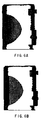
- a distance map of the image is calculated as illustrated in Figs. 6A and 6B.
- Figure 6A illustrates the distance map image
- Figure 6B illustrates the subsequent threshold image obtained by thresholding Figure 6A.
- darker pixels are closer to the film edge.
- the shortest connecting path of "breast object pixels" to the outside i.e., film edge
- the thresholding yields possible transition points which are then analyzed for presence of "sharp" concave angles. Then, the contouring routine need only track the pixels having the single gray value corresponding to the breast region.
- Figure 7 shows an example of a final segmented breast in a digital mammogram, showing an allowed connection at point A. At point B, the connection was not made since the concave angle was not sufficiently sharp. The degree of sharpness required to make the connection is empirically derived.
- Figure 8 is a graph illustrating the performance of the segmentation method, evaluated on 740 mammograms. The ratings were subjectively assigned by 3 observers. Note that 96% were considered acceptable for use as input to further computerized mammographic analysis methods. In the rating scale (x-axis) of Fig. 8, (1) corresponds to optimal, (2) to minor deviations, (3) to acceptable for CAD purposes, (4) to substantial deviations, and (5) to complete failure of segmentation.
- the segmentation method could be employed in an iterative manner as illustrated in Figure 10.
- various parameters of the method could be iteratively tried in order to segment the breast in images obtained from various film digitizers or direct digital devices.
- Figure 9 shows examples of how the computer-determined breast contour (found from breast segmentation) could be further used in such methods as mass detection, microcalcification detection, and skin analysis in computer-aided diagnosis schemes, and image enhancement.
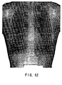
- segmentation can be used in other medical imaging applications including segmentation of the hand in bone radiographs as showed in Figure 11, and segmentation of the chest in images of the thorax as shown in Figure 12.
- both global and local thresholding can be used. Local thresholding is used to segment bone from skin.
- a number of ROIs ROI1-ROI5, in this example
- the corresponding pixel distributions for ROI1-ROI3 are shown in Figs. 14A-14C.
- the center pixel of ROI1 is set to a constant K 1 .
- a valley is found at gray level p 2 . If the center pixel in ROI2 has a gray value less than p 2 , then the center pixel is assigned a gray value of K 2 .
- ROI3 If the center pixel in ROI2 has a gray value greater than p 2 , then the center pixel is assigned a gray value of K 3 . In ROI3, a valley is found at gray level p 3 . The center pixel of ROI3 is assigned gray value K 2 or K 3 if its gray value is less than or greater than p 3 , respectively. It should be noted that ROI4 and ROI5 will have a single peak distribution similar to Fig. 14A as ROI4 is entirely within the bone and ROI5 is entirely within the skin.
- the advantage of the local thresholding is that the peak shown in ROI3 may be too small to be detected on a histogram of an entire image, as shown in Fig. 14D.
- FIG 15 is a more detailed schematic block diagram illustrating a system for implementing the method of the invention for automated segmentation of medical images.
- radiographic images of an object are obtained from an image acquisition device 150 which could be an x-ray exposure device and a laser digitizer, and input to the system.
- Each breast image is digitized and put into memory 151.
- the image data is first passed through a noise filtering circuit 152 and a local gray-value range circuit 153 in order to determine the initial potential regions of breast and non-breast.
- the data is then passed to the modified global histogram analysis circuit 154 and the region growing circuit 155 in order to determine a partial segmentation.
- Image data are passed to the morphological erosion circuit 156, the distance map circuit 157, and the initial contouring circuit 158 which determines the contour by evaluating the thresholded image data after the distance map is obtained, in order to determine the features for input to the contour connection circuit 159.
- the data are retained in image memory 160.
- the superimposing circuit 161 the results are either superimposed onto breast images, stored in file format, or shown with all non-breast regions set to a constant gray value. The results are then displayed on the display system 163 after passing through a digital-to-analog converter 162.
Landscapes
- Engineering & Computer Science (AREA)
- Physics & Mathematics (AREA)
- General Physics & Mathematics (AREA)
- Theoretical Computer Science (AREA)
- Computer Vision & Pattern Recognition (AREA)
- Nuclear Medicine, Radiotherapy & Molecular Imaging (AREA)
- General Health & Medical Sciences (AREA)
- Medical Informatics (AREA)
- Health & Medical Sciences (AREA)
- Radiology & Medical Imaging (AREA)
- Quality & Reliability (AREA)
- Multimedia (AREA)
- Apparatus For Radiation Diagnosis (AREA)
- Image Processing (AREA)
- Ultra Sonic Daignosis Equipment (AREA)
- Image Analysis (AREA)
Claims (32)
- Procédé automatisé de segmentation d'une image d'une caractéristique anatomique d'un sujet comprenant :la détermination d'une plage locale de valeurs de gris pour chaque pixel de ladite image de ladite caractéristique anatomique (30) ;l'obtention (40) d'un histogramme global en utilisant ladite image ;la modification (40) dudit histogramme global sur la base desdites plages locales de valeurs de gris pour obtenir un histogramme global modifié ; etla segmentation de ladite image en utilisant ledit histogramme global modifié.
- Procédé selon la revendication 1, dans lequel la modification dudit histogramme global comprend :la détermination des valeurs minimales locales de pixels dans ladite image ;la détermination des valeurs maximales locales de pixels dans ladite image ;la détermination d'une plage locale de valeurs de gris en utilisant lesdites valeurs minimales locales de pixels et locales maximales ; etla modification dudit histogramme global en utilisant ladite plage.
- Procédé selon la revendication 2, dans lequel la segmentation de ladite image comprend :la classification d'un pixel dans ladite image comme pixel non caractéristique sur la base de ladite plage locale de valeurs de gris, selon que ledit pixel est ou non au voisinage d'une crête de nuance de gris dudit histogramme modifié, et selon que ledit pixel fait partie ou non d'une région connectée ; etl'obtention d'une première image segmentée contenant des premiers pixels ayant une nuance de gris correspondant à un pixel caractéristique et de seconds pixels ayant au moins une valeur de gris correspondant audit pixel non caractéristique.
- Procédé selon la revendication 3, dans lequel la classification dudit pixel dans ladite image sur la base du fait que ledit pixel fait partie ou non de ladite région connectée comprend l'expansion de région.
- Procédé selon la revendication 3, comprenant :l'exécution d'une opération morphologique ouverte sur ladite première image segmentée pour obtenir une deuxième image segmentée ; etl'exécution d'un programme de contourage sur ladite deuxième image segmentée.
- Procédé selon la revendication 3, comprenant :la détermination d'une carte des distances de ladite première région segmentée.
- Procédé selon la revendication 6, dans lequel la détermination de ladite carte des distances comprend :la détermination d'un chemin de connexion pour chaque premier pixel sur ladite première image segmentée ; etle seuillage de ladite carte des distances sur la base dudit chemin de connexion.
- Procédé selon la revendication 7, dans lequel la détermination dudit chemin de connexion comprend :le calcul d'une distance à partir de chaque premier pixel jusqu'à un bord de ladite première image segmentée si un chemin de connexion correspondant ne croise pas une région de ladite première image segmentée correspondant à une région directement exposée de la dite image.
- Procédé selon la revendication 7, comprenant :le seuillage de ladite carte des distances pour générer une deuxième image segmentée ayant des pixels correspondant aux dits pixels caractéristiques ; etla détermination d'un contour de ladite caractéristique en utilisant ladite deuxième région segmentée.
- Procédé selon la revendication 9, dans lequel la détermination dudit contour comprend :l'analyse de points le long dudit contour ayant un chemin de connexion jusqu'à un bord de ladite deuxième image segmentée inférieur à une longueur prédéterminée et un angle concave.
- Procédé selon la revendication 9, dans lequel la déterminationdudit contour comprend :l'analyse de points le long dudit contour ayant un chemin de connexion jusqu'à un bord de ladite deuxième image segmentée traversant une zone de transition correspondant à une région dans ladite image entre une région directement exposée et une région non exposée avec une longueur inférieure à une longueur prédéterminée et un angle concave.
- Procédé de la revendication 1, comprenant :l'obtention d'un contour de ladite caractéristique dans ladite image segmentée ; etl'analyse de points le long dudit contour où ladite caractéristique se connecte à une zone de transition correspondant à une région dans ladite image entre une région directement exposée et une région non exposée.
- Procédé selon la revendication 1, comprenant :dans lequel la modification de 1' histogramme global comprend :la numérisation de ladite image de ladite caractéristique pour obtenir une image numérisée ; etl'exécution d'un filtrage du bruit sur ladite image numérisée pour obtenir une image filtrée ;la détermination de valeurs minimales locales de pixel dans ladite image filtrée ;la détermination de valeurs maximales locales de pixel dans ladite image filtrée ;la détermination d'une plage locale de valeurs de gris en utilisant lesdites valeurs minimales locales et maximales locales ; etla modification dudit histogramme global en utilisant ladite plage.
- Procédé selon la revendication 2, dans lequel la segmentation de ladite image comprend :la détermination d'une première image segmentée en classifiant les premiers pixels dans ladite image comme des pixels non caractéristiques sur la base de ladite plage de valeurs de gris ;la détermination d'une deuxième image segmentée en classifiant les deuxièmes pixels dans ladite image segmentée comme des pixels non caractéristiques sur la base du fait que les seconds pixels sont ou pas au voisinage d'une crête de valeur de gris dudit histogramme modifié ; etla détermination d'une troisième région segmentée en classifiant les troisièmes pixels dans ladite deuxième image segmentée comme des dits pixels non caractéristiques sur la base du fait que les troisièmes pixels font ou non partie d'une région connectée.
- Procédé selon la revendication 14, comprenant :l'obtention d'une quatrième image segmentée contenant des quatrièmes pixels ayant une valeur de gris correspondant à un pixel caractéristique et des cinquièmes pixels ayant au moins une valeur de gris correspondant à un pixel non caractéristique.
- Procédé selon la revendication 1, comprenant :l'obtention d'une image radiographique de sein ; etla segmentation dudit sein dans ladite image radiographique de sein.
- Procédé selon la revendication 16, comprenant :l'obtention d'un contour dudit sein dans ladite image segmentée ; et l'analyse de points le long dudit contour où ledit sein se connecte à une zone de transition correspondant à une région dans ladite image entre une région directement exposée et une région non exposée.
- Procédé selon la revendication 1, comprenant :l'obtention d'une image radiographique de main ; etla segmentation de ladite main à partir de ladite image en utilisant un seuillage local.
- Procédé selon la revendication 18, dans lequel ledit seuillage local comprend :le placement d'un nombre prédéterminé de régions d'intérêt, chacune ayant une pluralité de pixels sur ladite image de main ;la détermination d'une distribution desdits pixels dans chacune desdites régions d'intérêt ; etla définition de pixels sélectionnés dans chacune desdits régions d'intérêt à une valeur prédéterminée sur la base de distributions correspondantes desdits pixels.
- Procédé selon la revendication 7, dans lequel :ladite étape de segmentation de ladite image produit une image segmentée ; etledit procédé comprend en outre :la détermination d'une ligne de peau externe de ladite caractéristique en utilisant ladite image segmentée ;la détermination d'une ligne de peau interne de ladite caractéristique en utilisant ladite image segmentée ;la mesure d'une épaisseur de peau de ladite caractéristique en utilisant lesdites lignes de peau internes et externes.
- Procédé selon la revendication 20, comprenant :l'obtention d'une image de gradient de ladite image segmentée ; etla détermination de lignes de peau internes et externes en utilisant ladite image de gradient.
- Procédé selon la revendication 20, dans lequel la détermination de ladite ligne de peau interne comprend :l'obtention d'une courbe de valeurs de gris perpendiculaire à une bordure de ladite caractéristique ; etla détermination d'un minimum de gradient local dans ladite courbe de valeurs de gris comme ladite ligne de peau interne.
- Procédé selon la revendication 20, dans lequel la détermination d'une ligne de peau externe comprend :la détermination d'une ligne de peau externe initiale de ladite caractéristique ;le calcul d'une deuxième dérivée d'un laplacien de côté obscur ; etla détermination de ladite ligne de peau externe comme une arête dans ladite deuxième dérivée.
- Procédé selon la revendication 23, dans lequel la détermination de ladite ligne de peau externe comme ladite arête comprend :la détermination d'un squelette en échelle de gris de pixels dans ladite image de gradient ; etla détermination d'une arête de maxima dans ledit squelette comme ladite ligne de peau externe.
- Procédé selon la revendication 20, comprenant en outre la détermination de l'épaisseur de peau en utilisant lesdites lignes de peau internes et externes.
- Procédé selon la revendication 20, comprenant en outre la superposition desdites lignes de peau internes et externes sur ladite image.
- Procédé selon la revendication 1, dans lequel
ladite étape de segmentation de ladite image produit une image segmentée ; et
ledit procédé comprend en outre :la détermination d'une ligne de peau externe en utilisant ladite image segmentée ;le calcul d'une distance de pixels de ladite image en tant que pixels caractéristiques potentiels par rapport à ladite ligne de peau externe ;la détermination d'une valeur de gris moyenne desdits pixels caractéristiques potentiels en fonction de ladite distance ; etle renforcement d'une périphérie de ladite image en utilisant la valeur de gris moyenne. - Système pour segmenter automatiquement une image d'une caractéristique anatomique d'un sujet, comprenant :un dispositif d'acquisition d'image (150) ;des moyens pour déterminer un histogramme global de ladite image, connectés audit dispositif d'acquisition d'image ;des moyens pour déterminer une plage locale de valeurs de gris pour chaque pixel de ladite image, connectés aux dits moyens pour déterminer un histogramme global ;des moyens (154)pour modifier ledit histogramme global sur la base desdites plages locales de valeurs de gris ;des moyens pour segmenter ladite image sur la base dudit histogramme global segmenté.
- Système selon la revendication 28, comprenant en outre :des moyens pour définir les contours d'une image segmentée par lesdits moyens de segmentation ; etun écran.
- Système pour segmenter des images, selon la revendication 29, comprenant en outre :une mémoire d'images connectée audit dispositif d'acquisition ;un circuit de carte des distances connecté aux dits moyens pour modifier ledit histogramme global ;un circuit de connexion de contour connecté audit circuit de contourage.
- Système selon la revendication 30, comprenant :un circuit de filtrage du bruit connecté entre ladite mémoire d'images et lesdits moyens pour déterminer une plage locale ;un circuit d'expansion de région connecté aux dits moyens pour modifier ledit histogramme global ;un circuit d'érosion connecté entre ledit circuit d'expansion de région et ledit circuit de carte des distances ; etun circuit de superposition connecté entre ledit circuit de connexion de contour et ledit écran.
- Système selon la revendication 28, comprenant :un circuit d'identification de ligne de peau.
Applications Claiming Priority (3)
| Application Number | Priority Date | Filing Date | Title |
|---|---|---|---|
| US08/158,320 US5452367A (en) | 1993-11-29 | 1993-11-29 | Automated method and system for the segmentation of medical images |
| US158320 | 1993-11-29 | ||
| EP95903128A EP0731959B1 (fr) | 1993-11-29 | 1994-11-29 | Procede et systeme automatises de traitement d'images medicales |
Related Parent Applications (1)
| Application Number | Title | Priority Date | Filing Date |
|---|---|---|---|
| EP95903128.7 Division | 1995-06-01 |
Publications (2)
| Publication Number | Publication Date |
|---|---|
| EP1035508A1 EP1035508A1 (fr) | 2000-09-13 |
| EP1035508B1 true EP1035508B1 (fr) | 2003-07-30 |
Family
ID=22567579
Family Applications (2)
| Application Number | Title | Priority Date | Filing Date |
|---|---|---|---|
| EP95903128A Expired - Lifetime EP0731959B1 (fr) | 1993-11-29 | 1994-11-29 | Procede et systeme automatises de traitement d'images medicales |
| EP00111213A Expired - Lifetime EP1035508B1 (fr) | 1993-11-29 | 1994-11-29 | Procédé et système automatisés de segmentation d'images médicales |
Family Applications Before (1)
| Application Number | Title | Priority Date | Filing Date |
|---|---|---|---|
| EP95903128A Expired - Lifetime EP0731959B1 (fr) | 1993-11-29 | 1994-11-29 | Procede et systeme automatises de traitement d'images medicales |
Country Status (8)
| Country | Link |
|---|---|
| US (1) | US5452367A (fr) |
| EP (2) | EP0731959B1 (fr) |
| JP (1) | JPH09508814A (fr) |
| AT (2) | ATE246383T1 (fr) |
| AU (1) | AU692499B2 (fr) |
| CA (1) | CA2177477A1 (fr) |
| DE (2) | DE69432995T2 (fr) |
| WO (1) | WO1995014966A1 (fr) |
Families Citing this family (192)
| Publication number | Priority date | Publication date | Assignee | Title |
|---|---|---|---|---|
| US6266435B1 (en) | 1993-09-29 | 2001-07-24 | Shih-Ping Wang | Computer-aided diagnosis method and system |
| US6574357B2 (en) | 1993-09-29 | 2003-06-03 | Shih-Ping Wang | Computer-aided diagnosis method and system |
| US6075879A (en) * | 1993-09-29 | 2000-06-13 | R2 Technology, Inc. | Method and system for computer-aided lesion detection using information from multiple images |
| JPH08507249A (ja) * | 1993-12-30 | 1996-08-06 | フィリップス エレクトロニクス エヌ ベー | デジタルマンモグラムでの自動区画化及び皮膚ライン検出 |
| US5583659A (en) * | 1994-11-10 | 1996-12-10 | Eastman Kodak Company | Multi-windowing technique for thresholding an image using local image properties |
| US5572565A (en) * | 1994-12-30 | 1996-11-05 | Philips Electronics North America Corporation | Automatic segmentation, skinline and nipple detection in digital mammograms |
| JP3264932B2 (ja) * | 1995-01-31 | 2002-03-11 | ユナイテッド パーセル サービス オブ アメリカ インコーポレイテッド | テキストを含む画像における背景から前景を分離する方法及び装置 |
| US5657362A (en) * | 1995-02-24 | 1997-08-12 | Arch Development Corporation | Automated method and system for computerized detection of masses and parenchymal distortions in medical images |
| EP0813720A4 (fr) * | 1995-03-03 | 1998-07-01 | Arch Dev Corp | Technique de detection de lesions sur des images medicales et dispositif correspondant |
| US5807276A (en) * | 1995-03-09 | 1998-09-15 | Russin; Lincoln David | Biopsy device and method |
| US5795308A (en) * | 1995-03-09 | 1998-08-18 | Russin; Lincoln D. | Apparatus for coaxial breast biopsy |
| FR2733336A1 (fr) * | 1995-04-20 | 1996-10-25 | Philips Electronique Lab | Procede et dispositif de traitement d'images pour la detection automatique d'objets dans des images numerisees |
| US6256529B1 (en) * | 1995-07-26 | 2001-07-03 | Burdette Medical Systems, Inc. | Virtual reality 3D visualization for surgical procedures |
| US5889882A (en) * | 1996-03-21 | 1999-03-30 | Eastman Kodak Company | Detection of skin-line transition in digital medical imaging |
| CA2201057C (fr) * | 1996-03-29 | 2002-01-01 | Kenji Morimoto | Methode de traitement d'une image sectionnee d'un echantillon osseux comprenant la partie corticale et la partie spongieuse d'un os |
| US5815591A (en) * | 1996-07-10 | 1998-09-29 | R2 Technology, Inc. | Method and apparatus for fast detection of spiculated lesions in digital mammograms |
| US7650015B2 (en) | 1997-07-22 | 2010-01-19 | Image Processing Technologies. LLC | Image processing method |
| US5917929A (en) * | 1996-07-23 | 1999-06-29 | R2 Technology, Inc. | User interface for computer aided diagnosis system |
| US5796862A (en) * | 1996-08-16 | 1998-08-18 | Eastman Kodak Company | Apparatus and method for identification of tissue regions in digital mammographic images |
| DE19636949A1 (de) * | 1996-09-11 | 1998-03-12 | Siemens Ag | Verfahren zur Detektion von Kanten in einem Bildsignal |
| US5982916A (en) * | 1996-09-30 | 1999-11-09 | Siemens Corporate Research, Inc. | Method and apparatus for automatically locating a region of interest in a radiograph |
| US5757880A (en) * | 1997-01-08 | 1998-05-26 | Colomb; Denis | Apparatus, article of manufacture, and method for creation of an uncompressed image of compressed matter |
| US5859891A (en) * | 1997-03-07 | 1999-01-12 | Hibbard; Lyn | Autosegmentation/autocontouring system and method for use with three-dimensional radiation therapy treatment planning |
| US6246782B1 (en) | 1997-06-06 | 2001-06-12 | Lockheed Martin Corporation | System for automated detection of cancerous masses in mammograms |
| US5930327A (en) * | 1997-06-23 | 1999-07-27 | Trex Medical Corporation | X-ray image processing |
| US5984870A (en) * | 1997-07-25 | 1999-11-16 | Arch Development Corporation | Method and system for the automated analysis of lesions in ultrasound images |
| US6058322A (en) * | 1997-07-25 | 2000-05-02 | Arch Development Corporation | Methods for improving the accuracy in differential diagnosis on radiologic examinations |
| WO1999005640A1 (fr) * | 1997-07-25 | 1999-02-04 | Arch Development Corporation | Procede et systeme de segmentation des regions pulmonaires dans les radiographies thoraciques laterales |
| US6317617B1 (en) | 1997-07-25 | 2001-11-13 | Arch Development Corporation | Method, computer program product, and system for the automated analysis of lesions in magnetic resonance, mammogram and ultrasound images |
| US6300960B1 (en) * | 1997-08-04 | 2001-10-09 | Pixar Animation Studios | Realistic surface simulation in computer animation |
| US6246784B1 (en) | 1997-08-19 | 2001-06-12 | The United States Of America As Represented By The Department Of Health And Human Services | Method for segmenting medical images and detecting surface anomalies in anatomical structures |
| US6970587B1 (en) | 1997-08-28 | 2005-11-29 | Icad, Inc. | Use of computer-aided detection system outputs in clinical practice |
| US7308126B2 (en) * | 1997-08-28 | 2007-12-11 | Icad, Inc. | Use of computer-aided detection system outputs in clinical practice |
| US5999639A (en) | 1997-09-04 | 1999-12-07 | Qualia Computing, Inc. | Method and system for automated detection of clustered microcalcifications from digital mammograms |
| US6137898A (en) * | 1997-08-28 | 2000-10-24 | Qualia Computing, Inc. | Gabor filtering for improved microcalcification detection in digital mammograms |
| GB9724110D0 (en) * | 1997-11-15 | 1998-01-14 | Elekta Ab | Analysis of radiographic images |
| AU4318499A (en) | 1997-11-24 | 1999-12-13 | Burdette Medical Systems, Inc. | Real time brachytherapy spatial registration and visualization system |
| US6996549B2 (en) | 1998-05-01 | 2006-02-07 | Health Discovery Corporation | Computer-aided image analysis |
| WO1999057683A1 (fr) | 1998-05-04 | 1999-11-11 | The Johns Hopkins University | Procede et appareil de segmentation de petites structures dans des images |
| US6138045A (en) * | 1998-08-07 | 2000-10-24 | Arch Development Corporation | Method and system for the segmentation and classification of lesions |
| US6381352B1 (en) * | 1999-02-02 | 2002-04-30 | The United States Of America As Represented By The Secretary Of The Navy | Method of isolating relevant subject matter in an image |
| US6801645B1 (en) | 1999-06-23 | 2004-10-05 | Icad, Inc. | Computer aided detection of masses and clustered microcalcifications with single and multiple input image context classification strategies |
| US6941323B1 (en) | 1999-08-09 | 2005-09-06 | Almen Laboratories, Inc. | System and method for image comparison and retrieval by enhancing, defining, and parameterizing objects in images |
| US6898303B2 (en) | 2000-01-18 | 2005-05-24 | Arch Development Corporation | Method, system and computer readable medium for the two-dimensional and three-dimensional detection of lesions in computed tomography scans |
| US6901156B2 (en) * | 2000-02-04 | 2005-05-31 | Arch Development Corporation | Method, system and computer readable medium for an intelligent search workstation for computer assisted interpretation of medical images |
| US20030135102A1 (en) * | 2000-05-18 | 2003-07-17 | Burdette Everette C. | Method and system for registration and guidance of intravascular treatment |
| US20020159641A1 (en) * | 2001-03-14 | 2002-10-31 | Whitney Paul D. | Directed dynamic data analysis |
| FR2825816B1 (fr) * | 2001-06-08 | 2004-07-23 | Ge Med Sys Global Tech Co Llc | Procede d'affichage d'images d'organes |
| WO2002103065A2 (fr) * | 2001-06-20 | 2002-12-27 | Koninklijke Philips Electronics N.V. | Procede de segmentation d'images numeriques |
| WO2003039370A1 (fr) | 2001-11-05 | 2003-05-15 | Computerized Medical Systems, Inc. | Appareil et procede de reperage, de guidage, et de ciblage d'une therapie au moyen du rayonnement de faisceaux externes |
| AUPR891701A0 (en) * | 2001-11-16 | 2001-12-13 | Proteome Systems Ltd | Method for locating the edge of an object |
| US7123761B2 (en) * | 2001-11-20 | 2006-10-17 | Konica Corporation | Feature extracting method, subject recognizing method and image processing apparatus |
| US7054473B1 (en) | 2001-11-21 | 2006-05-30 | R2 Technology, Inc. | Method and apparatus for an improved computer aided diagnosis system |
| US7072498B1 (en) | 2001-11-21 | 2006-07-04 | R2 Technology, Inc. | Method and apparatus for expanding the use of existing computer-aided detection code |
| US20030103663A1 (en) * | 2001-11-23 | 2003-06-05 | University Of Chicago | Computerized scheme for distinguishing between benign and malignant nodules in thoracic computed tomography scans by use of similar images |
| GB0205000D0 (en) * | 2002-03-04 | 2002-04-17 | Isis Innovation | Unsupervised data segmentation |
| US6707878B2 (en) * | 2002-04-15 | 2004-03-16 | General Electric Company | Generalized filtered back-projection reconstruction in digital tomosynthesis |
| US7187800B2 (en) | 2002-08-02 | 2007-03-06 | Computerized Medical Systems, Inc. | Method and apparatus for image segmentation using Jensen-Shannon divergence and Jensen-Renyi divergence |
| US7260250B2 (en) * | 2002-09-30 | 2007-08-21 | The United States Of America As Represented By The Secretary Of The Department Of Health And Human Services | Computer-aided classification of anomalies in anatomical structures |
| US7577282B2 (en) | 2002-11-27 | 2009-08-18 | Hologic, Inc. | Image handling and display in X-ray mammography and tomosynthesis |
| US7123684B2 (en) | 2002-11-27 | 2006-10-17 | Hologic, Inc. | Full field mammography with tissue exposure control, tomosynthesis, and dynamic field of view processing |
| US10638994B2 (en) | 2002-11-27 | 2020-05-05 | Hologic, Inc. | X-ray mammography with tomosynthesis |
| US8571289B2 (en) | 2002-11-27 | 2013-10-29 | Hologic, Inc. | System and method for generating a 2D image from a tomosynthesis data set |
| FR2847698B1 (fr) * | 2002-11-27 | 2005-05-06 | Ge Med Sys Global Tech Co Llc | Procede de gestion de la dynamique d'une image radiologique numerique |
| US8565372B2 (en) | 2003-11-26 | 2013-10-22 | Hologic, Inc | System and method for low dose tomosynthesis |
| US7616801B2 (en) * | 2002-11-27 | 2009-11-10 | Hologic, Inc. | Image handling and display in x-ray mammography and tomosynthesis |
| US20040122707A1 (en) * | 2002-12-18 | 2004-06-24 | Sabol John M. | Patient-driven medical data processing system and method |
| US20040122704A1 (en) * | 2002-12-18 | 2004-06-24 | Sabol John M. | Integrated medical knowledge base interface system and method |
| US20040122787A1 (en) * | 2002-12-18 | 2004-06-24 | Avinash Gopal B. | Enhanced computer-assisted medical data processing system and method |
| US20040122708A1 (en) * | 2002-12-18 | 2004-06-24 | Avinash Gopal B. | Medical data analysis method and apparatus incorporating in vitro test data |
| US20040122709A1 (en) * | 2002-12-18 | 2004-06-24 | Avinash Gopal B. | Medical procedure prioritization system and method utilizing integrated knowledge base |
| US7187790B2 (en) * | 2002-12-18 | 2007-03-06 | Ge Medical Systems Global Technology Company, Llc | Data processing and feedback method and system |
| US20040122706A1 (en) * | 2002-12-18 | 2004-06-24 | Walker Matthew J. | Patient data acquisition system and method |
| US20040122719A1 (en) * | 2002-12-18 | 2004-06-24 | Sabol John M. | Medical resource processing system and method utilizing multiple resource type data |
| US7490085B2 (en) * | 2002-12-18 | 2009-02-10 | Ge Medical Systems Global Technology Company, Llc | Computer-assisted data processing system and method incorporating automated learning |
| US20040122705A1 (en) * | 2002-12-18 | 2004-06-24 | Sabol John M. | Multilevel integrated medical knowledge base system and method |
| US20040122702A1 (en) * | 2002-12-18 | 2004-06-24 | Sabol John M. | Medical data processing system and method |
| US20040122703A1 (en) * | 2002-12-19 | 2004-06-24 | Walker Matthew J. | Medical data operating model development system and method |
| US7321669B2 (en) * | 2003-07-10 | 2008-01-22 | Sarnoff Corporation | Method and apparatus for refining target position and size estimates using image and depth data |
| US7369638B2 (en) * | 2003-07-11 | 2008-05-06 | Siemens Medical Solutions Usa, Inc. | System and method for detecting a protrusion in a medical image |
| US20070122033A1 (en) * | 2003-12-10 | 2007-05-31 | Qingmao Hu | Methods and apparatus for binarising images |
| US7515743B2 (en) * | 2004-01-08 | 2009-04-07 | Siemens Medical Solutions Usa, Inc. | System and method for filtering a medical image |
| JP4549082B2 (ja) * | 2004-03-17 | 2010-09-22 | キヤノン株式会社 | 画像処理装置及び方法 |
| GB2412803A (en) * | 2004-03-30 | 2005-10-05 | Amersham Biosciences Niagara I | Image segmentation using local and remote pixel value comparison |
| US20060018524A1 (en) * | 2004-07-15 | 2006-01-26 | Uc Tech | Computerized scheme for distinction between benign and malignant nodules in thoracic low-dose CT |
| JP2006068373A (ja) * | 2004-09-03 | 2006-03-16 | Fuji Photo Film Co Ltd | 乳頭検出装置およびそのプログラム |
| US7662082B2 (en) | 2004-11-05 | 2010-02-16 | Theragenics Corporation | Expandable brachytherapy device |
| EP2602743B1 (fr) | 2004-11-15 | 2014-11-05 | Hologic, Inc. | Affichage et génération de géométrie de mise en correspondance de mammographies et d'images de tomosynthèse |
| EP3106094B1 (fr) | 2004-11-26 | 2021-09-08 | Hologic, Inc. | Systeme radiographique multimode integrant mammographie/tomosynthese |
| US20060136259A1 (en) * | 2004-12-17 | 2006-06-22 | General Electric Company | Multi-dimensional analysis of medical data |
| US20060136417A1 (en) * | 2004-12-17 | 2006-06-22 | General Electric Company | Method and system for search, analysis and display of structured data |
| DE102004061507B4 (de) * | 2004-12-21 | 2007-04-12 | Siemens Ag | Verfahren zur Korrektur von Inhomogenitäten in einem Bild sowie bildgebende Vorrichtung dazu |
| GB0510793D0 (en) * | 2005-05-26 | 2005-06-29 | Bourbay Ltd | Segmentation of digital images |
| GB0510792D0 (en) * | 2005-05-26 | 2005-06-29 | Bourbay Ltd | Assisted selections with automatic indication of blending areas |
| US20100284579A1 (en) * | 2005-07-27 | 2010-11-11 | Tsuyoshi Kobayashi | Abnormal shadow candidate detecting method and abnormal shadow candidate detecting apparatus |
| CN1907225B (zh) * | 2005-08-05 | 2011-02-02 | Ge医疗系统环球技术有限公司 | 用于脑内出血损伤分割的方法和设备 |
| US20070078873A1 (en) * | 2005-09-30 | 2007-04-05 | Avinash Gopal B | Computer assisted domain specific entity mapping method and system |
| US10008184B2 (en) | 2005-11-10 | 2018-06-26 | Hologic, Inc. | System and method for generating a 2D image using mammography and/or tomosynthesis image data |
| US8079946B2 (en) | 2005-11-18 | 2011-12-20 | Senorx, Inc. | Asymmetrical irradiation of a body cavity |
| US7899514B1 (en) | 2006-01-26 | 2011-03-01 | The United States Of America As Represented By The Secretary Of The Army | Medical image processing methodology for detection and discrimination of objects in tissue |
| WO2007095330A2 (fr) | 2006-02-15 | 2007-08-23 | Hologic Inc | Biopsie mammaire et localisation a l'aiguille a l'aide de systemes de tomosynthese |
| US20070206844A1 (en) * | 2006-03-03 | 2007-09-06 | Fuji Photo Film Co., Ltd. | Method and apparatus for breast border detection |
| DE102006021042A1 (de) * | 2006-04-28 | 2007-10-31 | Image Diagnost International Gmbh | Vorrichtung und Verfahren zur computergestützten Analyse sich hinsichtlich ihrer Bildcharakteristik unterscheidender Mammogramme |
| DE102006021036B4 (de) * | 2006-04-28 | 2010-04-08 | Image Diagnost International Gmbh | Vorrichtung und Verfahren zur computergestützten Analyse von Mammogrammen |
| US7961975B2 (en) * | 2006-07-31 | 2011-06-14 | Stc. Unm | System and method for reduction of speckle noise in an image |
| US8184927B2 (en) * | 2006-07-31 | 2012-05-22 | Stc.Unm | System and method for reduction of speckle noise in an image |
| JP4833785B2 (ja) * | 2006-09-29 | 2011-12-07 | 富士フイルム株式会社 | 放射線撮影装置および放射線撮影方法 |
| DE102006046191B4 (de) | 2006-09-29 | 2017-02-02 | Siemens Healthcare Gmbh | Streustrahlungskorrektur in der Radiographie und Computertomographie mit Flächendetektoren |
| US7873194B2 (en) | 2006-10-25 | 2011-01-18 | Rcadia Medical Imaging Ltd. | Method and system for automatic analysis of blood vessel structures and pathologies in support of a triple rule-out procedure |
| US7940977B2 (en) | 2006-10-25 | 2011-05-10 | Rcadia Medical Imaging Ltd. | Method and system for automatic analysis of blood vessel structures to identify calcium or soft plaque pathologies |
| US7983459B2 (en) | 2006-10-25 | 2011-07-19 | Rcadia Medical Imaging Ltd. | Creating a blood vessel tree from imaging data |
| US7860283B2 (en) | 2006-10-25 | 2010-12-28 | Rcadia Medical Imaging Ltd. | Method and system for the presentation of blood vessel structures and identified pathologies |
| US7940970B2 (en) | 2006-10-25 | 2011-05-10 | Rcadia Medical Imaging, Ltd | Method and system for automatic quality control used in computerized analysis of CT angiography |
| US8086002B2 (en) * | 2007-04-27 | 2011-12-27 | Three Palm Software | Algorithms for selecting mass density candidates from digital mammograms |
| US7630533B2 (en) | 2007-09-20 | 2009-12-08 | Hologic, Inc. | Breast tomosynthesis with display of highlighted suspected calcifications |
| US20090080773A1 (en) * | 2007-09-20 | 2009-03-26 | Mark Shaw | Image segmentation using dynamic color gradient threshold, texture, and multimodal-merging |
| US7792245B2 (en) * | 2008-06-24 | 2010-09-07 | Hologic, Inc. | Breast tomosynthesis system with shifting face shield |
| US7991106B2 (en) | 2008-08-29 | 2011-08-02 | Hologic, Inc. | Multi-mode tomosynthesis/mammography gain calibration and image correction using gain map information from selected projection angles |
| EP2161688B1 (fr) | 2008-09-03 | 2012-03-14 | Agfa Healthcare | Procédé de dérivation de la quantité de tissu dense dans une représentation d'image mammographique numérique |
| EP2356626B1 (fr) * | 2008-10-29 | 2013-05-29 | Koninklijke Philips Electronics N.V. | Analyse d'au moins une image médicale en trois dimensions |
| US8139832B2 (en) * | 2008-12-12 | 2012-03-20 | Hologic, Inc. | Processing medical images of the breast to detect anatomical abnormalities therein |
| US9579524B2 (en) | 2009-02-11 | 2017-02-28 | Hologic, Inc. | Flexible multi-lumen brachytherapy device |
| US9248311B2 (en) | 2009-02-11 | 2016-02-02 | Hologic, Inc. | System and method for modifying a flexibility of a brachythereapy catheter |
| US10207126B2 (en) | 2009-05-11 | 2019-02-19 | Cytyc Corporation | Lumen visualization and identification system for multi-lumen balloon catheter |
| WO2011007312A1 (fr) * | 2009-07-17 | 2011-01-20 | Koninklijke Philips Electronics N.V. | Imagerie multimodale de sein |
| GB2474319B (en) * | 2009-07-20 | 2014-05-07 | Matakina Technology Ltd | Method and system for analysing tissue from images |
| ES2699471T3 (es) * | 2009-08-03 | 2019-02-11 | Volpara Health Tech Limited | Método y sistema para analizar tejido a partir de imágenes |
| EP2485651B1 (fr) | 2009-10-08 | 2020-12-23 | Hologic, Inc. | Système de ponction-biopsie du sein |
| JP5468362B2 (ja) * | 2009-11-18 | 2014-04-09 | 株式会社東芝 | マンモグラフィ装置 |
| CN101866487B (zh) * | 2010-06-11 | 2014-01-08 | 中国科学院深圳先进技术研究院 | 从医学图像中提取身体部位的方法和系统 |
| US9352172B2 (en) | 2010-09-30 | 2016-05-31 | Hologic, Inc. | Using a guide member to facilitate brachytherapy device swap |
| EP2624761B1 (fr) | 2010-10-05 | 2021-07-14 | Hologic, Inc. | Imagerie du sein par rayon x en position debout avec un mode de tomodensitométrie, de multiples modes de tomosynthèse et un mode de mammographie |
| WO2015054518A1 (fr) | 2013-10-09 | 2015-04-16 | Hologic, Inc | Tomosynthèse du sein à rayons x améliorant la résolution spatiale y compris dans le sens de l'épaisseur du sein aplati |
| US20120133600A1 (en) | 2010-11-26 | 2012-05-31 | Hologic, Inc. | User interface for medical image review workstation |
| WO2012082861A2 (fr) | 2010-12-14 | 2012-06-21 | Hologic, Inc. | Système et procédé de fusion de données d'image tridimensionnelle provenant d'une pluralité de systèmes d'imagerie différents destinés à être utilisés en imagerie diagnostique |
| US8861814B2 (en) * | 2010-12-22 | 2014-10-14 | Chevron U.S.A. Inc. | System and method for multi-phase segmentation of density images representing porous media |
| US10342992B2 (en) | 2011-01-06 | 2019-07-09 | Hologic, Inc. | Orienting a brachytherapy applicator |
| WO2012109643A2 (fr) * | 2011-02-11 | 2012-08-16 | Emory University | Systèmes, procédés et supports d'enregistrement lisibles par ordinateur stockant des instructions destinées à classifier des images tomodensitométriques des seins |
| CA2829349C (fr) | 2011-03-08 | 2021-02-09 | Hologic, Inc. | Systeme et procede pour une imagerie de seins a double energie et/ou a injection d'un agent de contraste pour un depistage, un diagnostic et une biopsie |
| US8600107B2 (en) * | 2011-03-31 | 2013-12-03 | Smart Technologies Ulc | Interactive input system and method |
| FR2977054A1 (fr) * | 2011-06-23 | 2012-12-28 | St Microelectronics Grenoble 2 | Procede d'amelioration de la perception visuelle d'une image numerique |
| US8781187B2 (en) * | 2011-07-13 | 2014-07-15 | Mckesson Financial Holdings | Methods, apparatuses, and computer program products for identifying a region of interest within a mammogram image |
| JP2014534042A (ja) | 2011-11-27 | 2014-12-18 | ホロジック, インコーポレイテッドHologic, Inc. | マンモグラフィーおよび/またはトモシンセシス画像データを使用して2d画像を生成するためのシステムおよび方法 |
| US9805507B2 (en) | 2012-02-13 | 2017-10-31 | Hologic, Inc | System and method for navigating a tomosynthesis stack using synthesized image data |
| CN103093449A (zh) * | 2013-02-28 | 2013-05-08 | 重庆大学 | 一种多分辨率融合的射线图像增强方法 |
| EP2967473B1 (fr) | 2013-03-15 | 2020-02-19 | Hologic, Inc. | Système et procédé pour explorer une pile de tomosynthèse comprenant une mise au point automatique |
| JP6388347B2 (ja) | 2013-03-15 | 2018-09-12 | ホロジック, インコーポレイテッドHologic, Inc. | 腹臥位におけるトモシンセシス誘導生検 |
| CN104367331B (zh) * | 2013-08-15 | 2017-02-15 | 深圳市蓝韵实业有限公司 | 用于数字乳腺x射线机的全数字自动曝光方法 |
| EP3035850B1 (fr) | 2013-08-20 | 2020-05-13 | Densitas Incorporated | Méthodes et systèmes de détermination de densité mammaire |
| EP4278977A3 (fr) | 2013-10-24 | 2024-02-21 | Hologic, Inc. | Système et procédé de navigation pour une biopise du sein guidée par rayons x |
| US9443161B2 (en) * | 2014-02-24 | 2016-09-13 | Dimensions And Shapes, Llc | Methods and systems for performing segmentation and registration of images using neutrosophic similarity scores |
| JP6506769B2 (ja) | 2014-02-28 | 2019-04-24 | ホロジック, インコーポレイテッドHologic, Inc. | トモシンセシス画像スラブを生成し表示するためのシステムおよび方法 |
| US9626476B2 (en) | 2014-03-27 | 2017-04-18 | Change Healthcare Llc | Apparatus, method and computer-readable storage medium for transforming digital images |
| CN104680537B (zh) * | 2015-03-09 | 2018-05-15 | 苏州比特速浪电子科技有限公司 | 图像处理装置 |
| CN108471995B (zh) * | 2015-09-30 | 2022-03-29 | 上海联影医疗科技股份有限公司 | 确定医学图像中乳房区域的系统和方法 |
| CN108780571B (zh) * | 2015-12-31 | 2022-05-31 | 上海联影医疗科技股份有限公司 | 一种图像处理方法和系统 |
| US11076820B2 (en) | 2016-04-22 | 2021-08-03 | Hologic, Inc. | Tomosynthesis with shifting focal spot x-ray system using an addressable array |
| ES2978199T3 (es) | 2016-05-27 | 2024-09-06 | Hologic Inc | Detección de tumor de superficie e interna sincronizada |
| WO2018013703A1 (fr) | 2016-07-12 | 2018-01-18 | Mindshare Medical, Inc. | Système d'analyse médicale |
| US11246551B2 (en) | 2016-09-20 | 2022-02-15 | KUB Technologies, Inc. | System and method for computer aided detection (CAD) in a breast specimen radiograph |
| CN110621233B (zh) | 2017-03-30 | 2023-12-12 | 豪洛捷公司 | 用于处理乳房组织图像数据的方法 |
| CN110662489B (zh) | 2017-03-30 | 2024-08-02 | 豪洛捷公司 | 用于靶向对象增强以生成合成乳房组织图像的系统和方法 |
| EP3600047A1 (fr) | 2017-03-30 | 2020-02-05 | Hologic, Inc. | Système et procédé de synthèse et de représentation d'image de caractéristique multiniveau hiérarchique |
| WO2018236565A1 (fr) | 2017-06-20 | 2018-12-27 | Hologic, Inc. | Procédé et système d'imagerie médicale à auto-apprentissage dynamique |
| DE202018006903U1 (de) | 2017-08-16 | 2024-07-29 | Hologic Inc. | Techniken zur Patientenbewegungsartefaktkompensation bei Brustbildgebung |
| EP3449835B1 (fr) | 2017-08-22 | 2023-01-11 | Hologic, Inc. | Système et méthode de tomographie assistée par ordinateur pour imager de multiples cibles anatomiques |
| US11049606B2 (en) | 2018-04-25 | 2021-06-29 | Sota Precision Optics, Inc. | Dental imaging system utilizing artificial intelligence |
| US11090017B2 (en) | 2018-09-13 | 2021-08-17 | Hologic, Inc. | Generating synthesized projection images for 3D breast tomosynthesis or multi-mode x-ray breast imaging |
| WO2020068851A1 (fr) | 2018-09-24 | 2020-04-02 | Hologic, Inc. | Cartographie mammaire et localisation d'anomalie |
| CN112770674B (zh) | 2018-09-28 | 2024-08-06 | 豪洛捷公司 | 通过高密度元素抑制来合成乳腺组织的图像生成的系统和方法 |
| US10748290B2 (en) * | 2018-10-31 | 2020-08-18 | Fei Company | Smart metrology on microscope images |
| US12437390B2 (en) | 2018-11-24 | 2025-10-07 | Densitas Incorporated | System and method for assessing quality of medical images |
| DE202019006040U1 (de) | 2018-11-25 | 2024-08-06 | Hologic Inc. | Multimodalitätsaufhängungsprotokolle |
| KR20210148132A (ko) | 2019-03-29 | 2021-12-07 | 홀로직, 인크. | 스닙-트리거링된 디지털 영상 보고서 생성 |
| US11883206B2 (en) | 2019-07-29 | 2024-01-30 | Hologic, Inc. | Personalized breast imaging system |
| EP3832689A3 (fr) | 2019-12-05 | 2021-08-11 | Hologic, Inc. | Systèmes et procédés pour améliorer la durée de vie d'un tube à rayons x |
| CN111260631B (zh) * | 2020-01-16 | 2023-05-05 | 成都地铁运营有限公司 | 一种高效刚性接触线结构光光条提取方法 |
| US11471118B2 (en) | 2020-03-27 | 2022-10-18 | Hologic, Inc. | System and method for tracking x-ray tube focal spot position |
| US12505645B2 (en) | 2020-03-27 | 2025-12-23 | Hologic, Inc. | Systems and methods for correlating regions of interest in multiple imaging modalities |
| JP7671776B2 (ja) | 2020-03-27 | 2025-05-02 | ホロジック, インコーポレイテッド | 複数の画像化モダリティにおける関心対象領域を識別するシステム及び方法 |
| JP7551419B2 (ja) * | 2020-09-23 | 2024-09-17 | キヤノン株式会社 | 情報処理装置、情報処理方法及びプログラム |
| US20220164951A1 (en) | 2020-11-20 | 2022-05-26 | Hologic, Inc. | Systems and methods for using ai to identify regions of interest in medical images |
| US11786191B2 (en) | 2021-05-17 | 2023-10-17 | Hologic, Inc. | Contrast-enhanced tomosynthesis with a copper filter |
| US12186119B2 (en) | 2021-10-05 | 2025-01-07 | Hologic, Inc. | Interactive model interface for image selection in medical imaging systems |
| US12254586B2 (en) | 2021-10-25 | 2025-03-18 | Hologic, Inc. | Auto-focus tool for multimodality image review |
| CN118318247A (zh) | 2021-11-29 | 2024-07-09 | 豪洛捷公司 | 用于将感兴趣的对象相关的系统和方法 |
| US12414217B2 (en) | 2022-02-07 | 2025-09-09 | Hologic, Inc. | Systems and methods for adaptively controlling filament current in an X-ray tube |
| CN115553795B (zh) * | 2022-07-26 | 2026-03-24 | 科宁(天津)医疗设备有限公司 | 锥光束乳腺ct影像中计算机辅助自动去皮方法 |
| CN115998225A (zh) * | 2022-12-12 | 2023-04-25 | 珠海泰科医疗技术有限公司 | 一种内窥镜病灶区域曝光方法、内窥镜和存储介质 |
| CN117635609B (zh) * | 2024-01-25 | 2024-03-29 | 深圳市智宇精密五金塑胶有限公司 | 一种塑胶产品生产质量视觉检测方法 |
| ES3037026A1 (es) * | 2024-03-22 | 2025-09-26 | Sanchez Puebla Rodriguez Miguel Angel | Sistema experto para el analisis morfologico automatico de imagenes de celulas en la microglia de la retina |
| CN118941582B (zh) * | 2024-10-11 | 2024-12-06 | 大连千玺网络科技有限公司 | 一种地质勘察遥感影像优化分割方法 |
Family Cites Families (10)
| Publication number | Priority date | Publication date | Assignee | Title |
|---|---|---|---|---|
| US4581636A (en) * | 1984-04-02 | 1986-04-08 | Advanced Technology Laboratories, Inc. | Scan conversion apparatus and method |
| US4731863A (en) * | 1986-04-07 | 1988-03-15 | Eastman Kodak Company | Digital image processing method employing histogram peak detection |
| US4907156A (en) * | 1987-06-30 | 1990-03-06 | University Of Chicago | Method and system for enhancement and detection of abnormal anatomic regions in a digital image |
| US4952805A (en) * | 1987-08-20 | 1990-08-28 | Fuji Photo Film Co., Ltd. | Method of judging the presence or absence of a limited irradiation field, method of selecting a correct irradiation field, and method of judging correctness or incorrectness of an irradiation field |
| EP0360231B1 (fr) * | 1988-09-19 | 1996-08-28 | Fuji Photo Film Co., Ltd. | Méthode pour déterminer les plages de signal d'image désirées et méthode pour déterminer les régions d'image désirées |
| US5133020A (en) * | 1989-07-21 | 1992-07-21 | Arch Development Corporation | Automated method and system for the detection and classification of abnormal lesions and parenchymal distortions in digital medical images |
| CA2014918A1 (fr) * | 1989-09-06 | 1991-03-06 | James A. Mcfaul | Systeme de mammographie par balyage avec meilleure visualisation de la ligne cutanee |
| JP2845995B2 (ja) * | 1989-10-27 | 1999-01-13 | 株式会社日立製作所 | 領域抽出手法 |
| US5164993A (en) * | 1991-11-25 | 1992-11-17 | Eastman Kodak Company | Method and apparatus for automatic tonescale generation in digital radiographic images |
| US5268967A (en) * | 1992-06-29 | 1993-12-07 | Eastman Kodak Company | Method for automatic foreground and background detection in digital radiographic images |
-
1993
- 1993-11-29 US US08/158,320 patent/US5452367A/en not_active Expired - Lifetime
-
1994
- 1994-11-29 EP EP95903128A patent/EP0731959B1/fr not_active Expired - Lifetime
- 1994-11-29 AU AU12103/95A patent/AU692499B2/en not_active Ceased
- 1994-11-29 DE DE69432995T patent/DE69432995T2/de not_active Expired - Lifetime
- 1994-11-29 AT AT00111213T patent/ATE246383T1/de not_active IP Right Cessation
- 1994-11-29 WO PCT/US1994/013281 patent/WO1995014966A1/fr not_active Ceased
- 1994-11-29 CA CA002177477A patent/CA2177477A1/fr not_active Abandoned
- 1994-11-29 EP EP00111213A patent/EP1035508B1/fr not_active Expired - Lifetime
- 1994-11-29 AT AT95903128T patent/ATE232325T1/de not_active IP Right Cessation
- 1994-11-29 DE DE69432106T patent/DE69432106T2/de not_active Expired - Lifetime
- 1994-11-29 JP JP7515142A patent/JPH09508814A/ja not_active Ceased
Also Published As
| Publication number | Publication date |
|---|---|
| ATE232325T1 (de) | 2003-02-15 |
| CA2177477A1 (fr) | 1995-06-01 |
| US5452367A (en) | 1995-09-19 |
| EP0731959A1 (fr) | 1996-09-18 |
| DE69432995D1 (de) | 2003-09-04 |
| JPH09508814A (ja) | 1997-09-09 |
| AU692499B2 (en) | 1998-06-11 |
| WO1995014966A1 (fr) | 1995-06-01 |
| AU1210395A (en) | 1995-06-13 |
| DE69432106D1 (de) | 2003-03-13 |
| EP0731959B1 (fr) | 2003-02-05 |
| DE69432995T2 (de) | 2004-03-04 |
| EP1035508A1 (fr) | 2000-09-13 |
| EP0731959A4 (fr) | 1997-01-22 |
| ATE246383T1 (de) | 2003-08-15 |
| DE69432106T2 (de) | 2003-12-11 |
Similar Documents
| Publication | Publication Date | Title |
|---|---|---|
| EP1035508B1 (fr) | Procédé et système automatisés de segmentation d'images médicales | |
| EP0757544B1 (fr) | Detection assistee par ordinateur de masses et de distorsions parenchymateuses | |
| US5832103A (en) | Automated method and system for improved computerized detection and classification of massess in mammograms | |
| US6185320B1 (en) | Method and system for detection of lesions in medical images | |
| Raba et al. | Breast segmentation with pectoral muscle suppression on digital mammograms | |
| US5133020A (en) | Automated method and system for the detection and classification of abnormal lesions and parenchymal distortions in digital medical images | |
| US6014452A (en) | Method and system for using local attention in the detection of abnormalities in digitized medical images | |
| Petrick et al. | Automated detection of breast masses on mammograms using adaptive contrast enhancement and texture classification | |
| Anitha et al. | A dual stage adaptive thresholding (DuSAT) for automatic mass detection in mammograms | |
| US5815591A (en) | Method and apparatus for fast detection of spiculated lesions in digital mammograms | |
| US7903861B2 (en) | Method for classifying breast tissue density using computed image features | |
| US8086002B2 (en) | Algorithms for selecting mass density candidates from digital mammograms | |
| US20070206844A1 (en) | Method and apparatus for breast border detection | |
| Nawreen et al. | Lung cancer detection and classification using CT scan image processing | |
| US20130251207A1 (en) | Method and System to Detect the Microcalcifications in X-Ray Images Using Nonlinear Energy Operator | |
| Thamilarasi et al. | Lung segmentation in chest X-ray images using Canny with morphology and thresholding techniques | |
| Podsiadlo et al. | Automated selection of trabecular bone regions in knee radiographs | |
| Zhang et al. | Image enhancement and edge-based mass segmentation in mammogram | |
| Likitha et al. | Image Segmentation Techniques for Bone Cancer Identification in X-ray and MRI Imagery | |
| Alhabib et al. | Detection of partially overlapped masses in mammograms | |
| Arymurthy | A system for computer aided diagnosis of breast cancer based on mass analysis | |
| Lo et al. | Off-line mammography screening system embedded with hierarchically-coarse-to-fine techniques for the detection and segmentation of clustered microcalcifications | |
| Wang | Fuzzy segmentation of the breast region in mammograms | |
| de Almeida Duarte et al. | Segmenting mammographic microcalcifi cations using a semi-automatic procedure based on Otsu’s method and morphological fi filters | |
| Kasai et al. | An automated detection method of mammographic masses based on adaptive threshold technique utilizing multi-resolution processing |
Legal Events
| Date | Code | Title | Description |
|---|---|---|---|
| PUAI | Public reference made under article 153(3) epc to a published international application that has entered the european phase |
Free format text: ORIGINAL CODE: 0009012 |
|
| AC | Divisional application: reference to earlier application |
Ref document number: 731959 Country of ref document: EP |
|
| AK | Designated contracting states |
Kind code of ref document: A1 Designated state(s): AT BE CH DE DK ES FR GB GR IE IT LI LU MC NL PT SE |
|
| AX | Request for extension of the european patent |
Free format text: LT;SI |
|
| RIN1 | Information on inventor provided before grant (corrected) |
Inventor name: BICK, ULRICH Inventor name: GIGER, MARYELLEN L. |
|
| 17P | Request for examination filed |
Effective date: 20010219 |
|
| AKX | Designation fees paid |
Free format text: AT BE CH DE DK ES FR GB GR IE IT LI LU MC NL PT SE |
|
| 17Q | First examination report despatched |
Effective date: 20020308 |
|
| GRAH | Despatch of communication of intention to grant a patent |
Free format text: ORIGINAL CODE: EPIDOS IGRA |
|
| GRAH | Despatch of communication of intention to grant a patent |
Free format text: ORIGINAL CODE: EPIDOS IGRA |
|
| GRAA | (expected) grant |
Free format text: ORIGINAL CODE: 0009210 |
|
| AC | Divisional application: reference to earlier application |
Ref document number: 0731959 Country of ref document: EP Kind code of ref document: P |
|
| AK | Designated contracting states |
Designated state(s): AT BE CH DE DK ES FR GB GR IE IT LI LU MC NL PT SE |
|
| PG25 | Lapsed in a contracting state [announced via postgrant information from national office to epo] |
Ref country code: NL Free format text: LAPSE BECAUSE OF FAILURE TO SUBMIT A TRANSLATION OF THE DESCRIPTION OR TO PAY THE FEE WITHIN THE PRESCRIBED TIME-LIMIT Effective date: 20030730 Ref country code: BE Free format text: LAPSE BECAUSE OF FAILURE TO SUBMIT A TRANSLATION OF THE DESCRIPTION OR TO PAY THE FEE WITHIN THE PRESCRIBED TIME-LIMIT Effective date: 20030730 Ref country code: CH Free format text: LAPSE BECAUSE OF FAILURE TO SUBMIT A TRANSLATION OF THE DESCRIPTION OR TO PAY THE FEE WITHIN THE PRESCRIBED TIME-LIMIT Effective date: 20030730 Ref country code: FR Free format text: LAPSE BECAUSE OF FAILURE TO SUBMIT A TRANSLATION OF THE DESCRIPTION OR TO PAY THE FEE WITHIN THE PRESCRIBED TIME-LIMIT Effective date: 20030730 Ref country code: LI Free format text: LAPSE BECAUSE OF FAILURE TO SUBMIT A TRANSLATION OF THE DESCRIPTION OR TO PAY THE FEE WITHIN THE PRESCRIBED TIME-LIMIT Effective date: 20030730 Ref country code: AT Free format text: LAPSE BECAUSE OF FAILURE TO SUBMIT A TRANSLATION OF THE DESCRIPTION OR TO PAY THE FEE WITHIN THE PRESCRIBED TIME-LIMIT Effective date: 20030730 Ref country code: IT Free format text: LAPSE BECAUSE OF FAILURE TO SUBMIT A TRANSLATION OF THE DESCRIPTION OR TO PAY THE FEE WITHIN THE PRESCRIBED TIME-LIMIT;WARNING: LAPSES OF ITALIAN PATENTS WITH EFFECTIVE DATE BEFORE 2007 MAY HAVE OCCURRED AT ANY TIME BEFORE 2007. THE CORRECT EFFECTIVE DATE MAY BE DIFFERENT FROM THE ONE RECORDED. Effective date: 20030730 |
|
| REG | Reference to a national code |
Ref country code: GB Ref legal event code: FG4D |
|
| REG | Reference to a national code |
Ref country code: CH Ref legal event code: EP |
|
| REG | Reference to a national code |
Ref country code: IE Ref legal event code: FG4D |
|
| REF | Corresponds to: |
Ref document number: 69432995 Country of ref document: DE Date of ref document: 20030904 Kind code of ref document: P |
|
| PG25 | Lapsed in a contracting state [announced via postgrant information from national office to epo] |
Ref country code: GR Free format text: LAPSE BECAUSE OF FAILURE TO SUBMIT A TRANSLATION OF THE DESCRIPTION OR TO PAY THE FEE WITHIN THE PRESCRIBED TIME-LIMIT Effective date: 20031030 Ref country code: SE Free format text: LAPSE BECAUSE OF FAILURE TO SUBMIT A TRANSLATION OF THE DESCRIPTION OR TO PAY THE FEE WITHIN THE PRESCRIBED TIME-LIMIT Effective date: 20031030 Ref country code: DK Free format text: LAPSE BECAUSE OF FAILURE TO SUBMIT A TRANSLATION OF THE DESCRIPTION OR TO PAY THE FEE WITHIN THE PRESCRIBED TIME-LIMIT Effective date: 20031030 |
|
| PG25 | Lapsed in a contracting state [announced via postgrant information from national office to epo] |
Ref country code: ES Free format text: LAPSE BECAUSE OF FAILURE TO SUBMIT A TRANSLATION OF THE DESCRIPTION OR TO PAY THE FEE WITHIN THE PRESCRIBED TIME-LIMIT Effective date: 20031110 |
|
| PG25 | Lapsed in a contracting state [announced via postgrant information from national office to epo] |
Ref country code: LU Free format text: LAPSE BECAUSE OF NON-PAYMENT OF DUE FEES Effective date: 20031129 |
|
| PG25 | Lapsed in a contracting state [announced via postgrant information from national office to epo] |
Ref country code: MC Free format text: LAPSE BECAUSE OF NON-PAYMENT OF DUE FEES Effective date: 20031130 |
|
| NLV1 | Nl: lapsed or annulled due to failure to fulfill the requirements of art. 29p and 29m of the patents act | ||
| PG25 | Lapsed in a contracting state [announced via postgrant information from national office to epo] |
Ref country code: IE Free format text: LAPSE BECAUSE OF NON-PAYMENT OF DUE FEES Effective date: 20031201 |
|
| PG25 | Lapsed in a contracting state [announced via postgrant information from national office to epo] |
Ref country code: PT Free format text: LAPSE BECAUSE OF FAILURE TO SUBMIT A TRANSLATION OF THE DESCRIPTION OR TO PAY THE FEE WITHIN THE PRESCRIBED TIME-LIMIT Effective date: 20031230 |
|
| REG | Reference to a national code |
Ref country code: CH Ref legal event code: PL |
|
| PLBE | No opposition filed within time limit |
Free format text: ORIGINAL CODE: 0009261 |
|
| STAA | Information on the status of an ep patent application or granted ep patent |
Free format text: STATUS: NO OPPOSITION FILED WITHIN TIME LIMIT |
|
| 26N | No opposition filed |
Effective date: 20040504 |
|
| EN | Fr: translation not filed | ||
| REG | Reference to a national code |
Ref country code: IE Ref legal event code: MM4A |
|
| PGFP | Annual fee paid to national office [announced via postgrant information from national office to epo] |
Ref country code: DE Payment date: 20101124 Year of fee payment: 17 |
|
| PGFP | Annual fee paid to national office [announced via postgrant information from national office to epo] |
Ref country code: GB Payment date: 20101124 Year of fee payment: 17 |
|
| GBPC | Gb: european patent ceased through non-payment of renewal fee |
Effective date: 20111129 |
|
| REG | Reference to a national code |
Ref country code: DE Ref legal event code: R119 Ref document number: 69432995 Country of ref document: DE Effective date: 20120601 |
|
| PG25 | Lapsed in a contracting state [announced via postgrant information from national office to epo] |
Ref country code: GB Free format text: LAPSE BECAUSE OF NON-PAYMENT OF DUE FEES Effective date: 20111129 |
|
| PG25 | Lapsed in a contracting state [announced via postgrant information from national office to epo] |
Ref country code: DE Free format text: LAPSE BECAUSE OF NON-PAYMENT OF DUE FEES Effective date: 20120601 |