EP1068356B1 - Addressable protein arrays - Google Patents
Addressable protein arrays Download PDFInfo
- Publication number
- EP1068356B1 EP1068356B1 EP99916283A EP99916283A EP1068356B1 EP 1068356 B1 EP1068356 B1 EP 1068356B1 EP 99916283 A EP99916283 A EP 99916283A EP 99916283 A EP99916283 A EP 99916283A EP 1068356 B1 EP1068356 B1 EP 1068356B1
- Authority
- EP
- European Patent Office
- Prior art keywords
- solid support
- nucleic acid
- protein
- protein fusion
- proteins
- Prior art date
- Legal status (The legal status is an assumption and is not a legal conclusion. Google has not performed a legal analysis and makes no representation as to the accuracy of the status listed.)
- Expired - Lifetime
Links
- 238000003498 protein array Methods 0.000 title description 9
- 239000000523 sample Substances 0.000 claims abstract description 139
- 230000004927 fusion Effects 0.000 claims abstract description 130
- 239000007787 solid Substances 0.000 claims abstract description 87
- 108091034117 Oligonucleotide Chemical group 0.000 claims abstract description 65
- 125000006850 spacer group Chemical group 0.000 claims abstract description 44
- 238000000034 method Methods 0.000 claims abstract description 39
- 150000001875 compounds Chemical class 0.000 claims abstract description 28
- 230000003993 interaction Effects 0.000 claims abstract description 28
- 108090000623 proteins and genes Proteins 0.000 claims description 74
- 102000004169 proteins and genes Human genes 0.000 claims description 73
- 238000009396 hybridization Methods 0.000 claims description 40
- 150000007523 nucleic acids Chemical class 0.000 claims description 36
- 239000011521 glass Substances 0.000 claims description 35
- 108020004707 nucleic acids Proteins 0.000 claims description 30
- 102000039446 nucleic acids Human genes 0.000 claims description 30
- 230000027455 binding Effects 0.000 claims description 23
- 238000009739 binding Methods 0.000 claims description 23
- ZCCUUQDIBDJBTK-UHFFFAOYSA-N psoralen Chemical group C1=C2OC(=O)C=CC2=CC2=C1OC=C2 ZCCUUQDIBDJBTK-UHFFFAOYSA-N 0.000 claims description 13
- 238000012986 modification Methods 0.000 claims description 11
- 230000004048 modification Effects 0.000 claims description 11
- VYPSYNLAJGMNEJ-UHFFFAOYSA-N Silicium dioxide Chemical compound O=[Si]=O VYPSYNLAJGMNEJ-UHFFFAOYSA-N 0.000 claims description 8
- 230000001588 bifunctional effect Effects 0.000 claims description 7
- 238000000926 separation method Methods 0.000 claims description 7
- 150000001720 carbohydrates Chemical class 0.000 claims description 6
- 239000002245 particle Substances 0.000 claims description 6
- 125000002924 primary amino group Chemical group [H]N([H])* 0.000 claims description 6
- 102000004190 Enzymes Human genes 0.000 claims description 5
- 108090000790 Enzymes Proteins 0.000 claims description 5
- 238000004925 denaturation Methods 0.000 claims description 5
- 230000036425 denaturation Effects 0.000 claims description 5
- 229920003171 Poly (ethylene oxide) Polymers 0.000 claims description 4
- 238000002702 ribosome display Methods 0.000 claims description 4
- 239000000377 silicon dioxide Substances 0.000 claims description 4
- 230000001225 therapeutic effect Effects 0.000 claims description 3
- CZPWVGJYEJSRLH-UHFFFAOYSA-N Pyrimidine Chemical compound C1=CN=CN=C1 CZPWVGJYEJSRLH-UHFFFAOYSA-N 0.000 claims description 2
- 229920000233 poly(alkylene oxides) Polymers 0.000 claims description 2
- 238000003491 array Methods 0.000 abstract description 17
- 238000012216 screening Methods 0.000 abstract description 6
- 238000002360 preparation method Methods 0.000 abstract description 3
- 235000018102 proteins Nutrition 0.000 description 51
- 108020004414 DNA Proteins 0.000 description 40
- 239000000243 solution Substances 0.000 description 30
- 108091032973 (ribonucleotides)n+m Proteins 0.000 description 29
- CSCPPACGZOOCGX-UHFFFAOYSA-N Acetone Chemical compound CC(C)=O CSCPPACGZOOCGX-UHFFFAOYSA-N 0.000 description 27
- 125000005647 linker group Chemical group 0.000 description 23
- LOKCTEFSRHRXRJ-UHFFFAOYSA-I dipotassium trisodium dihydrogen phosphate hydrogen phosphate dichloride Chemical compound P(=O)(O)(O)[O-].[K+].P(=O)(O)([O-])[O-].[Na+].[Na+].[Cl-].[K+].[Cl-].[Na+] LOKCTEFSRHRXRJ-UHFFFAOYSA-I 0.000 description 20
- 239000002953 phosphate buffered saline Substances 0.000 description 20
- XLYOFNOQVPJJNP-UHFFFAOYSA-N water Chemical compound O XLYOFNOQVPJJNP-UHFFFAOYSA-N 0.000 description 19
- 238000013019 agitation Methods 0.000 description 16
- 239000000178 monomer Substances 0.000 description 15
- WEVYAHXRMPXWCK-UHFFFAOYSA-N Acetonitrile Chemical compound CC#N WEVYAHXRMPXWCK-UHFFFAOYSA-N 0.000 description 12
- OKKJLVBELUTLKV-UHFFFAOYSA-N Methanol Chemical compound OC OKKJLVBELUTLKV-UHFFFAOYSA-N 0.000 description 12
- JUJWROOIHBZHMG-UHFFFAOYSA-N Pyridine Chemical compound C1=CC=NC=C1 JUJWROOIHBZHMG-UHFFFAOYSA-N 0.000 description 12
- 108090000765 processed proteins & peptides Proteins 0.000 description 11
- 238000011160 research Methods 0.000 description 11
- YBJHBAHKTGYVGT-ZKWXMUAHSA-N (+)-Biotin Chemical compound N1C(=O)N[C@@H]2[C@H](CCCCC(=O)O)SC[C@@H]21 YBJHBAHKTGYVGT-ZKWXMUAHSA-N 0.000 description 10
- BLRPTPMANUNPDV-UHFFFAOYSA-N Silane Chemical compound [SiH4] BLRPTPMANUNPDV-UHFFFAOYSA-N 0.000 description 10
- 230000008878 coupling Effects 0.000 description 10
- 238000010168 coupling process Methods 0.000 description 10
- 238000005859 coupling reaction Methods 0.000 description 10
- 239000012153 distilled water Substances 0.000 description 10
- 239000007788 liquid Substances 0.000 description 10
- 229910000077 silane Inorganic materials 0.000 description 10
- WFDIJRYMOXRFFG-UHFFFAOYSA-N Acetic anhydride Chemical compound CC(=O)OC(C)=O WFDIJRYMOXRFFG-UHFFFAOYSA-N 0.000 description 9
- LFQSCWFLJHTTHZ-UHFFFAOYSA-N Ethanol Chemical compound CCO LFQSCWFLJHTTHZ-UHFFFAOYSA-N 0.000 description 9
- 229920001213 Polysorbate 20 Polymers 0.000 description 9
- 239000000256 polyoxyethylene sorbitan monolaurate Substances 0.000 description 9
- 235000010486 polyoxyethylene sorbitan monolaurate Nutrition 0.000 description 9
- WCUXLLCKKVVCTQ-UHFFFAOYSA-M Potassium chloride Chemical compound [Cl-].[K+] WCUXLLCKKVVCTQ-UHFFFAOYSA-M 0.000 description 8
- RXWNCPJZOCPEPQ-NVWDDTSBSA-N puromycin Chemical group C1=CC(OC)=CC=C1C[C@H](N)C(=O)N[C@H]1[C@@H](O)[C@H](N2C3=NC=NC(=C3N=C2)N(C)C)O[C@@H]1CO RXWNCPJZOCPEPQ-NVWDDTSBSA-N 0.000 description 8
- VHUUQVKOLVNVRT-UHFFFAOYSA-N Ammonium hydroxide Chemical compound [NH4+].[OH-] VHUUQVKOLVNVRT-UHFFFAOYSA-N 0.000 description 7
- 101710135898 Myc proto-oncogene protein Proteins 0.000 description 7
- 102100038895 Myc proto-oncogene protein Human genes 0.000 description 7
- 101710150448 Transcriptional regulator Myc Proteins 0.000 description 7
- 235000011114 ammonium hydroxide Nutrition 0.000 description 7
- 239000000872 buffer Substances 0.000 description 7
- 238000006243 chemical reaction Methods 0.000 description 7
- 101100440894 Arabidopsis thaliana CP33 gene Proteins 0.000 description 6
- CDBYLPFSWZWCQE-UHFFFAOYSA-L Sodium Carbonate Chemical compound [Na+].[Na+].[O-]C([O-])=O CDBYLPFSWZWCQE-UHFFFAOYSA-L 0.000 description 6
- 150000001413 amino acids Chemical class 0.000 description 6
- 125000003277 amino group Chemical group 0.000 description 6
- 239000000203 mixture Substances 0.000 description 6
- 239000013642 negative control Substances 0.000 description 6
- 125000003729 nucleotide group Chemical group 0.000 description 6
- 150000008300 phosphoramidites Chemical class 0.000 description 6
- UMJSCPRVCHMLSP-UHFFFAOYSA-N pyridine Natural products COC1=CC=CN=C1 UMJSCPRVCHMLSP-UHFFFAOYSA-N 0.000 description 6
- VXGRJERITKFWPL-UHFFFAOYSA-N 4',5'-Dihydropsoralen Natural products C1=C2OC(=O)C=CC2=CC2=C1OCC2 VXGRJERITKFWPL-UHFFFAOYSA-N 0.000 description 5
- 101100440919 Escherichia phage 186 CP80 gene Proteins 0.000 description 5
- IQFYYKKMVGJFEH-XLPZGREQSA-N Thymidine Chemical compound O=C1NC(=O)C(C)=CN1[C@@H]1O[C@H](CO)[C@@H](O)C1 IQFYYKKMVGJFEH-XLPZGREQSA-N 0.000 description 5
- JLCPHMBAVCMARE-UHFFFAOYSA-N [3-[[3-[[3-[[3-[[3-[[3-[[3-[[3-[[3-[[3-[[3-[[5-(2-amino-6-oxo-1H-purin-9-yl)-3-[[3-[[3-[[3-[[3-[[3-[[5-(2-amino-6-oxo-1H-purin-9-yl)-3-[[5-(2-amino-6-oxo-1H-purin-9-yl)-3-hydroxyoxolan-2-yl]methoxy-hydroxyphosphoryl]oxyoxolan-2-yl]methoxy-hydroxyphosphoryl]oxy-5-(5-methyl-2,4-dioxopyrimidin-1-yl)oxolan-2-yl]methoxy-hydroxyphosphoryl]oxy-5-(6-aminopurin-9-yl)oxolan-2-yl]methoxy-hydroxyphosphoryl]oxy-5-(6-aminopurin-9-yl)oxolan-2-yl]methoxy-hydroxyphosphoryl]oxy-5-(6-aminopurin-9-yl)oxolan-2-yl]methoxy-hydroxyphosphoryl]oxy-5-(6-aminopurin-9-yl)oxolan-2-yl]methoxy-hydroxyphosphoryl]oxyoxolan-2-yl]methoxy-hydroxyphosphoryl]oxy-5-(5-methyl-2,4-dioxopyrimidin-1-yl)oxolan-2-yl]methoxy-hydroxyphosphoryl]oxy-5-(4-amino-2-oxopyrimidin-1-yl)oxolan-2-yl]methoxy-hydroxyphosphoryl]oxy-5-(5-methyl-2,4-dioxopyrimidin-1-yl)oxolan-2-yl]methoxy-hydroxyphosphoryl]oxy-5-(5-methyl-2,4-dioxopyrimidin-1-yl)oxolan-2-yl]methoxy-hydroxyphosphoryl]oxy-5-(6-aminopurin-9-yl)oxolan-2-yl]methoxy-hydroxyphosphoryl]oxy-5-(6-aminopurin-9-yl)oxolan-2-yl]methoxy-hydroxyphosphoryl]oxy-5-(4-amino-2-oxopyrimidin-1-yl)oxolan-2-yl]methoxy-hydroxyphosphoryl]oxy-5-(4-amino-2-oxopyrimidin-1-yl)oxolan-2-yl]methoxy-hydroxyphosphoryl]oxy-5-(4-amino-2-oxopyrimidin-1-yl)oxolan-2-yl]methoxy-hydroxyphosphoryl]oxy-5-(6-aminopurin-9-yl)oxolan-2-yl]methoxy-hydroxyphosphoryl]oxy-5-(4-amino-2-oxopyrimidin-1-yl)oxolan-2-yl]methyl [5-(6-aminopurin-9-yl)-2-(hydroxymethyl)oxolan-3-yl] hydrogen phosphate Polymers Cc1cn(C2CC(OP(O)(=O)OCC3OC(CC3OP(O)(=O)OCC3OC(CC3O)n3cnc4c3nc(N)[nH]c4=O)n3cnc4c3nc(N)[nH]c4=O)C(COP(O)(=O)OC3CC(OC3COP(O)(=O)OC3CC(OC3COP(O)(=O)OC3CC(OC3COP(O)(=O)OC3CC(OC3COP(O)(=O)OC3CC(OC3COP(O)(=O)OC3CC(OC3COP(O)(=O)OC3CC(OC3COP(O)(=O)OC3CC(OC3COP(O)(=O)OC3CC(OC3COP(O)(=O)OC3CC(OC3COP(O)(=O)OC3CC(OC3COP(O)(=O)OC3CC(OC3COP(O)(=O)OC3CC(OC3COP(O)(=O)OC3CC(OC3COP(O)(=O)OC3CC(OC3COP(O)(=O)OC3CC(OC3COP(O)(=O)OC3CC(OC3CO)n3cnc4c(N)ncnc34)n3ccc(N)nc3=O)n3cnc4c(N)ncnc34)n3ccc(N)nc3=O)n3ccc(N)nc3=O)n3ccc(N)nc3=O)n3cnc4c(N)ncnc34)n3cnc4c(N)ncnc34)n3cc(C)c(=O)[nH]c3=O)n3cc(C)c(=O)[nH]c3=O)n3ccc(N)nc3=O)n3cc(C)c(=O)[nH]c3=O)n3cnc4c3nc(N)[nH]c4=O)n3cnc4c(N)ncnc34)n3cnc4c(N)ncnc34)n3cnc4c(N)ncnc34)n3cnc4c(N)ncnc34)O2)c(=O)[nH]c1=O JLCPHMBAVCMARE-UHFFFAOYSA-N 0.000 description 5
- 238000013459 approach Methods 0.000 description 5
- 229960002685 biotin Drugs 0.000 description 5
- 235000020958 biotin Nutrition 0.000 description 5
- 239000011616 biotin Substances 0.000 description 5
- 239000002981 blocking agent Substances 0.000 description 5
- 230000000295 complement effect Effects 0.000 description 5
- PCHJSUWPFVWCPO-UHFFFAOYSA-N gold Chemical compound [Au] PCHJSUWPFVWCPO-UHFFFAOYSA-N 0.000 description 5
- 239000010931 gold Substances 0.000 description 5
- 229910052737 gold Inorganic materials 0.000 description 5
- 239000012528 membrane Substances 0.000 description 5
- 239000002773 nucleotide Substances 0.000 description 5
- 239000013641 positive control Substances 0.000 description 5
- 102000004196 processed proteins & peptides Human genes 0.000 description 5
- 150000003839 salts Chemical class 0.000 description 5
- 229920006395 saturated elastomer Polymers 0.000 description 5
- 238000005406 washing Methods 0.000 description 5
- NLXLAEXVIDQMFP-UHFFFAOYSA-N Ammonium chloride Substances [NH4+].[Cl-] NLXLAEXVIDQMFP-UHFFFAOYSA-N 0.000 description 4
- 108091026890 Coding region Proteins 0.000 description 4
- FFEARJCKVFRZRR-BYPYZUCNSA-N L-methionine Chemical compound CSCC[C@H](N)C(O)=O FFEARJCKVFRZRR-BYPYZUCNSA-N 0.000 description 4
- 108091093037 Peptide nucleic acid Proteins 0.000 description 4
- DRTQHJPVMGBUCF-XVFCMESISA-N Uridine Chemical group O[C@@H]1[C@H](O)[C@@H](CO)O[C@H]1N1C(=O)NC(=O)C=C1 DRTQHJPVMGBUCF-XVFCMESISA-N 0.000 description 4
- 238000004458 analytical method Methods 0.000 description 4
- 229940095564 anhydrous calcium sulfate Drugs 0.000 description 4
- OMWQUXGVXQELIX-UHFFFAOYSA-N bitoscanate Chemical compound S=C=NC1=CC=C(N=C=S)C=C1 OMWQUXGVXQELIX-UHFFFAOYSA-N 0.000 description 4
- 230000000903 blocking effect Effects 0.000 description 4
- 239000003153 chemical reaction reagent Substances 0.000 description 4
- 239000005289 controlled pore glass Substances 0.000 description 4
- 238000000151 deposition Methods 0.000 description 4
- 238000010790 dilution Methods 0.000 description 4
- 239000012895 dilution Substances 0.000 description 4
- -1 for example Polymers 0.000 description 4
- 238000010438 heat treatment Methods 0.000 description 4
- 239000000463 material Substances 0.000 description 4
- 108020004999 messenger RNA Proteins 0.000 description 4
- 229930182817 methionine Natural products 0.000 description 4
- 230000000269 nucleophilic effect Effects 0.000 description 4
- 239000001103 potassium chloride Substances 0.000 description 4
- 235000011164 potassium chloride Nutrition 0.000 description 4
- 238000006884 silylation reaction Methods 0.000 description 4
- VKIGAWAEXPTIOL-UHFFFAOYSA-N 2-hydroxyhexanenitrile Chemical compound CCCCC(O)C#N VKIGAWAEXPTIOL-UHFFFAOYSA-N 0.000 description 3
- PEDCQBHIVMGVHV-UHFFFAOYSA-N Glycerine Chemical compound OCC(O)CO PEDCQBHIVMGVHV-UHFFFAOYSA-N 0.000 description 3
- 108091028043 Nucleic acid sequence Proteins 0.000 description 3
- 239000004698 Polyethylene Substances 0.000 description 3
- HEMHJVSKTPXQMS-UHFFFAOYSA-M Sodium hydroxide Chemical compound [OH-].[Na+] HEMHJVSKTPXQMS-UHFFFAOYSA-M 0.000 description 3
- 239000008351 acetate buffer Substances 0.000 description 3
- 230000002378 acidificating effect Effects 0.000 description 3
- 238000007605 air drying Methods 0.000 description 3
- 150000001412 amines Chemical class 0.000 description 3
- NSXIZAVGLZWTNH-UHFFFAOYSA-N aminooxyphosphonamidous acid Chemical compound NOP(N)O NSXIZAVGLZWTNH-UHFFFAOYSA-N 0.000 description 3
- 239000000908 ammonium hydroxide Substances 0.000 description 3
- 230000003321 amplification Effects 0.000 description 3
- 230000015572 biosynthetic process Effects 0.000 description 3
- 230000015556 catabolic process Effects 0.000 description 3
- 238000006731 degradation reaction Methods 0.000 description 3
- 230000001419 dependent effect Effects 0.000 description 3
- 238000006642 detritylation reaction Methods 0.000 description 3
- 239000003814 drug Substances 0.000 description 3
- 239000010410 layer Substances 0.000 description 3
- 238000003199 nucleic acid amplification method Methods 0.000 description 3
- 229950010131 puromycin Drugs 0.000 description 3
- 239000011541 reaction mixture Substances 0.000 description 3
- 230000035484 reaction time Effects 0.000 description 3
- 238000004007 reversed phase HPLC Methods 0.000 description 3
- 229910000029 sodium carbonate Inorganic materials 0.000 description 3
- 238000010561 standard procedure Methods 0.000 description 3
- 238000002198 surface plasmon resonance spectroscopy Methods 0.000 description 3
- 229940104230 thymidine Drugs 0.000 description 3
- QKNYBSVHEMOAJP-UHFFFAOYSA-N 2-amino-2-(hydroxymethyl)propane-1,3-diol;hydron;chloride Chemical compound Cl.OCC(N)(CO)CO QKNYBSVHEMOAJP-UHFFFAOYSA-N 0.000 description 2
- QGZKDVFQNNGYKY-UHFFFAOYSA-N Ammonia Chemical compound N QGZKDVFQNNGYKY-UHFFFAOYSA-N 0.000 description 2
- DWRXFEITVBNRMK-UHFFFAOYSA-N Beta-D-1-Arabinofuranosylthymine Natural products O=C1NC(=O)C(C)=CN1C1C(O)C(O)C(CO)O1 DWRXFEITVBNRMK-UHFFFAOYSA-N 0.000 description 2
- 108091003079 Bovine Serum Albumin Proteins 0.000 description 2
- 241000283707 Capra Species 0.000 description 2
- 102000012410 DNA Ligases Human genes 0.000 description 2
- 108010061982 DNA Ligases Proteins 0.000 description 2
- 239000003155 DNA primer Substances 0.000 description 2
- VEXZGXHMUGYJMC-UHFFFAOYSA-N Hydrochloric acid Chemical compound Cl VEXZGXHMUGYJMC-UHFFFAOYSA-N 0.000 description 2
- TWRXJAOTZQYOKJ-UHFFFAOYSA-L Magnesium chloride Chemical compound [Mg+2].[Cl-].[Cl-] TWRXJAOTZQYOKJ-UHFFFAOYSA-L 0.000 description 2
- BAVYZALUXZFZLV-UHFFFAOYSA-N Methylamine Chemical compound NC BAVYZALUXZFZLV-UHFFFAOYSA-N 0.000 description 2
- 241000283973 Oryctolagus cuniculus Species 0.000 description 2
- 229910019142 PO4 Inorganic materials 0.000 description 2
- OAICVXFJPJFONN-UHFFFAOYSA-N Phosphorus Chemical compound [P] OAICVXFJPJFONN-UHFFFAOYSA-N 0.000 description 2
- XUIMIQQOPSSXEZ-UHFFFAOYSA-N Silicon Chemical compound [Si] XUIMIQQOPSSXEZ-UHFFFAOYSA-N 0.000 description 2
- FAPWRFPIFSIZLT-UHFFFAOYSA-M Sodium chloride Chemical compound [Na+].[Cl-] FAPWRFPIFSIZLT-UHFFFAOYSA-M 0.000 description 2
- 101710137500 T7 RNA polymerase Proteins 0.000 description 2
- 108010006785 Taq Polymerase Proteins 0.000 description 2
- 108091023045 Untranslated Region Proteins 0.000 description 2
- OIRDTQYFTABQOQ-KQYNXXCUSA-N adenosine Chemical compound C1=NC=2C(N)=NC=NC=2N1[C@@H]1O[C@H](CO)[C@@H](O)[C@H]1O OIRDTQYFTABQOQ-KQYNXXCUSA-N 0.000 description 2
- 238000001042 affinity chromatography Methods 0.000 description 2
- 239000007864 aqueous solution Substances 0.000 description 2
- 239000011324 bead Substances 0.000 description 2
- IQFYYKKMVGJFEH-UHFFFAOYSA-N beta-L-thymidine Natural products O=C1NC(=O)C(C)=CN1C1OC(CO)C(O)C1 IQFYYKKMVGJFEH-UHFFFAOYSA-N 0.000 description 2
- DRTQHJPVMGBUCF-PSQAKQOGSA-N beta-L-uridine Chemical group O[C@H]1[C@@H](O)[C@H](CO)O[C@@H]1N1C(=O)NC(=O)C=C1 DRTQHJPVMGBUCF-PSQAKQOGSA-N 0.000 description 2
- 229940098773 bovine serum albumin Drugs 0.000 description 2
- 239000003795 chemical substances by application Substances 0.000 description 2
- 238000004132 cross linking Methods 0.000 description 2
- 230000008021 deposition Effects 0.000 description 2
- 238000001212 derivatisation Methods 0.000 description 2
- 238000013461 design Methods 0.000 description 2
- 238000001514 detection method Methods 0.000 description 2
- 238000011161 development Methods 0.000 description 2
- 238000010586 diagram Methods 0.000 description 2
- 238000006073 displacement reaction Methods 0.000 description 2
- 230000005284 excitation Effects 0.000 description 2
- 239000012634 fragment Substances 0.000 description 2
- 238000004128 high performance liquid chromatography Methods 0.000 description 2
- 125000002887 hydroxy group Chemical group [H]O* 0.000 description 2
- 238000000338 in vitro Methods 0.000 description 2
- XEEYBQQBJWHFJM-UHFFFAOYSA-N iron Substances [Fe] XEEYBQQBJWHFJM-UHFFFAOYSA-N 0.000 description 2
- 229910052742 iron Inorganic materials 0.000 description 2
- ZBKFYXZXZJPWNQ-UHFFFAOYSA-N isothiocyanate group Chemical group [N-]=C=S ZBKFYXZXZJPWNQ-UHFFFAOYSA-N 0.000 description 2
- 239000003446 ligand Substances 0.000 description 2
- 239000006166 lysate Substances 0.000 description 2
- 238000004949 mass spectrometry Methods 0.000 description 2
- 238000000329 molecular dynamics simulation Methods 0.000 description 2
- 239000008188 pellet Substances 0.000 description 2
- NBIIXXVUZAFLBC-UHFFFAOYSA-K phosphate Chemical compound [O-]P([O-])([O-])=O NBIIXXVUZAFLBC-UHFFFAOYSA-K 0.000 description 2
- 239000010452 phosphate Substances 0.000 description 2
- 238000003752 polymerase chain reaction Methods 0.000 description 2
- 210000001995 reticulocyte Anatomy 0.000 description 2
- 238000003345 scintillation counting Methods 0.000 description 2
- 238000002444 silanisation Methods 0.000 description 2
- 229910052710 silicon Inorganic materials 0.000 description 2
- 239000010703 silicon Substances 0.000 description 2
- 239000011780 sodium chloride Substances 0.000 description 2
- 239000001509 sodium citrate Substances 0.000 description 2
- NLJMYIDDQXHKNR-UHFFFAOYSA-K sodium citrate Chemical compound O.O.[Na+].[Na+].[Na+].[O-]C(=O)CC(O)(CC([O-])=O)C([O-])=O NLJMYIDDQXHKNR-UHFFFAOYSA-K 0.000 description 2
- 241000894007 species Species 0.000 description 2
- 238000003860 storage Methods 0.000 description 2
- 239000000758 substrate Substances 0.000 description 2
- 238000003786 synthesis reaction Methods 0.000 description 2
- 238000013519 translation Methods 0.000 description 2
- 230000014616 translation Effects 0.000 description 2
- DRTQHJPVMGBUCF-UHFFFAOYSA-N uracil arabinoside Chemical group OC1C(O)C(CO)OC1N1C(=O)NC(=O)C=C1 DRTQHJPVMGBUCF-UHFFFAOYSA-N 0.000 description 2
- 229940045145 uridine Drugs 0.000 description 2
- 102000040650 (ribonucleotides)n+m Human genes 0.000 description 1
- SJECZPVISLOESU-UHFFFAOYSA-N 3-trimethoxysilylpropan-1-amine Chemical compound CO[Si](OC)(OC)CCCN SJECZPVISLOESU-UHFFFAOYSA-N 0.000 description 1
- FWMNVWWHGCHHJJ-SKKKGAJSSA-N 4-amino-1-[(2r)-6-amino-2-[[(2r)-2-[[(2r)-2-[[(2r)-2-amino-3-phenylpropanoyl]amino]-3-phenylpropanoyl]amino]-4-methylpentanoyl]amino]hexanoyl]piperidine-4-carboxylic acid Chemical compound C([C@H](C(=O)N[C@H](CC(C)C)C(=O)N[C@H](CCCCN)C(=O)N1CCC(N)(CC1)C(O)=O)NC(=O)[C@H](N)CC=1C=CC=CC=1)C1=CC=CC=C1 FWMNVWWHGCHHJJ-SKKKGAJSSA-N 0.000 description 1
- HRPVXLWXLXDGHG-UHFFFAOYSA-N Acrylamide Chemical compound NC(=O)C=C HRPVXLWXLXDGHG-UHFFFAOYSA-N 0.000 description 1
- 102000057234 Acyl transferases Human genes 0.000 description 1
- 229920000936 Agarose Polymers 0.000 description 1
- 239000002126 C01EB10 - Adenosine Substances 0.000 description 1
- 108020004635 Complementary DNA Proteins 0.000 description 1
- PMPVIKIVABFJJI-UHFFFAOYSA-N Cyclobutane Chemical compound C1CCC1 PMPVIKIVABFJJI-UHFFFAOYSA-N 0.000 description 1
- KCXVZYZYPLLWCC-UHFFFAOYSA-N EDTA Chemical compound OC(=O)CN(CC(O)=O)CCN(CC(O)=O)CC(O)=O KCXVZYZYPLLWCC-UHFFFAOYSA-N 0.000 description 1
- 108060002716 Exonuclease Proteins 0.000 description 1
- 102000016397 Methyltransferase Human genes 0.000 description 1
- FULZLIGZKMKICU-UHFFFAOYSA-N N-phenylthiourea Chemical group NC(=S)NC1=CC=CC=C1 FULZLIGZKMKICU-UHFFFAOYSA-N 0.000 description 1
- GRYLNZFGIOXLOG-UHFFFAOYSA-N Nitric acid Chemical compound O[N+]([O-])=O GRYLNZFGIOXLOG-UHFFFAOYSA-N 0.000 description 1
- 101710163270 Nuclease Proteins 0.000 description 1
- 239000004677 Nylon Substances 0.000 description 1
- 108010033276 Peptide Fragments Proteins 0.000 description 1
- 102000007079 Peptide Fragments Human genes 0.000 description 1
- 108010010677 Phosphodiesterase I Proteins 0.000 description 1
- 239000004952 Polyamide Substances 0.000 description 1
- 102000001253 Protein Kinase Human genes 0.000 description 1
- 108700040121 Protein Methyltransferases Proteins 0.000 description 1
- 102100037968 Ribonuclease inhibitor Human genes 0.000 description 1
- 229920002684 Sepharose Polymers 0.000 description 1
- 108010090804 Streptavidin Proteins 0.000 description 1
- 108091023040 Transcription factor Proteins 0.000 description 1
- 102000040945 Transcription factor Human genes 0.000 description 1
- 101710100170 Unknown protein Proteins 0.000 description 1
- XSQUKJJJFZCRTK-UHFFFAOYSA-N Urea Natural products NC(N)=O XSQUKJJJFZCRTK-UHFFFAOYSA-N 0.000 description 1
- 230000009471 action Effects 0.000 description 1
- 238000007792 addition Methods 0.000 description 1
- 229960005305 adenosine Drugs 0.000 description 1
- 239000012670 alkaline solution Substances 0.000 description 1
- 150000001414 amino alcohols Chemical class 0.000 description 1
- UMGDCJDMYOKAJW-UHFFFAOYSA-N aminothiocarboxamide Natural products NC(N)=S UMGDCJDMYOKAJW-UHFFFAOYSA-N 0.000 description 1
- 229910021529 ammonia Inorganic materials 0.000 description 1
- 238000000376 autoradiography Methods 0.000 description 1
- 239000012472 biological sample Substances 0.000 description 1
- 239000005018 casein Substances 0.000 description 1
- BECPQYXYKAMYBN-UHFFFAOYSA-N casein, tech. Chemical compound NCCCCC(C(O)=O)N=C(O)C(CC(O)=O)N=C(O)C(CCC(O)=N)N=C(O)C(CC(C)C)N=C(O)C(CCC(O)=O)N=C(O)C(CC(O)=O)N=C(O)C(CCC(O)=O)N=C(O)C(C(C)O)N=C(O)C(CCC(O)=N)N=C(O)C(CCC(O)=N)N=C(O)C(CCC(O)=N)N=C(O)C(CCC(O)=O)N=C(O)C(CCC(O)=O)N=C(O)C(COP(O)(O)=O)N=C(O)C(CCC(O)=N)N=C(O)C(N)CC1=CC=CC=C1 BECPQYXYKAMYBN-UHFFFAOYSA-N 0.000 description 1
- 235000021240 caseins Nutrition 0.000 description 1
- 108091092328 cellular RNA Proteins 0.000 description 1
- 239000002738 chelating agent Substances 0.000 description 1
- 238000010382 chemical cross-linking Methods 0.000 description 1
- 238000004590 computer program Methods 0.000 description 1
- 238000011109 contamination Methods 0.000 description 1
- 239000006184 cosolvent Substances 0.000 description 1
- 239000003431 cross linking reagent Substances 0.000 description 1
- 239000008367 deionised water Substances 0.000 description 1
- 229910021641 deionized water Inorganic materials 0.000 description 1
- 238000010511 deprotection reaction Methods 0.000 description 1
- 238000009826 distribution Methods 0.000 description 1
- 229940079593 drug Drugs 0.000 description 1
- 229940000406 drug candidate Drugs 0.000 description 1
- 238000010828 elution Methods 0.000 description 1
- 238000005516 engineering process Methods 0.000 description 1
- DEFVIWRASFVYLL-UHFFFAOYSA-N ethylene glycol bis(2-aminoethyl)tetraacetic acid Chemical compound OC(=O)CN(CC(O)=O)CCOCCOCCN(CC(O)=O)CC(O)=O DEFVIWRASFVYLL-UHFFFAOYSA-N 0.000 description 1
- 238000001704 evaporation Methods 0.000 description 1
- 230000008020 evaporation Effects 0.000 description 1
- 102000013165 exonuclease Human genes 0.000 description 1
- 238000002474 experimental method Methods 0.000 description 1
- 238000009472 formulation Methods 0.000 description 1
- 125000000524 functional group Chemical group 0.000 description 1
- 238000007429 general method Methods 0.000 description 1
- 238000011534 incubation Methods 0.000 description 1
- 238000009830 intercalation Methods 0.000 description 1
- 230000004807 localization Effects 0.000 description 1
- 229920002521 macromolecule Polymers 0.000 description 1
- 229910001629 magnesium chloride Inorganic materials 0.000 description 1
- 238000001840 matrix-assisted laser desorption--ionisation time-of-flight mass spectrometry Methods 0.000 description 1
- 230000001404 mediated effect Effects 0.000 description 1
- 230000008018 melting Effects 0.000 description 1
- 238000002844 melting Methods 0.000 description 1
- 238000005374 membrane filtration Methods 0.000 description 1
- 229910052751 metal Inorganic materials 0.000 description 1
- 239000002184 metal Substances 0.000 description 1
- 238000002493 microarray Methods 0.000 description 1
- 230000004001 molecular interaction Effects 0.000 description 1
- 108010087904 neutravidin Proteins 0.000 description 1
- 239000002547 new drug Substances 0.000 description 1
- 229910017604 nitric acid Inorganic materials 0.000 description 1
- 102000044158 nucleic acid binding protein Human genes 0.000 description 1
- 108700020942 nucleic acid binding protein Proteins 0.000 description 1
- 229920001778 nylon Polymers 0.000 description 1
- 238000002515 oligonucleotide synthesis Methods 0.000 description 1
- 150000002894 organic compounds Chemical class 0.000 description 1
- 239000013610 patient sample Substances 0.000 description 1
- 230000000704 physical effect Effects 0.000 description 1
- 239000004033 plastic Substances 0.000 description 1
- 229920003023 plastic Polymers 0.000 description 1
- 229920002401 polyacrylamide Polymers 0.000 description 1
- 238000002264 polyacrylamide gel electrophoresis Methods 0.000 description 1
- 229920002647 polyamide Polymers 0.000 description 1
- 229920000573 polyethylene Polymers 0.000 description 1
- 229920000642 polymer Polymers 0.000 description 1
- 229920001184 polypeptide Polymers 0.000 description 1
- 230000004481 post-translational protein modification Effects 0.000 description 1
- 150000003141 primary amines Chemical class 0.000 description 1
- 108010057123 protein acyltransferase Proteins 0.000 description 1
- 235000004252 protein component Nutrition 0.000 description 1
- 108060006633 protein kinase Proteins 0.000 description 1
- 230000004850 protein–protein interaction Effects 0.000 description 1
- 239000003161 ribonuclease inhibitor Substances 0.000 description 1
- 239000002356 single layer Substances 0.000 description 1
- 150000003384 small molecules Chemical class 0.000 description 1
- 230000009870 specific binding Effects 0.000 description 1
- 239000004094 surface-active agent Substances 0.000 description 1
- 230000008685 targeting Effects 0.000 description 1
- 238000012360 testing method Methods 0.000 description 1
- 125000003396 thiol group Chemical group [H]S* 0.000 description 1
- 238000012546 transfer Methods 0.000 description 1
- 238000011282 treatment Methods 0.000 description 1
- 230000001960 triggered effect Effects 0.000 description 1
- GPRLSGONYQIRFK-MNYXATJNSA-N triton Chemical compound [3H+] GPRLSGONYQIRFK-MNYXATJNSA-N 0.000 description 1
- 235000012431 wafers Nutrition 0.000 description 1
Images


Classifications
-
- C—CHEMISTRY; METALLURGY
- C03—GLASS; MINERAL OR SLAG WOOL
- C03C—CHEMICAL COMPOSITION OF GLASSES, GLAZES OR VITREOUS ENAMELS; SURFACE TREATMENT OF GLASS; SURFACE TREATMENT OF FIBRES OR FILAMENTS MADE FROM GLASS, MINERALS OR SLAGS; JOINING GLASS TO GLASS OR OTHER MATERIALS
- C03C17/00—Surface treatment of glass, not in the form of fibres or filaments, by coating
- C03C17/28—Surface treatment of glass, not in the form of fibres or filaments, by coating with organic material
- C03C17/30—Surface treatment of glass, not in the form of fibres or filaments, by coating with organic material with silicon-containing compounds
-
- B—PERFORMING OPERATIONS; TRANSPORTING
- B82—NANOTECHNOLOGY
- B82Y—SPECIFIC USES OR APPLICATIONS OF NANOSTRUCTURES; MEASUREMENT OR ANALYSIS OF NANOSTRUCTURES; MANUFACTURE OR TREATMENT OF NANOSTRUCTURES
- B82Y30/00—Nanotechnology for materials or surface science, e.g. nanocomposites
-
- C—CHEMISTRY; METALLURGY
- C03—GLASS; MINERAL OR SLAG WOOL
- C03C—CHEMICAL COMPOSITION OF GLASSES, GLAZES OR VITREOUS ENAMELS; SURFACE TREATMENT OF GLASS; SURFACE TREATMENT OF FIBRES OR FILAMENTS MADE FROM GLASS, MINERALS OR SLAGS; JOINING GLASS TO GLASS OR OTHER MATERIALS
- C03C17/00—Surface treatment of glass, not in the form of fibres or filaments, by coating
- C03C17/34—Surface treatment of glass, not in the form of fibres or filaments, by coating with at least two coatings having different compositions
- C03C17/3405—Surface treatment of glass, not in the form of fibres or filaments, by coating with at least two coatings having different compositions with at least two coatings of organic materials
-
- C—CHEMISTRY; METALLURGY
- C07—ORGANIC CHEMISTRY
- C07K—PEPTIDES
- C07K17/00—Carrier-bound or immobilised peptides; Preparation thereof
- C07K17/02—Peptides being immobilised on, or in, an organic carrier
- C07K17/06—Peptides being immobilised on, or in, an organic carrier attached to the carrier via a bridging agent
-
- C—CHEMISTRY; METALLURGY
- C12—BIOCHEMISTRY; BEER; SPIRITS; WINE; VINEGAR; MICROBIOLOGY; ENZYMOLOGY; MUTATION OR GENETIC ENGINEERING
- C12Q—MEASURING OR TESTING PROCESSES INVOLVING ENZYMES, NUCLEIC ACIDS OR MICROORGANISMS; COMPOSITIONS OR TEST PAPERS THEREFOR; PROCESSES OF PREPARING SUCH COMPOSITIONS; CONDITION-RESPONSIVE CONTROL IN MICROBIOLOGICAL OR ENZYMOLOGICAL PROCESSES
- C12Q1/00—Measuring or testing processes involving enzymes, nucleic acids or microorganisms; Compositions therefor; Processes of preparing such compositions
- C12Q1/68—Measuring or testing processes involving enzymes, nucleic acids or microorganisms; Compositions therefor; Processes of preparing such compositions involving nucleic acids
- C12Q1/6813—Hybridisation assays
- C12Q1/6834—Enzymatic or biochemical coupling of nucleic acids to a solid phase
- C12Q1/6837—Enzymatic or biochemical coupling of nucleic acids to a solid phase using probe arrays or probe chips
-
- C—CHEMISTRY; METALLURGY
- C40—COMBINATORIAL TECHNOLOGY
- C40B—COMBINATORIAL CHEMISTRY; LIBRARIES, e.g. CHEMICAL LIBRARIES
- C40B30/00—Methods of screening libraries
- C40B30/04—Methods of screening libraries by measuring the ability to specifically bind a target molecule, e.g. antibody-antigen binding, receptor-ligand binding
-
- G—PHYSICS
- G01—MEASURING; TESTING
- G01N—INVESTIGATING OR ANALYSING MATERIALS BY DETERMINING THEIR CHEMICAL OR PHYSICAL PROPERTIES
- G01N33/00—Investigating or analysing materials by specific methods not covered by groups G01N1/00 - G01N31/00
- G01N33/48—Biological material, e.g. blood, urine; Haemocytometers
- G01N33/50—Chemical analysis of biological material, e.g. blood, urine; Testing involving biospecific ligand binding methods; Immunological testing
- G01N33/53—Immunoassay; Biospecific binding assay; Materials therefor
- G01N33/543—Immunoassay; Biospecific binding assay; Materials therefor with an insoluble carrier for immobilising immunochemicals
- G01N33/551—Immunoassay; Biospecific binding assay; Materials therefor with an insoluble carrier for immobilising immunochemicals the carrier being inorganic
- G01N33/552—Glass or silica
-
- G—PHYSICS
- G01—MEASURING; TESTING
- G01N—INVESTIGATING OR ANALYSING MATERIALS BY DETERMINING THEIR CHEMICAL OR PHYSICAL PROPERTIES
- G01N33/00—Investigating or analysing materials by specific methods not covered by groups G01N1/00 - G01N31/00
- G01N33/48—Biological material, e.g. blood, urine; Haemocytometers
- G01N33/50—Chemical analysis of biological material, e.g. blood, urine; Testing involving biospecific ligand binding methods; Immunological testing
- G01N33/68—Chemical analysis of biological material, e.g. blood, urine; Testing involving biospecific ligand binding methods; Immunological testing involving proteins, peptides or amino acids
- G01N33/6803—General methods of protein analysis not limited to specific proteins or families of proteins
- G01N33/6845—Methods of identifying protein-protein interactions in protein mixtures
-
- B—PERFORMING OPERATIONS; TRANSPORTING
- B01—PHYSICAL OR CHEMICAL PROCESSES OR APPARATUS IN GENERAL
- B01J—CHEMICAL OR PHYSICAL PROCESSES, e.g. CATALYSIS OR COLLOID CHEMISTRY; THEIR RELEVANT APPARATUS
- B01J2219/00—Chemical, physical or physico-chemical processes in general; Their relevant apparatus
- B01J2219/00274—Sequential or parallel reactions; Apparatus and devices for combinatorial chemistry or for making arrays; Chemical library technology
- B01J2219/00277—Apparatus
- B01J2219/00351—Means for dispensing and evacuation of reagents
- B01J2219/00378—Piezoelectric or ink jet dispensers
-
- B—PERFORMING OPERATIONS; TRANSPORTING
- B01—PHYSICAL OR CHEMICAL PROCESSES OR APPARATUS IN GENERAL
- B01J—CHEMICAL OR PHYSICAL PROCESSES, e.g. CATALYSIS OR COLLOID CHEMISTRY; THEIR RELEVANT APPARATUS
- B01J2219/00—Chemical, physical or physico-chemical processes in general; Their relevant apparatus
- B01J2219/00274—Sequential or parallel reactions; Apparatus and devices for combinatorial chemistry or for making arrays; Chemical library technology
- B01J2219/00277—Apparatus
- B01J2219/00351—Means for dispensing and evacuation of reagents
- B01J2219/00387—Applications using probes
-
- B—PERFORMING OPERATIONS; TRANSPORTING
- B01—PHYSICAL OR CHEMICAL PROCESSES OR APPARATUS IN GENERAL
- B01J—CHEMICAL OR PHYSICAL PROCESSES, e.g. CATALYSIS OR COLLOID CHEMISTRY; THEIR RELEVANT APPARATUS
- B01J2219/00—Chemical, physical or physico-chemical processes in general; Their relevant apparatus
- B01J2219/00274—Sequential or parallel reactions; Apparatus and devices for combinatorial chemistry or for making arrays; Chemical library technology
- B01J2219/00277—Apparatus
- B01J2219/00497—Features relating to the solid phase supports
- B01J2219/00527—Sheets
- B01J2219/00529—DNA chips
-
- B—PERFORMING OPERATIONS; TRANSPORTING
- B01—PHYSICAL OR CHEMICAL PROCESSES OR APPARATUS IN GENERAL
- B01J—CHEMICAL OR PHYSICAL PROCESSES, e.g. CATALYSIS OR COLLOID CHEMISTRY; THEIR RELEVANT APPARATUS
- B01J2219/00—Chemical, physical or physico-chemical processes in general; Their relevant apparatus
- B01J2219/00274—Sequential or parallel reactions; Apparatus and devices for combinatorial chemistry or for making arrays; Chemical library technology
- B01J2219/00583—Features relative to the processes being carried out
- B01J2219/00585—Parallel processes
-
- B—PERFORMING OPERATIONS; TRANSPORTING
- B01—PHYSICAL OR CHEMICAL PROCESSES OR APPARATUS IN GENERAL
- B01J—CHEMICAL OR PHYSICAL PROCESSES, e.g. CATALYSIS OR COLLOID CHEMISTRY; THEIR RELEVANT APPARATUS
- B01J2219/00—Chemical, physical or physico-chemical processes in general; Their relevant apparatus
- B01J2219/00274—Sequential or parallel reactions; Apparatus and devices for combinatorial chemistry or for making arrays; Chemical library technology
- B01J2219/00583—Features relative to the processes being carried out
- B01J2219/00596—Solid-phase processes
-
- B—PERFORMING OPERATIONS; TRANSPORTING
- B01—PHYSICAL OR CHEMICAL PROCESSES OR APPARATUS IN GENERAL
- B01J—CHEMICAL OR PHYSICAL PROCESSES, e.g. CATALYSIS OR COLLOID CHEMISTRY; THEIR RELEVANT APPARATUS
- B01J2219/00—Chemical, physical or physico-chemical processes in general; Their relevant apparatus
- B01J2219/00274—Sequential or parallel reactions; Apparatus and devices for combinatorial chemistry or for making arrays; Chemical library technology
- B01J2219/00583—Features relative to the processes being carried out
- B01J2219/00603—Making arrays on substantially continuous surfaces
- B01J2219/00605—Making arrays on substantially continuous surfaces the compounds being directly bound or immobilised to solid supports
- B01J2219/00608—DNA chips
-
- B—PERFORMING OPERATIONS; TRANSPORTING
- B01—PHYSICAL OR CHEMICAL PROCESSES OR APPARATUS IN GENERAL
- B01J—CHEMICAL OR PHYSICAL PROCESSES, e.g. CATALYSIS OR COLLOID CHEMISTRY; THEIR RELEVANT APPARATUS
- B01J2219/00—Chemical, physical or physico-chemical processes in general; Their relevant apparatus
- B01J2219/00274—Sequential or parallel reactions; Apparatus and devices for combinatorial chemistry or for making arrays; Chemical library technology
- B01J2219/00583—Features relative to the processes being carried out
- B01J2219/00603—Making arrays on substantially continuous surfaces
- B01J2219/00605—Making arrays on substantially continuous surfaces the compounds being directly bound or immobilised to solid supports
- B01J2219/0061—The surface being organic
-
- B—PERFORMING OPERATIONS; TRANSPORTING
- B01—PHYSICAL OR CHEMICAL PROCESSES OR APPARATUS IN GENERAL
- B01J—CHEMICAL OR PHYSICAL PROCESSES, e.g. CATALYSIS OR COLLOID CHEMISTRY; THEIR RELEVANT APPARATUS
- B01J2219/00—Chemical, physical or physico-chemical processes in general; Their relevant apparatus
- B01J2219/00274—Sequential or parallel reactions; Apparatus and devices for combinatorial chemistry or for making arrays; Chemical library technology
- B01J2219/00583—Features relative to the processes being carried out
- B01J2219/00603—Making arrays on substantially continuous surfaces
- B01J2219/00605—Making arrays on substantially continuous surfaces the compounds being directly bound or immobilised to solid supports
- B01J2219/00612—Making arrays on substantially continuous surfaces the compounds being directly bound or immobilised to solid supports the surface being inorganic
-
- B—PERFORMING OPERATIONS; TRANSPORTING
- B01—PHYSICAL OR CHEMICAL PROCESSES OR APPARATUS IN GENERAL
- B01J—CHEMICAL OR PHYSICAL PROCESSES, e.g. CATALYSIS OR COLLOID CHEMISTRY; THEIR RELEVANT APPARATUS
- B01J2219/00—Chemical, physical or physico-chemical processes in general; Their relevant apparatus
- B01J2219/00274—Sequential or parallel reactions; Apparatus and devices for combinatorial chemistry or for making arrays; Chemical library technology
- B01J2219/00583—Features relative to the processes being carried out
- B01J2219/00603—Making arrays on substantially continuous surfaces
- B01J2219/00605—Making arrays on substantially continuous surfaces the compounds being directly bound or immobilised to solid supports
- B01J2219/00623—Immobilisation or binding
- B01J2219/00626—Covalent
-
- B—PERFORMING OPERATIONS; TRANSPORTING
- B01—PHYSICAL OR CHEMICAL PROCESSES OR APPARATUS IN GENERAL
- B01J—CHEMICAL OR PHYSICAL PROCESSES, e.g. CATALYSIS OR COLLOID CHEMISTRY; THEIR RELEVANT APPARATUS
- B01J2219/00—Chemical, physical or physico-chemical processes in general; Their relevant apparatus
- B01J2219/00274—Sequential or parallel reactions; Apparatus and devices for combinatorial chemistry or for making arrays; Chemical library technology
- B01J2219/00583—Features relative to the processes being carried out
- B01J2219/00603—Making arrays on substantially continuous surfaces
- B01J2219/00605—Making arrays on substantially continuous surfaces the compounds being directly bound or immobilised to solid supports
- B01J2219/00632—Introduction of reactive groups to the surface
- B01J2219/00637—Introduction of reactive groups to the surface by coating it with another layer
-
- B—PERFORMING OPERATIONS; TRANSPORTING
- B01—PHYSICAL OR CHEMICAL PROCESSES OR APPARATUS IN GENERAL
- B01J—CHEMICAL OR PHYSICAL PROCESSES, e.g. CATALYSIS OR COLLOID CHEMISTRY; THEIR RELEVANT APPARATUS
- B01J2219/00—Chemical, physical or physico-chemical processes in general; Their relevant apparatus
- B01J2219/00274—Sequential or parallel reactions; Apparatus and devices for combinatorial chemistry or for making arrays; Chemical library technology
- B01J2219/00583—Features relative to the processes being carried out
- B01J2219/00603—Making arrays on substantially continuous surfaces
- B01J2219/00639—Making arrays on substantially continuous surfaces the compounds being trapped in or bound to a porous medium
- B01J2219/00641—Making arrays on substantially continuous surfaces the compounds being trapped in or bound to a porous medium the porous medium being continuous, e.g. porous oxide substrates
-
- B—PERFORMING OPERATIONS; TRANSPORTING
- B01—PHYSICAL OR CHEMICAL PROCESSES OR APPARATUS IN GENERAL
- B01J—CHEMICAL OR PHYSICAL PROCESSES, e.g. CATALYSIS OR COLLOID CHEMISTRY; THEIR RELEVANT APPARATUS
- B01J2219/00—Chemical, physical or physico-chemical processes in general; Their relevant apparatus
- B01J2219/00274—Sequential or parallel reactions; Apparatus and devices for combinatorial chemistry or for making arrays; Chemical library technology
- B01J2219/00583—Features relative to the processes being carried out
- B01J2219/00603—Making arrays on substantially continuous surfaces
- B01J2219/00659—Two-dimensional arrays
- B01J2219/00662—Two-dimensional arrays within two-dimensional arrays
-
- B—PERFORMING OPERATIONS; TRANSPORTING
- B01—PHYSICAL OR CHEMICAL PROCESSES OR APPARATUS IN GENERAL
- B01J—CHEMICAL OR PHYSICAL PROCESSES, e.g. CATALYSIS OR COLLOID CHEMISTRY; THEIR RELEVANT APPARATUS
- B01J2219/00—Chemical, physical or physico-chemical processes in general; Their relevant apparatus
- B01J2219/00274—Sequential or parallel reactions; Apparatus and devices for combinatorial chemistry or for making arrays; Chemical library technology
- B01J2219/00583—Features relative to the processes being carried out
- B01J2219/00603—Making arrays on substantially continuous surfaces
- B01J2219/00677—Ex-situ synthesis followed by deposition on the substrate
-
- B—PERFORMING OPERATIONS; TRANSPORTING
- B01—PHYSICAL OR CHEMICAL PROCESSES OR APPARATUS IN GENERAL
- B01J—CHEMICAL OR PHYSICAL PROCESSES, e.g. CATALYSIS OR COLLOID CHEMISTRY; THEIR RELEVANT APPARATUS
- B01J2219/00—Chemical, physical or physico-chemical processes in general; Their relevant apparatus
- B01J2219/00274—Sequential or parallel reactions; Apparatus and devices for combinatorial chemistry or for making arrays; Chemical library technology
- B01J2219/0068—Means for controlling the apparatus of the process
- B01J2219/00686—Automatic
- B01J2219/00691—Automatic using robots
-
- B—PERFORMING OPERATIONS; TRANSPORTING
- B01—PHYSICAL OR CHEMICAL PROCESSES OR APPARATUS IN GENERAL
- B01J—CHEMICAL OR PHYSICAL PROCESSES, e.g. CATALYSIS OR COLLOID CHEMISTRY; THEIR RELEVANT APPARATUS
- B01J2219/00—Chemical, physical or physico-chemical processes in general; Their relevant apparatus
- B01J2219/00274—Sequential or parallel reactions; Apparatus and devices for combinatorial chemistry or for making arrays; Chemical library technology
- B01J2219/0068—Means for controlling the apparatus of the process
- B01J2219/00702—Processes involving means for analysing and characterising the products
- B01J2219/00707—Processes involving means for analysing and characterising the products separated from the reactor apparatus
-
- B—PERFORMING OPERATIONS; TRANSPORTING
- B01—PHYSICAL OR CHEMICAL PROCESSES OR APPARATUS IN GENERAL
- B01J—CHEMICAL OR PHYSICAL PROCESSES, e.g. CATALYSIS OR COLLOID CHEMISTRY; THEIR RELEVANT APPARATUS
- B01J2219/00—Chemical, physical or physico-chemical processes in general; Their relevant apparatus
- B01J2219/00274—Sequential or parallel reactions; Apparatus and devices for combinatorial chemistry or for making arrays; Chemical library technology
- B01J2219/00718—Type of compounds synthesised
- B01J2219/0072—Organic compounds
- B01J2219/00722—Nucleotides
-
- B—PERFORMING OPERATIONS; TRANSPORTING
- B01—PHYSICAL OR CHEMICAL PROCESSES OR APPARATUS IN GENERAL
- B01J—CHEMICAL OR PHYSICAL PROCESSES, e.g. CATALYSIS OR COLLOID CHEMISTRY; THEIR RELEVANT APPARATUS
- B01J2219/00—Chemical, physical or physico-chemical processes in general; Their relevant apparatus
- B01J2219/00274—Sequential or parallel reactions; Apparatus and devices for combinatorial chemistry or for making arrays; Chemical library technology
- B01J2219/00718—Type of compounds synthesised
- B01J2219/0072—Organic compounds
- B01J2219/00729—Peptide nucleic acids [PNA]
-
- C—CHEMISTRY; METALLURGY
- C07—ORGANIC CHEMISTRY
- C07B—GENERAL METHODS OF ORGANIC CHEMISTRY; APPARATUS THEREFOR
- C07B2200/00—Indexing scheme relating to specific properties of organic compounds
- C07B2200/11—Compounds covalently bound to a solid support
-
- C—CHEMISTRY; METALLURGY
- C12—BIOCHEMISTRY; BEER; SPIRITS; WINE; VINEGAR; MICROBIOLOGY; ENZYMOLOGY; MUTATION OR GENETIC ENGINEERING
- C12Q—MEASURING OR TESTING PROCESSES INVOLVING ENZYMES, NUCLEIC ACIDS OR MICROORGANISMS; COMPOSITIONS OR TEST PAPERS THEREFOR; PROCESSES OF PREPARING SUCH COMPOSITIONS; CONDITION-RESPONSIVE CONTROL IN MICROBIOLOGICAL OR ENZYMOLOGICAL PROCESSES
- C12Q1/00—Measuring or testing processes involving enzymes, nucleic acids or microorganisms; Compositions therefor; Processes of preparing such compositions
- C12Q1/68—Measuring or testing processes involving enzymes, nucleic acids or microorganisms; Compositions therefor; Processes of preparing such compositions involving nucleic acids
- C12Q1/6813—Hybridisation assays
- C12Q1/6834—Enzymatic or biochemical coupling of nucleic acids to a solid phase
-
- C—CHEMISTRY; METALLURGY
- C40—COMBINATORIAL TECHNOLOGY
- C40B—COMBINATORIAL CHEMISTRY; LIBRARIES, e.g. CHEMICAL LIBRARIES
- C40B40/00—Libraries per se, e.g. arrays, mixtures
- C40B40/04—Libraries containing only organic compounds
- C40B40/06—Libraries containing nucleotides or polynucleotides, or derivatives thereof
-
- C—CHEMISTRY; METALLURGY
- C40—COMBINATORIAL TECHNOLOGY
- C40B—COMBINATORIAL CHEMISTRY; LIBRARIES, e.g. CHEMICAL LIBRARIES
- C40B60/00—Apparatus specially adapted for use in combinatorial chemistry or with libraries
- C40B60/14—Apparatus specially adapted for use in combinatorial chemistry or with libraries for creating libraries
-
- G—PHYSICS
- G01—MEASURING; TESTING
- G01N—INVESTIGATING OR ANALYSING MATERIALS BY DETERMINING THEIR CHEMICAL OR PHYSICAL PROPERTIES
- G01N2500/00—Screening for compounds of potential therapeutic value
- G01N2500/04—Screening involving studying the effect of compounds C directly on molecule A (e.g. C are potential ligands for a receptor A, or potential substrates for an enzyme A)
Definitions
- the invention relates to fixed arrays of nucleic acid-protein fusions and, in particular, RNA-protein fusions, on solid supports.
- proteins are known to interact specifically with other molecules based on the three-dimensional shapes and electronic distributions of those molecules.
- proteins interact selectively with other proteins, with nucleic acids, and with small-molecules.
- Modem pharmaceutical research relies on the study of these interactions; the development of new drugs depends on the discovery of compounds that bind specifically to biologically important molecules.
- proteins cannot always be easily attached to the planar surfaces traditionally used to make other fixed arrays, such as nucleic acid microchips. More importantly, because proteins can interact with the functional groups on the surfaces of these supports, the proximity of the protein to the surface can lead to disruption of the protein structure.
- the invention features a solid support including an array of immobilized capture probes; each of the capture probes includes a non-nucleosidic spacer group and an oligonucleotide sequence to which a nucleic acid-protein fusion is bound (for example, hybridized or covalently bound).
- the nucleic acid-protein fusion is an RNA-protein fusion
- the protein component is encoded by the nucleic acid (for example, the RNA).
- the spacer group can include a polyalkylene oxide, for example, polyethylene oxide.
- a preferred spacer group includes hexaethylene oxide.
- the capture probe may also include a photocleavable linker.
- the oligonucleotide sequence can include a modified base, such as 5-propyne pyrimidine. It can also include an internucleotide analog (such as 3'-phosphoramidate) or a carbohydrate modification (such as a 2'-O-methyl group).
- the nucleic acid-protein fusion can include a hybridization tag sequence.
- the hybridization tag sequence can also include a modified base, an internucleotide analog, or a carbohydrate modification.
- the capture probe further includes a reactive moiety (for example, a nucleophilic group), such as a primary amino group.
- a reactive moiety for example, a nucleophilic group
- the nucleic acid-protein fusion is covalently linked to the capture probe (for example, by photo-crosslinking); in one preferred approach, this is accomplished by including one or more psoralen moieties in the capture probe or in the capture probe-fusion hybridization reaction mixture.
- a preferred solid support is a glass or silica-based chip.
- the invention features a solid support including an array of immobilized capture probes; each of the capture probes is attached to the surface of the solid support through a non-nucleosidic spacer group, and each of the capture probes includes an oligonucleotide sequence to which a nucleic acid-protein fusion (for example, an RNA-protein fusion) is bound (for example, hybridized or covalently bound).
- a nucleic acid-protein fusion for example, an RNA-protein fusion
- the invention features a solid support including an array of immobilized capture probes; each of the capture probes includes a non-nucleosidic spacer group and an oligonucleotide sequence to which a ribosome display particle is bound (for example, hybridized or covalently bound).
- the invention features a method for preparing a solid support.
- the method includes the steps of: (a) preparing a capture probe by linking a spacer group to an oligonucleotide sequence; (b) attaching the capture probe to the solid support; and (c) binding (for example, hybridizing or covalently binding) a nucleic acid-protein fusion (for example, an RNA-protein fusion) to the capture probe.
- a nucleic acid-protein fusion for example, an RNA-protein fusion
- the invention also features a second general method for preparing a solid support.
- This method includes the steps of: (a) attaching a spacer group to a surface of the solid support; (b) attaching a bifunctional linker to the spacer group; (c) attaching a capture probe to the bifunctional linker; and (d) binding (for example, hybridizing or covalently binding) a nucleic acid-protein fusion (for example, an RNA-protein fusion) to the capture probe.
- the invention features a method for detecting an interaction between a protein and a compound.
- the method includes the steps of: (a) providing a solid support including an array of immobilized capture probes, where each of the capture probes includes a non-nucleosidic spacer group and an oligonucleotide sequence to which a nucleic acid-protein fusion is bound (for example, hybridized or covalently bound); (b) contacting the solid support with a candidate compound under conditions which allow an interaction between the protein portion of the nucleic acid-protein fusion and the compound; and (c) analyzing the solid support for the presence of the compound as an indication of an interaction between the protein and the compound.
- the invention features another method for detecting an interaction between a protein and a compound; this method involves the steps of: (a) providing a population of nucleic acid-protein fusions; (b) contacting the population of nucleic acid-protein fusions with a candidate compound under conditions which allow an interaction between the protein portion of the nucleic acid-protein fusion and the compound; (c) contacting the product of step (b) with a solid support that includes an array of immobilized capture probes, each of the capture probes including a non-nucleosidic spacer group and an oligonucleotide sequence to which a nucleic acid-protein fusion binds (for example, hybridizes or covalently binds); and (d) analyzing the solid support for the presence of the compound as an indication of an interaction between the protein and the compound.
- the nucleic acid-protein fusion is an RNA-protein fusion.
- the compound is labeled.
- Compounds that can be screened using these methods include, without limitation, proteins, drugs, therapeutics, enzymes, and nucleic acids.
- the invention features an array (for example, an addressable array) of nucleic acid-protein fusions including at least 10 2 different fusions/cm 2 .
- the nucleic acid-protein fusions are RNA-protein fusions, and the array includes at least 10 4 different fusions/cm 2 .
- the invention features a method for generating an addressable array of molecules.
- the method involves: (a) providing a solid support on which an array of nucleic acid molecules is immobilized; (b) contacting the solid support with a population of addressable molecules; and (c) allowing the addressable molecules to orient themselves on the solid support by sequence-dependent recognition and binding of the immobilized nucleic acid molecules.
- the addressable array of molecules is an array of nucleic acid-protein fusions (for example, an array of RNA-protein fusions); the addressable molecules orient themselves on the solid support by base pairing (for example, hybridization) with the immobilized nucleic acid molecules; the solid support is a glass or silica-based chip; and the nucleic acid molecules immobilized on the solid support are capture probes, each including a non-nucleosidic spacer group and an oligonucleotide sequence to which the addressable molecule binds.
- an “array” is meant a fixed pattern of immobilized objects on a solid surface or membrane.
- the array is made up of nucleic acid-protein fusion molecules (for example, RNA-protein fusion molecules) bound to capture nucleic acid sequences which themselves are immobilized on the solid surface or membrane.
- the array preferably includes at least 10 2 , more preferably at least 10 3 , and most preferably at least 10 4 different fusions, and these fusions are preferably arrayed on a 125 x 80 mm, and more preferably on a 10 x 10 mm, surface.
- an “addressable array” is meant that the locations, or addresses, on the solid support of the members of the array (for example, the nucleic acid-protein fusions) are known; the members of the array are referred to as “addressable molecules” and are utilized in methods for screening for subsequent molecular interactions (for example, for screening for interactions between the addressable nucleic acid-protein fusions and candidate therapeutics).
- nucleic acid-protein fusion is meant a nucleic acid covalently bound to a protein.
- nucleic acid is meant any two or more covalently bonded nucleotides or nucleotide analogs or derivatives.
- this term includes, without limitation, DNA, RNA, and PNA.
- protein is meant any two or more amino acids, or amino acid analogs or derivatives, joined by peptide or peptoid bond(s), regardless of length or post-translational modification. As used herein, this term includes, without limitation, proteins, peptides, and polypeptides.
- hybridization tag is meant a non-coding oligonucleotide sequence that differs sufficiently in sequence from other nucleic acid sequences in a given population or reaction mixture that significant cross-hybridization does not occur.
- hybridization tags When multiple hybridization tags are utilized in a single reaction mixture, these tags also preferably differ in sequence from one another such that each has a unique binding partner under the conditions employed.
- a “population” is meant more than one molecule.
- solid support any solid surface including, without limitation, any chip (for example, silica-based, glass, or gold chip), glass slide, membrane, bead, solid particle (for example, agarose, sepharose, or magnetic bead), column (or column material), test tube, or microtiter dish.
- the invention features support-based, addressable arrays of proteins, and methods for preparing and using these arrays.
- the arrays are prepared by fixing oligonucleotide sequences, the capture probes (or capture oligos), to a support in a defined array.
- the capture probes are then used to bind nucleic acid-protein fusions, such as RNA-protein fusions.
- binding may occur through base pairing (for example, through Watson-Crick base pairing, pseudo Watson-Crick base pairing involving modified bases, or Hoogsteen base pairing) between the nucleic acid component of the fusion and a complementary capture probe, or may occur through any other type of sequence-dependent recognition and binding of the capture probe (including, without limitation, polyamide-mediated nucleic acid groove binding or specific binding by nucleic acid-binding proteins such as transcription factors).
- base pairing for example, through Watson-Crick base pairing, pseudo Watson-Crick base pairing involving modified bases, or Hoogsteen base pairing
- sequence-dependent recognition and binding of the capture probe including, without limitation, polyamide-mediated nucleic acid groove binding or specific binding by nucleic acid-binding proteins such as transcription factors.
- a variety of materials can be used as the solid support. Examples of such materials include polymers (e.g., plastics), aminated surfaces, gold coated surfaces, nylon membranes, polyacrylamide pads deposited on solid surfaces, silicon, silicon-glass (e.g., microchips), silicon wafers, and glass (e.g., microscope slides). Microchips, and particularly glass microchips, represent a preferred solid support surface.
- the surface can be modified to provide a layer of amino groups.
- a glass microscope slide can be treated with a silylating agent such as trialkoxyaminosilane to provide a surface of primary amino groups that exists as a monolayer or 3-8 molecular layers. This reaction is illustrated in Figure 1.
- the silane-treated surface is then derivatized with a homobifunctional or heterobifunctional linker that permits the attachment of oligonucleotides at discrete positions.
- Phenylene 1,4-diisothiocyanate is a useful homobifunctional linker; amino-surfaces derivatized with this reagent have isothiocyanate functionalities that are available to covalently react with the primary amino groups on the termini of oligonucleotides to form stable thiourea bonds, as shown in Figure 1.
- the capture probes i.e., the oligonucleotide sequences that are to be attached to the surface, are selected from the reverse-complements of the nucleic acid components of the nucleic acid-protein fusions (the targets).
- Capture probes preferably have between 5 and 30 nucleotide units, and more preferably have about 20 nucleotide units.
- Considerations for the selection of the exact sequence for a particular capture probe include melting temperature (Tm), interference from competing target sequences, and potential secondary structure in the target sequence.
- Tm melting temperature
- each unique capture probe has the same Tm, i.e., they are isoenergetic, so a single hybridization and washing temperature can be used successfully for all capture-target pairs.
- Commercially available computer programs e.g., Oligo 4.0
- Oligo 4.0 can be used to help identify sets of capture probes with similar thermodynamic properties based on nearest neighbor treatments.
- the capture probes are modified before they are attached to the surface.
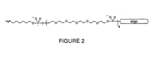
- One or more non-nucleosidic spacers such as polyethylene oxide, are added to the terminus of the oligo. Preferably, 1-20 spacers and, most preferably, 4 spacers are utilized. These spacers may be added to either the 5' or preferably the 3' end of the oligonucleotide.
- a nucleophilic moiety is then attached to the spacer group. The result is a derivatized capture probe, as shown in Figure 2.
- a preferred spacer monomer includes hexaethylene oxide.
- Non-nucleosidic spacers are preferred over nucleosidic spacers, such as poly-T, because non-nucleosidic spacers have greater flexibility.
- their physical properties can be tailored relatively easily, and it is possible to minimize specific and non-specific nucleic acid interactions.
- the spacers provide physical separation between the oligonucleotide and the solid surface and prevent interaction of the proteins with the support surface. This separation is important to ensure effective hybridization between the support-bound capture probe and the nucleic acid-protein fusion. In addition, the separation helps to minimize denaturation of the protein; the proteins are therefore able to adopt their native folded structures and remain functional.
- the spacer groups can be attached directly to the solid support surface, instead of to the capture probes.
- the spacer group can be attached to the amino groups on the surface.
- the bifunctional linker can then be attached to the other end of the spacer group.
- the capture probes may contain modifications that improve their hybridization properties and mismatch discrimination.
- they may contain base analogs, such as 5-propyne pyrimidines, internucleotide analogues such as peptide nucleic acids (PNA), in which the bases are connected by peptide-like linkages, or carbohydrate modifications.
- PNA peptide nucleic acids
- the capture probes are suspended in an aqueous alkaline solution, then applied to defined positions of the support surface; the nucleophilic moieties at the termini of the capture probes react with the active sites of the bifunctional linkers to form covalent bonds.
- the density of the capture probes can be controlled by adjusting reaction time and oligo concentration. Alternatively, the density can be controlled by doping the solution with capture oligos that lack nucleophilic moieties or doping with simple organic compounds that possess amine functional groups.
- biotin/streptavidin-linkers/spacers are non-nucleosidic and serve the attachment of the capture probe to the solid support / array, c.f. page 5531, left column.
- nucleic acid-protein fusion molecules are bound via base-pairing interactions, c.f. pages 5533 - 5539 and in particular for Figures 3 - 5.
- the capture probes can be applied using liquid deposition techniques, such as inkjet delivery, robotic quill delivery or spotting, and other similar deposition methods. They can also be applied using manual methods, such as pipetting.
- the feature sizes of the capture probes can range from one square micron (e.g., when robotic techniques are used) to one square millimeter (e.g., when a 0.2 microliter pipette is used).
- the result of the application of the capture probes is a defined, regular array of nucleic acid sequences.
- Blocking agents can also be selected to modify the surface energy, i.e., the hydrophobicity of the solid support surface.
- the hydrophobicity of the solid support surface is important because it affects the background signal level and the extent of unwanted interaction of the protein portion of a nucleic acid-protein fusion with the surface. Examples of blocking agents that modify hydrophobicity are methylamine, amino alcohols, and suitable amino-containing polyethylene oxide moieties.
- Non-covalent blocking agents can also be used to further minimize non-specific interactions between the fusion and the solid support (e.g., glass) surface.
- blocking agents include non-specific proteins such as BSA or casein, or similar commercially available blocking reagent formulations marketed for use with membranes.
- the capture probes arrayed on the surface of the solid support are then bound (for example, by hybridization) to nucleic acid-protein fusions, such as RNA-protein fusions.
- a solution containing the mixture of fusions is adjusted to an appropriate salt concentration, applied to the surface, and incubated at a suitable temperature to allow for efficient binding (for example, hybridization) between the capture probe and the target sequence.
- the solution may also contain surfactants such as TWEEN-20, TRITON X-100, or SDS (Sigma Chemical Co.) at concentrations of about 0.02% to about 1.0%; it may also include non-specific proteins, such as BSA.
- the experimental variables of salt concentration, temperature, and hybridization time are a function of the capture oligo design.
- a preferred range for the salt concentration is 25 mM to 2 M, with a concentration of about 750 mM being especially preferred.
- a preferred temperature range is from 5 °C to 70°C, with 30°C being especially preferred.
- Preferred reaction times can be from 1 to 24 hours, with 3 hours being especially preferred.
- the variables for each experiment are determined empirically by standard methods.
- the hybridization step can be performed in a simple chamber device that constrains the liquid sample and prevents evaporation.
- the solution may also contain one or more components to suppress nuclease degradation of the RNA moiety.
- Preferred additions include (a) metal chelators (e.g., EDTA or EGTA) at concentrations of between 1-10 mM, (b) placental RNase inhibitor protein (Promega) at concentrations of between 0.1-1 Unit/ ⁇ l; and (c) Anti-RNase protein (Ambion) at concentrations of between 0.1 - 1 Unit/ ⁇ l.
- metal chelators e.g., EDTA or EGTA
- placental RNase inhibitor protein Promega
- Anti-RNase protein A separate strategy to specifically suppress 5'-exonuclease degradation involves capping the 5'-terminus of the fusion RNA with a binding molecule. The capping strategy may be used in conjunction with one or more of the components listed above.
- a native or analog (e.g., PNA) nucleic acid sequence conplementary to the 5'-terminus of the fusion RNA is added to generate a stable duplex at the 5'-end.
- the complementary sequence is preferably between 10 - 50 bases in length, and most preferably abount 20 bases in length.
- This added nucleic acid sequence may also contain pendant groove-binding, intercalating, or cross-linking moieties.
- native or analog nucleic acid sequences may be added that form stable intermolecular hairpin, tetraloop, or pseudoknot secondary structures with the 5'-terminus of the RNA. In the latter case, these nucleic acids are preferably about 20 - 100 bases in length, with about 35 bases being especially preferred.
- the mixture of nucleic acid-protein fusions should be free of un-fused nucleic acids.
- Un-fused nucleic acids that are complementary to the capture probes will compete with the fusions for binding and will limit the amount of a given protein that can be displayed on the solid support.
- at least 1 % of the nucleic acid is fused to protein.
- hybridization tag sequences can include the same analogue units as are described above for the capture probes. In some cases, both the capture probe and the tag sequences can be modified so they hybridize preferentially with each other, thereby minimizing interference from the coding fusion sequences.
- unbound nucleic acid-protein fusion is washed away with a buffer that has a higher stringency and a lower salt concentration than that used for the hybridization step.
- the optimal buffer composition is determined empirically by standard methods. What remains upon completion of washing is an addressable array of proteins on the solid surface, attached via sequence-dependent recognition between the nucleic acid component of the fusion and the surface-bound capture oligo. The position of each protein is defined, because each fusion corresponds to the complementary capture probe.
- the nucleic acid component of the fusion may be covalently linked to a part of the solid support, the linker, or the capture probe.
- covalently linked fusions provide particularly robust and versatile addressable arrays that may be used, for example, in the screening methods described herein.
- Covalently linked fusion arrays may be generated by any standard approach. According to one general technique, the fusions are addressed to specific locations on a solid surface via hybridization with corresponding capture probes, and a chemical cross-linking or attachment reaction is triggered to fix the location of the fusions on the solid support.
- One method to achieve such a covalent link involves functionalizing the DNA capture oligos during chemical synthesis with one or more pendant psoralen moieties, preferably positioned near adenosine bases.
- the surface is exposed to long-wavelength UV light (for example, at 350 nm). Light of this wavelength triggers a photoreaction between psoralen and an adjacent thymidine or uridine base in the duplex region, forming a cyclobutane linkage and permanently attaching the fusion to the solid support.
- psoralen itself i.e., not linked to a capture probe
- the psoralen molecule intercalates between bases in double-stranded regions.
- the intercalated psoralen cross-links with thymidine or uridine bases (intrastrand and interstrand) in a bifunctional mode, forming covalent links between the capture probe and the nucleic acid component of the fusion.
- Other reactive, cross-linking reagents may also be used in place of psoralen in combination with triggering conditions appropriate for those reagents.
- Ordered, addressable arrays of peptide fragments can also be prepared.
- the fusion library is generated from short synthetic DNA sequences or fragments of cDNAs or genomic DNAs.
- ribosome display particles such as those described in Gold et al., WO 93/03172, can be hybridized to the solid support to generate the protein array. Again, these particles are immobilized on the solid support through a hybridization reaction between the capture oligo and the protein-coding RNA.
- the addressable protein arrays of the present invention have many uses. For example, a library of proteins can be displayed on a support, such as a microchip. This microchip can then be used to identify previously unknown protein-protein interactions.
- a probe protein can be detectably labeled, for example, with a radioisotope, chromophore, fluorophore, or chemiluminescent species, then incubated with the microchip. After the excess probe protein is washed away, the chip surface is analyzed for signal from the label. Detection of a signal indicates interaction of the labeled protein with one or more unique members of the protein library.
- the identity of proteins that are able to bind to the probe protein can then be determined from the location of the spots on the chip that become labeled due to binding of the probe.
- the same approach can also be used to screen protein libraries for protein-ligand interactions and protein-nucleic acid interactions.
- SPR surface plasmon resonance
- the reactive moiety on the oligonucleotide capture probe is a thiol group (rather than an amino group) and the gold surface need not be functionalized to achieve capture probe attachment.
- Mass spectrometry especially, Maldi-Tof
- Maldi-Tof can also be used to analyze species bound to unique members of the protein library.
- protein arrays Another application of protein arrays is the rapid determination of proteins that are chemically modified through the action of modifying enzymes such as protein kinases, acyl transferases, and methyl transferases.
- modifying enzymes such as protein kinases, acyl transferases, and methyl transferases.
- the location and hence the identity of those proteins that are substrates for the modifying enzyme may be readily determined. Further localization of the modification sites can be achieved using ordered displays of fragments of these proteins.
- the protein arrays can also be used to identify the unknown protein targets of therapeutically active compounds.
- a therapeutic compound may be applied to a protein array derived from cellular RNA. Detection of the captured therapeutic compound, either through its bound label or directly (for example, by mass spectrometry or surface plasmon resonance) reveals the compound's binding partner or partners.
- arrays can also be used in the development of protein-based diagnostics. For example, a solid support containing a variety of proteins associated with various illnesses can be prepared. A single patient sample, which might contain one or more proteins whose interactions with the support-bound proteins would be indicative of certain illnesses, can then be contacted with the support. Thus, a single sample can be used to simultaneously detect the presence of several conditions, or to distinguish between conditions.
- addressable arrays may be used to quantify target molecules in a sample.
- addressable arrays of single chain antibodies or antibody mimics may be used for quantifying a target protein (or proteins) in a biological sample.
- the arrays can also be used in the emerging fields of proteomics and functional genomics.
- the specific fusions that are identified as binding specifically to a probe molecule can be removed from the support surface.
- the fusion is released by disrupting hybridization with the capture probe.
- the specified fusion is physically separated from the rest of the fusions, then treated with a denaturing agent, such as a chemical reagent or heat, to disrupt the base pairing with the capture oligo. The liberated fusion is then recovered from the solution.
- the entire capture probe can be detached.
- a light-sensitive linker can be used to attach the capture probe to the solid surface.
- a laser beam of the appropriate wavelength can be used to cleave the linker, thus releasing the desired fusion.
- the fusion can be specifically recovered and manipulated, for example, using PCR, and further characterized.
- Silane treated slides from Example 1 are immersed in a freshly prepared 0.2% solution of phenylene 1,4-diisothiocyanate (Aldrich Chemical Co.) in 90% DMF/10% pyridine for two hours, with gentle agitation. The slides are washed sequentially with 90% DMF/10% pyridine, methanol, and acetone. After air drying, the functionalized slides are stored at 0°C in a vacuum desiccator over anhydrous calcium sulfate.
- phenylene 1,4-diisothiocyanate Aldrich Chemical Co.
- Oligonucleotides are chemically synthesized in the 3' ⁇ 5' direction by coupling standard phosphoramidite monomers with an automated DNA synthesizer. Typically, 500 angstrom controlled-pore glass supports are used at the 0.2 micromole scale.
- hexaethylene oxide phosphoramidite monomer (Glen Research) is added to the 5' terminus. The coupling wait time is extended to 15 minutes by modifying the synthesizer program. Additional hexaethylene oxide monomer units are added in the same way.
- C-6 Amino phosphoramidite (Glen Research) is then added to the 5' terminus; the coupling wait time is again extended to 15 minutes.
- Capture probe sequences are cleaved from the solid support and deprotected with ammonium hydroxide, concentrated to dryness, precipitated in ethanol, and purified by reverse-phase HPLC using an acetonitrile gradient in triethylammonium acetate buffer.
- the purified, amine-labeled capture probes from Example 3 are adjusted to a concentration of 500 micromolar in 100 mM sodium carbonate buffer (pH 9.0), and are applied to the derivatized glass surface from Example 2 at defined positions. For manual deposition, aliquots of 0.2 microliter each are applied with a pipetman. The array is incubated at room temperature in a moisture-saturated environment for at least two hours. The attachment reaction is terminated by immersing the glass surface in an aqueous 1% ammonia solution for five minutes with gentle agitation. The glass surface is then subjected to three 5-minute washes, using fresh portions of distilled water for each wash. The array is then soaked in 1 M phosphate buffered saline (PBS) solution for 2 hours at room temperature, then rinsed again for 5 minutes in distilled water.
- PBS phosphate buffered saline
- the ammonia solution from Example 4 is replaced with a 1-5% aqueous solution of a different primary amine-containing molecule. A small amount (10%) of methanol or acetonitrile cosolvent is added, if necessary.
- the glass surface is then subjected to three 5-minute washes, using fresh portions of distilled water for each wash.
- the surface is soaked in 1 M phosphate buffered saline (PBS) solution for 2 hours, then washed again for 5 minutes with distilled water.
- PBS phosphate buffered saline
- the glass surface is immersed in a dilute, aqueous solution of a protein-containing blocking solution for several minutes, then subjected to three 5-minute washes, using fresh portions of distilled water for each wash. Finally, the surface is air dried.
- RNA-protein fusions 50 microliters of a solution containing the RNA-protein fusions and consisting of 25 mM Tris-HCl (pH 8.0) and 100 mM potassium chloride are applied to the glass microchip surface in a chamber that can contain and seal the liquid.
- the solution is maintained at a specific temperature (determined by the capture oligo design) for at least three hours.
- non-hybridized RNA-protein fusions are removed by washing with 25 mM Tris-HCl (pH 8.0) and 50 mM potassium chloride for several minutes at the incubation temperature.
- the protein chip is subjected to two 15-minute washes, using a buffer that is more stringent and contains a lower salt concentration than the buffer used for the hybridization reaction.
- Example 7 Generation of an Exemplary FLAG and HA11 Fusion Chip
- exemplary FLAG and HA11 fusion chips were generated as follows.
- the silane treated slides were immersed in a freshly prepared 0.2% solution of phenylene 1,4-diisothiocyanate (Aldrich Chemical Co.) in 90% DMF / 10% pyridine for two hours, with gentle agitation. The slides were washed sequentially with 90% DMF / 10% pyridine, methanol, and acetone. After air drying, the functionalized slides were stored at 0°C in a vacuum desiccator over anhydrous calcium sulfate.
- phenylene 1,4-diisothiocyanate Aldrich Chemical Co.
- Capture oligos were then designed and synthesized by standard techniques.
- the RNA employed to make the FLAG epitope fusion (17 amino acids total) consisted of 5'-r(UAA UAC GAC UCA CUA UAG GGA CAA UUA CUA UUU ACA AUU ACA AUG GAC UAC AAG GAC GAU GAC GAU AAG GGC GGC UGG UCC CAC CCC CAG UUC GAG AAG) (SEQ ID NO: 1).
- RNA employed to make the HA1 1 epitope fusion (20 amino acids total) consisted of 5'-r(UAA UAC GAC UCA CUA UAG GGA CAA UUA CUA UUU ACA AUU ACA AUG UAC CCC UAC GAC GUG CCC GAC UAC GCC GGC GGC UGG UCC CAC CCC CAG UUC GAG AAG) (SEQ ID NO: 2).
- the following DNA linker which also contained the essential puromycin moiety at its 3'-end, was ligated to the 3'-terminus of the RNA message: 5'-d(AAAAAAAAAAAAAAAAAAAAAAAAAAAAAAAAAA CC) (SEQ ID NO: 3).
- Oligonucleotide capture probes were chemically synthesized in the 3' to 5' direction by coupling standard phosphoramidite monomers using an automated DNA synthesizer (PE BioSystems Expedite 8909). Typically, 500 angstrom controlled-pore glass supports were used at the 0.2 micromole scale. In the case of 5'-attachment, after the desired probe sequence had been assembled (using A, G, C, and T monomers), four hexaethylene oxide phosphoramidite monomers (Glen Research) were added to the 5'-terminus. The coupling wait time was extended to 15 minutes by modifying the synthesizer program. Additional hexaethylene oxide monomer units were added in the same way.
- the oligos were adjusted to a concentration of 250 micromolar in 50 mM sodium carbonate buffer (pH 9.0) containing 10% glycerol.
- the oligos were then robotically applied (MicroGrid, BioRobotics) to the derivatized glass surface described above at defined positions in a 5 x 5 x 16 array pattern (384 spots) within a 20 x 20mm area.
- the layout of these capture probes is shown schematically in Figure 3.
- a 16-pin tool was used to transfer the liquid, producing 200 micron features with a pitch of 600 microns.
- Each sub-grid of 24 spots represented a single capture probe (i.e., 24 duplicate spots).
- the array was incubated at room temperature in a moisture-saturated environment for 12-18 hours.
- the attachment reaction was terminated by immersing the glass surface in an aqueous 1% ammonia solution for five minutes with gentle agitation.
- the glass surface was then subjected to three 5-minute washes, using fresh portions of distilled water for each wash.
- the array was then soaked in a 10X PBS (phosphate buffered saline) solution for 2 hours at room temperature, and then rinsed again for 5 minutes in distilled water.
- 10X PBS phosphate buffered saline
- RNA-protein fusions between the peptides containing the FLAG and HA 11 epitopes and their corresponding mRNAs were produced as generally described by Szostak et al., WO 98/31700; and Roberts and Szostak, Proc. Natl. Acad. Sci. USA 94:12297-12302, 1997.
- the polymerase chain reaction using Taq polymerase was used to amplify the sequences 5'-TAA TAC GAC TCA CTA TAG GGA CAA TTA CTA TTT ACA ATT ACA ATG GAC TAC AAG GAC GAT GAC GAT AAG GGC GGC TGG TCC CAC CCC CAG TTC GAG AAG (SEQ ID NO: 12) and 5'-TAA TAC GAC TCA CTA TAG GGA CAA TTA CTA TTT ACA ATT ACA ATG TAC CCC TAC GAC GTG CCC GAC TAC GCC GGC GGC TGG TCC CAC CCC CAG TTC GAG AAG (SEQ ID NO: 13) for FLAG and HA11, respectively, using the oligonucleotide primers 5'-TAA TAC GAC TCA CTA TAG GGA CAA TTA CTA TTT ACA ATT (SEQ ID NO: 14) and 5'-AGCGGATGCCTTCTCTC
- RNA linker 5'-AAA AAA AAA AAA AAA AAA AAA AAA AAA CC (SEQ ID NO: 3) containing a 5' phosphate and a 3' puromycin by T4 DNA ligase (Promega) in the presence of an 80:20 mixture of the following two DNA splints: 5'-TGCAACGACCAACTTTTTTTTTTAGCGCATGC (SEQ ID NO: 16) and 5'-TGCAACGACCAACTTTTTTTTTNAGCGCATGC (SEQ ID NO: 17), each containing two biotin moieties at the 5' terminus.
- RNA-DNA chimera was purified by binding to Immobilized NeutrAvidin (Pierce), washing to remove unligated material, and eluting by displacement using the sequence 5'-GCATCCGCTAAAAAAAAAAGTTGGTCGTTGC (SEQ ID NO: 18). Subsequent translations were performed in rabbit reticulocyte lysate (Ambion) according to the manufacturer's instructions except that MgCl 2 (150 mM) and KCl (425 mM) were added after 30 minutes to promote the formation of the puromycin-peptide bond.
- RNA-peptide fusions were then purified by oligo dT affinity chromatography (Pharmacia), quantitated by scintillation counting of the incorporated vs. added 35 S methionine (Amersham), and concentrated to a low volume via membrane filtration (MicroCon).
- the FLAG and HA11-fusion chip was exposed to a phosphorimage screen (Molecular Dynamics) for 60 hours by direct contact between the screen and the chip. This allowed identification of the areas that contained hybridized fusions, since the peptides contained a 35 S methionine radiolabel which was detectable by the phosphor storage screen. As shown in Figure 4, analysis of the phosphorimage revealed that the fusions had successfully hybridized to their respective capture probes targeting specific areas of the RNA message (i.e., CPflag and CPha11).
- the four non-sense capture probes which were not compelementary to any region of the FLAG or HA11 RNA, did not give any appreciable signal (i.e., CPau1, CPau5, CPirs, CPkt3).
- the positive control capture probe CPtag produced the expected signal, but the corresponding positive control capture probe CPt7 did not, likely due to degradation (e.g., exonuclease contamination) of the 5'-region of the targeted RNA.
- a duplicate chip was probed with a monoclonal antibody that recognized the HA11 epitope. All of the following steps were performed at 4°C. Nonspecific sites were first blocked with a solution containing 1X PBS (phosphate buffered saline) + 1% BSA (bovine serum albumin, RNAse free grade, Ambion) + 0.02% Tween-20 for 1 hour under a coverslip. The blocking solution was removed and 50 microliters of HA. 11 monoclonal antibody (100:1 dilution, Berkeley Antibody Co.) in 1X PBS + 0.02% Tween-20 was applied to the chip under a coverslip.
- 1X PBS phosphate buffered saline
- BSA bovine serum albumin, RNAse free grade, Ambion
- the chip was subsequently analyzed at 10 micron pixel resolution with a confocal laser scanner (ScanArray 3000, General Scanning) using preset excitation and emission wavelengths tuned to the Cy3 fluorophore.
- the resulting fluorimage was in accord with the phosphorimage and demonstrated that the HA11 peptide, which was covalently linked to its RNA message and fixed to the chip surface, was functional and was available to interact with its binding partner (the HA11 monoclonal antibody).
- the HA11 monoclonal antibody was specific for its own epitope.
- the 3'-attachment capture probes generally provided a better signal than the 5'-attachment capture probes. Without being bound to a particular theory, this may reflect the greater accessibility of the epitope when it is oriented away from the chip surface.
- an exemplary Myc fusion chip was also generated as follows.
- the silane treated slides were immersed in a freshly prepared 0.2% solution of phenylene 1,4-diisothiocyanate (Aldrich Chemical Co.) in 90% DMF / 10% pyridine for two hours, with gentle agitation. The slides were washed sequentially with 90% DMF / 10% pyridine, methanol, and acetone. After air drying, the functionalized slides were stored at 0°C in a vacuum desiccator over anhydrous calcium sulfate.
- phenylene 1,4-diisothiocyanate Aldrich Chemical Co.
- the capture oligos were synthesized based on the Myc sequence.
- the RNA employed to make the c-myc fusion (33 amino acids total) consisted of the following sequence: 5'-r(UAAUACGACUCACUAUAGGGACAAUUACUAUUUACAAUUACA AUGGGGACAAUUACUAUUUACAAUUACAAUGGCUGAAGAACAGA AACUGAUCUCUGAAGAAGACCUGCUGCGUAAACGUCGUGAACAGC UGAAACACAAACUGGAACAGCUGCGUAACUCUUGCGCU)(SEQ ID NO: 19).
- RNA linker which also contains the essential puromycin moiety
- 5'-d(AAAAAAAAAAAAAAAAAAAAAAAAAAAAACC) SEQ ID NO: 3
- Three non-overlapping and thermodynamically isoenergetic 20-mer sequences along the RNA were identified to serve as capture points.
- dA25 (on the ligated DNA) was selected as a fourth target area.
- the targeted sequences began at nucleotide positions 1, 33, 80, and 125 (CP01, CP33, CP80 and CP125, respectively).
- a mismatch sequence derived from target sequence 33 and containing four internal and adjacent nucleotide mismatches, was also designed (CPmm).
- a non-sense sequence corresponding to the reverse-orientation of CP33, was also utilized as a negative control (CPns).
- CPns a negative control
- Oligonucleotide capture probes were chemically synthesized in the 3' to 5' direction by coupling standard phosphoramidite monomers with an automated DNA synthesizer (PE BioSystems Expedite 8909). Typically, 500 angstrom controlled-pore glass supports were used at the 0.2 micromole scale. After the desired probe sequence had been assembled (using A, G, C, and T monomers), hexaethylene oxide phosphoramidite monomer (Glen Research) was added to the 5'-terminus. The coupling wait time was extended to 15 minutes by modifying the synthesizer program. Additional hexaethylene oxide monomer units were added in the same way.
- the oligos were adjusted to a concentration of 500 micromolar in 100 mM sodium carbonate buffer (pH 9.0) and were applied to the derivatized glass surface at defined positions in a 6 x 6 array pattern (36 spots) within a 20 x 20mm area (as shown in Figure 6).
- CP01 was applied to locations A1, B1, C1 and A4, B4, C4.
- CP33 was applied to locations D1, E1, F1 and D4, E4, F4.
- CP80 was applied to locations A2, B2, C2 and A5, B5, C5.
- CP125 was applied to locations D2, E2, F2 and D5, E5, F5.
- Cpmm was applied to locations A3, B3, C3 and A6, B6, C6.
- Cpns was applied to locations D3, E3, F3 and D6, E6, F6.
- the array was incubated at room temperature in a moisture-saturated environment for 12-18 hours.
- the attachment reaction was terminated by immersing the glass surface in an aqueous 1% ammonia solution for five minutes with gentle agitation.
- the glass surface was then subjected to three 5-minute washes, using fresh portions of distilled water for each wash.
- the array was then soaked in a 10X PBS (phosphate buffered saline) solution for 2 hours at room temperature, and then rinsed again for 5 minutes in distilled water.
- 10X PBS phosphate buffered saline
- RNA-protein fusions between a 33 amino acid peptide containing the c-myc epitope and its mRNA were produced as described by Szostak et al., WO 98/31700; and Roberts and Szostak, Proc. Natl. Acad. Sci. USA 94:12297-12302, 1997.
- the polymerase chain reaction using Taq polymerase was used to amplify the sequence 5'-AGC GCA AGA GTT ACG CAG CTG TTC CAG TTT GTG TTT CAG CTG TTC ACG ACG TTT ACG CAG CAG GTC TTC AGA GAT CAG TTT CTG TTC TTC AGC CAT (SEQ ID NO: 26) using oligonucleotide primers 5'-AGC GCA AGA GTT ACG CAG CTG (SEQ ID NO: 27) and 5'-TAA TAC GAC TCA CTA TAG GGA CAA TTA CTA TTT ACA ATT ACA ATG GCT GAA GAA CAG AAA CT (SEQ ID NO: 28).
- RNA linker 5'-AAA AAA AAA AAA AAA AAA AAA AAA AAA CC (SEQ ID NO: 3) containing a 5' phosphate and a 3' puromycin by T4 DNA ligase (Promega) in the presence of a DNA splint with the sequence TTT TTT TTT TAG CGC AAG A (SEQ ID NO: 29).
- RNA-DNA chimera was purified by denaturing polyacrylamide gel electrophoresis (6% acrylamide). Translation was performed in rabbit reticulocyte lysate (Ambion) according to the manufacturer's instructions except that KCl (500 mM) was added after 30 minutes to promote the formation of the puromycin-peptide bond.
- the RNA-peptide fusion was purified by oligo dT affinity chromatography (Pharmacia), quantitated by scintillation counting of the incorporated vs. added 35 S methionine (Amersham), and dried to a pellet. 2.5 pmol of the c-myc fusion was produced.
- the dry myc-fusion pellet was taken up with 20 microliters of 5X SSC (saline sodium citrate) + 0.02% SDS, mixed, and then briefly centrifuged.
- 5X SSC saline sodium citrate
- the solution was applied to the slide described above, a coverslip was placed on top, and the slide was placed in a moisture-saturated chamber at room temperature. After 18 hours the coverslip was removed, and the slide was washed sequentially with stirred 500 mL portions of 5X SSC + 0.02% SDS, 2.5X SSC + 0.01% SSC, 2.5X SSC, and 1.25X SSC for 5 minutes each. After removal of liquid the slide was allowed to briefly air-dry.
- 5X SSC saline sodium citrate
- the glass chip was exposed to a phosphorimage screen (Molecular Dynamics) for four hours by direct contact between the screen and the chip. This allowed identification of the areas that contained hybridized myc-fusion, since the myc peptide contained a 35 S methionine radiolabel which was detectable by the phosphor storage screen.
- analysis of the phosphorimage revealed that the myc-fusion had successfully hybridized to each of the four capture probes that targeted the myc RNA message and DNA linker sequence.
- the non-sense capture probe which was not complementary to any region of the myc RNA, did not give any appreciable signal.
- the capture probe sequence that contained several mismatches produced only a small amount of signal.
- the chip was subsequently analyzed at 10 micron pixel resolution with a confocal laser scanner (ScanArray 3000, General Scanning) using preset excitation and emission wavelengths tuned to the Cy3 fluorophore.
- the resulting fluorimage was in accord with the phosphorimage and demonstrated that the myc peptide, which was covalently linked to its RNA message and fixed to the chip surface, was functional and was available to interact with its binding partner (the monoclonal antibody).
Landscapes
- Chemical & Material Sciences (AREA)
- Life Sciences & Earth Sciences (AREA)
- Health & Medical Sciences (AREA)
- Engineering & Computer Science (AREA)
- Molecular Biology (AREA)
- Organic Chemistry (AREA)
- Immunology (AREA)
- General Health & Medical Sciences (AREA)
- Biochemistry (AREA)
- Physics & Mathematics (AREA)
- Biomedical Technology (AREA)
- Proteomics, Peptides & Aminoacids (AREA)
- Medicinal Chemistry (AREA)
- Urology & Nephrology (AREA)
- Hematology (AREA)
- Chemical Kinetics & Catalysis (AREA)
- Materials Engineering (AREA)
- Microbiology (AREA)
- Biophysics (AREA)
- Biotechnology (AREA)
- General Physics & Mathematics (AREA)
- Analytical Chemistry (AREA)
- General Chemical & Material Sciences (AREA)
- Zoology (AREA)
- Genetics & Genomics (AREA)
- Pathology (AREA)
- Food Science & Technology (AREA)
- Nanotechnology (AREA)
- Wood Science & Technology (AREA)
- Cell Biology (AREA)
- Bioinformatics & Cheminformatics (AREA)
- Geochemistry & Mineralogy (AREA)
- Condensed Matter Physics & Semiconductors (AREA)
- Composite Materials (AREA)
- General Engineering & Computer Science (AREA)
- Crystallography & Structural Chemistry (AREA)
- Inorganic Chemistry (AREA)
- Bioinformatics & Computational Biology (AREA)
- Measuring Or Testing Involving Enzymes Or Micro-Organisms (AREA)
- Apparatus Associated With Microorganisms And Enzymes (AREA)
Abstract
Description
- The invention relates to fixed arrays of nucleic acid-protein fusions and, in particular, RNA-protein fusions, on solid supports.
- Certain macromolecules, such as proteins, are known to interact specifically with other molecules based on the three-dimensional shapes and electronic distributions of those molecules. For example, proteins interact selectively with other proteins, with nucleic acids, and with small-molecules. Modem pharmaceutical research relies on the study of these interactions; the development of new drugs depends on the discovery of compounds that bind specifically to biologically important molecules.
- The discovery of a single drug candidate can require the screening of thousands of compounds. It is therefore important to be able to screen large numbers of compounds rapidly and efficiently. One method for screening a large number of compounds is to fix possible binding partners, such as proteins, to a solid support.
- It is difficult to prepare arrays of isolated proteins on solid supports, however, for a variety of reasons. First of all, proteins cannot always be easily attached to the planar surfaces traditionally used to make other fixed arrays, such as nucleic acid microchips. More importantly, because proteins can interact with the functional groups on the surfaces of these supports, the proximity of the protein to the surface can lead to disruption of the protein structure.
- In general, the invention features a solid support including an array of immobilized capture probes; each of the capture probes includes a non-nucleosidic spacer group and an oligonucleotide sequence to which a nucleic acid-protein fusion is bound (for example, hybridized or covalently bound). In preferred embodiments, the nucleic acid-protein fusion is an RNA-protein fusion, and the protein component is encoded by the nucleic acid (for example, the RNA). The spacer group can include a polyalkylene oxide, for example, polyethylene oxide. A preferred spacer group includes hexaethylene oxide. The capture probe may also include a photocleavable linker.
- The oligonucleotide sequence can include a modified base, such as 5-propyne pyrimidine. It can also include an internucleotide analog (such as 3'-phosphoramidate) or a carbohydrate modification (such as a 2'-O-methyl group). The nucleic acid-protein fusion can include a hybridization tag sequence. The hybridization tag sequence can also include a modified base, an internucleotide analog, or a carbohydrate modification.
- In a preferred embodiment, the capture probe further includes a reactive moiety (for example, a nucleophilic group), such as a primary amino group. In another preferred embodiment, the nucleic acid-protein fusion is covalently linked to the capture probe (for example, by photo-crosslinking); in one preferred approach, this is accomplished by including one or more psoralen moieties in the capture probe or in the capture probe-fusion hybridization reaction mixture. A preferred solid support is a glass or silica-based chip.
- In a related aspect, the invention features a solid support including an array of immobilized capture probes; each of the capture probes is attached to the surface of the solid support through a non-nucleosidic spacer group, and each of the capture probes includes an oligonucleotide sequence to which a nucleic acid-protein fusion (for example, an RNA-protein fusion) is bound (for example, hybridized or covalently bound).
- In another related aspect, the invention features a solid support including an array of immobilized capture probes; each of the capture probes includes a non-nucleosidic spacer group and an oligonucleotide sequence to which a ribosome display particle is bound (for example, hybridized or covalently bound).
- In yet another related aspect, the invention features a method for preparing a solid support. The method includes the steps of: (a) preparing a capture probe by linking a spacer group to an oligonucleotide sequence; (b) attaching the capture probe to the solid support; and (c) binding (for example, hybridizing or covalently binding) a nucleic acid-protein fusion (for example, an RNA-protein fusion) to the capture probe.
- The invention also features a second general method for preparing a solid support. This method includes the steps of: (a) attaching a spacer group to a surface of the solid support; (b) attaching a bifunctional linker to the spacer group; (c) attaching a capture probe to the bifunctional linker; and (d) binding (for example, hybridizing or covalently binding) a nucleic acid-protein fusion (for example, an RNA-protein fusion) to the capture probe.
- In a second aspect, the invention features a method for detecting an interaction between a protein and a compound. The method includes the steps of: (a) providing a solid support including an array of immobilized capture probes, where each of the capture probes includes a non-nucleosidic spacer group and an oligonucleotide sequence to which a nucleic acid-protein fusion is bound (for example, hybridized or covalently bound); (b) contacting the solid support with a candidate compound under conditions which allow an interaction between the protein portion of the nucleic acid-protein fusion and the compound; and (c) analyzing the solid support for the presence of the compound as an indication of an interaction between the protein and the compound.
- Alternatively, the invention features another method for detecting an interaction between a protein and a compound; this method involves the steps of: (a) providing a population of nucleic acid-protein fusions; (b) contacting the population of nucleic acid-protein fusions with a candidate compound under conditions which allow an interaction between the protein portion of the nucleic acid-protein fusion and the compound; (c) contacting the product of step (b) with a solid support that includes an array of immobilized capture probes, each of the capture probes including a non-nucleosidic spacer group and an oligonucleotide sequence to which a nucleic acid-protein fusion binds (for example, hybridizes or covalently binds); and (d) analyzing the solid support for the presence of the compound as an indication of an interaction between the protein and the compound.
- In a preferred embodiment of each of the above methods, the nucleic acid-protein fusion is an RNA-protein fusion. In another preferred embodiment, the compound is labeled. Compounds that can be screened using these methods include, without limitation, proteins, drugs, therapeutics, enzymes, and nucleic acids.
- In a third aspect, the invention features an array (for example, an addressable array) of nucleic acid-protein fusions including at least 102 different fusions/cm2. Preferably, the nucleic acid-protein fusions are RNA-protein fusions, and the array includes at least 104 different fusions/cm2.
- In a related aspect, the invention features a method for generating an addressable array of molecules. The method involves: (a) providing a solid support on which an array of nucleic acid molecules is immobilized; (b) contacting the solid support with a population of addressable molecules; and (c) allowing the addressable molecules to orient themselves on the solid support by sequence-dependent recognition and binding of the immobilized nucleic acid molecules.
- In preferred embodiments of this method, the addressable array of molecules is an array of nucleic acid-protein fusions (for example, an array of RNA-protein fusions); the addressable molecules orient themselves on the solid support by base pairing (for example, hybridization) with the immobilized nucleic acid molecules; the solid support is a glass or silica-based chip; and the nucleic acid molecules immobilized on the solid support are capture probes, each including a non-nucleosidic spacer group and an oligonucleotide sequence to which the addressable molecule binds.
- As used herein, by an "array" is meant a fixed pattern of immobilized objects on a solid surface or membrane. Typically, the array is made up of nucleic acid-protein fusion molecules (for example, RNA-protein fusion molecules) bound to capture nucleic acid sequences which themselves are immobilized on the solid surface or membrane. The array preferably includes at least 102, more preferably at least 103, and most preferably at least 104 different fusions, and these fusions are preferably arrayed on a 125 x 80 mm, and more preferably on a 10 x 10 mm, surface. By an "addressable array" is meant that the locations, or addresses, on the solid support of the members of the array (for example, the nucleic acid-protein fusions) are known; the members of the array are referred to as "addressable molecules" and are utilized in methods for screening for subsequent molecular interactions (for example, for screening for interactions between the addressable nucleic acid-protein fusions and candidate therapeutics).
- By "nucleic acid-protein fusion" is meant a nucleic acid covalently bound to a protein. By "nucleic acid" is meant any two or more covalently bonded nucleotides or nucleotide analogs or derivatives. As used herein, this term includes, without limitation, DNA, RNA, and PNA. By "protein" is meant any two or more amino acids, or amino acid analogs or derivatives, joined by peptide or peptoid bond(s), regardless of length or post-translational modification. As used herein, this term includes, without limitation, proteins, peptides, and polypeptides.
- By "hybridization tag" is meant a non-coding oligonucleotide sequence that differs sufficiently in sequence from other nucleic acid sequences in a given population or reaction mixture that significant cross-hybridization does not occur. When multiple hybridization tags are utilized in a single reaction mixture, these tags also preferably differ in sequence from one another such that each has a unique binding partner under the conditions employed.
- By a "population" is meant more than one molecule.
- By a "solid support" is meant any solid surface including, without limitation, any chip (for example, silica-based, glass, or gold chip), glass slide, membrane, bead, solid particle (for example, agarose, sepharose, or magnetic bead), column (or column material), test tube, or microtiter dish.
-
- Figure 1 is a drawing showing the silylation of a glass surface, the derivatization of the resulting amino groups, and the attachment of a capture probe to the modified surface.
- Figure 2 is a drawing illustrating a capture probe containing a non-nucleosidic spacer group and a reactive moiety.
- Figure 3 is a schematic diagram of the layout of the FLAG and HA11 fusion chip capture probes utilized in Figures 4 and 5. In this Figure, t7, tag, au1, au5, flag, ha1, irs, and kt3 represent the capture probes CPt7 (positive control), CPtag (positive control), CPaul (negative control), CPau5 (negative control), CPflag, CPha11, CPirs (negative control), and CPkt3 (negative control), respectively.
- Figure 4 is a phosphorimage demonstrating hybridization of nucleic acid-protein fusions (FLAG and HA11) to capture probes immobilized on a chip.
- Figure 5 is a fluorimage demonstrating hybridization of nucleic acid-protein fusions (FLAG and HA11) to capture probes immobilized on a chip and subsequent recognition with anti-HA1 monoclonal antibodies.
- Figure 6 is a schematic diagram of the layout of the Myc fusion chip capture probes utilized in Figures 7 and 8. In this Figure, capture probes CP01, CP33, CP80, CP125, CPmm, and CPns (described herein) were arranged on the chip as follows: CP01 at locations A1, B1, C1, A4, B4, and C4; CP33 at locations D1, E1, F1, D4, E4, and F4; CP80 at locations A2, B2, C2, A5, B5, and C5; CP125 at locations D2, E2, F2, D5, E5, and F5; CPmm at locations A3, B3, C3, A6, B6, and C6; and CPns at locations D3, E3, F3, D6, E6, and F6.
- Figure 7 is a phosphorimage demonstrating hybridization of nucleic acid-protein fusions (Myc) to capture probes immobilized on a chip.
- Figure 8 is a fluorimage demonstrating hybridization of nucleic acid-protein fusions (Myc) to capture probes immobilized on a chip and subsequent recognition with anti-Myc monoclonal antibodies.
- The invention features support-based, addressable arrays of proteins, and methods for preparing and using these arrays. The arrays are prepared by fixing oligonucleotide sequences, the capture probes (or capture oligos), to a support in a defined array. The capture probes are then used to bind nucleic acid-protein fusions, such as RNA-protein fusions. Such binding may occur through base pairing (for example, through Watson-Crick base pairing, pseudo Watson-Crick base pairing involving modified bases, or Hoogsteen base pairing) between the nucleic acid component of the fusion and a complementary capture probe, or may occur through any other type of sequence-dependent recognition and binding of the capture probe (including, without limitation, polyamide-mediated nucleic acid groove binding or specific binding by nucleic acid-binding proteins such as transcription factors). The result of the binding interactions between the fusions and the capture probes is a defined, addressable array of proteins attached to a solid support.
- A variety of materials can be used as the solid support. Examples of such materials include polymers (e.g., plastics), aminated surfaces, gold coated surfaces, nylon membranes, polyacrylamide pads deposited on solid surfaces, silicon, silicon-glass (e.g., microchips), silicon wafers, and glass (e.g., microscope slides). Microchips, and particularly glass microchips, represent a preferred solid support surface.
- If the surface is not already aminated, it can be modified to provide a layer of amino groups. For example, a glass microscope slide can be treated with a silylating agent such as trialkoxyaminosilane to provide a surface of primary amino groups that exists as a monolayer or 3-8 molecular layers. This reaction is illustrated in Figure 1. The silane-treated surface is then derivatized with a homobifunctional or heterobifunctional linker that permits the attachment of oligonucleotides at discrete positions. Phenylene 1,4-diisothiocyanate is a useful homobifunctional linker; amino-surfaces derivatized with this reagent have isothiocyanate functionalities that are available to covalently react with the primary amino groups on the termini of oligonucleotides to form stable thiourea bonds, as shown in Figure 1.
- The capture probes, i.e., the oligonucleotide sequences that are to be attached to the surface, are selected from the reverse-complements of the nucleic acid components of the nucleic acid-protein fusions (the targets). Capture probes preferably have between 5 and 30 nucleotide units, and more preferably have about 20 nucleotide units. Considerations for the selection of the exact sequence for a particular capture probe include melting temperature (Tm), interference from competing target sequences, and potential secondary structure in the target sequence. Ideally, each unique capture probe has the same Tm, i.e., they are isoenergetic, so a single hybridization and washing temperature can be used successfully for all capture-target pairs. Commercially available computer programs (e.g., Oligo 4.0) can be used to help identify sets of capture probes with similar thermodynamic properties based on nearest neighbor treatments.
- The capture probes are modified before they are attached to the surface. One or more non-nucleosidic spacers, such as polyethylene oxide, are added to the terminus of the oligo. Preferably, 1-20 spacers and, most preferably, 4 spacers are utilized. These spacers may be added to either the 5' or preferably the 3' end of the oligonucleotide. A nucleophilic moiety is then attached to the spacer group. The result is a derivatized capture probe, as shown in Figure 2. A preferred spacer monomer includes hexaethylene oxide.
- Non-nucleosidic spacers are preferred over nucleosidic spacers, such as poly-T, because non-nucleosidic spacers have greater flexibility. In addition, their physical properties can be tailored relatively easily, and it is possible to minimize specific and non-specific nucleic acid interactions.
- The spacers provide physical separation between the oligonucleotide and the solid surface and prevent interaction of the proteins with the support surface. This separation is important to ensure effective hybridization between the support-bound capture probe and the nucleic acid-protein fusion. In addition, the separation helps to minimize denaturation of the protein; the proteins are therefore able to adopt their native folded structures and remain functional.
- Alternatively, the spacer groups can be attached directly to the solid support surface, instead of to the capture probes. For example, the spacer group can be attached to the amino groups on the surface. The bifunctional linker can then be attached to the other end of the spacer group.
- In addition to spacer groups, the capture probes may contain modifications that improve their hybridization properties and mismatch discrimination. For example, they may contain base analogs, such as 5-propyne pyrimidines, internucleotide analogues such as peptide nucleic acids (PNA), in which the bases are connected by peptide-like linkages, or carbohydrate modifications.
- The capture probes are suspended in an aqueous alkaline solution, then applied to defined positions of the support surface; the nucleophilic moieties at the termini of the capture probes react with the active sites of the bifunctional linkers to form covalent bonds. The density of the capture probes can be controlled by adjusting reaction time and oligo concentration. Alternatively, the density can be controlled by doping the solution with capture oligos that lack nucleophilic moieties or doping with simple organic compounds that possess amine functional groups.
- Guo et al., Nucleic Acids Research (1994), volume 22, number 24, teaches in Figure 1 on page 5458 the coupling of the capture probe, here named "hybridization sequence", to a nucleosidic linker (poly-thymidine oligonucleotide), said nucleosidic linker is fixed to the solid support via silanization chemistry. On page 5460, left column, third paragraph, it is stated, that this spacer ought to have a certain length for an efficient hybridization to occur. Maskos et al., Nucleic Acids Research (1992), volume 20, number 7, introduces non-nucleosidic spacer chemistry, namely polyethylene spacers / linkers to glass surfaces via silanization chemistry, said procedure being detailed on page 1680. As it is outlined on page 1682, paragraph on "hybridization to tethered oligonucleotides", this linker is the starting point for the generation of the hybridization sequence / capture probe. Niemeyer et al., Nucleic Acids Research (1994) describes a method for preparing a microarray where capture probes are fixed upon. The fixation of the capture probes to the surface of the array occurs via biotin/streptavidin linkers/spacers. These biotin/streptavidin-linkers/spacers are non-nucleosidic and serve the attachment of the capture probe to the solid support / array, c.f. page 5531, left column. To these support-attached capture probes, nucleic acid-protein fusion molecules are bound via base-pairing interactions, c.f. pages 5533 - 5539 and in particular for Figures 3 - 5.
- The capture probes can be applied using liquid deposition techniques, such as inkjet delivery, robotic quill delivery or spotting, and other similar deposition methods. They can also be applied using manual methods, such as pipetting. The feature sizes of the capture probes can range from one square micron (e.g., when robotic techniques are used) to one square millimeter (e.g., when a 0.2 microliter pipette is used). The result of the application of the capture probes is a defined, regular array of nucleic acid sequences.
- After a sufficient reaction time, the excess capture probe is washed away, and the remaining unreacted isothiocyanate groups are blocked off. Dilute ammonia can be used as the blocking agent, resulting in a surface of phenyl thiourea groups. Blocking agents can also be selected to modify the surface energy, i.e., the hydrophobicity of the solid support surface. The hydrophobicity of the solid support surface is important because it affects the background signal level and the extent of unwanted interaction of the protein portion of a nucleic acid-protein fusion with the surface. Examples of blocking agents that modify hydrophobicity are methylamine, amino alcohols, and suitable amino-containing polyethylene oxide moieties.
- Non-covalent blocking agents can also be used to further minimize non-specific interactions between the fusion and the solid support (e.g., glass) surface. Examples of such blocking agents include non-specific proteins such as BSA or casein, or similar commercially available blocking reagent formulations marketed for use with membranes.
- The capture probes arrayed on the surface of the solid support are then bound (for example, by hybridization) to nucleic acid-protein fusions, such as RNA-protein fusions. A solution containing the mixture of fusions is adjusted to an appropriate salt concentration, applied to the surface, and incubated at a suitable temperature to allow for efficient binding (for example, hybridization) between the capture probe and the target sequence. The solution may also contain surfactants such as TWEEN-20, TRITON X-100, or SDS (Sigma Chemical Co.) at concentrations of about 0.02% to about 1.0%; it may also include non-specific proteins, such as BSA.
- The experimental variables of salt concentration, temperature, and hybridization time are a function of the capture oligo design. A preferred range for the salt concentration is 25 mM to 2 M, with a concentration of about 750 mM being especially preferred. A preferred temperature range is from 5 °C to 70°C, with 30°C being especially preferred. Preferred reaction times can be from 1 to 24 hours, with 3 hours being especially preferred. The variables for each experiment are determined empirically by standard methods. The hybridization step can be performed in a simple chamber device that constrains the liquid sample and prevents evaporation.
- When RNA-protein fusions are utilized as addressable arrays, the solution may also contain one or more components to suppress nuclease degradation of the RNA moiety. Preferred additions include (a) metal chelators (e.g., EDTA or EGTA) at concentrations of between 1-10 mM, (b) placental RNase inhibitor protein (Promega) at concentrations of between 0.1-1 Unit/µl; and (c) Anti-RNase protein (Ambion) at concentrations of between 0.1 - 1 Unit/µl. A separate strategy to specifically suppress 5'-exonuclease degradation involves capping the 5'-terminus of the fusion RNA with a binding molecule. The capping strategy may be used in conjunction with one or more of the components listed above. In one particular capping approach, a native or analog (e.g., PNA) nucleic acid sequence conplementary to the 5'-terminus of the fusion RNA is added to generate a stable duplex at the 5'-end. The complementary sequence is preferably between 10 - 50 bases in length, and most preferably abount 20 bases in length. This added nucleic acid sequence may also contain pendant groove-binding, intercalating, or cross-linking moieties. Alternatively, native or analog nucleic acid sequences may be added that form stable intermolecular hairpin, tetraloop, or pseudoknot secondary structures with the 5'-terminus of the RNA. In the latter case, these nucleic acids are preferably about 20 - 100 bases in length, with about 35 bases being especially preferred.
- To the extent possible, the mixture of nucleic acid-protein fusions should be free of un-fused nucleic acids. Un-fused nucleic acids that are complementary to the capture probes will compete with the fusions for binding and will limit the amount of a given protein that can be displayed on the solid support. Preferably, at least 1 % of the nucleic acid (for example, the RNA message) is fused to protein.
- Unique non-coding regions can be incorporated into the nucleic acid component of the fusion for the specific purpose of being "captured" by the capture probe; these non-coding regions are referred to as "hybridization tag sequences." The hybridization tag sequences may include the same analogue units as are described above for the capture probes. In some cases, both the capture probe and the tag sequences can be modified so they hybridize preferentially with each other, thereby minimizing interference from the coding fusion sequences.
- Upon completion of the binding step, unbound nucleic acid-protein fusion is washed away with a buffer that has a higher stringency and a lower salt concentration than that used for the hybridization step. Again, the optimal buffer composition is determined empirically by standard methods. What remains upon completion of washing is an addressable array of proteins on the solid surface, attached via sequence-dependent recognition between the nucleic acid component of the fusion and the surface-bound capture oligo. The position of each protein is defined, because each fusion corresponds to the complementary capture probe.
- In addition, if desired, the nucleic acid component of the fusion may be covalently linked to a part of the solid support, the linker, or the capture probe. Such covalently linked fusions provide particularly robust and versatile addressable arrays that may be used, for example, in the screening methods described herein. Covalently linked fusion arrays may be generated by any standard approach. According to one general technique, the fusions are addressed to specific locations on a solid surface via hybridization with corresponding capture probes, and a chemical cross-linking or attachment reaction is triggered to fix the location of the fusions on the solid support. One method to achieve such a covalent link involves functionalizing the DNA capture oligos during chemical synthesis with one or more pendant psoralen moieties, preferably positioned near adenosine bases. After hybridizing the nucleic acid-protein fusion (for example, the RNA-protein fusion) to the support-bound capture oligos, the surface is exposed to long-wavelength UV light (for example, at 350 nm). Light of this wavelength triggers a photoreaction between psoralen and an adjacent thymidine or uridine base in the duplex region, forming a cyclobutane linkage and permanently attaching the fusion to the solid support. Alternatively, psoralen itself (i.e., not linked to a capture probe) may be included in the hybridization solution or in a subsequent separate solution. The psoralen molecule intercalates between bases in double-stranded regions. Upon irradiation with long-wavelength UV light, the intercalated psoralen cross-links with thymidine or uridine bases (intrastrand and interstrand) in a bifunctional mode, forming covalent links between the capture probe and the nucleic acid component of the fusion. Other reactive, cross-linking reagents may also be used in place of psoralen in combination with triggering conditions appropriate for those reagents.
- Ordered, addressable arrays of peptide fragments can also be prepared. To prepare these arrays, the fusion library is generated from short synthetic DNA sequences or fragments of cDNAs or genomic DNAs. In another variation, ribosome display particles, such as those described in Gold et al., WO 93/03172, can be hybridized to the solid support to generate the protein array. Again, these particles are immobilized on the solid support through a hybridization reaction between the capture oligo and the protein-coding RNA.
- The addressable protein arrays of the present invention have many uses. For example, a library of proteins can be displayed on a support, such as a microchip. This microchip can then be used to identify previously unknown protein-protein interactions. A probe protein can be detectably labeled, for example, with a radioisotope, chromophore, fluorophore, or chemiluminescent species, then incubated with the microchip. After the excess probe protein is washed away, the chip surface is analyzed for signal from the label. Detection of a signal indicates interaction of the labeled protein with one or more unique members of the protein library. The identity of proteins that are able to bind to the probe protein can then be determined from the location of the spots on the chip that become labeled due to binding of the probe. The same approach can also be used to screen protein libraries for protein-ligand interactions and protein-nucleic acid interactions.
- Other methods can be used to detect protein-protein, protein-ligand, or protein-nucleic acid interactions. For example, when the solid surface used to form the protein array is a gold layer, surface plasmon resonance (SPR) can be used to detect mass changes at the surface. When gold surfaces are employed, the reactive moiety on the oligonucleotide capture probe is a thiol group (rather than an amino group) and the gold surface need not be functionalized to achieve capture probe attachment. Mass spectrometry (especially, Maldi-Tof) can also be used to analyze species bound to unique members of the protein library.
- Another application of protein arrays is the rapid determination of proteins that are chemically modified through the action of modifying enzymes such as protein kinases, acyl transferases, and methyl transferases. By incubating the protein array with the enzyme of interest and a radioactively labeled substrate, followed by washing and autoradiography, the location and hence the identity of those proteins that are substrates for the modifying enzyme may be readily determined. Further localization of the modification sites can be achieved using ordered displays of fragments of these proteins.
- The protein arrays can also be used to identify the unknown protein targets of therapeutically active compounds. For example, a therapeutic compound may be applied to a protein array derived from cellular RNA. Detection of the captured therapeutic compound, either through its bound label or directly (for example, by mass spectrometry or surface plasmon resonance) reveals the compound's binding partner or partners. In addition, arrays can also be used in the development of protein-based diagnostics. For example, a solid support containing a variety of proteins associated with various illnesses can be prepared. A single patient sample, which might contain one or more proteins whose interactions with the support-bound proteins would be indicative of certain illnesses, can then be contacted with the support. Thus, a single sample can be used to simultaneously detect the presence of several conditions, or to distinguish between conditions. Alternatively, addressable arrays may be used to quantify target molecules in a sample. In one particular example, addressable arrays of single chain antibodies or antibody mimics may be used for quantifying a target protein (or proteins) in a biological sample. The arrays can also be used in the emerging fields of proteomics and functional genomics.
- The specific fusions that are identified as binding specifically to a probe molecule can be removed from the support surface. In one method, the fusion is released by disrupting hybridization with the capture probe. In one particular approach, the specified fusion is physically separated from the rest of the fusions, then treated with a denaturing agent, such as a chemical reagent or heat, to disrupt the base pairing with the capture oligo. The liberated fusion is then recovered from the solution.
- Alternatively, the entire capture probe can be detached. During solid support preparation, a light-sensitive linker can be used to attach the capture probe to the solid surface. Following identification of the active fusion, a laser beam of the appropriate wavelength can be used to cleave the linker, thus releasing the desired fusion. Following release from the surface by any of the above methods, the fusion can be specifically recovered and manipulated, for example, using PCR, and further characterized.
- There now follow particular examples of the preparation of protein arrays according to the invention. These examples are provided for the purpose of illustrating the invention, and should not be construed as limiting.
- Select grade, low-iron content, pre-cleaned 1 x 3 inch glass microscope slides (VWR Scientific) are prepared by heating with 1 M hydrochloric or nitric acid for 30 minutes at 70°C. The slides are then subjected to three 5-minutes washes, using fresh distilled water for each wash. A 1% solution of aminopropyltrimethoxysilane (Gelest, Inc.) in 95% acetone/5% water is prepared and allowed to hydrolyze for at least five minutes. The glass slides are immersed in the hydrolyzed silane solution for 2-20 minutes with gentle agitation. Excess silane is removed by subjecting the slides to ten 5-minute washes, using fresh portions of 95% acetone/5% water for each wash, using gentle agitation. The slides are then cured by heating at 110°C for 20-45 minutes.
- Silane treated slides from Example 1 are immersed in a freshly prepared 0.2% solution of phenylene 1,4-diisothiocyanate (Aldrich Chemical Co.) in 90% DMF/10% pyridine for two hours, with gentle agitation. The slides are washed sequentially with 90% DMF/10% pyridine, methanol, and acetone. After air drying, the functionalized slides are stored at 0°C in a vacuum desiccator over anhydrous calcium sulfate.
- Oligonucleotides are chemically synthesized in the 3'→5' direction by coupling standard phosphoramidite monomers with an automated DNA synthesizer. Typically, 500 angstrom controlled-pore glass supports are used at the 0.2 micromole scale. After the desired probe sequence has been assembled (using A, G, C, and T monomers), hexaethylene oxide phosphoramidite monomer (Glen Research) is added to the 5' terminus. The coupling wait time is extended to 15 minutes by modifying the synthesizer program. Additional hexaethylene oxide monomer units are added in the same way. C-6 Amino phosphoramidite (Glen Research) is then added to the 5' terminus; the coupling wait time is again extended to 15 minutes. The acetic anhydride capping step and the final acidic detritylation step are eliminated. Capture probe sequences are cleaved from the solid support and deprotected with ammonium hydroxide, concentrated to dryness, precipitated in ethanol, and purified by reverse-phase HPLC using an acetonitrile gradient in triethylammonium acetate buffer.
- The purified, amine-labeled capture probes from Example 3 are adjusted to a concentration of 500 micromolar in 100 mM sodium carbonate buffer (pH 9.0), and are applied to the derivatized glass surface from Example 2 at defined positions. For manual deposition, aliquots of 0.2 microliter each are applied with a pipetman. The array is incubated at room temperature in a moisture-saturated environment for at least two hours. The attachment reaction is terminated by immersing the glass surface in an aqueous 1% ammonia solution for five minutes with gentle agitation. The glass surface is then subjected to three 5-minute washes, using fresh portions of distilled water for each wash. The array is then soaked in 1 M phosphate buffered saline (PBS) solution for 2 hours at room temperature, then rinsed again for 5 minutes in distilled water.
- The ammonia solution from Example 4 is replaced with a 1-5% aqueous solution of a different primary amine-containing molecule. A small amount (10%) of methanol or acetonitrile cosolvent is added, if necessary.
- The glass surface is then subjected to three 5-minute washes, using fresh portions of distilled water for each wash. The surface is soaked in 1 M phosphate buffered saline (PBS) solution for 2 hours, then washed again for 5 minutes with distilled water. The glass surface is immersed in a dilute, aqueous solution of a protein-containing blocking solution for several minutes, then subjected to three 5-minute washes, using fresh portions of distilled water for each wash. Finally, the surface is air dried.
- 50 microliters of a solution containing the RNA-protein fusions and consisting of 25 mM Tris-HCl (pH 8.0) and 100 mM potassium chloride are applied to the glass microchip surface in a chamber that can contain and seal the liquid. The solution is maintained at a specific temperature (determined by the capture oligo design) for at least three hours. Excess, non-hybridized RNA-protein fusions are removed by washing with 25 mM Tris-HCl (pH 8.0) and 50 mM potassium chloride for several minutes at the incubation temperature. The protein chip is subjected to two 15-minute washes, using a buffer that is more stringent and contains a lower salt concentration than the buffer used for the hybridization reaction.
- Using the techniques essentially as described above, exemplary FLAG and HA11 fusion chips were generated as follows.
- For silylation of the glass microchip surface, pre-cleaned 1 x 3 inch glass microscope slides (Goldseal, #3010) were treated with Nanostrip (Cyantek) for 15 minutes, 10% aqueous NaOH at 70°C for 3 minutes, and 1% aqueous HCl for 1 minute, thoroughly rinsing with deionized water after each solution. The slides were then dried in a vacuum desiccator over anhydrous calcium sulfate for several hours. A 1% solution of aminopropytrimethoxysilane (Gelest, Inc.) in 95% acetone / 5% water was prepared and allowed to hydrolyze for 20 minutes. The glass slides were immersed in the hydrolyzed silane solution for 5 minutes with gentle agitation. Excess silane was removed by subjecting the slides to ten 5-minute washes, using fresh portions of 95% acetone / 5% water for each wash, with gentle agitation. The slides were then cured by heating at 110°C for 20 minutes.
- To derivatize with a homobifunctional linker, the silane treated slides were immersed in a freshly prepared 0.2% solution of phenylene 1,4-diisothiocyanate (Aldrich Chemical Co.) in 90% DMF / 10% pyridine for two hours, with gentle agitation. The slides were washed sequentially with 90% DMF / 10% pyridine, methanol, and acetone. After air drying, the functionalized slides were stored at 0°C in a vacuum desiccator over anhydrous calcium sulfate.
- Capture oligos were then designed and synthesized by standard techniques. In particular, the RNA employed to make the FLAG epitope fusion (17 amino acids total) consisted of 5'-r(UAA UAC GAC UCA CUA UAG GGA CAA UUA CUA UUU ACA AUU ACA AUG GAC UAC AAG GAC GAU GAC GAU AAG GGC GGC UGG UCC CAC CCC CAG UUC GAG AAG) (SEQ ID NO: 1). The RNA employed to make the HA1 1 epitope fusion (20 amino acids total) consisted of 5'-r(UAA UAC GAC UCA CUA UAG GGA CAA UUA CUA UUU ACA AUU ACA AUG UAC CCC UAC GAC GUG CCC GAC UAC GCC GGC GGC UGG UCC CAC CCC CAG UUC GAG AAG) (SEQ ID NO: 2). In addition, in each case, the following DNA linker, which also contained the essential puromycin moiety at its 3'-end, was ligated to the 3'-terminus of the RNA message: 5'-d(AAAAAAAAAAAAAAAAAAAAAAAAAAA CC) (SEQ ID NO: 3).
- Specific, non-interacting, and thermodynamically isoenergetic sequences along the target RNAs were identified to serve as capture points. The software program HybSimulator v2.2 (Advanced Gene Computing Technology, Inc.) facilitated the identifcation and analysis of potential capture probes. A single specific capture probe for each RNA was ultimately identified (CPflag and CPha11). In addition, two sequences common to each RNA (CPt7, CPtag) were also identified to serve as positive controls. Four non-sense sequences (CPaul, CPau5, CPirs, CPkt3) were generated as well to serve as negative controls. In total, eight unique sequences were selected. These oligonucleotides were prepared so that they could be attached to the chip surface at either the 3'- or 5'-terminus. Therefore, 16 capture probes were prepared comprising eight unique sequences. The following is a list of these capture probe sequences (5' to 3') (SEQ ID NOS: 4-11):
- CPt7:
- TGTAAATAGTAATTGTCCC
- CPtag:
- CTTCTCGAACTGGG
- CPaul:
- CCTGTAGGTGTCCAT
- CPau5:
- CAGGTAGAAGTCGGT
- CPflag:
- CATCGTCCTTGTAGTC
- CPha11:
- CGTCGTAGGGGTA
- CPirs:
- CCGCTCCTGATGTA
- CPkt3:
- TCGGGAGGCATTG.
- Oligonucleotide capture probes were chemically synthesized in the 3' to 5' direction by coupling standard phosphoramidite monomers using an automated DNA synthesizer (PE BioSystems Expedite 8909). Typically, 500 angstrom controlled-pore glass supports were used at the 0.2 micromole scale. In the case of 5'-attachment, after the desired probe sequence had been assembled (using A, G, C, and T monomers), four hexaethylene oxide phosphoramidite monomers (Glen Research) were added to the 5'-terminus. The coupling wait time was extended to 15 minutes by modifying the synthesizer program. Additional hexaethylene oxide monomer units were added in the same way. C-6 Amino phosphoramidite (Glen Research) was then added to the 5' terminus; the coupling wait time was again extended to 15 minutes. The acetic anhydride capping step and the final acidic detritylation were eliminated. In the case of 3'-attachment, oligonucleotide synthesis began with a controlled-pore glass support bearing orthoganally protected primary hydroxyl and amino functionalities (Glen Research). Chain elongation began on the hydroxyl group, and the amino group remained protected during oligomer assembly, only being unveiled during the final deprotection. The first four monomers to be added were hexaethylene oxide units, followed by the standard A, G, C, and T monomers. All capture oligo sequences were cleaved from the solid support and deprotected with ammonium hydroxide, concentrated to dryness, precipitated in ethanol, and purified by reverse-phase HPLC using an acetonitrile gradient in triethylammonium acetate buffer. Apppropriate fractions from the HPLC were collected, evaporated to dryness in a vacuum centrifuge, and then coevaporated with a portion of water.
- To attach the purified, amine-labeled capture oligos, the oligos were adjusted to a concentration of 250 micromolar in 50 mM sodium carbonate buffer (pH 9.0) containing 10% glycerol. The oligos were then robotically applied (MicroGrid, BioRobotics) to the derivatized glass surface described above at defined positions in a 5 x 5 x 16 array pattern (384 spots) within a 20 x 20mm area. The layout of these capture probes is shown schematically in Figure 3. A 16-pin tool was used to transfer the liquid, producing 200 micron features with a pitch of 600 microns. Each sub-grid of 24 spots represented a single capture probe (i.e., 24 duplicate spots). The array was incubated at room temperature in a moisture-saturated environment for 12-18 hours. The attachment reaction was terminated by immersing the glass surface in an aqueous 1% ammonia solution for five minutes with gentle agitation. The glass surface was then subjected to three 5-minute washes, using fresh portions of distilled water for each wash. The array was then soaked in a 10X PBS (phosphate buffered saline) solution for 2 hours at room temperature, and then rinsed again for 5 minutes in distilled water.
- RNA-protein fusions between the peptides containing the FLAG and HA 11 epitopes and their corresponding mRNAs were produced as generally described by Szostak et al., WO 98/31700; and Roberts and Szostak, Proc. Natl. Acad. Sci. USA 94:12297-12302, 1997. The polymerase chain reaction using Taq polymerase (Promega) was used to amplify the sequences 5'-TAA TAC GAC TCA CTA TAG GGA CAA TTA CTA TTT ACA ATT ACA ATG GAC TAC AAG GAC GAT GAC GAT AAG GGC GGC TGG TCC CAC CCC CAG TTC GAG AAG (SEQ ID NO: 12) and 5'-TAA TAC GAC TCA CTA TAG GGA CAA TTA CTA TTT ACA ATT ACA ATG TAC CCC TAC GAC GTG CCC GAC TAC GCC GGC GGC TGG TCC CAC CCC CAG TTC GAG AAG (SEQ ID NO: 13) for FLAG and HA11, respectively, using the oligonucleotide primers 5'-TAA TAC GAC TCA CTA TAG GGA CAA TTA CTA TTT ACA ATT (SEQ ID NO: 14) and
5'-AGCGGATGCCTTCTCGAACTGGGGGTGGGA (SEQ ID NO: 15). The resulting PCR products were transcribed in vitro using T7 RNA polymerase (Ambion) to produce an mRNA containing the coding region for the FLAG and HA11 epitopes and the TMV untranslated region. This RNA was ligated to a DNA linker 5'-AAA AAA AAA AAA AAA AAA AAA AAA AAA CC (SEQ ID NO: 3) containing a 5' phosphate and a 3' puromycin by T4 DNA ligase (Promega) in the presence of an 80:20 mixture of the following two DNA splints: 5'-TGCAACGACCAACTTTTTTTTTTAGCGCATGC (SEQ ID NO: 16) and 5'-TGCAACGACCAACTTTTTTTTTNAGCGCATGC (SEQ ID NO: 17), each containing two biotin moieties at the 5' terminus. The resulting RNA-DNA chimera was purified by binding to Immobilized NeutrAvidin (Pierce), washing to remove unligated material, and eluting by displacement using the sequence 5'-GCATCCGCTAAAAAAAAAAGTTGGTCGTTGC (SEQ ID NO: 18). Subsequent translations were performed in rabbit reticulocyte lysate (Ambion) according to the manufacturer's instructions except that MgCl2 (150 mM) and KCl (425 mM) were added after 30 minutes to promote the formation of the puromycin-peptide bond. The RNA-peptide fusions were then purified by oligo dT affinity chromatography (Pharmacia), quantitated by scintillation counting of the incorporated vs. added 35S methionine (Amersham), and concentrated to a low volume via membrane filtration (MicroCon). - For hybridization of the fusions to the immobilized capture probes, aliquots of each of the FLAG and HA11 fusions, corresponding to 1.0 picomole each, were combined and adjusted to 5X SSC (saline sodium citrate) + 0.02% Tween-20 in a volume of 20 microliters. The solution was applied to the glass chips described above, coverslips were placed on top, and the slides were placed in a moisture-saturated chamber at room temperature. After 18 hours the coverslips were removed, and the slides were washed sequentially with stirred 500 mL portions of 1X SSC + 0.02% Tween-20, 1X SSC + 0.02% Tween-20, and 1X SSC for 5 minutes each, followed by a brief rinse with 0.2X SSC. After removal of liquid the slides were allowed to briefly air-dry.
- To detect hybridization, the FLAG and HA11-fusion chip was exposed to a phosphorimage screen (Molecular Dynamics) for 60 hours by direct contact between the screen and the chip. This allowed identification of the areas that contained hybridized fusions, since the peptides contained a 35S methionine radiolabel which was detectable by the phosphor storage screen. As shown in Figure 4, analysis of the phosphorimage revealed that the fusions had successfully hybridized to their respective capture probes targeting specific areas of the RNA message (i.e., CPflag and CPha11). In addition, the four non-sense capture probes, which were not compelementary to any region of the FLAG or HA11 RNA, did not give any appreciable signal (i.e., CPau1, CPau5, CPirs, CPkt3). The positive control capture probe CPtag produced the expected signal, but the corresponding positive control capture probe CPt7 did not, likely due to degradation (e.g., exonuclease contamination) of the 5'-region of the targeted RNA. These results demonstrated the feasibility of addressing a mixture of peptides (as fusions) to specific locations on the surface of a chip. Both the 3'-attached capture probes and the 5'-attached capture probes were effective.
- A duplicate chip was probed with a monoclonal antibody that recognized the HA11 epitope. All of the following steps were performed at 4°C. Nonspecific sites were first blocked with a solution containing 1X PBS (phosphate buffered saline) + 1% BSA (bovine serum albumin, RNAse free grade, Ambion) + 0.02% Tween-20 for 1 hour under a coverslip. The blocking solution was removed and 50 microliters of HA. 11 monoclonal antibody (100:1 dilution, Berkeley Antibody Co.) in 1X PBS + 0.02% Tween-20 was applied to the chip under a coverslip. After 2 hours the coverslip was removed, and the chip was washed with three 50mL portions of 1X PBS + 0.02% Tween-20 for 5 minutes each, with gentle agitation. Excess liquid was removed and then 50 microliters of Cy3-labeled goat anti-mouse IgG (400:1 dilution, Amersham Pharmacia Biotech) in 1X PBS + 0.02% Tween-20 was added under a coverslip. After 1 hour the coverslip was removed, and the chip was washed in three 50mL portions of 1X PBS + 0.02% Tween-20 for 5 minutes each, with gentle agitation. Excess liquid was removed, and the chip was allowed to air-dry at room temperature. The chip was subsequently analyzed at 10 micron pixel resolution with a confocal laser scanner (ScanArray 3000, General Scanning) using preset excitation and emission wavelengths tuned to the Cy3 fluorophore. As shown in Figure 5, the resulting fluorimage was in accord with the phosphorimage and demonstrated that the HA11 peptide, which was covalently linked to its RNA message and fixed to the chip surface, was functional and was available to interact with its binding partner (the HA11 monoclonal antibody). Moreover, although both the FLAG-fusion and the HA11-fusion were presented on the chip surface, the HA11 monoclonal antibody was specific for its own epitope. In addition, the 3'-attachment capture probes generally provided a better signal than the 5'-attachment capture probes. Without being bound to a particular theory, this may reflect the greater accessibility of the epitope when it is oriented away from the chip surface.
- Using the techniques essentially as described above, an exemplary Myc fusion chip was also generated as follows.
- For silylation of the glass surface, select grade, low-iron content, pre-cleaned 25 x 75mm glass microscope slides (VWR Scientific, #48311-950) were used as supplied. A 1% solution of aminopropytrimethoxysilane (Gelest, Inc.) in 95% acetone / 5% water was prepared and allowed to hydrolyze for 20 minutes. The glass slides were immersed in the hydrolyzed silane solution for 5 minutes with gentle agitation. Excess silane was removed by subjecting the slides to ten 5-minute washes, using fresh portions of 95% acetone / 5% water for each wash, with gentle agitation. The slides were then cured by heating at 110°C for 20 minutes.
- To derivatize with a homobifunctional linker, the silane treated slides were immersed in a freshly prepared 0.2% solution of phenylene 1,4-diisothiocyanate (Aldrich Chemical Co.) in 90% DMF / 10% pyridine for two hours, with gentle agitation. The slides were washed sequentially with 90% DMF / 10% pyridine, methanol, and acetone. After air drying, the functionalized slides were stored at 0°C in a vacuum desiccator over anhydrous calcium sulfate.
- The capture oligos were synthesized based on the Myc sequence. In particular, the RNA employed to make the c-myc fusion (33 amino acids total) consisted of the following sequence:
5'-r(UAAUACGACUCACUAUAGGGACAAUUACUAUUUACAAUUACA AUGGGGACAAUUACUAUUUACAAUUACAAUGGCUGAAGAACAGA AACUGAUCUCUGAAGAAGACCUGCUGCGUAAACGUCGUGAACAGC UGAAACACAAACUGGAACAGCUGCGUAACUCUUGCGCU)(SEQ ID NO: 19). In addition, the following DNA linker, which also contains the essential puromycin moiety, was ligated to the 3'-terminus of the RNA message: 5'-d(AAAAAAAAAAAAAAAAAAAAAAAAAAACC) (SEQ ID NO: 3). Three non-overlapping and thermodynamically isoenergetic 20-mer sequences along the RNA were identified to serve as capture points. In addition, dA25 (on the ligated DNA) was selected as a fourth target area. The targeted sequences began at nucleotide positions 1, 33, 80, and 125 (CP01, CP33, CP80 and CP125, respectively). A mismatch sequence, derived from target sequence 33 and containing four internal and adjacent nucleotide mismatches, was also designed (CPmm). A non-sense sequence, corresponding to the reverse-orientation of CP33, was also utilized as a negative control (CPns). The following is a list of the capture probe sequences that were employed (5' to 3') (SEQ ID NOS: 20-25): - CP01:
- TTGTAAATAGTAATTGTCCC
- CP33:
- AGAGATCAGTTTCTGTTCTT
- CP80:
- AGTTTGTGTTTCAGCTGTTC
- CP125:
- TTTTTTTTTTTTTTTTTTTTTTTTT
- Cpmm:
- AGAGATCTCAATCTGTTCTT
- Cpns:
- TTCTTGTCTTTGACTAGAGA
- Oligonucleotide capture probes were chemically synthesized in the 3' to 5' direction by coupling standard phosphoramidite monomers with an automated DNA synthesizer (PE BioSystems Expedite 8909). Typically, 500 angstrom controlled-pore glass supports were used at the 0.2 micromole scale. After the desired probe sequence had been assembled (using A, G, C, and T monomers), hexaethylene oxide phosphoramidite monomer (Glen Research) was added to the 5'-terminus. The coupling wait time was extended to 15 minutes by modifying the synthesizer program. Additional hexaethylene oxide monomer units were added in the same way. C-6 Amino phosphoramidite (Glen Research) was then added to the 5' terminus; the coupling wait time was again extended to 15 minutes. The acetic anhydride capping step and the final acidic detritylation were eliminated. Capture oligo sequences were cleaved from the solid support and deprotected with ammonium hydroxide, concentrated to dryness, precipitated in ethanol, and purified by reverse-phase HPLC using an acetonitrile gradient in triethylammonium acetate buffer. Apppropriate fractions from the HPLC were collected, evaporated to dryness in a vacuum centrifuge, and then coevaporated with a portion of water.
- To attach these purified, amine-labeled capture oligos, the oligos were adjusted to a concentration of 500 micromolar in 100 mM sodium carbonate buffer (pH 9.0) and were applied to the derivatized glass surface at defined positions in a 6 x 6 array pattern (36 spots) within a 20 x 20mm area (as shown in Figure 6). CP01 was applied to locations A1, B1, C1 and A4, B4, C4. CP33 was applied to locations D1, E1, F1 and D4, E4, F4. CP80 was applied to locations A2, B2, C2 and A5, B5, C5. CP125 was applied to locations D2, E2, F2 and D5, E5, F5. Cpmm was applied to locations A3, B3, C3 and A6, B6, C6. Cpns was applied to locations D3, E3, F3 and D6, E6, F6. For manual deposition, aliquots of 0.2 microliter each were applied with a pipetman. The array was incubated at room temperature in a moisture-saturated environment for 12-18 hours. The attachment reaction was terminated by immersing the glass surface in an aqueous 1% ammonia solution for five minutes with gentle agitation. The glass surface was then subjected to three 5-minute washes, using fresh portions of distilled water for each wash. The array was then soaked in a 10X PBS (phosphate buffered saline) solution for 2 hours at room temperature, and then rinsed again for 5 minutes in distilled water.
- RNA-protein fusions between a 33 amino acid peptide containing the c-myc epitope and its mRNA were produced as described by Szostak et al., WO 98/31700; and Roberts and Szostak, Proc. Natl. Acad. Sci. USA 94:12297-12302, 1997. The polymerase chain reaction using Taq polymerase (Promega) was used to amplify the sequence 5'-AGC GCA AGA GTT ACG CAG CTG TTC CAG TTT GTG TTT CAG CTG TTC ACG ACG TTT ACG CAG CAG GTC TTC TTC AGA GAT CAG TTT CTG TTC TTC AGC CAT (SEQ ID NO: 26) using oligonucleotide primers 5'-AGC GCA AGA GTT ACG CAG CTG (SEQ ID NO: 27) and 5'-TAA TAC GAC TCA CTA TAG GGA CAA TTA CTA TTT ACA ATT ACA ATG GCT GAA GAA CAG AAA CT (SEQ ID NO: 28). The resulting PCR product was transcribed in vitro using T7 RNA polymerase (Ambion) to produce an mRNA containing the coding region for the c-myc epitope and the TMV untranslated region. This RNA was ligated to a DNA linker 5'-AAA AAA AAA AAA AAA AAA AAA AAA AAA CC (SEQ ID NO: 3) containing a 5' phosphate and a 3' puromycin by T4 DNA ligase (Promega) in the presence of a DNA splint with the sequence TTT TTT TTT TAG CGC AAG A (SEQ ID NO: 29). The resulting 154mer RNA-DNA chimera was purified by denaturing polyacrylamide gel electrophoresis (6% acrylamide). Translation was performed in rabbit reticulocyte lysate (Ambion) according to the manufacturer's instructions except that KCl (500 mM) was added after 30 minutes to promote the formation of the puromycin-peptide bond. The RNA-peptide fusion was purified by oligo dT affinity chromatography (Pharmacia), quantitated by scintillation counting of the incorporated vs. added 35S methionine (Amersham), and dried to a pellet. 2.5 pmol of the c-myc fusion was produced.
- To hybridize to the capture probes, the dry myc-fusion pellet was taken up with 20 microliters of 5X SSC (saline sodium citrate) + 0.02% SDS, mixed, and then briefly centrifuged. The solution was applied to the slide described above, a coverslip was placed on top, and the slide was placed in a moisture-saturated chamber at room temperature. After 18 hours the coverslip was removed, and the slide was washed sequentially with stirred 500 mL portions of 5X SSC + 0.02% SDS, 2.5X SSC + 0.01% SSC, 2.5X SSC, and 1.25X SSC for 5 minutes each. After removal of liquid the slide was allowed to briefly air-dry.
- To detect hybridization of the Myc fusions, the glass chip was exposed to a phosphorimage screen (Molecular Dynamics) for four hours by direct contact between the screen and the chip. This allowed identification of the areas that contained hybridized myc-fusion, since the myc peptide contained a 35S methionine radiolabel which was detectable by the phosphor storage screen. As shown in Figure 7, analysis of the phosphorimage revealed that the myc-fusion had successfully hybridized to each of the four capture probes that targeted the myc RNA message and DNA linker sequence. In addition, the non-sense capture probe, which was not complementary to any region of the myc RNA, did not give any appreciable signal. The capture probe sequence that contained several mismatches produced only a small amount of signal. These results demonstrated that it was possible to address a peptide (as a fusion) to a specific location on the surface of a chip.
- After phosphorimage analysis, the same chip was probed with a monoclonal antibody that recognized the c-myc epitope. All of the following steps were performed at 4°C. Nonspecific sites were first blocked with a solution containing 1X PBS (phosphate buffered saline) + 1% BSA (bovine serum albumin, Sigma Chemical Co.) +0.1 unit per microliter RNAse inhibitor (Ambion) for 1 hour under a coverslip. The blocking solution was removed, and 50 microliters of 9E10 monoclonal antibody in 1X PBS (400:1 dilution, Berkeley Antibody Co.) was applied to the chip under a coverslip. After 1 hour the coverslip was removed, and the chip was washed with three 50mL portions of 1X PBS for 5 minutes each, with gentle agitation. Excess liquid was removed, and then 50 microliters of Cy3-labeled goat anti-mouse IgG in 1X PBS (400:1 dilution, Amersham Pharmacia Biotech) was added under a coverslip. After 1 hour the coverslip was removed, and the chip was washed in three 50mL portions of 1X PBS for 5 minutes each, with gentle agitation. Excess liquid was removed, and the chip was allowed to air-dry at room temperature. The chip was subsequently analyzed at 10 micron pixel resolution with a confocal laser scanner (ScanArray 3000, General Scanning) using preset excitation and emission wavelengths tuned to the Cy3 fluorophore. As shown in Figure 8, the resulting fluorimage was in accord with the phosphorimage and demonstrated that the myc peptide, which was covalently linked to its RNA message and fixed to the chip surface, was functional and was available to interact with its binding partner (the monoclonal antibody).
- From the foregoing description, it will be apparent that variations and modifications may be made to the invention described herein to adopt it to various usages and conditions. Such embodiments are also within the scope of the following claims.
-
- <110> Phlyos, Inc.
- <120> ADDRESSABLE PROTEIN ARRAYS
- <130> 50036/009WO2
- <150> 60/080,686
<151> 1998-04-03 - <160> 29
- <170> FastSEQ for Windows Version 3.0
- <210> 1
<211> 99
<212> RNA
<213> Artificial Sequence - <220>
<223> Oligonucleotide employed to construct FLAG epitope fusion - <400> 1

- <210> 2
<211> 102
<212> RNA
<213> Artificial Sequence - <220>
<223> Oligonucleotide employed to construct HA11 epitope fusion - <400> 2

- <210> 3
<211> 29
<212> DNA
<213> Artificial Sequence - <220>
<223> Oligonucleotide used for attaching puromycin - <400> 3
aaaaaaaaaa aaaaaaaaaa aaaaaaacc 29 - <210> 4
<211> 19
<212> DNA
<213> Artificial Sequence - <220>
<223> Oligonucleotide used for chip attachment - <400> 4
tgtaaatagt aattgtccc 19 - <210> 5
<211> 14
<212> DNA
<213> Artificial Sequence - <220>
<223> Oligonucleotide used for chip attachment - <400> 5
cttctcgaac tggg 14 - <210> 6
<211> 15
<212> DNA
<213> Artificial Sequence - <220>
<223> Oligonucleotide used for chip attachment - <400> 6
cctgtaggtg tccat 15 - <210> 7
<211> 15
<212> DNA
<213> Artificial Sequence - <220>
<223> Oligonucleotide used for chip attachment - <400> 7
caggtagaag tcggt 15 - <210> 8
<211> 16
<212> DNA
<213> Artificial Sequence - <220>
<223> Oligonucleotide used for chip attachment - <400> 8
catcgtcctt gtagtc 16 - <210> 9
<211> 13
<212> DNA
<213> Artificial Sequence - <220>
<223> Oligonucleotide used for chip attachment - <400> 9
cgtcgtaggg gta 13 - <210> 10
<211> 14
<212> DNA
<213> Artificial Sequence - <220>
<223> Oligonucleotide used for chip attachment - <400> 10
ccgctcctga tgta 14 - <210> 11
<211> 13
<212> DNA
<213> Artificial Sequence - <220>
<223> Oligonucleotide used for chip attachment - <400> 11
tcgggaggca ttg 13 - <210> 12
<211> 99
<212> DNA
<213> Artificial Sequence - <220>
<223> FLAG amplification sequence - <400> 12

- <210> 13
<211> 102
<212> DNA
<213> Artificial Sequence - <220>
<223> HA11 amplification sequence - <400> 13

- <210> 14
<211> 39
<212> DNA
<213> Artificial Sequence - <220>
<223> Oligonucleotide used for PCR - <400> 14
taatacgact cactataggg acaattacta tttacaatt 39 - <210> 15
<211> 30
<212> DNA
<213> Artificial Sequence - <220>
<223> Oligonucleotide used for PCR - <400> 15
agcggatgcc ttctcgaact gggggtggga 30 - <210> 16
<211> 32
<212> DNA
<213> Artificial Sequence - <220>
<223> Oligonucleotide used as a splint containing biotin moiety at 5' terminus - <400> 16
tgcaacgacc aacttttttt tttagcgcat gc 32 - <210> 17
<211> 32
<212> DNA
<213> Artificial Sequence - <220>
<223> Oligonucleotide used as a splint containing biotin moiety at 5' terminus - <400> 17
tgcaacgacc aacttttttt ttnagcgcat gc 32 - <210> 18
<211> 31
<212> DNA
<213> Artificial Sequence - <220>
<223> Oigonucleotide used for elution displacement - <400> 18
gcatccgcta aaaaaaaaag ttggtcgttg c 31 - <210> 19
<211> 169
<212> RNA
<213> Artificial Sequence - <220>
<223> Oligonucleotide used to make c-myc fusion - <400> 19

- <210> 20
<211> 20
<212> DNA
<213> Artificial Sequence - <220>
<223> Capture probe sequence - <400> 20
ttgtaaatag taattgtccc 20 - <210> 21
<211> 20
<212> DNA
<213> Artificial Sequence - <220>
<223> Capture probe sequence - <400> 21
agagatcagt ttctgttctt 20 - <210> 22
<211> 20
<212> DNA
<213> Artificial Sequence - <220>
<223> Capture probe sequence - <400> 22
agtttgtgtt tcagctgttc 20 - <210> 23
<211> 25
<212> DNA
<213> Artificial Sequence - <220>
<223> Capture probe sequence - <400> 23
tttttttttt tttttttttt ttttt 25 - <210> 24
<211> 20
<212> DNA
<213> Artificial Sequence - <220>
<223> Capture probe sequence - <400> 24
agagatctca atctgttctt 20 - <210> 25
<211> 20
<212> DNA
<213> Artificial Sequence - <220>
<223> Capture probe sequence - <400> 25
ttcttgtctt tgactagaga 20 - <210> 26
<211> 99
<212> DNA
<213> Artificial Sequence - <220>
<223> c-myc epitope amplification sequence - <400> 26

- <210> 27
<211> 21
<212> DNA
<213> Artificial Sequence - <220>
<223> Oligonucleotide used for PCR - <400> 27
agcgcaagag ttacgcagct g 21 - <210> 28
<211> 62
<212> DNA
<213> Artificial Sequence - <220>
<223> Oligonucleotide used for PCR - <400> 28

- <210> 29
<211> 19
<212> DNA
<213> Artificial Sequence - <220>
<223> Oligonucleotide used as a splint - <400> 29
tttttttttt agcgcaaga 19
Claims (25)
- A solid support comprising an array of immobilized capture probes, each of said capture probes comprising a non-nucleosidic spacer group and an oligonucleotide sequence, wherein each of said capture probes is attached to the surface of said solid support through the non-nucleosidic spacer group, wherein the non-nucleosidic spacer group provides physical separation between the oligonucleotide and the surface of the solid support, thereby preventing interaction of the proteins with the support surface, ensuring effective hybridization between the support linked capture probe and the nucleic acid-protein fusion and helping to minimize denaturation of the proteins, wherein the proteins are able to adopt their native folded structures and remain functional, wherein a nucleic acid-protein fusion is covalently linked to said capture probe.
- A solid support comprising an array of immobilized capture probes, each of said capture probes comprising a non-nucleosidic spacer group and an oligonucleotide sequence, wherein a ribosome display particle is bound to said oligonucleotide sequence and wherein the non-nucleosidic spacer group provides physical separation between the oligonucleotide and the surface of the solid support, thereby preventing interaction of the proteins with the support surface, ensuring effective hybridization between the support linked capture probe and the ribosome display particle and helping to minimize denaturation of the proteins, wherein the proteins are able to adopt their native folded structures and remain functional.
- The solid support of claim 1, wherein said nucleic acid-protein fusion is an RNA-protein fusion.
- The solid support of claim 1, wherein said capture probe is bound to said nucleic acid-protein fusion by base pairing.
- The solid support of claim 1, wherein said protein is encoded by said nucleic acid.
- The solid support of claim 1 or 2, wherein said spacer group comprises a polyalkylene oxide, polyethylene oxide, or hexaethylene oxide.
- The solid support of claim 1 or 2, wherein said capture probe comprises a photocleavable linker.
- The solid support of claim 1 or 2, wherein said oligonucleotide sequence comprises a modified base, an internucleotide analog, wherein said internucleotide analog is a modification of the internucleotide linkage, or a carbohydrate modification.
- The solid support of claim 8, wherein said modified base is 5-propyne pyrimidine, or said carbohydrate modification is a 2'-O-methyl group.
- The solid support of claim 1, wherein said nucleic acid-protein fusion comprises a hybridization tag sequence.
- The solid support of claim 10, wherein said hybridization tag sequence comprises a modified base, an internucleotide analog, or a carbohydrate modification.
- The solid support of claim 1 or 2, wherein said capture probe further comprises a reactive moiety.
- The solid support of claim 12, wherein said reactive moiety is a primary amino group.
- The solid support of claim 1 or 2, wherein said solid support is a glass or silica-based chip.
- The solid support of claim 1, wherein the nucleic acid of said nucleic acid-protein fusion encodes the protein of said nucleic acid-protein fusion.
- The solid support of claim 1, wherein said capture probe comprises one or more psoralen moieties.
- A method for preparing a solid support, said method comprising the steps of:(a) attaching a linker and a capture probe to a solid support, wherein said capture probe comprises a non-nucleosidic spacer group and an oligonucleotide sequence; and(b) binding a nucleic acid-protein fusion to said capture probe,wherein the non-nucleosidic spacer group provides physical separation between the oligonucleotide and the surface of the solid support, thereby preventing interaction of the proteins with the support surface, ensuring effective hybridization between the support linked capture probe and the nucleic acid-protein fusion and helping to minimize denaturation of the proteins, wherein the proteins are able to adopt their native folded structures and remain functional.
- The method of claim 17, wherein said capture probe is attached to the solid support by first attaching the non-nucleosidic spacer group to the surface of the solid support, second attaching a bifunctional linker to the non-nucleosidic spacer group, and third attaching the oligonucleotide sequence to the bifunctional linker.
- The method of claim 17, wherein said nucleic acid-protein fusion is an RNA-protein fusion.
- A method for detecting an interaction between a protein and a compound, said method comprising the steps of:(a) providing a solid support comprising an array of immobilized linkers and capture probes, each of said capture probes comprising a non-nucleosidic spacer group and an oligonucleotide sequence, wherein a nucleic acid-protein fusion is bound to said oligonucleotide sequence;(b) contacting said solid support with a candidate compound under conditions which allow an interaction between the protein portion of said nucleic acid-protein fusion and said compound; and(c) analyzing said solid support for the presence of said compound as an indication of an interaction between said protein and said compound,
wherein the non-nucleosidic spacer group provides physical separation between the oligonucleotide and the surface of the solid support, thereby preventing interaction of the proteins with the support surface, ensuring effective hybridization between the support linked capture probe and the nucleic acid-protein fusion and helping to minimize denaturation of the proteins, wherein the proteins are able to adopt their native folded structures and remain functional. - The method of claim 20, wherein said nucleic acid-protein fusion is an RNA-protein fusion.
- The method of claim 20, wherein said compound is labeled.
- The method of claim 20, wherein said compound is a protein, a therapeutic, an enzyme, or a nucleic acid.
- The method of claim 17, wherein the nucleic acid of said nucleic acid-protein fusion encodes the protein of said nucleic acid-protein fusion.
- The method of claim 20, wherein the nucleic acid of said nucleic acid-protein fusion encodes the protein of said nucleic acid-protein fusion.
Applications Claiming Priority (3)
| Application Number | Priority Date | Filing Date | Title |
|---|---|---|---|
| US8068698P | 1998-04-03 | 1998-04-03 | |
| US80686P | 1998-04-03 | ||
| PCT/US1999/007203 WO1999051773A1 (en) | 1998-04-03 | 1999-03-31 | Addressable protein arrays |
Publications (4)
| Publication Number | Publication Date |
|---|---|
| EP1068356A1 EP1068356A1 (en) | 2001-01-17 |
| EP1068356A4 EP1068356A4 (en) | 2001-09-05 |
| EP1068356B1 true EP1068356B1 (en) | 2006-09-27 |
| EP1068356B8 EP1068356B8 (en) | 2007-01-03 |
Family
ID=22158971
Family Applications (1)
| Application Number | Title | Priority Date | Filing Date |
|---|---|---|---|
| EP99916283A Expired - Lifetime EP1068356B8 (en) | 1998-04-03 | 1999-03-31 | Addressable protein arrays |
Country Status (9)
| Country | Link |
|---|---|
| US (2) | US6537749B2 (en) |
| EP (1) | EP1068356B8 (en) |
| JP (1) | JP2002510505A (en) |
| AT (1) | ATE340870T1 (en) |
| AU (1) | AU3463699A (en) |
| CA (1) | CA2323638A1 (en) |
| DE (1) | DE69933369D1 (en) |
| IL (1) | IL138668A0 (en) |
| WO (1) | WO1999051773A1 (en) |
Families Citing this family (337)
| Publication number | Priority date | Publication date | Assignee | Title |
|---|---|---|---|---|
| US6458530B1 (en) * | 1996-04-04 | 2002-10-01 | Affymetrix Inc. | Selecting tag nucleic acids |
| CA2255599C (en) | 1996-04-25 | 2006-09-05 | Bioarray Solutions, Llc | Light-controlled electrokinetic assembly of particles near surfaces |
| US6349144B1 (en) | 1998-02-07 | 2002-02-19 | Biodiscovery, Inc. | Automated DNA array segmentation and analysis |
| US6990221B2 (en) * | 1998-02-07 | 2006-01-24 | Biodiscovery, Inc. | Automated DNA array image segmentation and analysis |
| JP2002522091A (en) | 1998-08-17 | 2002-07-23 | フィロス インク. | Methods for generating nucleic acid lacking the 3 'untranslated region to optimize cellular RNA protein fusion formation |
| US7115396B2 (en) * | 1998-12-10 | 2006-10-03 | Compound Therapeutics, Inc. | Protein scaffolds for antibody mimics and other binding proteins |
| US6818418B1 (en) * | 1998-12-10 | 2004-11-16 | Compound Therapeutics, Inc. | Protein scaffolds for antibody mimics and other binding proteins |
| US6849397B2 (en) * | 1999-05-04 | 2005-02-01 | Wisconsin Alumni Research Foundation | Label-free detection of nucleic acids via surface plasmon resonance |
| DE19923966C2 (en) * | 1999-05-25 | 2003-04-24 | Phylos Inc | Detection system for the separation of sample components, its production and use |
| AU778194B2 (en) | 1999-07-12 | 2004-11-18 | Bristol-Myers Squibb Company | C-terminal protein tagging |
| CA2382157C (en) | 1999-08-18 | 2012-04-03 | Illumina, Inc. | Compositions and methods for preparing oligonucleotide solutions |
| US6436665B1 (en) | 1999-08-27 | 2002-08-20 | Phylos, Inc | Methods for encoding and sorting in vitro translated proteins |
| US6731781B1 (en) * | 1999-09-30 | 2004-05-04 | Biodiscovery, Inc. | System and method for automatically processing microarrays |
| US6839454B1 (en) * | 1999-09-30 | 2005-01-04 | Biodiscovery, Inc. | System and method for automatically identifying sub-grids in a microarray |
| US7099502B2 (en) * | 1999-10-12 | 2006-08-29 | Biodiscovery, Inc. | System and method for automatically processing microarrays |
| DE19959857C1 (en) * | 1999-12-10 | 2001-06-28 | Aventis Res & Tech Gmbh & Co | Test system for the detection of analytes and a method for the production and use |
| FR2802550B1 (en) * | 1999-12-17 | 2004-03-05 | Centre Nat Rech Scient | BIOCUCES, PREPARATION AND USES |
| EP1244812A1 (en) * | 1999-12-21 | 2002-10-02 | LION Bioscience AG | Branched compound for use in nucleic acid detection and analysis reactions |
| DE60118527D1 (en) * | 2000-01-24 | 2006-05-18 | Compound Therapeutics Inc | SENSITIVE AND MULTIPLEXED DIAGNOSTIC TESTS FOR PROTEIN ANALYSIS |
| US7022479B2 (en) | 2000-01-24 | 2006-04-04 | Compound Therapeutics, Inc. | Sensitive, multiplexed diagnostic assays for protein analysis |
| US7816098B2 (en) | 2000-01-31 | 2010-10-19 | Sense Proteomic Limited | Methods of making and using a protein array |
| DE60142765D1 (en) * | 2000-01-31 | 2010-09-23 | Sense Proteomic Ltd | METHOD FOR PRODUCING PROTEIN EXPRESSION ARRAYS AND APPLYING THE SAME IN FAST SCREENING |
| WO2001064882A2 (en) | 2000-02-29 | 2001-09-07 | Millennium Pharmaceuticals, Inc. | 1983, 52881, 2398, 45449, 50289, and 52872, g protein-coupled receptors and uses therefor |
| GB2360282A (en) * | 2000-03-17 | 2001-09-19 | Bioinvent Int Ab | Making and using micro-arrays of biological materials |
| EP1136567A1 (en) * | 2000-03-24 | 2001-09-26 | Advanced Array Technologies S.A. | Method and kit for the screening, the detection and /or the quantification of transcriptional factors |
| US8288322B2 (en) | 2000-04-17 | 2012-10-16 | Dyax Corp. | Methods of constructing libraries comprising displayed and/or expressed members of a diverse family of peptides, polypeptides or proteins and the novel libraries |
| WO2001083826A2 (en) * | 2000-05-03 | 2001-11-08 | Massachusetts Institute Of Technology | Methods and reagents for assembling molecules on solid supports |
| US7410761B2 (en) | 2000-05-19 | 2008-08-12 | Proteonova, Inc. | System for rapid identification and selection of nucleic acids and polypeptides, and method thereof |
| US6962781B1 (en) | 2000-05-19 | 2005-11-08 | Proteonova, Inc. | In vitro evolution of nucleic acids and encoded polypeptide |
| ATE369442T1 (en) * | 2000-05-19 | 2007-08-15 | Richard B Williams | IN VITRO EVOLUTION OF NUCLEIC ACIDS AND ENCODED POLYPEPTIDES |
| US20040229271A1 (en) | 2000-05-19 | 2004-11-18 | Williams Richard B. | Compositions and methods for the identification and selection of nucleic acids and polypeptides |
| US9709559B2 (en) | 2000-06-21 | 2017-07-18 | Bioarray Solutions, Ltd. | Multianalyte molecular analysis using application-specific random particle arrays |
| US7270954B1 (en) * | 2000-06-30 | 2007-09-18 | Iris Biotechnologies, Inc. | Hybridization of target DNA with immobilized nucleic acid analogs |
| DE10033194C2 (en) * | 2000-07-07 | 2002-07-18 | Aventis Res & Tech Gmbh & Co | Bio probes and their use |
| GB0016836D0 (en) * | 2000-07-07 | 2000-08-30 | Lee Helen | Improved dipstick assays (1) |
| EP1350107A2 (en) * | 2000-08-11 | 2003-10-08 | Nanotype GmbH | Method and device for characterising and/or for detecting a bonding complex |
| AU2001279948A1 (en) * | 2000-08-17 | 2002-04-08 | Sense Proteomic Limited | Rapid profiling of the interactions between a chemical entity and proteins in a given proteome |
| US7094568B2 (en) | 2000-08-17 | 2006-08-22 | Sense Proteomic Ltd. | Method for producing proteins tagged at the N- or C-terminus |
| US6980674B2 (en) | 2000-09-01 | 2005-12-27 | Large Scale Proteomics Corp. | Reference database |
| US6539102B1 (en) | 2000-09-01 | 2003-03-25 | Large Scale Proteomics | Reference database |
| AU2001292959A1 (en) | 2000-09-22 | 2002-04-02 | Clontech Laboratories, Inc. | Highly sensitive proteomic analysis methods and kits and systems for practicing the same |
| EP2141243A3 (en) | 2000-10-16 | 2010-01-27 | Brystol-Myers Squibb Company | Protein scaffolds for antibody mimics and other binding proteins |
| US6902933B2 (en) | 2000-11-03 | 2005-06-07 | Regents Of The University Of Michigan | Surface transfection and expression procedure |
| EP1330516B1 (en) * | 2000-11-03 | 2007-01-17 | The Regents Of The University Of Michigan | Surface transfection and expression procedure |
| US6897067B2 (en) | 2000-11-03 | 2005-05-24 | Regents Of The University Of Michigan | Surface transfection and expression procedure |
| US7439056B2 (en) | 2000-11-08 | 2008-10-21 | Surface Logix Inc. | Peelable and resealable devices for arraying materials |
| US7351575B2 (en) | 2000-11-08 | 2008-04-01 | Surface Logix, Inc. | Methods for processing biological materials using peelable and resealable devices |
| US6803205B2 (en) | 2000-11-08 | 2004-10-12 | Surface Logix, Inc. | Methods of measuring enzyme activity using peelable and resealable devices |
| US7001740B2 (en) | 2000-11-08 | 2006-02-21 | Surface Logix, Inc. | Methods of arraying biological materials using peelable and resealable devices |
| US7371563B2 (en) | 2000-11-08 | 2008-05-13 | Surface Logix, Inc. | Peelable and resealable devices for biochemical assays |
| US6967074B2 (en) | 2000-11-08 | 2005-11-22 | Surface Logix, Inc. | Methods of detecting immobilized biomolecules |
| JP4516273B2 (en) * | 2000-11-15 | 2010-08-04 | ミナーヴァ・バイオテクノロジーズ・コーポレーション | Oligonucleotide identifier |
| US6905816B2 (en) | 2000-11-27 | 2005-06-14 | Intelligent Medical Devices, Inc. | Clinically intelligent diagnostic devices and methods |
| US6800453B2 (en) | 2001-01-23 | 2004-10-05 | President And Fellows Of Harvard College | Nucleic-acid programmable protein arrays |
| DE10126798A1 (en) * | 2001-06-01 | 2002-12-19 | Nanotype Gmbh | Highly parallel determination of analytes, useful e.g. for diagnostic detection of proteins, by immobilization then contacting with surface carrying specific probes |
| US7981842B2 (en) * | 2001-06-08 | 2011-07-19 | Panomics, Inc. | Method for detecting transcription factor-protein interactions |
| US6924113B2 (en) * | 2001-06-08 | 2005-08-02 | Panomics, Inc. | Method and kit for isolating DNA probes that bind to activated transcription factors |
| EP1401850A1 (en) | 2001-06-20 | 2004-03-31 | Nuevolution A/S | Nucleoside derivatives for library preparation |
| US7262063B2 (en) | 2001-06-21 | 2007-08-28 | Bio Array Solutions, Ltd. | Directed assembly of functional heterostructures |
| AU2002310502A1 (en) * | 2001-06-21 | 2003-01-08 | Phylos, Inc. | In vitro protein interaction detection systems |
| US9777312B2 (en) | 2001-06-30 | 2017-10-03 | Enzo Life Sciences, Inc. | Dual polarity analysis of nucleic acids |
| US20040161741A1 (en) | 2001-06-30 | 2004-08-19 | Elazar Rabani | Novel compositions and processes for analyte detection, quantification and amplification |
| US7172905B2 (en) | 2001-08-07 | 2007-02-06 | The University Of Chicago | Polypeptide immobilization |
| DE10145226A1 (en) * | 2001-09-13 | 2003-04-10 | Lifebits Ag | Manufacture of carrier-bound molecules |
| US20040142325A1 (en) | 2001-09-14 | 2004-07-22 | Liat Mintz | Methods and systems for annotating biomolecular sequences |
| CA2463725A1 (en) | 2001-10-12 | 2003-11-06 | Spectral Genomics, Inc. | Compilations of nucleic acids and arrays and methods of using them |
| US7439346B2 (en) | 2001-10-12 | 2008-10-21 | Perkinelmer Las Inc. | Nucleic acids arrays and methods of use therefor |
| CA2497740C (en) | 2001-10-15 | 2011-06-21 | Bioarray Solutions, Ltd. | Multiplexed analysis of polymorphic loci by probe elongation-mediated detection |
| AU2002365404A1 (en) * | 2001-11-27 | 2003-06-10 | Compound Therapeutics, Inc. | Solid-phase immobilization of proteins and peptides |
| WO2003064604A2 (en) * | 2002-01-25 | 2003-08-07 | The Board Of Trustees Of The Leland Stanford Junior University | Translation profiling |
| US10731151B2 (en) | 2002-03-15 | 2020-08-04 | Nuevolution A/S | Method for synthesising templated molecules |
| US20030228709A1 (en) * | 2002-03-25 | 2003-12-11 | Kozlowski Roland Zbignieiw | Arrays |
| US6916621B2 (en) | 2002-03-27 | 2005-07-12 | Spectral Genomics, Inc. | Methods for array-based comparitive binding assays |
| US20030194709A1 (en) * | 2002-04-10 | 2003-10-16 | Xing Yang | Hydrophobic zone device |
| PT1497418E (en) | 2002-04-19 | 2013-01-25 | Dsm Ip Assets Bv | Phospholipases, nucleic acids encoding them and methods for making and using them |
| US7226771B2 (en) | 2002-04-19 | 2007-06-05 | Diversa Corporation | Phospholipases, nucleic acids encoding them and methods for making and using them |
| JP2004028953A (en) * | 2002-06-28 | 2004-01-29 | Dna Chip Research Inc | A substrate for immobilizing a biological substance or a chemical substance, a method for immobilizing a biological substance or a chemical substance, and a method for coating the substrate |
| EP1521961A2 (en) * | 2002-07-05 | 2005-04-13 | The University of Chicago | Characterization of biochips containing self-assembled monolayers with maldi-tof-ms |
| EP1539980B1 (en) | 2002-08-01 | 2016-02-17 | Nuevolution A/S | Library of complexes comprising small non-peptide molecules and double-stranded oligonucleotides identifying the molecules |
| US7041453B2 (en) | 2002-08-22 | 2006-05-09 | Bioarray Solutions Ltd. | Molecular constructs and methods of use for detection of biochemical reactions |
| US7512496B2 (en) * | 2002-09-25 | 2009-03-31 | Soheil Shams | Apparatus, method, and computer program product for determining confidence measures and combined confidence measures for assessing the quality of microarrays |
| EP2348124B1 (en) | 2002-10-30 | 2013-12-11 | Nuevolution A/S | Synthesis of a bifunctional complex |
| US20030153013A1 (en) * | 2002-11-07 | 2003-08-14 | Ruo-Pan Huang | Antibody-based protein array system |
| AU2003298655A1 (en) | 2002-11-15 | 2004-06-15 | Bioarray Solutions, Ltd. | Analysis, secure access to, and transmission of array images |
| JP2004184312A (en) * | 2002-12-05 | 2004-07-02 | Yokogawa Electric Corp | Biopolymer detection method and biochip |
| AU2003294377A1 (en) | 2002-12-18 | 2004-07-29 | Ciphergen Biosystems, Inc. | Serum biomarkers in lung cancer |
| WO2004056994A2 (en) | 2002-12-19 | 2004-07-08 | Nuevolution A/S | Quasirandom structure and function guided synthesis methods |
| US7695723B2 (en) | 2002-12-31 | 2010-04-13 | Sygnis Bioscience Gmbh & Co. Kg | Methods of treating neurological conditions with hematopoietic growth factors |
| US7785601B2 (en) | 2002-12-31 | 2010-08-31 | Sygnis Bioscience Gmbh & Co. Kg | Methods of treating neurological conditions with hematopoietic growth factors |
| DK1587907T3 (en) | 2003-01-07 | 2011-04-04 | Dyax Corp | Kunitz domain library |
| WO2004064972A2 (en) | 2003-01-16 | 2004-08-05 | Hk Pharmaceuticals, Inc. | Capture compounds, collections thereof and methods for analyzing the proteome and complex compositions |
| WO2004074501A2 (en) * | 2003-02-21 | 2004-09-02 | Nuevolution A/S | A method for obtaining structural information about an encoded molecule |
| US20070026397A1 (en) | 2003-02-21 | 2007-02-01 | Nuevolution A/S | Method for producing second-generation library |
| CN102618564B (en) | 2003-03-06 | 2014-10-29 | 维莱尼姆公司 | Amylases, nucleic acids encoding them and methods for making and using them |
| CA3007908A1 (en) | 2003-03-07 | 2005-04-14 | Dsm Ip Assets B.V. | Hydrolases, nucleic acids encoding them and methods for making and using them |
| US7915201B2 (en) | 2003-03-20 | 2011-03-29 | Nuevolution A/S | Ligational encoding of small molecules |
| HUE030493T2 (en) | 2003-04-04 | 2017-05-29 | Basf Enzymes Llc | Pectate lyases, Nucleic Acids encoding them and methods for making and using them |
| US7341838B2 (en) | 2003-04-17 | 2008-03-11 | Biosite Incorporated | Polypeptides related to natriuretic peptides and methods of their identification and use |
| US7960148B2 (en) | 2003-07-02 | 2011-06-14 | Verenium Corporation | Glucanases, nucleic acids encoding them and methods for making and using them |
| EP2128270B1 (en) | 2003-08-08 | 2012-10-03 | Genenews Inc. | Osteoarthritis biomarkers and uses thereof |
| CA2535526C (en) | 2003-08-11 | 2015-09-29 | Diversa Corporation | Laccases, nucleic acids encoding them and methods for making and using them |
| AU2004288132A1 (en) * | 2003-08-12 | 2005-05-19 | Massachusetts Institute Of Technology | Sample preparation methods and devices |
| US20050277204A1 (en) * | 2003-08-12 | 2005-12-15 | Massachusetts Institute Of Technology | Sample preparation methods and devices |
| US11118215B2 (en) | 2003-09-18 | 2021-09-14 | Nuevolution A/S | Method for obtaining structural information concerning an encoded molecule and method for selecting compounds |
| CA2539824C (en) | 2003-09-22 | 2015-02-03 | Xinwen Wang | Surface immobilized polyelectrolyte with multiple functional groups capable of covalently bonding to biomolecules |
| CA2544041C (en) | 2003-10-28 | 2015-12-08 | Bioarray Solutions Ltd. | Optimization of gene expression analysis using immobilized capture probes |
| US7794948B2 (en) | 2003-11-07 | 2010-09-14 | Vermilllion, Inc. | Biomarkers for alzheimer's disease |
| CA2494571C (en) * | 2003-12-02 | 2010-02-09 | F.Hoffmann-La Roche Ag | Oligonucleotides containing molecular rods |
| EP1718763B1 (en) | 2003-12-05 | 2011-09-21 | McGill University | Serum biomarkers for chagas disease |
| EP1711196A4 (en) | 2003-12-05 | 2011-09-14 | Bristol Myers Squibb Co | Inhibitors of type 2 vascular endothelial growth factor receptors |
| CN1925855B (en) | 2003-12-19 | 2010-06-16 | 普莱希科公司 | Compounds and methods for developing Ret modulators |
| WO2005086679A2 (en) | 2004-03-05 | 2005-09-22 | Medical College Of Ohio | Methods and compositions for assessing nucleic acids and alleles |
| DE602005017370D1 (en) | 2004-03-22 | 2009-12-10 | Nuevolution As | LIGATION CODING USING OLIGONUCLEOTIDE COMPONENTS |
| DE602005023286D1 (en) * | 2004-03-31 | 2010-10-14 | Kazuo Shinya | PROCEDURE FOR BIOSUBSTANCE INTEGRATION, FOR STRUCTURE ANALYSIS OR / AND IDENTIFICATION USING A MARKING SUBSTANCE |
| WO2005106036A2 (en) | 2004-04-12 | 2005-11-10 | Medical College Of Ohio | Methods and compositions for assaying analytes |
| AU2005241003B2 (en) * | 2004-04-14 | 2011-05-19 | President And Fellows Of Harvard College | Nucleic-acid programmable protein arrays |
| US7396654B2 (en) | 2004-04-15 | 2008-07-08 | University Of Florida Research Foundation, Inc. | Neural proteins as biomarkers for traumatic brain injury |
| AU2005236075A1 (en) | 2004-04-26 | 2005-11-03 | Children's Medical Center Corporation | Platelet biomarkers for the detection of disease |
| EP1742627A4 (en) | 2004-05-06 | 2009-08-26 | Plexxikon Inc | Pde4b inhibitors and uses therefor |
| US8911942B2 (en) | 2004-05-20 | 2014-12-16 | Quest Diagnostics Investments Incorporated | Single label comparative hybridization |
| AU2005264938B2 (en) | 2004-06-16 | 2011-11-24 | Verenium Corporation | Compositions and methods for enzymatic decolorization of chlorophyll |
| US7498342B2 (en) | 2004-06-17 | 2009-03-03 | Plexxikon, Inc. | Compounds modulating c-kit activity |
| US7848889B2 (en) | 2004-08-02 | 2010-12-07 | Bioarray Solutions, Ltd. | Automated analysis of multiplexed probe-target interaction patterns: pattern matching and allele identification |
| WO2006026754A2 (en) | 2004-09-03 | 2006-03-09 | Plexxikon, Inc. | Bicyclic heteroaryl pde4b inhibitors |
| WO2006047417A2 (en) | 2004-10-21 | 2006-05-04 | University Of Florida Research Foundation, Inc. | Detection of cannabinoid receptor biomarkers and uses thereof |
| CA2593184A1 (en) | 2005-01-06 | 2006-07-13 | Eastern Virginia Medical School | Apolipoprotein a-ii isoform as a biomarker for prostate cancer |
| US8021888B2 (en) | 2005-01-27 | 2011-09-20 | Quest Diagnostics Investments Incorporated | Rapid comparative genomic hybridization using acoustic surface waves |
| CN103497993A (en) | 2005-02-07 | 2014-01-08 | 基因信息公司 | Mild osteoarthritis biomarkers and uses thereof |
| EP2886658A1 (en) | 2005-03-10 | 2015-06-24 | BASF Enzymes LLC | Lyase enzymes, nucleic acids encoding them and methods for making and using them |
| CA2611859C (en) | 2005-03-15 | 2015-03-31 | Verenium Corporation | Cellulases, nucleic acids encoding them and methods for making and using them |
| US8048638B2 (en) | 2005-04-01 | 2011-11-01 | University Of Florida Research Foundation, Inc. | Biomarkers of liver injury |
| ES2732071T3 (en) | 2005-04-01 | 2019-11-20 | Univ Florida | Biomarkers of liver lesions |
| US7846941B2 (en) | 2005-05-17 | 2010-12-07 | Plexxikon, Inc. | Compounds modulating c-kit and c-fms activity and uses therefor |
| EP1904852A2 (en) | 2005-06-24 | 2008-04-02 | Ciphergen Biosystems, Inc. | Biomarkers for ovarian cancer: beta-2 microglobulin |
| TW200730825A (en) | 2005-10-21 | 2007-08-16 | Genenews Inc | Method and apparatus for correlating levels of biomarker products with disease |
| ATE492651T1 (en) | 2005-11-10 | 2011-01-15 | Bristol Myers Squibb Pharma Co | MOESIN, CAVEOLIN 1 AND YES-ASSOCIATED PROTEIN 1 AS PREDICTIVE MARKERS OF RESPONSE TO DASATINIB IN BREAST CANCER |
| US8076074B2 (en) | 2005-11-29 | 2011-12-13 | Quest Diagnostics Investments Incorporated | Balanced translocation in comparative hybridization |
| PT3305900T (en) | 2005-12-01 | 2021-10-27 | Nuevolution As | Enzymatic encoding methods for efficient synthesis of large libraries |
| WO2007070809A2 (en) * | 2005-12-12 | 2007-06-21 | Mcgill University | Biomarkers for babesia |
| SG176453A1 (en) * | 2006-01-03 | 2011-12-29 | Harvard College | Small molecule printing |
| EP1987142A4 (en) | 2006-02-02 | 2009-07-15 | Verenium Corp | Esterases and related nucleic acids and methods |
| NZ595497A (en) | 2006-02-10 | 2013-09-27 | Verenium Corp | Cellulolytic enzymes, nucleic acids encoding them and methods for making and using them |
| TR201809912T4 (en) | 2006-02-14 | 2018-07-23 | Bp Corp North America Inc | Xylanases, nucleic acids encoding them, and methods for making and using them. |
| AU2007223086B2 (en) | 2006-03-07 | 2013-09-26 | Basf Enzymes Llc | Aldolases, nucleic acids encoding them and methods for making and using them |
| MX2008011477A (en) | 2006-03-07 | 2008-09-23 | Cargill Inc | Aldolases, nucleic acids encoding them and methods for making and using them. |
| JP5357746B2 (en) | 2006-03-11 | 2013-12-04 | ザ ボード オブ トラスティーズ オブ ザ レランド スタンフォード ジュニア ユニバーシティー | Β-2 microglobulin as a biomarker for peripheral arterial disease |
| JP4984990B2 (en) * | 2006-03-28 | 2012-07-25 | 富士通株式会社 | Method for producing functional molecules |
| US8178316B2 (en) | 2006-06-29 | 2012-05-15 | President And Fellows Of Harvard College | Evaluating proteins |
| SG175602A1 (en) | 2006-07-05 | 2011-11-28 | Catalyst Biosciences Inc | Protease screening methods and proteases identified thereby |
| EP2041574B1 (en) | 2006-07-14 | 2016-11-16 | The Regents of The University of California | Cancer biomarkers and methods of use threof |
| CA2659745A1 (en) * | 2006-08-02 | 2008-02-07 | California Institute Of Technology | Methods and systems for detecting and/or sorting targets |
| DK2069389T3 (en) | 2006-08-04 | 2015-01-12 | Bp Corp North America Inc | Glucanases, nucleic acids encoding them, and processes for their preparation and use |
| EP2057274B1 (en) | 2006-09-21 | 2013-12-11 | DSM IP Assets B.V. | Phospholipases, nucleic acids encoding them and methods for making and using them |
| ES2531135T3 (en) | 2006-09-21 | 2015-03-11 | Basf Enzymes Llc | Phytases, nucleic acids that encode them and methods for their production and use |
| US20110143953A1 (en) * | 2006-10-16 | 2011-06-16 | Arizona Board Of Regents, A Body Corporate Of The State Of Arizona | Synthetic Antibodies |
| US8618248B2 (en) | 2006-10-31 | 2013-12-31 | President And Fellows Of Harvard College | Phosphopeptide compositions and anti-phosphopeptide antibody compositions and methods of detecting phosphorylated peptides |
| WO2008063888A2 (en) | 2006-11-22 | 2008-05-29 | Plexxikon, Inc. | Compounds modulating c-fms and/or c-kit activity and uses therefor |
| MX2009005466A (en) | 2006-11-22 | 2009-08-17 | Adnexus A Bristol Myers Sqibb | Targeted therapeutics based on engineered proteins for tyrosine kinases receptors, including igf-ir. |
| US8288110B2 (en) * | 2006-12-04 | 2012-10-16 | Perkinelmer Health Sciences, Inc. | Biomarkers for detecting cancer |
| PL2479267T3 (en) | 2006-12-21 | 2017-06-30 | Basf Enzymes Llc | Amylases and glucoamylases, nucleic acids encoding them and methods for making and using them |
| NZ598285A (en) | 2007-01-30 | 2013-10-25 | Syngenta Participations Ag | Enzymes for the treatment of lignocellulosics, nucleic acids encoding them and methods for making and using them |
| GB2456236B8 (en) * | 2007-03-22 | 2009-12-09 | Heptares Therapeutics Ltd | Stable muscarinic receptor mutants |
| EP2210099A4 (en) | 2007-03-27 | 2011-03-16 | Vermillion Inc | BIOMARKERS FOR OVARY CANCER |
| CA2683443A1 (en) | 2007-04-13 | 2008-10-23 | Catalyst Biosciences, Inc. | Modified factor vii polypeptides and uses thereof |
| US8916745B2 (en) | 2007-04-27 | 2014-12-23 | The Regents Of The University Of California | Plant CO2 sensors, nucleic acids encoding them, and methods for making and using them |
| EP2162552A4 (en) | 2007-05-11 | 2010-06-30 | Univ Johns Hopkins | BIOMARKERS FOR MELANOMES |
| WO2009012283A1 (en) | 2007-07-17 | 2009-01-22 | Plexxikon Inc. | Compounds and methods for kinase modulation, and indications therefor |
| US7507539B2 (en) | 2007-07-30 | 2009-03-24 | Quest Diagnostics Investments Incorporated | Substractive single label comparative hybridization |
| JP2010536383A (en) * | 2007-08-23 | 2010-12-02 | アプライド バイオシステムズ, エルエルシー | Method of modifying support surface for particle immobilization and use of immobilized particles for nucleic acid analysis |
| BRPI0817811A2 (en) | 2007-10-03 | 2014-10-14 | Verenium Corp | Xylanases, nucleic acids that encode the same and methods for the manufacture and use of the same. |
| US7811810B2 (en) | 2007-10-25 | 2010-10-12 | Industrial Technology Research Institute | Bioassay system including optical detection apparatuses, and method for detecting biomolecules |
| US7767441B2 (en) * | 2007-10-25 | 2010-08-03 | Industrial Technology Research Institute | Bioassay system including optical detection apparatuses, and method for detecting biomolecules |
| US8883146B2 (en) | 2007-11-30 | 2014-11-11 | Abbvie Inc. | Protein formulations and methods of making same |
| CN101932728B (en) | 2007-11-30 | 2013-06-19 | 西门子医疗保健诊断公司 | Adiponectin receptor fragments and methods of use |
| US8420081B2 (en) | 2007-11-30 | 2013-04-16 | Abbvie, Inc. | Antibody formulations and methods of making same |
| EP2229473A4 (en) | 2007-12-07 | 2011-02-02 | Steven A Goldstein | Identification of toxin ligands |
| GB0724051D0 (en) * | 2007-12-08 | 2008-01-16 | Medical Res Council | Mutant proteins and methods for producing them |
| GB0724860D0 (en) | 2007-12-20 | 2008-01-30 | Heptares Therapeutics Ltd | Screening |
| ES2334084B1 (en) | 2007-12-21 | 2011-01-24 | Inst Cientifico Tecnol Navarra | GENETIC MARKERS FOR THE FORECAST OF MULTIPLE SCLEROSIS. |
| CN104651381A (en) | 2008-01-03 | 2015-05-27 | 巴斯夫酶有限责任公司 | Transferases and oxidoreductases, nucleic acids encoding them and methods for making and using them |
| US8420088B2 (en) | 2008-01-28 | 2013-04-16 | Novartis Ag | Methods and compositions using FGF23 fusion polypeptides |
| TW200936156A (en) | 2008-01-28 | 2009-09-01 | Novartis Ag | Methods and compositions using Klotho-FGF fusion polypeptides |
| GB0802474D0 (en) * | 2008-02-11 | 2008-03-19 | Heptares Therapeutics Ltd | Mutant proteins and methods for selecting them |
| AU2009213141A1 (en) | 2008-02-14 | 2009-08-20 | Bristol-Myers Squibb Company | Targeted therapeutics based on engineered proteins that bind EGFR |
| WO2008068534A2 (en) * | 2008-03-05 | 2008-06-12 | Heptares Therapeutics Limited | Crystal structure of a betal -adremergi c receptor and uses thereof |
| US20110160070A1 (en) | 2008-03-10 | 2011-06-30 | Lineagen, Inc. | Copd biomarker signatures |
| WO2009114815A1 (en) | 2008-03-13 | 2009-09-17 | Dyax Corp | Libraries of genetic packages comprising novel hc cdr3 designs |
| CN101550441B (en) * | 2008-04-01 | 2011-11-23 | 博尔诚(北京)科技有限公司 | Method for high-flux analysis of specimen containing biological marker and sample bank preparation |
| JP5780951B2 (en) | 2008-04-24 | 2015-09-16 | ダイアックス コーポレーション | A library of genetic packages containing new HCCR1, CDR2 and CDR3 designs, and new LCCR1, CDR2 and CDR3 designs |
| WO2009131702A2 (en) | 2008-04-25 | 2009-10-29 | Dyax Corp. | Antibodies against fcrn and use thereof |
| SG190572A1 (en) | 2008-04-29 | 2013-06-28 | Abbott Lab | Dual variable domain immunoglobulins and uses thereof |
| WO2009142773A2 (en) | 2008-05-22 | 2009-11-26 | Bristol-Myers Squibb Company | Multivalent fibronectin based scaffold domain proteins |
| CA2726087A1 (en) | 2008-06-03 | 2009-12-10 | Tariq Ghayur | Dual variable domain immunoglobulins and uses thereof |
| WO2010123720A1 (en) | 2009-04-23 | 2010-10-28 | Siemens Healthcare Diagnostics Inc. | Monomeric and dimeric forms of adiponectin receptor fragments and methods of use |
| US8173198B2 (en) * | 2008-07-23 | 2012-05-08 | Life Technologies Corporation | Deposition of metal oxides onto surfaces as an immobilization vehicle for carboxylated or phophated particles or polymers |
| EP2316030B1 (en) | 2008-07-25 | 2019-08-21 | Wagner, Richard W. | Protein screeing methods |
| JP5781436B2 (en) | 2008-08-11 | 2015-09-24 | バンヤン・バイオマーカーズ・インコーポレーテッド | Biomarker detection methods and assays for neurological conditions |
| US12601749B2 (en) | 2008-08-11 | 2026-04-14 | Banyan Biomarkers, Inc. | Biomarker detection process and assay of neurological condition |
| DK2329259T3 (en) | 2008-08-29 | 2016-05-09 | Janssen Biotech Inc | MARKERS AND PROCEDURES FOR ASSESSING AND TREATING ULCERATIVE COLITIS AND RELATED DISEASES USING A 20-GEN PANEL |
| US8198062B2 (en) | 2008-08-29 | 2012-06-12 | Dsm Ip Assets B.V. | Hydrolases, nucleic acids encoding them and methods for making and using them |
| US20100093563A1 (en) | 2008-09-22 | 2010-04-15 | Robert Anthony Williamson | Methods and vectors for display of molecules and displayed molecules and collections |
| CA2679750A1 (en) * | 2008-09-23 | 2010-03-23 | Nexus Dx, Inc. | Methods for detecting nucleic acids in a sample |
| EP2169078A1 (en) | 2008-09-26 | 2010-03-31 | Fundacion Gaiker | Methods and kits for the diagnosis and the staging of colorectal cancer |
| NZ591543A (en) | 2008-09-30 | 2012-11-30 | Abbott Lab | Improved antibody libraries |
| CA2740440A1 (en) | 2008-10-14 | 2010-04-22 | Dyax Corp. | Use of igf-ii/igf-iie binding proteins for the treatment and prevention of systemic sclerosis-associated pulmonary fibrosis |
| US20120052066A1 (en) | 2008-11-07 | 2012-03-01 | Cesar Calderon | Markers and methods for assessing and treating lupus patients susceptible to photoprovocation |
| TWI496582B (en) | 2008-11-24 | 2015-08-21 | 必治妥美雅史谷比公司 | Bispecific egfr/igfir binding molecules |
| US8349325B2 (en) | 2008-12-23 | 2013-01-08 | Abbott Laboratories | Soluble FMS-like tyrosine kinase-1 (sFLT-1) antibody and related composition, kit, methods of using, and materials and method for making |
| US20110251099A1 (en) | 2008-12-30 | 2011-10-13 | Sudha Visvanathan | SERUM MARKERS PREDICTING CLINICAL RESPONSE TO ANTI-TNFa ANTIBODIES IN PATIENTS WITH ANKYLOSING SPONDYLITIS |
| WO2010087985A2 (en) | 2009-01-28 | 2010-08-05 | Yale University | Novel markers for detection of complications resulting from in utero encounters |
| TWI541021B (en) | 2009-03-05 | 2016-07-11 | 艾伯維有限公司 | Il-17 binding proteins |
| CN102369296A (en) | 2009-03-09 | 2012-03-07 | 生物蛋白有限公司 | Mirac proteins |
| MY172424A (en) | 2009-04-03 | 2019-11-25 | Hoffmann La Roche | Propane- i-sulfonic acid {3- (4-chloro-phenyl)-1h-pyrrolo [2, 3-b] pyridine-3-carconyl] -2, 4-difluoro-phenyl} -amide compositions and uses thereof |
| NZ596459A (en) | 2009-05-21 | 2013-11-29 | Verenium Corp | Phytases, nucleic acids encoding them and methods for making and using them |
| CA2764153C (en) | 2009-06-02 | 2021-07-27 | The Board Of Regents Of The University Of Texas System | Identification of small molecules recognized by antibodies in subjects with neurodegenerative diseases |
| GB0910725D0 (en) | 2009-06-22 | 2009-08-05 | Heptares Therapeutics Ltd | Mutant proteins and methods for producing them |
| AU2010291902A1 (en) | 2009-09-14 | 2012-04-05 | Dyax Corp. | Libraries of genetic packages comprising novel HC CDR3 designs |
| WO2011035205A2 (en) | 2009-09-18 | 2011-03-24 | Calmune Corporation | Antibodies against candida, collections thereof and methods of use |
| AU2010298000A1 (en) | 2009-09-25 | 2012-04-05 | Signature Genomics Laboratories Llc | Multiplex (+/-) stranded arrays and assays for detecting chromosomal abnormalities associated with cancer and other diseases |
| US8759259B2 (en) | 2009-10-16 | 2014-06-24 | The Board Of Regents Of The University Of Texas System | Compositions and methods for producing cyclic peptoid libraries |
| UA111708C2 (en) | 2009-10-16 | 2016-06-10 | Бандж Ойлз, Інк. | METHOD OF OIL REFINING |
| UA109884C2 (en) | 2009-10-16 | 2015-10-26 | A POLYPEPTIDE THAT HAS THE ACTIVITY OF THE PHOSPHATIDYLINOSYTOL-SPECIFIC PHOSPHOLIPASE C, NUCLEIC ACID, AND METHOD OF METHOD | |
| CN106220623A (en) | 2009-11-06 | 2016-12-14 | 普莱希科公司 | Compounds and methods for and indication thereof for kinases regulation |
| SMT201800552T1 (en) | 2010-01-06 | 2018-11-09 | Dyax Corp | Plasma kallikrein binding proteins |
| EP2531224B1 (en) | 2010-01-26 | 2019-06-05 | Bioregency, Inc. | Compositions and methods relating to argininosuccinate synthetase |
| US20120296403A1 (en) | 2010-02-10 | 2012-11-22 | Novartis Ag | Methods and compounds for muscle growth |
| US20110229921A1 (en) | 2010-03-18 | 2011-09-22 | Abbott Laboratories | METHODS OF ASSAYING URINARY NEUTROPHIL GELATINASE-ASSOCIATED LIPOCALIN (uNGAL) IN THE PROGNOSIS OF CADAVERIC KIDNEY TRANSPLANT FUNCTION IN A PATIENT, INCLUDING A PATIENT DIAGNOSED WITH DELAYED GRAFT FUNCTION (DGF), A METHOD OF ASSAYING uNGAL IN THE ASSESSMENT OF RISK OF DGF IN A PATIENT DIAGNOSED WITH EARLY GRAFT FUNCTION (EGF), AND RELATED KITS |
| EP3540059A1 (en) | 2010-04-16 | 2019-09-18 | Nuevolution A/S | Bi-functional complexes and methods for making and using such complexes |
| US12571798B2 (en) | 2010-04-27 | 2026-03-10 | The Regents Of The University Of California | Cancer biomarkers and methods of use thereof |
| TW201138808A (en) | 2010-05-03 | 2011-11-16 | Bristol Myers Squibb Co | Serum albumin binding molecules |
| AR081246A1 (en) | 2010-05-14 | 2012-07-18 | Abbott Lab | PROTEINS OF UNION TO IL-1 |
| JP6023703B2 (en) | 2010-05-26 | 2016-11-09 | ブリストル−マイヤーズ スクイブ カンパニーBristol−Myers Squibb Company | Fibronectin based scaffold proteins with improved stability |
| US9551714B2 (en) | 2010-06-25 | 2017-01-24 | Abbott Laboratories | Materials and methods for assay of anti-hepatitis C virus (HCV) antibodies |
| WO2012006500A2 (en) | 2010-07-08 | 2012-01-12 | Abbott Laboratories | Monoclonal antibodies against hepatitis c virus core protein |
| UY33492A (en) | 2010-07-09 | 2012-01-31 | Abbott Lab | IMMUNOGLOBULINS WITH DUAL VARIABLE DOMAIN AND USES OF THE SAME |
| EP3252072A3 (en) | 2010-08-03 | 2018-03-14 | AbbVie Inc. | Dual variable domain immunoglobulins and uses thereof |
| CN103328500B (en) | 2010-08-04 | 2018-01-26 | 西兹尔生物技术有限公司 | Method and compound for diagnosis and the treatment of cancer |
| EP2428583A1 (en) | 2010-09-10 | 2012-03-14 | Bioftalmik, S.L. | Method of prognosis for the success of filtering surgery and for determining the progress of cicatrization |
| US9096871B2 (en) | 2010-10-06 | 2015-08-04 | Bp Corporation North America Inc. | Variant CBH I polypeptides with reduced product inhibition |
| WO2012051519A2 (en) | 2010-10-14 | 2012-04-19 | The Johns Hopkins University | Biomarkers of brain injury |
| US20120115244A1 (en) | 2010-11-09 | 2012-05-10 | Abbott Laboratories | Materials and methods for immunoassay of pterins |
| PE20141060A1 (en) | 2010-12-21 | 2014-09-26 | Abbvie Inc | IL-1 ALPHA AND BETA DUAL VARIABLE DOMAIN IMMUNOGLOBULINS AND THEIR USE |
| AU2011352207B2 (en) | 2010-12-31 | 2016-03-03 | Bioatla, Llc | Comprehensive monoclonal antibody generation |
| US9624213B2 (en) | 2011-02-07 | 2017-04-18 | Plexxikon Inc. | Compounds and methods for kinase modulation, and indications therefor |
| AR085279A1 (en) | 2011-02-21 | 2013-09-18 | Plexxikon Inc | SOLID FORMS OF {3- [5- (4-CHLORINE-PHENYL) -1H-PIRROLO [2,3-B] PIRIDINA-3-CARBONIL] -2,4-DIFLUOR-PHENIL} -AMIDE OF PROPANE ACID-1- SULFONIC |
| BR112013023724A2 (en) | 2011-03-15 | 2019-09-24 | Univ Utah Res Found | methods for treating disease or symptom, screening for an agent or a combination of agents, and for determining the effectiveness of an agent and treatment |
| ES2926988T3 (en) | 2011-03-15 | 2022-10-31 | X Body Inc | Antibody screening methods |
| EP3415528B1 (en) | 2011-04-13 | 2021-05-26 | Bristol-Myers Squibb Company | Fc fusion proteins comprising novel linkers or arrangements |
| CN103732738A (en) | 2011-04-28 | 2014-04-16 | 小利兰斯坦福大学托管委员会 | Identification of polynucleotides associated with a sample |
| CN103649334A (en) | 2011-05-12 | 2014-03-19 | 特拉斯雷神诺癌症医药有限公司 | KIAA1456 expression predicts survival in colon cancer patients |
| US20140141458A1 (en) | 2011-05-12 | 2014-05-22 | The Johns Hopkins University | Assay reagents for a neurogranin diagnostic kit |
| EP2709669A1 (en) | 2011-05-17 | 2014-03-26 | Bristol-Myers Squibb Company | Methods for maintaining pegylation of polypeptides |
| JP6113151B2 (en) | 2011-05-17 | 2017-04-12 | プレキシコン インコーポレーテッドPlexxikon Inc. | Kinase regulation and its indications |
| AU2012262007B2 (en) | 2011-06-02 | 2017-06-22 | Takeda Pharmaceutical Company Limited | Fc receptor binding proteins |
| WO2013027191A1 (en) | 2011-08-25 | 2013-02-28 | Novartis Ag | Methods and compositions using fgf23 fusion polypeptides |
| HK1200322A1 (en) | 2011-10-24 | 2015-08-07 | Abbvie Inc. | Immunobinders directed against sclerostin |
| RU2014121043A (en) | 2011-10-24 | 2015-12-10 | Эббви Инк. | BISPECIFIC IMMUNO-BINDING AGENTS AIMED AGAINST TNF AND IL-17 |
| JP2015504038A (en) | 2011-10-31 | 2015-02-05 | ブリストル−マイヤーズ スクイブ カンパニーBristol−Myers Squibb Company | Fibronectin binding domain with reduced immunogenicity |
| CN104053788A (en) | 2011-11-28 | 2014-09-17 | 加泰罗尼亚调查和高级研究机构 | Prognostic method and kit for colorectal cancer |
| AU2012347972B2 (en) | 2011-12-05 | 2018-05-10 | X-Body, Inc. | PDGF receptor beta binding polypeptides |
| WO2013092960A1 (en) | 2011-12-22 | 2013-06-27 | Fundacion Gaiker | Methods and kits for the diagnosis of colorectal cancer |
| US8993248B2 (en) | 2011-12-31 | 2015-03-31 | Abbott Laboratories | Truncated human vitamin D binding protein and mutation and fusion thereof and related materials and methods of use |
| CN104204807B (en) | 2012-01-20 | 2016-11-09 | 阿德莱德研究及创新控股有限公司 | Biomarker and application thereof for cancer of the stomach |
| GB2500243A (en) * | 2012-03-15 | 2013-09-18 | Isogenica Ltd | Identifying members of immobilised peptide libraries comprising protein-DNA complexes |
| US9150570B2 (en) | 2012-05-31 | 2015-10-06 | Plexxikon Inc. | Synthesis of heterocyclic compounds |
| WO2013190081A1 (en) | 2012-06-22 | 2013-12-27 | Proyecto De Biomedicina Cima, S.L. | Methods and reagents for the prognosis of cancer |
| UY34905A (en) | 2012-07-12 | 2014-01-31 | Abbvie Inc | IL-1 UNION PROTEINS |
| CA2884737A1 (en) | 2012-08-16 | 2014-02-20 | The Trustees Of Columbia University In The City Of New York | Diagnostic markers of indolent prostate cancer |
| ES2984405T3 (en) | 2012-09-13 | 2024-10-29 | Bristol Myers Squibb Co | Fibronectin-based scaffold domain proteins that bind to myostatin |
| ES2791627T3 (en) | 2012-11-12 | 2020-11-05 | Inst Catalana Recerca Estudis Avancats | Colorectal Cancer Prognosis Methods and Kits |
| EP2929351B1 (en) * | 2012-12-10 | 2019-10-09 | The University of Queensland | Cell-free biofragment compositions and related systems, devices, and methods |
| AU2013202668B2 (en) | 2012-12-24 | 2014-12-18 | Adelaide Research & Innovation Pty Ltd | Inhibition of cancer growth and metastasis |
| EP2951206A2 (en) | 2013-02-01 | 2015-12-09 | Bristol-Myers Squibb Company | Fibronectin based scaffold proteins |
| EP2953968B1 (en) | 2013-02-06 | 2018-07-25 | Bristol-Myers Squibb Company | Fibronectin type iii domain proteins with enhanced solubility |
| US10065987B2 (en) | 2013-02-12 | 2018-09-04 | Bristol-Myers Squibb Company | High pH protein refolding methods |
| US9267171B2 (en) | 2013-02-28 | 2016-02-23 | New York University | DNA photolithography with cinnamate crosslinkers |
| JP2016512241A (en) | 2013-03-14 | 2016-04-25 | アボット・ラボラトリーズAbbott Laboratories | HCVNS3 recombinant antigen for improved antibody detection and mutants thereof |
| WO2014143343A1 (en) | 2013-03-14 | 2014-09-18 | Abbott Laboratories | Hcv core lipid binding domain monoclonal antibodies |
| MX362075B (en) | 2013-03-14 | 2019-01-07 | Abbott Lab | Hcv antigen-antibody combination assay and methods and compositions for use therein. |
| GB2525568B (en) | 2013-03-15 | 2020-10-14 | Abvitro Llc | Single cell barcoding for antibody discovery |
| US9157910B2 (en) | 2013-03-15 | 2015-10-13 | Abbott Laboratories | Assay with increased dynamic range |
| US9005901B2 (en) | 2013-03-15 | 2015-04-14 | Abbott Laboratories | Assay with internal calibration |
| CA3210220A1 (en) | 2013-05-10 | 2014-11-13 | Johns Hopkins University | Compositions for ovarian cancer assessment having improved specificity |
| MY191368A (en) | 2013-06-28 | 2022-06-20 | X Body Inc | Target antigen discovery, phenotypic screens and use thereof for identification of target cell specific target epitopes |
| US10534003B2 (en) | 2013-07-17 | 2020-01-14 | The Johns Hopkins University | Multi-protein biomarker assay for brain injury detection and outcome |
| EP3779440A1 (en) | 2013-09-23 | 2021-02-17 | X-Body, Inc. | Methods and compositions for generation of binding agents against cell surface antigens |
| ES2736127T3 (en) | 2014-03-20 | 2019-12-26 | Bristol Myers Squibb Co | Type III domains of seroalbumin binding fibronectin |
| ES2913205T3 (en) | 2014-05-13 | 2022-06-01 | Bioatla Inc | Conditionally active biological proteins |
| ES2753573T3 (en) | 2014-06-27 | 2020-04-13 | Abbott Lab | Compositions and methods for detecting human pegivirus 2 (HPGV-2) |
| DK4074735T3 (en) | 2014-08-28 | 2025-07-14 | Bioatla Inc | CONDITIONALLY ACTIVE CHIMERIC ANTIGEN RECEPTORS FOR MODIFIED T-CELLS |
| US11111288B2 (en) | 2014-08-28 | 2021-09-07 | Bioatla, Inc. | Conditionally active chimeric antigen receptors for modified t-cells |
| WO2016036916A1 (en) | 2014-09-03 | 2016-03-10 | Bioatla, Llc | Discovering and producing conditionally active biologic proteins in the same eukaryotic cell production hosts |
| EP3950944A1 (en) | 2014-09-15 | 2022-02-09 | AbVitro LLC | High-throughput nucleotide library sequencing |
| WO2016044697A1 (en) | 2014-09-19 | 2016-03-24 | The Johns Hopkins University | Biomarkers of cognitive dysfunction |
| ES2828713T3 (en) | 2014-10-29 | 2021-05-27 | Abbott Lab | Target Anti-HCV Antibody Detection Assays Using NS3 Capture Peptides |
| AU2015356643B2 (en) | 2014-12-04 | 2019-01-24 | Novartis Ag | Methods and compositions using Klotho variant polypeptides |
| WO2016090323A1 (en) | 2014-12-05 | 2016-06-09 | Prelude, Inc. | Dcis recurrence and invasive breast cancer |
| WO2016117700A1 (en) * | 2015-01-22 | 2016-07-28 | アークレイ株式会社 | Target analysis chip and target analysis method |
| US11041865B2 (en) | 2015-02-20 | 2021-06-22 | The Johns Hopkins University | Biomarkers of myocardial injury |
| EP3262217A4 (en) | 2015-02-24 | 2018-07-18 | BioAtla LLC | Conditionally active biological proteins |
| WO2016164641A1 (en) | 2015-04-08 | 2016-10-13 | Plexxikon Inc. | Compounds and methods for kinase modulation, and indications therefor |
| AU2016291558A1 (en) | 2015-07-10 | 2018-02-08 | West Virginia University | Markers of stroke and stroke severity |
| US10829484B2 (en) | 2015-07-28 | 2020-11-10 | Plexxikon Inc. | Compounds and methods for kinase modulation, and indications therefor |
| CN108290941A (en) | 2015-09-23 | 2018-07-17 | 百时美施贵宝公司 | The seralbumin associativity fibronectin type III domain of fast dissociation rate |
| MX2018006856A (en) | 2015-12-07 | 2018-08-01 | Plexxikon Inc | Compounds and methods for kinase modulation, and indications therefor. |
| US11998540B2 (en) | 2015-12-11 | 2024-06-04 | The General Hospital Corporation | Compositions and methods for treating drug-tolerant glioblastoma |
| CN108463558A (en) * | 2016-01-22 | 2018-08-28 | 爱科来株式会社 | Target analysis method and target analysis chip |
| PL3455261T3 (en) | 2016-05-13 | 2022-12-12 | Bioatla, Inc. | ANTI-ROR2 ANTIBODY, ANTIBODY FRAGMENTS, THEIR IMMUNOCJUGATES AND THEIR APPLICATIONS |
| EP3246415A1 (en) | 2016-05-18 | 2017-11-22 | Asociación Centro de Investigación Cooperativa en Biociencias - CIC bioGUNE | Methods for the prognosis of prostate cancer |
| TW201815766A (en) | 2016-09-22 | 2018-05-01 | 美商普雷辛肯公司 | Compounds and methods for IDO and TDO modulation and indications thereof |
| US11078298B2 (en) | 2016-10-28 | 2021-08-03 | Banyan Biomarkers, Inc. | Antibodies to ubiquitin C-terminal hydrolase L1 (UCH-L1) and glial fibrillary acidic protein (GFAP) and related methods |
| JP7132233B2 (en) | 2016-11-11 | 2022-09-06 | アセンダント ディーエックス, エルエルシー | Compositions and Methods for Diagnosing and Differentiating Systemic Juvenile Idiopathic Arthritis and Kawasaki Disease |
| EP3558991A2 (en) | 2016-12-23 | 2019-10-30 | Plexxikon Inc. | Compounds and methods for cdk8 modulation and indications therefor |
| ES2688737A1 (en) | 2017-05-04 | 2018-11-06 | Universidad Del País Vasco / Euskal Herriko Unibertsitatea | METHOD FOR DIAGNOSTICING UNSTABLE ATEROSCLEROTIC PLATE |
| KR102550778B1 (en) | 2017-05-26 | 2023-07-03 | 에이비비트로 엘엘씨 | High-throughput polynucleotide library sequencing and transcriptome analysis |
| WO2018226846A1 (en) | 2017-06-07 | 2018-12-13 | Plexxikon Inc. | Compounds and methods for kinase modulation |
| EP3669004A1 (en) | 2017-08-16 | 2020-06-24 | Medimmune, LLC | Compositions and methods for treatment of atopic dermatitis and treatment selection |
| MA50079A (en) | 2017-09-07 | 2020-07-15 | Juno Therapeutics Inc | METHODS FOR IDENTIFYING CELLULAR CHARACTERISTICS RELATING TO RESPONSES ASSOCIATED WITH CELL THERAPY |
| US11709156B2 (en) | 2017-09-18 | 2023-07-25 | Waters Technologies Corporation | Use of vapor deposition coated flow paths for improved analytical analysis |
| US11709155B2 (en) | 2017-09-18 | 2023-07-25 | Waters Technologies Corporation | Use of vapor deposition coated flow paths for improved chromatography of metal interacting analytes |
| US12181452B2 (en) | 2017-09-18 | 2024-12-31 | Waters Technologies Corporation | Use of vapor deposition coated flow paths for improved chromatography of metal interacting analytes |
| US12180581B2 (en) | 2017-09-18 | 2024-12-31 | Waters Technologies Corporation | Use of vapor deposition coated flow paths for improved chromatography of metal interacting analytes |
| CN112272775A (en) | 2018-06-04 | 2021-01-26 | 雅芳产品公司 | Protein biomarkers for identifying and treating aging skin and skin conditions |
| US20220187301A1 (en) | 2018-09-14 | 2022-06-16 | Prelude Corporation | Method of selection for treatment of subjects at risk of invasive breast cancer |
| WO2020109316A1 (en) | 2018-11-26 | 2020-06-04 | Asociación Centro De Investigación Cooperativa En Biociencias-Cic Biogune | Methods for diagnosing and/or treating acute or chronic liver, kidney or lung disease |
| EP3674421A1 (en) | 2018-12-28 | 2020-07-01 | Asociación Centro de Investigación Cooperativa en Biociencias - CIC bioGUNE | Methods for the prognosis of prostate cancer |
| CN114072499B (en) | 2019-04-30 | 2024-08-06 | Encodia公司 | Methods and related kits for preparing analytes |
| CN111041025B (en) | 2019-12-17 | 2021-06-18 | 深圳市瑞吉生物科技有限公司 | mRNA targeting molecule based on binding N-acetylgalactosamine polypeptide and preparation method thereof |
| US11918936B2 (en) | 2020-01-17 | 2024-03-05 | Waters Technologies Corporation | Performance and dynamic range for oligonucleotide bioanalysis through reduction of non specific binding |
| CN111744019B (en) | 2020-07-01 | 2023-08-04 | 深圳瑞吉生物科技有限公司 | Mannose-based mRNA targeted delivery system and its application |
| WO2022003146A1 (en) | 2020-07-03 | 2022-01-06 | Fundació Institut D'investigació Biomèdica De Bellvitge (Idibell) | Methods for predicting the risk of local invasion and/or metastasis induced by an antiangiogenic treatment |
| WO2022020359A1 (en) | 2020-07-24 | 2022-01-27 | The Regents Of The University Of Michigan | Compositions and methods for detecting and treating high grade subtypes of uterine cancer |
| US12352734B2 (en) | 2020-09-24 | 2025-07-08 | Waters Technologies Corporation | Chromatographic hardware improvements for separation of reactive molecules |
| EP4222498A4 (en) | 2020-09-29 | 2024-10-16 | The Johns Hopkins University | Integrated proteomic biomarkers for the detection of aggressive prostate cancer |
| WO2023122723A1 (en) | 2021-12-23 | 2023-06-29 | The Broad Institute, Inc. | Panels and methods for diagnosing and treating lung cancer |
| WO2025106804A1 (en) | 2023-11-16 | 2025-05-22 | Massachusetts Institute Of Technology | Compositions and methods for characterizing hepatocyte stress |
| WO2025217069A1 (en) | 2024-04-08 | 2025-10-16 | Beth Israel Deaconess Medical Center, Inc. | Compositions and methods for treating cardiotoxicity |
Citations (1)
| Publication number | Priority date | Publication date | Assignee | Title |
|---|---|---|---|---|
| WO2000022172A1 (en) * | 1998-10-13 | 2000-04-20 | Clontech Laboratories, Inc. | Nucleic acid arrays |
Family Cites Families (30)
| Publication number | Priority date | Publication date | Assignee | Title |
|---|---|---|---|---|
| US4599303A (en) * | 1983-12-12 | 1986-07-08 | Hri Associates, Inc. | Nucleic acid hybridization assay employing probes crosslinkable to target sequences |
| US5143854A (en) | 1989-06-07 | 1992-09-01 | Affymax Technologies N.V. | Large scale photolithographic solid phase synthesis of polypeptides and receptor binding screening thereof |
| AU638762B2 (en) | 1989-10-05 | 1993-07-08 | Optein Inc | Cell-free synthesis and isolation of novel genes and polypeptides |
| US5623065A (en) * | 1990-08-13 | 1997-04-22 | Isis Pharmaceuticals, Inc. | Gapped 2' modified oligonucleotides |
| US5843701A (en) * | 1990-08-02 | 1998-12-01 | Nexstar Pharmaceticals, Inc. | Systematic polypeptide evolution by reverse translation |
| US5545698A (en) * | 1990-08-31 | 1996-08-13 | University Of Minnesota | Polyethylene glycol derivatives for solid-phase applications |
| EP0834575B1 (en) | 1990-12-06 | 2001-11-28 | Affymetrix, Inc. (a Delaware Corporation) | Identification of nucleic acids in samples |
| DE69132531T2 (en) | 1990-12-06 | 2001-09-13 | Affymetrix, Inc. (N.D.Ges.D.Staates Delaware) | Compounds and their use in a binary synthesis strategy |
| WO1993003172A1 (en) | 1991-08-01 | 1993-02-18 | University Research Corporation | Systematic polypeptide evolution by reverse translation |
| EP0636186B1 (en) * | 1992-04-03 | 1998-11-25 | The Perkin-Elmer Corporation | Probe composition and method |
| US5372930A (en) * | 1992-09-16 | 1994-12-13 | The United States Of America As Represented By The Secretary Of The Navy | Sensor for ultra-low concentration molecular recognition |
| DE69433811T2 (en) * | 1993-01-07 | 2005-06-23 | Sequenom, Inc., San Diego | DNA SEQUENCING BY MASS SPECTROMONY |
| US5677196A (en) * | 1993-05-18 | 1997-10-14 | University Of Utah Research Foundation | Apparatus and methods for multi-analyte homogeneous fluoro-immunoassays |
| US5635602A (en) | 1993-08-13 | 1997-06-03 | The Regents Of The University Of California | Design and synthesis of bispecific DNA-antibody conjugates |
| US6156501A (en) * | 1993-10-26 | 2000-12-05 | Affymetrix, Inc. | Arrays of modified nucleic acid probes and methods of use |
| ATE214633T1 (en) * | 1993-10-28 | 2002-04-15 | Houston Advanced Res Ct | MICROFABRICATED POROUS FLOW DEVICE |
| WO1995011922A1 (en) | 1993-10-29 | 1995-05-04 | Affymax Technologies N.V. | In vitro peptide and antibody display libraries |
| DE69534347T2 (en) | 1994-01-31 | 2006-05-24 | Trustees Of Boston University, Boston | Libraries of polyclonal antibodies |
| US5561043A (en) | 1994-01-31 | 1996-10-01 | Trustees Of Boston University | Self-assembling multimeric nucleic acid constructs |
| US5627024A (en) | 1994-08-05 | 1997-05-06 | The Scripps Research Institute | Lambdoid bacteriophage vectors for expression and display of foreign proteins |
| WO1997027317A1 (en) * | 1996-01-23 | 1997-07-31 | Affymetrix, Inc. | Nucleic acid analysis techniques |
| US5556752A (en) * | 1994-10-24 | 1996-09-17 | Affymetrix, Inc. | Surface-bound, unimolecular, double-stranded DNA |
| US5559000A (en) | 1995-01-18 | 1996-09-24 | The Scripps Research Institute | Encoded reaction cassette |
| US5545531A (en) | 1995-06-07 | 1996-08-13 | Affymax Technologies N.V. | Methods for making a device for concurrently processing multiple biological chip assays |
| US5935793A (en) * | 1996-09-27 | 1999-08-10 | The Chinese University Of Hong Kong | Parallel polynucleotide sequencing method using tagged primers |
| US6261804B1 (en) | 1997-01-21 | 2001-07-17 | The General Hospital Corporation | Selection of proteins using RNA-protein fusions |
| ATE332368T1 (en) | 1997-01-21 | 2006-07-15 | Gen Hospital Corp | SELECTION OF PROTEINS USING RNA-PROTEIN FUSIONS |
| US5972615A (en) * | 1998-01-21 | 1999-10-26 | Urocor, Inc. | Biomarkers and targets for diagnosis, prognosis and management of prostate disease |
| US5985575A (en) | 1998-05-20 | 1999-11-16 | Wisconsin Alumni Research Foundation | Tethered function assay for protein function |
| US6436665B1 (en) * | 1999-08-27 | 2002-08-20 | Phylos, Inc | Methods for encoding and sorting in vitro translated proteins |
-
1999
- 1999-03-31 CA CA002323638A patent/CA2323638A1/en not_active Abandoned
- 1999-03-31 US US09/282,734 patent/US6537749B2/en not_active Expired - Lifetime
- 1999-03-31 AT AT99916283T patent/ATE340870T1/en not_active IP Right Cessation
- 1999-03-31 EP EP99916283A patent/EP1068356B8/en not_active Expired - Lifetime
- 1999-03-31 JP JP2000542484A patent/JP2002510505A/en active Pending
- 1999-03-31 AU AU34636/99A patent/AU3463699A/en not_active Abandoned
- 1999-03-31 DE DE69933369T patent/DE69933369D1/en not_active Expired - Lifetime
- 1999-03-31 IL IL13866899A patent/IL138668A0/en unknown
- 1999-03-31 WO PCT/US1999/007203 patent/WO1999051773A1/en not_active Ceased
-
2003
- 2003-01-22 US US10/348,627 patent/US20030143616A1/en not_active Abandoned
Patent Citations (1)
| Publication number | Priority date | Publication date | Assignee | Title |
|---|---|---|---|---|
| WO2000022172A1 (en) * | 1998-10-13 | 2000-04-20 | Clontech Laboratories, Inc. | Nucleic acid arrays |
Non-Patent Citations (3)
| Title |
|---|
| MASKOS U. ET AL: "Oligonucleotide hybridizations on glass supports: a novel linker for oligonucleotide synthesis and hybridsation properties of oligonucleotides synthesized in situ", NUCLEIC ACIDS RESEARCH, vol. 20, no. 7, pages 1679 - 1684 * |
| NIEMEYER C. ET AL: "Oligonucleotide-directed self-assembly of proteins: semisynthetic DNA-streptavidin hybrid molecules as connectors for the generation of macroscopic arrays and the construction of supramolecular bioconjugates", NUCLEIC ACIDS RESEARCH, vol. 22, no. 25, 1994, pages 5530 - 5539 * |
| SLOMKOWSKI S. ET AL: "Two-dimensional Latex Assemblies and their potential application in diagnostics", TRENDS IN POLYMER SCIENCE, vol. 3, no. 9, pages 297 - 304 * |
Also Published As
| Publication number | Publication date |
|---|---|
| ATE340870T1 (en) | 2006-10-15 |
| DE69933369D1 (en) | 2006-11-09 |
| IL138668A0 (en) | 2001-10-31 |
| EP1068356A4 (en) | 2001-09-05 |
| WO1999051773A9 (en) | 2000-02-03 |
| EP1068356A1 (en) | 2001-01-17 |
| US20030143616A1 (en) | 2003-07-31 |
| AU3463699A (en) | 1999-10-25 |
| CA2323638A1 (en) | 1999-10-14 |
| US6537749B2 (en) | 2003-03-25 |
| JP2002510505A (en) | 2002-04-09 |
| US20020182597A1 (en) | 2002-12-05 |
| EP1068356B8 (en) | 2007-01-03 |
| WO1999051773A1 (en) | 1999-10-14 |
Similar Documents
| Publication | Publication Date | Title |
|---|---|---|
| EP1068356B1 (en) | Addressable protein arrays | |
| AU779653B2 (en) | Methods for encoding and sorting in vitro translated proteins | |
| US6413722B1 (en) | Polymer coated surfaces for microarray applications | |
| JP6768547B2 (en) | Multiplexed analysis of test samples | |
| US6037124A (en) | Carboxylated polyvinylidene fluoride solid supports for the immobilization of biomolecules and methods of use thereof | |
| WO2001059161A2 (en) | Analyte assays employing universal arrays | |
| JP4388369B2 (en) | Sorting and immobilization system for nucleic acids using a synthetic binding system | |
| US9266726B2 (en) | Method for making biochips | |
| AU771491B2 (en) | Method for characterizing nucleic acid fragments | |
| US20030082571A1 (en) | Linear nucleic acid and sequence therefor | |
| AU2004200234A1 (en) | Addressable protein arrays | |
| JPWO2005001086A1 (en) | Immobilized mRNA-puromycin conjugate and uses thereof | |
| CN1413262A (en) | Detection system for analyzing molecular interactions, production and utilization thereof | |
| US20030138633A1 (en) | Plastic slides for the fabrication of biochips | |
| US20100167955A1 (en) | Microarray including layer comprising dna molecule and method of manufacturing the same |
Legal Events
| Date | Code | Title | Description |
|---|---|---|---|
| PUAI | Public reference made under article 153(3) epc to a published international application that has entered the european phase |
Free format text: ORIGINAL CODE: 0009012 |
|
| 17P | Request for examination filed |
Effective date: 20001031 |
|
| AK | Designated contracting states |
Kind code of ref document: A1 Designated state(s): AT BE CH CY DE DK ES FI FR GB GR IE IT LI LU MC NL PT SE |
|
| RIC1 | Information provided on ipc code assigned before grant |
Free format text: 7C 12Q 1/68 A |
|
| A4 | Supplementary search report drawn up and despatched |
Effective date: 20010724 |
|
| AK | Designated contracting states |
Kind code of ref document: A4 Designated state(s): AT BE CH CY DE DK ES FI FR GB GR IE IT LI LU MC NL PT SE |
|
| 17Q | First examination report despatched |
Effective date: 20020304 |
|
| RAP1 | Party data changed (applicant data changed or rights of an application transferred) |
Owner name: COMPOUND THERAPEUTICS, INC. |
|
| GRAP | Despatch of communication of intention to grant a patent |
Free format text: ORIGINAL CODE: EPIDOSNIGR1 |
|
| GRAS | Grant fee paid |
Free format text: ORIGINAL CODE: EPIDOSNIGR3 |
|
| GRAA | (expected) grant |
Free format text: ORIGINAL CODE: 0009210 |
|
| AK | Designated contracting states |
Kind code of ref document: B1 Designated state(s): AT BE CH CY DE DK ES FI FR GB GR IE IT LI LU MC NL PT SE |
|
| PG25 | Lapsed in a contracting state [announced via postgrant information from national office to epo] |
Ref country code: NL Free format text: LAPSE BECAUSE OF FAILURE TO SUBMIT A TRANSLATION OF THE DESCRIPTION OR TO PAY THE FEE WITHIN THE PRESCRIBED TIME-LIMIT Effective date: 20060927 Ref country code: LI Free format text: LAPSE BECAUSE OF FAILURE TO SUBMIT A TRANSLATION OF THE DESCRIPTION OR TO PAY THE FEE WITHIN THE PRESCRIBED TIME-LIMIT Effective date: 20060927 Ref country code: IT Free format text: LAPSE BECAUSE OF FAILURE TO SUBMIT A TRANSLATION OF THE DESCRIPTION OR TO PAY THE FEE WITHIN THE PRESCRIBED TIME-LIMIT;WARNING: LAPSES OF ITALIAN PATENTS WITH EFFECTIVE DATE BEFORE 2007 MAY HAVE OCCURRED AT ANY TIME BEFORE 2007. THE CORRECT EFFECTIVE DATE MAY BE DIFFERENT FROM THE ONE RECORDED. Effective date: 20060927 Ref country code: FI Free format text: LAPSE BECAUSE OF FAILURE TO SUBMIT A TRANSLATION OF THE DESCRIPTION OR TO PAY THE FEE WITHIN THE PRESCRIBED TIME-LIMIT Effective date: 20060927 Ref country code: CH Free format text: LAPSE BECAUSE OF FAILURE TO SUBMIT A TRANSLATION OF THE DESCRIPTION OR TO PAY THE FEE WITHIN THE PRESCRIBED TIME-LIMIT Effective date: 20060927 Ref country code: BE Free format text: LAPSE BECAUSE OF FAILURE TO SUBMIT A TRANSLATION OF THE DESCRIPTION OR TO PAY THE FEE WITHIN THE PRESCRIBED TIME-LIMIT Effective date: 20060927 Ref country code: AT Free format text: LAPSE BECAUSE OF FAILURE TO SUBMIT A TRANSLATION OF THE DESCRIPTION OR TO PAY THE FEE WITHIN THE PRESCRIBED TIME-LIMIT Effective date: 20060927 |
|
| REG | Reference to a national code |
Ref country code: GB Ref legal event code: FG4D |
|
| REG | Reference to a national code |
Ref country code: CH Ref legal event code: EP |
|
| REG | Reference to a national code |
Ref country code: IE Ref legal event code: FG4D |
|
| RAP2 | Party data changed (patent owner data changed or rights of a patent transferred) |
Owner name: ADNEXUS THERAPEUTICS, INC. |
|
| REF | Corresponds to: |
Ref document number: 69933369 Country of ref document: DE Date of ref document: 20061109 Kind code of ref document: P |
|
| PG25 | Lapsed in a contracting state [announced via postgrant information from national office to epo] |
Ref country code: SE Free format text: LAPSE BECAUSE OF FAILURE TO SUBMIT A TRANSLATION OF THE DESCRIPTION OR TO PAY THE FEE WITHIN THE PRESCRIBED TIME-LIMIT Effective date: 20061227 Ref country code: DK Free format text: LAPSE BECAUSE OF FAILURE TO SUBMIT A TRANSLATION OF THE DESCRIPTION OR TO PAY THE FEE WITHIN THE PRESCRIBED TIME-LIMIT Effective date: 20061227 |
|
| PG25 | Lapsed in a contracting state [announced via postgrant information from national office to epo] |
Ref country code: DE Free format text: LAPSE BECAUSE OF FAILURE TO SUBMIT A TRANSLATION OF THE DESCRIPTION OR TO PAY THE FEE WITHIN THE PRESCRIBED TIME-LIMIT Effective date: 20061228 |
|
| NLT2 | Nl: modifications (of names), taken from the european patent patent bulletin |
Owner name: ADNEXUS THERAPEUTICS, INC. Effective date: 20061102 |
|
| PG25 | Lapsed in a contracting state [announced via postgrant information from national office to epo] |
Ref country code: ES Free format text: LAPSE BECAUSE OF FAILURE TO SUBMIT A TRANSLATION OF THE DESCRIPTION OR TO PAY THE FEE WITHIN THE PRESCRIBED TIME-LIMIT Effective date: 20070107 |
|
| NLV1 | Nl: lapsed or annulled due to failure to fulfill the requirements of art. 29p and 29m of the patents act | ||
| PG25 | Lapsed in a contracting state [announced via postgrant information from national office to epo] |
Ref country code: PT Free format text: LAPSE BECAUSE OF FAILURE TO SUBMIT A TRANSLATION OF THE DESCRIPTION OR TO PAY THE FEE WITHIN THE PRESCRIBED TIME-LIMIT Effective date: 20070313 |
|
| REG | Reference to a national code |
Ref country code: CH Ref legal event code: PL |
|
| EN | Fr: translation not filed | ||
| PLBE | No opposition filed within time limit |
Free format text: ORIGINAL CODE: 0009261 |
|
| STAA | Information on the status of an ep patent application or granted ep patent |
Free format text: STATUS: NO OPPOSITION FILED WITHIN TIME LIMIT |
|
| 26N | No opposition filed |
Effective date: 20070628 |
|
| GBPC | Gb: european patent ceased through non-payment of renewal fee |
Effective date: 20070331 |
|
| PG25 | Lapsed in a contracting state [announced via postgrant information from national office to epo] |
Ref country code: MC Free format text: LAPSE BECAUSE OF NON-PAYMENT OF DUE FEES Effective date: 20070331 |
|
| PG25 | Lapsed in a contracting state [announced via postgrant information from national office to epo] |
Ref country code: GR Free format text: LAPSE BECAUSE OF FAILURE TO SUBMIT A TRANSLATION OF THE DESCRIPTION OR TO PAY THE FEE WITHIN THE PRESCRIBED TIME-LIMIT Effective date: 20061228 Ref country code: GB Free format text: LAPSE BECAUSE OF NON-PAYMENT OF DUE FEES Effective date: 20070331 Ref country code: FR Free format text: LAPSE BECAUSE OF FAILURE TO SUBMIT A TRANSLATION OF THE DESCRIPTION OR TO PAY THE FEE WITHIN THE PRESCRIBED TIME-LIMIT Effective date: 20070525 |
|
| PG25 | Lapsed in a contracting state [announced via postgrant information from national office to epo] |
Ref country code: IE Free format text: LAPSE BECAUSE OF NON-PAYMENT OF DUE FEES Effective date: 20070402 |
|
| PG25 | Lapsed in a contracting state [announced via postgrant information from national office to epo] |
Ref country code: FR Free format text: LAPSE BECAUSE OF FAILURE TO SUBMIT A TRANSLATION OF THE DESCRIPTION OR TO PAY THE FEE WITHIN THE PRESCRIBED TIME-LIMIT Effective date: 20060927 |
|
| PG25 | Lapsed in a contracting state [announced via postgrant information from national office to epo] |
Ref country code: LU Free format text: LAPSE BECAUSE OF NON-PAYMENT OF DUE FEES Effective date: 20070331 Ref country code: CY Free format text: LAPSE BECAUSE OF FAILURE TO SUBMIT A TRANSLATION OF THE DESCRIPTION OR TO PAY THE FEE WITHIN THE PRESCRIBED TIME-LIMIT Effective date: 20060927 |