-
This is a continuation-in-part of co-pending U.S. Patent Application
Serial No. 08/060,699, filed May 10, 1993 which, in turn, is a continuation of
U.S. Patent Application Serial No. 07/915,068, filed July 16, 1992 and now
abandoned.
BACKGROUND OF THE INVENTION
-
The present invention relates generally to methods for the alleviation
of symptoms associated with inflammatory disease states, and more particularly to the
inhibition of inflammatory processes involved in the multiple sclerosis disease state
through administration of a pharmaceutically effective amount of an antibody
substance immunologically reactive with molecules expressed on the surface of
leukocytes.
-
Inflammation is a body process central to a number of diseases and is
the body's primary defense against infection. The inflammatory process involves an
orchestrated series of events initiated in response to tissue damage. In all cases, this
cellular damage ultimately leads to the influx of white blood cells (leukocytes) to the
site of injury. As leukocytes arrive at the site of injury, they become metabolically
activated and begin to secrete specific proteins (mediators) that are generally of
defensive significance, for example, in the eradication of bacteria.
-
If unregulated, the inflammatory state may persist as a condition known
as chronic inflammation. In this setting, the mediators produced may amplify the
inflammatory response and cause damage to otherwise normal tissue. Depending
upon the body site, such tissue damage may result in chronic diseases such as
arthritis, multiple sclerosis, asthma, emphysema, ulcerative colitis, and various
autoimmune diseases.
-
Multiple sclerosis (MS) is a chronic disease characterized by recurrent
attacks of neurologic dysfunction due to lesions in the central nervous system. These
lesions, termed "plaques," represent areas of axonal demyelination which are the
hallmark of multiple sclerosis. The lesions contain inflammatory cells such as
lymphocytes, macrophages and neutrophils in areas where myelin is being destroyed.
The classic clinical features of multiple sclerosis include impaired vision and
weakness or paralysis of one or more limbs. After a number of years, patients
experience a slow, steady deterioration of neurologic function. The disease course
is unpredictable and involves exacerbations and remissions in 75% of patients.
Although a few patients die within the first few years of onset, the average duration
of disease is greater than 30 years.
-
There are an estimated 250,000 cases of multiple sclerosis in the
United States, with approximately 10,000 new cases occurring each year. The cause
is unknown but epidemiology implicates immunologic or infectious factors resulting
in a chronic inflammatory brain condition. Multiple sclerosis is a disease of young
adults, with 66% of cases occurring at ages 20-40; 60% of the patients are women.
Over one million physician visits occur annually for multiple sclerosis in the United
States alone; however, there is currently no effective treatment for multiple sclerosis.
Therapy is directed toward the reduction of the severity of acute episodes and
prevention of relapses. In acute flare-ups, steroids reduce severity and speed
recovery. Experimental therapy with other immunosuppressive agents, such as
cyclophosphamide, has been tried, but with limited success.
-
A model for human multiple sclerosis is experimental allergic
encephalomyelitis (EAE), an acute inflammatory and demyelinating disease of the
central nervous system (CNS). [Rose, et al., Clin. Immunol. Immunopathol., 59: 1-15
(1991)]. For both EAE and MS, there is considerable evidence that immunological
and inflammatory processes contribute to the pathogenesis of the disease. [Hauser,
et al., Ann Neurol 13:418-425 (1983); Traugott, et al., Cell. Immunol., 68:261-275
(1982); Rose, et al., Clin. Immunol. Immunopathol., 45:405-423 (1987). This is
supported by the presence of perivascular mononuclear cellular infiltrates in lesions
and macrophage-dependent phagocytosis of myelin in the CNS white matter.
[Prineas, et al., Lab. Invest., 38:409-421 (1978); Alvord, JC Koetsier, editor;
Handbook of Clinical Neurol, 3(47):467-502, Koetsier (ed.); Amsterdam; Elsevier
Science Publishers BV (1985)]. The mechanisms by which these blood cells first
recognize the brain as a target organ and then traverse the blood brain barrier are not
well understood.
-
The migration of blood cells into extravascular sites of inflammation
involves a complex series of events including: i) recognition of an intravascular
chemotactic stimulus, ii) adherence to endothelium, iii) diapedesis across the
endothelium, and iv) migration through subendothelial connective tissue.
[Snyderman, et al., Science, 213:830-837 (1981)]. Endothelial cells (EC) found on
lumenal surfaces of blood vessels are the first cells that leukocytes encounter during
migration from the blood to the extravascular space. Molecules expressed by both
the leukocytes and by the endothelial cells are important in regulating the adhesive
interaction between these two cell types.
-
One family of leukocyte receptors, variously designated "leukocyte
integrins," "leukointegrins," and "CD11/CD18 integrins," are involved in cell-cell
and cell-protein interactions of all leukocytes. [Hynes, Cell, 48:549-554 (1987);
Beatty, et al., J. Immunol., 131:2913-2918 (1983)]. The CD11/CD18 antigen family
consists of three heterodimers, each containing a unique α-chain (CD11a, CD11b, or
CD11c), and a common β-chain (CD18). [Sanchez-Madrid, et al., J. Exp. Med.,
158:1785-1803 (1983)]. The CD11a/CD18 integrin is referred to as LFA-1; the
CD11b/CD18 integrin is referred to as Mac-1; the CD11c/CD18 integrin is referred
to as p 150,95.
-
Numerous murine hybridomas have been generated which produce
monoclonal antibodies of varying isotype to the common β chain (CD18) of the
leukocyte integrins. These include: mAb 1B4 [IgG2a; Wright, et al., Proc. Nat'l.
Acad. Sci. USA, 80:5699-5703 (1983)]; mAb 60.3 [IgG2a; Beatty, et al., J.
Immunol., 131:2913-2918 (1983)]: mAb TS1/18 [IgG1; Sanchez-Madrid, supra];
mAb H52 [Hildreth, et al., Science 244:1075-1803 (1989)]; and ATCC T1B 218
[IgG2a Kappa; Springer, et al., J. Exp. Med., 158:586-602 (1983)]. Production of
chimeric and humanized monoclonal antibodies against human CD18 is referred to
in Law, et al., European Patent Application 440 351 A2, published August 7, 1992.
-
Monoclonal antibodies directed against the common β-chain of
leukocyte integrins completely inhibit adherence of PMNCs to un-activated endothelial
cells and certain matrix proteins in vitro. [Harlan, et al., Blood, 66:167-178 (1985);
Zimmerman, et al., J. Clin. Invest., 81:531-537 (1988); Bohnsack, et al., J. Exp.
Med., 171:1221-1237 (1990); Luscinskas, et al., J. Immunol., 142:2257-2263
(1989)]. Moreover, the systemic administration of anti-CD11/CD18 antibodies inhibits
tissue accumulation of PMNCs. [Springer, et al., Nature, 346:425-434 (1990);
Jutila, et al., Transplantation, 48:727-731 (1989); Arfors, et al., Blood, 60:338-340
(1987); Price, et al., J. Immunol., 139:4174-4177 (1987); and, Doerschuk, et al., J.
Immunol., 144:2327-2333 (1990)].
-
Arfors, et al., supra, studied the effect of the anti-CD18 antibody,
mAb 60.3, on induced PMN accumulation in vivo and found that both PMN
accumulation and PMN-dependent plasma leakage were abolished in the inflammatory
skin lesions of rabbits treated with mAb 60.3 prior to intra dermal injection with the
chemotactic factors fMLP, leukotriene (LTB4) and C5a. These chemotactic agents
cause a significant increase in albumin extravasation; extravasation of PMNs at
chemotaxin-injective sites is followed by plasma leakage. Curiously, histamine-induced
PMN-independent plasma leakage was unaffected by pre-treatment with mAb
60.3.
-
Hernandez, et al., Am. J. Physiol., 253:H699 (1987) investigated
whether PMNs mediate the increase in microvascular permeability produced by
ischemia-reperfusion (I/R) by treating cats with either saline, anti-neutrophil serum
(ANS), or mAb 60.3. The results indicated that both PMN depletion and prevention
of PMN adherence significantly attenuated the increase in microvascular permeability
induced by I/R and that prevention of neutrophil adherence with mAb 60.3 afforded
protection against I/R-induced microvascular injury. See also, U.S. Patent No.
4,797,277. Similarly, Vedder, et al.. J. Clin. Invest., 81:939 (1988) concluded that
increased leukocyte adhesiveness plays an important role in the development of
multiple organ injury and death after general ischemia-reperfusion and that this injury
may be significantly reduced by blocking leukocyte adherence functions with the mAb
60.3. Their results, however, did not rule out the possibility that other leukocytes
or other leukocyte adherence functions may also be involved, as CD18 is present on
all leukocytes.
-
Doerschuk, et al., supra, reported on the differential effects of mAb
60.3 on neutrophil (PMN) migration to either pulmonary or peritoneal sites of
inflammation in rabbits. Inflammation in the abdominal wall of rabbits was induced
following implantation of polyvinyl sponges containing one of the following stimuli:
Streptococcus pneumoniae, endotoxin (E. coli), hydrochloric acid or phorbol
myristate acetate (PMA). Peritoneal inflammation was induced by instillation of S.
pneumoniae and pulmonary inflammation was induced by instilling intrabronchially
either S. pneumoniae, hydrochloric acid, E. coli endotoxin, or PMA. Rabbits were
pre-treated with either intra venous injection of mAb 60.3 or saline 20 minutes prior
to initiating inflammation. The results demonstrated that mAb 60.3 inhibited PMN
emigration in response to S. pneumoniae, hydrochloric acid, E. coli endotoxin, or
PMA into both the abdominal wall and the peritoneal cavity. However, in the same
animals, mAb 60.3 did not alter PMN emigration into the alveolar space, visceral
pleura, or bronchial epithelium in response to S. pneumoniae or hydrochloric acid.
In contrast, mAb 60.3 markedly inhibited PMN emigration into the alveolar space in
response to E. coli endotoxin and abolished lung emigration in response to PMA.
The results demonstrated that there are as yet unidentified organ-specific and
stimulus-specific differences in PMN adherence mechanisms that could be due to
differences in response of pulmonary and systemic endothelium to identical stimuli
or, to the site-specific production of secondary mediators that affect the PMN or
endothelial cell.
-
In addition to U.S. Patent No. 4,797,277, which addresses methods for
reperfusion therapy with anti-leukocyte-integrin antibody preparations, a number of
quite generalized proposals for therapeutic uses of such antibodies have recently been
made. PCT WO 92/04034, published March 19, 1992, generically proposes the use
of anti-CD18 antibodies in the treatment of endotoxin shock. PCT WO 92/03473,
published March 5, 1992, proposes use of CD18 peptide fragments and antibodies
thereto in the treatment of a "disease". European Patent Application No. 440 451,
supra, illustrates usefulness of recombinant humanized anti-CD18 antibodies in the
treatment of rabbit dermal inflammation but projects utility in treatment of
inflammation in "lung, central nervous system, kidney, joints, endocardium,
pericardium, eyes, ears, skin, gastrointestinal tract and urogenital systems."
Although multiple sclerosis is proposed as one of many disease states which "may be
responsive to recombinant human anti-CD18 antibody," no studies of the effect of
such antibodies on any chronic inflammatory disease states are reported in the
published European Patent Application.
-
With particular reference to MS, Raine et al., Lab. Investigations,
63)4:476-489 (1990) noted that a variety of adhesion molecules appear to be involved
in the lymphocyte homing and cell triggering processes extant in the disease state.
Raine et al. further noted that disruption of any of the involved ligand pairs [e.g.,
LFA-1/ICAM-1, CD-2/LFA-3, CD4/class II MHC (HLA-DR)] might lead to
alterations in lymphocyte traffic, effector cell activation, or effector cell function.
The same research group later reported, however, that while treatment of EAE mice
with anti-ICAM-1 antibodies resulted in some reduction in the degree of
inflammation, treatment with anti-LFA-1 antibodies was lethal at high doses and had
no effect on CNS changes at lower doses. See, Cannella et al., J. Neuropathol. Exp.
Neurol., 51:382 (May, 1992). See also, Racke et al., Journal of Cellular
Biochemistry, 17A:355 (1993) reporting that low doses of anti-ICAM-1 and anti-LFA-1
antibodies resulted in more severe disease in EAE mice, while higher doses
appeared to delay disease onset and only treatment with both antibody preparations
allowed observation of suppression of clinical signs; and Welsh et al., J.
Neuroimmunology, 43:161-168 (1993) reporting that anti-LFA-1 antibody treatment
augmented the severity of disease symptoms in adoptively transferred EAE.
-
There thus continues to exist a need in the art for therapeutic materials
and regimens for the treatment of inflammation associated with multiple sclerosis.
BRIEF SUMMARY OF THE INVENTION
-
The present invention provides novel and effective methods for the
treatment of inflammatory processes and the alleviation of symptoms associated with
the multiple sclerosis disease state comprising administering a therapeutically effective
amount of an anti-CD 18 and/or anti-LFA-1 antibody substance. Alternatively stated,
the present invention addresses the use of anti-CD18 and/or anti-LFA-1 antibodies for
the manufacture of a medicament for alleviation of symptoms associated with the
multiple sclerosis disease state.
-
Antibody substances useful in practice of the present invention include
monoclonal and polyclonal antibodies, antibody fragments, single chain antibodies,
chimeric and/or CDR-grafted (including humanized) antibodies and the like which are
specifically immunoreactive with one or more epitopes presented by the common β-chain
(CD18) of human leukocyte integrins. Antibodies may be of any class or
subclass including IgG, IgA, IgD, IgE and/or IgM. Among the preferred antibodies
for practice of the invention is mouse monoclonal antibody mAB 60.3 of the IgG2a
isotype.
-
Also provided by the present invention are monoclonal anti-human
CD18 antibodies produced by hybridoma cell lines designated 23F2G (A.T.C.C. HB
11081) 23I11B and 22F12C. These and antibodies produced by hybridomas 22B3B
and 22J4A are characterized as competitive inhibitors of the binding of mAb 60.3 to
human LFA-1 on Hut78 cells. The 23F2G antibody produced by A.T.C.C. HB
11081 has correspondingly been "humanized" and transformed mammalian cells
expressing humanized forms of the mouse 23F2G antibody have been deposited under
accession Nos. A.T.C.C. CRL 11397 and CRL 11398.
-
Other aspects and advantages of the present invention will be apparent
upon consideration of the enclosed detailed description of embodiments thereof,
reference being had to the drawing wherein:
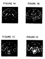
- Figures 1A, 1B, 1C and 1D and Figures 2A, 2B, 2C, 2D, 2E, 2F and
2G present magnetic resonance images illustrating results of practice of the invention
in the Experimental Allergic Encephalomyelitis (EAE) model system;
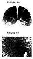
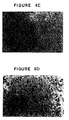
- Figures 3A and 3B and Figures 4A, 4B, 4C and 4D represent
histological results of practice of the invention in the EAE model system; and
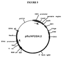
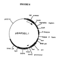
- Figure 5 and 6 are schematic representations of expression vectors
encoding humanized heavy and light chains, respectively, of an antibody of the
invention.
-
DETAILED DESCRIPTION
-
The following detailed description illustrates practice of the invention
through use of the mouse monoclonal anti-CD18 antibody mAb 60.3, mouse
monoclonal antibody 23F2G and humanized antibody 23F2G to alleviate progression
of Experimental Allergic Encephalomyelitis (EAE) in macaque monkeys. Non-human
primates induced to develop EAE, an acute inflammatory disease of the central
nervous system and a model of the human disease MS, were chosen because of the
many immunological and neurophysiological similarities EAE shares with MS (Rose,
et al., supra.) and because the CD18 antigen recognized by mAb 60.3 is present in
high density on monkey peripheral blood leukocytes (Rose, et al., Clin. Immunol.
Immunopathol., 44:93-106 (1987).
-
Example 1 relates to the induction of EAE in monkeys. Example 2
describes treatment by mAb 60.3 antibody infusion and Example 3 relates to the
evaluation of disease progression as monitored by a combination of clinical
evaluation, blood analysis, and magnetic resonance imaging, including
immunocytochemical staining of frozen tissue sections obtained post-mortem.
Example 4 describes the preparation of hybridoma cell lines producing anti-human
CD18 antibodies and Example 5 relates use of monoclonal antibodies from hybridoma
23F2G in treatment of EAE. Example 6 addresses humanization of antibody 23F2G
and Example 7 describes additional treatments of EAE macaque monkeys with
antibody 23F2G and humanized 23F2G.
EXAMPLE 1
Induction of EAE
-
Eighteen male monkeys Macaca fascicularis weighing 2-3 kg were
sensitized to myelin basic protein (BP) by the intra dermal injection of 0.1 ml of an
emulsion containing 5.0 mg of monkey BP and 0.5 mg of heat-killed M. tuberculosis.
Each animal was obtained and housed by the Regional Primate Research Center at the
University of Washington, Seattle. The Primate Center conforms to the National
Institutes of Health Guide for the Care and Use of Laboratory Animals. Ten days
after sensitization to BP, each animal was outfitted with a femoral catheter and tether
to facilitate blood sampling and the administration of treatments or anesthetics for
magnetic resonance imaging (MRI). The animals were maintained on intravenous
heparin (18 units/hour) to prevent blood clots from forming in the intra venous lines.
-
The animals were randomly admitted to specific treatment groups
before they were sensitized to BP. This was done to schedule MRI and to prevent
the severity of clinical signs at the onset from influencing the choice therapy.
Primate Center personnel, responsible for deciding when animals should be treated
or sacrificed, were blinded regarding the treatment history of each animal. Table 1
below provides a statement of criteria for determination of clinical signs and grades
of EAE in the animals.
| CLINICAL SIGNS AND GRADES OF EAE IN MACAQUES |
| Score | Signs |
| ? | Prodromal signs, including weight loss, anorexia, yawning, slow response to stimuli, irritability |
| ± | Mild neurologic signs, including "headache" (acute distress), "apathy" (indifference), hypokinesia, drooling, clumsiness in using limbs, nystagmus |
| + | Moderate neurologic signs, including akinesia, blindness, ataxia, tremor, ptosis, seizures, paresis, incontinence |
| + + | Severe neurologic signs including somnolence, paraplegia, hemiplegia, or quadriplegia |
| + + + | Moribund state with semicoma, coma, decerebration or decortication |
| D | Death |
EXAMPLE 2
Treatment By Antibody Infusion
-
At the onset of definite clinical signs (± to +) six EAE animals
received a bolus injection of mAb 60.3 (2 mg/kg) and dexamethasone (4 mg/kg).
Dexamethasone was administered to reduce the incidence of severe acute EAE which
is rapidly fatal within 48 hours unless treated immediately. Severe acute EAE occurs
in approximately 25% of animals induced to develop EAE and is caused by edema
in areas involving or impinging upon the brain stem or cerebral cortex. Following
injection of the bolus, these six animals were treated by continuous infusion of mAb
60.3 (2 mg/kg/day) for seven days. Over the seven day treatment period, the dose
of dexamethasone, which started at 4 mg/kg, was halved every two days until only
1 mg had been in effect for three days. Six control EAE animals were treated only
with dexamethasone, following the same protocol outlined above, and six additional
controls were treated with continuous infusion of saline.
-
Animals that improved and/or stabilized as a result of therapy were
sacrificed six weeks after the onset of clinical signs. Animals that experienced a
clinical relapse (clinical signs ≥ + +) some time after the cessation of treatment and
before six weeks had elapsed, were sacrificed shortly after the onset of the relapse.
Animals that did not respond well to therapy and continued to deteriorate clinically
(clinical signs ≥ + +) were sacrificed without completing the treatment program.
The statistical significance of differences in survival times in each treatment group
was determined by two-tailed Student t analysis.
EXAMPLE 3
Evaluation of Disease Progression
-
Disease progression was monitored by a combination of clinical
evaluation, blood analysis and magnetic resonance imaging. Clinical evaluations were
performed twice daily.
-
As indicated in Table 2 below, all of the animals in this study
developed clinical signs of EAE 13-26 days after sensitization to myelin basic protein.
-
In Table 2, listing of animal numbers in boldface identified animals
scanned by MRI; the Onset/Grade column reflects the day of onset after sensitization
and the severity of clinical signs (see Table 1) at onset; the Survival/Grade column
reflects days of survival post-onset and severity of clinical signs at time of sacrifice.
| THERAPEUTIC RESULTS |
| Animals | Onset/Grade | Treatment | Survival/Grade |
| 89070 | 17 days/+ | 60.3+dex | 2 days/D |
| 89075 | 21 days/+ | 60.3+dex | 42 days/± |
| 89080 | 21 days/+ | 60.3+dex | 42 days/± |
| 89069 | 18 days/± | 60.3+dex | 42 days/± |
| 89071 | 18 days/± | 60.3+dex | 21 days/++ |
| 89074 | 22 days/+ | 60.3+dex | 13 days/+ |
| 87143 | 21 days/± | dex alone | 17 days/++ |
| 87125 | 24 days/± | dex alone | 1 day/+ + |
| 87209 | 21 days/+ | dex alone | 2 days/+ + |
| 89077 | 16 days/± | dex alone | 1 day/D |
| 87200 | 22 days/+ | dex alone | 13 days/++ |
| 87069 | 26 days/± | dex alone | 9 days/+ + |
| 84228 | 13 days/+ | saline | 5 days/D |
| 84218 | 19 days/+ | saline | 3 days/D |
| 84291 | 25 days/+ | saline | 1 day/D |
| 86140 | 26 days/+ | saline | 3 days/D |
| 86187 | 19 days/+ | saline | 1 day/+ + |
| 86209 | 15 days/+ | saline | 1 day/ + + |
-
As indicated in Table 2, all eighteen animals developed clinical signs
of EAE which ranged in severity from ± to + at the onset of disease. Six of the
eighteen EAE animals (33%) died of severe acute EAE 1-5 days after onset. Four
of the six had been treated with saline, one with dexamethasone, and one with a
combination of dexamethasone and mAb 60.3. The remaining twelve animals were
sacrificed at various times after onset either because they had deteriorated clinically
or because they had completed the six-week post-treatment protocol.
-
Out of six mAb-treated animals, five either improved or stabilized with
continuing, but not worsening, clinical signs. Animal 89071 improved, but suffered
an acute relapse two weeks after the cessation of treatment and was sacrificed at 21
days. Animal 89074 was sacrificed at 13 days, one week after the cessation of
treatment, to investigate an abnormal lipid signal detected by magnetic resonance
spectroscopy (MRS), a technique being developed for studies of demyelination in the
nonhuman primate [Richards et al., New York, Soc. Mag. Reson. Med. 2: p.1043
(1990)]. This animal had improved as a result of therapy and was clinically stable
at the time of sacrifice. Animals 89075, 89080, and 89069 were all clinically stable
six weeks after onset. The mean survival time of mAb 60.3-treated animals was 30
days. Control animals treated with saline (excluding animal 87220) survived 1-5 days
after onset, with a mean survival time of 3 days. Animals treated with
dexamethasone survived 1-17 days after onset, with a mean survival time of 7 days,
3 of 6 dying of their disease 1-2 days after onset, and 3 of 6 surviving 9,13 and 17
days, respectively, until they were sacrificed due to deteriorating clinical signs
(≥ + +).
Magnetic Resonance Imaging
-
MRI was used to map the anatomic distribution of the lesions in all six
of the mAb-treated EAE animals, two dexamethasone-treated animals, and two
saline-treated animals. Conventional spin-warp imaging was performed using a
General Electric CSI-II NMR imager/spectrometer (2 Tesla magnet) as described in
Rose et al., Biomed. and Pharmacother., 43:347-353 (1989). Images were acquired
once a week before the onset of clinical signs and twice a week after the onset of
clinical signs. The animals were anesthetized with ketamine and positioned in the
magnet so that the center of the brain was at magnet iso-center. Coronal T2-weighted
images (TE=80 msec, TR=3000 msec) were used to find abnormal and normal brain
regions. Lesions were detected in 60% of the scanned animals on or before the onset
of clinical signs. Most lesions were localized in the cerebral white matter, optic
tracts, visual radiations or brainstem. The lesions were identified as areas of
increased brightness (white) in a T2-weighted MR image. Of the four control animals
that were scanned, only one survived long enough to obtain post-treatment scans. In
this dexamethasone-treated animal (87069) a brainstem lesion, detected one day
before the onset of clinical signs, progressed in size and intensity until the animal was
sacrificed nine days later as shown in Figure 1. Plate A represents a "normal" brain
scan, 21 days after sensitization; Plate B reveals detection of a brainstem lesion two
days after onset of clinical signs i.e., 28 days post sensitization: Plates C and D
illustrate gradual progression of the brainstem lesion 35 and 38 days after
sensitization, respectively.
-
Following treatment with mAb 60.3, the MRI lesions detected in 5 of
6 animals diminished in both size and intensity. Three animals (89075, 89071, and
89074) had evidence of brainstem lesions at the onset of clinical signs. Although
such lesions are frequently fatal if untreated [Shaw et al., Ann. Neurol. 24:738-748
(1988)], these lesions disappeared in all three animals following treatment with mAb
60.3. Figure 2 demonstrates the progression of the brainstem lesion in animal 89071.
In the Figure, Plate A illustrates a normal brain scan 15 days post sensitization; Plate
B shows a brainstem lesion at the onset of clinical signs, i.e., 18 days after
sensitization; Plates C through F illustrate gradual resolution of the brainstem lesion
through antibody treatment as monitored at 22, 25, 29 and 32 days post sensitization;
Plate G illustrates reappearance of a lesion in the same region which was
accompanied by clinical relapse 39 days post sensitization and 14 days after cessation
of treatment. Enlarging lesions are identified as prominent areas of increased
intensity (white) on the scans. As noted above, two weeks after the treatment was
discontinued, 89071 clinically relapsed and MR images demonstrated a larger, more
intense lesion in the brainstem. The relapse was not treated and the animal was
sacrificed.
-
Animal 89080 developed a major hemorrhagic lesion in the lateral
geniculate nucleus and striatum. Hemorrhagic lesions are usually fatal and usually
do not respond to treatment (Shaw et al., supra) but this lesion almost completely
disappeared following administration of mAb 60.3. Animal 89069 had the mildest
disease of any of the animals, as determined by MRI, with several small, but intense,
sub-cortical white matter lesions that disappeared following treatment.
-
Two of the longest surviving animals (89080 and 89069) developed a
chronic disease characterized by the appearance of new, smaller, MRI-detectable
lesions 25 and 22 days, respectively, after the cessation of treatment. These
secondary lesions were not as severe as the first lesions and were accompanied by
milder clinical signs which were not treated. Animal 89074 was sacrificed 13 days
after the onset of disease, to investigate an abnormal lipid signal detected by MRS.
At the time of sacrifice, six days after stopping treatment, his initial lesions were
gone, but a new smaller subclinical lesion had appeared on the opposite side of the
brain. No new lesions were ever detected in animal 89075 after cessation of
treatment.
-
Cryostat sections of cerebellum or cerebrum were stained with
hematoxylin and eosin and by the immunoperoxidase technique in conjunction with
mAbs to human leukocyte membrane antigens in order to analyze the composition of
the cellular infiltrates. Immunocytochemical staining with mAb 60.3, which
recognizes a CD18 determinant common to all leukocytes, was used to confirm the
presence of leukocytes in EAE lesions. A tissue section obtained from the brain of
animal 89070 and stained with mAb 60.3 is shown in Figure 3. In Panel A, at 390X
magnification and stained with rabbit anti-mouse IgG, murine IgG coated cells are
revealed in the lumen of blood vessels but not in brain tissue; and, in Panel B, at
390X magnification and stained with mAb 60.3, massive infiltration of PMBCs is
seen in the exudate and extending out into adjacent white matter.
-
Animal 89070 was the only mAb-treated animal to die before
completion of treatment. Brain tissue taken from this animal was stained with a goat
anti-mouse Ig antibody in order to determine whether the treatment mAb had entered
the CNS before the animal died. MAb 60.3 reacts with both leukocytes and brain
microglia. If this mAb had crossed the blood-brain-barrier during the treatment
period, it was expected that a staining pattern similar to that shown in Figure 3, Panel
B might be obtained. Alternatively, if the treatment mAb entered the CNS, bound
only to blood leukocytes, the microglia would not have been stained. Instead, murine
Ig-coated cells were only detectable in the vessel lumen, and not at all in the
perivascular spaces. The absence of any positively stained leukocytes in the lesion
stained with goat anti-mouse Ig, suggests that the animal died of an inflammatory
condition existing before treatment was initiated and that the treatment antibody was
given too late to be effective.
Effect of Treatments on Leukocyte Counts
-
The hematologic hallmarks of untreated EAE are a progressive
leukocytosis and lymphopenia prior to the onset of clinical signs. The leukocytosis
can represent as much as a four-fold increase in the number of circulating PMNC and
no significant changes in the frequency or absolute numbers of monocytes,
eosinophils, or basophils. Following successful treatments of EAE, the absolute
numbers of lymphocytes and PMNC return, fairly soon after the initiation of
treatment, to pre-sensitization levels. Thus, continuous monitoring of blood
leukocytes provides a useful measure of disease progression which complements the
clinical evaluation and magnetic resonance imaging.
-
Blood was drawn on a weekly basis until the onset of clinical disease
and then more frequently. One-half ml was sent to the hematology laboratory for
WBC and differential counts. The results are set out in Table 3 below providing
white blood cell number (x10
3) per mm
3 whole blood. "NA" indicates that data was
not available. A dashed line indicates that the animal was sacrificed at this time.
| WBC ANALYSES OF MAB-TREATED EAE ANIMALS |
| ANIMALS: | 89070 | 89075 | 89080 | 89069 | 89071 | 89074 |
| Day |
| Presens | 8.1 | 8.7 | 14.0 | 8.0 | 7.8 | 13.3 |
| Onset | 14.6 | 16.2 | 16.4 | 9.3 | 12.3 | 18.1 |
| Day 2 | --- | 27.1 | 33.2 | 22.2 | 22.6 | 49.9 |
| Day 4 | | 49.6 | 53.1 | 31.6 | 41.4 | 72.0 |
| Day 7 | | 61.2 | 61.7 | 16.4 | 90.7 | 88.6 |
| Day 10 | | 20.7 | 18.2 | 20.0 | 12.8 | 14.6 |
| Day 14 | | 33.3 | 9.6 | 8.2 | 18.4 | 13.4 |
| Day 21 | | 8.7 | 12.9 | 8.9 | 37.1 | --- |
| Day 28 | | 8.7 | 19.8 | 6.7 | --- |
| Day 35 | | 7.9 | 16.4 | 6.9 |
| Day 42 | | NA | NA | 8.6 |
WBC ANALYSIS OF CONTROL EAE ANIMALS
DEXAMETHASONE CONTROL |
| Animal: | 87143 | 87125 | 87209 | 89077 | 87200 | 87069 |
| Presens | 4.7 | 6.1 | 6.7 | 13.5 | 8.9 | 14.1 |
| Onset | 9.6 | 9.0 | 14.6 | 32.2 | 12.4 | 16.3 |
| Day 2 | 3.1 | --- | --- | --- | 5.4 | NA |
| Day 4 | 3.7 | | | | 3.6 | NA |
| Day 7 | 3.8 | | | | 7.0 | NA |
| Day 10 | 8.7 | | | | 10.7 | --- |
| Day 14 | 7.9 | | | | --- |
| Day 21 | --- |
| SALINE CONTROL |
| Animal: | 84228 | 84218 | 84291 | 86140 | 86187 | 86209 |
| Presens | 11.0 | 7.0 | 13.3 | 9.7 | 8.3 | 9.7 |
| Onset | 14.3 | 9.0 | 21.9 | 23.6 | 14.2 | 3.5 |
| Day 2 | 20.2 | 8.4 | 22.5 | 29.0 | --- | --- |
| Day 4 | 30.1 | 8.0 | --- | --- |
| Day 7 | --- | --- |
-
In animals treated with mAb 60.3 and dexamethasone, a delayed
resolution of the leukocytosis was observed as compared to animals treated with
dexamethasone alone as revealed in Table 3. In two of the dexamethasone-treated
animals that survived 9 days or longer, the white blood cell counts dropped to pre-sensitization
levels two days after the initiation of treatment. In mAb 60.3-treated
animals, a pronounced leukocytosis (4-12 fold above presensitization values) was
observed which lasted for the entire treatment period. In these animals, the WBC
returned to presensitization levels 3-7 days only after the treatment period had ended.
Transient episodes of lymphopenia and leukocytosis continued to be observed in the
surviving animals, and could be correlated in some cases with episodes of clinical
relapse.
-
Reaction of the treatment mAb with blood leukocytes was confirmed
by staining cells with anti-murine Kappa chain mAb (Becton-Dickinson, San Jose,
CA). Flow cytometric analysis was performed with a FACSCAN Analyzer (Becton
Dickinson, San Jose, CA). Forward and right-angle scatter gates were set on
lymphocytes or large polymorphonuclear granulocytes for analysis of staining
patterns. The results for lymphocytes are set out in Table 4 below.
| % MURINE Ig-COATED CELLS |
| ANIMALS: | 89070 | 89075 | 89080 | 89069 | 89071 | 89074 |
| Day |
| Presens | 0 | 0 | 0 | 0 | 0 | 0 |
| Onset | 0 | 0 | 0 | 0 | 0 | 0 |
| Day 2 | --- | 96.6 | 97.7 | 94.2 | 96.7 | 98.2 |
| Day 4 | | 93.8 | 95.2 | 91.2 | 97.0 | 49.8 |
| Day 7 | | 95.2 | 99.1 | 94.9 | 95.4 | 27.2 |
| Day 10 | | 0 | 2.6 | 11.2 | 19.6 | 1.4 |
-
The dose of mAb given to the animals was in all instances sufficient
to coat >95% of circulating white blood cells and, as indicated in Table 5, to
maintain detectable levels of free antibody in circulation. Data in the Table represent
micrograms of circulating murine IgG per mm
3 whole blood.
| CIRCULATING MURINE IgG |
| Day |
| ANIMALS: | 89070 | 89075 | 89080 | 89069 | 89071 | 89074 |
| Presens | 0 | 0 | 0 | 0 | 0 | 0 |
| Onset | 0 | 0 | 0 | 0 | 0 | 0 |
| Day 2 | --- | 65 | 70 | 15 | 28 | 34 |
| Day 4 | | 80 | 70 | 65 | 25 | 15 |
| Day 7 | | 90 | 58 | 4 | 7 | 28 |
| Day 10 | | 0 | 0 | 0 | 0 | 0 |
-
After 7 days, the injected mAb was no longer detectable in the circulation. The
disappearance of the murine Ig coincided with the appearance of monkey anti-murine
Ig antibodies in the circulation of each animal.
Neuropathology
-
To confirm the diagnosis of EAE, the brains and spinal cords of each
BP-sensitized animal were examined postmortem. The animals were euthanized by
Nembutal overdose and brains and spinal cords were removed and dissected so that
fresh-frozen samples could be obtained before the larger blocks were fixed in 10%
neutral formalin. Representative blocks of tissue were embedded in paraffin and
sections were stained with hematoxylin and eosin, gallocyanin-Darrow red, luxol fast
blue (LFB) combined with periodic acid Schiff (PAS) and hematoxylin and with
Holmes' axon stain alone or combined with LFB to differentiate demyelination from
necrosis. Frozen sections of mirror-image blocks were stained with Oil Red O
(ORO) [Kil, Stain Technology, 47:271, (1972)] to identify lipid-filled macrophages.
-
All eighteen EAE animals were evaluated histologically postmortem for
evidence of EAE. All had microscopic evidence of either mild to severe acute EAE
or hyperacute EAE that generally correlated with the severity and duration of the
disease. All but 2 of the 12 control animals showed either predominantly hyperacute
lesions characterized by hemorrhages, subtotal necrosis, demyelination, and diffuse
massive infiltrations of neutrophils into the CNS parenchyma; or severe acute
perivascular lesions which were more compact, more confined to the perivascular
spaces than the hyperacute lesions and composed predominantly of lymphocytes and
macrophages, but some neutrophils, and with abundant demyelination as well as some
axonal debris. The two exceptional control monkeys, with EAE of 13-17 days
duration, had a mixture of lesions ranging from mild to severe demyelinating lesions,
composed predominantly of lymphocytes and macrophages with abundant myelin
debris and little or no axonal reaction. All but one of the mAb-treated monkeys had
a combination of: (a) older well developed demyelinated plaques with abundant
myelin debris, sudanophilic lipid and little or no axonal reaction or lymphocytic
infiltrate; and (b) more recent lesions corresponding to the terminal untreated
relapses, which were compact, confined to perivascular spaces, with varying
proportions of lymphocytes, monocytes, or neutrophils (consistent with diagnoses of
mild to severe acute EAE) and some myelin debris. These two types of lesions,
found in the same animal (#89069), are shown in Figure 4.
-
In Figure 4, Panel A, at two-fold magnification, numerous
inflammatory (area 1) and/or demyelinating (area 2) lesions can be ascertained in the
white matter; in Panel B, at 325X magnification, area 2 of Panel A is revealed as
being composed predominantly of macrophages scattered diffusely through the tissue
and about blood vessels, with the small black granules constituting PAS positive
glyco-lipoproteins; and in Panels C and D, at 130X and 325X magnification,
respectively, area 1 of Panel A is revealed as being composed predominantly of
PMNCs and no PAS positive insoluble glycoproteins.
Summary Of Results
-
Monitoring disease progression by a combination of clinical evaluation,
blood cell fluctuations, and magnetic resonance imaging (MRI) revealed the
following. Antibody treated animals survived significantly longer than either
dexamethasone- or saline-treated animals (p < 0.02; p < 0.001; respectively). The
effectiveness of this therapy is further confirmed by visualization by magnetic
resonance imaging (MRI). The almost complete disappearance of major lesions in
the brainstems of three different animals was unprecedented as brainstem lesions are
rapidly fatal in untreated animals. The disappearance of MRI-detectable lesions
following administration of mAb 60.3 demonstrates a potent effect on the
inflammatory reactions producing edema in the brain lesions. MAb 60.3 is known
to inhibit plasma leakage in vivo [Arfors et al., Blood, 60: 338-340, (1987)],
probably the most important factor in improvement in MRI-detectable lesions, as
changes in the tissue water characteristics can be visualized by changes in image
intensity on the MRI scans.
-
The severity of clinical signs at the onset of EAE was not uniform
because individual monkeys responded differently to sensitization with myelin BP.
Nevertheless, there was no correlation between the severity of clinical signs at the
onset of EAE and the ability of animals to respond to treatment. Of the six mAB-treated
animals, four were + at the onset of clinical signs and 2 were ±. Of the
twelve control animals (six dexamethasone-treated and six saline-treated), 8 were +
and 4 were ±, essentially the same distribution as the mAb-treated group. No
correlation was found between the severity of clinical signs at the onset and the
potentially stressful procedure of MRI examination under general anesthesia. Of the
ten animals scanned by MRI, six were + at the onset of clinical disease and four
were ±. One might have expected the additional handling involved in scanning the
animals, as well as the frequent administration of anesthetics, to result in a
generalized suppression of the immune response. [Wall et al., Lab Animals,
19:138-144 (1985), Thomas et al., Clin. Exp. Immunol., 47:457-466 (1982)].
However, the results do not support this hypothesis.
-
The dexamethasone-treated animals did have longer mean survival time
(7 days) than the saline-treated controls (3 days), but the difference was not
statistically significant (p < 0.1). Thus, although dexamethasone slowed down the
disease process, it had little apparent effect on the final outcome of the disease. Co-administration
of mAb 60.3, on the other hand, significantly prolonged survival and
in some cases completely reversed the clinical and MRI appearances of EAE.
EXAMPLE 4
-
Six to twelve week old Balb/c mice (Charles River Biotechnical
Services, Inc., Wilmington, Massachusetts, IACUC #901103) were immunized with
human T cell line Hut 78 to generate anti-CD 18 antibodies. For each of two
hybridoma-generating fusions (designated fusions 22 and 23) two Balb/c mice were
bled retro-orbitally for the collection of pre-immune serum on day 0. On day 2, each
animal received a total of 5 x 10⁶ Hut 78 cells in 0.2 ml sterile PBS intravenously.
The mice were then immunized at two week intervals for six weeks and then at
monthly intervals for three months. The final monthly boost was of glutaraldehyde
fixed Hut 78 cells. Cells were fixed by mixing equal volumes of cells (2 x 10⁷) and
glutaraldehyde (0.1 % so that the final concentration of the fixative was equal to
0.05%. The mixture was incubated with swirling for thirty seconds and an equal
volume of 0.2M glycine (in PBS) was added to block the reaction. The reaction
mixture was washed once with 0.2M glycine (in PBS) and twice with PBS. Cells
were resuspended in PBS at a concentration of 5 x 10⁶ cells per 0.2 ml PBS.
Immune serum was collected via retro-orbital bleeding on day 56 and tested by FACS
to determine its reactivity with the immunogen (Hut 78 cells). Three days prior to
the projected fusion date, the animals were immunized by intravenous injection with
a final dose of 5 x 10⁶ glutaraldehyde fixed cells.
-
For each fusion, the spleen from the mouse displaying the highest
immune serum titer against the immunogen was removed sterilely. A single-cell
suspension was formed by grinding the spleen between the frosted ends of two glass
microscope slides submerged in serum free RPMI 1640, supplemented with 2 mM L-glutamine,
1 mM sodium pyruvate, 100 units/ml penicillin, and 100 µg/ml
streptomycin (RPMI)(Gibco, Canada). The cell suspension was filtered through
sterile 70-mesh Nitex cell strainer (Becton Dickinson, Parsippany, New Jersey), and
washed twice by centrifuging at 200 g for 5 minutes and resuspending the pellet in
20 ml serum free RPMI. Thymocytes taken from 3 naive Balb/c mice were prepared
in a similar manner.
-
NS-1 myeloma cells, kept in log phase in RPMI with 11% fetal bovine
serum (FBS)(Hyclone Laboratories, Inc., Logan, Utah) for three days prior to fusion,
were centrifuged at 200 g for 5 minutes, and the pellet was washed twice as described
in the foregoing paragraph. After washing, each cell suspension brought to a final
volume of 10 ml in serum free RPMI, and 10 µl was diluted 1:100. 20 µl of each
dilution was removed, mixed with 20 µl 0.4% trypan blue stain in 0.85% saline
(Gibco), loaded onto a hemacytometer (Baxter Healthcare Corp., Deerfield, Illinois)
and counted.
-
For each fusion, 2 x 10⁸ spleen cells were combined with 4 x 10⁷ NS-1
cells, centrifuged and the supernatant was aspirated. the cell pellet was dislodged by
tapping the tube and 2 ml of 37°C PEG 1500 (50% in 75 mM Hepes, pH
8.0)(Boehringer Mannheim) was added with stirring over the course of 1 minute,
followed by adding 14 ml of serum free RPMI over 7 minutes. An additional 16 ml
RPMI was added and the cells were centrifuged at 200 g for 10 minutes. After
discarding the supernatant, the pellet was resuspended in 200 ml RPMI containing
15% FBS, 100 µM sodium hypoxanthine, 0.4 µM aminopterin, 16 µM thymidine
(HAT) (Gibco), 25 units/ml IL-6 (Boehringer Mannheim) and 1.5 x 10⁶
thymocytes/ml. The suspension was dispensed into ten 96-well flat bottom tissue
culture plates (Corning, United Kingdom) at 200 µl/well. Cells in plates were fed
on days 2, 4 and 6 days post fusion by aspirating approximately 100 µl/well plating
medium described above except containing 10 units/ml IL-6 and lacking thymocytes.
-
On day 10 culture supernatants were taken from each well, pooled by
column or row and analyzed for the presence of an antibody capable of competing
with an FITC-60.3 conjugate (prepared according to Goding, in "Monoclonal
Antibodies: Principles and Practice," 2d Ed., p. 241-280, Academic Press, New
York, 1986) for binding to Hut78 cells. The competition assay was performed as
follows. Twenty-five µl of a cell suspension containing 10 x 106 Hut78 cells/ml was
combined with 25 µl of a sub-saturating dose of FITC-60.3 (x 2560 ≅ 0.8 µg/ml)
and 50 ml µl of fusion supernatant and incubated for 30 min at 4°C, washed by
adding culture media to the cell mixture and centrifuging at 1800 rpm for three
minutes. The supernatant was removed by aspiration and the pellet was washed twice
with flow cytometric analysis (FACS) medium containing RPMI, 2% PBS and 0.1%
NaN3. The reaction mixture was fixed by addition of 1% paraformaldehyde (pH 7.2
in PBS) and transferred to polystyrene tubes for FACS using a Becton Dickenson
FACscan analyzer. Of the 960 hybridomas generated in fusion 22, the supernatant
of hybridoma No. 22F12C inhibited binding of FITC-60.3 by about 90%;
supernatants from hybridoma Nos. 22J4A and 22B3B inhibited binding by about 40%
and 10% respectively. Two hybridomas from fusion 23, 23F2G and 23I11B,
produced antibodies which inhibited FITC-60.3 binding by ≥ 95%. These five
hybridomas were cloned. Hybridoma cell line 23F2G was deposited with the
American Type Culture Collection, 12301 Parklawn Drive, Rockville, M.D. 20852
U.S.A. on June 30, 1992 and accorded accession number A.T.C.C. HB 11081. The
isotype of the antibody produced by hybridoma cell line 23F2G (referred to herein
as mAb 23F2G) was determined to be IgG2a. Hybridomas 22F12C and 23I11B also
produced antibodies of the IgG2a isotype, while hybridomas 22B3B and 22J4A
produced IgG1 antibodies.
-
Antibodies from hybridomas 22F12C, 23F2G and 23I11B were all
found, in varying degrees, to: (1) block adhesion of human T cells to activated
HUVEC monolayers; (2) block aggregation of 13-acetate (PMA) activated Con A
blast cells phorbol 12-myristate; and (3) induce demargination of white blood cells
in rabbits. The ability of the IgG1 antibodies produced by hybridomas 22B3B and
22J4A to block adhesion is as yet undetermined; mAb 22J4A was able to block
aggregation, but mAb 22B3B was not; both antibodies induced demargination, but to
a lesser degree than the others or mAb 60.3. Based on the screening results noted
above, it appears likely that, notwithstanding the ability to compete for binding of
mAb 60.3 to LFA-1 on Hut 78 cells, mAb 22B3B and 22J4A are specific for an
epitope present on the CD11a component of LFA-1 or an epitope associated with the
CD11a/CD18 heterodimer rather than with an epitope of CD18.
-
A comparison was made between the amino acid sequence of the
variable region of mAb 60.3 presented by L. Harris at San Francisco, California on
May 26-27, 1992 in a poster presented at a conference entitled "Cell Adhesion
Molecules In Inflammation" organized by International Business Communications
(South Natick, Mass.) and the amino acid sequence of the variable region of mAb
23F2G. Gross differences were found, despite the fact that 23F2G competed with
60.3 for binding to Hut78 cells. While these differences are suggestive of the
possibility that the antibodies bind adjacent, different epitopes on CD18, confirmation
of this possibility will have to await definitive epitope mapping studies.
EXAMPLE 5
-
Hybridoma 23F2G was amplified by the ascites method and antibody
was purified by affinity chromatography on Protein A under pyrogen-free conditions.
From 25 ml of ascites, 120 mg of antibody preparation was isolated. This
preparation contained only about 4 endotoxin units per mg.
-
Two Macaca fascicularis monkeys were then involved in a study to
preliminarily determine the ability of mAb 23F2G to alleviate EAE symptoms. The
EAE induction procedures were essentially as in Example 1. Two animals, Nos.
89161 and 89186, sensitized to 1.0 mg of purified monkey basic protein and
developed hyperacute hemorrhagic EAE 18 and 20 days post sensitization,
respectively. Animal 89161 died within 12 hours of onset and before treatment with
dexamethasone could be initiated. Animal 89186 was treated for a week with both
dexamethasone (3 days at 4 mg/kg, 2 days at 2 mg/kg and 2 days at 1 mg/kg) and
mAb 23F2G (7 days at 2 mg/kg, i.v.). The animal improved dramatically with
complete resolution of paralysis three days after initiation of treatment. Lesions
detected by MRI resolved four days after cessation of treatment and the animal
improved dramatically with complete resolution of paralysis. Eight days following
cessation of treatment, the animal relapsed and was sacrificed. Hematologic analysis
revealed that mAb 23F2G caused steady demargination of leukocytes which peaked
on the last day of treatment. The white blood cell count rose from 22.7 x 10³ on the
day of clinical onset to 82 x 103/mm3 on the last day of treatment. The antibody
showed saturation of blood lymphocytes at the dose administered.
EXAMPLE 6
Humanization of 23F2G Antibody Variable Regions
A. Determination of Variable Region Sequences
-
Total RNA was isolated from the hybridoma cell line 23F2G
(A.T.C.C. HB 11081) and first strand cDNA was synthesized using the total RNA
as a template. The first strand cDNA was in turn used as a template for PCR
reactions to obtain double-stranded DNA fragments encoding the variable regions of
both the heavy and light chains of mAb 23F2G.
-
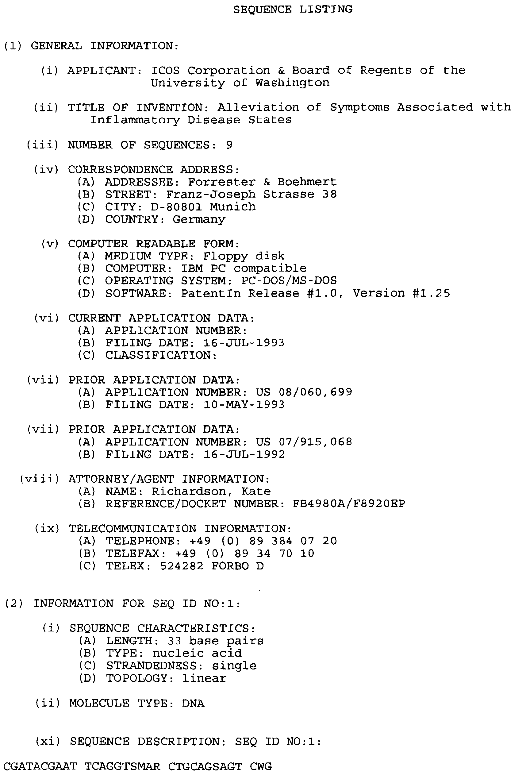
The forward primer used to clone the heavy chain variable region was
primer HFR1-4, the sequence of which is set out below in IUPAC nomenclature as
SEQ ID NO: 1.
- Forward Primer HFR1-4 (SEQ ID NO: 1)
CGATACGAATTCAGGTSMARCTGCAGSAGTCWG
The 3' nucleotides of the primer correspond to the region encoding the first seven
amino acids of mature mouse immunoglobulin heavy chains, while the underlined
nucleotides of the primer encode an EcoRI site to facilitate cloning of the PCR
fragment generated. The reverse primer used to clone the heavy variable region
sequences was primer HG2A-1, the sequence of which is set out below in IUPAC
nomenclature as SEQ ID NO: 2.
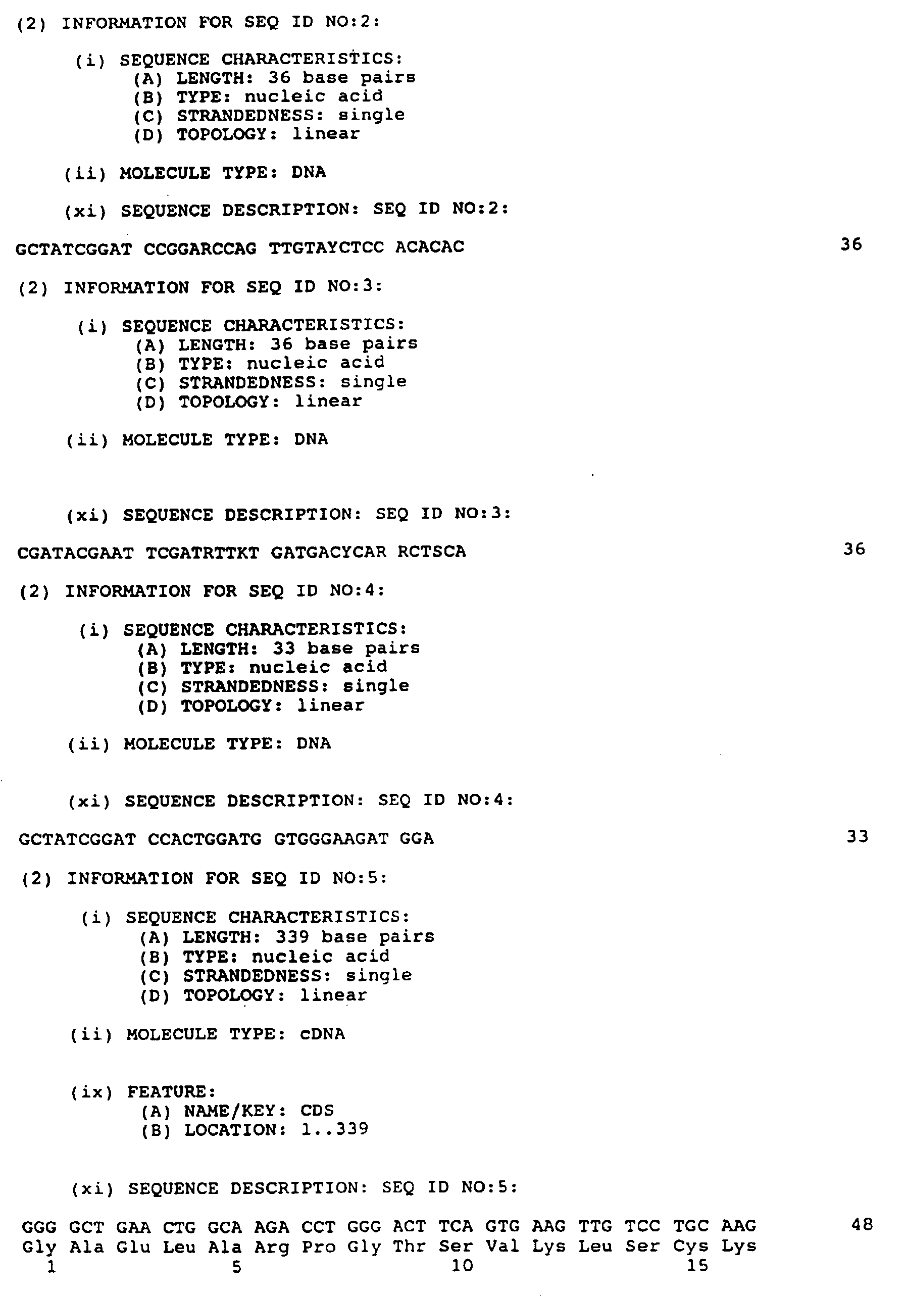
- Reverse Primer HG2A-1 (SEQ ID NO: 2)
GCTATCGGATCCGGARCCAGTTGTAYCTCCACACAC
The 3' end of primer HG2A-1 encodes the complement of codons 127-136 in the
mouse IgG2A constant region [according to the numbering system of Kabat et al.,
"Sequences of Proteins of Immunological Interest", U.S. Department of Health and
Human Services, NIH Publication No. 91-3242 (1991)] and the underlined nucleotides
of the primer encode a BamHI site. -
-
The forward primer used to generate PCR fragments encoding the light
chain variable region was primer LFR1-3, the sequence of which is set out below in
IUPAC nomenclature as SEQ ID NO: 3.
- Forward Primer LFR1-3 (SEQ ID NO: 3)
CGATACGAATTCGATRTTKTGATGACYCARRCTSCA
The 3' end of primer LFR1-3 corresponds to the mature amino-terminal region of the
mouse immunoglobulin light chain (residues 1-8) and the underlined nucleotides
encode an EcoRI site to facilitate cloning. The reverse primer for the light chain
variable region was primer LKC-1, the sequence of which is set out below as SEQ
ID NO: 4.
- Reverse Primer LKC-1 (SEQ ID NO: 4)
GCTATCGGATCCACTGGATGGTGGGAAGATGGA
The 3' end of primer LKC-1 is complementary to codons 116-122 in the constant
region of the mouse kappa light chain gene (according to the numbering system of
Kabat et al., supra). The underlined nucleotides in the primer encode a BamHI site.-
-
The resulting light and heavy chain PCR fragments were ligated into
different vectors and eight to twelve independent clones were sequenced on both
strands. The DNA and amino acid sequences corresponding to the heavy chain
variable region of 23F2G are shown in SEQ ID NO: 5. The seven amino-terminal
residues of the heavy chain sequence are indefinite because they correspond to the
forward primer used in the PCR reaction. Amino acid residues 1 to 15, 31 to 45, 53
to 84 and 94 to 103 of SEQ ID NO: 5 comprise the framework regions of the mouse
23F2G heavy chain variable domain while amino acid residues 16 to 30, 46 to 52 and
35 to 93 of SEQ ID NO: 5 comprise the CDR regions. Comparison of the translated
heavy chains variable sequence to the different subgroups of variable regions of
mouse heavy chains [Kabat et al., supra] indicates that the heavy chain variable
sequence of 23F2G belongs in mouse subgroup II. The DNA and deduced amino
acid sequences of the light chains variable region of 23F2G are shown in SEQ ID
NO: 6. The eight amino-terminal residues of the light chain were elucidated through
amino-terminal sequencing of the light chain protein. Amino acid residues 1 to 30.
36 to 49, 67 to 98 and 110 to 120 of SEQ ID NO: 6 comprise the framework regions
of the mouse 23F2G light chain variable domain while amino acid residues 31 to 35,
50 to 66 and 99 to 109 of SEQ ID NO: 5 comprise the CDR regions. The light chain
variable region of 23F2G is most similar to the subgroup III mouse light chain
variable regions [Kabat et al., supra].
B. Design of Humanized Heavy and Light Chain Variable Regions
-
In choosing human frameworks, two general criteria were used: (1)
the human frameworks chosen were as homologous as possible to those of 23F2G in
order to increase the probability that the CDR regions would retain their correct
conformations and consequently their affinity towards the antigen [see Kabat, et al.,
supra, and Kirkham et al., EMBO J., 11:603-609 (1992)] and (2) the human
framework regions chosen contained a minimal number of unusual residues that could
potentially provoke an immune response against the antibody in a human.
-
Because the heavy chain of mAb 23F2G resides in the mouse subgroup
II sequences, it is most homologous to sequences in human subgroup I. Therefore,
a framework from the human subgroup I set of heavy chain variable sequences was
chosen for use in a humanized version of mAb 23F2G. Rather than choose a
particular framework from existing sequenced human antibodies, it was decided to use
a consensus sequence for the human subgroup I variable region framework (Kabat,
et al., supra) so that the unusual residues often found in particular frameworks were
avoided. The combined framework regions of the heavy chain of 23F2G, excluding
the amino-terminal seven amino acids, have 70% sequence identity to the human
subgroup I consensus sequence. In comparison, the combined 23F2G heavy chain
framework regions were less homologous to the human subgroup II and III consensus
sequence (52% and 58%, respectively).
-
Replacement of rodent antibody framework regions with human
framework sequences can result in an antibody with significantly reduced binding
affinity. In such cases, it is necessary to substitute certain human framework residues
with those in the original rodent antibody. Based on an analysis of published
antibody humanization studies including Co et al., J. Immunol., 148:1149-1154
(1992); Carter et al., Proc. Nat'l. Acad. Sci. (USA), 89:4285-4289 (1992); Maeda
et al., Hum. Antibod. Hybridomas, 2:124-134 (1991); Kettleborough et al., Protein
Engineering, 4(7):773-783 (1991); Tempest et al., BIO/TECHNOLOGY, 9:266-271
(1991); Gorman et al., Proc. Nat'l. Acad. Sci. (USA), 88:4181-4185 (1991); Queen
et al., Proc. Nat'l. Acad. Sci. (USA), 86:10029-10033 (1989); and Co et al., Proc.
Nat'l. Acad. Sci. (USA), 88:2869-2873 (1991), it was decided to retain the framework
residues of mouse 23F2G at positions 66, 67 and 93 (numbering system of Kabat et
al., supra) which correspond to amino acid positions 60, 61 and 90 of SEQ ID NO:
5, rather than use the human subgroup I residues at these positions in the humanized
heavy chain.
-
A similar rationale was followed in humanizing the 23F2G light chain
variable domain. The variable region framework sequences of the light chain of
23F2G are 71%, 69% and 72% identical to the human subgroup I, II, and III kappa
consensus sequences, respectively. It was decided to use the human kappa subgroup
I consensus framework sequence because it was highly homologous to that of 23F2G
and because subgroup I frameworks have most commonly been used to humanize light
chain variable regions. No mouse-specific residues were substituted in the light chain
human framework regions.
-
The sequences designed for the humanized form of the 23F2G light and
heavy variable regions consisted of the framework regions of the human variable
region framework sequences as described above and the CDR regions of mouse mAb
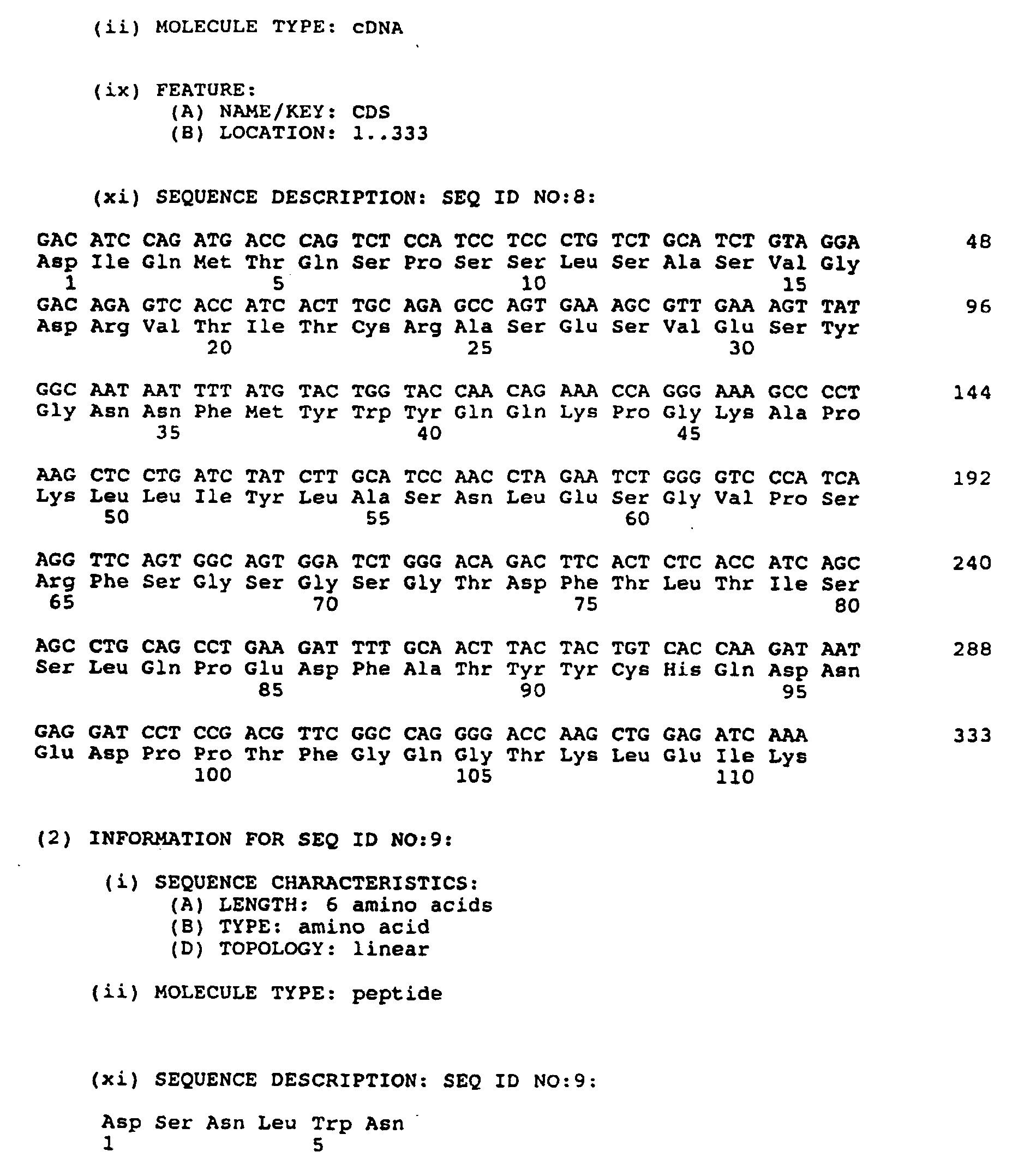
23F2G. The DNA and deduced amino acid sequences of these humanized heavy and
light chains variable regions are presented in SEQ ID NOs: 7 and 8, respectively.
Amino acids residues 1 to 30, 36 to 49, 67 to 98 and 110 to 120 of SEQ ID NO: 7
comprise the framework regions of the humanized heavy chain variable domain, while
amino acids residues 31 to 35, 50 to 66 and 99 to 109 of SEQ ID NO: 7 comprise
the CDR regions. Amino acids residues 1 to 23, 39 to 53, 61 to 92 and 101 to 111
of SEQ ID NO: 8 comprise the framework regions of the humanized light chain
variable domain, while amino acids residues 24 to 38, 54 to 60 and 93 to 100 of SEQ
ID NO: 8 comprise the CDR regions.
C. Assembly of Humanized 23F2G Variable Region Domains
-
Two DNA fragments which together encoded the humanized version
of the heavy chain variable region of 23F2G were assembled from complementary
synthetic oligonucleotides 50-60 nucleotides in length. Sequences included in addition
to sequences encoding the variable domain were (1) a HindIII restriction site at the
5' end to facilitate attachment to a promoter sequence, (2) an optimal translation
initiation sequence just upstream of the initiator methionine, (3) DNA encoding a
signal peptide, (4) a splice donor site just 3' to the variable region, and (5) an EcoRI
site at the 3' end to facilitate attachment to the constant region DNA segment. The
DNA sequence encoding the signal peptide was designed to be the same as that
encoding the signal peptide associated with several human heavy chain subgroup I
sequences (Kabat et al., supra). Two DNA fragments together encoding the
humanized light chain variable region DNA segment were also constructed from
complementary synthetic oligonucleotides. The assembled heavy and light chain
variable region DNA fragments were then cloned into pSK+ (Stratagene, La Jolla,
CA) and their sequence was confirmed.
D. Construction of a Humanized 23F2G Heavy Chain Expression Vector
-
A plasmid for expression of the humanized heavy chain of 23F2G was
constructed by ligating together the two DNA fragments containing the humanized
heavy chain variable region sequence, the constant regions of the human IgG4 gene
[cloned from a human T cell line (Hut 78)], and the expression vector pRc/CMV
(Invitrogen). The expression vector pRc/CMV contains the immediate early promoter
from the cytomegalovirus (CMV), a polylinker region downstream of the promoter,
and a neomycin resistance cassette. Specifically, the following four fragments were
isolated and ligated: (1) the 5.5 kb HindIII/XbaI fragment from pRc/CMV, (2) a 6
kb EcoRI/XbaI fragment containing the IgG4 sequences, (3) a ∼ 200 bp HindIII/XhoI
fragment containing the 5' half of the humanized heavy chain variable region
sequence, and (4) a ∼ 300 bp XhoI/EcoRI fragment containing the 3' half of the
heavy chain variable region sequence.
-
The ligation mix was used to transform E. coli, and a correct clone was
confirmed by restriction digest and was designated pRc/HF2GH.2. A circular map
of pRc/HF2GH.2 is shown in FIGURE 5. Large scale plasmid preparations of
pRc/HF2GH.2 were performed using the alkaline lysis method as described in
Sambrook et al., Molecular Cloning: A Laboratory Manual, Cold Spring Harbor
Press (1989) and the plasmid was twice-banded in CsCl-ethidium bromide gradients.
E. Construction of a Humanized 23F2G Light Chain Expression Vector
-
A plasmid for expression of the humanized light chain was constructed
by ligating together two DNA fragments containing the humanized light chain variable
region sequence, the human kappa gene, a dihydrofolate reductase (DHFR) gene
expression cassette, and a part of the pRc/CMV vector containing the strong promoter
from CMV.
-
In constructing a plasmid containing the humanized light chain gene of
23F2G and DHFR sequences, several intermediate plasmids were first constructed.
The first intermediate plasmid constructed, pRc/HF2GL. 1, was similar to that made
for expression of the heavy chain gene in that the variable and constant region coding
sequences were cloned between the HindIII and XbaI sites of pRc/CMV. To create
pRc/HF2GL.1 the following 5 fragments were ligated together: (1) HindIII/XbaI
digested pRc/CMV, (2) a ∼ 200 bp HindIII/Asp718 fragment containing the 5' half
of the humanized light chain variable region sequence, (3) a ∼ 200 bp Asp718/EcoRI
fragment containing the 3' half of variable region gene, (4) a ∼ 400 bp EcoRI/SacI
fragment containing the 5' half of kappa gene fragment (cloned from a human
genomic DNA library) and (5) a ∼ 350 bp SacI/XbaI fragment containing the 3' half
of kappa gene fragment (also cloned from the human genomic DNA library).
-
A second intermediate plasmid, pSl1190-dhfr, was made by inserting
a 1.7kb SphI/BamHI fragment from pSV2-dhfr (ATCC 37146) into the corresponding
site in the polylinker of the sequencing vector pSL1190 (Pharmacia, Piscataway, NJ).
The SV40-DHFR sequences within pSL1190-dhfr were completely sequenced and
were found to contain only minor differences from previously published sequence of
these elements. In particular, the DHFR coding sequence within pSL1190-dhfr was
found to encode the same polypeptide as the previously published sequence of mouse
DHFR [Simonsen and Levinson, Proc. Natl. Acad. Sci. USA, 80:2495-2499 (1983)].
-
To create a final plasmid (pD/HF2GL.1) for expression of humanized
23F2G light chain, the following four fragments were ligated: (1) the 3.1 kb
BamHI/HindIII fragment from pRc/CMV, (2) the 0.753 bp XbaI/Asp718 fragment
from pRc/CMV, (3) the 1.2 kb fragment from pRc/HF2GL.1 (containing the
complete humanized light chain gene), and (4) the 1.7 kb Asp718/BamHI fragment
(containing SV40 promoter-DHFR sequences) from pSL1190-dhfr. A circular map
of pD/HF2GL.1 is shown in FIGURE 6. Large scale preparations and purifications
of these plasmids were performed as described above for the heavy chain expression
plasmid.
F. Transfection of the CHO Cell Line DXB11 and Gene Amplification
-
Lipofection was used to introduce both the heavy and light chain
expression plasmids into the dhfr CHO cell line DXB11. Similar dhfr CHO cell
lines are described in Urlaub, et al, Proc. Natl. Acad. Sci. USA, 77:4216-4220
(1980). Selection for cells that were able to grow in the absence of nucleosides
(selects for DHFR) and that were resistant to G418 (neomycin) ensured that
transfectants contained both plasmids.
-
A pool of three transfectants was subjected first to 25nM and then
100nM methotrexate (Sigma, St. Louis, MO) to select cells that had undergone
amplification of the DHFR gene. After limited dilution cloning, one resulting cell
line, designated 6E6 (A.T.C.C. CRL 11398) was found to produce humanized 23F2G
at a rate of about 1 ng/cell/day and accumulate 5-10 mg antibody per liter of growth
medium. Another cell line derived by transfection of the dhfr CHO cell line [Urlaub,
et al., Cell, 33: 405-412 (1983)] was isolated in a similar manner to cell line 6E6 and
6E6 and was named B13-24 (A.T.C.C. CRL 11397). Cell line B13-24, which is
resistant to 200nM methotrexate, produces over 100 mg/l of antibody at a rate of
about 25 pg/cell/day.
-
The 6E6 and B13-24 cell lines can be cultured in Dulbecco's modified
Earls Medium/Ham's F-12 (1:1 mix) with 5% heat inactivated fetal bovine serum at
37°C, 6% carbon dioxide and 100% humidity, and at a density of between 2x10⁵ and
6x10⁵ cells per milliliter. The media for the 6E6 cell line includes 100nM
methotrexate while the media for B13-24 cell line includes 200nM methotrexate.
-
Humanized 23F2G antibody was purified from the 6E6 and B13-24 cell
lines as follows. Cell harvest fluid was first passed through a Protein A column,
which binds IgGs. The column was washed with 35mM Tris buffer, 0.1% Tween
20, pH 7.85 and then with 50mM citrate, pH 5.0. The antibody was eluted with
50mM citrate, 0.02% Tween 20, pH 3.0, and kept under acidic conditions at room
temperature for 15 minutes. The eluate was then chilled to 2-8°C and neutralized
with cold 1M Tris buffer. The Protein A eluate was adjusted to 0.5M ammonium
sulfate, 25mM Tris, pH 8.0 and loaded onto a Phenyl Sepharose column. The
column was then washed with loading buffer (0.5M ammonium sulfate, 25mM Tris,
pH 8.0), and eluted with 0.1M ammonium sulfate, 25mM Tris, pH 8.0. The eluate
was then diluted 1/10 with purified water to prepare it for the next step. The diluted
phenyl sepharose eluant was loaded onto a DEAE Sepharose Fast Flow column that
has been equilibrated with 10mM Tris, pH 7.5. After loading, the column was
washed with 10mM Tris, pH 7.5, followed by 10mM Tris, 50mM NaCl, pH 7.5.
The antibody was eluted with 10mM Tris, 200mM NaCl, pH 7.5. The DEAE eluate
is loaded onto a Sephadex G-25 Superfine column, and washed through with the final
formulation buffer (50mM sodium acetate, 120mM NaCl, 0.02% Tween 20, pH 5.6).
The eluted antibody was then diluted to the required concentration.
G. The Heavy Chains Derived from Cell Lines 6E6 and B13-24
-
During the course of analysis of mRNA derived from cell line 6E6 it
was noted that most (but not all) of the RNA encoding the humanized 23F2G heavy
chain was longer than expected. Subsequent cloning and sequencing of several
cDNAs encoding the heavy chain revealed that a region in the DNA near that
encoding the C-terminus had undergone a recombination event such that the sequence
encoding the Gly-Lys sequence typically found at the C-termini of IgG4 heavy chains
was missing and was replaced by the sequence encoding Asp-Ser-Asn-Leu-Trp-Asn
(SEQ ID NO: 9).
-
The heavy chain mRNA derived from the cell line B13-24 is the
expected size and therefore the C-terminal coding region of the heavy chain produced
by cell line B13-24 is likely to be intact.
H. Affinity of the Humanized 23F2G Antibodies
-
Humanized 23F2G antibody competes effectively with flourescent
murine 23F2G antibody in a cell binding assay. Fluorescent murine 23F2G antibody
at 0.4 µg/ml was mixed with increasing amount of humanized 23F2G antibody.
These antibody mixtures were incubated with the LFA-1 positive cell line HUT-78.
After washing the cells were examined by flow cytometry for mean fluorescent
intensity. In this assay humanized 23F2G antibody (at 1.01 µg/ml) bound to LFA-1
positive cells as efficiently as murine 23F2G antibody.
EXAMPLE 7
-
In order to further characterize the effects of murine monoclonal
antibody 23F2G (referred to in this example as "M23F2G") and the humanized
antibody obtained from CHO 6E6 cells of Example 6 ("Hu23F2G"), additional
Macaca Fascicularis
monkeys were sensitized to myelin BP in the manner of Example 1 using 0.1 ml of
a freshly prepared, somewhat more potent emulsion comprising 0.1-0.4 mg of
monkey BP and 0.5 mg of heat-killed M. tuberculosis. M23F2G was purified from
ascites fluid as in Example 5 and contained 5.2 endotoxin units per mg. Hu23F2G
was purified from CHO cell 6E6 (A.T.C.C. CRL 11398) culture supernatant by
affinity chromatography on Protein A under pyrogen-free conditions and contained
1.2 endotoxin units per mg.
-
Treatment was started on the day of onset of disease. Treatment
groups consisted of dexamethasone (4 mg/kg/day for 3 days) alone, dexamethasone
plus M23F2G (2 mg/kg/day for 7 days), or dexamethasone plus Hu23F2G (2
mg/kg/day for 7 days). Treatment assignment of animals was made prior to the onset
of clinical disease. Seven animals were treated with dexamethasone, five with
M23F2G, and four with Hu23F2G.
-
The endpoints used to evaluate the effects of treatment were: overall
survival after onset of disease up to a maximum of 42 days; clinical score following
treatment; and lesion burden (determined by MRI) following treatment. Clinical signs
were monitored daily until time of death or sacrifice. Animals were scanned by MRI
once a week. A modified clinical scoring system (vis-a-vis the previous scoring
system in Table 1) was used to reflect the severity of disease and is set out in Table
6, below. Pre-treatment MRI scan data was obtained at onset (± 3 days) of clinical
disease; post-treatment scan data was obtained on 10 days after clinical onset (± 3
days). MRI scans were analyzed by the software package NIH Image Version 1.44
(Bethesda, MD) to quantitate areas of abnormality. These MRI lesions reflect brain
edema and cellular infiltration caused by EAE and allow a non-invasive method of
assessing the effect of treatment on brain inflammation.
| CLINICAL SIGNS AND GRADES OF EAE IN MACAQUES |
| Score | Clinical Sign |
| 1 | Anorexia, weight loss |
| 2 | Inactivity, slow response, facial rash, hyperactivity, yawning |
| 3 | Apathy, drooling |
| 4 | Nystagmus, transient tremor |
| 5 | Hypokinesis, clumsiness |
| 6 | Ataxia, pupillary signs, ptosis strabismus, blindness |
| 7 | Head tilt, tremor |
| 8 | Seizure, body twisting, paresis |
| 9 | Paraplegia, hemiplegia, quadriplegia, lethargy, semicoma |
| 10 | Death |
-
Survival data are shown in Table 7 below. The mean survival for the
dexamethasone group was 23.4 days, compared with 34.8 days in the M23F2G group
and 25.5 days in the Hu23F2G group. No statistical significance could be assigned
to the survival scores for the antibody treated animals compared to those of the
dexamethasone treated animals, likely due to the potency of the BP preparation.
| SURVIVAL AFTER TREATMENT (DAYS) |
| (Animal No.) Dexamethasone | | (Animal No.) M23F2G | | (Animal No.) Hu23F2G |
| T89241 | 24 | T89341 | 42 | 91339 | 11 |
| 91444 | 8 | 91424 | 42 | F91307 | 42 |
| 91449 | 42 | 91425 | 6 | M91353 | 42 |
| 92144 | 42 | 92150 | 42 | 92136 | 7 |
| 92152 | 3 | 91331 | 42 |
| F91337 | 3 |
| F91304 | 42 |
| Mean | 23.4 | | 34.8 | | 25.5 |
| SD | ± 18.7 | | ± 16.1 | | ±19.1 |
-
Clinical score and lesion area were determined at onset of disease (±
3 days) and ten days post treatment (± 3 days), except for two animals in the
dexamethasone group (92152 and F91337) that died three days after onset, and the
post scan was obtained on the day of death. The values for each of the 3 treatment
groups are shown in Table 8. Clinical score values shown in Table 8 were those
obtained on the same day as the scan. There was no significant difference between
clinical scores at onset of disease between the three treatment groups; however,
statistically significant differences were noted post-treatment in both clinical score and
lesion area. [1-p for clinical scores, onset versus post; Dex=.73, M23F2G=.004,
Hu23F2G=.0001; 1-p for lesion area, onset versus post, Dex=.27, M23F2G=.007,
Hu23F2G=.07].
| CLINICAL SCORE AND LESION AREA VALUES |
| | Clinical Score | Lesion Area |
| Animal (Dex) | Onset | Post (d10+3) | % change | Onset | Post (d10+3) | % change |
| T89241 | 7 | 2 | -71.4 | 5237 | 3512 | -33 |
| 91444 | 4 | 5 | +25.0 | 8441 | 6607 | -22 |
| 91449 | 7 | 7 | 0 | 2514 | 3315 | +32 |
| 92144 | 4 | 4 | 0 | 7916 | 5575 | -30 |
| 92152 | 4 | 9 | +125 | 9209 | 12904 | +40 |
| F91337 | 7 | 9 | +28 | 4566 | 3566 | -22 |
| F91304 | 8 | 8 | 0 | 10135 | 4342 | -57 |
| Mean | 5.86 | 6.29 | +15.2% | 6860 | 5688 | -13.1% |
| SD | ±1.77 | ±2.69 | | ±2788 | ±3406 |
| (M23F2G) |
| T89341 | 7 | 0 | -100 | 1853 | 0 | -100 |
| 91424 | 6 | 5 | -16.7 | 10998 | 0 | -100 |
| 91425 | 4 | 4 | 0 | 7107 | 845 | -88 |
| 92150 | 4 | 0 | -100 | 499 | 151 | -70 |
| 91331 | 6 | 0 | -100 | 6237 | 269 | -96 |
| Mean | 5.4 | 1.8 | -63.3% | 5339 | 253 | -90.8% |
| SD | ±1.34 | ±2.49 | | ±4229 | ±350 |
| (Hu23F2G) |
| 91339 | 5 | 2 | -60 | 3773 | 569 | -85 |
| F91307 | 9 | 2 | -77.8 | 6855 | 230 | -97 |
| M91353 | 6 | 0 | -100 | 14870 | 4219 | -72 |
| 92136 | 6 | 3 | -50 | 20268 | 9344 | -54 |
| Mean | 6.5 | 1.75 | -71.95% | 11441.5 | 3591 | -77% |
| SD | ±1.73 | ±1.26 | | ±7517 | ±4240 |
-
In summary, M23F2G and Hu23F2G have the same beneficial effect
on monkey EAE as antibody 60.3 in terms of survival. Analyses of clinical severity
and MRI endpoints also supports a beneficial effect of M23F2G and Hu23F2G
compared to dexamethasone alone in monkey EAE.
-
The foregoing examples describe a method for treatment for inhibiting
inflammatory processes and alleviating symptoms associated with inflammatory
disease states by administering a reagent, such as an antibody, that blocks leukocyte
adhesion and migration to inflammatory sites. While the present invention has been
described in terms of specific methods and compositions, it is understood that
variations and modifications will occur to those skilled in the art upon consideration
of the description of the present invention. As one example, while two anti-CD18
mAbs, 60.3 and 23F2G, have been demonstrated to be useful in practice of methods
of the invention, it is expected that any antibody which competes with mAb 60.3
and/or with 23F2G for binding to LFA-1 will also be useful, whether or not such
antibody recognizes on epitope present on CD18. Accordingly, it is intended in the
appended claims to cover all such equivalent variations which come within the scope
of the invention as claimed.