EP1072224A2 - Synchronisation cardiaque rétrospective avec balayages cinématrographiques pour un balayeur a multicouche - Google Patents
Synchronisation cardiaque rétrospective avec balayages cinématrographiques pour un balayeur a multicouche Download PDFInfo
- Publication number
- EP1072224A2 EP1072224A2 EP00306245A EP00306245A EP1072224A2 EP 1072224 A2 EP1072224 A2 EP 1072224A2 EP 00306245 A EP00306245 A EP 00306245A EP 00306245 A EP00306245 A EP 00306245A EP 1072224 A2 EP1072224 A2 EP 1072224A2
- Authority
- EP
- European Patent Office
- Prior art keywords
- patient
- recorded
- image slices
- ekg data
- data
- Prior art date
- Legal status (The legal status is an assumption and is not a legal conclusion. Google has not performed a legal analysis and makes no representation as to the accuracy of the status listed.)
- Withdrawn
Links
- 230000000747 cardiac effect Effects 0.000 title claims abstract description 39
- 238000000034 method Methods 0.000 claims abstract description 28
- 230000000875 corresponding effect Effects 0.000 claims abstract description 11
- 238000001454 recorded image Methods 0.000 claims abstract description 7
- 230000002596 correlated effect Effects 0.000 claims abstract description 6
- 238000002591 computed tomography Methods 0.000 claims description 54
- 238000003384 imaging method Methods 0.000 description 9
- 238000013170 computed tomography imaging Methods 0.000 description 6
- 230000002308 calcification Effects 0.000 description 5
- 238000010894 electron beam technology Methods 0.000 description 4
- 230000033001 locomotion Effects 0.000 description 4
- 238000005259 measurement Methods 0.000 description 4
- 238000010586 diagram Methods 0.000 description 3
- 230000007246 mechanism Effects 0.000 description 3
- 230000001360 synchronised effect Effects 0.000 description 3
- OYPRJOBELJOOCE-UHFFFAOYSA-N Calcium Chemical compound [Ca] OYPRJOBELJOOCE-UHFFFAOYSA-N 0.000 description 2
- 206010029216 Nervousness Diseases 0.000 description 2
- 230000002238 attenuated effect Effects 0.000 description 2
- 239000008280 blood Substances 0.000 description 2
- 210000004369 blood Anatomy 0.000 description 2
- 229910052791 calcium Inorganic materials 0.000 description 2
- 239000011575 calcium Substances 0.000 description 2
- 230000001419 dependent effect Effects 0.000 description 2
- 238000011156 evaluation Methods 0.000 description 2
- 230000005855 radiation Effects 0.000 description 2
- 238000012360 testing method Methods 0.000 description 2
- 238000010009 beating Methods 0.000 description 1
- 230000005540 biological transmission Effects 0.000 description 1
- 231100001012 cardiac lesion Toxicity 0.000 description 1
- 238000007435 diagnostic evaluation Methods 0.000 description 1
- 238000002059 diagnostic imaging Methods 0.000 description 1
- 230000036541 health Effects 0.000 description 1
- 230000006872 improvement Effects 0.000 description 1
- 229910052751 metal Inorganic materials 0.000 description 1
- 239000002184 metal Substances 0.000 description 1
- 230000004048 modification Effects 0.000 description 1
- 238000012986 modification Methods 0.000 description 1
- 210000004165 myocardium Anatomy 0.000 description 1
- 230000008569 process Effects 0.000 description 1
- 238000012545 processing Methods 0.000 description 1
- 230000000284 resting effect Effects 0.000 description 1
- 239000000523 sample Substances 0.000 description 1
- 238000010998 test method Methods 0.000 description 1
- 238000004846 x-ray emission Methods 0.000 description 1
Images

Classifications
-
- A—HUMAN NECESSITIES
- A61—MEDICAL OR VETERINARY SCIENCE; HYGIENE
- A61B—DIAGNOSIS; SURGERY; IDENTIFICATION
- A61B6/00—Apparatus or devices for radiation diagnosis; Apparatus or devices for radiation diagnosis combined with radiation therapy equipment
- A61B6/54—Control of apparatus or devices for radiation diagnosis
- A61B6/541—Control of apparatus or devices for radiation diagnosis involving acquisition triggered by a physiological signal
-
- A—HUMAN NECESSITIES
- A61—MEDICAL OR VETERINARY SCIENCE; HYGIENE
- A61B—DIAGNOSIS; SURGERY; IDENTIFICATION
- A61B6/00—Apparatus or devices for radiation diagnosis; Apparatus or devices for radiation diagnosis combined with radiation therapy equipment
- A61B6/02—Arrangements for diagnosis sequentially in different planes; Stereoscopic radiation diagnosis
- A61B6/03—Computed tomography [CT]
- A61B6/032—Transmission computed tomography [CT]
-
- A—HUMAN NECESSITIES
- A61—MEDICAL OR VETERINARY SCIENCE; HYGIENE
- A61B—DIAGNOSIS; SURGERY; IDENTIFICATION
- A61B6/00—Apparatus or devices for radiation diagnosis; Apparatus or devices for radiation diagnosis combined with radiation therapy equipment
- A61B6/40—Arrangements for generating radiation specially adapted for radiation diagnosis
- A61B6/4064—Arrangements for generating radiation specially adapted for radiation diagnosis specially adapted for producing a particular type of beam
- A61B6/4085—Cone-beams
Definitions
- This invention relates generally to methods for noninvasive diagnostic imaging of a patient, and more particularly to methods for collecting still images of a patient's moving heart utilizing a CT scanning device.
- a physician screens otherwise healthy patients for cardiac lesions and plaque by examining and scoring x-ray images of the patient's heart. For example, calcium-containing plaque can be readily observed in such images and risk levels associated with an observed amount of plaque estimated by a trained physician.
- CT computed tomography
- an x-ray source projects a fan-shaped beam which is collimated to lie within an X-Y plane of a Cartesian coordinate system and generally referred to as the "imaging plane".
- the x-ray beam passes through the object being imaged, such as a patient.
- the beam after being attenuated by the object, impinges upon an array of radiation detectors.
- the intensity of the attenuated beam radiation received at the detector array is dependent upon the attenuation of the x-ray beam by the object.
- Each detector element of the array produces a separate electrical signal that is a measurement of the beam attenuation at the detector location.
- the attenuation measurements from all the detectors are acquired separately to produce a transmission profile.
- the x-ray source and the detector array are rotated with a gantry within the imaging plane and around the object to be imaged so that the angle at which the x-ray beam intersects the object constantly changes.
- a group of x-ray attenuation measurements, i.e., projection data, from the detector array at one gantry angle is referred to as a "view”.
- a "scan" of the object comprises a set of views made at different gantry angles, or view angles, during one revolution of the x-ray source and detector.
- the projection data is processed to construct an image that corresponds to a two-dimensional slice taken through the object.
- CT numbers integers called "Hounsfield units”
- a CT scanner is employed in a spiral scanning mode. A patient being evaluated remains still on a table of the CT scanner as the table continuously advances through the scanner along a z-axis, i.e., along the axis of rotation of the gantry.
- a patient is hooked to an EKG device and the scanner is synchronized to a signal derived from the patient's EKG.
- This method represents an improvement over the preceding method, because it provides a level of control over, or at least knowledge of a z-axis location of an imaging plane relative to the patient's heart during specified portions of a cardiac cycle.
- synchronizing the motions of the table and the rotating gantry to a patient's heartbeat increases the complexity of a CT scanner, even when the patient's heartbeat is relatively constant.
- most patients are unfamiliar with the testing environment and understandably become nervous during the test. This nervousness introduces a level of variability into the patient's heartbeat on a time scale can impact the test procedure.
- an electron beam is scanned across a curved metal plate to produce x-ray emissions that are directed towards the patient. Because only an electron beam is scanned, there is no need for a rotating gantry and the heart can be scanned very rapidly.
- electron-beam scanners are rare and expensive in comparison to CT scanners.
- a method for obtaining a set of still images of a moving heart of a patient utilizing a multi-slice CT scanner and an EKG data recorder comprising: simultaneously recording EKG data of the patient's cardiac cycle utilizing the EKG data recorder while cine scanning and recording plural CT image slices of the patient's heart utilizing the multi-slice CT scanner; correlating the recorded cine scanned CT image slices with the recorded EKG data; and selecting segmented images of the set of recorded image slices corresponding to a selected portion of the patient's cardiac cycle as indicated by the correlated, recorded EKG data, the selected segmented images thereby becoming the set of still image slices.
- the selected portion of the cardiac cycle may is preferably a portion in which the heart is relatively stationary.
- the CT scanner may have a rotating gantry, a detector, and an x-ray source on the rotating gantry projecting an x-ray beam at a detector, and the detector may acquire a fan angle of data for each image slice, and wherein cine scanning plural CT image slices of the patient's heart may comprise the step of collecting projection data from an axial scan having a duration at least as long as one complete cardiac cycle plus a time required for rotation of the gantry through 180° plus an angle equal to or less than one fan angle.
- the selected portion of the patient's cardiac cycle may be a portion of the cardiac cycle immediately prior to R-peaks of the recorded EKG data.
- Selecting segmented images of the set of recorded image slices may comprise the step of selecting segments of the set of collected image slices ending immediately prior to the EKG R-peaks or may comprise the step of selecting segmented images collected during rotation of the gantry through at least 180° plus an angle equal to or less than one fan angle.
- the multi-slice CT scanner may comprise a moveable table on which the patient is resting, and the method may further comprise stepping the table so that EKG data and CT image slices are recorded simultaneously at the plurality of positions.
- the selected portion of the patient's cardiac cycle may be a portion of the cardiac cycle in which the heart is relatively stationary.
- the CT scanner may have a rotating gantry, a detector, and an x-ray source on the rotating gantry projecting an x-ray beam at a detector, and the detector may acquire a fan angle of data for each image slice, and wherein cine scanning plural CT image slices of the patient's heart may comprise the step of collecting projection data from an axial scan having a duration at least as long as one complete cardiac cycle plus a time required for rotation of the gantry through 180° plus an angle equal to or less than one fan angle.
- the selected portion of the patient's cardiac cycle may be a portion of the cardiac cycle immediately prior to R-peaks of the recorded EKG data.
- Selecting segmented images of the set of recorded image slices may comprises the step of selecting segments of the set of collected image slices ending immediately prior to the EKG R-peaks or may comprise the step of selecting segmented images collected during rotation of the gantry through at least 180° plus one fan angle.
- Cine scanning and recording plural CT image slices of the patient's heart may comprise simultaneously cine scanning and acquiring data for at least four image slices.
- Cine scanning and recording data for at least four image slices may comprise acquiring data for at least four images slices each having a thickness of between 2.5 and 3.75 mm.
- Stepping the table may comprise stepping the table a distance of a total of the thicknesses of the simultaneously acquired image slices.
- Selecting segments of the set of collected image slices may comprise selected segments having at least 120 mm coverage of the patient's heart, and simultaneously recording EKG data of the patient's cardiac cycles while scanning and recording plural CT image slices of the patient's heart may comprise scanning for a total of no more than 30 seconds.
- a system for obtaining a set of still image slices of a moving heart of a patient said apparatus configured to: simultaneously record EKG data of a patient's cardiac cycle while cine scanning and recording plural CT image slices of the patient's heart; and correlate a recording time of the recorded EKG data with the recorded plural CT image slices.
- the system may comprise an EKG apparatus configured to record EKG data and to record, in association with the recorded EKG data, an indication of a start of a simultaneous recording of CT image slices.
- the system may further comprise a display screen configured to display a CT image slice from the recording of CT image slices and a portion of the simultaneously recorded EKG data, including an indication of a portion of the EKG data corresponding to a time of recording of the CT image slice being displayed.
- the system may comprise a multi-slice CT scanner configured to simultaneously record at least four image slices each having a thickness of between 2.5 and 3.75 mm.
- a system for retrospectively correlating recorded CT images and recorded EKG data said system being configured to record EKG data of a patient's cardiac cycle; and said system being configured to be responsive to a CT image scanner to record an indication correlating a start of a CT scan with a simultaneously recorded portion of recorded EKG data.
- the system may be configured to record the EKG data and the indication correlating a start of the CT scan with a simultaneously recorded portion of the recorded EKG data on a floppy disk.
- a method for obtaining a set of still image slices of a moving heart of a patient utilizing a multi-slice CT scanner and an EKG data recorder includes steps of simultaneously recording EKG data of the patient's cardiac utilizing the EKG data recorder while cine scanning and recording plural CT image slices of the patient's heart utilizing the multi-slice CT scanner; correlating the recorded cine scanned CT image slices with the recorded EKG data; and selecting segmented images of the set of recorded image slices corresponding to a selected portion of the patient's cardiac cycle as indicated by the correlated, recorded EKG data, the selected segmented images thereby becoming the set of still image slices.
- the selected segments thus become the set of still image slices.
- This method embodiment does not require that the CT scanner be synchronized to the EKG of the patient, because the acquired EKG data and scanned image data can be synchronized after they are recorded. Moreover, no special CT scanning technology, such as electron-beam scanning, is required, yet a suitable set of still images of the patient's heart can be acquired in a period of time in which a normal patient is able to hold his or her breath.
- a computed tomograph (CT) imaging system 10 is shown as including a gantry 12 representative of a "third generation" CT scanner.
- Gantry 12 has an x-ray source 14 that projects a beam of x-rays 16 toward a detector array 18 on the opposite side of gantry 12.
- Detector array 18 is formed by detector elements 20, which together sense the projected x-rays that pass through an object 22, for example a medical patient.
- Detector array 18 is fabricated in a multi-slice configuration. This type of configuration is found in at least one known commercial imaging system.
- Each detector element 20 produces an electrical signal that represents the intensity of an impinging x-ray beam and hence the attenuation of the beam as it passes through patient 22.
- gantry 12 and the components mounted thereon rotate about a center of rotation 24.
- Control mechanism 26 includes an x-ray controller 28 that provides power and timing signals to x-ray source 14 and a gantry motor controller 30 that controls the rotational speed and position of gantry 12.
- a data acquisition system (DAS) 32 in control mechanism 26 samples analog data from detector elements 20 and converts the data to digital signals for subsequent processing.
- An image reconstructor 34 receives sampled and digitized x-ray data from DAS 32 and performs high speed image reconstruction. The reconstructed image is applied as an input to a computer 36 that stores the image in a mass storage device 38.
- DAS data acquisition system
- Computer 36 also receives commands and scanning parameters from an operator via console 40 that has a keyboard.
- An associated cathode ray tube display 42 allows the operator to observe the reconstructed image and other data from computer 36.
- the operator supplied commands and parameters are used by computer 36 to provide control signals and information to DAS 32, x-ray controller 28 and gantry motor controller 30.
- computer 36 operates a table motor controller 44 that controls a motorized table 46 to position patient 22 in gantry 12. Particularly, table 46 moves portions of patient 22 through gantry opening 48.
- EKG data of the cardiac cycles of patient 22 is recorded while plural CT image slices of the patient 22 heart are scanned and recorded with a multi-slice CT scanner.
- EKG data is obtained by connecting EKG probes 50 from an EKG recorder 52 to patient 22.
- EKG recorder 52 comprises a floppy disk drive 54 that is utilized for recording EKG data from patient 22 on a diskette.
- EKG recorder 52 also monitors a signal from CT imaging system 10 indicating a start of a scan, for example, a signal from computer 36 instructing x-ray controller 28 to energize x-ray source 14.
- An indication of the occurrence of this signal is recorded on the diskette in floppy disk drive 54 with the EKG data to indicate a time at which the EKG data coincides with the start of a scan. Recording a start of scan signal in this manner permits later correlation of the EKG data taken by EKG recorder 52 and image data obtained by CT imaging system 10 after completion of a recording.
- "recording plural CT image slices" is intended to encompass recording of data representative of plural CT image slices.
- helical scan capability is provided so that table 46 continuously moves portions of patient 22 through gantry opening 48, i.e., in a z -direction, as image data is being collected.
- imaging system 10 provides cine rather than helical scans of patient 22, i.e., table 46 is held motionless while axial scanning is performed and image data is continuously recorded.
- Imaging system 10 is therefore configured to provide such scans, for example, by programming computer 36 of imaging system 10 to send appropriate signals to table motor controller 44, x-ray controller 28, and gantry motor controller 30 to carry out a cine scanning function upon entry of a command at operator console 40 after patient 22 and table 46 are positioned for scanning with x-ray source 14 and detector array 18.
- One known multi-slice scanner has 16 detector rows that are combinable to provide image slices having thicknesses of 1.25 mm or multiples thereof.
- This scanner also has the capability to simultaneously record up to 4 image slices at the same time.
- Image slice thicknesses of approximately 3.0 mm such as those that are desirable for calcification scoring can be approximated with the detector 18 and DAS 32 configuration of such a scanner by combining detector rows in thicknesses of either 2.5 or 3.75 mm.
- Configurations in which more than 4 slices can obtained simultaneously are possible with suitable detector 18 and DAS 32 hardware, and such configurations can be advantageously employed in practicing the present invention.
- the still images of a patient's heart that are obtained are "segmented images.” These are images made in the shortest period of time needed to make an image with a mechanical CT scanner.
- a segmented image is computed from data obtained during a half rotation 58 (i.e., 180°) of gantry 12 plus an additional amount equal to or less than one fan angle 56, which is the detector width angle.
- gantry 12 rotates once every 0.8 seconds and through a fan angle 56 in about 0.13 seconds.
- a segmented image in this embodiment corresponds to 0.53 seconds of continuous data. It will be understood that better images are obtained with faster gantry rotation rates, which make shorter scanning periods possible.
- Optimum segmented images for calcification scoring are those obtained during the most stationary period of the cardiac cycle. This period is selected by noting that the human heart demonstrates minimum motion from the time it is almost full of blood until the systole, which is when the myocardium contracts quickly to pump blood out.
- scanning system 10 collects cine image data for a time duration at least equal to the time required for rotation of gantry 12 through one fan angle 56 plus one half rotation 58, plus one complete cardiac cycle.
- a segmented image is created using data from the segment corresponding to the most stationary portion of the cardiac cycle.
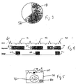
- an EKG data signal 60 is correlated with image data 62.
- a large spike R1 in EKG data signal 60 corresponds to an R-peak, which is an indication that systole is imminent.
- a segment 64 ending very close to an EKG R-peak is optimal for creating an image with a minimum of motion artifacts. Because a plurality of image slices are collected by CT imaging system 10 simultaneously, selecting a set of segmented image slices 64 taken at the optimum time produces the best images for scoring.
- At least one known multi-slice scanner 10 has hardware capable of rotating gantry 12 a full 360° in 0.8 seconds.
- a cine scan of two full rotations (1.6 seconds) at a first location of patient 46 suffices to provide four slices of the patient's heart because the time required for two full rotations is greater than a half rotation 58 plus a fan angle 56, plus one complete cardiac cycle.
- table 46 is stepped to another position by computer 36 and table motor controller 44 between cine scans of 1.6 second duration.
- Each step advances table 46 a distance four times the slice width, so that each cine scan (other than the first) provides image slices contiguous with, but not overlapping the image slices obtained during the cine scan immediately preceding the most recent table 46 step.
- the stepping distance is adjusted in accordance with the number of slices recorded simultaneously during a cine scan.
- Each table step can be accomplished in 1.5 seconds with known motorized tables 46 and controllers 44.
- Most patients 22 to be screened for calcification would be able to hold their breath this length of time without much difficulty.
- 3.75 mm segmented image slices a total of 120 mm of coverage is obtained in this amount of time, a coverage sufficient for calcification scoring. If patient 22 can hold his or her breath for a longer period, more slices can be obtained from a four-slice system 10. For example, 30 seconds is sufficient to obtain 40 segmented images. It will be understood that the total amount of coverage obtainable is dependent upon the number of slices available per cine scan, the width of the slices, and the amount of time patient 22 can hold his or her breath.
- Each cine scan in the scan-step-scan-step procedure produces a set of image data.
- three such sets of image data 62, 66, and 70 are indicated in Figure 4.
- Sets of segmented image slices 64, 68, and 72 are selected for the still images of the heart at portions of each set of image data 62, 66, and 70.
- Segmented image slices 64, 68, and 72 each immediately precede R-peaks R1, R2, and R3 in Figure 4.
- exposures 62, 66, and 70 are shown as being discontinuous in Figure 4, it is not necessary to turn off the x-ray beam or stop collecting image data during steps of table 46. However, if continuous image data is obtained from scanner 10, it is useful for the recorded image data to include indications distinguishing data taken during cine scans and data taken while table 46 is stepping.
- FIG. 5 is a drawing of an embodiment of a workstation display 74 that facilitates manual selection of image data segments 64, 68, and 72 from exposures 62, 66, and 70.
- a portion of EKG data signal 60 is shown above a reconstructed, segmented image 76.
- Operator controls such as a mouse or keyboard (neither of which are shown), are operated to step segmented image 76 in time through the recorded scanned image data.
- EKG data signal 60 is scrolled, and a portion 78 of EKG data signal 60 that corresponds to a presently displayed segmented image 76 appears highlighted on display 74.
- segmented images 76 correlated with a selected portion R of a cardiac cycle are located, and corresponding images from each slice taken by multi-slice scanner 10 are selected at the same time.
- a set of segmented still images are selected for scoring.
- a table z -location 80 or another suitable indication of scanning plane location is recorded along with the image data and displayed on workstation screen 74 with each segmented image 76.
- data from EKG recorder 52 need not be recorded on a floppy disk.
- data is transferred from EKG recorder 52 to a workstation either via a serial port or via a network connection.
- computer 36 is programmed to provide the workstation functions for selecting segmented image data 64, 68, and 72, using operator console 40 and display screen 42.
- CT system described herein is a "third generation" system in which both the x-ray source and detector rotate with the gantry.
- CT systems including "fourth generation" systems wherein the detector is a full-ring stationary detector and only the x-ray source rotates with the gantry, may be used if individual detector elements are corrected to provide substantially uniform responses to a given x-ray beam.
Landscapes
- Health & Medical Sciences (AREA)
- Life Sciences & Earth Sciences (AREA)
- Engineering & Computer Science (AREA)
- Medical Informatics (AREA)
- Heart & Thoracic Surgery (AREA)
- Molecular Biology (AREA)
- Biophysics (AREA)
- Nuclear Medicine, Radiotherapy & Molecular Imaging (AREA)
- Optics & Photonics (AREA)
- Pathology (AREA)
- Radiology & Medical Imaging (AREA)
- Biomedical Technology (AREA)
- Physics & Mathematics (AREA)
- High Energy & Nuclear Physics (AREA)
- Surgery (AREA)
- Animal Behavior & Ethology (AREA)
- General Health & Medical Sciences (AREA)
- Public Health (AREA)
- Veterinary Medicine (AREA)
- Physiology (AREA)
- Pulmonology (AREA)
- Theoretical Computer Science (AREA)
- Apparatus For Radiation Diagnosis (AREA)
- Magnetic Resonance Imaging Apparatus (AREA)
Applications Claiming Priority (2)
| Application Number | Priority Date | Filing Date | Title |
|---|---|---|---|
| US36251199A | 1999-07-28 | 1999-07-28 | |
| US362511 | 1999-07-28 |
Publications (2)
| Publication Number | Publication Date |
|---|---|
| EP1072224A2 true EP1072224A2 (fr) | 2001-01-31 |
| EP1072224A3 EP1072224A3 (fr) | 2001-06-27 |
Family
ID=23426398
Family Applications (1)
| Application Number | Title | Priority Date | Filing Date |
|---|---|---|---|
| EP00306245A Withdrawn EP1072224A3 (fr) | 1999-07-28 | 2000-07-21 | Synchronisation cardiaque rétrospective avec balayages cinématrographiques pour un balayeur a multicouche |
Country Status (3)
| Country | Link |
|---|---|
| EP (1) | EP1072224A3 (fr) |
| JP (1) | JP2001061835A (fr) |
| IL (1) | IL137251A0 (fr) |
Cited By (10)
| Publication number | Priority date | Publication date | Assignee | Title |
|---|---|---|---|---|
| US6370217B1 (en) | 1999-05-07 | 2002-04-09 | General Electric Company | Volumetric computed tomography system for cardiac imaging |
| US6393091B1 (en) | 1999-12-13 | 2002-05-21 | General Electric Company | Methods and apparatus for non-uniform temporal cardiac imaging |
| US6421552B1 (en) | 1999-12-27 | 2002-07-16 | Ge Medical Systems Global Technology Company, Llc | Methods and apparatus for estimating cardiac motion using projection data |
| EP1114616A3 (fr) * | 2000-01-07 | 2003-03-12 | GE Medical Systems Global Technology Company LLC | Dispositif d'imagerie par tomographie à rayonnement réduit |
| EP1340460A1 (fr) * | 2002-02-28 | 2003-09-03 | GE Medical Systems Global Technology Company LLC | Système et procédé d'imagerie avec une table à vitesse variable pour l'imagerie thoracique |
| US6639965B1 (en) | 1999-09-30 | 2003-10-28 | General Electric Company | Methods and apparatus for cardiac imaging with conventional computed tomography |
| CN100425202C (zh) * | 2002-01-10 | 2008-10-15 | 株式会社日立医药 | X射线ct成像方法和x射线ct设备 |
| US7477928B2 (en) | 2002-05-17 | 2009-01-13 | Ge Medical Systems Global Technology Company, Llc | Method and system for associating an EKG waveform with a CT image |
| US7545903B2 (en) | 2003-07-16 | 2009-06-09 | Koninklijke Philips Electronics N.V. | Reconstruction of an image of a moving object from volumetric data |
| CN106163405A (zh) * | 2014-02-12 | 2016-11-23 | 三星电子株式会社 | 断层扫描设备及由断层扫描设备显示断层扫描图像的方法 |
Families Citing this family (3)
| Publication number | Priority date | Publication date | Assignee | Title |
|---|---|---|---|---|
| US6728566B1 (en) * | 2001-11-21 | 2004-04-27 | Koninklijke Philips Electronics, N.V. | Vessel tracking and tree extraction method and apparatus |
| US7894570B2 (en) * | 2003-11-25 | 2011-02-22 | Koninklijke Philips Electronics N.V. | Automatic dose adaptation as a function of patient body mass index in CT calcium scoring |
| JP5022612B2 (ja) * | 2006-03-07 | 2012-09-12 | ジーイー・メディカル・システムズ・グローバル・テクノロジー・カンパニー・エルエルシー | X線ct装置 |
Family Cites Families (5)
| Publication number | Priority date | Publication date | Assignee | Title |
|---|---|---|---|---|
| US4182311A (en) * | 1977-04-22 | 1980-01-08 | Varian Associates, Inc. | Method and system for cardiac computed tomography |
| JPS5917334A (ja) * | 1982-07-21 | 1984-01-28 | 株式会社東芝 | 心拍連動画像診断装置 |
| JP2529949B2 (ja) * | 1986-08-12 | 1996-09-04 | 株式会社東芝 | 同期画像再構成装置 |
| JPH053867A (ja) * | 1991-06-28 | 1993-01-14 | Toshiba Corp | 三次元画像診断装置 |
| DE19622075C2 (de) * | 1996-05-31 | 1999-10-14 | Siemens Ag | Verfahren und Gerät zur radiologischen Untersuchung von Herzphasen eines Patienten |
-
2000
- 2000-07-11 IL IL13725100A patent/IL137251A0/xx unknown
- 2000-07-21 EP EP00306245A patent/EP1072224A3/fr not_active Withdrawn
- 2000-07-26 JP JP2000224690A patent/JP2001061835A/ja not_active Withdrawn
Cited By (13)
| Publication number | Priority date | Publication date | Assignee | Title |
|---|---|---|---|---|
| US6370217B1 (en) | 1999-05-07 | 2002-04-09 | General Electric Company | Volumetric computed tomography system for cardiac imaging |
| US6639965B1 (en) | 1999-09-30 | 2003-10-28 | General Electric Company | Methods and apparatus for cardiac imaging with conventional computed tomography |
| US6393091B1 (en) | 1999-12-13 | 2002-05-21 | General Electric Company | Methods and apparatus for non-uniform temporal cardiac imaging |
| US6421552B1 (en) | 1999-12-27 | 2002-07-16 | Ge Medical Systems Global Technology Company, Llc | Methods and apparatus for estimating cardiac motion using projection data |
| EP1114616A3 (fr) * | 2000-01-07 | 2003-03-12 | GE Medical Systems Global Technology Company LLC | Dispositif d'imagerie par tomographie à rayonnement réduit |
| CN100425202C (zh) * | 2002-01-10 | 2008-10-15 | 株式会社日立医药 | X射线ct成像方法和x射线ct设备 |
| EP1340460A1 (fr) * | 2002-02-28 | 2003-09-03 | GE Medical Systems Global Technology Company LLC | Système et procédé d'imagerie avec une table à vitesse variable pour l'imagerie thoracique |
| US7848790B2 (en) | 2002-02-28 | 2010-12-07 | General Electric Company | System and method of imaging using a variable speed for thorax imaging |
| US7477928B2 (en) | 2002-05-17 | 2009-01-13 | Ge Medical Systems Global Technology Company, Llc | Method and system for associating an EKG waveform with a CT image |
| US7545903B2 (en) | 2003-07-16 | 2009-06-09 | Koninklijke Philips Electronics N.V. | Reconstruction of an image of a moving object from volumetric data |
| US7924972B2 (en) | 2003-07-16 | 2011-04-12 | Koninklijke Philips Electronics N.V. | Reconstruction of an image of a moving object from volumetric data |
| CN106163405A (zh) * | 2014-02-12 | 2016-11-23 | 三星电子株式会社 | 断层扫描设备及由断层扫描设备显示断层扫描图像的方法 |
| EP3104782A4 (fr) * | 2014-02-12 | 2017-12-27 | Samsung Electronics Co., Ltd. | Appareil de tomographie et méthode d'affichage d'une image tomographique par l'appareil de tomographie |
Also Published As
| Publication number | Publication date |
|---|---|
| JP2001061835A (ja) | 2001-03-13 |
| EP1072224A3 (fr) | 2001-06-27 |
| IL137251A0 (en) | 2001-07-24 |
Similar Documents
| Publication | Publication Date | Title |
|---|---|---|
| EP1088517B1 (fr) | Procédé et dispositif pour l'imagerie cardiaque sans mouvement par tomodensitomètrie | |
| US6370217B1 (en) | Volumetric computed tomography system for cardiac imaging | |
| JP4630440B2 (ja) | スカウト画像をベースとした心臓石灰化計数のための方法及び装置 | |
| US5459769A (en) | Procedure for monitoring contrast agent application in a CT imaging system | |
| JP4578675B2 (ja) | 投影データを用いて心運動を評価する方法及び装置 | |
| JP4567130B2 (ja) | Ct像発生方法 | |
| US6639965B1 (en) | Methods and apparatus for cardiac imaging with conventional computed tomography | |
| US6480560B2 (en) | Methods and apparatus for motion gating using CT projection data | |
| US6307910B1 (en) | Methods and apparatus for reduced radiation coronary computed tomography imaging | |
| JP4429677B2 (ja) | 周期的に運動する器官のct画像形成方法 | |
| US6434215B1 (en) | EKG-less cardiac image reconstruction | |
| US20040042581A1 (en) | R-peak detection corrections for improved cardiac gated reconstruction | |
| EP1108392B1 (fr) | Procédés et appareils pour imagerie cardiaque temporaire non uniforme | |
| EP1095619B1 (fr) | Reconstruction hybride pour imagerie cardiaque à pas élevé, multicouche, hélicoidale | |
| JP2001061835A (ja) | マルチスライス型スキャナのシネ・スキャンによる遡及的心同期法 | |
| US6137858A (en) | Radiation tomography method and apparatus | |
| US6470208B1 (en) | Method and apparatus for controlling x-ray exposure during gated cardiac scanning | |
| JP2005125084A (ja) | 放射線撮像装置及び撮像方法 |
Legal Events
| Date | Code | Title | Description |
|---|---|---|---|
| PUAI | Public reference made under article 153(3) epc to a published international application that has entered the european phase |
Free format text: ORIGINAL CODE: 0009012 |
|
| AK | Designated contracting states |
Kind code of ref document: A2 Designated state(s): AT BE CH CY DE DK ES FI FR GB GR IE IT LI LU MC NL PT SE |
|
| AX | Request for extension of the european patent |
Free format text: AL;LT;LV;MK;RO;SI |
|
| PUAL | Search report despatched |
Free format text: ORIGINAL CODE: 0009013 |
|
| AK | Designated contracting states |
Kind code of ref document: A3 Designated state(s): AT BE CH CY DE DK ES FI FR GB GR IE IT LI LU MC NL PT SE |
|
| AX | Request for extension of the european patent |
Free format text: AL;LT;LV;MK;RO;SI |
|
| RIC1 | Information provided on ipc code assigned before grant |
Free format text: 7A 61B 6/03 A, 7A 61B 6/00 B |
|
| 17P | Request for examination filed |
Effective date: 20011227 |
|
| AKX | Designation fees paid |
Free format text: DE NL |
|
| STAA | Information on the status of an ep patent application or granted ep patent |
Free format text: STATUS: THE APPLICATION IS DEEMED TO BE WITHDRAWN |
|
| 18D | Application deemed to be withdrawn |
Effective date: 20040203 |