EP1106145A2 - Procédé et appareil pour augmenter la masse osseuse de la crête de la mâchoire - Google Patents
Procédé et appareil pour augmenter la masse osseuse de la crête de la mâchoire Download PDFInfo
- Publication number
- EP1106145A2 EP1106145A2 EP00310568A EP00310568A EP1106145A2 EP 1106145 A2 EP1106145 A2 EP 1106145A2 EP 00310568 A EP00310568 A EP 00310568A EP 00310568 A EP00310568 A EP 00310568A EP 1106145 A2 EP1106145 A2 EP 1106145A2
- Authority
- EP
- European Patent Office
- Prior art keywords
- ridge
- stent
- bone
- patient
- area
- Prior art date
- Legal status (The legal status is an assumption and is not a legal conclusion. Google has not performed a legal analysis and makes no representation as to the accuracy of the status listed.)
- Withdrawn
Links
- 238000000034 method Methods 0.000 title claims abstract description 45
- 230000003416 augmentation Effects 0.000 title claims abstract description 29
- 210000001909 alveolar process Anatomy 0.000 claims abstract description 65
- 210000000988 bone and bone Anatomy 0.000 claims abstract description 65
- 239000000463 material Substances 0.000 claims abstract description 38
- 230000001054 cortical effect Effects 0.000 claims abstract description 17
- 239000008280 blood Substances 0.000 claims description 16
- 210000004369 blood Anatomy 0.000 claims description 16
- 210000001847 jaw Anatomy 0.000 claims description 15
- 210000001519 tissue Anatomy 0.000 claims description 9
- 239000011505 plaster Substances 0.000 claims description 8
- 239000000203 mixture Substances 0.000 claims description 7
- 210000004373 mandible Anatomy 0.000 claims description 6
- NIXOWILDQLNWCW-UHFFFAOYSA-N acrylic acid group Chemical group C(C=C)(=O)O NIXOWILDQLNWCW-UHFFFAOYSA-N 0.000 claims description 4
- 210000002050 maxilla Anatomy 0.000 claims description 3
- 239000004575 stone Substances 0.000 claims description 3
- 238000003780 insertion Methods 0.000 claims description 2
- 230000037431 insertion Effects 0.000 claims description 2
- 238000009736 wetting Methods 0.000 claims 2
- 238000004080 punching Methods 0.000 claims 1
- 230000003190 augmentative effect Effects 0.000 abstract description 21
- 210000004195 gingiva Anatomy 0.000 abstract description 13
- 239000011796 hollow space material Substances 0.000 abstract description 3
- 238000001356 surgical procedure Methods 0.000 description 13
- 238000000605 extraction Methods 0.000 description 12
- 230000000740 bleeding effect Effects 0.000 description 11
- 239000007943 implant Substances 0.000 description 5
- 206010003694 Atrophy Diseases 0.000 description 3
- 230000037444 atrophy Effects 0.000 description 3
- 230000008468 bone growth Effects 0.000 description 3
- 239000000316 bone substitute Substances 0.000 description 2
- 238000010276 construction Methods 0.000 description 2
- 238000002513 implantation Methods 0.000 description 2
- 239000007788 liquid Substances 0.000 description 2
- 239000012528 membrane Substances 0.000 description 2
- 230000003239 periodontal effect Effects 0.000 description 2
- 206010065687 Bone loss Diseases 0.000 description 1
- 206010033372 Pain and discomfort Diseases 0.000 description 1
- FAPWRFPIFSIZLT-UHFFFAOYSA-M Sodium chloride Chemical compound [Na+].[Cl-] FAPWRFPIFSIZLT-UHFFFAOYSA-M 0.000 description 1
- 229940035676 analgesics Drugs 0.000 description 1
- 239000000730 antalgic agent Substances 0.000 description 1
- 239000003242 anti bacterial agent Substances 0.000 description 1
- 229940088710 antibiotic agent Drugs 0.000 description 1
- 230000003115 biocidal effect Effects 0.000 description 1
- 239000005312 bioglass Substances 0.000 description 1
- 210000001185 bone marrow Anatomy 0.000 description 1
- 230000008859 change Effects 0.000 description 1
- YZIYKJHYYHPJIB-UUPCJSQJSA-N chlorhexidine gluconate Chemical compound OC[C@@H](O)[C@@H](O)[C@H](O)[C@@H](O)C(O)=O.OC[C@@H](O)[C@@H](O)[C@H](O)[C@@H](O)C(O)=O.C1=CC(Cl)=CC=C1NC(=N)NC(=N)NCCCCCCNC(=N)NC(=N)NC1=CC=C(Cl)C=C1 YZIYKJHYYHPJIB-UUPCJSQJSA-N 0.000 description 1
- 230000001010 compromised effect Effects 0.000 description 1
- 239000004053 dental implant Substances 0.000 description 1
- 230000002542 deteriorative effect Effects 0.000 description 1
- 239000008187 granular material Substances 0.000 description 1
- 230000012010 growth Effects 0.000 description 1
- 230000035876 healing Effects 0.000 description 1
- 230000036541 health Effects 0.000 description 1
- 210000001981 hip bone Anatomy 0.000 description 1
- 239000000411 inducer Substances 0.000 description 1
- 208000014674 injury Diseases 0.000 description 1
- 230000002452 interceptive effect Effects 0.000 description 1
- 230000005012 migration Effects 0.000 description 1
- 238000013508 migration Methods 0.000 description 1
- 230000025712 muscle attachment Effects 0.000 description 1
- 239000002245 particle Substances 0.000 description 1
- 229940098804 peridex Drugs 0.000 description 1
- 230000002980 postoperative effect Effects 0.000 description 1
- 230000008569 process Effects 0.000 description 1
- 230000001737 promoting effect Effects 0.000 description 1
- 108090000623 proteins and genes Proteins 0.000 description 1
- 102000004169 proteins and genes Human genes 0.000 description 1
- 238000007493 shaping process Methods 0.000 description 1
- 230000035939 shock Effects 0.000 description 1
- 235000021055 solid food Nutrition 0.000 description 1
- 239000000126 substance Substances 0.000 description 1
- 230000009885 systemic effect Effects 0.000 description 1
- 238000002560 therapeutic procedure Methods 0.000 description 1
- 210000000332 tooth crown Anatomy 0.000 description 1
- 230000008733 trauma Effects 0.000 description 1
- 230000001720 vestibular Effects 0.000 description 1
Images


Classifications
-
- A—HUMAN NECESSITIES
- A61—MEDICAL OR VETERINARY SCIENCE; HYGIENE
- A61C—DENTISTRY; APPARATUS OR METHODS FOR ORAL OR DENTAL HYGIENE
- A61C8/00—Means to be fixed to the jaw-bone for consolidating natural teeth or for fixing dental prostheses thereon; Dental implants; Implanting tools
- A61C8/0003—Not used, see subgroups
- A61C8/0004—Consolidating natural teeth
- A61C8/0006—Periodontal tissue or bone regeneration
-
- Y—GENERAL TAGGING OF NEW TECHNOLOGICAL DEVELOPMENTS; GENERAL TAGGING OF CROSS-SECTIONAL TECHNOLOGIES SPANNING OVER SEVERAL SECTIONS OF THE IPC; TECHNICAL SUBJECTS COVERED BY FORMER USPC CROSS-REFERENCE ART COLLECTIONS [XRACs] AND DIGESTS
- Y10—TECHNICAL SUBJECTS COVERED BY FORMER USPC
- Y10S—TECHNICAL SUBJECTS COVERED BY FORMER USPC CROSS-REFERENCE ART COLLECTIONS [XRACs] AND DIGESTS
- Y10S623/00—Prosthesis, i.e. artificial body members, parts thereof, or aids and accessories therefor
- Y10S623/902—Method of implanting
- Y10S623/908—Bone
Definitions
- the present invention relates to dental surgical procedures in general and, more particularly, to a method and apparatus for augmenting a patient's alveolar ridge or jaw bone using bone graft material.
- Figures 1 through 3 illustrate the deteriorating effect of tooth extraction on the alveolar ridge.
- Figure 1 illustrates a tooth of a patient, comprised of a crown 10 and root 20 seated in the alveolar bone or jaw bone 30.
- the buccal and lingual portions of the alveolar bone are surrounded by a layer of tissue known as the gingiva or gum 40.
- the crown 10 and root 20 are supported by the elevated portion of the alveolar ridge or jaw bone 30 and gingiva 40 which, in the ideal case, hold all of the teeth in place such that a level gum line 50 and crown line 60 are maintained.
- the root is removed from the alveolar bone 30 by separating the surface of the root 20 from the periodontal membrane 70.
- Figure 2 represents the portion of the jaw bone shortly after extraction of the crown 10 and root 20. As is shown, the periodontal membrane clots such that bleeding ceases and a socket 90 remains in the alveolar bone 40 in the shape of the extracted root 20.
- the buccal and lingual portions of the alveolar bone 30 are composed of soft trabecular bone which has the unique characteristic of being capable of absorbing the shocks caused by the movement of teeth during speech, eating, etc.
- the removal of a tooth and the resulting absence of frequent bone pressure stimuli in the area causes the alveolar bone 30 to shrink in that area, with the subsequent loss of 40 to 60 percent of the alveolar ridge's former height measured at the gum line 50.
- Fig. 3 shows an endulous extraction site with loss of buccal and lingual portions of the alveolar bone 30, two years after the extraction of the tooth represented in Fig. 1. After initial 40-60% loss, the alveolar bone 30 continues to atrophy at a bone loss rate of one-half to one percent per year.
- Bone replacement graft material has been used to immediately fill a socket 90 at an extraction site after a root 20 extraction, in order to promote bone growth and therefore avoid this atrophy. Bone growth is promoted via the bone graft material's intermixing with the patient's own marrow blood at the extraction site 90. While methods of applying bone graft materials to a newly extracted root site are known, a method for applying bone graft materials to an area of jaw bone which has already atrophied is not known and would have obvious benefits.
- a method for performing ridge augmentation on an atrophied alveolar ridge of the jaw bone of a patient utilizing synthetic bone alloplast comprises the steps of (1) constructing a denture-stent with a fitted hollow space on the underside of the stent conforming to the desired height, width and extent of ridge augmentation, (2) reflecting gingiva tissue covering the alveolar ridge at the site to be augmented, (3) inserting synthetic bone or other graft material into reflected gingiva tissue of the alveolar ridge and onto the bleeding cortical plate of the jaw bone in the area where ridge augmentation is desired, (4) suturing the reflected gingiva tissue, (5) placing the preconstructed stent over the area of the alveolar ridge containing the synthetic bone graft material immediately after the area has been sutured and (6) fixing the stent denture to the jaw bone when necessary.
- Phase I comprises the construction of a provisional denture-stent.
- a dental surgeon constructs a provisional denture-stent with a "hollow" space on the underside of the stent to accommodate what will become the newly augmented ridge area.
- the patient's existing denture is never to be used as the Phase I denture-stent.
- the dental surgeon constructs either a partial denture-stent or a full denture-stent depending on the extent of the augmentation. When only a part of the alveolar ridge is to be augmented, the dental surgeon makes a partial denture-stent.
- the dental surgeon constructs a full denture-stent.
- denture-stent includes both full and partial denture-stents.
- a method for constructing a denture-stent for use in the present invention is illustrated.
- a portion 28 of the patient's alveolar ridge of the lower jaw bone 30 is shown facing outward from the lingual portion of the jaw bone.
- the alveolar ridge 30 is atrophied such that augmentation is desired in an area (represented by 100) between the alveolar ridge and the desired gum line 50.
- the dental surgeon commences the construction of the denture-stent in the manner known in the art.
- Fig. 4b the dental surgeon constructs a stone or plaster model 120 of the atrophic alveolar ridge shown in Fig. 4a.
- Model 120 is constructed using methods known in the art. With continued reference to Fig. 4b, the dental surgeon places wax on model 120 in the area representing the portion of the alveolar ridge 30 to be augmented (100). The resulting shape of the underside of the wax represents a wax impression 130 of the area of the alveolar ridge 30 to be augmented.
- the dental surgeon uses the wax impression 130 formed in Fig. 4b to create a plaster impression 140 having the same shape as the wax impression 130 of Fig. 4b.
- the dental surgeon uses the plaster impression 140 to create a final master model 150 of the ridge to be augmented using methods known in the art.
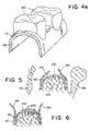
- the dental surgeon constructs the denture-stent 110 over the master model 150 of Fig. 4c. As is shown, the hollow underside 155 of the denture-stent conforms to the shape of the plaster impression 140 of Fig. 4c. Further, the dental surgeon adds prosthetic teeth 160 to the top of the denture-stent 110.
- the denture-stent 110 is constructed of a wax and acrylic mixture.
- Figure 4e represents the final denture-stent 110 prior to surgery and ridge augmentation and after final try-in of the denture-stent of Fig. 4d.
- the final denture-stent 110 is constructed of hard acrylic or plastic.
- the hollow underside 155 of the final denture-stent has a shape that conforms to the shape of the desired ridge augmentation.
- Phase II includes the surgical implantation of bone graft material into the patient's atrophic alveolar ridge 30 thereby filling the hollow-space 100 with a bone replacement graft.
- the denture-stent 110 is placed after the surgery is completed.
- Fig. 5 the first step of the Phase 11 surgical procedure, as performed on the mandible (lower jaw bone), is illustrated.
- the ridge of either the maxilla (upper jaw bone) or the mandible can be augmented using this procedure.
- the alveolar ridge of the mandible is shown positioned between the patient's tongue 170 and the patient's lip 180.
- the dental surgeon makes a crestal full-thickness incision 140 in the gingiva tissue 150 at the location along the alveolar ridge where augmentation is desired.
- the cortical plate 160 of the atrophied alveolar or trabecular bone 120 is exposed.
- a lingual split -thickness incision reflecting to a full-thickness incision at the crest of the ridge going in a buccal direction is made. Again, the cortical plate 160 will be exposed.
- the bone graft material is now added to the exposed alveolar ridge.
- any bone graft material such as autogenous bone or synthetics, alone or mixed with autogenous bone (e.g., Bioglass® or Bio Oss®)
- prepackaged synthetic bone HTR® manufactured by Bioplant®, Inc. is preferably used in Phase II of the ridge augmentation procedure.
- HTR® is a synthetic bone alloplast which promotes bone growth by acting as a scaffold which supports the creation of dense lamina bone.
- HTR®-24 which comprises granular particles of a larger size (750 microns in diameter), is preferably utilized.
- HTR-24® is prepackaged in Bioplant® 0.25 Gram straight syringes, Item #01-81002, which are available from the manufacturer.
- the granular form of HTR®-24 must be wetted (i.e., hydrated) in order to change the consistency of HTR® to a more useable and formable paste-like substance that will not migrate when placed on bleeding bone.
- the dental surgeon may additionally mix the graft material with the patient's own bone (e.g., from the hip bone) in order to promote faster and more effective growth of bone in the alveolar ridge.
- U.S. Patent Application Serial No. 08/831, 941 describing the syringe tip and a method for using the same is hereby incorporated in its entirety by reference.
- the HTR® can be wetted with liquid antibiotic, liquid bone-inducer protein (e.g., BMPs), or sterile saline solution
- blood from the patient's alveolar marrow is preferably used to wet the HTR®.
- the dental surgeon uses a round surgical bur to punch holes 200 into the exposed cortical plate 160 of the alveolar ridge 120.
- the holes 200 are spaced approximately 2-4 mm apart.
- the use of the bur to create holes promotes marrow bleeding 210 via the small bleeding points 200 of alveolar marrow which brings to the area the precurser ("Pluri potential") cells that will form new bone.
- the dental surgeon draws the bleeding marrow 210 into the syringe 260 containing granular HTR® through the syringe filter tip 280.
- the granular HTR® is, thereby, wetted by the blood to form a blood wetted mixture with HTR® 270.
- the filter 280 in the tip is sized to allow the necessary bone forming cells into the syringe 260 to mix with the graft material. Excess blood can be expelled from the syringe once a sufficient quantity of blood is mixed with the HTR®.
- the blood wetted HTR® 270 is permitted to congeal for three to four minutes, at the conclusion of which time the blood wetted HTR® mixture 270 will have a viscous, paste-like consistency which can more easily be formed on the alveolar ridge 120 and not migrate off the ridge 120.
- the dental surgeon thereafter removes the filter tip 280 of the syringe 260 and expels the wetted HTR® 270 onto the exposed and bleeding cortical plate 160.
- the dental surgeon expels an amount of wetted HTR® 270 sufficient to provide the required augmentation of the atrophied area of jaw bone. Because the HTR® granules are wetted, they will stay in place without migration and can be molded to generally conform with the desired width, height and shape of the alveolar ridge consistent with the gum line 50.
- the wetted HTR® 270 By placing the wetted HTR® 270 on that portion of the cortical plate 160 where the marrow holes 200 have been punched, the wetted HTR® 270 and the alveolar marrow 280 will more readily interact to form dense lamina bone.
- Figure 9 illustrates the suture and denture-stent placement operations.
- the dental surgeon In order for the newly augmented ridge to form properly, the dental surgeon immediately places the Phase I denture-stent 110 over the new ridge as shown in Fig. 9. If the newly augmented alveolar ridge does not sufficiently fill the hollowed space of the denture-stent, the dental surgeon relines the denture-stent 110 with a soft-line material. Alternately, if the newly augmented area of alveolar ridge impinges upon the hollow underside of the denture-stent, the dental surgeon relieves (removes) a portion of the denture-stent 110 adjacent to the hollow area using a large round bur on the under side soft line of the denture-stent 110.
- the denture-stent 110 will hold the newly augmented ridge in shape while the alveolar bone 120 fuses with the wetted HTR® 270 to form dense lamina bone.
- the dental surgeon checks the fit and bite of the denture-stent.
- a vestibular extension 305 may be needed at this point as well as a possible frenectomy to eliminate interfering muscle attachments.
- the dental surgeon is satisfied with the present fit of the enture-stent 110, he secures the denture-stent 110 to the patient's alveolar ridge preferably using a palatal screw 310.
- circumferential wiring can be used on the mandible.
- the screw 310 insures that the denture-stent remains in place, as it is important that the denture-stent not be removed for 7 to 10 days following the Phase II surgery in order to promote initial healing.
- the present method advantageously allows the patient to immediately function with the Phase I denture-stent.
- the edentulous individual who may have been unable to support a full or partial denture before the Phase II surgery, aesthetically benefits at once since the patient leaves the dental surgeon's office with a prosthesis and aesthetic tooth or teeth which are properly aligned with the gum line 50 and crown line 60. The patient never goes without teeth. Additionally, the pain and discomfort normally associated with dental surgery is minimized in that the stent acts to hold the HTR mixture in place and to prevent any remnant bleeding or trauma to the area which might occur following suturing without the denture-stent in place.
- Phase III of the ridge augmentation method comprises follow-up procedures.
- the dental surgeon instructs the patient to avoid eating solid foods for approximately 7 to 10 days.
- the dental surgeon also prescribes systemic antibiotics and analgesics for about 10 days.
- the dental surgeon sees the patient and inspects the patient's bite in order to be sure that the dental stent is properly mounted on the patient's alveolar ridge.
- the dental surgeon removes the denture-stent 110 and cleans the surgical area using, e.g., a Peridex rinse.
- the denture-stent 110 is then again placed onto the patient's alveolar ridge, now as a functioning full or partial denture.
- the dental surgeon sees the patient again and inspects the effected alveolar ridge area and denture-stent 110 fitting.
- the dental surgeon may redo the soft-line of the underside 155 of the denture-stent 110 if necessary to maintain the denture-stent's proper fit on the patient's alveolar ridge.
- the dental surgeon fits the patient for a new full or partial denture in the normal manner, i.e, the dental surgeon will make a normal denture without a hollow on the underside fitting the newly restored jaw bone.
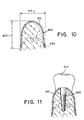
- Figure 10 illustrates the augmented alveolar ridge at about 18 months after the Phase II surgery.
- Both the height 400 and the width 410 of the alveolar ridge increased as a result of the dense lamina bone 420 which forms over the original bony ridge 430 during the 18 month interval.
- the patient may continue to function normally with a full or partial denture or, as shown in Fig. 11, the patient may be considered for implant therapy whereby an implant screw 500 may be inserted into the patients' newly augmented alveolar ridge in the normal manner to support a prosthetic crown 510.
Landscapes
- Health & Medical Sciences (AREA)
- Life Sciences & Earth Sciences (AREA)
- Engineering & Computer Science (AREA)
- Biomedical Technology (AREA)
- Developmental Biology & Embryology (AREA)
- Oral & Maxillofacial Surgery (AREA)
- Orthopedic Medicine & Surgery (AREA)
- Dentistry (AREA)
- Epidemiology (AREA)
- Animal Behavior & Ethology (AREA)
- General Health & Medical Sciences (AREA)
- Public Health (AREA)
- Veterinary Medicine (AREA)
- Dental Preparations (AREA)
- Prostheses (AREA)
Applications Claiming Priority (2)
| Application Number | Priority Date | Filing Date | Title |
|---|---|---|---|
| US09/449,879 US6402518B1 (en) | 1999-11-30 | 1999-11-30 | Method and apparatus for performing ridge augmentation |
| US449879 | 1999-11-30 |
Publications (2)
| Publication Number | Publication Date |
|---|---|
| EP1106145A2 true EP1106145A2 (fr) | 2001-06-13 |
| EP1106145A3 EP1106145A3 (fr) | 2003-01-08 |
Family
ID=23785844
Family Applications (1)
| Application Number | Title | Priority Date | Filing Date |
|---|---|---|---|
| EP00310568A Withdrawn EP1106145A3 (fr) | 1999-11-30 | 2000-11-29 | Procédé et appareil pour augmenter la masse osseuse de la crête de la mâchoire |
Country Status (2)
| Country | Link |
|---|---|
| US (2) | US6402518B1 (fr) |
| EP (1) | EP1106145A3 (fr) |
Cited By (2)
| Publication number | Priority date | Publication date | Assignee | Title |
|---|---|---|---|---|
| WO2003037209A1 (fr) * | 2001-10-30 | 2003-05-08 | Schulter Carl W | Forme biocompatible et son procede de fabrication |
| RU2485911C1 (ru) * | 2012-03-05 | 2013-06-27 | Алексей Викторович Васильев | Способ протезирования больных с выраженными врожденными аномалиями развития и приобретенными деформациями альвеолярных гребней костей верхней и нижней челюстей |
Families Citing this family (20)
| Publication number | Priority date | Publication date | Assignee | Title |
|---|---|---|---|---|
| US7771482B1 (en) | 2000-05-09 | 2010-08-10 | Ben-Zion Karmon | Method for tissue expansion and regeneration using bioresorbable inflatable devices |
| US8622739B2 (en) * | 2001-05-09 | 2014-01-07 | Ben-Zion Karmon | Method for enlarging a jaw bone using a hollow dental implant having a side perforation |
| SE522984C2 (sv) * | 2002-07-25 | 2004-03-23 | Nobel Biocare Ab | Anordning vid två eller flera implantat försedda med tillväxtstimulerande substans(-er) |
| CA2510288A1 (fr) * | 2004-09-02 | 2006-03-02 | Odontis Ltd. | Regeneration des os |
| US9259320B2 (en) | 2008-08-26 | 2016-02-16 | Andy Boiangiu | Apparatus and method for bone augmentation |
| US9861482B2 (en) | 2008-08-26 | 2018-01-09 | Andy Boiangiu | Dental bone implant and implant method |
| US7964208B2 (en) * | 2009-02-25 | 2011-06-21 | Warsaw Orthopedic, Inc. | System and methods of maintaining space for augmentation of the alveolar ridge |
| US20100291508A1 (en) * | 2009-05-13 | 2010-11-18 | Jensen Ole T | Biocompatible shell for bone treatment |
| US20110014587A1 (en) * | 2009-07-16 | 2011-01-20 | Warsaw Orthopedic, Inc. | System and methods of preserving an oral socket |
| US9539068B2 (en) * | 2009-07-24 | 2017-01-10 | Warsaw Orthopedic, Inc. | Implantable screw and system for socket preservation |
| US20110165536A1 (en) * | 2010-01-06 | 2011-07-07 | Rainbow Medical Ltd. | Alveolar ridge augmentation |
| US8574302B2 (en) | 2010-12-20 | 2013-11-05 | Warsaw Orthopedic, Inc. | Bone augmentation device |
| US8470046B2 (en) | 2011-04-25 | 2013-06-25 | Warsaw Orthopedic, Inc. | Bone augmentation device and method |
| US9539069B2 (en) | 2012-04-26 | 2017-01-10 | Zimmer Dental, Inc. | Dental implant wedges |
| US9554877B2 (en) | 2012-07-31 | 2017-01-31 | Zimmer, Inc. | Dental regenerative device made of porous metal |
| WO2017010811A1 (fr) * | 2015-07-13 | 2017-01-19 | 한양대학교 산학협력단 | Tissu osseux alvéolaire personnalisé et procédé de production dudit tissu |
| IL243401A (en) | 2015-12-29 | 2017-12-31 | Zion Karmon Ben | Instruments and methods for lifting Schneider membrane |
| IL248472A0 (en) | 2016-10-13 | 2017-01-31 | Zion Karmon Ben | Devices for tissue growth |
| WO2019213354A1 (fr) | 2018-05-03 | 2019-11-07 | The United States Of America As Represented By The Secretary Of The Navy | Matrice d'augmentation de crête dentaire avec système de guidage de foret chirurgical d'implant dentaire intégré |
| CN112716643A (zh) * | 2020-12-28 | 2021-04-30 | 浙江大学 | 一种区域功能特异性临床牙周缺损修复模块 |
Family Cites Families (18)
| Publication number | Priority date | Publication date | Assignee | Title |
|---|---|---|---|---|
| US4251215A (en) * | 1979-09-10 | 1981-02-17 | Gulf South Research Institute | Phosphonitrilic fluoroelastomer lined denture |
| US4820306A (en) * | 1981-06-22 | 1989-04-11 | Sterling Drug Inc. | Method for augmentation of the alveolar ridge |
| US4787906A (en) * | 1987-03-02 | 1988-11-29 | Haris Andras G | Controlled tissue growth and graft containment |
| US5073114A (en) * | 1988-02-23 | 1991-12-17 | Detsch Steven G | Bone growing method and composition |
| US5700479A (en) * | 1988-12-23 | 1997-12-23 | Guidor Ab | Surgical element and method for selective tissue regeneration |
| JP2905592B2 (ja) * | 1989-04-05 | 1999-06-14 | ダブリュ.エル.ゴア アンド アソシエイツ,インコーポレイティド | 歯周囲の病気および骨の欠陥を処置する方法および物品 |
| US5433607A (en) * | 1991-07-15 | 1995-07-18 | Institut Straumann Ag | Implant for attaching a substitute tooth or the like to a jaw |
| US5397235A (en) | 1993-07-02 | 1995-03-14 | Dental Marketing Specialists, Inc. | Method for installation of dental implant |
| US5372503A (en) | 1993-04-27 | 1994-12-13 | Dental Marketing Specialists, Inc. | Method for installation of a dental implant |
| FR2695551B1 (fr) * | 1992-09-15 | 1994-10-21 | Antoine Robert | Dispositif d'expansion pour reconstruction orale et procédé d'installation du dispositif d'expansion sous la gencive d'un patient dans le but d'effectuer la reconstruction orale dans des cas de perte osseuse. |
| US6019764A (en) * | 1993-08-02 | 2000-02-01 | Bartee; Barry K. | Method of treating alveolar bone defects |
| DE69426530D1 (de) * | 1993-10-04 | 2001-02-08 | Maurizio Tonetti | Pharmazeutische zusammensetzung zur förderung der gewebeheilung und -regenerierung und darreichungssatz |
| FR2713090A1 (fr) * | 1993-12-03 | 1995-06-09 | Scortecci Gerard | Dispositif utilisé pour la régénération tissulaire guidée. |
| DE4414675C1 (de) * | 1994-04-27 | 1995-09-28 | Kirsch Axel | Abdeckeinrichtung für Knochendefektstellen und Verfahren zu deren Herstellung |
| US6071284A (en) * | 1995-10-30 | 2000-06-06 | Biomedical Enterprises, Inc. | Materials collection system and uses thereof |
| US5839899A (en) * | 1996-03-01 | 1998-11-24 | Robinson; Dane Q. | Method and apparatus for growing jaw bone utilizing a guided-tissue regeneration plate support and fixation system |
| DE19741273C1 (de) * | 1997-09-19 | 1999-03-11 | Johann Flaum | Hilfsmittel zur Anregung des Zahnwachstums und zur Kieferregenerierung |
| US6030218A (en) * | 1999-04-12 | 2000-02-29 | Robinson; Dane Q. | Osseo-integrated sub-periosteal implant |
-
1999
- 1999-11-30 US US09/449,879 patent/US6402518B1/en not_active Expired - Lifetime
-
2000
- 2000-11-29 EP EP00310568A patent/EP1106145A3/fr not_active Withdrawn
-
2002
- 2002-04-18 US US10/126,203 patent/US20020110785A1/en not_active Abandoned
Non-Patent Citations (1)
| Title |
|---|
| None |
Cited By (4)
| Publication number | Priority date | Publication date | Assignee | Title |
|---|---|---|---|---|
| WO2003037209A1 (fr) * | 2001-10-30 | 2003-05-08 | Schulter Carl W | Forme biocompatible et son procede de fabrication |
| US6645250B2 (en) | 2001-10-30 | 2003-11-11 | Carl W. Schulter | Biocompatible form and method of fabrication |
| US6911046B2 (en) | 2001-10-30 | 2005-06-28 | Cagenix, Inc. | Biocompatible form and method of fabrication |
| RU2485911C1 (ru) * | 2012-03-05 | 2013-06-27 | Алексей Викторович Васильев | Способ протезирования больных с выраженными врожденными аномалиями развития и приобретенными деформациями альвеолярных гребней костей верхней и нижней челюстей |
Also Published As
| Publication number | Publication date |
|---|---|
| US6402518B1 (en) | 2002-06-11 |
| EP1106145A3 (fr) | 2003-01-08 |
| US20020110785A1 (en) | 2002-08-15 |
Similar Documents
| Publication | Publication Date | Title |
|---|---|---|
| US6402518B1 (en) | Method and apparatus for performing ridge augmentation | |
| Yilmaz et al. | Alveolar ridge reconstruction and/or preservation using root form bioglass cones | |
| Solar | Preserving alveolar ridge anatomy following tooth removal in conjunction with immediate implant placement: the Bio-Col technique | |
| Fugazzotto et al. | Long-term success of sinus augmentation using various surgical approaches and grafting materials. | |
| US6244868B1 (en) | Integrated guided-tissue-regeneration barrier for root-form dental implants | |
| Raghoebar et al. | Bone grafting of the floor of the maxillary sinus for the placement of endosseous implants | |
| EP1223888B1 (fr) | Procede pour fournir un dispositif de protection de la crete gingivale et d'insertion d'implant | |
| EP3421006B1 (fr) | Procédé de fabrication d'une prothèse dentaire | |
| Novaes Jr et al. | Soft tissue management for primary closure in guided bone regeneration: surgical technique and case report. | |
| Cordaro et al. | Ridge augmentation procedures in implant patients: a staged approach | |
| Novaes Jr et al. | Bone formation over a TiAl6V4 (IMZ) implant placed into an extraction socket in association with membrane therapy (Gengiflex) | |
| Levy et al. | Initial healing in the dog of submerged versus non‐submerged porous‐coated endosseous dental implants | |
| US7314375B2 (en) | Provisional dental implant for preparing an alveolus | |
| RU2098040C1 (ru) | Литая культевая штифтовая вкладка | |
| Petrungaro | Immediate restoration of multiple tooth implants for aesthetic implant restorations | |
| Sullivan | Implant placement in the aesthetic zone following an autogenous bone graft from an intraoral site: a case study | |
| CN216221761U (zh) | 一种具有舌侧牙龈仿生结构的假牙模型组合 | |
| Pereira Nunes et al. | Maxillary Sinus Elevation Using the Bone Ring Technique with Immediate Implantation: A Case Report. | |
| WO2009118725A1 (fr) | Recouvrement d'une alvéole dentaire | |
| CA2313540A1 (fr) | Barriere integree de guidage de regeneration tissulaire pour implants dentaires en forme de racine | |
| Shen | The use of different implant modalities in the atrophied ridge | |
| Assadawy | Immediate Dental Implant Practical Steps at Your Fingertips | |
| Roberts | Placement of plate-form implants using osteotomes | |
| Yang et al. | Reconstruction of Hard and Soft Tissue in the Edentulous Area Using Implants with Submerged Healing Abutments Combined Guided Bone Regeneration Procedure: Two Cases Report | |
| Carlos et al. | Immediate Implant Placement in Compromised Maxillary Anterior and Bicuspid |
Legal Events
| Date | Code | Title | Description |
|---|---|---|---|
| PUAI | Public reference made under article 153(3) epc to a published international application that has entered the european phase |
Free format text: ORIGINAL CODE: 0009012 |
|
| AK | Designated contracting states |
Kind code of ref document: A2 Designated state(s): AT BE CH CY DE DK ES FI FR GB GR IE IT LI LU MC NL PT SE TR |
|
| AX | Request for extension of the european patent |
Free format text: AL;LT;LV;MK;RO;SI |
|
| PUAL | Search report despatched |
Free format text: ORIGINAL CODE: 0009013 |
|
| AK | Designated contracting states |
Kind code of ref document: A3 Designated state(s): AT BE CH CY DE DK ES FI FR GB GR IE IT LI LU MC NL PT SE TR |
|
| AX | Request for extension of the european patent |
Free format text: AL;LT;LV;MK;RO;SI |
|
| AKX | Designation fees paid | ||
| REG | Reference to a national code |
Ref country code: DE Ref legal event code: 8566 |
|
| STAA | Information on the status of an ep patent application or granted ep patent |
Free format text: STATUS: THE APPLICATION IS DEEMED TO BE WITHDRAWN |
|
| 18D | Application deemed to be withdrawn |
Effective date: 20030709 |