BACKGROUND OF THE INVENTION
Field of the Invention
-
This invention relates to polypeptide ligands that bind to receptors implicated in
cellular growth. In particular, it relates to polypeptide ligands that bind to the p185HER2
receptor.
Description of Background and Related Art
-
Cellular protooncogenes encode proteins that are thought to regulate normal cellular
proliferation and differentiation. Alterations in their structure or amplification of their
expression lead to abnormal cellular growth and have been associated with carcinogenesis
(Bishop JM, Science 235:305-311 [1987]); (Rhims JS, Cancer Detection and Prevention 11:139-149
[1988]); (Nowell PC, Cancer Res. 46:2203-2207 [1886]); (Nicolson GL, Cancer Res.
47:1473-1487 [1987]). Protooncogenes were first identified by either of two approaches.
First, molecular characterization of the genomes of transforming retroviruses showed that
the genes responsible for the transforming ability of the virus in many cases were altered
versions of genes found in the genomes of normal cells. The normal version is the
protooncogene, which is altered by mutation to give rise to the oncogene. An example of such
a gene pair is represented by the EGF receptor and the v-erb-B gene product. The virally
encoded v-erb-B gene product has suffered truncation and other alterations that render it
constitutively active and endow it with the ability to induce cellular transformation (Yarden et
al., Ann. Rev. Biochem. 57:443-478, 1988).
-
The second method for detecting cellular transforming genes that behave in a
dominant fashion involves transfection of cellular DNA from tumor cells of various species
into nontransformed target cells of a heterologous species. Most often this was done by
transfection of human, avian, or rat DNAs into the murine NIH 3T3 cell line (Bishop JM,
Science 235:305-311 [1987]); (Rhims JS, Cancer Detection and Prevention 11:139-149 [1988]);
(Nowell PC, Cancer. Res. 46:2203-2207 [1986]); (Nicolson GL, Cancer. Res. 47:1473-1487
[1987]); (Yarden et al., Ann. Rev. Biochem. 57:443-478 [1988]). Following several cycles of
genomic DNA isolation and retransfection, the human or other species DNA was molecularly
cloned from the murine background and subsequently characterized. In some cases, the same
genes were isolated following transfection and cloning as those identified by the direct
characterization of transforming viruses. In other cases, novel oncogenes were identified. An
example of a novel oncogene identified by this transfection assay is the neu oncogene. It was
discovered by Weinberg and colleagues in a transfection experiment in which the initial DNA
was derived from a carcinogen-induced rat neuroblastoma (Padhy et al., Cell 28:865-871
[1982]); (Schechter et al., Nature 312:513-516 [1984]). Characterization of the rat neu
oncogene revealed that it had the structure of a growth factor receptor tyrosine kinase, had
homology to the EGF receptor, and differed from its normal counterpart, the neu
protooncogene, by an activating mutation in its transmembrane domain (Bargmann et al., Cell
45:649-657 [1986]). The human counterpart to neu is the HER2 protooncogene, also designated
c-erb-B2 (Coussens et al., Science 230:1137-1139 [1985]), W089/06692).
-
The association of the HER2 protooncogene with cancer was established by yet a
third approach, that is, its association with human breast cancer. The HER2 protooncogene
was first discovered in cDNA libraries by virtue of its homology with the EGF receptor, with
which it shares structural similarities throughout (Yarden et al., Ann. Rev. Biochem. 57:443-478
[1988]). When radioactive probes derived from the cDNA sequence encoding p185HER2
were used to screen DNA samples from breast cancer patients, amplification of the HER2
protooncogene was observed in about 30% of the patient samples (Slamon et al., Science
235:177-182 [1987]). Further studies have confirmed this original observation and extended it
to suggest an important correlation between HER2 protooncogene amplification and/or
overexpression and worsened prognosis in ovarian cancer and non-small cell lung cancer
(Slamon et al., Science 244:707-712 [1989]); (Wright et al., Cancer Res 49:2087-2090, 1989);
(Paik et al., J. Clin. Oncology 8:103-112 [1990]); (Berchuck et al., Cancer Res. 50:4087-4091,
1990); (Kern et al., Cancer Res. 50:5184-5191, 1990).
-
The association of HER2 amplification/overexpression with aggressive malignancy,
as described above, implies that it may have an important role in progression of human
cancer; however, many tumor-related cell surface antigens have been described in the past,
few of which appear to have a direct role in the genesis or progression of disease (Schlom et
al. Cancer Res. 50:820-827, 1990); (Szala et al., Proc. Natl. Acad. Sci. 98:3542-3546).
-
Among the protooncogenes are those that encode cellular growth factors which act
through endoplasmic kinase phosphorylation of cytoplasmic protein. The HER1 gene (or erb-B1)
encodes the epidermal growth factor (EGF) receptor. The β-chain of platelet-derived
growth factor is encoded by the c-sis gene. The granulocyte-macrophage colony stimulating
factor is encoded by the c-fms gene. The neu protooncogene has been identified in
ethylnitrosourea-induced rat neuroblastomas. The HER2 gene encodes the 1,255 amino acid
tyrosine kinase receptor-like glycoprotein p185HER2 that has homology to the human epidermal
growth factor receptor.
-
The known receptor tyrosine kinases all have the same general structural motif: an
extracellular domain that binds ligand, and an intracellular tyrosine kinase domain that is
necessary for signal transduction and transformation. These two domains are connected by
a single stretch of approximately 20 mostly hydrophobic amino acids, called the
transmembrane spanning sequence. This transmembrane spanning sequence is thought to
play a role in transferring the signal generated by ligand binding from the outside of the cell to
the inside. Consistent with this general structure. the human p185HER2 glycoprotein. which is
located on the cell surface, may be divided into three principal portions: an extracellular
domain, or ECD (also known as XCD); a transmembrane spanning sequence; and a
cytoplasmic, intracellular tyrosine kinase domain. While it is presumed that the extracellular
domain is a ligand receptor, the p185HER2 ligand has not yet been positively identified.
-
No specific ligand binding to p185HER2 has been identified, although Lupu et al.,
(Science 249:1552-1555,1989) describe an inhibitory 30 kDa glycoprotein secreted from human
breast cancer cells which is alleged to be a putative ligand for p185HER2. Lupu et al., Science,
249:1552-1555 (1990); Proceedings of the American Assoc. for Cancer Research, Vol 32, Abs
297, March 1991) reported the purification of a 30 kD factor from MDA-MB-231 cells and a 75
kD factor from SK-BR-3 cells that stimulates p185HER2. The 75 kD factor reportedly induced
phosphorylation of p185HER2 and modulated cell proliferation and colony formation of SK-BR-3
cells overexpressing the p185HER2 receptor. The 30 kD factor competes with muMAb 4D5 for
binding to p185HER2, its growth effect on SK-BR-3 cells was dependent on 30 kD
concentration (stimulatory at low concentrations and inhibitory at higher concentrations).
Furthermore, it stimulated the growth of MDA-MB-468 cells (EGF-R positive, p185HER2
negative), it stimulated phosphosylation of the EGF receptor and it could be obtained from SK-BR-3
cells. In the rat neu system, Yarden et al., (Biochemistry, 30:3543-3550, 1991) describe
a 35 kDa glycoprotein candidate ligand for the neu encoded receptor secreted by ras
transformed fibroblasts. Dobashi et al., Proc. Natl. Acad. Sci. USA, 88:8582-8586 (1991);
Biochem. Biophys. Res. Commun.; 179:1536-1542 (1991) described a neu protein-specific
activating factor (NAF) which is secreted by human T-cell line ATL-2 and which has a
molecular weight in the range of 8-24 kD. A 25 kD ligand from activated macrophages was
also described (Tarakhovsky, et al., J. Cancer Res., 2188-2196 (1991).
-
Methods for the in vivo assay of tumors using HER2 specific monoclonal antibodies
and methods of treating tumor cells using HER2 specific monoclonal antibodies are described in
WO89/06692.
-
There is a current and continuing need in the art to identify the actual ligand or ligands
that activate p185HER2, and to identify their biological role(s), including their roles in cell-growth
and differentiation, cell-transformation and the creation of malignant neoplasms.
-
Accordingly, it is an object of this invention to identify and purify one or more novel
p185HER2 ligand polypeptide(s) that bind and stimulate p185HER2.
-
It is another object to provide nucleic acid encoding novel p185HER2 binding ligand
polypeptides and to use this nucleic acid to produce a p185HER2 binding ligand polypeptide in
recombinant cell culture for therapeutic or diagnostic use, and for the production of therapeutic
antagonists for use in certain metabolic disorders including, but not necessarily restricted to
the killing, inhibition and/or diagnostic imaging of tumors and tumorigenic cells.
-
It is a further object to provide derivatives and modified forms of novel glycoprotein
ligands, including amino acid sequence variants, fusion polypeptides combining a p185HER2
binding ligand and a heterologous protein and covalent derivatives of a p185HER2 binding ligand.
-
It is an additional object to prepare immunogens for raising antibodies against
p185HER2 binding ligands, as well as to obtain antibodies capable of binding to such ligands, and
antibodies which bind a p185HER2 binding ligand and prevent the ligand from activating
p185HER2. It is a further object to prepare immunogens comprising a p185HER2 binding ligand
fused with an immunogenic heterologous polypeptide.
-
These and other objects of the invention will be apparent to the ordinary artisan upon
consideration of the specification as a whole.
SUMMARY OF THE INVENTION
-
In accordance with the objects of this invention, we have identified and isolated novel
ligand families which bind to p185HER2. These ligands are denominated the heregulin (HRG)
polypeptides, and include HRG-α, HRG-β1, HRG-β2, HRG-β3 and other HRG polypeptides
which cross-react with antibodies directed against these family members andlor which are
substantially homologous as defined infra. A preferred HRG is the ligand disclosed in Fig. 4
and its fragments, further designated HRG-α. Other preferred HRGs are the ligands and
their fragments disclosed in Figure 8, and designated HRG-β1, HRG-β2 disclosed in Figure
12, and HRG-β3 disclosed in Figure 13.
-
In another aspect, the invention provides a composition comprising HRG which is
isolated from its source environment, in particular HRG that is free of contaminating human
polypeptides. HRG is purified by absorption to heparin sepharose, cation (e.g. polyaspartic
acid) exchange resins, and reversed phase HPLC.
-
HRG or HRG fragments (which also may be synthesized by in vitro methods) are
fused (by recombinant expression or an in vitro peptidyl bond) to an immunogenic polypeptide
and this fusion polypeptide, in turn, is used to raise antibodies against an HRG epitope. Anti-HRG
antibodies are recovered from the serum of immunized animals. Alternatively,
monoclonal antibodies are prepared from cells in vitro or from in vivo immunized animals in
conventional fashion. Preferred antibodies identified by routine screening will bind to HRG, but
will not substantially cross-react with any other known ligands such as EGF, and will prevent
HRG from activating p185HER2. In addition, anti-HRG antibodies are selected that are
capable of binding specifically to individual family members of the HRG family, e.g. HRG-α,
HRG-β1, HRG-β2, HRG-β3, and thereby may act as specific antagonists thereof.
-
HRG also is derivatized in vitro to prepare immobilized HRG and labeled HRG,
particularly for purposes of diagnosis of HRG or its antibodies, or for affinity purification of
HRG antibodies. Immobilized anti-HRG antibodies are useful in the diagnosis (in vitro or in
vivo) or purification of HRG. In one preferred embodiment, a mixture of HRG and other
peptides is passed over a column to which the anti-HRG antibodies are bound.
-
Substitutional, deletional, or insertional variants of HRG are prepared by in vitro or
recombinant methods and screened, for example, for immuno-crossreactivity with the native
forms of HRG and for HRG antagonist or agonist activity.
-
In another preferred embodiment, HRG is used for stimulating the activity of
p185HER2 in normal cells. In another preferred embodiment, a variant of HRG is used as an
antagonist to inhibit stimulation of p185HER2.
-
HRG, its derivatives, or its antibodies are formulated into physiologically acceptable
vehicles, especially for therapeutic use. Such vehicles include sustained-release formulations
of HRG or HRG variants. A composition is also provided comprising HRG and a
pharmaceutically acceptable carrier, and an isolated polypeptide comprising HRG fused to a
heterologous polypeptide.
-
In still other aspects, the invention provides an isolated nucleic acid encoding an HRG,
which nucleic acid may be labeled or unlabeled with a detectable moiety, and a nucleic acid
sequence that is complementary, or hybridizes under stringent conditions to, a nucleic acid
sequence encoding an HRG.
-
The nucleic acid sequence is also useful in hybridization assays for HRG nucleic acid
and in a method of determining the presence of an HRG, comprising hybridizing the DNA (or
RNA) encoding (or complementary to) an HRG to a test sample nucleic acid and determining
the presence of an HRG. The invention also provides a method of amplifying a nucleic acid
test sample comprising priming a nucleic acid polymerase (chain) reaction with nucleic acid
(DNA or RNA) encoding (or complementary to) a HRG.
-
In still further aspects, the nucleic acid is DNA and further comprises a replicable
vector comprising the nucleic acid encoding an HRG operably linked to control sequences
recognized by a host transformed by the vector; host cells transformed with the vector; and a
method of using a nucleic acid encoding an HRG to effect the production of HRG, comprising
expressing HRG nucleic acid in a culture of the transformed host cells and recovering an HRG
from the host cell culture.
-
In further embodiments, the invention provides a method for producing HRG comprising
inserting into the DNA of a cell containing the nucleic acid encoding an HRG a transcription
modulatory element in sufficient proximity and orientation to an HRG nucleic acid to influence
(suppress or stimulate) transcription thereof, with an optional further step comprising culturing
the cell containing the transcription modulatory element and an HRG nucleic acid.
-
In still further embodiments, the invention provides a cell comprising the nucleic acid encoding
an HRG and an exogenous transcription modulatory element in sufficient proximity and orientation to
an HRG nucleic acid to influence transcription thereof; and a host cell containing the nucleic acid
encoding an HRG operably linked to exogenous control sequences recognized by the host cell.
BRIEF DESCRIPTION OF THE DRAWINGS
-
-
Figure 1
- Purification of Heregulin on PolyAspartic Acid column.
-
PolyAspartic acid column chromography of heregulin-α was conducted and the elution
profile of proteins measured at A214. The 0.6 M NaCI pool from the heparin Sepharose
purification step was diluted to 0.2 M NaCI with water and loaded onto the polyaspartic acid
column equilibrated in 17 mM Na phosphate, pH 6.8 with 30% ethanol. A linear NaCI gradient
from 0.3 to 0.6 M was initiated at 0 time and was complete at 30 minutes. Fractions were
tested in HRG tyrosine autophosphorylation assay. The fractions corresponding to peak C
were pooled for further purification on C4 reversed phase HPLC.
- Figure 2
- C4 Reversed Phase Purification of Heregulin-2.
- Panel A: Pool C from the polyaspartic acid column was applied to a C4 HPLC
column (SynChropak RP-4) equilibrated in 0.1% TFA and the proteins eluted with a
linear acetonitrile gradient at 0.25%/minute. The absorbance trace for the run
numbered C4-17 is shown. One milliliter fractions were collected for assay.
- Panel B: Ten microliter aliquots of the fractions were tested in HRG tyrosine
autophosphorylation assay. Levels of phosphotyrosine in the p185HER2 protein were
quantitated by a specific antiphosphotyrosine antibody and displayed in arbitrary
units on the abscissa.
- Panel C: Ten microliter fractions were taken and subjected to SDS gel
electrophoresis on 4-20% acrylamide gradient gels according to the procedure of
Laemmli (Nature, 227:680-685,1970). The molecular weights of the standard proteins
are indicated to the left of the lane containing the standards. The major peak of
tyrosine phosphorylation activity found in fraction 17 was associated with a
prominent 45,000 Da band (HRG-α).
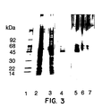
- Figure 3.
- SDS Polyacrylamide Gel Showing Purification of Heregulin-α.
-
Molecular weight markers are shown in
Lane 1. Aliquots from the MDA-MB-231
conditioned media (Lane 2), the 0.6M NaCI pool from the heparin Sepharose column (Lane 3),
Pool C from the polyaspartic acid column (Lane 4) and
Fraction 17 from the HPLC column
(C4-17) (Lane 5) were electrophoresed on a 4-20% gradient gel and silver stained.
Lanes 6
and 7 contained buffer only and shows the presence of gel artifacts in the 50-65 KDa
molecular weight region.
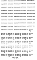
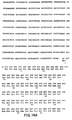
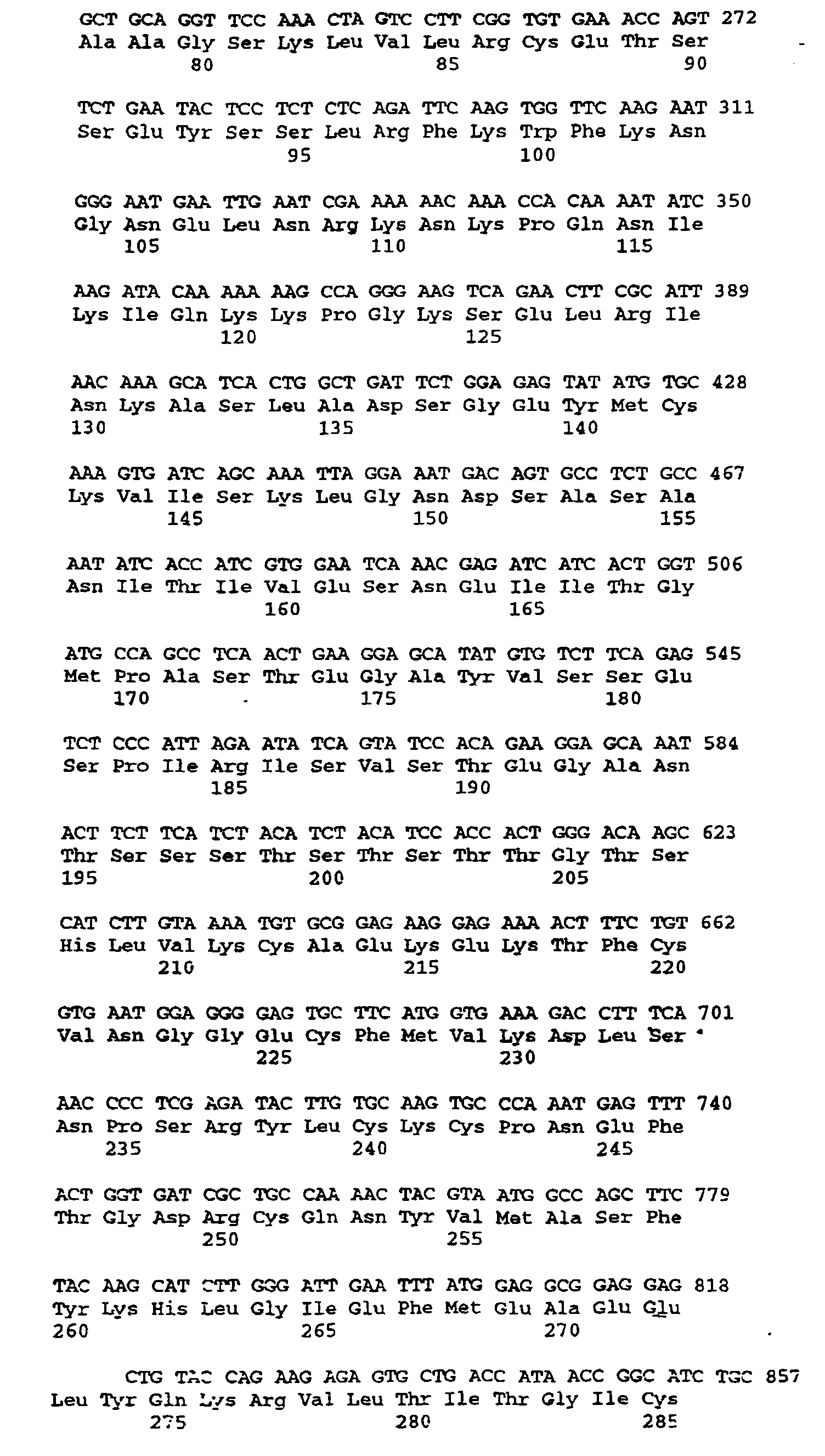
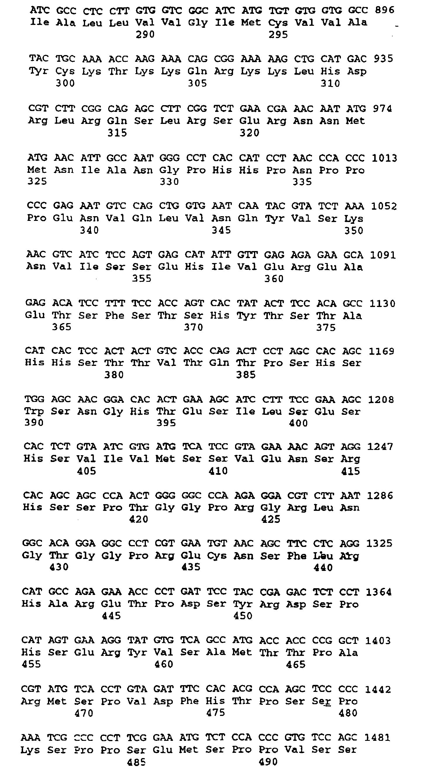
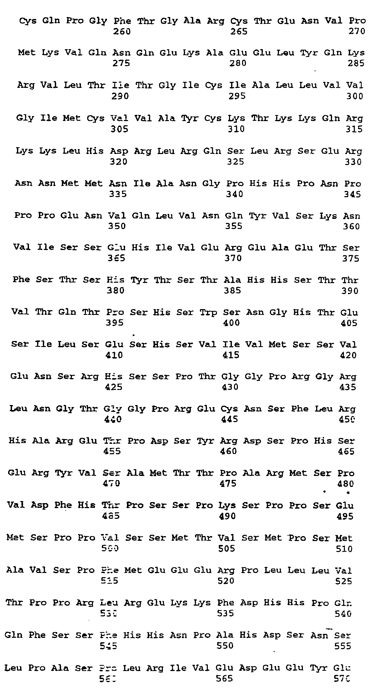
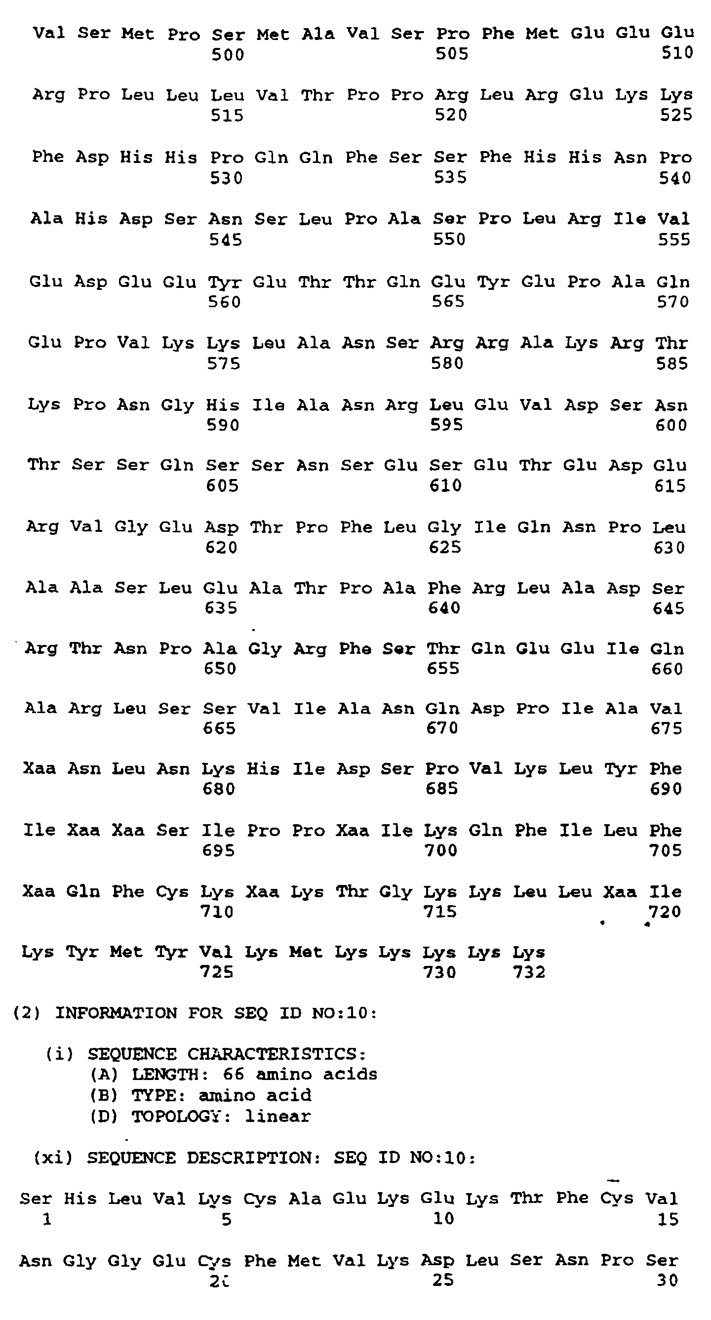
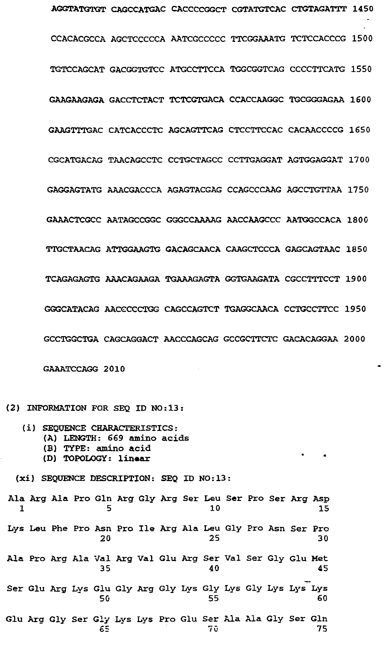
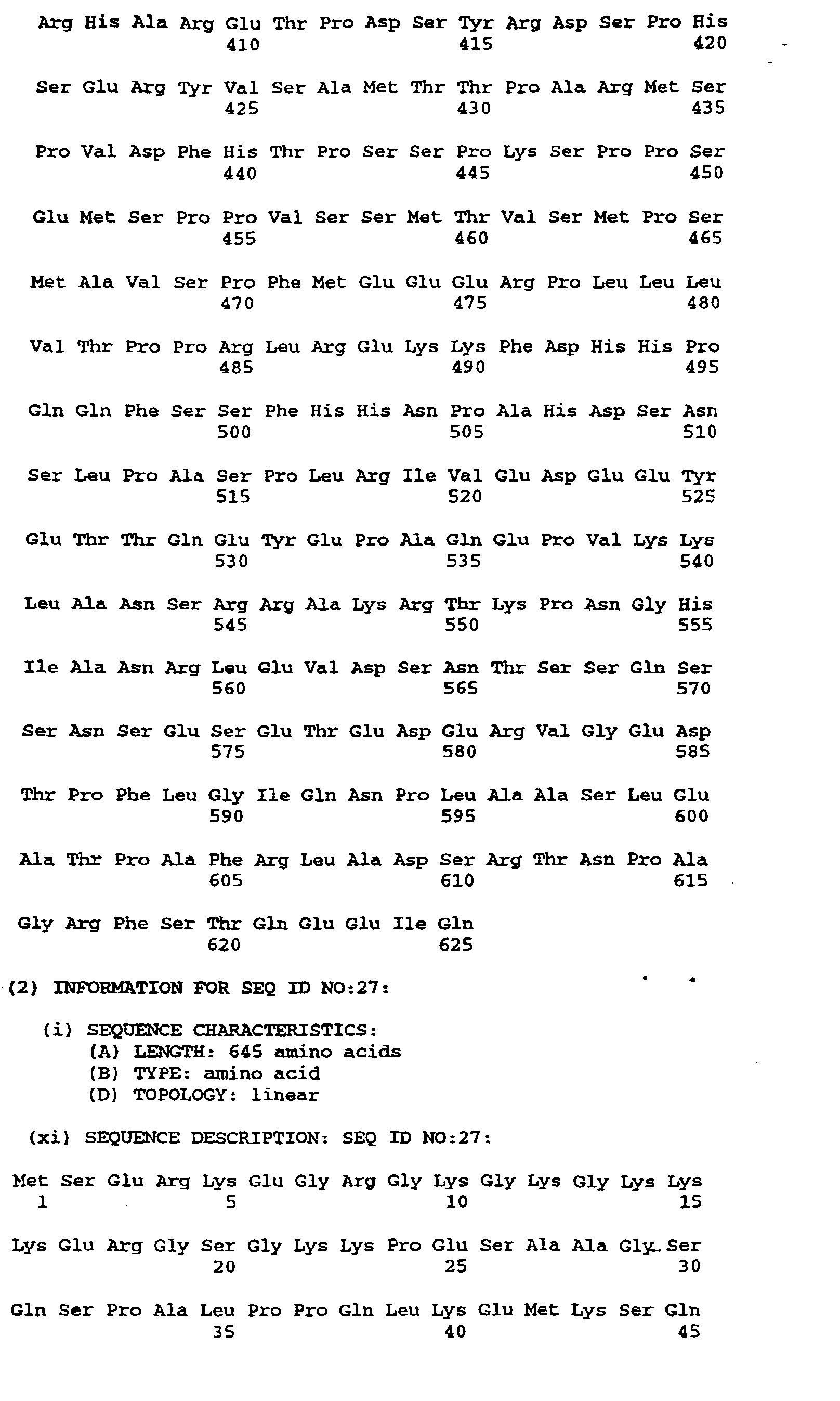
- Figures 4a-4d depict the deduced amino acid sequence of the cDNA contained in λgt10her16
(SEQ ID NO:12 and SEQ ID NO:13). The nucleotides are numbered at the top left of each line
and the amino acids written in three letter code are numbered at the bottom left of each line.
The nucleotide sequence corresponding to the probe is nucleotides 681-720. The probable
transmembrane domain is amino acids 287-309. The six cysteines of the EGF motif are 226,
234, 240, 254, 256 and 265. The five potential three-amino acid N-linked glycosylation sites
are 164-166, 170-172, 208-210, 437-439 and 609-611. The serine-threonine potential O-glycosylation
sites are 209-221. Serine-glycine dipeptide potential glycosaminoglycan addition
sites are amino acids 42-43, 64-65 and 151-152. The initiating methionine(MET) is at position
#45 of figure 4 although the processed N-terminal residue is S46.
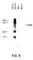
- Figure 5 Northern blot analysis of MDA-MB-231 and SKBR3 RNAs Labeled from left to
right are the following: 1) MDA-MB-231 polyA minus-RNA, (RNA remaining after polyA-containing
RNA is removed); 2) MDA-MB-231 polyA plus-mRNA (RNA which contains polyA);
3) SKBR3 polyA minus-RNA; and, 4) SKBR3 polyA plus-mRNA. The probe used for this
analysis was a radioactively (32P) labelled internal xho1 DNA restriction endonuclease
fragment from the cDNA portion of λgt10her16.
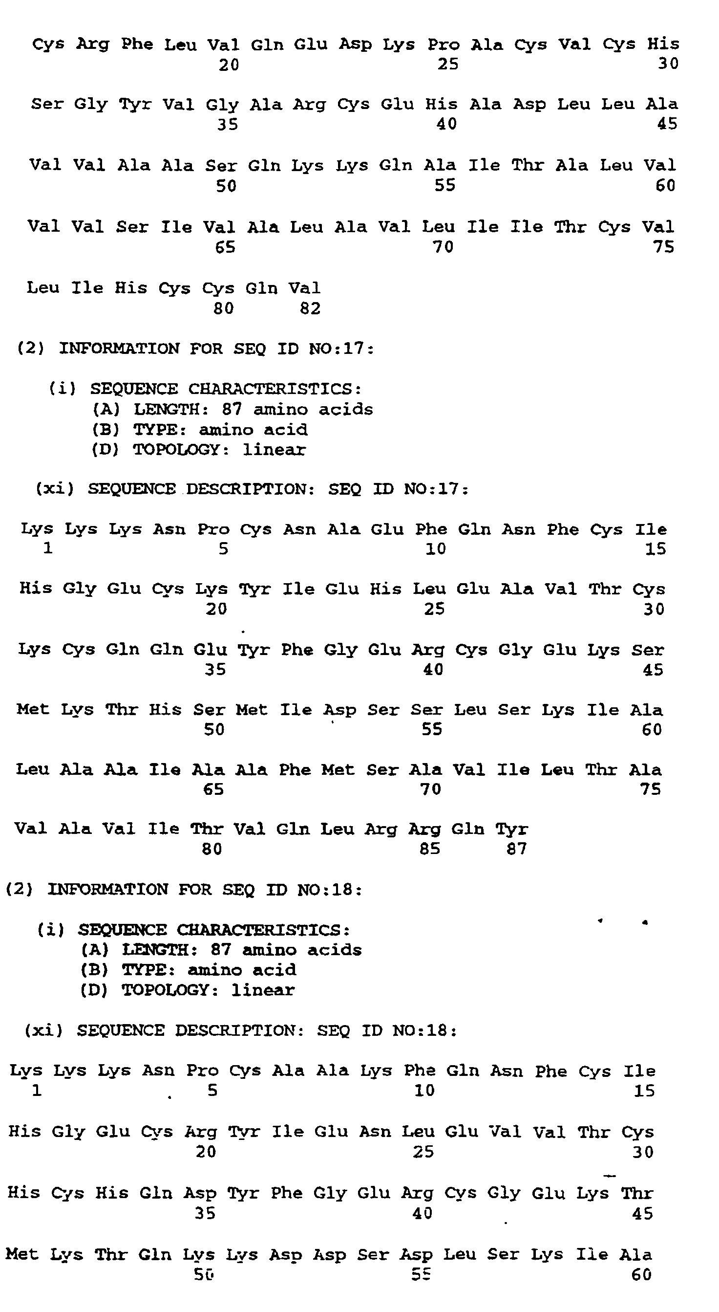
- Figure 6 Sequence Comparisons in the EGF Family of Proteins.
Sequences of several EGF-like proteins (SEQ ID NOS: 14, 15, 16, 17, 18, and 19)
around the cysteine domain are aligned with the sequence of HRG-α. The location in figure 6
of the cysteines and the invariant glycine and arginine residues at positions 238 and 264
clearly show that HRG-α is a member of the EGF family. The region in figure 6 of highest
amino acid identity of the family members relative to HRG-α (30-40%) is found between Cys
234 and Cys 265. The strongest identity (40%) is with the heparin-binding EGF (HB-EGF)
species. HRG-α has a unique 3 amino acid insert between Cys 240 and Cys 254. Potential
transmembrane domains are boxed (287-309). Bars indicate the carboxy-terminal sites for
EGF and TGF-alpha where proteolytic cleavage detaches the mature growth factors from
their transmembrane assodated preforms. HB-EGF is heparin binding-epidermal growth
factor; EGF is epidermal growth factor; TGF-alpha is transforming growth factor alpha; and
schwannoma is the schwannoma-derived growth factor. The residue numbers in Fig. 6 reflect
the Fig. 4 convention.
- Figure 7 Stimulation of Cell Growth by HRG-α.
Three different cell lines were tested for growth responses to 1 nM HRG-α. Cell
protein was quantitated by crystal violet staining and the responses normalized to control,
untreated cells.
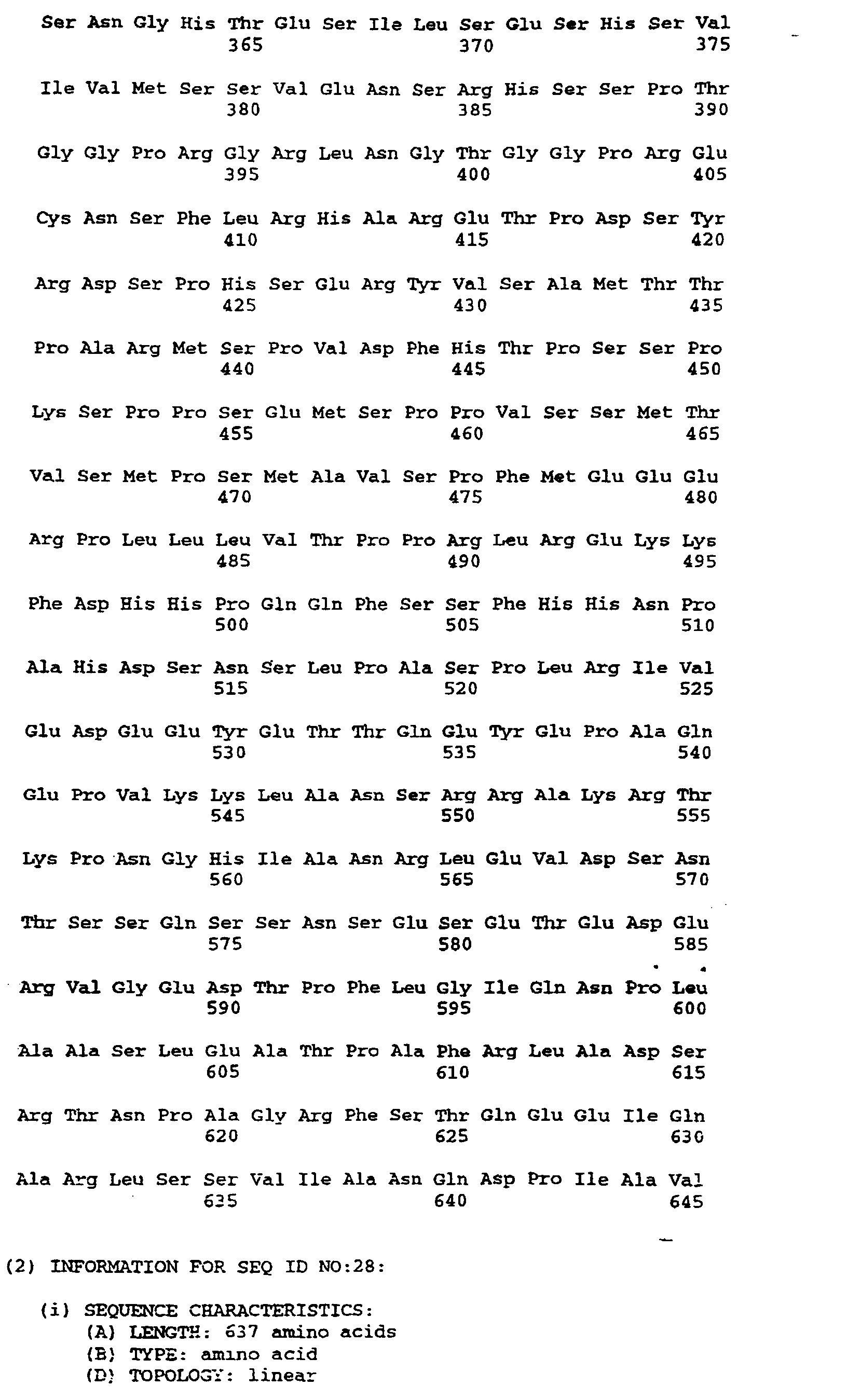
- Figures 8a-8d (SEQ ID NO:7) depict the entire potential coding DNA nucleotide sequence of the
heregulin-β1 and the deduced amino acid sequence of the cDNA contained in λher 11.1dbl
(SEQ ID NO:9). The nucleotides are numbered at the top left of each line and the amino acids
written in three letter code are numbered at the bottom left of each line. The probable
transmembrane amino acid domain is amino acids 278-300. The six cysteines of the EGF
motif are 212, 220, 226, 240, 242 and 251. The five potential three-amino acid N-linked
glycosylation sites are 150-152, 156-158, 196-198, 428-430 and 600-612. The serine-threonine
potential O-glycosylation sites are 195-207. Serine-glycine dipeptide potential
glycosaminoglycan addition sites are amino acids 28-29, 50-51 and 137-138. The initiating
methionine (MET) is at position #31. HRG-β1 is processed to the N-terminal residue S32.
- Figure 9 depicts a comparison of the amino acid sequences of heregulin-α and -β1. A dash (-)
indicates no amino acid at that position. (SEQ ID NO:8 and SEQ ID NO:9). This Fig. uses the
numbering convention of Figs. 4 and 6.
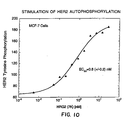
- Figure 10 shows the stimulation of HER2 autophosphorylation using recombinant HRG-α as
measured by HER2 tyrosine phosphorylation.
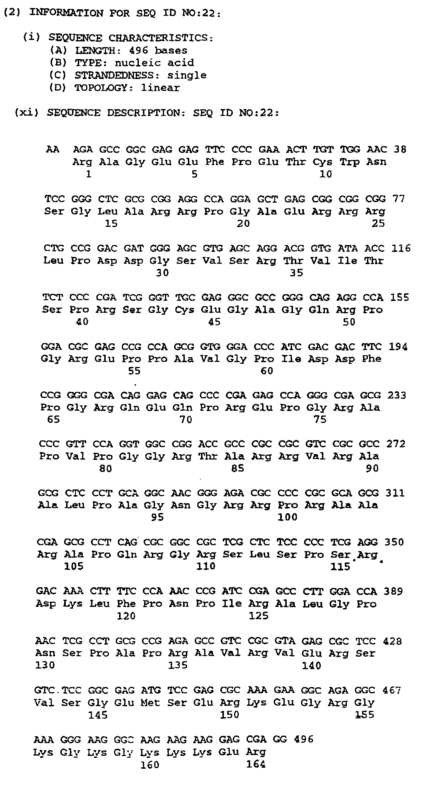
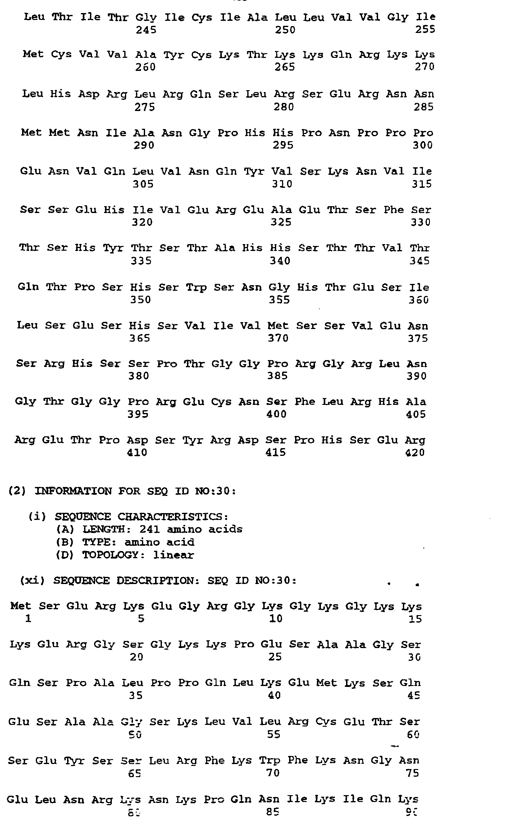
- Figure 11 depicts the nucleotide and inputed amino acid sequence of λl5'her13 (SEQ ID NO:22);
the amino acid residue numbering convention is unique to this figure.
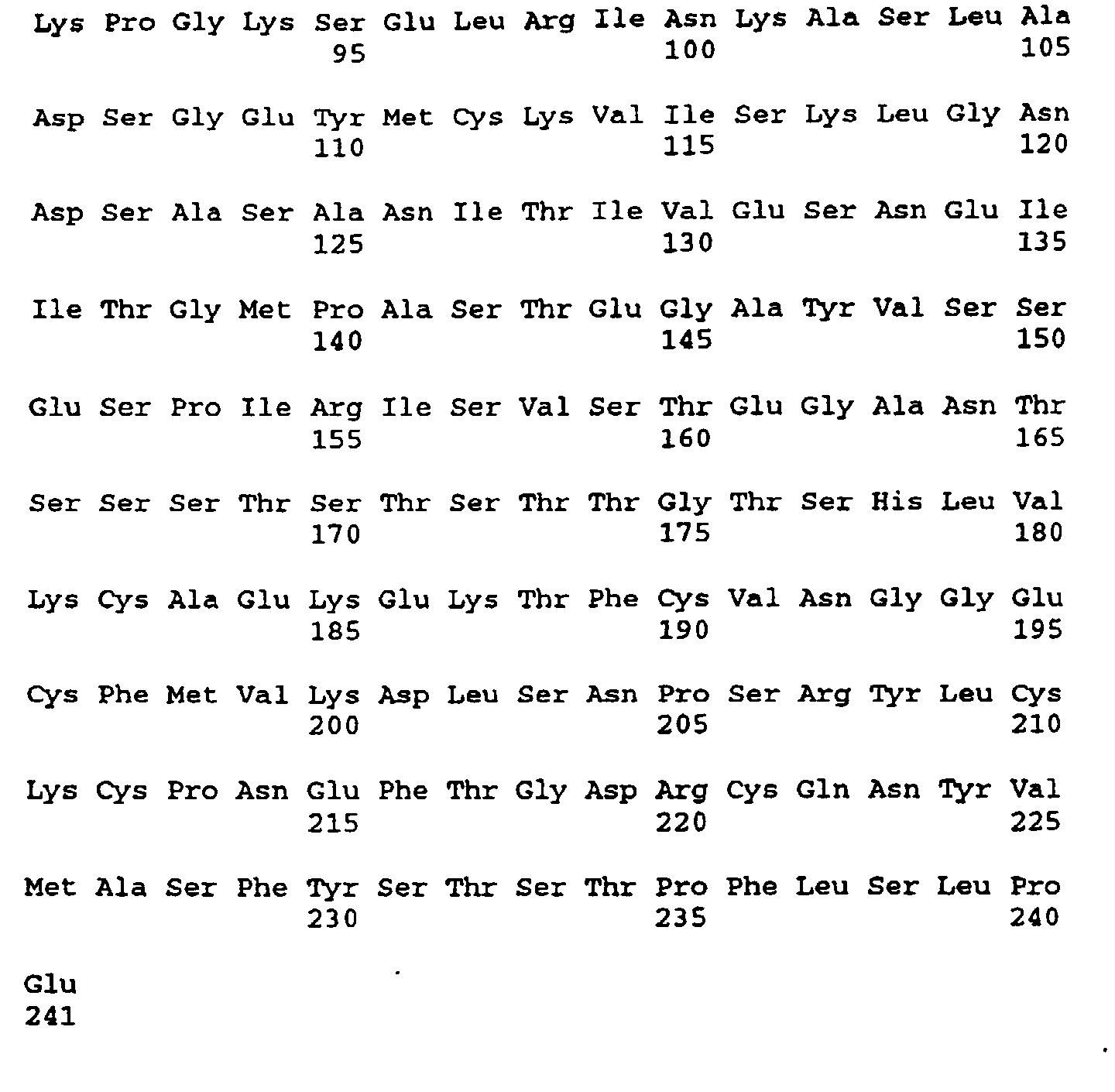
- Figure 12a-12e depict the nucleotide sequence of λher76, encoding heregulin-β2 (SEQ ID
NO:23). This figure commences amino acid residue numbering with the expressed N-terminal
MET; the N-terminus is S2.
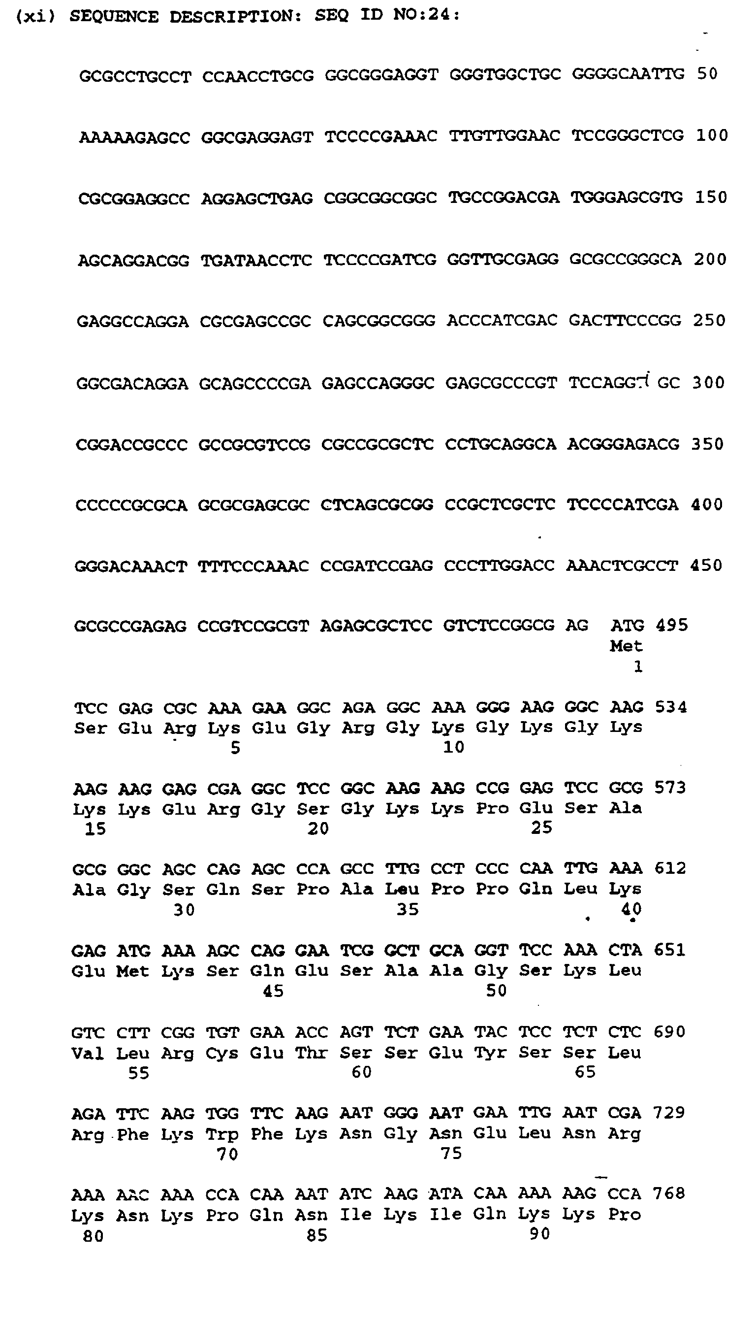
- Figures 13a-13c depict the nucleotide sequence of λher78, encoding heregulin-β3 (SEQ ID
NO:24). This figure uses the amino acid numbering convention of Fig. 12; S2 is the processed
N-terminus.
- Figures 14a-14d depict the nucleotide sequence of λher84, encoding a heregulin-β2-like
polypeptide (SEQ ID N0:25). This figure uses the amino acid numbering convention of Fig. 12;
S2 is the processed N-terminus.
- Figure 15a-15c depict the amino acid homologies between the known heregulins (α, β1, β2, β2-like
and β3 in descending order) and illustrates the amino acid insertions, deletions or substitutions that
distinguish the different forms (SEQ ID NOS:26-30). This figure uses the amino acid numbering
convention of Figs. 12-14.
-
DETAILED DESCRIPTION OF THE PREFERRED EMBODIMENTS
I.
Definitions
-
In general, the following words or phrases have the indicated definition when used in
the description, examples, and claims.
-
Heregulin ("HRG") is defined herein to be any isolated polypeptide sequence which
possesses a biological activity of a polypeptide disclosed in Figs. 4, 8, 12, 13, or 15, and
fragments, alleles or animal analogues thereof or their animal analogues. HRG excludes any
polypeptide heretofore identified, including any known polypeptide which is otherwise
anticipatory under 35 U.S.C. 102, as well as polypeptides obvious over such known
polypeptides under 35 U.S.C. 103, including in particular EFG, TFG-α, amphiregulin (Plowman
et al. Mol. Cell. Biol. 10:1969 (1990), HB-EGF (Higashimaya et al., Science 251:936 [1991]),
schwannoma factor or polypeptides obvious thereover.
-
"Biological activity" for the purposes herein means an in vivo effector or antigenic
function that is directly or indirectly performed by an HRG polypeptide (whether in its native
or denatured conformation), or by any subsequence thereof. Effector functions include
receptor binding or activation, induction of differentiation, mitogenic or growth promoting
activity, immune modulation, DNA regulatory functions and the like, whether presently known
or inherent. Antigenic functions include possession of an epitope or antigenic site that is
capable of cross-reacting with antibodies raised against a naturally occurring or denatured
HRG polypeptide or fragment thereof.
-
Biologically active HRG includes polypeptides having both an effector and antigenic
function, or only one of such functions. HRG includes antagonist polypeptides to HRG,
provided that such antagonists include an epitope of a native HRG. A principal known
effector function of HRG is its ability to bind to p185HER2 and activate the receptor tyrosine
kinase.
-
HRG Includes the translated amino acid sequence of full length human-HRGs
(proHRG) set forth herein in the Figures; deglycosylated or unglycosylated derivatives; amino
acid sequence variants; and covalent derivatives of HRG, provided that they possess
biological actvity. While the native proform of HRG is probably a membrane-bound
polypeptide, soluble forms, such as those forms lacking a functional transmembrane domain
(proHRG or its fragments), are also included within this definition.
-
Fragments of intact HRG are included within the definition of HRG. Two principal
domains are included within the fragments. These are the growth factor domain ("GFD"),
homologous to the EGF family and located at about residues S216-A227 to N268-R286 (Fig. 9,
HRG-α; the GFD domains for other HRGs (Fig. 15) are the homologous sequences.).
Preferably, the GFDs for HRG-α, β1, β2, β2-like and β3 are, respectively, G175-K241, G175-K246,
G175-K238, G175-K238 and G175-E241 (Fig. 15).
-
Another fragment of interest is the N-terminal domain ("NTD"). The NTD extends
from the N-terminus of processed HRG (S2) to the residue adjacent to an N-terminal residue
of the GFD, i.e., about T172-C182 (Fig. 15) and preferably T174. An additional group of
fragments are NTD-GFD domains, equivalent to the extracellular domains of HRG-α and β1-β2.
Another fragment is the C-terminal peptide ("CTP") located about 20 residues N-terminal
to the first residue of the transmembrane domain, either alone or in combination with the C-terminal
remainder of the HRG.
-
In preferred embodiments, antigenically active HRG is a polypeptide that binds with an
affinity of at least about 107 l/mole to an antibody raised against a naturally occurring HRG
sequence. Ordinarily the polypeptide binds with an affinity of at least about 108 l/mole. Most
preferably, the antigenically active HRG is a polypeptide that binds to an antibody raised
against one of HRGs in its native conformation. HRG in its native conformation generally is
HRG as found in nature which has not been denatured by chaotropic agents, heat or other
treatment that substantially modifies the three dimensional structure of HRG as determined,
for example, by migration on nonreducing, nondenaturing sizing gels. Antibody used in this
determination is rabbit polyclonal antibody raised by formulating native HRG from a non-rabbit
species in Freund's complete adjuvant, subcutaneously injecting the formulation into
rabbits, and boosting the immune response by intraperitoneal injection of the formulation until
the titer of anti-HRG antibody plateaus.
-
Ordinarily, biologically active HRG will have an amino acid sequence having at least
75% amino acid sequence identity with an HRG sequence, more preferably at least 80%, even
more preferably at least 90%, and most preferably at least 95%. Identity or homology with
respect to an HRG sequence is defined herein as the percentage of amino acid residues in the
candidate sequence that are identical with HRG residues in Figs. 15, after aligning the
sequences and introducing gaps, if necessary, to achieve the maximum percent homology, and
not considering any conservative substitutions to be identical residues. None of N-terminal,
C-terminal or internal extensions, deletions, or insertions into HRG sequence shall be construed
as affecting homology.
-
Thus, the biologically active HRG polypeptides that are the subject of this invention
include each expressed or processed HRG sequence; fragments thereof having a consecutive
sequence of at least 5, 10, 15, 20, 25, 30 or 40 amino acid residues; amino acid sequence
variants of HRG wherein an amino acid residue has been inserted N- or C-terminal to, or
within, HRG sequence or its fragment as defined above; amino acid sequence variants of HRG
sequence or its fragment as defined above wherein a residue has been substituted by another
residue. HRG polypeptides include those containing predetermined mutations by, e.g., site-directed
or PCR mutagenesis. HRG includes HRG from such as species as rabbit, rat,
porcine, non-human primate, equine, murine, and ovine HRG and alleles or other naturally
occurring variants of the foregoing; derivatives of HRG or its fragments as defined above
wherein HRG or its fragments have been covalently modified by substitution, chemical,
enzymatic, or other appropriate means with a moiety other than a naturally occurring amino
acid (for example a detectable moiety such as an enzyme or radioisotope); glycosylation
variants of HRG (insertion of a glycosylation site or deletion of any glycosylation site by
deletion, insertion or substitution of an appropriate residue); and soluble forms of HRG, such
as HRG-GFD or those that lack a functional transmembrane domain.
-
Of particular interest are fusion proteins that contain HRG-NTD but are free of the
GFD ordinarily associated with the HRG-NTD in question. The first 23 amino acids of the
NTD are dominated by charged residues and contain a sequence (GKKKER; residues 13-18,
Fig. 15) that closely resembles the consensus sequence motif for nuclear targeting (Roberts,
Biochim. Biophys. Acta. 1008:263 [1989]). Accordingly, the HRG includes fusions in which the
NTD, or at least a polypeptide comprising its first about 23 residues, is fused at a terminus
to a non-HRG polypeptide or to a GFD of another HRG family member. The non-HRG
polypeptide in this embodiment is a regulatory protein, a growth factor such as EGF or TGF-α,
or a polypeptide ligand that binds to a cell receptor, particularly a cell surface receptor
found on the surface of a cell whose regulation is desired, e.g. a cancer cell.
-
In another embodiment, one or more of residues 13-18 independently are varied to
produce a sequence incapable of nuclear targeting. For example G13 is mutated to any other
naturally occurring residue including P, L, I, V, A, M, F, K, D or S; any one or more of K14-K16
are mutated to any other naturally occurring residue including R,H,D,E,N or Q; E17 to any
other naturally occurring residue including D, R, K, H, N or Q; and R18 to any other naturally
occurring residue including K, H, D, E, N or Q. All or any one of residues 13-18 are deleted as
well, or extraneous residues are inserted adjacent to these residues; for example residues
inserted adjacent to residue 13-18 which are the same as the above- suggested substitutions
for the residues themselves.
-
In another embodiment, enzymes or a nuclear regulatory protein such as a
transcriptional regulatory factor is fused to HRG-NTD, HRG-NTD-GFD, or HRG-GFD. The
enzyme or factor is fused to the N- or C- terminus, or inserted between the NTD and GFD
domains, or is substituted for the region of NTD between the first about 23 residues and the
GFD.
-
"Isolated" HRG means HRG which has been identified and is free of components of its
natural environment. Contaminant components of its natural environment include materials
which would interfere with diagnostic or therapeutic uses for HRG, and may include proteins,
hormones, and other substances. In preferred embodiments, HRG will be purified (1) to
greater than 95% by weight of protein as determined by the Lowry method or other validated
protein determination method, and most preferably more than 99% by weight, (2) to a degree
sufficient to obtain at least 15 residues of N-terminal or internal amino acid sequence by use
of the best commercially available amino acid sequenator marketed on the filing date hereof,
or (3) to homogeneity by SDS-PAGE using Coomassie blue or, preferably, silver stain.
Isolated HRG includes HRG in situ within heterologous recombinant cells since at least one
component of HRG natural environment will not be present. Isolated HRG includes HRG from
one species in a recombinant cell culture of another species since HRG in such circumstances
will be devoid of source polypeptides. Ordinanly, however, isolated HRG will be prepared by
at least one purification step.
-
In accordance with this invention, HRG nucleic acid is RNA or DNA containing greater
than ten bases that encodes a biologically or antigenically active HRG, is complementary to
nucleic acid sequence encoding such HRG; or hybridizes to nucleic acid sequence encoding such
HRG and remains stably bound to it under stringent conditions.
-
Preferably, HRG nucleic acid encodes a polypeptide sharing at least 75% sequence
identity, more preferably at least 80%, still more preferably at least 85%, even more
preferably at 90%, and most preferably 95%, with an HRG sequence. Preferably, the HRG
nucleic acid that hybridizes contains at least 20, more preferably at least about 40, and most
preferably at least about 90 bases. Such hybridizing or complementary nucleic acid, however,
is further defined as being novel under 35 U.S.C. 102 and unobvious under 35 U.S.C. 103 over
any prior art nucleic acid and excludes nucleic acid encoding EGF, TGF-α, amphiregulin, HB-EGF,
schwannoma factor or fragments or variants thereof which would have been obvious as
of the filing date hereof.
-
Isolated HRG nucleic acid includes a nucleic acid that is free from at least one
contaminant nucleic acid with which it is ordinarily associated in the natural source of HRG
nucleic acid. Isolated HRG nucleic acid thus is present in other than in the form or setting in
which it is found in nature. However, isolated HRG encoding nucleic acid includes HRG nucleic
add in ordinarily HRG-expressing cells where the nucleic acid is in a chromosomal location
different from that of natural cells or is otherwise flanked by a different DNA sequence than
that found in nature. Nucleic acid encoding HRG may be used In specific hybridization assays,
particularly those portions of HRG encoding sequence that do not hybridize with other known
DNA sequences, for example those encoding the EGF-like molecules of figure 6.
-
"Stringent conditions" are those that (1) employ low ionic strength and-high
temperature for washing, for example, 0.015 M NACI/0.0015 M sodium citrate/0/1%
NaDodSO4 at 50° C; (2) employ during hybridization a denaturing agent such as formamide,
for example, 50% (vol/vol) formamide with 0.1% bovine serum albumin, 0.1% Ficoll, 0.1%
polyvinylpyrrolidone, 50 mM sodium phosphate buffer at pH 6.5 with 750 mM NaCI, 75 mM
sodium citrate at 42° C; or (3) employ 50% formamide, 5 x SSC (0.75 M NaCI, 0.075 M
sodium citrate), 50 mM sodium phosphate (pH 6.8), 0.1% sodium pyrophosphate, 5 x
Denhardt's solution, sonicated salmon sperm DNA (50 g/ml), 0.1% SDS, and 10% dextran
sulfate at 42°C, with washes at 42°C in 0.2 x SSC and 0.1% SDS.
-
Particular HRG-α nucleic acids are nucleic acids or oligonucleotides consisting of or
comprising a nucleotide sequence selected from Figs. 4a-4d and containing greater than 17
bases (when excluding nucleic acid sequences of human small polydisperse circular DNA
(HUMPC125), chicken c-mos proto-oncogene homolog (CHKMOS), basement membrane
heparin sulfate proteoglycan (HUMBMHSP) and human lipocortin 2 pseudogene (complete cds-like
region, HUMLIP2B), ordinarily greater than 20 bases, preferably greater than 25 bases,
together with the complementary sequences thereof.
-
Particular HRG-β1, -β2 or -β3 nucleic acids are nucleic acids or oligonucleotides
consisting of or comprising a nucleotide sequence selected from Figs. 8a-8d, 12a-12e or 13a-13c
and containing greater than 20 bases, but does not include the polyA sequence found at the 3'
end of each gene as noted in the Figures, together with the complements to such sequences.
Preferably the sequence contains contains greater than 25 bases. HRG-β sequences also
may exclude the human small polydisperse circular DNA sequence (HUMP-C125).
-
In other embodiments, the HRG nucleotide sequence contains a 15 or more base HRG
sequence and is selected from within the sequence encoding the HRG domain extending from
the N-terminus of the GFD to the N-terminus of the transmembrane sequence (or the
complement of that nucleic acid sequence). For example, with respect to HRG-α, the
nucleotide sequence is selected from within the sequence 678-869 (Fig. 4b) and contains a
sequence of 15 or more bases from this section of the HRG nucleic acid.
-
In other embodiments, the HRG nucleic acid sequence is greater than 14 bases and is
selected from a nucleotide sequence unique to each subtype, for instance a nucleic acid
sequence encoding an amino acid sequence that is unique to each of the HRG subtypes (or the
complement of that nucleic acid sequence). These sequences are useful in diagnostic assays
for expression of the various subtypes, as well as specific amplification of the subtype DNA.
For example, the HRG-α sequence of interest would be selected from the sequence encoding
the unique N-terminus or GFD-transmembrane joining sequence, e.g. about bp771-860.
Similarly, a unique HRG-β1 sequence is that which encodes the last 15 C-terminal amino acid
residues; this sequence is not found in
HRG-α.
-
in general, the length of the HRG-α or β sequence beyond greater than the above-indicated
number of bases Is immaterial since all of such nucleic acids are useful as probes or
amplification primers. The selected HRG sequence may contain additional HRG sequence,
either the normal flanking sequence or other regions of the HRG nucleic acid, as well as other
nucleic acid sequences. For purposes of hybridization, only the HRG sequence is material.
-
The term 'control sequences" refers to DNA sequences necessary for the expression
of an operably linked coding sequence in a particular host organism. The control sequences
that are suitable for prokaryotes, for example, include a promoter, optionally an operator
sequence, a ribosome binding site, and possibly, other as yet poorly understood sequences.
Eukaryotic cells are known to utilize promoters, polyadenylation signals, and enhancers.
-
Nucleic acid is "operably linked" when it Is placed into a functional relationship with
another nucleic acid sequence. For example, DNA for a presequence or secretory leader is
operably linked to DNA for a polypeptide if it is expressed as a preprotein that participates in
the secretion of the polypeptide; a promoter or enhancer is operably linked to a coding
sequence if it affects the transcription of the sequence; or a ribosome binding site is operably
linked to a coding sequence if it is positioned so as to facilitate translation. Generally,
'operably linked" means that the DNA sequences being linked are contiguous and, in the case
of a secretory leader, contiguous and in reading phase. However enhancers do not have to be
contiguous. Linking is accomplished by ligation at convenient restriction sites. If such sites do
not exist, then synthetic oligonucleotide adaptors or linkers are used in accord with
conventional practice.
-
An "exogenous" element is defined herein to mean nucleic acid sequence that is foreign
to the cell, or homologous to the cell but in a position within the host cell nucleic acid in which
the element Is ordinarily not found.
-
As used herein, the expressions "cell", "cell line", and "cell culture" are used
interchangeably, and all such designations include progeny. Thus, the words "transformants"
and "transformed cells" include the primary subject cell and cultures derived therefrom without
regard for the number of transfers. It is also understood that all progeny may not be precisely
identical in DNA content, due to deliberate or inadvertent mutations. Mutant progeny that
have the same function or biological activity as screened for in the originally transformed cell
are included. It will be dear from the context where distinct designations are intended.
-
"Plasmids" are designated by a lower case "p" preceded and/or followed by capital
letters and/or numbers. The starting plasmids herein are commercially available, are publicly
available on an unrestricted basis, or can be constructed from such available plasmids in
accord with published procedures. In addition, other equivalent plasmids are known in the art
and will be apparent to the ordinary artisan.
-
"Restriction Enzyme Digestion" of DNA refers to catalytic cleavage of the DNA with
an enzyme that acts only at certain locations in the DNA. Such enzymes are called
restriction endonucleases, and the sites for which each is specific is called a restriction site.
The various restriction enzymes used herein are commercially available and their reaction
conditions, cofactors, and other requirements as established by the enzyme suppliers are used.
Restriction enzymes commonly are designated by abbreviations composed of a capital letter
followed by other letters representing the microorganism from which each restriction enzyme
originally was obtained, and then a number designating the particular enzyme. In general,
about 1 µg of plasmid or DNA fragment is used with about 1-2 units of enzyme in about 20 µl
of buffer solution. Appropriate buffers and substrate amounts for particular restriction
enzymes are specified by the manufacturer. Incubation of about 1 hour at 37°C is ordinarily
used, but may vary in accordance with the supplier's instructions. After incubation, protein or
polypeptide is removed by extraction with phenol and chloroform, and the digested nucleic acid
is recovered from the aqueous fraction by precipitation with ethanol. Digestion with a
restriction enzyme may be followed with bacterial alkaline phosphatase hydrolysis of the
terminal 5' phosphates to prevent the two restriction cleaved ends of a DNA fragment from
"circularizing" or forming a closed loop that would impede insertion of another DNA fragment
at the restriction site. Unless otherwise stated, digestion of plasmids is not followed by 5'
terminal dephosphorylation. Procedures and reagents for dephosphorylation are conventional
as described in sections 1.56-1.61 of Sambrook et al., (Molecular Cloning: A Laboratory Manual
New York: Cold Spring Harbor Laboratory Press, 1989).
-
"Ligation" refers to the process of forming phosphodiester bonds between two nucleic
acid fragments. To ligate the DNA fragments together, the ends of the DNA fragments must
be compatible with each other. In some cases, the ends will be directly compatible after
endonuclease digestion. However, it may be necessary to first convert the staggered ends
commonly produced after endonuclease digestion to blunt ends to make them compatible for
ligation. To blunt the ends, the DNA is treated in a suitable buffer for at least 15 minutes at
15°C with about 10 units of the Klenow fragment of DNA polymerase I or T4 DNA
polymerase in the presence of the four deoxyribonucleotide triphosphates. The DNA is then
purified by phenol-chloroform extraction and ethanol precipitation. The DNA fragments that
are to be ligated together are put in solution in about equimolar amounts. The solution will also
contain ATP, ligase buffer, and a ligase such as T4 DNA ligase at about 10 units per 05 µg
of DNA. If the DNA is to be ligated into a vector, the vector is first linearized by digestion
with the appropriate restriction endonuclease(s). The linearized fragment is then treated with
bacterial alkaline phosphatase, or calf intestinal phosphatase to prevent self-ligation during
the ligation step.
-
The technique of "polymerase chain reaction," or "PCR," as used herein generally
refers to a procedure wherein minute amounts of a specific piece of nucleic acid, RNA and/or
DNA, are amplified as described in U.S. Pat. No. 4,683,195, issued 28 July 1987. Generally,
sequence information from the ends of the region of interest or.beyond needs to be available,
such that oligonucleotide primers can be designed; these primers will be identical or similar in
sequence to opposite strands of the template to be amplified. The 5' terminal nucleotides of
the two primers may coincide with the ends of the amplified material. PCR can be used to
amplify specific RNA sequences, specific DNA sequences from total genomic DNA, and cDNA
transcribed from total cellular RNA, bacteriophage or plasmid sequences, etc. See generally
Mullis et al., Cold Spring Harbor Symp. Quant. Biol. 51: 263 (1987); Erlich, ed., PCR
Technology, (Stockton Press, NY, 1989). As used herein, PCR is considered to be one, but
not the only, example of a nucleic acid polymerase reaction method for amplifying a nucleic
acid test sample, comprising the use of a known nucleic acid (DNA or RNA) as a primer, and
utilizes a nucleic acid polymerase to amplify or generate a specific piece of nucleic acid or to
amplify or generate a specific piece of nucleic acid which is complementary to a particular
nucleic acid.
-
The "HRG tyrosine autophosphorylation assay" to detect the presence of HRG
ligands was used to monitor the purification of a ligand for the p185HER2 receptor. This assay
is based on the assumption that a specific ligand for the p185HER2 receptor will stimulate
autophosphorylation of the receptor, in analogy with EGF and its stimulation of EGF receptor
autophosphorylation. MDA-MB-453 cells or MCF7 cells which contain high levels of p185HER2
receptors but negligible levels of human EGF receptors, were obtained from the American
Type Culture Collection, Rockville, Md. (ATCC No HTB-131) and maintained in tissue culture
with 10% fetal calf serum in DMEM/Hams F12 (1:1) media. For assay, the cells were
trypsinized and plated at about 150,000 cells/well in 24 well dishes (Costar). After incubation
with serum containing media overnight, the cells were placed in serum free media for 2-18
hours before assay. Test samples of 100 uL aliquots were added to each well. The cells
were incubated for 5-30 minutes (typically 30 min) at 37°C and the media removed. The
cells in each well were treated with 100 uL SDS gel denaturing buffer (Seprosol, Enpotech,
Inc.) and the plates heated at 100°C for 5 minutes to dissolve the cells and denature the
proteins. Aliquots from each well were electrophoresed on 5-20% gradient SDS gels (Novex,
Encinitas, CA) according to the manufacturer's directions. After the dye front reached the
bottom of the gel, the electrophoresis was terminated and a sheet of PVDF membrane
(ProBlott, ABI) was placed on the gel and the proteins transferred from the gel to the
membrane in a blotting chamber (BioRad) at 200 mAmps for 30-60 min. After blotting, the
membranes were incubated with Tris buffered saline containing 0.1% Tween 20 detergent
buffer with 5% BSA for 2-18 hrs to block nonspecific binding, and then treated with a mouse
anti-phosphotyrosine antibody (Upstate Biological Inc., N.Y.). Subsequently, the membrane
blots were treated with goat anti-mouse antibody conjugated to alkaline phosphatase. The
gels were developed using the ProtoBlot System from Promega. After drying the membranes,
the density of the bands corresponding to p185HER2 in each sample lane was quantitated with
a Hewlett Packard ScanJet Plus Scanner attached to a Macintosh computer. The number
of receptors per cell in the MDA-MB-453 or MCF-7cells is such that under these experimental
conditions the p185HER2 receptor protein is the major protein which is labeled.
-
"Protein microsequencing" was accomplished based upon the following procedures.
Proteins from the final HPLC step were either sequenced directly by automated Edman
degradation with a model 470A Applied Biosystems gas phase sequencer equipped with a
120A PTH amino acid analyzer or sequenced after digestion with various chemicals or
enzymes. PTH amino acids were integrated using the ChromPerfect data system (Justice
Innovations, Palo Alto, CA). Sequence interpretation was performed on a VAX 11/785 Digital
Equipment Corporation computer as described (Henzel et al., J. Chromatography 404:41-52
(1987)). In some cases, aliquots of the HPLC fractions were electrophoresed on 5-20% SDS
polyacrylamide gels, electrotransferred to a PVDF membrane (ProBlott, ABI, Foster City,
CA) and stained with Coomassie Brilliant Blue (Matsudaira, P., J. Biol. Chem. 262:10035-10038,
1987). The specific protein was excised from the blot for N terminal sequencing. To
determine internal protein sequences, HPLC fractions were dried under vacuum (SpeedVac),
resuspended in appropriate buffers, and digested with cyanogen bromide, the lysine-specific
enzyme Lys-C (Wako Chemicals, Richmond, VA) or Asp-N (Boehringer Mannheim,
Indianapolis, Ind.). After digestion, the resultant peptides were sequenced as a mixture or
were resolved by HPLC on a C4 column developed with a propanol gradient in 0.1% TFA
before sequencing as described above.
II. USE AND PREPARATION OF HRG POLYPEPTIDES
1. PREPARATION OF HRG POLYPEPTIDES INCLUDING VARIANTS
-
The system to be employed in preparing HRG polypeptides will depend upon the
particular HRG sequence selected. If the sequence is sufficiently small HRG is prepared by in
vitro polypeptide synthetic methods. Most commonly, however, HRG is prepared in
recombinant cell culture using the host-vector systems described below.
-
In general, mammalian host cells will be employed, and such hosts may or may not
contain post-translational systems for processing HRG prosequences in the normal fashion. If
the host cells contain such systems then it will be possible to recover natural subdomain
fragments such as HRG-GFD OR HRG-NTD-GFD from the cultures. If not, then the proper
processing can be accomplished by transforming the hosts with the required enzyme(s) or by
cleaving the precursor in vitro. However, it is not necessary to transform cells with DNA
encoding the complete prosequence for a selected HRG when it is desired to only produce
fragments of HRG sequences such as an HRG-GFD. For example, to prepare HRG-GFD a
start codon is ligated to the 5' end of DNA encoding an HRG-GFD,this DNA is used to
transform host cells and the product expressed directly as the Met N-terminal form (if
desired, the extraneous Met may be removed in vitro or by endogenous N-terminal
demethionylases). Alternatively, HRG-GFD is expressed as a fusion with a signal sequence
recognized by the host cell, which will process and secrete the mature HRG-GFD as is further
described below. Amino acid sequence variants of native HRG-GFD sequences are produced
in the same way.
-
HRG-NTD is produced in the same fashion as the full length molecule but from
expression of DNA encoding only HRG-NTD, with the stop codon after one of S172-C182 (Fig.
15).
-
In addition, HRG variants are expressed from DNA encoding protein in which both the
GFD and NTD domains are in their proper orientation but which contain an amino acid
insertion, deletion or substitution at the NTD-GFD joining site (for example located within the
sequence S172-C182. In another embodiment a stop codon is positioned at the 3' end of the
NTD-GFD-encoding sequence (after any residue T/Q222-T245 of Fig. 15). The result is a
soluble form of HRG-α or -β1 or -β2 which lacks its transmembrane sequence (this sequence
also may be an internal signal sequence but will be referred to as a transmembrane sequence).
In further variations of this embodiment, an internal signal sequence of another polypeptide is
substituted in place of the native HRG transmembrane domain, or a cytoplasmic domain of
another cell membrane polypeptide, e.g. receptor kinase, is substituted for the HRG-α or HRG
β1-β2 cytoplasmic peptide.
-
In a still further embodiment, the NTD, GFD and transmembrane domains of HRG and
other EGF family members are substituted for one another, e.g. the NTD equivalent region of
EGF is substituted for the NTD of HRG, or the GFD of HRG is substituted for EGF in the
processed, soluble proform of EGF. Alternatively, an HRG or EGF family member
transmembrane domain is fused onto the C-terminal E236 of HRG-β3.
-
In a further variant, the HRG sequence spanning K241 to the C-terminus is fused at
its N-terminus to the C-terminus of a non-HRG polypeptide.
-
Another embodiment comprises the functional or structural deletion of the proteolytic
processing site in CTP, the GFD-transmembrane spanning domain. For example, the putative
C-terminal lysine (K241) of processed HRG-α or β1-β2 is deleted, substituted with another
residue, a residue other than K or R inserted between K241 and R242, or other disabling
mutation is made in the prosequence.
-
In another embodiment, the domain of any EGF family member extending from (a) its
cysteine corresponding to (b) C221 to the C-terminal residue of the family member is
substituted for the analogous domain of HRG-α or -β1 or -β2 (or fused to the C-terminus of
HRG-β3). Such variants will be processed free of host cells in the same fashion as the family
member rather than as the parental HRG. In more refined embodiments other specific
cleavage sites (e.g. protease sites) are substituted into the CTP or GFD-transmembrane
spanning domain (about residues T/Q222-T245, Fig. 15). For example, amphiregulin sequence
E84-K99 or TGFα sequence E44-K58 is substituted for HRG-α residues E223-K241.
-
In a further embodiment, a variant (termed HRG-NTDxGFD) is prepared wherein (1)
the lysine residue found in the NTD-GFD joining sequence VKC (residues 180-182, Figure 15) is
deleted or (preferably) substituted by another residue other than R such as H, A, T or S and
(2) a stop codon is introduced in the sequence RCT or RCQ (residues 220-222, Figure 15) in
place of C, or T (for HRG-α) or Q (for HRG-beta).
-
A preferred HRG-α ligand with binding affinity to p185HER2 comprises amino acids
226-265 of figure 4. This HRG-α ligand further may comprise up to an additional 1-20 amino
acids preceding amino acid 226 from figure 4 and 1-20 amino acids following amino acid 265
from figure 4. A preferred HRG-β ligand with binding affinity to p185HER2 comprises amino
acids 226-265 of figure 8. This HRG-β ligand may comprise up to an additional 1-20 amino
acids preceding amino acid 226 from figure 8 and 1-20 amino acids following amino acid 265
from figure 8.
-
GFD sequences include those in which one or more residues corresponding to another
member of the EGF family are deleted or substituted or have a residue inserted adjacent
thereto. For example, F216 of HRG is substituted by Y, L202 with E, F189 with Y, or S203-P205
is deleted.
-
HRG also includes NTD-GFD having its C-terminus at one of the first about 1 to 3
extracellular domain residues (QKR, residues 240-243, HRE-α, Figure 15) or first about 1-2
transmembrane region residues. In addition, in some HRG-GFD variants the codons are
modified at the GFD-transmember proproteolysis site by substitution, insertion or deletion.
The GFD proteolysis site is the domain that contains the GFD C-terminal residue and about 5
residues N- and 5 residues C-terminal from this residue. At this time neither the natural C-terminal
residue for HRG-α or HRG-β has been identified. although it is known that Met-227
terminal and Val-229 terminal HRG-α-GFD are biologically active. The native C-terminus for
HRG-α-GFD is probably Met-227, Lys-228, Val-229, Gln-230, Asn-231 or Gln-232, and for
HRG β1-β2-GFD is probably Met-226, Ala-227, Ser-228, Phe-229, Trp-230, Lys 231or (for
HRG-β1) K240 or (for HRG-β2) K246. The native C-terminus is determined readily by C-terminal
sequencing, although it is not critical that HRG-GFD have the native terminus so long
as the GFD sequence possesses the desired activity. In some embodiments of HRG-GFD
variants, the amino acid change(s) in the CTP are screened for their ability to resist
proteolysis in vitro and inhibit the protease responsible for generation of HRG-GFD.
-
If it is desired to prepare the full length HRG polypeptides and the 5' or 3' ends of the
given HRG are not described herein, it may be necessary to prepare nucleic acids in which the
missing domains are supplied by homologous regions from more complete HRG nucleic acids.
Alternatively, the missing domains can be obtained by probing libraries using the DNAs
disclosed in the Figures or fragments thereof.
A. Isolation of DNA Encoding Heregulin
-
The DNA encoding HRG may be obtained from any cDNA library prepared from
tissue believed to possess HRG mRNA and to express it at a detectable level. HRG DNA
also is obtained from a genomic library.
-
Libraries are screened with probes or analytical tools designed to identify the gene of
interest or the protein encoded by it. For cDNA expression libraries, suitable probes include
monoclonal or polyclonal antibodies that recognize and specifically bind to HRG;
oligonucleotides of about 20-80 bases in length that encode known or suspected portions of
HRG cDNA from the same or different species; and/or complementary or homologous cDNAs
or fragments thereof that encode the same or a hydridizing gene. Appropriate probes for
screening genomic DNA libraries include, but are not limited to, oligonucleotides; cDNAs or
fragments thereof that encode the same or hybridizing DNA; and/or homologous genomic
DNAs or fragments thereof. Screening the cDNA or genomic library with the selected probe
may be conducted using standard procedures as described in chapters 10-12 of Sambrook et
al., supra.
-
An alternative means to isolate the gene encoding HRG is to use polymerase chain
reaction (PCR) methodology as described in section 14 of Sambrook et al., supra. This
method requires the use of oligonucleotide probes that will hybridize to HRG. Strategies for
selection of oligonucleotides are described below.
-
Another alternative method for obtaining the gene of interest is to chemically
synthesize it using one of the methods described in Engels et al. (Agnew. Chem. Int. Ed. Engl.,
28: 716-734,1989). These methods include triester, phosphite, phosphoramidite and H-Phosphonate
methods, PCR and other autoprimer methods, and oligonucleotide syntheses on
solid supports. These methods may be used if the entire nucleic acid sequence of the gene is
known, or the sequence of the nucleic acid complementary to the coding strand is available, or
alternatively, if the target amino acid sequence is known, one may infer potential nucleic acid
sequences using known and preferred coding residues for each amino acid residue.
-
A preferred method of practicing this invention is to use carefully selected
oligonucleotide sequences to screen cDNA libraries from various tissues, preferably human
breast, colon, salivary gland, placental, fetal, brain, and carcinoma cell lines. Other biological
sources of DNA encoding an heregulin-like ligand include other mammals and birds. Among the
preferred mammals are members of the following orders: bovine, ovine, equine, murine, and
rodentia.
-
The oligonucleotide sequences selected as probes should be of sufficient length and
sufficiently unambiguous that false positives are minimized. The actual nucleotide
sequence(s) is usually based on conserved or highly homologous nucleotide sequences or
regions of HRG-α. The oligonucleotides may be degenerate at one or more positions. The use
of degenerate oligonucleotides may be of particular importance where a library is screened
from a species in which preferential codon usage in that species is not known. The
oligonucleotide must be labeled such that it can be detected upon hybridization to DNA in the
library being screened. The preferred method of labeling is to use 32P-labeled ATP with
polynucleotide kinase, as is well known in the art, to radiolabel the oligonucleotide. However,
other methods may be used to label the oligonucleotide, including, but not limited to,
biotinylation or enzyme labeling.
-
Of particular interest is HRG nucleic acid that encodes the full-length propolypeptide.
In some preferred embodiments, the nucleic acid sequence includes the native HRG signal
transmembrane sequence. Nucleic acid having all the protein coding sequence is obtained by
screening selected cDNA or genomic libraries, and, if necessary, using conventional primer
extension procedures as described in section 7.79 of Sambrook et al., supra, to detect
precursors and processing intermediates of mRNA that may not have been reverse-transcribed
into cDNA.
-
HRG encoding DNA is used to isolate DNA encoding the analogous ligand from other
animal species via hybridization employing the methods discussed above. The preferred
animals are mammals, particularly bovine, ovine, equine, feline, canine and rodentia, and more
specifically rats, mice and rabbits.
B. Amino Acid Sequence Variants of Heregulin
-
Amino acid sequence variants of HRG are prepared by introducing appropriate
nucleotide changes into HRG DNA, or by in vitro synthesis of the desired HRG polypeptide.
Such variants include, for example, deletions from, or insertions or substitutions of, residues
within the amino acid sequence shown for human HRG sequences. Any combination of
deletion, insertion, and substitution can be made to arrive at the final construct, provided that
the final construct possesses the desired characteristics. The amino acid changes also may
alter post-translational processes of HRG-α, such as changing the number or position of
glycosylation sites, altering the membrane anchoring characteristics, altering the intra-cellular
location of HRG by inserting, deleting, or otherwise affecting the transmembrane sequence of
native HRG, or modifying its susceptibility to proteolytic cleavage.
-
In designing amino acid sequence variants of HRG, the location of the mutation site
and the nature of the mutation will depend on HRG characteristic(s) to be modified. The sites
for mutation can be modified individually or in series, e.g., by (1) substituting first with
conservative amino acid choices and then with more radical selections depending upon the
results achieved, (2) deleting the target residue, or (3) inserting residues of other ligands
adjacent to the located site.
-
A useful method for identification of HRG residues or regions for mutagenesis is called
"alanine scanning mutagenesis" as described by Cunningham and Wells (Science, 244:1081-1085,
1989). Here, a residue or group of target residues are identified (e.g., charged residues
such as arg, asp, his, lys, and glu) and replaced by a neutral or negatively charged amino acid
(most preferably alanine or polyalanine) to affect the interaction of the amino acids with the
surrounding aqueous environment in or outside the cell. Those domains demonstrating
functional sensitivity to the substitutions then are refined by introducing further or other
variants at or for the sites of substitution. Thus, while the site for introducing an amino acid
sequence variation is predetermined, the nature of the mutation per se need not be
predetermined. For example, to optimize the performance of a mutation at a given site, ala
scanning or random mutagenesis may be conducted at the target codon or region and the
expressed HRG variants are screened for the optimal combination of desired activity.
-
There are two principal variables in the construction of amino acid sequence variants:
the location of the mutation site and the nature of the mutation. These are variants from
HRG sequence, and may represent naturally occurring alleles (which will not require
manipulation of HRG DNA) or predetermined mutant forms made by mutating the DNA, either
to arrive at an allele or a variant not found in nature. In general, the location and nature of
the mutation chosen will depend upon HRG characteristic to be modified. Obviously, such
variations that, for example, convert HRG into a known receptor ligand, are not included
within the scope of this invention, nor are any other HRG variants or polypeptide sequences
that are not novel and unobvious over the prior art.
-
Amino acid sequence deletions generally range from about 1 to 30 residues, more
preferably about 1 to 10 residues, and typically about 1 to 5 contiguous residues. Deletions
may be introduced into regions of low homology with other EGF family precursors to modify
the activity of HRG. Deletions from HRG in areas of substantial homology with other EGF
family sequences will be more likely to modify the biological activity of HRG more significantly.
The number of consecutive deletions will be selected so as to preserve the tertiary structure
of HRG In the affected domain, e.g., cysteine crosslinking, beta-pleated sheet or alpha helix.
-
Amino acid sequence insertions include amino- and/or carboxyl-terminal fusions
ranging in length from one residue to polypeptides containing a hundred or more residues, as
well as intrasequence insertions of single or multiple amino acid residues. Intrasequence
insertions (i.e., insertions within HRG sequence) may range generally from about 1 to 10
residues, more preferably 1 to 5, and most preferably 1 to 3. Examples of terminal insertions
include HRG with an N-terminal methionyl residue (an artifact of the direct expression of HRG
in bacterial recombinant cell culture), and fusion of a heterologous N-terminal signal sequence
to the N-terminus of HRG to facilitate the secretion of mature HRG from recombinant host
cells. Such signal sequences generally will be obtained from, and thus be homologous to, the
intended host cell species. Suitable sequences include STII or Ipp for E. coli, alpha factor for
yeast, and viral signals such as herpes gD for mammalian cells.
-
Other insertional variants of HRG include the fusion to the N- or C-terminus of HRG
to an immunogenic polypeptide, e.g., bacterial polypeptides such as beta-lactamase or an
enzyme encoded by the E. coli trp locus, or yeast protein, bovine serum albumin, and
chemotactic polypeptides. C-terminal fusions of HRG-NTD-GFD with proteins having a long
half-life such as immunoglobulin constant regions (or other immunoglobulin regions), albumin, or
ferritin, as described in WO 89/02922, published 6 April 1989 are included.
-
Another group of variants are amino acid substitution variants. These variants have
at least one amino acid residue in the HRG molecule removed and a different residue inserted
in its place. The sites of greatest interest for substitutional mutagenesis include sites
identified as the active site(s) of HRG, and sites where the amino acids found in HRG ligands
from various species are substantially different in terms of side-chain bulk, charge, and/or
hydrophobicity.
-
The amino terminus of the cytoplasmic region of HRG may be fused to the carboxy
terminus of heterologous transmembrane domains and receptors, to form a fusion polypeptide
useful for intracellular signaling of a ligand binding to the heterologous receptor.
-
Other sites of interest are those in which particular residues of HRG-like ligands
obtained from various species are identical. These positions may be important for the
biological activity of HRG. These sites, especially those falling within a sequence of at least
three other identically conserved sites, are substituted in a relatively conservative manner.
Such conservative substitutions are shown in Table 1 under the heading of "preferred
substitutions". If such substitutions result in a change in biological activity, then more
substantial changes, denominated exemplary substitutions in Table 1, or as further described
below in reference to amino acid classes, are introduced and the products screened.
| Original Residue | Exemplary Substitutions | Preferred Substitutions |
| Ala (A) | val; leu; ile | val |
| Arg (R) | lys; gln; asn | lys |
| Asn (N) | gln; his; lys; arg | gln |
| Asp (D) | glu | glu |
| Cys (C) | ser | ser |
| Gln (Q) | asn | asn |
| Glu (E) | asp | asp |
| Gly (G) | pro | pro |
| His (H) | asn; gln; lys; arg | arg |
| Ile (I) | leu; val; met; ala; phe; norleucine | leu |
| Leu (L) | norleucine;ile;val; met; ala; phe | ile |
| Lys (K) | arg;gln;asn | arg |
| Met (M) | leu;phe;ile | leu |
| Phe (F) | leu; val; ile; ala | leu |
| Pro (P) | gly | gly |
| Ser (S) | thr | thr |
| Thr (T) | ser | ser |
| Trp (W) | tyr | tyr |
| Tyr (Y) | trp; phe; thr; ser | phe |
| Val (V) | ile;leu;met;phe; ala; norleucine | leu |
-
Substantial modifications in function or immunological identity of HRG are
accomplished by selecting substitutions that differ significantly in their effect on maintaining
- (a) the structure of the polypeptide backbone in the area of the substitution, for example, as
a sheet or helical conformation, (b) the charge or hydrophobicity of the molecule at the target
site, or (c) the bulk of the side chain. Naturally occurring residues are divided into groups
based on common side chain properties:
- 1) hydrophobic: norleucine, met, ala, val, leu, ile;
- 2) neutral hydrophilic: cys, ser, thr;
- 3) acidic: asp, glu;
- 4) basic: asn, gln, his, lys, arg;
- 5) residues that influence chain orientation: gly, pro; and
- 6) aromatic: trp, tyr, phe.
-
-
Non-conservative substitutions will entail exchanging a member of one of these
classes for another. Such substituted residues may be introduced into regions of HRG that
are homologous with other receptor ligands, or, more preferably, into the non-homologous
regions of the molecule.
-
In one embodiment of the invention, it is desirable to inactivate one or more protease
cleavage sites that are present in the molecule. These sites are identified by inspection of the
encoded amino acid sequence. Where potential protease cleavage sites are identified, e.g. at
K241 R242, they are rendered inactive to proteolytic cleavage by substituting the targeted
residue with another residue, preferably a basic residue such as glutamine or a hydrophylic
residue such as serine; by deleting the residue; or by inserting a prolyl residue immediately
after the residue.
-
In another embodiment, any methionyl residue other than the starting methionyl
residue, or any residue located within about three residues N- or C-terminal to each such
methionyl residue, is substituted by another residue (preferably in accord with Table 1) or
deleted. We have found that oxidation of the 2 GFD M residues in the courses of E. coli
expression appears to severely reduce GFD activity. Thus, these M residues are mutated in
accord with Table 1. Alternatively, about 1-3 residues are inserted adjacent to such sites.
-
Any cysteine residues not involved in maintaining the proper conformation of HRG
also may be substituted, generally with serine, to improve the oxidative stability of the
molecule and prevent aberrant crosslinking.
-
Sites particularly suited for substitutions, deletions or insertions, or use as fragments,
include, numbered from the N-terminus of HRG-α of Figure 4:
- 1) potential glycosaminoglycan addition sites at the serine-glycine dipeptides at 42-43,
64-65, 151-152;
- 2) potential asparagine-linked glycosylation at positions 164, 170, 208 and 437, sites
(NDS) 164-166, (NIT) 170-172, (NTS) 208-210, and NTS (609-611);
- 3) potential O-glycosylation in a cluster of serine and threonine at 209-218;
- 4) cysteines at 226, 234, 240, 254, 256 and 265;
- 5) transmembrane domain at 287-309;
- 6) loop 1 delineated by cysteines 226 and 240;
- 7) loop 2 delineated by cysteines 234 and 254;
- 8) loop 3 delineated by cysteines 256 and 265; and
- 9) potential protease processing sites at 2-3, 8-9, 23-24, 33-34, 36-37, 45-46, 48-49, 62-63,
66-67, 86-87, 110-111, 123-124, 134-135, 142-143, 272-273, 278-279 and 285-286;
-
-
Analogous regions in HRG-β1 may be determined by reference to figure 9 which aligns
analogous amino acids in HRG-α and HRG-β1. The analogous HRG-β1 amino acids may be
mutated or modified as discussed above for HRG-α. Analogous regions in HRG-β2 may be
determined by reference to figure 15 which aligns analogous amino acids in HRG-α, HRG-β1
and HRG-β2. The analogous HRG-β2 amino acids may be mutated or modified as discussed
above for HRG-α or HRG-β1. Analogous regions in HRG-β3 may be determined by
reference to figure 15 which aligns analogous amino acids in HRG-α, HRG-β1 and HRG-β2.
The analogous HRG-β3 amino acids may be mutated or modified as discussed above for
HRG-α, HRG-β1, or HRG-β2.
-
DNA encoding amino acid sequence variants of HRG is prepared by a variety of
methods known in the art. These methods include, but are not limited to, isolation from a
natural source (in the case of naturally occurring amino acid sequence variants) or
preparation by oligonucleotide-mediated (or site-directed) mutagenesis, PCR mutagenesis, and
cassette mutagenesis of an earlier prepared variant or a non-variant version of HRG. These
techniques may utilize HRG nucleic acid (DNA or RNA), or nucleic acid complementary to
HRG nucleic acid.
-
Oligonucleotide-mediated mutagenesis is a preferred method for preparing substitution,
deletion, and insertion variants of HRG DNA. This technique is well known in the art as
described by Adelman et al., DNA, 2:183 (1983).
-
Generally, oligonucleotides of at least 25 nucleotides in length are used. An optimal
oligonucleotide will have 12 to 15 nucleotides that are completely complementary to the
template on either side of the nucleotide(s) coding for the mutation. This ensures that the
oligonucleotide will hybridize properly to the single-stranded DNA template molecule. The
oligonucleotides are readily synthesized using techniques known in the art such as that
described by Crea et al. (Proc. Natl. Acad. Sci. USA, 75:5765,1978).
-
Single-stranded DNA template may also be generated by denaturing double-stranded
plasmid (or other) DNA using standard techniques.
-
For alteration of the native DNA sequence (to generate amino acid sequence
variants, for example), the oligonucleotide is hybridized to the single-stranded template under
suitable hybridization conditions. A DNA polymerizing enzyme, usually the Klenow fragment
of DNA polymerase I, is then added to synthesize the complementary strand of the template
using the oligonucleotide as a primer for synthesis. A heteroduplex molecule is thus formed
such that one strand of DNA encodes the mutated form of HRG, and the other strand (the
original template) encodes the native, unaltered sequence of HRG. This heteroduplex molecule
is then transformed into a suitable host cell, usually a prokaryote such as E. coli JM101. After
the cells are grown, they are plated onto agarose plates and screened using the oligonucleotide
primer radiolabeled with 32P-phosphate to identify the bacterial colonies that contain the
mutated DNA. The mutated region is then removed and placed in an appropriate vector for
protein production, generally an expression vector of the type typically employed for
transformation of an appropriate host.
-
The method described immediately above may be modified such that a homoduplex
molecule is created wherein both strands of the plasmid contain the mutation(s). The
modifications are as follows: the single-stranded oligonucleotide is annealed to the single-stranded
template as described above. A mixture of three deoxyribonucleotides,
deoxyriboadenosine (dATP), deoxyriboguanosine (dGTP), and deoxyribothymidine (dTTP), is
combined with a modified thio-deoxyribocytosine called dCTP-(aS)
(Amersham Corporation). This mixture is added to the template-oligonucleotide complex.
Upon addition of DNA polymerase to this mixture, a strand of DNA identical to the template
except for the mutated bases is generated. In addition, this new strand of DNA will contain
dCTP-(aS) instead of dCTP, which serves to protect it from restriction endonuclease
digestion. After the template strand of the double-stranded heteroduplex is nicked with an
appropriate restriction enzyme, the template strand can be digested with Exolll nuclease or
another appropriate nuclease past the region that contains the site(s) to be mutagenized. The
reaction is then stopped to leave a molecule that is only partially single-stranded. A complete
double-stranded DNA homoduplex is then formed using DNA polymerase in the presence of all
four deoxyribonucleotide triphosphates, ATP, and DNA ligase. This homoduplex molecule can
then be transformed into a suitable host cell such as E. coli 101, as described above.
-
Explanary substitutions common to any HRG include S2T or D; E3D or K; R4 K or E;
K5R or E; E6D or K; G7P or Y; R8K or D; G9P or Y; K10R or E; G11P or Y; K12R or E; G19P
or Y; S20T or F; G21P or Y; K22 or E; K23R or E; Q38D; S107N; G108P; N120K; D121K; S122
T; N126S; I126L; T127S; A163V; N164K; T165-T174; any residue to I, L, V, M, F, D, E, R or
K; G175V or P; T176S or V; S177K or T; H178K or S; L179F or I; V180L or S; K181R or E; A
183N or V; E184K or D; K185R or E; E186D or Y; K187R or D; T188S or Q; F189Y or S; V191L
or D; N192Q or H; G193P or A; G194P or A; E195D or K; F197Y or I; M198V or Y; V199L or T;
K200V or R; D201E or K; L202E or K; S203A or T; N204Δ; N204Q; P205Δ; P205G; S206T or
R; R207K or A; Y208P or F; L209I or D; K211I or D; F216Y or I; T217 H or S; G218A or P;
A/D219K or R; R220K or A; A235/240/232V or F; E236/241/233D or K; E237/242/234D or
K; L238/243/235I or T; Y239/244/236F or T; Q240/245/237N or K; K241/246/238H or R;
R242/247/238H or K; V243/248/239L or T; L244/249/240I or S; T245/250/241S or I;
I246/251/242V or T and T247/252/243S or I. Specifically with respect to HRG-α, T222S, K
or V; E223D, R or Q; N224Q, K or F; V225A, R or D; P226G, I K or F; M227V, T, R or Y;
K228R, H or D; V229L, K or D; Q230N, R or Y; N231Q, K or Y; Q232N, R or Y; E233D, K or
T and K 234R, H or D (adjacent K/R mutations are paired in alternative embodiments to
create new proteolysis sites). Specifically with respect to HRG-β (any member), Q222N, R
or Y; N223Q, K or Y; Y224F, T or R; V225A, K or D; M226V, T or R; A227V, K, Y or D;
S228T, Y or R; F229Y, I or K and Y230F, T or R are suitable variants. Specifically with
respect to HRG-β1, K231 R or D, H232R or D; L233I, K, F or Y; G234P, R, A or S; 12351, K, F
or Y; E236D, R or A; F237I, Y, K or A; M238V, T, R or A and E239D, R or A are suitable
variants. Specifically with respect to HRG-β1 and HRG-β2, K231R or D are suitable
variants. Alternatively, each of these residues may be deleted or the indicated substituents
inserted adjacent thereto. In addition, about from 1-10 variants are combined to produce
combinations. These changes are made in the proHRG, NTD, GFD, NTD-GFD or other
fragments or fusions. Q213-G215, A219 and the about 11-21 residues C-terminal to C221
differ among the various HRG classes. Residues at these are interchanged among HRG
classes or EGF family members, are deleted, or a residue inserted adjacent thereto.
-
DNA encoding HRG-α mutants with more than one amino acid to be substituted may
be generated in one of several ways. If the amino acids are located close together in the
polypeptide chain, they may be mutated simultaneously using one oligonucleotide that codes
for all of the desired amino acid substitutions. If, however, the amino acids are located some
distance from each other (separated by more than about ten amino acids), it is more difficult
to generate a single oligonucleotide that encodes all of the desired changes. Instead, one of
two alternative methods may be employed.
-
PCR mutagenesis is also suitable for making amino acid variants of HRG-α. While
the following discussion refers to DNA, it is understood that the technique also finds
application with RNA. The PCR technique generally refers to the following procedure (see
Erlich, supra, the chapter by R. Higuchi, p. 61-70). When small amounts of template DNA are
used as starting material in a PCR, primers that differ slightly in sequence from the
corresponding region in a template DNA can be used to generate relatively large quantities of
a specific DNA fragment that differs from the template sequence only at the positions where
the primers differ from the template. For introduction of a mutation into a plasmid DNA, one
of the primers is designed to overlap the position of the mutation and to contain the mutation;
the sequence of the other primer must be identical to a stretch of sequence of the opposite
strand of the plasmid, but this sequence can be located anywhere along the plasmid DNA. It is
preferred, however, that the sequence of the second primer is located within 200 nucleotides
from that of the first, such that in the end the entire amplified region of DNA bounded by the
primers can be easily sequenced. PCR amplification using a primer pair like the one just
described results in a population of DNA fragments that differ at the position of the mutation
specified by the primer, and possibly at other positions, as template copying is somewhat
error-prone.
-
If the ratio of template to product material is extremely low, the vast majority of
product DNA fragments incorporate the desired mutation(s). This product material is used to
replace the corresponding region in the plasmid that served as PCR template using standard
DNA technology. Mutations at separate positions can be introduced simultaneously by either
using a mutant second primer, or performing a second PCR with different mutant primers and
ligating the two resulting PCR fragments simultaneously to the vector fragment in a three (or
more)-part ligation.
-
Another method for preparing variants, cassette mutagenesis, is based on the
technique described by Wells et al. (Gene, 34: 315,1985). The starting material is the plasmid
(or other vector) comprising HRG DNA to be mutated. The codon(s) in HRG DNA to be
mutated are identified. There must be a unique restriction endonuclease site on each side of
the identified mutation site(s). If no such restriction sites exist, they may be generated using
the above-described oligonucleotide-mediated mutagenesis method to introduce them at
appropriate locations in HRG DNA. After the restriction sites have been introduced into the
plasmid, the plasmid is cut at these sites to linearize it. A double-stranded oligonucleotide
encoding the sequence of the DNA between the restriction sites but containing the desired
mutation(s) is synthesized using standard procedures. The two strands are synthesized
separately and then hybridized together using standard techniques. This double-stranded
oligonucleotide is referred to as the cassette. This cassette is designed to have 3' and 5' ends
that are compatible with the ends of the linearized plasmid, such that it can be directly ligated
to the plasmid. This plasmid now contains the mutated HRG DNA sequence.
C. Insertion of DNA into a Cloning or Expression Vehicle
-
The cDNA or genomic DNA encoding native or variant HRG is inserted into a
replicable vector for further cloning (amplification of the DNA) or for expression. Many
vectors are available, and selection of the appropriate vector will depend on 1) whether it is to
be used for DNA amplification or for DNA expression, 2) the size of the DNA to be inserted
into the vector, and 3) the host cell to be transformed with the vector. Each vector contains
various components depending on its function (amplification of DNA or expression of DNA)
and the host cell for which it is compatible. The vector components generally include, but are
not limited to, one or more of the following: a signal sequence, an origin of replication, one or
more marker genes, an enhancer element, a promoter, and a transcription termination
sequence.
(i) Signal Sequence Component
-
In general, the signal sequence may be a component of the vector, or it may be a part
of HRG DNA that is inserted into the vector. The native HRG DNA is believed to encode a
signal sequence at the amino terminus (5' end of the DNA encoding HRG) of the polypeptide
that is cleaved during post-translational processing of the polypeptide to form the mature
HRG polypeptide ligand that binds to p185HER2 receptor, although a conventional signal
structure is not apparent. Native proHRG is. secreted from the cell but may remain lodged in
the membrane because it contains a transmembrane domain and a cytoplasmic region in the
carboxyl terminal region of the polypeptide. Thus, in a secreted, soluble version of HRG the
carboxyl terminal domain of the molecule, including the transmembrane domain, is ordinarily
deleted. This truncated variant HRG polypeptide may be secreted from the cell, provided that
the DNA encoding the truncated variant encodes a signal sequence recognized by the host.
-
HRG of this invention may be expressed not only directly, but also as a fusion with a
heterologous polypeptide, preferably a signal sequence or other polypeptide having a specific
cleavage site at the N-and/or C-terminis of the mature protein or polypeptide. In general, the
signal sequence may be a component of the vector, or it may be a part of HRG DNA that is
inserted into the vector. Included within the scope of this invention are HRG with the native
signal sequence deleted and replaced with a heterologous signal sequence. The heterologous
signal sequence selected should be one that is recognized and processed, i.e., cleaved by a
signal peptidase, by the host cell. For prokaryotic host cells that do not recognize and
process the native HRG signal sequence, the signal sequence is substituted by a prokaryotic
signal sequence selected, for example, from the group of the alkaline phosphatase,
penicillinase, Ipp, or heat-stable enterotoxin II leaders. For yeast secretion the native HRG
signal sequence may be substituted by the yeast invertase, alpha factor, or acid phosphatase
leaders. In mammalian cell expression the native signal sequence is satisfactory, although
other mammalian signal sequences may be suitable.
(ii) Origin of Replication Component
-
Both expression and cloning vectors generally contain a nucleic acid sequence that
enables the vector to replicate in one or more selected host cells. Generally, in cloning vectors
this sequence Is one that enables the vector to replicate independently of the host
chromosomal DNA, and includes origins of replication or autonomously replicating sequences.
Such sequences are well known for a variety of bacteria, yeast, and viruses. The origin of
replication from the plasmid pBR322 is suitable for most Gram-negative bacteria, the 2µ
plasmid origin is suitable for yeast, and various viral origins (SV40, polyoma, adenovirus,
VSV or BPV) are useful for cloning vectors in mammalian cells. Generally, the origin of
replication component is not needed for mammalian expression vectors (the SV40 origin may
typically be used only because it contains the early promoter).
-
Most expression vectors are "shuttle" vectors, Le., they are capable of replication in
at least one class of organisms but can be transfected into another organism for expression.
For example, a vector is cloned in E. coli and then the same vector is transfected into yeast
or mammalian cells for expression even though it is not capable of replicating independently of
the host cell chromosome.
-
DNA may also be amplified by insertion into the host genome. This is readily
accomplished using Bacillus species as hosts, for example, by including in the vector a DNA
sequence that is complementary to a sequence found in Bacillus genomic DNA. Transfection
of Bacillus with this vector results in homologous recombination with the genome and insertion
of HRG DNA. However, the recovery of genomic DNA encoding HRG is more complex than
that of an exogenously replicated vector because restriction enzyme digestion is required to
excise HRG DNA. DNA can be amplified by PCR and directly transfected into the host cells
without any replication component.
(iii) Selection Gene Component
-
Expression and cloning vectors should contain a selection gene, also termed a
selectable marker. This gene encodes a protein necessary for the survival or growth of
transformed host cells grown in a selective culture medium. Host cells not transformed with
the vector containing the selection gene will not survive in the culture medium. Typical
selection genes encode proteins that (a) confer resistance to antibiotics or other toxins, e.g.,
ampicillin, neomycin, methotrexate, or tetracycline, (b) complement auxotrophic deficiencies,
or (c) supply critical nutrients not available from complex media, e.g., the gene encoding D-alanine
racemase for Bacilli.
-
One example of a selection scheme utilizes a drug to arrest growth of a host cell.
Those cells that are successfully transformed with a heterologous gene express a protein
conferring drug resistance and thus survive the selection regimen. Examples of such dominant
selection use the drugs neomycin (Southern et al., J. Molec. Appl. Genet. 1: 327,1982),
mycophenolic acid (Mulligan et al., Science 209: 1422, 1980) or hygromycin (Sugden et al., Mol.
Cell. Biol. 5: 410-413,1985). The three examples given above employ bacterial genes under
eukaryotic control to convey resistance to the appropriate drug G418 or neomycin (geneticin),
xgpt (mycophenolic acid), or hygromycin, respectively.
-
Another example of suitable selectable markers for mammalian cells are those that
enable the identification of cells competent to take up HRG nucleic acid, such as dihydrofolate
reductase (DHFR) or thymidine kinase. The mammalian cell transformants are placed under
selection pressure which only the transformants are uniquely adapted to survive by virtue of
having taken up the marker. Selection pressure is imposed by culturing the transformants
under conditions in which the concentration of selection agent in the medium is successively
changed, thereby leading to amplification of both the selection gene and the DNA that encodes
HRG. Amplification is the process by which genes in greater demand for the production of a
protein critical for growth are reiterated in tandem within the chromosomes of successive
generations of recombinant cells. Increased quantities of HRG are synthesized from the
amplified DNA.
-
For example, cells transformed with the DHFR selection gene are first identified by
culturing all of the transformants in a culture medium that contains methotrexate (Mtx), a
competitive antagonist of DHFR. An appropriate host cell when wild-type DHFR is employed
is the Chinese hamster ovary (CHO) cell line deficient in DHFR activity, prepared and
propagated as described by Urlaub and Chasin, Proc. Natl. Acad. Sci. USA, 77: 4216, 1980.
The transformed cells are then exposed to increased levels of methotrexate. This leads to
the synthesis of multiple copies of the DHFR gene, and, concomitantly, multiple copies of other
DNA comprising the expression vectors, such as the DNA encoding HRG. This amplification
technique can be used with any otherwise suitable host, e.g., ATCC No. CCL61 CHO-K1,
notwithstanding the presence of endogenous DHFR if, for example, a mutant DHFR gene that
is highly resistant to Mtx is employed (EP 117,060). Alternatively, host cells (particularly
wild-type hosts that contain endogenous DHFR) transformed or co-transformed with DNA
sequences encoding HRG, wild-type DHFR protein, and another selectable marker such as
aminoglycoside 3' phosphotransferase (APH) can be selected by cell growth in medium
containing a selection agent for the selectable marker such as an aminoglycosidic antibiotic,
e.g., kanamycin, neomycin, or G418 (see U.S. Pat. No. 4,965,199).
-
A suitable selection gene for use in yeast is the trp1 gene present in the yeast plasmid
YRp7 (Stinchcomb et al., Nature, 282: 39, 1979; Kingsman et al., Gene, 7: 141, 1979; or
Tschemper et al., Gene, 10: 157, 1980). The trp 1 gene provides a selection marker for a
mutant strain of yeast lacking the ability to grow in tryptophan, for example, ATCC No.
44076 or PEP4-1 (Jones, Genetics, 85: 12, 1977). The presence of the trp1 lesion in the yeast
host cell genome then provides an effective environment for detecting transformation by
growth in the absence of tryptophan. Similarly, Leu2-deficient yeast strains (ATCC 20,622
or 38,626) are complemented by known plasmids bearing the Leu2 gene.
(iv) Promoter Component
-
Expression and cloning vectors usually contain a promoter that is recognized by the
host organism and is operably linked to HRG nucleic acid. Promoters are untranslated
sequences located upstream (5') to the start codon of a structural gene (generally within
about 100 to 1000 bp) that control the transcription and translation of a particular nucleic acid
sequence, such as HRG to which they are operably linked. Such promoters typically fall into
two classes, inducible and constitutive. Inducible promoters are promoters that initiate
increased levels of transcription from DNA under their control in response to some change in
culture conditions, e.g., the presence or absence of a nutrient or a change in temperature. At
this time a large number of promoters recognized by a variety of potential host cells are well
known. These promoters are operably linked to DNA encoding HRG by removingthe promoter
from the source DNA by restriction enzyme digestion and inserting the isolated promoter
sequence into the vector. Both the native HRG promoter sequence and many heterologous
promoters may be used to direct amplification and/or expression of HRG DNA. However,
heterologous promoters are preferred, as they generally permit greater transcription and
higher yields of expressed HRG as compared to the native HRG promoter.
-
Promoters suitable for use with prokaryotic hosts include the β-lactamase and
lactose promoter systems (Chang et al., Nature, 275: 615, 1978; and Goeddel et al., Nature 281: 544, 1979), alkaline phosphatase, a tryptophan (trp) promoter system (Goeddel, Nucleic
Acids Res., 8: 4057, 1980 and EP 36,776) and hybrid promoters such as the tac promoter
(deBoer et al., Proc. Natl. Acad. Sci. USA 80: 21-25, 1983). However, other known bacterial
promoters are suitable. Their nucleotide sequences have been published, thereby enabling a
skilled worker operably to ligate them to DNA encoding HRG (Siebenlist et al., Cell 20: 269,
1980) using linkers or adaptors to supply any required restriction sites. Promoters for use in
bacterial systems also generally will contain a Shine-Dalgamo (S.D.) sequence operably linked
to the DNA encoding HRG.
-
Suitable promoting sequences for use with yeast hosts include the promoters for 3-phosphoglycerate
kinase (Hitzeman et al., J. Biol. Chem., 255: 2073, 1980) or other glycolytic
enzymes (Hess et al., J. Adv. Enzyme Reg 7: 149, 1968; and Holland, Biochemistry 17: 4900,
1978), such as enolase, glyceraldehyde-3-phosphate dehydrogenase, hexokinase, pyruvate
decarboxylase, phosphofructokinase, glucose-6-phosphate isomerase, 3-phosphoglycerate
mutase, pyruvate kinase, triosephosphate isomerase, phosphoglucose isomerase, and
glucokinase.
-
Other yeast promoters, which are inducible promoters having the additional
advantage of transcription controlled by growth conditions, are the promoter regions for
alcohol dehydrogenase 2, isocytochrome C, acid phosphatase, degradative enzymes
associated with nitrogen metabolism, metallothionein, glyceraldehyde-3-phosphate
dehydrogenase, and enzymes responsible for maltose and galactose utilization. Suitable
vectors and promoters for use in yeast expression are further described in Hitzeman et al.,
EP 73,657A. Yeast enhancers also are advantageously used with yeast promoters.
-
Promoter sequences are known for eukaryotes. Virtually all eukaryotic genes have
an AT-rich region located approximately 25 to 30 bases upstream from the site where
transcription is initiated. Another sequence found 70 to 80 bases upstream from the start of
transcription of many genes is a CXCAAT (SEQ ID NO:1) region where X may be any
nucleotide. At the 3' end of most eukaryotic genes is an AATAAA sequence (SEQ ID NO:2)
that may be the signal for addition of the poly A tall to the 3' end of the coding sequence. All
of these sequences are suitably inserted into mammalian expression vectors.
-
HRG gene transcription from vectors in mammalian host cells is controlled by
promoters obtained from the genomes of viruses such as polyoma virus, fowlpox virus (UK
2,211,504, published 5 July 1989), adenovirus (such as Adenovirus 2), bovine-papilloma virus,
avian sarcoma virus, cytomegalovirus, a retrovirus, hepatitis-B virus and most preferably
Simian Virus 40 (SV40), from heterologous mammalian promoters, e.g., the actin promoter or
an immunoglobulin promoter, from heat-shock promoters, and from the promoter normally
associated with HRG sequence, provided such promoters are compatible with the host cell
systems.
-
The early and late promoters of the SV40 virus are conveniently obtained as an
SV40 restriction fragment that also contains the SV40 viral origin of replication (Fiers et al.,
Nature, 273:113 (1978); Mulligan and Berg, Science, 209: 1422-1427 (1980); Pavlakis et al.,
Proc. Natl. Acad. Sci. USA, 78: 7398-7402 (1981)). The immediate early promoter of the
human cytomegalovirus is conveniently obtained as a Hindlll E restriction fragment
(Greenaway et al., Gene, 18: 355-360 (1982)). A system for expressing DNA in mammalian
hosts using the bovine papilloma virus as a vector is disclosed in U.S. Pat. No. 4,419,446. A
modification of this system is described in U.S. Pat. No. 4,601,978. See also Gray et al.,
Nature, 295: 503-508 (1982) on expressing cDNA encoding Immune interferon in monkey cells;
Reyes et al., Nature, 297: 598-601 (1982) on expression of human β-interferon cDNA in mouse
cells under the control of a thymidine kinase promoter from herpes simplex virus; Canaani and
Berg, Proc. Natl. Acad. Sci. USA, 79: 5166-5170 (1982) on expression of the human interferon
β1 gene in cultured mouse and rabbit cells; and Gorman et al., Proc. Natl. Acad. Sci. USA, 79:
6777-6781 (1982) on expression of bacterial CAT sequences in CV-1 monkey kidney cells,
chicken embryo fibroblasts, Chinese hamster ovary cells, HeLa cells, and mouse NIH-3T3
cells using the Rous sarcoma virus long terminal repeat as a promoter.
(v) Enhancer Element Component
-
Transcription of a DNA encoding HRG of this invention by higher eukaryotes is often
increased by inserting an enhancer sequence into the vector. Enhancers are cis-acting
elements of DNA, usually about from 10-300 bp, that act on a promoter to increase its
transcription. Enhancers are relatively orientation and position independent having been found
5' (Laimins et al., Proc. Natl. Acad. Sci. USA, 78: 993, 1981) and 3' (Lusky et al, Mol. Cell
Bio., 3: 1108, 1983) to the transcription unit, within an intron (Banerji et al., Cell, 33: 729,
1983) as well as within the coding sequence itself (Osbome et al., Mol. Cell Bio., 4: 1293,
1984). Many enhancer sequences are now known from mammalian genes (globin, elastase,
albumin, α-fetoprotein and insulin). Typically, however, one will use an enhancer from a
eukaryotic cell virus. Examples include the SV40 enhancer on the late side of the replication
origin (bp 100-270), the cytomegalovirus early promoter enhancer, the polyoma enhancer on
the late side of the replication origin, and adenovirus enhancers (see also Yaniv, Nature, 297:
17-18 (1982)) on enhancing elements for activation of eukaryotic promoters. The enhancer
may be spliced into the vector at a position 5' or 3' to HRG DNA, but is preferably located at
a site 5' from the promoter.
(vi) Transcription Termination Component
-
Expression vectors used in eukaryotic host cells (yeast, fungi, insect, plant, animal,
human, or nucleated cells from other multicellular organisms) will also contain sequences
necessary for the termination of transcription and for stabilizing the mRNA. Such sequences
are commonly available from the 5' and, occasionally 3' untranslated regions of eukaryotic or
viral DNAs or cDNAs. These regions contain nucleotide segments transcribed as
polyadenylated fragments in the untranslated portion of the mRNA encoding HRG. The 3'
untranslated regions also include transcription termination sites.
-
Construction of suitable vectors containing one or more of the above listed
components the desired coding and control sequences employs standard ligation techniques.
Isolated plasmids or DNA fragments are cleaved, tailored, and religated in the form desired to
generate the plasmids required.
-
For analysis to confirm correct sequences in plasmids constructed, the ligation
mixtures are used to transform E. coli K12 strain 294 (ATCC 31,446) and successful
transformants selected by ampicillin or tetracycline resistance where appropriate. Plasmids
from the transformants are prepared, analyzed by restriction endonuclease digestion, and/or
sequenced by the method of Messing et al., Nucleic Acids Res. 9: 309 (1981) or by the method
of Maxam et al., Methods in Enzymology 65:499 (1980).
-
Particularly useful in the practice of this invention are expression vectors that
provide for the transient expression in mammalian cells of DNA encoding HRG. In general,
transient expression involves the use of an expression vector that is able to replicate
efficiently in a host cell, such that the host cell accumulates many copies of the expression
vector and, in turn, synthesizes high levels of a desired polypeptide encoded by the expression
vector. Transient expression systems, comprising a suitable expression vector and a host
cell, allow for the convenient positive identification of polypeptides encoded by cloned DNAs,
as well as for the rapid screening of such polypeptides for desired biological or physiological
properties. Thus, transient expression systems are particularly useful in the invention for
purposes of identifying analogs and variants of HRG that have HRG-like activity. Such a
transient expression system is described in EP 309,237 published 29 March 1989. Other
methods, vectors, and host cells suitable for adaptation to the synthesis of HRG in
recombinant vertebrate cell culture are described in Gething et al., Nature 293: 620-625, 1981;
Mantei et al., Nature, 281: 40-46, 1979; Levinson et al., EP 117,060 and EP 117,058. A
particularly useful expression plasmid for mammalian cell culture expression of HRG is pRK5
(EP pub. no. 307,247).
D. Selection and Transformation of Host Cells
-
Suitable host cells for cloning or expressing the vectors herein are the prokaryote,
yeast, or higher eukaryote cells described above. Suitable prokaryotes include eubacteria,
such as Gram-negative or Gram-positive organisms, for example, E. coli, Bacilli such as B.
subtilis, Pseudomonas species such as P. aeruginosa, Salmonella typhimurium, or Serratia
marcescans. One preferred E. coli cloning host is E. coli 294 (ATCC 31;446); although other
strains such as E. coli B, E. coli x1776 (ATCC 31,537), and E. coli W3110 (ATCC 27,325) are
suitable. These examples are illustrative rather than limiting. Preferably the host cell should
secrete minimal amounts of proteolytic enzymes. Alternatively, in vitro methods of cloning,
e.g., PCR or other nucleic acid polymerase reactions, are suitable.
-
In addition to prokaryotes, eukaryotic microbes such as filamentous fungi or yeast are
suitable hosts for HRG-encoding vectors. Saccharomyces cerevisiae, or common baker's
yeast, is the most commonly used among lower eukaryotic host microorganisms. However, a
number of other genera, species, and strains are commonly available and useful herein, such
as Schizosaccharomyces pombe (Beach and Nurse, Nature, 290: 140 (1981); EP 139,383,
published May 2, 1985), Kluyveromyces hosts (U.S.S.N. 4,943,529) such as, e.g., K. lactis
(Louvencourt et al., J. Bacteriol., 737 (1983); K. fragilis, K. bulgaricus, K. thermotolerans, and
K. marxianus, yarrowia (EP 402,226); Pichia pastoris (EP 183,070), Sreekrishna et al., J.
Basic Microbiol., 28: 265-278 (1988); Candida, Trichoderma reesia (EP 244,234); Neurospora
crassa (Case et al., Proc. Natl. Acad. Sci. USA, 76: 5259-5263 (1979), and filamentous fungi
such as, e.g, Neurospora, Penicillium, Tolypocladium (WO 91/00357, published 10 January
1991), and Aspergillus hosts such as A. nidulans (Ballance et al., Biochem. Biophys. Res.
Commun., 112: 284-289 (1983); Tilbum et al., Gene, 26: 205-221 (1983); Yelton et al., Proc.
Natl. Acad. Sci. USA, 81:1470-1474 (1984) and A. niger (Kelly and Hynes, EMBO J., 4: 475-479
(1985)).
-
Suitable host cells for the expression of glycosylated HRG polypeptide are derived
from multicellular organisms. Such host cells are capable of complex processing and
glycosylation activities. In principle, any higher eukaryotic cell culture is workable, whether
from vertebrate or invertebrate culture. Examples of invertebrate cells include plant and
insect cells. Numerous baculoviral strains and variants and corresponding permissive insect
host cells from hosts such as Spodoptera frugiperda (caterpillar), Aedes aegypti (mosquito),
Aedes albopictus (mosquito), Drosophila melanogaster (fruitfly), and Bombyx mori host cells
have been identified (see, e.g., Luckow et al., Bio/Technology, 6: 47-55 (1988); Miller et al., in
Genetic Engineering, Setlow, J.K. et al., eds., Vol. 8 (Plenum Publishing, 1986), pp. 277-279; and
Maeda et al., Nature, 315: 592-594 (1985)). A variety of such viral strains are publicly
available, e.g., the L-1 variant of Autographa californica NPV and the Bm-5 strain of Bombyx
mori NPV, and such viruses may be used as the virus herein according to the present
invention, particularly for transfection of Spodoptera frugiperda cells. Plant cell cultures of
cotton, corn, potato, soybean, petunia, tomato, and tobacco can be utilized as hosts.
Typically, plant cells are transfected by incubation with certain strains of the bacterium
Agrobacterium tumefaciens, which has been previously manipulated to contain HRG DNA.
During incubation of the plant cell culture with A. tumefaciens, the DNA encoding HRG is
transferred to the plant cell host such that it is transfected, and will, under appropriate
conditions, express HRG DNA. In addition, regulatory and signal sequences compatible with
plant cells are available, such as the nopaline synthase promoter and polyadenylation signal
sequences (Depicker et al., J. Mol. Appl. Gen., 1:561 [1982]). In addition, DNA segments
isolated from the upstream region of the T-DNA 780 gene are capable of activating or
increasing transcription levels of plant-expressible genes in recombinant DNA-containing plant
tissue (see EP 321,196, published 21 June 1989).
-
However, interest has been greatest in vertebrate cells, and propagation of
vertebrate cells in culture (tissue culture) has become a routine procedure in recent years
(Tissue Culture, Academic Press, Kruse and Patterson, editors (1973)). Examples of useful
mammalian host cell lines are monkey kidney CV1 line transformed by SV40 (COS-7, ATCC
CRL 1651); human embryonic kidney line (293 or 293 cells subcloned for growth in suspension
culture, Graham et al., J. Gen Virol., 36: 59, 1977); baby hamster kidney cells (BHK, ATCC
CCL 10); Chinese hamster ovary cells/-DHFR (CHO, Urlaub and Chasin, Proc. Natl. Acad.
Sci. USA, 77:4216 [1980]); mouse sertoli cells (TM4, Mather, Biol. Reprod., 23:243-251 [1980]);
monkey kidney cells (CV1 ATCC CCL 70); African green monkey kidney cells (VERO-76,
ATCC CRL-1587); human cervical carcinoma cells (HELA, ATCC CCL 2); canine kidney
cells (MDCK, ATCC CCL 34); buffalo rat liver cells (BRL 3A, ATCC CRL 1442); human
lung cells (W138, ATCC CCL 75); human liver cells (Hep G2, HB 8065); mouse mammary
tumor (MMT 060562, ATCC CCL51); TRI cells (Mather et al., Annals N.Y. Acad. Sci.,
383:44-68 (1982]); MRC 5 cells; FS4 cells; and a human hepatoma cell line (Hep G2).
Preferred host cells are human embryonic kidney 293 and Chinese hamster ovary cells.
-
Host cells are transfected and preferably transformed with the above-described
expression or cloning vectors of this invention and cultured in conventional nutrient media
modified as appropriate for inducing promoters, selecting transformants, or amplifying the
genes encoding the desired sequences.
-
Transfection refers to the taking up of an expression vector by a host cell whether or
not any coding sequences are in fact expressed. Numerous methods of transfection are
known to the ordinarily skilled artisan, for example, CaPO4 and electroporation. Successful
transfection is generally recognized when any indication of the operation of this vector occurs
within the host cell.
-
Transformation means introducing DNA into an organism so that the DNA is
replicable, either as an extrachromosomal element or by chromosomal integration. Depending
on the host cell used, transformation is done using standard techniques appropriate to such
cells. The calcium treatment employing calcium chloride, as described in section 1.82 of
Sambrook et al., supra, is generally used for prokaryotes or other cells that contain
substantial cell-wall barriers. Infection with Agrobacterium tumefaciens is used for
transformation of certain plant cells, as described by Shaw et al., Gene, 23:315 (1983) and
WO 89/05859, published 29 June 1989. For mammalian cells without such cell walls, the
calcium phosphate precipitation method described in sections 16.30-16.37 of Sambrook et al,
supra, is preferred. General aspects of mammalian cell host system transformations have
been described by Axel in U.S. Pat. No. 4,399,216, issued 16 August 1983. Transformations
into yeast are typically carried out according to the method of Van Solingen et al., J. Bact.,
130:946 (1977) and Hsiao et al., Proc. Natl. Acad. Sci. (USA), 76: 3829 (1979). However,
other methods for introducing DNA into cells such as by nuclear injection, electroporation, or
protoplast fusion may also be used.
E. Culturing the Host Cells
-
Prokaryotic cells used to produce HRG polypeptide of this invention are cultured in
suitable media as described generally in Sambrook et al., supra.
-
The mammalian host cells used to produce HRG of this invention may be cultured in a
variety of media. Commercially available media such as Ham's F10 (Sigma), Minimal
Essential Medium ([MEM], Sigma), RPMI-1640 (Sigma), and Dulbecco's Modified Eagle's
Medium ([DMEM], Sigma) are suitable for culturing the host cells. In addition, any of the media
described in Ham and Wallace, Meth. Enz., 58: 44 (1979), Bames and Sato, Anal. Biochem.,
102:255 (1980), U.S. Pat. Nos. 4,767,704; 4,657,866; 4,927,762; or 4,560,655; WO 90/03430;
WO 87/00195 and U.S. Pat. Re. 30,985, may be used as culture media for the host cells. Any
of these media may be supplemented as necessary with hormones and/or other growth
factors (such as insulin, transferrin, or epidermal growth factor), salts (such as sodium
chloride, calcium, magnesium, and phosphate), buffers (such as HEPES), nucleosides (such as
adenosine and thymidine), antibiotics (such as Gentamycin™ drug), trace elements (defined
as inorganic compounds usually present at final concentrations in the micromolar range), and
glucose or an equivalent energy source. Any other necessary supplements may also be
included at appropriate concentrations that would be known to those skilled in the art. The
culture conditions, such as temperature, pH, and the like, are those previously used with the
host cell selected for expression, and will be apparent to the ordinarily skilled artisan.
-
The host cells referred to in this disclosure encompass cells in in vitro culture as well
as cells that are within a host animal.
-
It is further envisioned that HRG of this invention may be produced by homologous
recombination, or with recombinant production methods utilizing control elements introduced
into cells already containing DNA encoding HRG currently in use in the field. For example, a
powerful promoter/enhancer element, a suppressor, or an exogenous transcription modulatory
element is inserted in the genome of the intended host cell in proximity and orientation
sufficient to influence the transcription of DNA encoding the desired HRG. The control
element does not encode HRG of this invention, but the DNA is present in the host cell genome.
One next screens for cells making HRG of this invention, or increased or decreased levels of
expression, as desired.
F. Detecting Gene Amplification/Expression
-
Gene amplification andlor expression may be measured in a sample directly, for
example, by conventional Southern blotting, Northern blotting to quantitate the transcription
of mRNA (Thomas, Proc. Natl. Acad. Sci. USA, 77:5201-5205 [1980]), dot blotting (DNA
analysis), or in situ hybridization, using an appropriately labeled probe based on the sequences
provided herein. Various labels may be employed, most commonly radioisotopes, particularly
32P. However, other techniques may also be employed, such as using biotin-modified
nudeotides for introduction into a polynucleotide. The biotin then serves as the site for binding
to avidin or antibodies which may be labeled with a wide variety of labels, such as
radionuclides, fluorescers, enzymes, or the Ike. Alternatively, antibodies may be employed
that can recognize specific duplexes, including DNA duplexes, RNA duplexes, and DNA-RNA
hybrid duplexes or DNA-protein duplexes. The antibodies in turn may be labeled and the
assay may be carried out where the duplex is bound to a surface, so that upon the formation
of duplex on the surface, the presence of antibody bound to the duplex can be detected.
-
Gene expression, alternatively, may be measured by immunological methods, such as
immunohistochemical staining of tissue sections and assay of cell culture or body fluids, to
quantitate directly the expression of gene product. With immunohistochemical staining
techniques, a cell sample is prepared, typically by dehydration and fixation, followed by
reaction with labeled antibodies specific for the gene product coupled where the labels are
usually visually detectable such as enzymatic labels, fluorescent labels, luminescent labels,
and the like. A particularly sensitive staining technique suitable for use in the present
invention is described by Hsu et al., Am. J. Clin. Path., 75: 734-738 (1980).
-
Antibodies useful for immunohistochemical staining and/or assay of sample fluids may
be either monoclonal or polyclonal, and may be prepared in any mammal. Conveniently, the
antibodies may be prepared against a native HRG polypeptide or against a synthetic peptide
based on the DNA sequences provided herein as described further in Section 4 below.
G. Purification of The Heregulin Polypeptide
-
HRG is recovered from a cellular membrane fraction. Alternatively, a proteolyticalLy
cleaved or a truncated expressed soluble HRG fragment or subdomain are recovered from the
culture medium as soluble polypeptides.
-
When HRG is expressed in a recombinant cell other than one of human origin, HRG is
completely free of proteins or polypeptides of human origin. However, it is desirable to purify
HRG from recombinant cell proteins or polypeptides to obtain preparations that are
substantially homogeneous as to HRG. As a first step, the culture medium or lysate is
centrifuged to remove particulate cell debris. The membrane and soluble protein fractions are
then separated. HRG is then purified from both the soluble protein fraction (requiring the
presence of a protease) and from the membrane fraction of the culture lysate, depending on
whether HRG is membrane bound. The following procedures are exemplary of suitable
purification procedures: fractionation on immunoaffinity or ion-exchange columns; ethanol
precipitation; reversed phase HPLC; chromatography on silica, heparin sepharose or on a
cation exchange resin such as DEAE; chromatofocusing; SDS-PAGE; ammonium sulfate
precipitation; and gel filtration using, for example, Sephadex G-75.
-
HRG variants in which residues have been deleted, inserted or substituted are
recovered in the same fashion as the native HRG, taking account of any.substantial changes
in properties occasioned by the variation. For example, preparation of a HRG fusion with
another protein or polypeptide, e.g., a bacterial or viral antigen, facilitates purification; an
immunoaffinity column containing antibody to the antigen can be used to adsorb the fusion.
Immunoaffinity columns such as a rabbit polyclonal anti-HRG column can be employed to
absorb HRG variant by binding it to at least one remaining immune epitope. A protease
inhibitor such as phenylmethylsulfonylfluoride (PMSF) also may be useful to inhibit proteolytic
degradation during purification, and antibiotics may be included to prevent the growth of
adventitious contaminants. One skilled in the art will appreciate that purification methods
suitable for native HRG may require modification to account for changes in the character of
HRG variants upon expression in recombinant cell culture.
H. Covalent Modifications of HRG
-
Covalent modifications of HRG polypeptides are included within the scope of this
invention. Both native HRG and amino acid sequence variants of HRG optionally are
covalently modified. One type of covalent modification included within the scope of this
invention is a HRG polypeptide fragment. HRG fragments, such as HRG-GDF, having up to
about 40 amino acid residues are conveniently prepared by chemical synthesis, or by
enzymatic or chemical cleavage of the full-length HRG polypeptide or HRG variant
polypeptide. Other types of covalent modifications of HRG or fragments thereof are
introduced into the molecule by reacting targeted amino acid residues of HRG or fragments
thereof with an organic derivatizing agent that is capable of reacting with selected side chains
or the N- or C-terminal residues.
-
Cysteinyl residues most commonly are reacted with α-haloacetates (and
corresponding amines), such as chloroacetic acid or chloroacetamide, to give carboxymethyl
or carboxyamidomethyl derivatives. Cysteinyl residues also are derivatized by reaction with
bromotrifluoroacetone, α-bromo-β-(5-imidozoyl)propionic acid, chloroacetyl phosphate, N-alkylmaleimides,
3-nitro-2-pyridyl disulfide, methyl 2-pyridyl disulfide, p-chloromercuribenzoate,
2-chloromercuri-4-nitrophenol, or chloro-7-nitrobenzo-2-oxa-1,3-diazole.
-
Histidyl residues are derivatized by reaction with diethylpyrocarbonate at pH 5.5-7.0
because this agent is relatively specific for the histidyl side chain. Para-bromophenacyl
bromide also is useful; the reaction is preferably performed in 0.1M sodium cacodylate at pH
6.0.
-
Lysinyl and amino terminal residues are reacted with succinic or other carboxylic acid
anhydrides. Derivatization with these agents has the effect of reversing the charge of the
lysinyl residues. Other suitable reagents for derivatizing α-amino-containing residues include
imidoesters such as methyl picolinimidate; pyridoxal phosphate; pyridoxal; chloroborohydride;
trinitrobenzenesulfonic acid; O-methylisourea; 2,4-pentanedione; and transaminase-catalyzed
reaction with glyoxylate.
-
Arginyl residues are modified by reaction with one or several conventional reagents,
among them phenyiglyoxal, 2,3-butanedione, 1,2-cyclohexanedione, and ninhydrin.
Derivatization of arginine residues requires that the reaction be performed in alkaline
conditions because of the high pKa of the guanidine functional group. Furthermore, these
reagents may react with the groups of lysine as well as the arginine epsilon-amino group.
-
The specific modification of tyrosyl residues may be made, with particular interest in
introducing spectral labels into tyrosyl residues by reaction with aromatic diazonium
compounds or tetranitromethane. Most commonly, N-acetylimidizole and tetranitromethane
are used to form O-acetyl tyrosyl species and 3-nitro derivatives, respectively. Tyrosyl
residues are iodinated using 125I or 131I to prepare labeled proteins for use in
radioimmunoassay, the chloramine T method described above being suitable.
-
Carboxyl side groups (aspartyl or glutamyl) are selectively modified by reaction with
carbodiimides (R'-N=C=N-R'), where R and R' are different alkyl groups, such as 1-cyclohexyl-3-(2-morpholinyl-4-ethyl)
carbodiimide or 1-ethyl-3-(4-azonia-4,4-dimethylpentyl) carbodiimide.
Furthermore, aspartyl and glutamyl residues are converted to asparaginyl and glutaminyl
residues by reaction with ammonium ions.
-
Derivatization with bifunctional agents is useful for crosslinking HRG to a water-insoluble
support matrix or surface for use in the method for purifying anti-HRG antibodies,
and vice versa. Commonly used crosslinking agents include, e.g., 1,1-bis(diazoacetyl)-2-phenylethane,
glutaraldehyde, N-hydroxysuccinimide esters, for example, esters with 4-azidosalicylic
acid, homobifunctional imidoesters, including disuccinimidyl esters such as 3,3'-dithiobis(succinimidylpropionate),
and bifunctional maleimides such as bis-N-maleimido-1,8-octane.
Derivatizing agents such as methyl-3-[(p-azidophenyl)-dithio]propioimidate yield
photoactivatable intermediates that are capable of forming crosslinks in the presence of light.
Alternatively, reactive water-insoluble matrices such as cyanogen bromide-activated
carbohydrates and the reactive substrates described in U.S. Pat. Nos. 3,969,287; 3,691,016;
4,195,128; 4,247,642; 4,229,537; and 4,330,440 are employed for protein immobilization.
-
Glutaminyl and asparaginyl residues are frequently deamidated to the corresponding
glutamyl and aspartyl residues, respectively. Alternatively, these residues are deamidated
under mildly acidic conditions. Either form of these residues falls within the scope of this
invention.
-
Other modifications include hydroxylation of proline and lysine, phosphorylation of
hydroxyl groups of seryl or threonyl residues, methylation of the α-amino groups of lysine,
arginine, and histidine side chains (T.E. Creighton, Proteins: Structure and Molecular
Properties, W.H. Freeman & Co., San Francisco, pp. 79-86 [1983]), acetylation of the N-terminal
amine, and amidation of any C-terminal carboxyl group.
-
HRG optionally is fused with a polypeptide heterologous to HRG. The heterologous
polypeptide optionally is an anchor sequence such as that found in the decay accelerating
system (DAF); a toxin such as ricin, pseudomonas exotoxin, gelonin, or ether polypeptide that
will result in target cell death. These heterologous polypeptides are covalently coupled to HRG
through side chains or through the terminal residues. Similarly, HRG is conjugated to other
molecules toxic or inhibitory to a target mammalian cell, e.g. such as tricothecenes, or
antisense DNA that blocks expression of target genes.
-
HRG also is covalently modified by altering its native glycosylation pattern. One or
more carbohydrate substitutents are modified by adding, removing or varying the
monosaccharide components at a given site, or by modifying residues in HRG such that
glycosylation sites are added or deleted.
-
Glycosylation of polypeptides is typically either N-linked or O-linked. N-linked refers
to the attachment of the carbohydrate moiety to the side chain of an asparagine residue.
The tri-peptide sequences asparagine-X-serine and asparagine-X-threonine, where X is any
amino acid except proline, are the recognition sequences for enzymatic attachment of the
carbohydrate moiety to the asparagine side chain. Thus, the presence of either of these tri-peptide
sequences in a polypeptide creates a potential glycosylation site. 0-linked
glycosylation refers to the attachment of one of the sugars N-acetylgalactosamine,
galactose, or xylose, to a hydroxyamino acid, most commonly serine or threonine, although 5-hydroxyproline
or 5-hydroxylysine may also be used.
-
Glycosylation sites are added to HRG by altering its amino acid sequence to contain
one or more of the above-described tri-peptide sequences (for N-linked glycosylation sites).
The alteration may also be made by the addition of, or substitution by, one or more serine or
threonine residues to HRG (for O-linked glycosylation sites). For ease, HRG is preferably
altered through changes at the DNA level, particularly by mutating the DNA encoding HRG at
preselected bases such that codons are generated that will translate into the desired amino
acids.
-
Chemical or enzymatic coupling of glycosides to HRG increases the number of
carbohydrate substituents. These procedures are advantageous in that they do not require
production of the polypeptide in a host cell that is capable of N- and O- linked glycosylation.
Depending on the coupling mode used, the sugar(s) may be attached to (a) arginine and
histidine, (b) free carboxyl groups, (c) free sulfhydryl groups such as those of cysteine, (d)
free hydroxyl groups such as those of serine, threonine, or hydroxyproline, (e) aromatic
residues such as those of phenylalanine, tyrosine, or tryptophan, or (f) the amide group of
glutamine. These methods are described in WO 87/05330, published 11 September 1987, and in
Aplin and Wriston (CRC Crit. Rev. Biochem., pp. 259-306 [1981]).
-
Carbohydrate moieties present on an HRG also are removed chemically or
enzymatically. Chemical deglycosylation requires exposure of the polypeptide to the
compound trifluoromethanesulfonic add, or an equivalent compound. This treatment results in
the cleavage of most or all sugars except the linking sugar (N-acetylglucosamine or N-acetylgalactosamine),
while leaving the polypeptide intact. Chemical deglycosylation is
described by Hakimuddin et al. (Arch. Biochem. Biophys., 259:52 [1987]) end by Edge et al.
(Anal. Biochem., 118:131 [1981]). Carbohydrate moieties are removed from HRG by a variety
of endo- and exo- glycosidases as described by Thotakura et al. (Meth. Enzymol., 138:350
[1987]).
-
Glycosylation added during expression in cells also is suppressed by tunicamycin as
described by Duskin et al. (J. Biol. Chem., 257:3105 [1982]). Tunicamycin blocks the formation
of protein-N-glycoside linkages.
-
HRG also is modified by linking HRG to various nonproteinaceous polymers, e.g.,
polyethylene glycol, polypropylene glycol or polyoxyalkylenes, in the manner set forth in U.S.
Pat. Nos. 4,640,835; 4,496,689; 4,301,144; 4,670,417; 4,791,192 or 4,179,337.
-
One preferred way to increase the in vivo circulating half life of non-membrane bound
HRG is to conjugate it to a polymer that confers extended half-life, such as polyethylene
glycol (PEG). (Maxfield, et al, Polymer 16,505-509 [1975]; Bailey, F. E., et al, in Nonionic
Surfactants [Schick, M. J., ed.] pp.794-821 [1967]; Abuchowski, A. et al., J. Biol. Chem.
252:3582-3586 [1977]; Abuchowski, A. et al., Cancer Biochem. Biophys. 7:175-186 [1984]; Katre,
N.V. et al., Proc. Natl. Acad. Sci., 84:1487-1491 [1987]; Goodson, R. et al. Bio Technology,
8:343-346:[1990]). Conjugation to PEG also has been reported to have reduced
immunogenicity and toxicity (Abuchowski, A. et al., J. Biol. Chem., 252:3578-3581 [1977]).
-
HRG also is entrapped in microcapsules prepared, for example, by coacervation
techniques or by interfacial polymerization (for example, hydroxymethylcellulose or gelatin-microcapsules
and poly-[methylmethacylate] microcapsules, respectively), in colloidal drug
delivery systems (for example, liposomes, albumin microspheres, microemulsions, nanoparticles
and nanocapsules), or in macroemulsions. Such techniques are disclosed in
Remington's Pharmaceutical Sciences, 16th edition, Osol, A., Ed., (1980).
-
HRG is also useful in generating antibodies, as standards in assays for HRG (e.g., by
labeling HRG for use as a standard in a radioimmunoassay, enzyme-linked immunoassay, or
radioreceptor assay), in affinity purification techniques, and in competitive-type receptor
binding assays when labeled with radioiodine, enzymes, fluorophores, spin labels, and the like.
-
Those skilled in the art will be capable of screening variants in order to select the
optimal variant for the purpose intended. For example, a change in the immunological
character of HRG, such as a change in affinity for a given antigen or for the HER2 receptor,
is measured by a competitive-type immunoassay using a standard or control such as a native
HRG (in particular native HRG-GFD). Other potential modifications of protein or polypeptide
properties such as redox or thermal stability, hydrophobicity, susceptibility to proteolytic
degradation, stability in recombinant cell culture or in plasma, or the tendency to aggregate
with carriers or into multimers are assayed by methods well known in the art.
1. Therepeutic use of Heregulin Ligands
-
While the role of the p185HER2 and its ligands is unknown in normal cell growth and
differentiation, it is an object of the present invention to develop therapeutic uses for the
p185HER2 ligands of the present invention in promoting normal growth and development and in
inhibiting abnormal growth, specifically in malignant or neoplastic tissues.
2. Therapeutic Compositions and Administration of HRG
-
Therapeutic formulations of HRG or HRG antibody are prepared for storage by
mixing the HRG protein having the desired degree of purity with optional physiologically
acceptable carriers, excipients, or stabilizers (Remington's Pharmaceutical Sciences, supra),
in the form of lyophilized cake or aqueous solutions. Acceptable carriers, excipients or
stabilizers are nontoxic to recipients at the dosages and concentrations employed, and include
buffers such as phosphate, citrate, and other organic acids; antioxidants including ascorbic
acid; low molecular weight (less than about 10 residues) polypeptides (to prevent methoxide
formation); proteins, such as serum albumin, gelatin, or immunoglobulins; hydrophilic polymers
such as polyvinylpyrrolidone; amino acids such as glycine, glutamine, asparagine, arginine or
lysine; monosaccharides, disaccharides, and other carbohydrates including glucose, mannose,
or dextrins; chelating agents such as EDTA; sugar alcohols such as mannitol or sorbitol; salt-forming
counterions such as sodium; and/or nonionic surfactants such as Tween, Pluronics or
polyethylene glycol (PEG).
-
HRG or HRG antibody to be used for in vivo administration must be sterile. This is
readily accomplished by filtration through sterile filtration membranes, prior to or following
lyophilization and reconstitution. HRG or antibody to an HRG ordinarily will be stored in
lyophilized form or in solution.
-
Therapeutic HRG, or HRG specific antibody compositions generally are placed into a
container having a sterile access port, for example, an intravenous solution bag or vial having
a stopper pierceable by a hypodermic injection needle.
-
HRG, its antibody or HRG variant when used as an antagonist may be optionally
combined with or administered in concert with other agents known for use in the treatment of
malignacies. When HRG is used as an agonist to stimulate the HER2 receptor, for example in
tissue cultures, it may be combined with or administered in concert with other compositions
that stimulate growth such as PDGF, FGF, EGF, growth hormone or other protein growth
factors.
-
The route of HRG or HRG antibody administration is in accord with known methods,
e.g., injection or infusion by intravenous, intraperitoneal, intracerebral, intramuscular,
intraocular, intraarterial, or intralesional routes, or by sustained release systems as noted
below. HRG is administered continuously by infusion or by bolus injection. HRG antibody is
administered in the same fashion, or by administration into the blood stream or lymph.
-
Suitable examples of sustained-release preparations include semipermeable matrices
of solid hydrophobic polymers containing the protein, which matrices are in the form of shaped
articles, e.g. films, or microcapsules. Examples of sustained-release matrices include
polyesters, hydrogels [e.g., poly(2-hydroxyethyl-methacrylate) as described by Langer et al.,
J. Biomed. Mater. Res., 15:167-277 (1981) and Larger, Chem. Tech., 12:98-105 (1982) or
poly(vinylalcohol)], polylactides (U.S. Pat. No. 3,773,919, EP 58,481), copolymers of L-glutamic
acid and gamma ethyl-L-glutamate (Sidman et al., Biopolymers, 22:547-556 [1983]),
non-degradable ethylene-vinyl acetate (Langer et al., supra), degradable lactic acid-glycolic
acid copolymers such as the Lupron Depot™ (injectable micropheres composed of lactic acid-glycolic
acid copolymer and leuprolide acetate), and poly-D-(-)-3-hydroxybutyric acid (EP
133,988). While polymers such as ethylene-vinyl acetate and lactic acid-glycolic add enable
release of molecules for over 100 days, certain hydrogels release proteins for shorter time
periods. When encapsulated proteins remain in the body for a long time, they may denature or
aggregate as a result of exposure to moisture at 37°C, resulting in a loss of biological activity
and possible changes in immunogenicity. Rational strategies can be devised for protein
stabilization depending on the mechanism involved. For example, if the aggregation mechanism
is discovered to be intermolecular S-S bond formation through-thio-disulfide interchange,
stabilization may be achieved by modifying sulfhydryl residues, lyophilizing from acidic
solutions, controlling moisture content, using appropriate additives, and developing specific
polymer matrix compositions.
-
Sustained-release HRG or antibody compositions also include liposomally entrapped
HRG or antibody. Liposomes containing HRG or antibody are prepared by methods known per
se: DE 3,218,121; Epstein et al., Proc. Natl. Acad. Sci. USA, 82:3688-3692 (1985); Hwang et
al., Proc. Natl. Acad. Sci. USA, 77:4030-4034 (1980); EP 52,322; EP 36,676; EP 88,046; EP
143,949; EP 142,641; Japanese patent application 83-118008; U.S. Pat. No. 4,485,045 and
4,544,545; and EP 102,324. Ordinarily the liposomes are of the small (about 200-800
Angstroms) unilamelar type in which the lipid content is greater than about 30 mol. %
cholesterol, the selected proportion being adjusted for the optimal HRG therapy. Uposomes
with enhanced circulation time are disclosed in U.S. Pat. No. 5,013,556.
-
Another use of the present invention comprises incorporating HRG polypeptide or
antibody into formed articles. Such articles can be used in modulating cellular growth and
development. In addition, cell growth and division and tumor invasion may be modulated with
these articles.
-
An effective amount of HRG or antibody to be employed therapeutically will depend,
for example, upon the therapeutic objectives, the route of administration, and the condition of
the patient. Accordingly, it will be necessary for the therapist to titer the dosage and modify
the route of administration as required to obtain the optimal therapeutic effect. A typical daily
dosage might range from about 1 µg/kg to up to 100 mg/kg or more, depending on the factors
mentioned above. Typically, the clinician will administer HRG or antibody until a dosage is
reached that achieves the desired effect. The progress of this therapy is easily monitored by
conventional assays.
3. Heregulin Antibody Preparation and Therapeutic Use
-
The antibodies of this invention are obtained by routine screening. Polyclonal
antibodies to HRG generally are raised in animals by multiple subcutaneous (sc) or
intraperitoneal (ip) injections of HRG and an adjuvant. It may be useful to conjugate HRG or
an HRG fragment containing the target amino acid sequence to a protein that is immunogenic
in the species to be immunized, e.g., keyhole limpet hemocyanin, serum albumin, bovine
thyroglobulin, or soybean trypsin inhibitor using a bifunctional or derivatizing agent, for
example, maleimidobenzoyl sulfosuccinimide ester (conjugation through cysteine residues), N-hydroxysuccinimide
(through lysine residues), glutaraldehyde, succinic anhydride, SOCl2, or
R1N = C = NR, where R and R1 are different alkyl groups.
-
The route and schedule of immunizing an animal or removing and culturing antibody-producing
cells are generally in keeping with established and conventional techniques for
antibody stimulation and production. While mice are frequently immunized, it is contemplated
that any mammalian subject including human subjects or antibody-producing cells obtained
therefrom can be immunized to generate antibody producing cells.
-
Subjects are typically immunized against HRG or its immunogenic conjugates or
derivatives by combining 1 mg or 1 µg of HRG immunogen (for rabbits or mice, respectively)
with 3 volumes of Freund's complete adjuvant and injecting the solution intradermally at
multiple sites. One month later the subjects are boosted with 1/5 to 1/10 the original amount
of immunogen in Freund's complete adjuvant (or other suitable adjuvant) by subcutaneous
injection at multiple sites. 7 to 14 days later animals are bled and the serum is assayed for
anti-HRG antibody titer. Subjects are boosted until the titer plateaus. Preferably, the subject
is boosted with a conjugate of the same HRG, but conjugated to a different protein and/or
through a different cross-linking agent. Conjugates also can be made in recombinant cell
culture as protein fusions. Also, aggregating agents such as alum are used to enhance the
immune response.
-
After immunization, monoclonal antibodies are prepared by recovering immune
lymphoid cells-typically spleen cells or lymphocytes from lymph node tissue-from immunized
animals and immortalizing the cells in conventional fashion, e.g., by fusion with myeloma cells
or by Epstein-Barr (EB)-virus transformation and screening for clones expressing the desired
antibody. The hybridoma technique described originally by Kohler and Milstein, Eur. J. Immunol.
6:511 (1976) has been widely applied to produce hybrid cell lines that secrete high levels of
monoclonal antibodies against many specific antigens.
-
It is possible to fuse cells of one species with another. However, it is preferable that
the source of the immunized antibody producing cells and the myeloma be from the same
species.
-
Hybridoma cell lines producing antiHRG are identified by screening the culture
supematants for antibody which binds to HRG. This is routinely accomplished by conventional
immunoassays using soluble HRG preparations or by FACS using cell-bound HRG and labelled
candidate antibody.
-
The hybrid cell lines can be maintained in culture in vitro in cell culture media. The cell
lines of this invention can be selected and/or maintained in a composition comprising the
continuous cell line in hypoxanthine-aminopterin thymidine (HAT) medium. In fact, once the
hybridoma cell line is established, it can be maintained on a variety of nutritionally adequate
media. Moreover, the hybrid cell lines can be stored and preserved in any number of
conventional ways, including freezing and storage under liquid nitrogen. Frozen cell lines can be
revived and cultured indefinitely with resumed synthesis and secretion of monoclonal antibody.
The secreted antibody is recovered from tissue culture supernatant by conventional methods
such as precipitation, ion exchange chromatography, affinity chromatography, or the like.
The antibodies described herein are also recovered from hybridoma cell cultures by
conventional methods for purification of IgG or IgM as the case may be that heretofore have
been used to purify these immunoglobulins from pooled plasma, e.g., ethanol or polyethylene
glycol precipitation procedures. The purified antibodies are sterile filtered, and optionally are
conjugated to a detectable marker such as an enzyme or spin label for use in diagnostic
assays of HRG in test samples.
-
While mouse monoclonal antibodies routinely are used, the invention is not so limited; in
fact, human antibodies may be used and may prove to be preferable. Such antibodies can be
obtained by using human hybridomas (Cote et al., Monoclonal Antibodies and Cancer
Therapy, Alan R. Liss, p. 77 (1985)). Chimeric antibodies, Cabilly et al., (Morrison et al., Proc.
Natl. Acad. Sci., 81:6851 (1984); Neuberger et al., Nature 312:604 (1984); Takeda et al.,
Nature 314:452 (1985)) containing a murine anti-HRG variable region and a human constant
region of appropriate biological activity (such as ability to activate human complement and
mediate ADCC) are within the scope of this invention, as are humanized anti-HRG
antibodiesproduced by conventional CRD-gratting methods.
-
Techniques for creating recombinant DNA versions of the
antigen-binding regions of antibody molecules (known as Fab or variable regions fragments)
which bypass the generation of monoclonal antibodies are encompassed within the practice of
this invention. One extracts antibody-specific messenger RNA molecules from immune
system cells taken from an immunized subject, transcribes these into complementary DNA
(cDNA), and clones the cDNA into a bacterial expression system and selects for the desired
binding characteristic. The Scripps/Stratagene method uses a bacteriophage lambda vector
system containing a leader sequence that causes the expressed Fab protein to migrate to the
periplasmic space (between the bacterial cell membrane and the cell walk or to be secreted.
One can rapidly generate and screen great numbers of functional Fab fragments to identify
those which bind HRG with the desired characteristics.
-
Antibodies specific to HRG-α, HRG-β1, HRG-β2 and HRG-β3 may be produced and
used in the manner described above. HRG-α, HRG-β1, HRG-β2 and HRG-β3 specific
antibodies of this invention preferably do not cross-react with other members of the EGF
family (Fig. 6) or with each other.
-
Antibodies capable of specifically binding to the HRG-NTD, HRG-GFD or HRG-CTP
are of particular interest. Also of interest are antibodies capable of specifically binding to the
proteolytic processing sites between the GFD and transmembrane domains. These antibodies
are identified by methods that are conventional per se. For example, a bank of candidate
antibodies capable of binding to HRG-ECD or proHRG are obtained by the above methods
using immunization with full proHRG. These can then be subdivided by their ability to bind to
the various HRG domains using conventional mapping techniques. Less preferably, antibodies
specific for a predetermined domain are initially raised by immunizing the subject with a
polypeptide comprising substantially only the domain in question, e.g. HRG-GFD free of NTD or
CTP polypeptides. These antibodies will not require mapping unless binding to a particular
epitope is desired.
-
Antibodies that are capable of binding to proteolytic processing sites are of particular
interest. They are produced either by immunizing with an HRG fragment that includes the
CTP processing site, with intact HRG, or with HRG-NTD-GFD and then screening for the
ability to block or inhibit proteolytic processing of HRG into the NTD-GFD fragment by
recombinant host cells or isolated cell lines that are otherwise capable of processing HRG to
the fragment. These antibodies are useful for suppressing the release of NTD-GFD and
therefore are promising for use in preventing the release of NTD-GFD and stimulation of the
HER-2 receptor. They also are useful in controlling cell growth and replication. Anti-GFD
antibodies are useful for the same reasons, but may not be as efficient biologically as
antibodies directed against a processing site.
-
Antibodies are selected that are capable of binding only to one of the members of the
HRG family, e.g. HRG-alpha or any one of the HRG-beta isoforms. Since each of the HRG
family members has a distinct GFD-transmembrane domain cleavage site, antibodies directed
specifically against these unique sequences will enable the highly specific inhibition of each of
the GFDs or processing sites, and thereby refine the desired biological response. For example,
breast carcinoma cells which are HER-2 dependent may in fact be activated only by a single
GFD isotype or, if not, the activating GFD may originate only from a particular processing
sequence, either on the HER-2 bearing cell itself or on a GFD-generating cell. The identification
of the target activating GFD or processing site is a straight-forward matter of analyzing
HER-2 dependent carcinomas, e.g., by analyzing the tissues for the presence of a particular
GFD family member associated with the receptor, or by analyzing the tissues for expression
of an HRG family member (which then would serve as the therapeutic target). These
selective antibodies are produced In the same fashion as described above, either by
immunization with the target sequence or domain, or by selecting from a bank of antibodies
having broader specificity.
-
As described above, the antibodies should have high specificity and affinity for the
target sequence. For example, the antibodies directed against GFD sequences should have
greater affinity for the GFD than GFD has for the HER-2 receptor. Such antibodies are
selected by routine screening methods.
4. Non-Therapeutic Uses of Heregulin and its Antibodies
-
The nucleic acid encoding HRG may be used as a diagnostic for tissue specific typing.
For example, such procedures as in situ hybridization, and Northem and Southern blotting, and
PCR analysis may be used to determine whether DNA andlor RNA encoding HRG are
present in the cell type(s) being evaluated. In particular, the nucleic acid may be useful as a
specific probe for certain types of tumor cells such as, for example, mammary gland, gastric
and colon adenocarcinomas, salivary gland and other tissues containing the p185HER2.
-
Isolated HRG may be used in quantitative diagnostic assays as a standard or control
against which samples containing unknown quantities of HRG may be compared.
-
Isolated HRG may be used as a growth factor for in vitro cell culture, and invivo to
promote the growth of cells containing p185HER2 or other analogous receptors.
-
HRG antibodies are useful in diagnostic assays for HRG expression in specific cells or
tissues. The antibodies are labeled In the same fashion as HRG described above and/or are
immobilized on an insoluble matrix.
-
HRG antibodies also are useful for the affinity purification of HRG from recombinant
cell culture or natural sources. HRG antibodies that do not detectably cross-react with other
HRG can be used to purify HRG free from other known ligands or contaminating protein.
-
Suitable diagnostic assays for HRG and its antibodies are well known per se. Such
assays include competitive and sandwich assays, and steric inhibition assays. Competitive
and sandwich methods employ a phase-separation step as an integral part of the method
while steric inhibition assays are conducted in a single reaction mixture. Fundamentally, the
same procedures are used for the assay of HRG and for substances that bind HRG, although
certain methods will be favored depending upon the molecular weight of the substance being
assayed. Therefore, the substance to be tested is referred to herein as an analyte,
irrespective of its status otherwise as an antigen or antibody, and proteins that bind to the
analyte are denominated binding partners, whether they be antibodies, cell surface receptors,
or antigens.
-
Analytical methods for HRG or its antibodies all use one or more of the following
reagents: labeled analyte analogue, immobilized analyte analogue, labeled binding partner,
immobilized binding partner and steric conjugates. The labeled reagents also are known as
"tracers."
-
The label used (and this is also useful to label HRG encoding nucleic acid for use as a
probe) is any detectable functionality that does not interfere with the binding of analyte and
its binding partner. Numerous labels are known for use in immunoassay, examples including
moieties that may be detected directly, such as fluorochrome, chemiluminescent, and
radioactive labels, as well as moieties, such as enzymes, that must be reacted or derivatized
to be detected. Examples of such labels include the radioisotopes 32P, 14C, 125I, 3H, and
131I, fluorophores such as rare earth chelates or fluorescein and its derivatives, rhodamine
and its derivatives, dansyl, umbelliferone, luciferases, e.g., firefly luciferase and bacterial
luciferase (U.S. Pat. No. 4,737,456), luciferin, 2,3-dihydrophthalazinediones, horseradish
peroxidase (HRP), alkaline phosphatase, β-galactosidase, glucoamylase, lysozyme,
saccharide oxidases, e.g., glucose oxidase, galactose oxidase, and glucose-6-phosphate
dehydrogenase, heterocyclic oxidases such as uricase and xanthine oxidase, coupled with an
enzyme that employs hydrogen peroxide to oxidize a dye precursor such as HRP,
lactoperoxidase, or microperoxidase, biotin/avidin, spin labels, bacteriophage labels, stable
free radicals, and the like.
-
Conventional methods are available to bind these labels covalently to proteins or
polypeptides. For instance, coupling agents such as dialdehydes, carbodiimides, dimaleimides,
bis-imidates, bis-diazotized benzidine, and the like may be used to tag the antibodies with the
above-described fluorescent, chemiluminescent, and enzyme labels. See, for example, U.S.
Pat. Nos. 3,940,475 (fluorimetry) and 3,645,090 (enzymes); Hunter et al., Nature, 144:945
(1962); David et al., Biochemistry, 13:1014-1021 (1974); Pain et al., J. Immunol. Methods,
40:219-230 (1981); and Nygren, J. Histochem. and Cytochem., 30:407-412 (1982). Preferred
labels herein are enzymes such as horseradish peroxidase and alkaline phosphatase. The
conjugation of such label, including the enzymes, to the antibody is a standard manipulative
procedure for one of ordinary skill in immunoassay techniques. See, for example, O'Sullivan et
al., "Methods for the Preparation of Enzyme-antibody Conjugates for Use in Enzyme
Immunoassay," in Methods in Enzymology, ed. J.J. Langone and H. Van Vunakis, Vol. 73
(Academic Press, New York, New York, 1981), pp. 147-166. Such bonding methods are
suitable for use with HRG or its antibodies, all of which are proteinaceous.
-
Immobilization of reagents is required for certain assay methods. Immobilization
entails separating the binding partner from any analyte that remains free in solution. This
conventionally is accomplished by either insolubilizing the binding partner or analyte analogue
before the assay procedure, as by adsorption to a water-insoluble matrix or surface (Bennich
et al., U.S. Pat. No. 3,720,760), by covalent coupling (for example, using glutaraldehyde cross-linking),
or by insolubilizing the partner or analogue afterward, e.g., by immunoprecipitation.
-
Other assay methods, known as competitive or sandwich assays, are well
established and widely used in the commercial diagnostics industry.
-
Competitive assays rely on the ability of a tracer analogue to compete with the test
sample analyte for a limited number of binding sites on a common binding partner. The binding
partner generally is insolubilized before or after the competition and then the tracer and
analyte bound to the binding partner are separated from the unbound tracer and analyte.
This separation is accomplished by decanting (where the binding partner was preinsolubilized)
or by centrifuging (where the binding partner was precipitated after the competitive reaction).
The amount of test sample analyte is inversely proportional to the amount of bound tracer as
measured by the amount of marker substance. Dose-response curves with known amounts of
analyte are prepared and compared with the test results to quantitatively determine the
amount of analyte present in the test sample. These assays are called ELISA systems when
enzymes are used as the detectable makers.
-
Another species of competitive assay, called a "homogeneous" assay, does not
require a phase separation. Here, a conjugate of an enzyme with the analyte is prepared and
used such that when anti-analyte binds to the analyte the presence of the anti-analyte
modifies the enzyme activity. In this case, HRG or its immunologically active fragments are
conjugated with a bifunctional organic bridge to an enzyme such as peroxidase. Conjugates
are selected for use with anti-HRG so that binding of the anti-HRG antibody inhibits or
potentiates the enzyme activity of the label. This method per se is widely practiced under the
name of EMIT.
-
Steric conjugates are used in steric hindrance methods for homogeneous assay.
These conjugates are synthesized by covalently linking a low-molecular-weight hapten to a
small analyte so that antibody to hapten substantially is unable to bind the conjugate at the
same time as anti-analyte. Under this assay procedure the analyte present in the test
sample will bind anti-analyte, thereby allowing anti-hapten to bind the conjugate, resulting in a
change in the character of the conjugate hapten, e.g., a change in fluorescence when the
hapten is a fluorophore.
-
Sandwich assays particularly are useful for the determination of HRG or HRG
antibodies. In sequential sandwich assays an immobilized binding partner is used to adsorb
test sample analyte, the test sample is removed as by washing, the bound analyte is used to
adsorb labeled binding partner, and bound material is then separated from residual tracer.
The amount of bound tracer is directly proportional to test sample analyte. In "simultaneous"
sandwich assays the test sample is not separated before adding the labeled binding partner.
A sequential sandwich assay using an anti-HRG monoclonal antibody as one antibody and a
polyclonal anti-HRG antibody as the other is useful in testing samples for HRG activity.
-
The foregoing are merely exemplary diagnostic assays for HRG and antibodies.
Other methods now or hereafter developed for the determination of these analytes are
included within the scope hereof, including the bioassays described above.
-
HRG polypeptides may be used for affinity purification of receptors such as the
p185HER2 and other similar receptors that have a binding affinity for HRG, and more
specifically HRG-α, HRG-β1, HRG-β2 and HRG-β3. HRG-α, HRG-β1, HRG-β2 and HRG-β3
may be used to form fusion polypeptides wherein HRG portion is useful for affinity binding
to nucleic acids and to heparin.
-
HRG polypeptides may be used as ligands for competitive screening of potential
agonists or antagonists for binding to p185HER2. HRG variants are useful as standards or
controls in assays for HRG provided that they are recognized by the analytical system
employed, e.g. an anti-HRG antibody. Antibody capable of binding to denatured HRG or a
fragment thereof, is employed in assays in which HRG is denatured prior to assay, and in this
assay the denatured HRG or fragment is used as a standard or control. Preferably. HRG-α,
HRG-β1, HRG-β2 and HRG-β3 are detectably labelled and a competition assay for bound
p185HER2 is conducted using standard assay procedures.
-
The methods and procedures described herein with HRG-α may be applied similarly to
HRG-β1, HRG-β2 and HRG-β3 and to other novel HRG ligands and to their variants. The
following examples are offered by way of illustration and not by way of limitation.
EXAMPLES
-
Example 1
Preparation of Breast Cancer Cell Supernatants
-
Heregulin-α was isolated from the supernatant of the human breast carcinoma MDA-MB-231.
HRG was released into and isolated from the cell culture medium.
a. Cell Culture
-
MDA-MB-231, human breast carcinoma cells, obtainable from the American Type
Culture Collection (ATCC HTB 26), were initially scaled-up from 25 cm2 tissue culture flasks
to 890 cm2 plastic roller bottles (Coming, Coming, N Y) by serial passaging and the seed train
was maintained at the roller bottle scale. To passage the cells and maintain the seed train,
flasks and roller bottles were first rinsed with phosphate buffered saline (PBS) and then
incubated with trypsin/EDTA (Sigma, St. Louis, Mo) for 1-3 minutes at 37°C. The detached
cells were then pipetted several times in fresh culture medium containing fetal bovine serum
(FBS), (Gibco, Grand Island, NY) to break up cell dumps and to inactivate the trypsin. The
cells were finally split at a ratio of 1:10 into fresh medium, transferred into new flasks or
bottles, incubated at 37°C, and allowed to grow until nearly confluent. The growth medium in
which the cells were maintained was a combined DME/Ham's-F-12 medium formulation
modified with respect to the concentrations of some amino acids, vitamins, sugars, and salts,
and supplemented with 5% FBS. The same basal medium is used for the serum-free ligand
production and is supplemented with 0.5% Primatone RL (Sheffield, Norwich, NY).
b. Large Scale Production
-
Large scale MDA-MB-231 cell growth was obtained by using Percell Biolytica
microcarriers (Hyclone Laboratories, Logan, UT) made of weighted cross-linked gelatin. The
microcarriers were first hydrated, autoclaved, and rinsed according to the manufacturer's
recommendations. Cells from 10 roller bottles were trypsinized and added into an inoculation
spinner vessel which contained three liters of growth medium and 10-20 g of hydrated
microcarriers. The cells were stirred gently for about one hour and transferred into a ten-liter
instrumented fermenter containing seven liters of growth medium. The culture was agitated
at 65-75 rpm to maintain the microcarriers in suspension. The fermenter was controlled at
37°C and the pH was maintained at 7.0-7.2 by the addition of sodium carbonate and CO2. Air
and oxygen gases were sparged to maintain the culture at about 40% of air saturation. The
cell population was monitored microscopically with a fluorescent vital stain (fluorescein
diacetate) and compared to trypan blue staining to assess the relative cell viability and the
degree of microcarrier invasion by the cells. Changes in cell-microcarrier aggregate size were
monitored by microscopic photography.
-
Once the microcarriers appeared 90-100% confluent, the culture was washed with
serum-free medium to remove the serum. This was accomplished by stopping the agitation
and other controls to allow the carriers to settle to the bottom of the vessel. Approximately
nine liters of the culture supernatant were pumped out of the vessel and replaced with an
equal volume of serum-free medium (the same basal medium described as above supplemented
either with or without Primatone RL). The microcarriers were briefly resuspended and the
process was repeated until a 1000 fold removal of FBS was achieved. The cells were then
incubated in the serum-free medium for 3-5 days. The glucose concentration in the culture
was monitored daily and supplemented with additions of glucose as needed to maintain the
concentration in the fermenter at or above 1 g/L. At the time of harvest, the microcarriers
were settled as described above and the supernatant was aseptically removed and stored at
2-8°C for purification. Fresh serum-free medium was replaced into the fermenter, the
microcarriers were resuspended, and the culture was incubated and harvested as before.
This procedure could be repeated four times.
Example 2
Purification of Growth Factor Activity
-
Conditioned media (10-20 liters) from MDA-MB-231 cells was clarified by
centrifugation at 10,000 rpm in a Sorvall Centrifuge, filtered through a 0.22 micron filter and
then concentrated 10-50 (approx. 25) fold with a Minitan Tangential Flow Unit (Millipore
Corp.) with a 10 kDa cutoff polysulfone membrane at room temperature. Alternatively,
media was concentrated with a 2.5L Amicon Stirred Cell at 4°C with a YM3 membrane.
After concentration, the media was again centrifuged at 10,000 rpm and the supernatant
frozen in 35-50 ml aliquots at -80°C.
-
Heparin Sepharose was purchased from Pharmacia (Piscataway, NJ) and was
prepared according to the directions of the manufacturer. Five milliliters of the resin was
packed into a column and was extensively washed (100 column volumes) and equilibrated with
phosphate buffered saline (PBS). The concentrated conditioned media was thawed, filtered
through a 0.22 micron filter to remove particulate material and loaded onto the heparin-Sepharose
column at a flow rate of 1 ml / min. The normal load consisted of 30-50 mls of 40-fold
concentrated media. After loading, the column was washed with PBS until the
absorbance at 280 nm returned to baseline before elution of protein was begun. The column
was eluted at 1 ml/min with successive salt steps of 0.3 M, 0.6 M, 0.9 M and (optionally) 2.0 M
NaCl prepared in PBS. Each step was continued until the absorbance returned to baseline,
usually 6-10 column volumes. Fractions of 1 milliliter volume were collected. All of the
fractions corresponding to each wash or salt step were pooled and stored for subsequent
assay in the MDA-MB-453 cell assay.
-
The majority of the tyrosine phosphorylation stimulatory activity was found in the
0.6M NaCI pool which was used for the next step of purification. Active fractions from the
heparin-Sepharose chromatography were thawed, diluted three fold with deionized (MilliQ)
water to reduce the salt concentration and loaded onto a polyaspartic acid column (PolyCAT
A, 4.6 x 100 mm, PolyLC, Columbia, MD.) equilibrated in 17 mM Na phosphate, pH 6.8. All
buffers for this purification step contained 30% ethanol to improve the resolution of protein on
this column. After loading, the column was washed with equilibration buffer and was eluted
with a linear salt gradient from 0.3 M to 0.6 M NaCI in 17 mM Na phospfiate, pH 6.8, buffer.
The column was loaded and developed at 1 ml/min and 1 ml fractions were collected during the
gradient elution. Fractions were stored at 4°C. Multiple heparin-Sepharose and PolyCat
columns were processed in order to obtain sufficient material for the next purification step. A
typical absorbance profile from a PolyCat A column is shown in Figure 1. Aliquots of 10-25
µL were taken from each fraction for assay and SDS gel analysis.
-
Tyrosine phosphorylation stimulatory activity was found throughout the eluted
fractions of the PolyCAT A column with a majority of the activity found in the fractions
corresponding to peak C of the chromatogram (salt concentration of approximately 0.45M
NaCI). These fractions were pooled and adjusted to 0.1% trifluoracetic acid (TFA) by
addition of 0.1 volume of 1% TFA. Two volumes of deionized water were added to dilute the
ethanol and salt from the previous step and the sample was subjected to further purification
on high pressure liquid chromatography (HPLC) utilizing a C4 reversed phase column
(SynChropak RP-4, 4.6 x100 mm) equilibrated in a buffer consisting of 0.1% TFA in water
with 15% acetonitrile. The HPLC procedure was carried out at room temperature with a
flow rate of 1 ml/min. After loading of the sample, the column was re-equilibrated in 0.1%
TFA/15% acetonitrile. A gradient of acetonitrile was established such that over a 10 minute
period of time the acetonitrile concentration increased from 15 to 25% (1%/min).
Subsequently, the column was developed with a gradient from 25 to 40% acetonitrile over 60
min time (0.25%/min). Fractions of 1 ml were collected, capped to prevent evaporation, and
stored at 4°C. Aliquots of 10 to 50 µL were taken, reduced to dryness under vacuum
(SpeedVac), and reconstituted with assay buffer (PBS with 0.1% bovine serum albumin) for
the tyrosine phosphorylation assay. Additionally, aliquots of 10 to 50 µL were taken and dried
as above for analysis by SDS gel electrophoresis. A typical HPLC profile is shown in Figure
2.
-
A major peak of activity was found In fraction 17 (Figure 2B). By SDS gel analysis,
fraction 17 was found to contain a single major protein species which comigrated with the
45,000 dalton molecular weight standard (Figs. 2C, 3). In other preparations, the presence of
the 45,000 dalton protein comigrated with the stimulation of tyrosine phosphorylation activity
in the MDA-MB-453 cell assay. The chromatographic properties of the 45,000 dalton protein
were atypical; in contrast to many other proteins in the preparation, the 45,000 dalton protein
did not elute from the reversed phase column within 2 or 3 fractions. Instead, it was eluted
over 5-10 fractions. This is possibly due to extensive post-translational modifications.
a. Protein Sequence Determination
-
Fractions containing the 45,000 dalton protein were dried under vacuum for amino acid
sequencing. Samples were redissolved in 70% formic acid and loaded into an Applied
Biosystems, Inc. Model 470A vapor phase sequencer for N-terminal sequencing. No
discemable N-terminal sequence was obtained, suggesting that the N-terminal residue was
blocked. Similar results were obtained when the protein was first run on an SDS gel,
transblotted to ProBlott membrane and the 45,000 dalton band excised after localization by
rapid staining with Coomassie Brilliant Blue.
-
Internal amino acid sequence was obtained by subjecting fractions containing the
45,000 dalton protein to partial digestion using either cyanogen bromide, to cleave at
methionine residues, Lysine-C to cleave at the C-terminal side of lysine residues, or Asp-N to
cleave at the N-terminal side of aspartic acid residues. Samples after digestion were
sequenced directly or the peptides were first resolved by HPLC chromatography on a
Synchrom C4 column (4000A, 2 x 100 mm) equilibrated in 0.1% TFA and eluted with a 1-propanol
gradient in 0.1% TFA. Peaks from the chromatographic run were dried under
vacuum before sequencing.
-
Upon sequencing of the peptide in the peak designated number 15 (lysine C-15),
several amino acids were found on each cycle of the run. After careful analysis, it was clear
that the fraction contained the same basic peptide with several different N-termini, giving rise
to the multiple amino acids in each cycle. After deconvolution, the following sequence was
determined (SEQ ID NO.3):
(Residues in brackets were uncertain while an X represents a cycle in which it was not
possible to identify the amino acid.)
-
The initial yield was 8.5 pmoles. This sequence comprising 24 amino acids did not
correspond to any previously known protein. Residue 1 was later found from the cDNA
sequence to be Cys and residue 9 was found to be correct. The unknown amino acids at
positions 15 and 22 were found to be Cys and Cys, respectively.
-
Sequencing on samples after cyanogen bromide and Asp-N digestions, but without
separation by HPLC, were performed to corroborate the cDNA sequence. The sequences
obtained are given in Table I and confirm the sequence for the 45,000 protein deduced from the
cDNA sequence. The N-terminal of the protein appears to be blocked with an unknown
blocking group. On one occasion, direct sequencing of the 45,000 dalton band from a PVDF
blot revealed this sequence with a very small initial yield (02 pmole)(SEQ ID NO:4):
(Residues which could not be determined are represented by "X", while tentative residues are
in parentheses). This corresponds to a sequence starting at the serine at position 46 near the
present N-terminal of HRG cDNA sequence; this suggests that the N terminus of the 45,000
protein is at or before this point in the sequence.
Examples 3
Cloning and Sequencing of Human Heregulin
-
The cDNA cloning of the p185
HER2 ligand was accomplished as follows. A portion of
the lysine C-15 peptide amino acid sequence was decoded in order to design a probe for
cDNA's encoding the 45kD HRG-α ligand. The following 39 residue long eight fold degenerate
deoxyoligonucleotide corresponding to the amino acid sequence(SEQ ID NO:5) NH2-...AEKEKTFXVNGGE
was chemically synthesized (SEQ ID NO:6):
The unknown amino acid residue designated by X in the amino acid sequence was assigned as
cysteine for design of the probe. This probe was radioactively phosphorylated and employed
to screen by low stringency hybridization an oligo dT primed cDNA library constructed from
human MDA-MB-231 cell mRNA in λgt10 (Huyng
et al., 1984, In DNA Cloning, Vol 1: A
Practical Approach (D. Glover, ed) pp.49-78. IRL Press, Oxford). Two positive clones
designated λgt10her16 and λgt10her13 were identified. DNA sequence analysis revealed that
these two clones were identical.
-
The 2010 basepair cDNA nucleotide sequence of λgt10her16 (Fig. 4) contains a single
long open reading frame of 669 amino acids beginning with alanine at nucleotide positions 3-5
and ending with glutamine at nucleotide positions 2007-2009. No stop codon was found in the
translated sequence; however, later analysis of heregulin β-type clones indicates that
methionine encoded at nucleotide positions 135-137 was the initiating methionine. Nucleotide
sequence homology with the probe is found between and including bases 681-719. Homology
between those amino acids encoded by the probe and those flanking the probe with the amino
acid sequence determined for the lysine C-15 fragment verify that the isolated clone encodes
at least the lysine C-15 fragment of the 45kD protein.
-
Hydropathy analysis shows the existence of a strongly hydrophobic amino acid region
including residues 287-309 (Fig. 4) indicating that this protein contains a transmembrane or
internal signal sequence domain and thus is anchored to the membrane of the cell.
-
The 669 amino acid sequence encoded by the 2010bp cDNA sequence contains
potential sites for asparagine-linked glycosylation (Winzler, R. in Hormonal Proteins and
Peptides, (Li, C.H. ed) pp 1-15 Academic Press, New York (1973)) at positions asparagine
164, 170, 208, 437 and 609. A potential O-glycosylation site (Marshall,R.D. (1974) Biochem.
Soc. Symp. 40:17-26) is presented in the region including a cluster of serine and threonine
residues at amino acid positions 209-218. Three sites of potential glycosaminoglycan addition
(Goldstein, L.A., et al. (1989) Cell 56:1063-1072) are positioned at the serine-glycine dipeptides
occurring at amino acids 42-43, 64-65 and 151-152. Glycosylation probably accounts for the
discrepancy between the calculated NW of about 26KD for the NTD-GFD (extracellular)
region of HRG and the observed NW of about 45 KD for purified HRG.
-
This amino acid sequence shares a number of features with the epidermal growth
factor (EGF) family of transmembrane bound growth factors (Carpenter,G., and Cohen,S.
(1979) Ann. Rev. Biochem.48: 193-216; Massenque, J.(1990) J. Biol. Chem. 265: 21393-21396)
including 1) the existence of a proform of each growth factor from which the mature form is
proteolytically released (Gray,A., Dull, T.J., and Ullrich, A. (1983) Nature 303, 722-725; Bell,
G.I. et al., (1986) Nuc. Acid Res., 14: 8427-8446; Derynck, R. et al. (1984) Cell: 287-297); 2)
the conservation of six cysteine residues characteristically positioned -over a span of
approximately 40 amino acids (the EGF-like structural motif) (Savage,R.C., et al. (1973) J.
Biol. Chem. 248: 7669-7672); HRG-α cysteines 226, 234, 240, 254, 256 and 265 ); and, 3) the
existence of a transmembrane domain occurring proximally on the carboxy-terminal side of
the EGF homologous region (Fig. 4 and 6).
-
Alignment of the amino acid sequences in the region of the EGF motif and flanking
transmembrane domain of several human EGF related proteins (Fig. 6) shows that between
the first and sixth cysteine of the EGF motif HRG is most similar (50%) to the heparin binding
EGF-like growth factor (HB-EGF) (Higashiyama, S. et al. (1991) Science 251: 936-939). In
this same region HRG is ~35% homologous to amphiregulin (AR) (Plowman, G.D. et al., (1990)
Mol. Cell. Biol. 10: 1969-1981), ~32% homologous to transforming growth factor α (TGF α)
(8), 27% homologous with EGF (Bell, G.I. et al., (1986) Nuc. Acid Res., 14: 8427-8446); and
39% homologous to the schwanoma-derived growth factor (Kimura, H., et al., Nature,
348:257-260, 1990). Disulfide linkages between cysteine residues in the EGF motif have been
determined for EGF (Savage, R.C. et al. (1973) J. Biol. Chem. 248: 7669-7672). These
disulfides define the secondary structure of this region and demarcate three loops. By
numbering the cysteines beginning with 1 on the amino-terminal end, loop 1 is delineated by
cysteines 1 and 3; loop 2 by cysteines 2 and 4; and loop 3 by cysteines 5 and 6. Although the
exact disulfide configuration in the region for the other members of the family has not been
determined, the strict conservation of the six cysteines, as well as several other residues i.e.,
glydne 238 and 262 and arginine at position 264, indicate that they too most likely have the
same arrangement. HRG-α and EGF both have 13 amino acids in loop 1. HB-EGF,
amphregulin (AR) and TGF α have 12 amino acids in loop 1. Each member has 10 residues in
loop 2 except HRG-α which has 13. All five members have 8 residues in the third loop.
-
EGF, AR, HB-EGF and TGF-α are all newly synthesized as membrane anchored
proteins by virtue of their transmembrane domains. The proproteins are subsequently
processed to yield mature active molecules. In the case of TGF-α there is evidence that the
membrane associated proforms of the molecules are also biologically active (Brachmann,
R.,et al. (1989) Cell 56: 691-700), a trait that may also be the case for HRG-α. EGF Is
synthesized as a 1168 amino acid transmembrane bound proEGF that is deaved on the amino-terminal
end between arginine 970 and asparagine 971 and at the carboxy-terminal end
between arginine 1023 and histidine 1024 (Carpenter,G., and Cohen,S. (1979) Ann. Rev.
Biochem.48: 193-216) to yield the 53 amino acid mature EGF molecule containing the three
loop, 3 disulfide bond signature structure. The 252 amino acid proAR is cleaved between
aspartic acid 100 and serine 101 and between lysine 184 and serine 185 to yield an 84 amino
acid form of mature AR and a 78 amino acid form is generated by NH2-terminal cleavage
between glutamine 106 and valine 107 (Plowman, G.D. et al., (1990) Mol. Cell. Biol. 10: 1969-1981).
HB-EGF is processed from its 208 amino acid primary translation product to its
proposed 84 amino acid form by cleavage between arginine 73 and valine 74 and a second site
approximately 84 amino acids away in the carboxy-terminal direction (Higashiyama, S., et
al., and Klagsbum, M. (1991) Science 251: 936-939). The 160 amino acid proform of TGF a is
processed to a mature 50 amino acid protein by cleavages between alanine 39 and valine 40
on one side and downstream cleavage between alanine 89 and valine 90 (Derynck et al.,
(1984) Cell: 38: 287-297). For each of the above described molecules COOH-terminal
processing occurs in the area bounded by the sixth cysteine of the EGF motif and the
beginning of the transmembrane domain.
-
The residues between the first and sixth cysteines of HRGs are most similar (45%)
to heparin-binding EGF-like growth factor (HB-EGF). In this same region they are 35%
identical to amphiregulin (AR), 32% identical to TGF-α, and 27% identical with EGF. Outside
of the EGF motif there is little similarity between HRGs and other members of the EGF
family. EGF, AR, HB-EGF and TGF-α are all derived from membrane anchored proproteins
which are processed on both sides of the EGF structural unit, yielding 50-84 amino acid mature
proteins (16-19). Like other EGF family members, the HRGs appear to be derived from a
membrane-bound proform but require only a single cleavage, C-terminal to the cysteine
cluster, to produce mature protein.
-
HRG may exert its biological function by binding to its receptor and triggering the
transduction of a growth modulating signal This it may accomplish as a soluble molecule or
perhaps as its membrane anchored form such as is sometimes the case with TGF α
(Brachmann, R., et al., (1989) Cell 56: 691-700). Conversely, or in addition to stimulating
signal transduction, HRG may be internalized by a target cell where it may then interact with
the controlling regions of other regulatory genes and thus directly deliver its message to the
nucleus of the cell. The possibility that HRG mediates some of its effects by a mechanism
such as this is suggested by the fact that a potential nuclear location signal (Roberts,
Biochem-Biophys Acta (1989) 1008: 263-280) exists in the region around the three lysine
residues at positions 58-60 (Fig. 4).
-
The isolation of full-length cDNA of HRG-α is accomplished by employing the DNA
sequence of Fig 4 to select additional cDNA sequences from the cDNA library constructed
from human MDA-MB-231. Full-length cONA clones encoding HRG-α are obtained by
identifying cDNAs encoding HRG-α longer in both the 3' and 5' directions and then splicing
together a composite of the different cDNAs. Additional cDNA libraries are constructed as
required for this purpose. Following are three types of cDNA libraries that may be
constructed: 1) Oligo-dT primed where predominately stretches of polyadenosine residues are
primed, 2) random primed using short synthetic deoxyoligonucleotides non-specific for any
particular region of the mRNA, and 3) specifically primed using short synthetic
deoxyoligonucleotides specific for a desired region of the mRNA. Methods for the isolation of
such cDNA libraries were previously described.
Example 4
Detection of HRG-α mRNA Expression by Northern Analyses
-
Northern blot analysis of MDA-MB-231 and SK-BR-3 cell mRNA under high stringency
conditions shows at least five hybridizing bands in MDA-MB-231 mRNA where a 6.4Kb band
predominates: other weaker bands are at 9.4, 6.9, 2.8 and 1.8Kb (Fig. 5). No hybridizing band
is seen in SK-BR-3 mRNA (this cell line overepresses p185HER2). The existence of these
multiple messages in MDA-MB-231 cells indicates either alternative splicing of the gene,
various processing of the genes' primary transcript or the existance of a transcript of another
homologous message. One of these messages may encode a soluble non-transmembrane
bound form of HRG-α. Such messages (Fig. 5) may be used to produce cDNA encoding soluble
non-transmembrane bound forms of HRG-α.
Example 5
Cell Growth Stimulation by Heregulin-α
-
Several different breast cancer cell lines expressing the EGF receptor or the
p185HER2 receptor were tested for their sensitivity to growth inhibition or stimulation by ligand
preparations. The cell lines tested were: SK-BR-3 (ATCC HTB 30), a cell line which
overexpresses p185HER2; MDA-MB-468 (ATCC HTB 132), a line which overexpresses the
EGF receptor; and MCF-7 cells (ATCC HTB 22) which have a moderate level of p185HER2
expression. These cells were maintained in culture and passaged according to established cell
culture techniques. The cells were grown in a 1:1 mixture of DMEM and F-12 media with 10%
fetal bovine serum. For the assay, the stock cultures were treated with trypsin to detach the
cells from the culture dish, and dispensed at a level of about 20000 cells/well in a ninety-six
well microtiter plate. During the course of the growth assay they were maintained in media
with 1% fetal bovine serum. The test samples were sterilized by filtration through 0.22 micron
filters and they were added to quadruplicate wells and the cells incubated for 3-5 days at
37°C. At the end of the growth period, the media was aspirated from each well and the cells
treated with crystal violet (Lewis, G. et al., Cancer Research, 347:5382-5385 [1987]). The
amount of crystal violet absorbance which is proportional to the number of cells in each well
was measured on a Flow Plate Reader. Values from replicate wells for each test sample
were averaged. Untreated wells on each dish served as controls. Results were expressed as
percent of growth relative to the control cells.
-
The purified HRG-α ligand was tested for activity in the cell growth assay and the
results are presented in Figure 7. At a concentration of approximately 1 nM ligand, both of
the cell lines expressing the p185HER2 receptor (SK-BR-3 and MCF-7) showed stimulation of
growth relative to the controls while the cell type (MDA-MB-468) expressing only the EGF
receptor did not show an appreciable response. These results were consistent to those
obtained from the autophosphorylation experiments with the various cell lines. These results
established that HRG-α ligand is specific for the p185HER2 receptor and does not show
appreciable interaction with the EGF receptor at these concentrations.
-
HRG does not compete with antibodies directed against the extra-cellular domain of
p185HER2, but anti-p185HER2 Mabs 2C4 and 7F3 (which are antipreliferative in their own
right) do antagonize HRG.
Example 6
Cloning and Sequencing of Heregulin-β1
-
The isolation of HRG-β1 cDNA was accomplished by employing a hybridizing
fragment of the DNA sequence encoding HRG-α to select additional cDNA sequences from
the cDNA library constructed from human MDA-MB-231 cells. Clone λher11.1dbl (heregulin-β1)
was identified in a λgt10 oligo-dT primed cDNA library derived from MDA MB231 polyA+
mRNA. Radioactively labelled synthetic DNA probes corresponding to the 5' and 3' ends of
λher16 (HRG-α) were employed in a hybridization reaction under high stringency conditions to
isolate the λher11.1dbl clone. The DNA nucleotide sequence of the λher11.1dbl clone is shown
in figure 8 (SEQ ID NO:9) HRG-β1 amino acid sequence is homologous to HRG-α from its
amino-terminal end at position Asp 15 of HRG-α through the 3'end of HRG-α except at the
positions described below. In addition. HRG-β1 encoding DNA extends 189 base pairs longer
than λher16 in the 3' direction and supplies a stop codon after Val 675. At nucleotide position
247 of λher11.1dbl there is a G substituted for A thereby resulting in the substitution of
Gln(Q) in place of Arg(R) in HRG-β1 as shown in the second line of Figure 9 (SEQ ID NO:8
and SEQ ID NO:9).
-
In the area of the EGF motif there are additional differences between HRG-α and
HRG-β1. These differences are illustrated below in an expanded view of the homology
between HRG-α and HRG-β1 in the region of the EGF motif or the GFD (growth factor
domain). The specific sequence shown corresponds to HRG-α amino acids 221-286 shown in
figure 9. Asterisks indicate identical residues in the comparison below (SEQ ID NO:10 and
SEQ ID NO:11).
Example 7
Expression of Heregulins in E. Coli
-
HRG-α and HRG-β1 have been expressed in E. coli using the DNA sequences of
Figures 4 and 8 encoding heregulin under the control of the alkaline phosphatase promotor and
the STII leader sequence. In the initial characterization of heregulin activity, the precise
natural amino and carboxy termini of the heregulin molecule were not precisely defined.
However, after comparsion of heregulin to EGF and TGF-α sequences, we expected that
shortened forms of heregulin starting around Ser 221 and ending around Glu 277 of figure 4
may have biological activity. Analogous regions of all heregulins may be identified and
expressed. One shortened form was constructed to have an N-terminal Asp residue followed
by the residues 221 to 277 of HRG-α. Due to an accidental frame shift mutation following Glu
277, HRG-α sequence was extended by 13 amino acids on the carboxy terminal end. Thus,
the carboxy-terminal end was Glu 277 of HRG-α followed by the thirteen amino acid sequence
RPNARLPPGVFYC (SEQ ID NO:20).
-
Expression of this construct was induced by growth of the cells in phosphate depleted
medium for about 20 hours. Recombinant protein was purified by harvesting cell paste and
resuspending in 10 mM Tris (pH8), homogenizing, incubating at 4°C. for 40 minutes and
followed by centrifuging at 15 K rpm (Sorvall). The supematant was concentrated on a 30K
ultrafiltration membrane (Amicon) and the filtrate was applied to a MonoQ column
equilibrtated in 10 mM Tris pH8. The flow-through fractions from the MonoQ column were
adjusted to 0.05% TFA (trifluoroacetic acid) and subjected to C4 reversed phase HPLC.
Elution was with a gradient of 10-25% acetonitrile in 0.1% TFA/H2O. The solvent was
removed by lyophilization and purified protein was resuspended in 0.1% bovine serum albumin
in phosphate buffered saline. Figure 10 depicts HER2 receptor autophophorylation data with
MCF-7 cells in response to the purified E. coli-derived protein. This material demonstrated full
biological activity with an EC50 of 0.8 nM. The purified material was also tested in the cell
growth assays (Example 5) and was found to be a potent stimulator of cell growth.
-
The recombinant expression vector for synthesis of HRG-β1 was constructed in a
manner similar to HRG-α. The expression vector contained DNA encoding HRG-β1 amino
acids from Ser207 through Leu273 (Figure 4). This DNA encoding HRG-β1 was recombinantly
spliced into the expression vector downstream from the alkaline phosphatase promoter and
STII leader sequence. An additional serine residue was spliced on the carboxy terminus as a
result of the recombinant construction process. The expression vector encoding HRG-β1 was
used to transform E. coli and expressed in phosphate depleted medium. Induced E. coil were
pelleted, resuspended in 10mM Tris (pH7.5) and sonicated. Cell debris was pelleted by
centrifugation and the supernatant was filtered through a sterile filter before assay. The
expression of HRG-β1 was confirmed by the detection of protein having the ability to
stimulate autophosphorylation of the HER2 receptor in MCF-7 cells.
-
A similar expression vector was constructed as described for HRG-β1 (above) with a
C terminal tyrosine residue instead of the serine residue. This vector was transformed into E.
coli and expressed as before. Purification of this recombinant protein was achieved as
described for recombinant HRG-α. Mass spectrometric analysis revealed that the purified
protein consisted of forms which were shorter than expected. Amino acid sequencing showed
that the protein had the desired N-terminal residue (Ser) but it was found by mass
spectrometry to be truncated at the C terminus The majority (>80%) of the protein
consisted of a form 51 amino acids long with a C terminal methionine (MET 271) (SEQ ID
NO:9). A small amount of a shorter form (49 residues) truncated at VAL 269 was also
detected. However, both the shortened forms showed full biological activity in the HER2
receptor autophosphorylation assay.
Example 8
ISOLATION OF HEREGULIN β2 and β3 VARIANTS
-
Heregulin-β2 and -β3 variants were isolated in order to obtain cDNA clones that
extend further in the 5' direction. A specifically primed cDNA library was constructed in
λgt10 by employing the chemically synthesized antisense primer
3' CCTTCCCGTTCTTCTTCCTCGCTCC (SEQ ID NO:21). This primer located
between nucleotides 167-190 in the sequence of λher16 (figure 4). The isolation of clone
λ5'her13 (not to be confused with λher13) was achieved by hybridizing a synthetic DNA
probe corresponding to the 5'end of λher16 under high stringency conditions with the
specifically primed cDNA library. The nucleotide sequence of λ5'her13 is shown in figure 11
(SEQ ID NO:22). The 496 base pair nucleotide sequence of λ5'her13 is homologous to the
sequence of λher16 between nucleotides 309-496 of λ5'her13 and 3-190 of λher16. λ5'her13
extends by 102 amino acids the open reading frame of λher16.
-
The isolation of variant heregulin-β forms was accomplished by probing a newly
prepared oligodT primed λgt10 MDA-MB-231 mRNA-derived cDNA library with synthetic
probes corresponding to the 5' end of λ5'her13 and the cysteine rich EGF-like region of λher16.
Three variants of heregulin-β were identified, isolated and sequenced. The amino acid
homologies between all heregulins is shown in figure 15 (SEQ ID NOS:26-30).
-
HRG polypeptides λher76 (heregulin-β2) (SEQ ID NO:23), λher78 (heregulin-β3)
(SEQ ID NO:24) and λher84 (heregulin β2-like) (SEQ ID NO:25) are considered variants of
λher11.1 dbl) (heregulin-β1) because although the deduced amino acid sequence is identical
between cysteine 1 and cysteine 6 of the EGF-like motif their sequences diverge before the
predicted transmembrane domain which probably begins with amino acid 248 in λher11.1dbl.
-
The nucleotide sequences and deduced amino acid sequences of λher76, λher78 and λher84
are shown in figures 12, 13 and 14.
-
The variants each contain a TGA stop codon 148 bases 5' of the first methionine
codon in their sequences. Therefore the ATG codon at nucleotide position 135-137 of λher16
and the corresponding ATG in the other heregulin clones may be defined as the initiating
methionine (amino acid 1). Clones λher11.1dbl, λher76, λher84 and λher78 all encode
glutamine at amino acid 38 (Figure 15) whereas clone her16 encodes arginine (Figure 4,
position 82).
-
The deduced amino acid sequence of λher76 (heregulin-β1) reveals a full-length clone
encoding 637 amino acids. It shares an identical deduced amino acid sequence as λher11.1dbl
except that residues corresponding to amino acids 232-239 of λher11.1dbl have been deleted.
The deduced amino acid sequence of λher84 shows that it posesses the same amino acid
sequence as λher76 from the initiating methionine (amino acid 1, Figure 15) through the EGF-like
area and transmembrane domain. However, λher84 comes to an early stop codon at
arginine 421 (λher84 numbering). Thereafter the 3' untranslated sequence diverges. The
deduced amino acid sequence of λher78 (heregulin-β3) is homologous with heregulins-β1 and
-β2 through amino acid 230 where the sequence diverges for eleven amino acids then
terminates. Thus heregulin-β3 has no transmembrane region. The 3' untranslated sequence is
not homologous to the other clones.
Example 9
EXPRESSION OF HEREGULIN β FORMS
-
In order to express heregulin-β forms in mammalian cells, full-length cDNA nucleotide
sequences from λher76 (heregulin-β2) or λher84 were subcloned into the mammalian
expression vector pRK5.1. This vector is a derivative of pRK5 that contains a
cytomegalovirus promoter followed by a 5' intron, a cloning polylinker and an SV40 early
polyadenylation signal. COS7, monkey or human kidney 293 cells were transfected and
conditioned medium was assayed in the MCF-7 cell p185/her2 autophosphorylation assay. A
positive response confirmed the expression of the cDNA's from λher76 (heregulin-β2) and
λher84 (heregulin-β3).
-
Supematants from a large scale transient expression experiment were concentrated
on a YM10 membrane (Amicon) and applied to a heparin Sepharose column as described in
Example 1. Activity (tyrosine phosphorylation assay) was detected in the 0.6M NaCI elution
pool and was further purifed on a polyaspartic acid column, as previously described By SDS
gel analysis and activity assays, the active fractions of this column were highly purified and
contained a single band of protein with an apparent molecular weight of 45,000 daltons. Thus,
the expressed protein has chromatographic and structural properties which are very similar to
those of the native form of heregulin originally isolated from the MDA 231 cells. Small scale
transient expression experiments with constructs made from λher84 cDNA also revealed
comparable levels of activity in the cell supernatants from this variant form. The expression
of the transmembrane-minus variant, heregulin-β3, is currently under investigation.
Example 10
-
proHRG-α and proHRG-β1 cDNAs were spliced into Epstein Barr virus derived
expression vectors containing a cytomegalovirus promoter. rHRGs were purified (essentially
as described in Example 2) from the serum free conditioned medium of stably transfected
CEN4 cells [human kidney 293 cells (ATCC No. 1573) expressing the Epstein Barr virus
EBNA-1 transactivator. In other experiments full length proHRG-α, -β1 and -β2 transient
expression constructs provided p185HER2 phosphorylation activity in the conditioned medium of
transfected COS7 monkey kidney cells. However, similar constructs of full length proHRG-β3
failed to yield activity suggesting that the hydrophobic domain missing in proHRG-β3 but
present in the other proHRGs is necessary for secretion of mature protein. Truncated
versions of proHRG-α (63 amino acids, serin 177 to tyrosine 239) and proHRG-β1 (68 amino
acids, serine 177 to tyrosine 241) each encoding the GFD structural unit and immediate
flanking regions were also expressed in E. coli; homologous truncated versions of HRG-β3 are
expected to be expressed as active molecules. These truncated proteins were purified from
the periplasmic space and culture broth of E. coli. transformed with expression vectors
designed to secrete recombinant proteins (C.N. Change, M. Rey, B. Bochenr, H. Heyneker, G.
Gray, Gene, 55:189 [1987]). These proteins also stimulated tyrosine phosphorylation of
p185HER2 but not p107HER1, indicating that the biological activity of HRG resides in the EGF-like
domain of the protein and that carbohydrate moieties are not essential for activity in this
assay. The NTD does not inhibit or suppress this activity.
Example 11
-
Various human tissues were examined for the presence of HRG mRNA. Transcripts
were found in breast, ovary, testis, prostate, heart, skeletal muscle, lung, liver, kidney,
salivary gland, small intestine, and spleen but not in stomach, pancreas, uterus or placenta.
While most of these tissues display the same three classes of transcripts as the MDA-MB-231
cells (6.6 kb, 2.5 kb and 1.8 kb), only the 6.6 kb message was observed for in heart and
skeletal muscle. In brain a single transcript of 2.2 kb is observed and in testis the 6.6 kb
transcript appears along with others of 22 kb, 1.9 kb and 1.5 kb. The tissue specific
expression pattern observed for HRG differs from that of p185
HER2; for example, adult liver,
spleen, and brain contain HRG but not p185
HER2 transcripts whereas stomach, pancreas,
uterus and placenta contain p185
HER2 transcripts but lack HRG mRNA.