EP1125568A2 - Verfahren und Vorrichtung zum Verbesseren sowie zur Aufrechterhaltung des Kammerwasserablusses in einem Auge - Google Patents
Verfahren und Vorrichtung zum Verbesseren sowie zur Aufrechterhaltung des Kammerwasserablusses in einem Auge Download PDFInfo
- Publication number
- EP1125568A2 EP1125568A2 EP01810106A EP01810106A EP1125568A2 EP 1125568 A2 EP1125568 A2 EP 1125568A2 EP 01810106 A EP01810106 A EP 01810106A EP 01810106 A EP01810106 A EP 01810106A EP 1125568 A2 EP1125568 A2 EP 1125568A2
- Authority
- EP
- European Patent Office
- Prior art keywords
- schlemm
- support element
- canal
- section
- lumen
- Prior art date
- Legal status (The legal status is an assumption and is not a legal conclusion. Google has not performed a legal analysis and makes no representation as to the accuracy of the status listed.)
- Withdrawn
Links
- 210000001742 aqueous humor Anatomy 0.000 title claims abstract description 33
- 238000000034 method Methods 0.000 title claims abstract description 16
- 210000003786 sclera Anatomy 0.000 claims abstract description 18
- 210000001519 tissue Anatomy 0.000 claims abstract description 16
- 210000002555 descemet membrane Anatomy 0.000 claims abstract description 15
- 239000000463 material Substances 0.000 claims description 24
- 210000004087 cornea Anatomy 0.000 claims description 10
- 229920002385 Sodium hyaluronate Polymers 0.000 claims description 7
- 229940010747 sodium hyaluronate Drugs 0.000 claims description 7
- YWIVKILSMZOHHF-QJZPQSOGSA-N sodium;(2s,3s,4s,5r,6r)-6-[(2s,3r,4r,5s,6r)-3-acetamido-2-[(2s,3s,4r,5r,6r)-6-[(2r,3r,4r,5s,6r)-3-acetamido-2,5-dihydroxy-6-(hydroxymethyl)oxan-4-yl]oxy-2-carboxy-4,5-dihydroxyoxan-3-yl]oxy-5-hydroxy-6-(hydroxymethyl)oxan-4-yl]oxy-3,4,5-trihydroxyoxane-2- Chemical compound [Na+].CC(=O)N[C@H]1[C@H](O)O[C@H](CO)[C@@H](O)[C@@H]1O[C@H]1[C@H](O)[C@@H](O)[C@H](O[C@H]2[C@@H]([C@@H](O[C@H]3[C@@H]([C@@H](O)[C@H](O)[C@H](O3)C(O)=O)O)[C@H](O)[C@@H](CO)O2)NC(C)=O)[C@@H](C(O)=O)O1 YWIVKILSMZOHHF-QJZPQSOGSA-N 0.000 claims description 7
- 238000002513 implantation Methods 0.000 claims description 6
- 210000002159 anterior chamber Anatomy 0.000 claims description 4
- 239000004744 fabric Substances 0.000 claims 1
- 238000002347 injection Methods 0.000 description 6
- 239000007924 injection Substances 0.000 description 6
- 239000000523 sample Substances 0.000 description 4
- 239000007943 implant Substances 0.000 description 3
- 239000000243 solution Substances 0.000 description 3
- 229910001220 stainless steel Inorganic materials 0.000 description 3
- 239000010935 stainless steel Substances 0.000 description 3
- 239000000560 biocompatible material Substances 0.000 description 2
- 230000004087 circulation Effects 0.000 description 2
- 238000003780 insertion Methods 0.000 description 2
- 230000037431 insertion Effects 0.000 description 2
- 239000004033 plastic Substances 0.000 description 2
- BASFCYQUMIYNBI-UHFFFAOYSA-N platinum Chemical compound [Pt] BASFCYQUMIYNBI-UHFFFAOYSA-N 0.000 description 2
- 238000011144 upstream manufacturing Methods 0.000 description 2
- 241000949648 Angulus Species 0.000 description 1
- 201000004569 Blindness Diseases 0.000 description 1
- 206010010774 Constipation Diseases 0.000 description 1
- 241001295925 Gegenes Species 0.000 description 1
- 208000010412 Glaucoma Diseases 0.000 description 1
- BQCADISMDOOEFD-UHFFFAOYSA-N Silver Chemical compound [Ag] BQCADISMDOOEFD-UHFFFAOYSA-N 0.000 description 1
- 230000004323 axial length Effects 0.000 description 1
- 230000017531 blood circulation Effects 0.000 description 1
- 230000000295 complement effect Effects 0.000 description 1
- 210000000795 conjunctiva Anatomy 0.000 description 1
- 238000005520 cutting process Methods 0.000 description 1
- 210000004207 dermis Anatomy 0.000 description 1
- 201000010099 disease Diseases 0.000 description 1
- 208000037265 diseases, disorders, signs and symptoms Diseases 0.000 description 1
- 239000000835 fiber Substances 0.000 description 1
- PCHJSUWPFVWCPO-UHFFFAOYSA-N gold Chemical compound [Au] PCHJSUWPFVWCPO-UHFFFAOYSA-N 0.000 description 1
- 229910052737 gold Inorganic materials 0.000 description 1
- 239000010931 gold Substances 0.000 description 1
- 238000007373 indentation Methods 0.000 description 1
- 239000002655 kraft paper Substances 0.000 description 1
- 238000004519 manufacturing process Methods 0.000 description 1
- 210000001328 optic nerve Anatomy 0.000 description 1
- 231100000915 pathological change Toxicity 0.000 description 1
- 230000036285 pathological change Effects 0.000 description 1
- 230000000149 penetrating effect Effects 0.000 description 1
- 229910052697 platinum Inorganic materials 0.000 description 1
- 238000003825 pressing Methods 0.000 description 1
- 238000009958 sewing Methods 0.000 description 1
- 229910052709 silver Inorganic materials 0.000 description 1
- 239000004332 silver Substances 0.000 description 1
- 238000001356 surgical procedure Methods 0.000 description 1
- 210000001585 trabecular meshwork Anatomy 0.000 description 1
- 238000004804 winding Methods 0.000 description 1
Images


Classifications
-
- A—HUMAN NECESSITIES
- A61—MEDICAL OR VETERINARY SCIENCE; HYGIENE
- A61F—FILTERS IMPLANTABLE INTO BLOOD VESSELS; PROSTHESES; DEVICES PROVIDING PATENCY TO, OR PREVENTING COLLAPSING OF, TUBULAR STRUCTURES OF THE BODY, e.g. STENTS; ORTHOPAEDIC, NURSING OR CONTRACEPTIVE DEVICES; FOMENTATION; TREATMENT OR PROTECTION OF EYES OR EARS; BANDAGES, DRESSINGS OR ABSORBENT PADS; FIRST-AID KITS
- A61F9/00—Methods or devices for treatment of the eyes; Devices for putting in contact-lenses; Devices to correct squinting; Apparatus to guide the blind; Protective devices for the eyes, carried on the body or in the hand
- A61F9/007—Methods or devices for eye surgery
- A61F9/00781—Apparatus for modifying intraocular pressure, e.g. for glaucoma treatment
-
- A—HUMAN NECESSITIES
- A61—MEDICAL OR VETERINARY SCIENCE; HYGIENE
- A61F—FILTERS IMPLANTABLE INTO BLOOD VESSELS; PROSTHESES; DEVICES PROVIDING PATENCY TO, OR PREVENTING COLLAPSING OF, TUBULAR STRUCTURES OF THE BODY, e.g. STENTS; ORTHOPAEDIC, NURSING OR CONTRACEPTIVE DEVICES; FOMENTATION; TREATMENT OR PROTECTION OF EYES OR EARS; BANDAGES, DRESSINGS OR ABSORBENT PADS; FIRST-AID KITS
- A61F2210/00—Particular material properties of prostheses classified in groups A61F2/00 - A61F2/26 or A61F2/82 or A61F9/00 or A61F11/00 or subgroups thereof
- A61F2210/0004—Particular material properties of prostheses classified in groups A61F2/00 - A61F2/26 or A61F2/82 or A61F9/00 or A61F11/00 or subgroups thereof bioabsorbable
Definitions
- the invention relates to a method for improvement of the aqueous humor outflow in one eye and on a device to maintain the aqueous humor outflow.
- a method is known from US Pat. No. 5,360,399 which the upstream of Schlemm's canal and as a result pathological changes partially drain the aqueous humor or completely obstructing trabecular tissue using a highly viscous solution injected into Schlemm's canal about hydraulically stretched and consequently the trabecular tissue in some places for the required drain of the aqueous humor is opened.
- the invention has for its object to provide a method as well as to create a generic device, which one regulates the pressure in one eye Circulation of the aqueous humor and the required Outflow of the same through the natural canal system improved and is maintained.
- the method according to the invention is characterized by a first lamellar incision on the surface of the Sclera with the first swung open in the direction of the cornea Scleral lobes as well as the first recess, a second provided within the first recess Incision with the second scleral flap swung open and analog trained second recess and one in this area exposed section of Schlemm's canal, whereby on both sides of the second recess in the lumen and optionally at least one in each of the exposed sections from the tissue of Schlemm's canal and / or aqueous humor degradable support element implanted and after disconnection of the second scleral lobe the first scleral lobe a support surface designed analogously to the second incision and the subcleral space created in front of it completely sealed with a viscous medium.
- the device according to the preamble of claim 9 is characterized by at least one ring-shaped, spherical or tubular and in the lumen of Schlemmschen Channel arranged support element, which is made of degradable Material, in particular from one of the tissues of the Schlemm's canal and / or degradable from the aqueous humor Material is made.
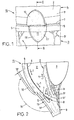
- Fig. 1 shows a larger scale and in a schematic View shown section of a whole with 15 designated eye and you can recognize a section the iris 2 (iris), the cornea 4 (cornea), the dermis 3 (sclera), a section of the circular feast Channel 5 (sinus venosus sclerae) and that from a variety channel system 3 'formed by channels for the to be derived Aqueous humor.
- a first one can be seen in FIG parabolic incision in the Sclera 3, a trained analogous to the incision and in First scleral flap 10 swung open in the direction of the cornea 4 as well as a first recess 11 designed analogously to the same with circumferential side wall 11 '.
- the first scleral flap 10 is by means not shown in the pivoted-up position held.
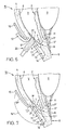
- FIG. 2 shows the section of the eye 15 according to that in FIG drawn line II-II on average and in larger Scale shown and you can see a section of the sclera 3, a section of the cornea 4 with the Descemet membrane 6 (Descemet's membrane) and the Schwalbeschen line 7 (Schwalbe's line), a section of the iris 2 and a Part of the lens 9, which by means of the zonular fibers 9 ' connected to the sclera 3. Furthermore one recognizes the according First scleral flap 10 swung open in the direction of arrow 16 with the analog recess 11 and the Schlemm's canal 5 with the upstream trabecular tissue 8 (trabecular meshwork).
- the circulation is also indicated by several arrows 1 of the aqueous humor (humor aquosus) and with the arrows 1 'shows the natural outflow via the trabecular tissue 8.
- this circulates continuously Refreshing aqueous humor according to arrow direction 1 from the back chamber H to the anterior chamber V and is in the chamber angle V ' (angulus irido-cornealis) according to arrows 1 'essentially through the trabecular tissue 8 into Schlemm's canal 5 and from there via the natural channel system 3 '(Fig.1) in derived a venous system, not shown.
- Constipation or the like only partially functional or inoperable trabecular tissue 8 the natural outflow of aqueous humor can be limited in this way that the pressure inside the eye 15 increases and consequently the blood circulation and thus the function of the Optic nerve (not shown) is restricted.
- This Disease is commonly known as "glaucoma" which can lead to blindness of the affected eye.
- the one not shown The conjunctiva of the eye is withdrawn using suitable means and therefore a sufficient section of the sclera 3 for the first approximately parabolic incision (incision) exposed.
- the first incision After the first incision, it becomes the incision appropriately trained first scleral flaps 10 in Direction of the cornea 4 folded and consequently the first Recess 11 with the circumferential side wall 11 'exposed.
- the depth of the first cut for example 3mm x 3mm is preferably chosen so that the thickness 10 'of the the first scleral flap shown in profile cross-section in FIG 10 about 1/3 of the natural thickness in this area Scleral 3 corresponds. Schlemm's channel 5 is in this first phase (Fig.2) still covered by the sclera 3.
- FIG. 3 shows the section of the eye 15 according to FIG. 1 with a arranged within the first parabolic incision second parabolic incision.
- the incision becomes a parabolic one second scleral flap 12 opened in the direction of the cornea 4 and thereby an analog of the second scleral flap 12 trained second recess 13 exposed.
- the depth of the second incision in sclera 3 is selected, for example, that with the second scleral flap 12 swung open Schlemm's channel 5 in the area of an exposed and in the Entire with 18 designated section is accessible.
- this phase are the two within the exposed section 18 arranged opposite each other at a distance Entrances 17 and 17 'of Schlemm's canal 5 for insertion an appropriately trained probe (Fig.5) accessible.
- Figure 3 is still in the form of one over the entire Width of the second scleral flap 12 extends approximately groove-shaped Indentation 5 'of Schlemm's canal 5 is shown.
- the portion of the eye 15 is according to that in Figure 3 drawn line IV-IV with the two according to the direction of the arrow 16 (Fig.3) and arrow direction 16 '(Fig.4) swung open Scleral lobes 10 and 12 shown in section.
- the two Scleral lobes 10, 12 are not for further surgery represented means held in this position.
- the Depth of cut and the thickness 12 'of the thickness shown in FIG second scleral flap 12 shown in profile cross section is selected, for example, so that the two inputs 17, 17 '(FIG. 3) is easily accessible via the exposed section 18 are.
- a suitable Medium preferably a highly viscous sodium hyaluronate solution (high viscosity sodium hyaluronate) injected.
- a suitable Medium preferably a highly viscous sodium hyaluronate solution (high viscosity sodium hyaluronate) injected.
- the lumen 19 of Schlemm's canal 5 is at least expanded over the axial length of an inserted probe 24. 5 shows the section of the eye 15 according to FIG.
- sclera 3 in shown on a larger scale and you can partially recognize them shown sclera 3 with the two in the direction of the cornea 4 scleral flaps swung open or up 10 and 12, the second recess 13 and the lateral contact surface 14 on the sclera 3 with the canal system 3 '.
- the injection device 25 For injecting the highly viscous sodium hyaluronate solution 5 shows the injection device 25 schematically, which in the illustrated embodiment with the probe arranged on an arcuate connector 29 24 introduced into the entrance 17 of Schlemm's canal 5 is. After expanding the lumen 19 on one side the injection device 25 is pulled out with the probe 24 and rotated accordingly in a manner not shown in the other, opposite entrance 17 'of the Schlemm's channel 5 for injecting the medium or introduced to expand the lumen 19.
- the injection device shown as an exemplary embodiment in FIG 25 stands over a feed line connected to it 28 with a schematically represented pressure source 26 in the form of a single-chamber syringe or the like.
- Pressure source 26 is the medium to be injected according to Arrow direction 27 through the elements 28, 29 and 24 in the Lumen 19 of Schlemm's canal 5 pressed and this accordingly stretched. After widening the Schlemm canal 5, the injection device 25 is shown in FIG Way removed again.
- the injection device 25 is not Subject of the present invention and is therefore not described in detail described.
- a small force in the area of the Schwalbeschen line 7 second scleral flap 12 from the facing inside of the Descemet membrane 6 can be solved.
- the approximately gap-shaped Opening 21 extends in a manner not shown over the entire width of the exposed section 18 and the second recess 13 (Fig.8). With the in Fig. 7 shown gap-shaped opening 21 (window) an additional connection between the front chamber V of the Eye 15 and the second recess 13 made.
- the aqueous humor is thus complementary to the natural drain over the trabecular tissue 8 according to arrow direction 1 'additionally still through the largely transparent and partially Chamber-permeable Descemet membrane 6 according to the direction of the arrow 1 "(Fig.7) and the gap-shaped opening 21 in the recess 13 derived.
- the roughly analogous to the second scleral lobe 12 formed flat recess 13 forms when folded down Scleral flap 10 is essentially a collecting basin (Fig.12) for the aqueous humor.
- the aqueous humor is from the recess 13 designed as a collecting basin (reservoir) through the two side entrances 17, 17 'in the Schlemmschen Channel 5 and from there via the channel system 3 '(Fig.1 and Fig.3) derived.
- the second scleral flap 12 is preferably, as in Fig.7 shown schematically, except for a remaining section 12.1 using a suitable surgical cutting instrument severed. However, there is also the possibility that the second scleral flap 12 is separated first and then by pressing against the Schwalbesche line 7 Swabs 20 the remaining section 12.1 of the facing inside of the Descemet membrane 6 to form the gap-shaped opening 21 is released.
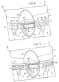
- FIG. 8 and 9 each show the section of the eye 15 severed scleral flap 12 and you can see the exposed Section 18 of Schlemm's canal 5 and the two opposite inputs 17 and 17 'arranged to each other.
- In the lumen 19 of Schlemm's canal 5 are on both sides of the exposed section 18 two at a distance mutually arranged support elements implanted.
- With one Embodiment according to Figure 8 are on one side two approximately ring-shaped support elements 30 and on the opposite other side two spherical support elements 33 implanted.
- Fig. 9 there is a mesh on one side trained support member 45 and on the opposite on the other side, a support element designed as a helical spring 50 implanted.
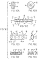
- the first support element 30 is in a spatial view as well as shown on a larger scale.
- the one Bore 32 penetrating support member 30 preferably an approximately circular or elliptical outer shape.
- the support element, for example in the form of a ring 30 is preferably from a self to the Lumen 19 of Schlemm's channel 5 matching material.
- the width 31 of the circular or elliptical Support element 30 is selected, for example, so that the same positionally implantable in the lumen 19 and an in axial direction of the lumen 19 oriented tilting of the Support element 30 is excluded.
- the 10B is the second spherical support element 33 in spatial view and on a larger scale shown.
- the spherical support element 33 is of at least one, but preferably several in the circumferential direction bores 34 distributed to each other.
- FIG. 10C shows the third support element 35 in a view, which for example consists of a flexible tube is produced and in the profile cross-section (not shown) either an annular or elliptical shape having.
- the support member 35 is in the axial direction penetrated by a bore 36.
- the bore 36 is with a number spaced apart in the axial direction Inlet openings 37 in connection.
- the support element 35 is with respect to the theoretical longitudinal axis X. its location and orientation as shown schematically in Fig. 10C shown designed to move freely.
- the support member 35 can result from insertion into Schlemm's canal 5 the flexibility automatically matches the internal shape of the lumen 19 adjust.
- the flexibility of the tubular support element 35 is so limited, however, that an automatic Kinking is excluded.
- Fig. 10D shows this in view and on a larger scale fourth support element 40, which is also an elongated, flexible tube 41 is formed.
- the support element 40 is from a bore oriented in the axial direction 41 'penetrated, the bore 41' with a number Openings spaced from each other in the axial direction 42,42 'communicates.
- On the support element 40 are still several in the axial direction at a distance from each other arranged support parts 43, 43 'and 43 "are arranged, preferably molded.
- Orient with the in the axial direction Bore 41 'communicating openings 42,42' are preferably diametrical to each other on the longer side (not designated) of the elliptical in cross section Support element 40 arranged.
- the support element 40 is in particular for implantation in the area of the second recess 13 of the sclera 3 exposed section 18 of Schlemmschen Channel 5 formed. That along the drawn in Fig.10D Line E-E shown in section according to Fig.10E Support element 40 has an approximately the profile cross section of the Schlemm's channel 5 correspondingly trained elliptical outer shape. The function of the fourth support element 40 will be explained in more detail below in connection with Fig.11 and Fig.12 described.
- the mesh network is a further variant Support element 45 shown in view.
- a plurality of threads 46 (filaments) helical winding mesh are corresponding between the threads 46 Distances 47,47 'and 47 "provided by which the Aqueous humor can be derived.
- the support member 45 is preferably analogous to the support element shown in FIG. 10C 35 in relation to its outer shape on the lumen 19 of the Schlemm's canal 5 self-adaptively deformable.
- Fig. 10G shows this in view and on a larger scale
- Support element 50 which for example consists of a single, helically wound thread 51 (filament) is made. With the support element 50, the aqueous humor through the provided between the individual turns Distances 52 and 52 'derived.
- the support member 50 is preferably analogous to the support element shown in FIG. 10C 35 in relation to its outer shape on the lumen 19 of the Schlemm's canal 5 self-adaptively deformable.
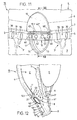
- FIG. 11 shows the section of the eye 15 with the two Sides of the second recess 13 in the lumen 19 of Schlemmschen Channel 5 spaced from each other and annular trained support elements 30. Furthermore, one can see that in the area of the second recess 13 of the sclera 3 in the exposed Schlemm's channel 5 inserted support element 40.
- the support member 40 is such in the exposed section 18 of Schlemm's canal 5 arranged that the over the entire width of the second recess 13 from the second Scleral flaps 12 loosened section 12.1 on the support parts 43,43 ', 43 "of the support element 40.
- the first scleral flap 10 After removal of the second scleral flap 12 and implantation of the individual support elements becomes the first scleral flap 10 folded down and schematically as in Fig.12 shown on the parabolic circumferential support surface 14 placed.
- the first scleral flap 10 is then in partially known, not shown in more detail the sclera 3 is sewn.
- the severed second Scleral flap 12 created behind the first scleral flap 10 sub-scleral space 13 'in the form of the flat recess 13 is preferably with before sewing completely high viscosity medium (high viscosity sodium hyaluronate) filled. This prevents a connecting contact the inside 10 "of the first scleral flap folded down 10 with the inner surface 13 "of the recess 13.
- Figure 12 is the portion of the eye 15 with the implanted Support element 40 according to the line drawn in Figure 11 XII-XII on average as well as on a larger scale and with folded scleral flap 10 shown. Still recognizes 12 that is released from the Descemet membrane 6 and on the support parts 43, 43 ', 43 "of the support element 40 lying section 12.1 of the second scleral flap 12. By means of of the support parts 43, 43 ', 43 "is a closing of the gap-shaped Opening 21 over the entire width of the exposed Part 18 of Schlemm's canal 5 prevented.
- the slit-shaped opening 21 an additional connection between the chamber angle V ' the front chamber V and the second recess 13.
- the aqueous humor can thus be in addition to the natural drain according to arrow direction 1 'over the trabecular tissue 8 also according to arrow direction 1 "due to the largely transparent and partially permeable Descemet membrane 6 and through the gap-shaped opening 21 (window) derived into the recess 13 become.
- the roughly analogous to the second scleral lobe 12 trained flat recess 13 forms when folded down Scleral flap 10 a sub-scleral space 13 'or a Collecting basin (reservoir) for the aqueous humor.
- the aqueous humor is from the sub-scleral space 13 'through the two connected lateral entrances 17, 17 'into the lumen 19 of Schlemm's canal 5 and from there into the canal system 3 'derived.
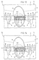
- FIG. 13 shows the portion of the eye 15 in the lumen 19 of Schlemm's canal 5 implanted at a distance from one another, ring-shaped support elements 30 (Fig.10A).
- the lumen 19 can, however also several spaced and spherical trained support elements 33 (Fig.10B) are implanted.
- Fig.10F mesh trained support element 45
- Fig. 14 shows the section of the eye 15 with Schlemm's canal 5, in which on one side in the lumen 19 for example two annular and spaced apart Support elements 30 are implanted according to Fig. 10A. On the other opposite side are in the lumen 19th two spaced-apart and spherical Support elements 33 implanted according to Fig. 10B. At this embodiment is in the exposed section 18 of Schlemm's channel 5, which is designed as a coil spring Support element 50 implanted according to Fig.10G.
- each implantable in the lumen 19 of Schlemm's canal 5 Support elements 30, 33, 35, 45 or 50 made of degradable, in particular from one of the tissues of Schlemm's canal 5 and / or material that is biodegradable from the aqueous humor are manufactured.
- a material grouping is preferably used as the material chosen, which is approximately within 2 to 12 Is biodegradable months after implantation.
- the individual support element 30, 33, 35, 45 or 50 can be a material such as a cross-linked sodium hyaluronate (cross linked sodium hyaluronate) is used become.
- biocompatible material for example made of plastic, stainless steel or stainless steel such as silver, gold or Platinum is made.
Landscapes
- Health & Medical Sciences (AREA)
- Ophthalmology & Optometry (AREA)
- Heart & Thoracic Surgery (AREA)
- Surgery (AREA)
- Engineering & Computer Science (AREA)
- Biomedical Technology (AREA)
- Nuclear Medicine, Radiotherapy & Molecular Imaging (AREA)
- Vascular Medicine (AREA)
- Life Sciences & Earth Sciences (AREA)
- Animal Behavior & Ethology (AREA)
- General Health & Medical Sciences (AREA)
- Public Health (AREA)
- Veterinary Medicine (AREA)
- Prostheses (AREA)
- External Artificial Organs (AREA)
Abstract
Description
- Fig.1
- ein in schematischer Ansicht und in grösserem Massstab dargestelltes Teilstück eines Auges mit einem ersten parabolischen Einschnitt in der Sklera und aufgeklapptem ersten Skleralappen;
- Fig.2
- das entlang der Linie II-II im Schnitt dargestellte Teilstück des Auges gemäss Fig.1 mit aufgeklapptem ersten Skleralappen;
- Fig.3
- das in Ansicht dargestellte Teilstück des Auges gemäss Fig.1 mit einem innerhalb des ersten parabolischen Einschnitts angeordneten zweiten parabolischen Einschnitt sowie aufgeklapptem zweiten Skleralappen;
- Fig.4
- das entlang der Linie IV-IV im Schnitt dargestellte Teilstück des Auges gemäss Fig.3 mit den beiden aufgeklappten Skleralappen und freigelegtem Schlemmschen Kanal;
- Fig.5
- eine in das freigelegte Teilstück des Schlemmschen Kanals eingeführte Vorrichtung zum Injizieren eines das Lumen des Schlemmschen Kanals dehnenden Mediums;
- Fig.6
- das Teilstück des Auges gemäss Fig.4 mit einem im Bereich der beiden aufgeklappten Skleralappen mit geringer Kraft gegen die Schwalbesche Linie wirkenden Tupfer;
- Fig.7
- das Teilstück des Auges gemäss Fig.6 bei welchem die Descemet-Membran von der Kornea gelöst und der zweite Skleralappen abgetrennt ist;
- Fig.8
- das Teilstück des Auges gemäss Fig.4 mit in das Lumen des Schlemmschen Kanals implantierten, beispielsweise ringförmig oder kugelförmig ausgebildeten Stützelementen;
- Fig.9
- das Teilstück des Auges gemäss Fig.4 mit in das Lumen des Schlemmschen Kanals implantierten, beispielsweise in Form eines Netzgeflecht oder einer Schraubenfeder ausgebildeten Stützelementen;
- Fig.10
- einzelne Ausführungsbeispiele der zu implantierenden Stützelemente;
- Fig.10A
- das in räumlicher Ansicht sowie in grösserem Massstab dargestellte und ringförmig ausgebildete Stützelement;
- Fig.10B
- das in räumlicher Ansicht sowie in grösserem Massstab dargestellte und kugelförmig ausgebildete Stützelement;
- Fig.10C
- ein in Ansicht sowie in grösserem Massstab dargestelltes und röhrchenförmig ausgebildetes Stützelement;
- Fig.10D
- ein in Ansicht sowie in grösserem Massstab dargestelltes und röhrchenförmig ausgebildetes Stützelement für das freigelegte Teilstück des Schlemmschen Kanals;
- Fig.10E
- das gemäss der Linie E-E im Schnitt dargestellte Stützelement gemäss Fig.10D;
- Fig.10F
- ein in Ansicht sowie in grösserem Massstab dargestelltes Stützelement in Form eines Netzgeflechts;
- Fig.10G
- ein in Ansicht sowie in grösserem Massstab dargestelltes Stützelement in Form einer Schraubenfeder;
- Fig.11
- das Teilstück des Auges gemäss Fig.8 mit implantierten Stützelementen gemäss Fig.10A sowie mit dem im freigelegten Teilstück des Schlemmschen Kanals implantierten Stützelement gemäss Fig.10D;
- Fig.12
- das gemäss der Linie XII-XII in Fig.11 im Schnitt dargestellte Teilstück des Auges mit dem gemäss Fig.10E im Schnitt dargestellten Stützelement und heruntergeklapptem ersten Skleralappen;
- Fig.13
- das Teilstück des Auges gemäss Fig.8 mit den implantierten Stützelementen gemäss Fig.10A sowie dem im freigelegten Teilstück des Schlemmschen Kanals implantierten Stützelement gemäss Fig.10F; und
- Fig.14
- das Teilstück des Auges gemäss Fig.8 mit den implantierten Stützelementen gemäss Fig.lOA und Fig.10B sowie dem im freigelegten Teilstück des Schlemmschen Kanals implantierten Stützelement gemäss Fig.10G.
Claims (21)
- Verfahren zum Verbessern des Kammerwasserabflusses in einem Auge (15), bei welchem das abgesonderte Kammerwasser im Bereich des Kammerwinkels (V') der Vorderkammer (V) über das Trabekulargewebe (8) in den Schlemmschen Kanal (5) und anschliessend über das natürliche Kanalsystem abgeleitet wird, gekennzeichnet durch einen ersten lamellaren Einschnitt an der Oberfläche der Sklera (3) mit in Richtung der Kornea (4) aufgeschwenktem ersten Skleralappen (10) sowie analog ausgebildeter erster Ausnehmung (11), einen innerhalb der ersten Ausnehmung (11) vorgesehenen zweiten Einschnitt mit aufgeschwenktem zweiten Skleralappen (12) und analog ausgebildeter zweiter Ausnehmung (13) sowie ein in diesem Bereich freigelegtes Teilstück (18) des Schlemmschen Kanals (5), wobei zu beiden Seiten der zweiten Ausnehmung (13) in das Lumen (19) sowie wahlweise in das freigelegte Teilstück (18) jeweils mindestens ein von dem Gewebe des Schlemmschen Kanals 5 und/oder vom Kammerwasser abbaubares Stützelement implantiert und nach dem Abtrennen des zweiten Skleralappens (12) der erste Skleralappen (10) auf eine analog dem zweiten Einschnitt ausgebildete Auflagefläche (14) gelegt und der dadurch gebildete subsklerale Raum (13') vor dem vollständigen Verschliessen mit viskosem Medium gefüllt wird.
- Verfahren nach Anspruch 1, dadurch gekennzeichnet, dass das Lumen (19) zu beiden Seiten des freigelegten Teilstücks (18) von mindestens zwei in axialer Richtung des Schlemmschen Kanals (5) im Abstand zueinander angeordneten und etwa ringförmig ausgebildeten Stützelementen (30) aus biolytisch abbaubarem Material in gedehnter Stellung gehalten wird.
- Verfahren nach Anspruch 1, dadurch gekennzeichnet, dass das Lumen (19) zu beiden Seiten des freigelegten Teilstücks (18) von mindestens zwei in axialer Richtung des Schlemmschen Kanals (5) im Abstand zueinander angeordneten und etwa kugelförmig ausgebildeten Stützelementen (33) aus biolytisch abbaubarem Material in gedehnter Stellung gehalten wird.
- Verfahren nach Anspruch 1, dadurch gekennzeichnet, dass das Lumen (19) des Schlemmschen Kanals (5) zu beiden Seiten des freigelegten Teilstücks (18) jeweils von mindestens einem länglichen, röhrchenförmig ausgebildeten Stützelement (35) aus biolytisch abbaubarem Material in gedehnter Stellung gehalten wird.
- Verfahren nach Anspruch 1, dadurch gekennzeichnet, dass das Lumen (19) des Schlemmschen Kanals (5) zu beiden Seiten des freigelegten Teilstücks (18) jeweils von mindestens einem länglichen, als Netzgeflecht ausgebildeten Stützelement (45) aus biolytisch abbaubarem Material in gedehnter Stellung gehalten wird.
- Verfahren nach Anspruch 1, dadurch gekennzeichnet, dass das Lumen (19) des Schlemmschen Kanals (5) zu beiden Seiten des freigelegten Teilstücks (18) jeweils von mindestens einem länglichen, als Schraubenfeder ausgebildeten Stützelement (50) aus biolytisch abbaubarem Material in gedehnter Stellung gehalten wird.
- Verfahren nach einem der Ansprüche 1 bis 6, dadurch gekennzeichnet, dass das Lumen (19) zu beiden Seiten des freigelegten Teilstücks (18) jeweils mittels ring- und/oder kugelförmiger sowie im Abstand zueinander angeordneter Stützelemente (30;33) und das freigelegte Teilstück (18) des Schlemmschen Kanals (5) durch ein längliches Stützelement (35;45;50) jeweils aus biolytisch abbaubarem Material in gedehnter Stellung gehalten wird.
- Verfahren nach Anspruch 1, dadurch gekennzeichnet, dass zur Bildung einer spaltförmigen Öffnung (21) der zweite Skleralappen (12) oder ein Teilstück (12.1) desselben im Bereich der Schwalbeschen Linie (7) von der teilweise kammerwasserdurchlässigen Descemet-Membran (6) gelöst wird, wobei das abgelöste Teilstück (12.1) mittels mehrerer Auflageteile (43, 43',43") eines in dem freigelegten Teilstück (18) des Schlemmschen Kanals (5) angeordneten Stützelements (40) in Offenstellung gehalten wird.
- Vorrichtung zum Verbessern und zur Aufrechterhaltung des Kammerwasserabflusses in einem Auge (15), bei welchem im Lumen (19) des Schlemmschen Kanals (5) mindestens ein Stützelement angeordnet ist, gekennzeichnet durch mindestens ein ringförmig, kugelförmig oder röhrchenförmig ausgebildetes und im Lumen (19) des Schlemmschen Kanals (5) angeordnetes Stützelement (30;33;35;45;50), welches aus abbaubarem Material, insbesondere aus einem von dem Gewebe des Schlemmschen Kanals (5) und/oder von dem Kammerwasser abbaubarem Material hergestellt ist.
- Vorrichtung nach Anspruch 9, dadurch gekennzeichnet, dass das mit einer Durchgangsbohrung (31) versehene Stützelement (30) im Profilquerschnitt kreisring- oder ellipsenringförmig ausgebildet ist und aus einem in Bezug auf den Profilquerschnitt des Schlemmschen Kanals (5) verformbaren und biolytisch abbaubarem Material hergestellt ist.
- Vorrichtung nach Anspruch 10, dadurch gekennzeichnet, dass zur Vermeidung einer Kippbewegung des implantierten Stützelements (30) dessen Breite (31) grösser als die Höhe des im Profilquerschnitt etwa ellipsenförmigen Schlemmschen Kanals (5) ausgebildet ist.
- Vorrichtung nach Anspruch 9, dadurch gekennzeichnet, dass das Stützelement (33) als Kugel ausgebildet ist und mindestens eine Durchgangsbohrung (34), vorzugsweise eine Vielzahl in Umfangsrichtung verteilt zueinander angeordnete Durchgangsbohrungen (34) aufweist und aus biolytisch abbaubarem Material hergestellt ist.
- Vorrichtung nach Anspruch 9, dadurch gekennzeichnet, dass das mit einer Durchgangsbohrung (36) versehene Stützelement (35) als ein im Profilquerschnitt kreisring- oder ellipsenringförmiges und mit einer Anzahl im Abstand zueinander angeordneter Öffnungen (37) versehenes Röhrchen ausgebildet ist, welches in Bezug auf die theoretische Längsachse (X) hinsichtlich der Lage und Orientierung flexibel sowie frei beweglich ausgebildet und aus biolytisch abbaubarem Material hergestellt ist.
- Vorrichtung nach Anspruch 9, dadurch gekennzeichnet, dass das mit einer Durchgangsbohrung (41') versehene Stützelement (40) als ellipsenförmiges Röhrchen ausgebildet ist und an der Breitseite eine Anzahl in axialer Richtung im Abstand zueinander angeordnete Öffnungen (42,42') und mindestens zwei in axialer Richtung im Abstand zueinander angeordnete sowie an der Schmalseite radial nach aussen abstehend angeformte Auflageteilten (43,43',43") aufweist.
- Vorrichtung nach Anspruch 14, dadurch gekennzeichnet, dass das mit den angeformten Auflageteilten (43,43',43") versehene Stützelement (40) aus biolytisch abbaubarem Material hergestellt ist.
- Vorrichtung nach Anspruch 9, dadurch gekennzeichnet, dass das längliche Stützelement (45) als flexibles sowie hinsichtlich der Lage und Orientierung frei bewegliches Netzgeflecht ausgebildet ist.
- Vorrichtung nach Anspruch 16, dadurch gekennzeichnet, dass das längliche Stützelement (45) aus biolytisch abbaubarem Material hergestellt ist.
- Vorrichtung nach Anspruch 9, dadurch gekennzeichnet, dass das längliche Stützelement (50) in Form einer flexiblen Schraubenfeder ausgebildet sowie hinsichtliche der Lage und Orientierung frei beweglich ist.
- Vorrichtung nach Anspruch 18, dadurch gekennzeichnet, dass das längliche Stützelement (50) aus biolytisch abbaubarem Material hergestellt ist.
- Vorrichtung nach Anspruch 9, dadurch gekennzeichnet, dass das Stützelement (30;33;35;45;50) aus einem innerhalb eine Zeitdauer von 2 bis 12 Monaten nach der Implantation von dem Gewebe des Schlemmschen Kanals (5) und/oder von dem Kammerwasser biolytisch abbaubarem Material besteht.
- Vorrichtung nach Anspruch 20, dadurch gekennzeichnet, dass das einzelne Stützelement (30;33;35; 45;50) aus vernetztem Natriumhyaluronat hergestellt ist.
Applications Claiming Priority (2)
| Application Number | Priority Date | Filing Date | Title |
|---|---|---|---|
| US503884 | 1995-07-20 | ||
| US09/503,884 US6375642B1 (en) | 2000-02-15 | 2000-02-15 | Method of and device for improving a drainage of aqueous humor within the eye |
Publications (2)
| Publication Number | Publication Date |
|---|---|
| EP1125568A2 true EP1125568A2 (de) | 2001-08-22 |
| EP1125568A3 EP1125568A3 (de) | 2003-09-03 |
Family
ID=24003914
Family Applications (1)
| Application Number | Title | Priority Date | Filing Date |
|---|---|---|---|
| EP01810106A Withdrawn EP1125568A3 (de) | 2000-02-15 | 2001-02-02 | Verfahren und Vorrichtung zum Verbesseren sowie zur Aufrechterhaltung des Kammerwasserablusses in einem Auge |
Country Status (5)
| Country | Link |
|---|---|
| US (1) | US6375642B1 (de) |
| EP (1) | EP1125568A3 (de) |
| JP (1) | JP2001245916A (de) |
| AU (1) | AU1002701A (de) |
| CA (1) | CA2331868A1 (de) |
Cited By (5)
| Publication number | Priority date | Publication date | Assignee | Title |
|---|---|---|---|---|
| US8845572B2 (en) | 2009-11-13 | 2014-09-30 | Grieshaber Ophthalmic Research Foundation | Method and device for the treatment of glaucoma |
| JP2014193366A (ja) * | 2003-08-05 | 2014-10-09 | Glaukos Corp | 緑内障治療装置 |
| US8951221B2 (en) | 2009-08-20 | 2015-02-10 | Grieshaber Ophthalmic Research Foundation | Method and device for the treatment of glaucoma |
| EP2837366A4 (de) * | 2012-04-12 | 2016-03-09 | Zakharov Ivan Dmitrievich | Drainagevorrichtung und verfahren zur herstellung davon |
| WO2021204312A2 (de) | 2020-04-09 | 2021-10-14 | aixtent GmbH | Verfahren zur herstellung eines implantats zum einführen in ein auge insbesondere zum einführen in den schlemmschen kanal eines auges |
Families Citing this family (108)
| Publication number | Priority date | Publication date | Assignee | Title |
|---|---|---|---|---|
| US20050119601A9 (en) * | 1999-04-26 | 2005-06-02 | Lynch Mary G. | Shunt device and method for treating glaucoma |
| KR20020035476A (ko) * | 1999-04-26 | 2002-05-11 | 지엠피 비젼 솔루션즈 인코포레이티드 | 측로 장치 및 녹내장 치료 방법 |
| US20040111050A1 (en) * | 2000-04-14 | 2004-06-10 | Gregory Smedley | Implantable ocular pump to reduce intraocular pressure |
| US20050277864A1 (en) * | 2000-04-14 | 2005-12-15 | David Haffner | Injectable gel implant for glaucoma treatment |
| US20020143284A1 (en) * | 2001-04-03 | 2002-10-03 | Hosheng Tu | Drug-releasing trabecular implant for glaucoma treatment |
| US7708711B2 (en) * | 2000-04-14 | 2010-05-04 | Glaukos Corporation | Ocular implant with therapeutic agents and methods thereof |
| US20050049578A1 (en) * | 2000-04-14 | 2005-03-03 | Hosheng Tu | Implantable ocular pump to reduce intraocular pressure |
| US20030060752A1 (en) * | 2000-04-14 | 2003-03-27 | Olav Bergheim | Glaucoma device and methods thereof |
| US6533768B1 (en) * | 2000-04-14 | 2003-03-18 | The Regents Of The University Of California | Device for glaucoma treatment and methods thereof |
| US6638239B1 (en) | 2000-04-14 | 2003-10-28 | Glaukos Corporation | Apparatus and method for treating glaucoma |
| US9603741B2 (en) | 2000-05-19 | 2017-03-28 | Michael S. Berlin | Delivery system and method of use for the eye |
| FR2813521B1 (fr) * | 2000-09-01 | 2003-06-13 | Ioltechnologie Production | Drain a glaucome |
| US6666841B2 (en) * | 2001-05-02 | 2003-12-23 | Glaukos Corporation | Bifurcatable trabecular shunt for glaucoma treatment |
| AU2002258754B2 (en) | 2001-04-07 | 2006-08-17 | Glaukos Corporation | Glaucoma stent and methods thereof for glaucoma treatment |
| US6981958B1 (en) | 2001-05-02 | 2006-01-03 | Glaukos Corporation | Implant with pressure sensor for glaucoma treatment |
| US7488303B1 (en) | 2002-09-21 | 2009-02-10 | Glaukos Corporation | Ocular implant with anchor and multiple openings |
| US7431710B2 (en) | 2002-04-08 | 2008-10-07 | Glaukos Corporation | Ocular implants with anchors and methods thereof |
| US7678065B2 (en) * | 2001-05-02 | 2010-03-16 | Glaukos Corporation | Implant with intraocular pressure sensor for glaucoma treatment |
| WO2002089699A2 (en) | 2001-05-03 | 2002-11-14 | Glaukos Corporation | Medical device and methods of use for glaucoma treatment |
| US7331984B2 (en) | 2001-08-28 | 2008-02-19 | Glaukos Corporation | Glaucoma stent for treating glaucoma and methods of use |
| US20030097151A1 (en) * | 2001-10-25 | 2003-05-22 | Smedley Gregory T. | Apparatus and mitochondrial treatment for glaucoma |
| US7163543B2 (en) * | 2001-11-08 | 2007-01-16 | Glaukos Corporation | Combined treatment for cataract and glaucoma treatment |
| US20030153863A1 (en) * | 2002-02-13 | 2003-08-14 | Patel Anilbhai S. | Implant system for glaucoma surgery |
| US6939298B2 (en) | 2002-02-28 | 2005-09-06 | Gmp Vision Solutions, Inc | Device and method for monitoring aqueous flow within the eye |
| US7186232B1 (en) | 2002-03-07 | 2007-03-06 | Glaukoa Corporation | Fluid infusion methods for glaucoma treatment |
| US7951155B2 (en) | 2002-03-15 | 2011-05-31 | Glaukos Corporation | Combined treatment for cataract and glaucoma treatment |
| US20040147870A1 (en) * | 2002-04-08 | 2004-07-29 | Burns Thomas W. | Glaucoma treatment kit |
| US9301875B2 (en) * | 2002-04-08 | 2016-04-05 | Glaukos Corporation | Ocular disorder treatment implants with multiple opening |
| US20040024345A1 (en) * | 2002-04-19 | 2004-02-05 | Morteza Gharib | Glaucoma implant with valveless flow bias |
| US20040193095A1 (en) * | 2003-03-29 | 2004-09-30 | Shadduck John H. | Implants for treating ocular hypertension, methods of use and methods of fabrication |
| ATE439107T1 (de) | 2003-04-16 | 2009-08-15 | Iscience Interventional Corp | Mikrochirurgische instrumente für die ophthalmologie |
| US20040225250A1 (en) | 2003-05-05 | 2004-11-11 | Michael Yablonski | Internal shunt and method for treating glaucoma |
| US7291125B2 (en) * | 2003-11-14 | 2007-11-06 | Transcend Medical, Inc. | Ocular pressure regulation |
| US20050250788A1 (en) * | 2004-01-30 | 2005-11-10 | Hosheng Tu | Aqueous outflow enhancement with vasodilated aqueous cavity |
| SE0401182D0 (sv) * | 2004-05-05 | 2004-05-05 | Q Med Ab | Novel use of a viscoelastic composition |
| BRPI0519087B8 (pt) * | 2004-12-16 | 2021-06-22 | Iscience Interventional Corp | instrumento para inserção de um implante no canal de schlemm do olho |
| AR054647A1 (es) | 2005-02-21 | 2007-07-11 | Maldonado Bas Arturo | Dispositivo para drenaje de humor acuoso en casos de glaucoma |
| EP1979023B1 (de) | 2006-01-17 | 2015-07-22 | Transcend Medical, Inc. | Glaukombehandlungsvorrichtung |
| EP1978892B1 (de) * | 2006-01-17 | 2017-11-15 | Novartis Ag | Behandlungsvorrichtung zur wirkstofffreisetzung |
| US20070293807A1 (en) * | 2006-05-01 | 2007-12-20 | Lynch Mary G | Dual drainage pathway shunt device and method for treating glaucoma |
| US7909789B2 (en) * | 2006-06-26 | 2011-03-22 | Sight Sciences, Inc. | Intraocular implants and methods and kits therefor |
| AU2007319383A1 (en) | 2006-11-10 | 2008-05-22 | Glaukos Corporation | Uveoscleral shunt and methods for implanting same |
| JP4957258B2 (ja) * | 2007-01-15 | 2012-06-20 | 富士通株式会社 | 歩数計数装置および歩数計数方法 |
| EP2173289A4 (de) * | 2007-07-17 | 2010-11-24 | Transcend Medical Inc | Augenimplantat mit hydrogelausdehnungsfähigkeiten |
| US8968396B2 (en) | 2007-07-23 | 2015-03-03 | Powervision, Inc. | Intraocular lens delivery systems and methods of use |
| US20090082862A1 (en) | 2007-09-24 | 2009-03-26 | Schieber Andrew T | Ocular Implant Architectures |
| US7740604B2 (en) * | 2007-09-24 | 2010-06-22 | Ivantis, Inc. | Ocular implants for placement in schlemm's canal |
| US20170360609A9 (en) | 2007-09-24 | 2017-12-21 | Ivantis, Inc. | Methods and devices for increasing aqueous humor outflow |
| US8734377B2 (en) | 2007-09-24 | 2014-05-27 | Ivantis, Inc. | Ocular implants with asymmetric flexibility |
| US8808222B2 (en) | 2007-11-20 | 2014-08-19 | Ivantis, Inc. | Methods and apparatus for delivering ocular implants into the eye |
| US8512404B2 (en) * | 2007-11-20 | 2013-08-20 | Ivantis, Inc. | Ocular implant delivery system and method |
| US8337509B2 (en) * | 2007-11-20 | 2012-12-25 | Ivantis, Inc. | Methods and apparatus for delivering ocular implants into the eye |
| EP2259833A1 (de) | 2008-03-05 | 2010-12-15 | Ivantis, INC. | Verfahren und vorrichtung zur behandlung von glaukom |
| CH700161A2 (de) * | 2008-12-22 | 2010-06-30 | Grieshaber Ophthalmic Res Foun | Implantat zum einführen in den schlemmschen kanal eines auges. |
| EP2548538B1 (de) | 2009-01-28 | 2020-04-01 | Alcon Inc. | Implantationssysteme für augenimplantate mit steifheitseigenschaften |
| AU2010256558B2 (en) | 2009-06-03 | 2013-10-03 | Forsight Labs, Llc | Anterior segment drug delivery |
| JP5662436B2 (ja) * | 2009-06-25 | 2015-01-28 | オプトノール リミティド | 軟組織において空間を維持する繊維マトリックス |
| WO2011006078A1 (en) | 2009-07-09 | 2011-01-13 | Ivantis, Inc. | Single operator device for delivering an ocular implant |
| AU2010271218B2 (en) | 2009-07-09 | 2017-02-02 | Alcon Inc. | Ocular implants and methods for delivering ocular implants into the eye |
| JP2013508096A (ja) | 2009-10-23 | 2013-03-07 | イバンティス インコーポレイテッド | 眼内移植システムおよび眼内移植方法 |
| US20110105990A1 (en) * | 2009-11-04 | 2011-05-05 | Silvestrini Thomas A | Zonal drug delivery device and method |
| US8529492B2 (en) | 2009-12-23 | 2013-09-10 | Trascend Medical, Inc. | Drug delivery devices and methods |
| US8529622B2 (en) | 2010-02-05 | 2013-09-10 | Sight Sciences, Inc. | Intraocular implants and related kits and methods |
| US8545430B2 (en) | 2010-06-09 | 2013-10-01 | Transcend Medical, Inc. | Expandable ocular devices |
| WO2011163505A1 (en) | 2010-06-23 | 2011-12-29 | Ivantis, Inc. | Ocular implants deployed in schlemm's canal of the eye |
| US9668915B2 (en) | 2010-11-24 | 2017-06-06 | Dose Medical Corporation | Drug eluting ocular implant |
| CA2827306C (en) | 2011-02-23 | 2018-03-27 | Grieshaber Ophthalmic Research Foundation | Implant for the treatment of glaucoma |
| US8747299B2 (en) | 2011-06-02 | 2014-06-10 | Grieshaber Ophtalmic Research Foundation | Method and device for the pathology analysis of the Schlemm's canal |
| US8657776B2 (en) | 2011-06-14 | 2014-02-25 | Ivantis, Inc. | Ocular implants for delivery into the eye |
| CN102824238B (zh) * | 2011-06-16 | 2014-07-09 | 王宁利 | 一种施莱姆氏管(Schlemm)扩张支架及其组合体 |
| EP4193907B1 (de) | 2011-09-13 | 2026-04-15 | Glaukos Corporation | Intraokularer physiologischer sensor |
| RU2740680C2 (ru) | 2011-09-14 | 2021-01-19 | Форсайт Вижн5, Инк. | Глазное вкладочное устройство и способы |
| US8663150B2 (en) | 2011-12-19 | 2014-03-04 | Ivantis, Inc. | Delivering ocular implants into the eye |
| RU2493787C2 (ru) * | 2011-12-20 | 2013-09-27 | Али Гумярович Амиров | Способ фиксации склерального лоскута при антиглаукоматозных операциях |
| US9216109B2 (en) | 2012-03-20 | 2015-12-22 | Sight Sciences, Inc. | Ocular delivery systems and methods |
| US9173775B2 (en) | 2012-03-26 | 2015-11-03 | Glaukos Corporation | System for delivering multiple ocular implants |
| US9358156B2 (en) | 2012-04-18 | 2016-06-07 | Invantis, Inc. | Ocular implants for delivery into an anterior chamber of the eye |
| US10085633B2 (en) | 2012-04-19 | 2018-10-02 | Novartis Ag | Direct visualization system for glaucoma treatment |
| US9241832B2 (en) | 2012-04-24 | 2016-01-26 | Transcend Medical, Inc. | Delivery system for ocular implant |
| ES2633185T3 (es) | 2012-09-17 | 2017-09-19 | Novartis Ag | Dispositivos de implante ocular expansibles |
| EP2911623B1 (de) | 2012-10-26 | 2019-08-14 | Forsight Vision5, Inc. | Ophthalmisches system für die verzögerte freisetzung von arzneimitteln ins auge |
| WO2014078288A1 (en) | 2012-11-14 | 2014-05-22 | Transcend Medical, Inc. | Flow promoting ocular implant |
| US10617558B2 (en) | 2012-11-28 | 2020-04-14 | Ivantis, Inc. | Apparatus for delivering ocular implants into an anterior chamber of the eye |
| US9730638B2 (en) | 2013-03-13 | 2017-08-15 | Glaukos Corporation | Intraocular physiological sensor |
| US9592151B2 (en) | 2013-03-15 | 2017-03-14 | Glaukos Corporation | Systems and methods for delivering an ocular implant to the suprachoroidal space within an eye |
| US10517759B2 (en) | 2013-03-15 | 2019-12-31 | Glaukos Corporation | Glaucoma stent and methods thereof for glaucoma treatment |
| EP3785668A1 (de) | 2013-03-15 | 2021-03-03 | Alcon Inc. | Aufbewahrungs- und ladevorrichtung für intraokularlinsen und verfahren zur verwendung |
| US9987163B2 (en) | 2013-04-16 | 2018-06-05 | Novartis Ag | Device for dispensing intraocular substances |
| US20150342875A1 (en) | 2014-05-29 | 2015-12-03 | Dose Medical Corporation | Implants with controlled drug delivery features and methods of using same |
| US10709547B2 (en) | 2014-07-14 | 2020-07-14 | Ivantis, Inc. | Ocular implant delivery system and method |
| US10299958B2 (en) | 2015-03-31 | 2019-05-28 | Sight Sciences, Inc. | Ocular delivery systems and methods |
| US20160296532A1 (en) | 2015-04-13 | 2016-10-13 | Forsight Vision5, Inc. | Ocular Insert Composition of a Semi-Crystalline or Crystalline Pharmaceutically Active Agent |
| JP6837475B2 (ja) | 2015-08-14 | 2021-03-03 | イバンティス インコーポレイテッド | 圧力センサを備えた眼用インプラントおよび送達システム |
| WO2017040853A1 (en) | 2015-09-02 | 2017-03-09 | Glaukos Corporation | Drug delivery implants with bi-directional delivery capacity |
| US11938058B2 (en) | 2015-12-15 | 2024-03-26 | Alcon Inc. | Ocular implant and delivery system |
| US11116625B2 (en) | 2017-09-28 | 2021-09-14 | Glaukos Corporation | Apparatus and method for controlling placement of intraocular implants |
| AU2018346229B2 (en) | 2017-10-06 | 2024-07-18 | Glaukos Corporation | Systems and methods for delivering multiple ocular implants |
| USD846738S1 (en) | 2017-10-27 | 2019-04-23 | Glaukos Corporation | Implant delivery apparatus |
| EP4371535A3 (de) | 2018-02-22 | 2024-08-14 | Alcon Inc. | Augenimplantat und freisetzungssystem |
| WO2021007559A1 (en) | 2019-07-10 | 2021-01-14 | Anand Doraiswamy | Eye stents and delivery systems |
| US11504270B1 (en) | 2019-09-27 | 2022-11-22 | Sight Sciences, Inc. | Ocular delivery systems and methods |
| WO2021230887A1 (en) | 2020-05-15 | 2021-11-18 | Eyeflow, Inc. | Method and apparatus for implant in the conventional aqueous humor outflow pathway of a mammalian eye |
| RU2736395C1 (ru) * | 2020-02-19 | 2020-11-16 | федеральное государственное автономное учреждение "Национальный медицинский исследовательский центр "Межотраслевой научно-технический комплекс "Микрохирургия глаза" имени академика С.Н. Федорова" Министерства здравоохранения Российской Федерации | Способ фиксации склерального лоскута при проведении антиглаукоматозных операций с имплантацией дренирующего устройства |
| CN111772920B (zh) * | 2020-07-22 | 2025-01-07 | 苏州朗目医疗科技有限公司 | 青光眼引流装置及其引流植入物 |
| AU2022205382B2 (en) | 2021-01-11 | 2026-02-19 | Alcon Inc. | Systems and methods for viscoelastic delivery |
| EP4346720A4 (de) | 2021-05-28 | 2025-01-01 | Sight Sciences, Inc. | Intraokulare vorrichtungen, systeme und verfahren |
| CN115006103B (zh) * | 2022-06-10 | 2023-07-18 | 健诺维(成都)生物科技有限公司 | 眼部植入管 |
| WO2025101534A1 (en) | 2023-11-06 | 2025-05-15 | Aquea Health, Inc. | Eye stents and delivery systems and methods |
Citations (2)
| Publication number | Priority date | Publication date | Assignee | Title |
|---|---|---|---|---|
| US898947A (en) | 1906-07-16 | 1908-09-15 | Curtain Supply Co | Shade-fixture. |
| US5360399A (en) | 1992-01-10 | 1994-11-01 | Robert Stegmann | Method and apparatus for maintaining the normal intraocular pressure |
Family Cites Families (8)
| Publication number | Priority date | Publication date | Assignee | Title |
|---|---|---|---|---|
| EP0654256B1 (de) * | 1993-02-26 | 2000-08-02 | Santen Pharmaceutical Co., Ltd. | Biologisch abbaubarer sklerastopfen |
| US5626558A (en) * | 1995-05-05 | 1997-05-06 | Suson; John | Adjustable flow rate glaucoma shunt and method of using same |
| DE19705815C2 (de) * | 1997-02-15 | 1999-02-11 | Heidelberg Engineering Optisch | Medizinisches Gerät zur Mikrochirurgie am Auge |
| FR2759577B1 (fr) * | 1997-02-17 | 1999-08-06 | Corneal Ind | Implant de sclerectomie profonde |
| US5882327A (en) * | 1997-04-17 | 1999-03-16 | Jacob; Jean T. | Long-term glaucoma drainage implant |
| EP0898947A3 (de) * | 1997-08-15 | 1999-09-08 | GRIESHABER & CO. AG SCHAFFHAUSEN | Verfahren und Vorrichtung zum Verbessern des Kammerwasserabflusses in einem Auge |
| US6558342B1 (en) * | 1999-06-02 | 2003-05-06 | Optonol Ltd. | Flow control device, introducer and method of implanting |
| AU2001261262A1 (en) * | 2000-11-01 | 2002-05-15 | Glaukos Corporation | Glaucoma treatment device |
-
2000
- 2000-02-15 US US09/503,884 patent/US6375642B1/en not_active Expired - Lifetime
-
2001
- 2001-01-03 AU AU10027/01A patent/AU1002701A/en not_active Abandoned
- 2001-01-22 CA CA002331868A patent/CA2331868A1/en not_active Abandoned
- 2001-02-02 EP EP01810106A patent/EP1125568A3/de not_active Withdrawn
- 2001-02-08 JP JP2001032379A patent/JP2001245916A/ja not_active Withdrawn
Patent Citations (2)
| Publication number | Priority date | Publication date | Assignee | Title |
|---|---|---|---|---|
| US898947A (en) | 1906-07-16 | 1908-09-15 | Curtain Supply Co | Shade-fixture. |
| US5360399A (en) | 1992-01-10 | 1994-11-01 | Robert Stegmann | Method and apparatus for maintaining the normal intraocular pressure |
Cited By (9)
| Publication number | Priority date | Publication date | Assignee | Title |
|---|---|---|---|---|
| JP2014193366A (ja) * | 2003-08-05 | 2014-10-09 | Glaukos Corp | 緑内障治療装置 |
| US8951221B2 (en) | 2009-08-20 | 2015-02-10 | Grieshaber Ophthalmic Research Foundation | Method and device for the treatment of glaucoma |
| US8845572B2 (en) | 2009-11-13 | 2014-09-30 | Grieshaber Ophthalmic Research Foundation | Method and device for the treatment of glaucoma |
| US9561132B2 (en) | 2009-11-13 | 2017-02-07 | Grieshaber Ophthalmic Research Foundation | Method and device for the treatment of glaucoma |
| EP2837366A4 (de) * | 2012-04-12 | 2016-03-09 | Zakharov Ivan Dmitrievich | Drainagevorrichtung und verfahren zur herstellung davon |
| WO2021204312A2 (de) | 2020-04-09 | 2021-10-14 | aixtent GmbH | Verfahren zur herstellung eines implantats zum einführen in ein auge insbesondere zum einführen in den schlemmschen kanal eines auges |
| DE102020002231A1 (de) | 2020-04-09 | 2021-10-14 | aixtent GmbH | Verfahren zur Herstellung eines Implantats zum Einführen in ein Auge insbesondere zum Einführen in den Schlemmschen Kanal eines Auges |
| WO2021204312A3 (de) * | 2020-04-09 | 2021-12-02 | aixtent GmbH | Verfahren zur herstellung eines implantats zum einführen in ein auge insbesondere zum einführen in den schlemmschen kanal eines auges |
| DE102020002231B4 (de) | 2020-04-09 | 2022-02-17 | aixtent GmbH | Verfahren zur Herstellung eines Implantats zum Einführen in den Schlemmschen Kanal eines Auges, Implantat und Anordnung mit einem Implantat |
Also Published As
| Publication number | Publication date |
|---|---|
| EP1125568A3 (de) | 2003-09-03 |
| AU1002701A (en) | 2001-08-16 |
| US6375642B1 (en) | 2002-04-23 |
| JP2001245916A (ja) | 2001-09-11 |
| CA2331868A1 (en) | 2001-08-15 |
Similar Documents
| Publication | Publication Date | Title |
|---|---|---|
| DE69838955T2 (de) | Implantat mit durchflussregelung | |
| EP2361067B1 (de) | Implantat zum einführen in den schlemmschen kanal eines auges | |
| DE102014102457B4 (de) | Glaukom-Drainage-Implantat | |
| DE60225815T2 (de) | Glaukom-stent für die glaukom-behandlung | |
| EP1114627A1 (de) | Verfahren und Vorrichtung zum Verbessern des Kammerwasserabflusses in einem Auge. | |
| DE69633074T2 (de) | Intraokulares implantat, einführgerät sowie implantierungsverfahren | |
| DE69120949T2 (de) | Glaukomimplantat | |
| DE60034123T2 (de) | Durchflussregelvorrichtung und zuführungsvorrichtung | |
| EP1109519B1 (de) | Glaukomstent | |
| DE102020002231B4 (de) | Verfahren zur Herstellung eines Implantats zum Einführen in den Schlemmschen Kanal eines Auges, Implantat und Anordnung mit einem Implantat | |
| EP2677981B1 (de) | Implantat zur behandlung von glaukom | |
| DE60019740T2 (de) | Shunteinrichtung und verfahren zur glaukombehandlung | |
| EP0550791B1 (de) | Vorrichtung zur Aufrechterhaltung des natürlichen Augen-Innendruckes | |
| AT413332B (de) | Drainageimplantat zur ableitung von kammerwasser aus der vorderen augenkammer in die episkleralen venen | |
| DE69106126T2 (de) | Kanalikuläres implantat mit einem zusammenfaltbaren abschnitt. | |
| AT409586B (de) | Drainageimplantat zur ableitung von kammerwasser aus der vorderen augenkammer in den schlemm'schen kanal | |
| DE69502497T2 (de) | Sklerotomie-implantat | |
| DE69513530T2 (de) | Implantat zur verminderung des augeninnendruckes mit temporär flusseinschränkender dichtung | |
| EP0898947A2 (de) | Verfahren und Vorrichtung zum Verbessern des Kammerwasserabflusses in einem Auge | |
| DE3512440A1 (de) | Hydrogel-haarseil | |
| DE102007004906A1 (de) | Augenimplantat | |
| DE68921468T2 (de) | Vorrichtung und reagens für die kataraktchirurgie. | |
| EP1184010A2 (de) | Kammerwasser-Drainagevorrichtung | |
| DE60201010T2 (de) | Element zur presbyopiekorrektur | |
| CH700142A1 (de) | Implantat zum einführen in den schlemmschen kanal eines auges. |
Legal Events
| Date | Code | Title | Description |
|---|---|---|---|
| PUAI | Public reference made under article 153(3) epc to a published international application that has entered the european phase |
Free format text: ORIGINAL CODE: 0009012 |
|
| AK | Designated contracting states |
Kind code of ref document: A2 Designated state(s): AT BE CH CY DE DK ES FI FR GB GR IE IT LI LU MC NL PT SE TR |
|
| AX | Request for extension of the european patent |
Free format text: AL PAYMENT 20010207;LT PAYMENT 20010207;LV PAYMENT 20010207;MK;RO PAYMENT 20010207;SI PAYMENT 20010207 |
|
| PUAL | Search report despatched |
Free format text: ORIGINAL CODE: 0009013 |
|
| AK | Designated contracting states |
Kind code of ref document: A3 Designated state(s): AT BE CH CY DE DK ES FI FR GB GR IE IT LI LU MC NL PT SE TR |
|
| AX | Request for extension of the european patent |
Extension state: AL LT LV MK RO SI |
|
| 17P | Request for examination filed |
Effective date: 20031117 |
|
| AKX | Designation fees paid |
Designated state(s): AT BE CH CY DE DK ES FI FR GB GR IE IT LI LU MC NL PT SE TR |
|
| AXX | Extension fees paid |
Extension state: SI Payment date: 20010207 Extension state: RO Payment date: 20010207 Extension state: LV Payment date: 20010207 Extension state: LT Payment date: 20010207 Extension state: AL Payment date: 20010207 |
|
| RAP1 | Party data changed (applicant data changed or rights of an application transferred) |
Owner name: ALCON GRIESHABER AG |
|
| 17Q | First examination report despatched |
Effective date: 20050204 |
|
| STAA | Information on the status of an ep patent application or granted ep patent |
Free format text: STATUS: THE APPLICATION IS DEEMED TO BE WITHDRAWN |
|
| 18D | Application deemed to be withdrawn |
Effective date: 20050615 |