EP1138768A2 - Apoptin-bindendes Protein AAP-5 - Google Patents
Apoptin-bindendes Protein AAP-5 Download PDFInfo
- Publication number
- EP1138768A2 EP1138768A2 EP01201137A EP01201137A EP1138768A2 EP 1138768 A2 EP1138768 A2 EP 1138768A2 EP 01201137 A EP01201137 A EP 01201137A EP 01201137 A EP01201137 A EP 01201137A EP 1138768 A2 EP1138768 A2 EP 1138768A2
- Authority
- EP
- European Patent Office
- Prior art keywords
- apoptin
- aap
- nucleic acid
- apoptosis
- anyone
- Prior art date
- Legal status (The legal status is an assumption and is not a legal conclusion. Google has not performed a legal analysis and makes no representation as to the accuracy of the status listed.)
- Withdrawn
Links
Images




Classifications
-
- C—CHEMISTRY; METALLURGY
- C07—ORGANIC CHEMISTRY
- C07K—PEPTIDES
- C07K14/00—Peptides having more than 20 amino acids; Gastrins; Somatostatins; Melanotropins; Derivatives thereof
- C07K14/435—Peptides having more than 20 amino acids; Gastrins; Somatostatins; Melanotropins; Derivatives thereof from animals; from humans
- C07K14/46—Peptides having more than 20 amino acids; Gastrins; Somatostatins; Melanotropins; Derivatives thereof from animals; from humans from vertebrates
- C07K14/47—Peptides having more than 20 amino acids; Gastrins; Somatostatins; Melanotropins; Derivatives thereof from animals; from humans from vertebrates from mammals
- C07K14/4701—Peptides having more than 20 amino acids; Gastrins; Somatostatins; Melanotropins; Derivatives thereof from animals; from humans from vertebrates from mammals not used
- C07K14/4747—Apoptosis related proteins
-
- A—HUMAN NECESSITIES
- A61—MEDICAL OR VETERINARY SCIENCE; HYGIENE
- A61K—PREPARATIONS FOR MEDICAL, DENTAL OR TOILETRY PURPOSES
- A61K38/00—Medicinal preparations containing peptides
-
- A—HUMAN NECESSITIES
- A61—MEDICAL OR VETERINARY SCIENCE; HYGIENE
- A61K—PREPARATIONS FOR MEDICAL, DENTAL OR TOILETRY PURPOSES
- A61K48/00—Medicinal preparations containing genetic material which is inserted into cells of the living body to treat genetic diseases; Gene therapy
Definitions
- Apoptosis is an active and programmed physiological process for eliminating superfluous, altered or malignant cells (Earnshaw, 1995, Duke et al., 1996). Apoptosis is characterized by shrinkage of cells, segmentation of the nucleus, condensation and cleavage of DNA into domain-sized fragments, in most cells followed by internucleosomal degradation. The apoptotic cells fragment into membrane-enclosed apoptotic bodies. Finally, neighbouring cells and/or macrophages will rapidly phagocytose these dying cells (Wyllie et al., 1980, White, 1996).
- DAPI apoptotic DNA staining DNA
- the apoptotic process can be initiated by a variety of regulatory stimuli (Wyllie, 1995, White 1996, Levine, 1997). Changes in the cell survival rate play an important role in human pathogenesis of diseases, e.g. in cancer development and auto-immune diseases, where enhanced proliferation or decreased cell death (Kerr et al., 1994, Paulovich, 1997) is observed.
- a variety of chemotherapeutic compounds and radiation have been demonstrated to induce apoptosis in tumor cells, in many instances via wild-type p53 protein (Thompson, 1995, Bellamy et al., 1995, adherer, 1995, McDonell et al., 1995).
- tumorigenic DNA viruses can inactivate p53 by directly binding to it (Teodoro, 1997).
- An example of such an agent is the large T antigen of the tumor DNA virus SV40.
- a high expression level of the proto-oncogene Bcl-2 or Bcr-abl is associated with a strong resistance to various apoptosis-inducing chemotherapeutic agents (Hockenberry 1994, Sachs and Lotem, 1997).
- Apoptin is a small protein derived from chicken anemia virus (CAV; Noteborn and De Boer, 1995, Noteborn et al., 1991, Noteborn et al., 1994; 1998a), which can induce apoptosis in human malignant and transformed cell lines, but not in untransformed human cell cultures.
- CAV chicken anemia virus
- Apoptin fails to induce programmed cell death in normal lymphoid, dermal, epidermal, endothelial and smooth-muscle cells. However, when normal cells are transformed they become susceptible to apoptosis by Apoptin. Long-term expression of Apoptin in normal human fibroblasts revealed that Apoptin has no toxic or transforming activity in these cells (Danen-van Oorschot, 1997 and Noteborn, 1996).
- Apoptin In normal cells, Apoptin was found predominantly in the cytoplasm, whereas in transformed or malignant cells i.e. characterized by hyperplasia, metaplasia or dysplasia, it was located in the nucleus, suggesting that the localization of Apoptin is related to its activity (Danen-van Oorschot et al. 1997). Apoptin-induced apoptosis occurs in the absence of functional p53 (Zhuang et al., 1995a), and cannot be blocked by Bcl-2, Bcr-abl (Zhuang et al., 1995), or the Bcl-2-associating protein BAG-1 (Danen-Van Oorschot, 1997a, Noteborn, 1996).
- Apoptin is a therapeutic compound for the selective destruction of tumor cells, or other hyperplasia, metaplasia or dysplasia, especially for those tumor cells which have become resistant to (chemo)-therapeutic induction of apoptosis, due to the lack of functional p53 and (over)-expression of Bcl-2 and other apoptosis-inhibiting agents (Noteborn and Pietersen, 1998). It appears, that even pre-malignant, minimally transformed cells, are sensitive to the death-inducing effect of Apoptin. In addition, Noteborn and Zhang (1998) have shown that Apoptin-induced apoptosis can be used as diagnosis of cancer-prone cells and treatment of cancer-prone cells.
- the invention provides novel therapeutic possibilities, for example novel combinatorial therapies or novel therapeutic compounds that can work alone, sequentially to, or jointly with Apoptin, especially in those cases wherein p53 is (partly) non-functional.
- the invention provides an isolated or recombinant nucleic acid or functional equivalent or fragment thereof encoding an Apoptin-associating proteinaceous substance capable of providing apoptosis, alone or in combination with other apoptosis inducing substances, such as Apoptin.
- Proteinaceous substance herein is defined as a substance comprising a peptide, polypeptide or protein, optionally having been modified by for example glycosylation, myristilation, phosphorylation, the addition of lipids, by homologous or heterologous di- or multi-merisation, or any other (posttranslational) modifications known in the art.
- Apoptin-associating herein is defined as belonging to the cascade of substances specifically involved in the cascade of events found in the apoptosis pathway as inducible by Apoptin, preferably those substances that are specifically involved in the p53-independent apoptosis pathway.
- the invention provides an isolated or recombinant nucleic acid or functional equivalent or fragment thereof encoding an Apoptin-associating proteinaceous substance capable of providing apoptosis derived from a cDNA library, preferably a vertebrate cDNA library, such as derivable from poultry, but more preferably a mammalian cDNA library, preferably wherein said cDNA library comprises human cDNA.
- a cDNA library preferably a vertebrate cDNA library, such as derivable from poultry, but more preferably a mammalian cDNA library, preferably wherein said cDNA library comprises human cDNA.
- An Apoptin-associating proteinaceous substance obtained by determining a vertebrate homologue (preferably human) of an Apoptin-associating proteinaceous substance derived from an invertebrate cDNA library is also included.
- the invention provides an isolated or recombinant nucleic acid or functional equivalent or fragment thereof encoding an Apoptin-associating proteinaceous substance capable of providing apoptosis capable of hybridising to a nucleic acid molecule encoding an Apoptin-associating proteinaceous substance capable of providing apoptosis as shown in figure 1 and/or 9, in particular encoding a novel protein capable of providing apoptosis or functional equivalent or functional fragment thereof called Apoptin-associating protein 5, abbreviated herein also as AAP-5.
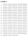
- Figure 1 shows a fragment of the complete AAP-5 fragment as depicted in Figure 9.
- Both nucleotide sequences encode a protein ( Figure 2 and Figure 10) with at least the capability of binding to Apoptin and providing apoptosis (as disclosed herein within the experimental part).
- an isolated or recombinant nucleic acid or functional equivalent or fragment thereof encoding an additional Apoptin-associating proteinaceous substance capable of associating with the partial or full-length AAP-5 protein is herewith also provided, means and methods to arrive at such an additional protein located in the Apoptin cascade follow those of the detailed description given herein.
- Knowledge derived from studying the partial or full-length AAP-5 is exploited to determine a functional pathway in which partial or full-length AAP-5 is involved, thus allowing the design of further therapeutic means of intervening in such a pathway.
- the invention provides an isolated or recombinant nucleic acid or functional equivalent or fragment thereof encoding an Apoptin-associating proteinaceous substance capable of providing apoptosis being at least 60% homologous, preferably at least 70%, more preferably at least 80%, even more preferably at least 90% and most preferably at least 95% homologous to a nucleic acid molecule, or to a functional equivalent or functional fragment thereof, encoding an Apoptin-associating proteinaceous substance as shown in figure 1 or 9.
- the invention provides a vector comprising a nucleic acid according to the invention.
- a vector comprising a nucleic acid according to the invention. Examples of such a vector are given in the detailed description given herein; such as vector pMT2SM-AAP-5, pMT2SM vector expressing Myc-tagged AAP-5 cDNA, a plasmid expressing an Apoptin-associating protein fragment, and so on.
- This and other vectors are for example useful in finding additional Apoptin-associating proteinaceous substances from the cascade, as defined above.
- the invention provides a vector comprising a nucleic acid according to the invention, said vector comprising a gene-delivery vehicle, making the invention very useful in gene therapy.
- a gene delivery vehicle By equipping a gene delivery vehicle with a nucleic acid according to the invention, and by targeting said vehicle to a cell or cells that have been over-proliferating and/or have shown decreased death rates, said gene delivery vehicle provides said cell or cells with the necessary means for apoptosis, providing far reaching therapeutic possibilities.
- the invention provides a host cell comprising a nucleic acid or a vector according to the invention.
- a host cell according to the invention comprises transduced bacterial or yeast cells as described in the detailed description herein.
- a host cell according to the invention which is a transduced eukaryotic cell such as a yeast cell or a vertebrate cell, such as mammalian or Cos cells transduced with a nucleic acid or vector according to the invention.
- Said cells are in general capable to express or produce a proteinaceous substance capable of providing apoptosis with the ability to associate with Apoptin.
- the invention furthermore provides an isolated or recombinant Apoptin-associating proteinaceous substance capable of providing apoptosis.
- expression of such Apoptin-associating proteinaceous substance in cells such as tumour cells, or other over-proliferating cells, induces the apoptotic process. It can do so alone, or in the presence of other apoptosis inducing substances such as Apoptin, and especially so independent of p53, showing that also in those cases where (functional) p53 is absent apoptosis can be induced by a substance according to the invention.
- the invention provides a proteinaceous substance according to the invention encoded by a nucleic acid according to the invention, for example comprising at least a part of an amino acid sequence as shown in figure 2 or 10 (AAP-5) or a functional equivalent or functional fragment thereof capable of providing apoptosis alone or in combination with other apoptosis inducing substances such as Apoptin.
- a nucleic acid according to the invention for example comprising at least a part of an amino acid sequence as shown in figure 2 or 10 (AAP-5) or a functional equivalent or functional fragment thereof capable of providing apoptosis alone or in combination with other apoptosis inducing substances such as Apoptin.
- the invention also provides an isolated or synthetic antibody specifically recognising a proteinaceous substance or functional equivalent or functional fragment thereof according to the invention.
- an antibody is for example obtainable by immunising an experimental animal with an Apoptin-associating proteinaceous substance or an immunogenic fragment or equivalent thereof and harvesting polyclonal antibodies from said immunised animal, or obtainable by other methods known in the art such as by producing monoclonal antibodies, or (single chain) antibodies or binding proteins expressed from recombinant nucleic acid derived from a nucleic acid library, for example obtainable via phage display techniques. Examples of such an antibody are given in the detailed description herein.
- the invention also provides a proteinaceous substance specifically recognisable by such an antibody according to the invention, for example obtainable via immunoprecipitation, Western Blotting, or other immunological techniques known in the art.
- the invention provides use of a nucleic acid, vector, host cell, or proteinaceous substance according to the invention for the induction of tumor-specific apoptosis, as for example shown in figure 3.
- apoptosis is p53-independent.
- use is also provided further comprising use of a nucleic acid encoding Apoptin or a functional equivalent or fragment thereof or use of Apoptin or a functional equivalent or fragment thereof.
- combining these Apoptin-inducing substances increases the percentage apoptosis of treated tumour cells.
- the invention provides herewith a pharmaceutical composition comprising a nucleic acid, vector, host cell, or proteinaceous substance according to the invention.
- a pharmaceutical composition according to the invention is provided further comprising a nucleic acid encoding Apoptin or a functional equivalent or fragment thereof.
- Such a pharmaceutical composition is in particular provided for the induction of apoptosis, for example wherein said apoptosis is p53-independent, for the treatment of a disease where enhanced cell proliferation or decreased cell death is observed, as is in general the case when said disease comprises cancer or auto-immune disease.
- the invention provides a method for treating an individual carrying a disease where enhanced cell proliferation or decreased cell death is observed comprising treating said individual with a pharmaceutical composition according to the invention.
- these compositions comprise a factor of an apoptosis pathway, which is specific for transformed cells. Therefore, these compositions are essential for new treatments, but also for diagnosis of diseases related with aberrances in the apoptotic process, such as cancer, cancer-proneness and auto-immune diseases.
- the yeast-2 hybrid system (Durfee et al., 1993) to identify Apoptin-associating cellular compounds, which are essential in the induction of apoptosis.
- the used system is an in vivo strategy to identify human proteins capable of physically associating with Apoptin. It has been used to screen cDNA libraries for clones encoding proteins capable of binding to a protein of interest (Fields and Song, 1989, Yang et al., 1992).
- the invention provides for example a novel Apoptin-associating protein, which is named Apoptin-associating protein 5 abbreviated as AAP-5..
- the invention also provides a method for inducing apoptosis through interference with the function of this newly discovered AAP-5 protein or functional equivalents or fragments thereof and/or the induction of apoptosis by means of (over)expression of AAP-5 or related gene or functional equivalents or fragments thereof.
- the invention also provides an anti-tumor therapy based on the interference with the function of AAP-5-like proteins and/or its (over)expression.
- the presence of AAP-5-like protein can result in the induction of the opposite process of cell transformation, namely apoptosis.
- the invention furthermore provides the mediator of Apoptin-induced apoptosis.
- the invention provides a therapy for cancer, autoimmune diseases or related diseases which is based on AAP-5-like proteins alone or in combination with Apoptin and/or Apoptin-like compounds.
- plasmid pET-16b-VP3 (Noteborn, unpublished results) was treated with Nde I and Bam HI.
- the 0.4 kb Nde I -Bam HI DNA fragment was isolated from low-melting-point agarose.
- Plasmid pGBT9 (Clontech Laboratories, Inc, Palo Alto, USA) was treated with the restriction enzymes Eco RI and Bam HI.
- the about 5.4-kb DNA fragment was isolated and ligated to an Eco RI -Nde I linker and the 0.4-kb DNA fragment containing the Apoptin-encoding sequences starting from its own ATG-initiation codon.
- the final construct containing a fusion gene of the GAL4-binding domain sequence and Apoptin under the regulation of the yeast promoter ADH was called pGBT-VP3 and was proven to be correct by restriction-enzyme analysis and DNA-sequencing according to the Sanger method (1977).
- the expression vector pACT containing the cDNAs from Epstein-Barr-virus-transformed human B cells fused to the GAL4 transcriptional activation domain, was used for detecting Apoptin-associating proteins.
- the pACT c-DNA library is derived from the lambda-ACT cDNA library, as described by Durfee et al. 1993.
- the E.coli strain JM109 was the transformation recipient for the plasmid pGBT9 and pGBT-VP3.
- the bacterial strain electromax/DH10B was used for the transformation needed for the recovery of the Apoptin-associating pACT-cDNAs, and was obtained from GIBCO-BRL, USA.
- the yeast strain Y190 was used for screening the cDNA library, and all other transformations, which are part of the used yeast-two-hybrid system.
- Luria Broth (LB) plates for E.coli were supplemented with ampicillin (50 microgram per ml).
- Yeast YPD and SC media were prepared as described by Rose et al. (1990).
- the yeast strain Y190 was made competent and transformed according to the methods described by Klebe et al. (1983). The yeast cells were first transformed with pGBT-VP3 and subsequently transformed with pACT-cDNA, and these transformed yeast cells were grown on histidine-minus plates, also lacking leucine and tryptophan.
- Hybond-N filters were layed on yeast colonies, which were histidine-positive and allowed to wet completely. The filters were lifted and submerged in liquid nitrogen to permeabilize the yeast cells. The filters were thawed and layed with the colony side up on Whattman 3MM paper in a petridish with Z-buffer (Per liter: 16.1 gr Na 2 HPO 4 .7H 2 O, 5.5 gr NaH 2 PO 4 .H 2 O, 0.75 gr KCl and 0,246 gr MgSO 4 .7H 2 O, pH 7.0) containing 0.27% beta-mercapto-ethanol and 1 mg/ml X-gal. The filters were incubated for at least 15 minutes or during night.
- Total DNA from yeast cells which were histidine- and beta-galactosidase-positive, was prepared by using the glusulase-alkaline lysis method as described by Hoffman and Winston (1987) and used to transform Electromax/DH10B bacteria via electroporation using a Bio-Rad GenePulser according the manufacturer's specifications.
- Transformants were plated on LB media containing the antibiotic agent ampicillin.
- the subclone containing the sequence encoding Apoptin-associating protein was partially sequenced using dideoxy NTPs according to the Sanger-method, which was performed by Eurogentec, Seraing, Belgium or BaseClear, Leiden, The Netherlands).
- the used sequencing primer was a pACT-specific 17-mer comprising of the DNA-sequence 5'-TACCACTACAATGGATG-3' or internal primers comprising the described novel AAP-5 sequences.
- Eurogentec In order to generate polyclonal antisera against the protein predicted to be encoded by the partial AAP-5 clone, Eurogentec (Belgium) designed three peptides predicted to be antigenic. These peptides were, for AAP-5: 1) Residues 27-41: C GGATHVYRYHRGESK; 2) Residues 49-63: GNGQRKDRKKTSLGP C ; and 3) Residues 71-85: EHAPEASQPAENISK C . (Note: numbering same as in Figure 2, and underlined C residues are added for technical reasons and are not part of the actual AAP sequence). These peptides were synthesized at Eurogentec and all subsequent antibody syntheses were also performed there.
- the three peptides were coupled to KLH and injected as a cocktail into two separate specific pathogen free rabbits with an immunization schedule of one injection and three subsequent boosts. Blood samples were taken before and after immunization. The sera were tested in-house by ELISA for specific reactivity to the peptide cocktail. The titers from each rabbit were high. Furthermore, for certain subsequent purposes, the AAP-5 antibodies were immune-purified using peptide cocktail coupled to immobilized diaminodipropylamine agarose columns (Pierce) according to the manufacturer's protocol.
- the AAP-5 antibody of the two generated with the highest affinity was selected for further use.
- We tested the efficacy of this antibody by transfecting 6 cm plates of sub-confluent primate COS-7 and human U2OS cells using the calcium phosphate co-precipitation method with 5 ⁇ g of the AAP-5-myc construct (partial clone), and as a control, untransfected cells.
- Detection in this case was performed with the anti-myc tag monoclonal antibody 9E10 (Evan et al.).
- the purified AAP-5 antibody was tested for utility in immunofluorescence by including glass coverslips in the above transfections. Coverslips were fixed with 4% paraformaldehyde, blocked with normal goat serum, incubated in a concentration of 1:10, washed, incubated in a 1:100 dilution of conjugated goat-anti-rabbit Ig, mounted in DAPI/DABCO/glycerol and visualized with fluorescence microscopy.
- RNA expression pattern of AAP-5 we tested commercially-manufactured Northern blots (Invitrogen, cat. 1999, #D2801-50) containing poly-A+ RNA from human fetal brain, liver, lung, muscle tissue and from adult lung.
- the DNA probe was derived from a Xho I restriction fragment encompassing the entire insert of the AAP-5-myc partial clone. This probe was labelled with 32 P-dATP using the MegaPrime kit of Amersham. All prehybridization, hybridization and washing steps were done according to the Northern blot manufacturer's recommendation. Blots were subjected to autoradiography and developed using standard automated methods.
- the complete ORF of AAP-5 was amplified from a human brain cDNA library (Clontech - Marathon Ready cDNA).
- the RT-PCR was performed according to the manufacturer's instructions (Clontech - Advantage®2 PCR kit).
- RT-PCR Sample volume 50 ⁇ l Brain cDNA 2 ⁇ l 5'-primer (50 ⁇ M) 1 ⁇ l 3'-primer (50 ⁇ M) 1 ⁇ l 10x cDNA PCR reaction buffer 5 ⁇ l dNTP mix (10 mM) 1 ⁇ l 50x Advantage 2 Polymerase mix 1 ⁇ l Steril water 39 ⁇ l
- the sequences for the RT-PCR primers were generated from AAP-5 genomic sequence identified on chromosome 11.
- the RT-PCR primers were as follow:
- the PCR products were cloned into the pCR®4-TOPO vector according to the instructions of the TOPO-TA cloning® kit from Invitrogen.
- the sequences of the PCR products were generated with the Applied Biosystem (ABI) Prism®BigDyeTMTerminator sequencing kit and analysed on a ABI 310 capillary sequencer.
- the sequencing primers were the PCR primers (5' and 3') and the following:
- the amount of DNA in the two-way co-transfection was 10 ⁇ g, and the partial clones were used (except AAP-3, of which a full-length sequence was used). Pull-downs were performed by IP'ing with the appropriate AAP antibody and performing Western blot analysis with the anti-myc tag 9E10. Polyclonal antibodies directed against AAP-5, AAP-4 and AAP-3 were used.
- Antibodies against AAP-3 and AAP-4 were obtained as described in the section " Generation and testing of antibodies”.
- Peptides used for raising antibodies against AAP-3 1) IYQRSGERPVTAGEE, 2) DEQVPDSIDAREIFD and 3) RSINDPEHPLTLEEL.
- Peptides used to raise antibodies against AAP-4 1) EESTPVHDSPGKDDA, 2) DSFKTKDSFRTAKSK and 3) IDIDISSRRREDQSL.
- AAP-5 In order to characterize the full-length AAP-5 protein, we expressed constructs encoding the full-length clones in cells and analyzed them by immunofluorescence staining. AAP-5 was analyzed as described in the results section under "Over-expression of the novel AAP-5 protein in human transformed cells induces the apoptotic process", using Saos-2 and U20S tumor cells. AAP-5 was stained with 9E10 to detect the fused myc tag. As comparison controls, Apoptin was expressed alone and in combination with AAP-5, and a construct expressing LacZ was included as a negative control. For these experiments, we examined cells after 2 and 5. days of transfection.
- apoptosis in transformed cells, such as cell lines derived from human tumors.
- transformed cells such as cell lines derived from human tumors.
- a yeast genetic screen was carried out.
- the final construct called pGBT-VP3, was analyzed by restriction-enzyme analysis and sequencing of the fusion area between Apoptin and the GAL4-DNA-binding domain.
- a gene(fragment) encoding an Apoptin-associating protein is determined by transactivation of a GAL4-responsive promoter in yeast.
- the Apoptin gene is fused to the GAL4-DNA-binding domain of plasmid pGBT-VP3, whereas all cDNAs derived from the transformed human B cells are fused to the GAL4-activation domain of plasmid pACT. If one of the proteinaceous substances encoded by said cDNAs binds to Apoptin, the GAL4-DNA-binding domain will be in the vicinity of the GAL4-activation domain resulting in the activation of the GAL4-responsive promoter, which regulates the reporter genes HIS3 and LacZ.
- the yeast clones containing plasmid expressing Apoptin and a plasmid expressing an Apoptin-associating protein fragment can grow on a histidine-minus medium and will stain blue in a beta-galactosidase assay. Subsequently, the plasmid with the cDNA insert encoding the Apoptin-associating protein can be isolated and characterized.
- yeast colony which upon transformation with pGBT-VP3 and pACT-cDNA was able to grow on a histidine-minus medium (also lacking leucine and tryptophan) and stained blue in a beta-galactosidase assay.
- Plasmid DNA was isolated from the positive yeast colony, which was transformed in bacteria. By means of a filter-hybridization assay using a pACT-specific labeled DNA-probe, the clone containing pACT plasmid could be determined. Subsequently, pACT DNA was isolated and digested with restriction enzyme Xho I, which resulted in the presence of an approximately 1.0-kbp cDNA insert. Finally, the cDNA insert of the pACT plasmid containing the cDNA insert was sequenced by using the Sanger method (Sanger et al., 1977).
- the yeast genetic screen for Apoptin-associating proteins resulted in the detection of a cDNA clone comprising a single type of protein, namely a novel protein called Apoptin-associating protein 5, abbreviated as AAP-5.
- the determined DNA sequence part of the AAP-5 cDNA clone is shown in Figure 1.
- the amino acid sequence, derived from the detected DNA sequence of clone AAP-5 is given in Figure 2.
- Construction of an expression vector for the identification of AAP-5 protein in mammalian cells Construction of an expression vector for the identification of AAP-5 protein in mammalian cells.
- the DNA plasmid pMT2SM contains the adenovirus 5 major late promoter (MLP) and the SV40 ori enabling high levels of expression of foreign genes in transformed mammalian cells, such as SV-40-transformed Cos cells. Furthermore, the pMT2SM vector contains a Myc-tag (amino acids: EQKLISEEDL) which is in frame with the foreign-gene product. This Myc-tag enables the recognition of e.g. Apoptin-associating proteins by means of the Myc-tag-specific 9E10 antibody.
- MLP major late promoter
- SV40 ori enabling high levels of expression of foreign genes in transformed mammalian cells, such as SV-40-transformed Cos cells.
- Myc-tag amino acids: EQKLISEEDL
- the pMT2SM vector expressing Myc-tagged AAP-5 cDNA was constructed as follows.
- the pACT-AAP-5 cDNA clone was digested with the restriction enzyme Xho I and the cDNA insert was isolated.
- the expression vector pMT2SM was digested with Xho I and treated with calf intestine alkaline phosphatase and ligated to the isolated AAP-5 cDNA inserts.
- sequence analysis the pMT2SM constructs containing the AAP-5 cDNA in the correct orientation was identified.
- Myc-tagged AAP-5 protein was analyzed by transfection of Cos cells with plasmid pMT2SM-AAP-5. As negative control, Cos cells were mock-transfected. Two days after transfection, the cells were lysed and Western-blot analysis was carried out using the Myc-tag-specific antibody 9E10.
- the Cos cells transfected with pMT2SM-AAP-5 were proven to synthesise a specific Myc-tagged AAP-5 product with the size of approximately 18 kDa. As expected, the lysates of the mock-transfected Cos cells did not contain a protein product reacting with the Myc-tag-specific antibodies.
- Cos cells were co-transfected with plasmid pCMV-VP3 encoding Apoptin and with plasmid pMT2SM-AAP-5.
- plasmid pMT2SM-AAP-5 As a negative control, cells were transfected with pCMV-VP3 expressing Apoptin and a plasmid pcDNA3.1.LacZ-myc/His-LacZ encoding the myc-tagged beta-galactosidase, which does not associate with Apoptin.
- the cells were lysed in a buffer consisting of 50 mM Tris (7.5), 250 mM NaCl, 5 mM EDTA, 0.1 % Triton X100, 1 mg/ml Na 4 P 2 O 7 and freshly added protease inhibitors such as PMSF, Trypsine-inhibitor, Leupeptine and Na 3 VO 4 .
- the specific proteins were immuno-precipitated as described by Noteborn et al. (1998) using the Myc-tag-specific antibodies 9E10, and analyzed by Western blotting.
- Immunoprecipitation of the Myc-tagged AAP-5 products was accompanied by the immunoprecipatation of Apoptin product of 16 kDa.
- immunoprecipitation of myc-tagged beta-galactosidase did not result in a detectable co-precipitation of the Apoptin protein.
- immunoprecipitation of the Apoptin protein by means of a polyclonal antibody directed against the C-terminal part of Apoptin (Noteborn and Danen, unpublished results), was accompanied by the immunoprecipitation of the AAP-5 product of 18 kDa, but not by beta-galactosidase protein.
- novel determined AAP-5 protein is able to specifically associate with Apoptin not only in the yeast background, but also in a mammalian transformed cellular system.
- AAP-5 carries apoptotic activity.
- AAP-5 protein can induce apoptosis in p53-minus Saos-2 cells indicates that AAP-5 can induce p53-independent apoptosis. These results imply that AAP-5 can be used as anti-tumor agent in cases where other (chemo)therapeutic agents will fail. Furthermore, the finding that both Apoptin and AAP-5 induce a p53-independent pathway indicates that AAP-5 fits in the Apoptin-induced apoptotic pathway.
- an Apoptin-associating protein namely the novel AAP-5 protein, which is mainly present in the nucleus and able to induce (p53-independent) apoptosis in human tumor cells.
- the putative BRCA-binding protein BRIP1 shows some amino-acid sequence homologies with AAP-5.
- the AAP-5 antibody of the two generated with the highest affinity was selected for further use. We tested the efficacy of this antibody by transfecting primate COS-7 and human U2OS cells with the partial AAP-5-myc construct.
- AAP-5-myc DNA transfections Western analysis showed that an approximately 17 or 12 kD (COS and U2OS, respectively) AAP-5-myc protein was detected strongly only in samples where the DNA was transfected. (These size differences may be a result of cell-type-specific post-translational modifications.) Similarly, in immunoprecipitation experiments, AAP-5-myc was also strongly detected. Finally, we could detect the presence of transfected AAP-5-myc diffusely in the cytoplasm and nucleus of human Saos-2 tumor cells using the AAP-5 antibody in immunofluorescence analysis.
- AAP-5 is not detectably expressed in the fetal tissue RNA examined, there is an approximately 1.8-2 kb message expressed in adult lung. This mRNA size is consistent with what is predicted to be expressed from the full-length gene, including post-transcriptional modifications.
- the genetic yeast screen with pGBT-VP3 as bait plasmid and pACT plasmid containing cDNAs from transformed human B cells also delivered other Apoptin-associating proteins, which also encode Apoptin-associating proteins. These Apoptin-associating proteins were called AAP-3 and AAP-4 (see co-pending application EP01200163.2 and EP00204396.6 respectively, which are incorporated herein by reference).
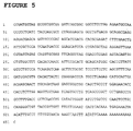
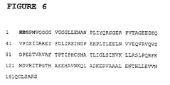
- the DNA sequence of AAP-3 is shown in Figure 5, whereas the AAP-3 cDNA-encoded amino acid sequence is shown in figure 6.
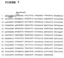
- the DNA sequence of AAP-4 is shown in Figure 7, whereas the AAP-4 cDNA-encoded amino acid sequence is shown in figure 8.
- both AAP-3 and AAP-4 are able to associate with Apoptin not only in the yeast background, but also in a mammalian transformed cellular system.
- Immunofluorescense assays of human transformed Saos-2 cells and normal diploid VH10 fibroblasts expressing AAP-3 revealed that AAP-3 is located in both cell types predominantly in the cytoplasm and nucleus, but in lower percentages also mainly in the nucleus or mainly in the cytoplasm.
- Co-synthesis of AAP-3 and Apoptin in both cell types showed a clear nuclear localization of AAP-3 and Apoptin.
- Tumor cells that have become apoptotic showed a nuclear localization of Apoptin and a peri-nuclear staining pattern of AAP-3.
- normal diploid VH10 cells synthesizing both Apoptin and AAP-3 did not undergo apoptosis.
- AAP-4 is present in the nucleus and able to induce (p53-independent) apoptosis in human tumor cells.
- AAP-5 in both Saos-2 and U2OS cells exhibited a primarily cytoplasmic localization.
- the staining was diffuse, but in addition most of the cells also contained very prominent bodies in the cytoplasm, clustering in particular around the nucleus. These bodies had a semi-regular, almost crystalline structure, and tended to cluster together into larger aggregates. In some cells a faint nuclear, diffuse staining could also be discerned.
- Apoptin it appeared that Apoptin formed a "shell" around the outside of the crystalline-like bodies, and the overall number of these bodies appeared to be decreased compared to the situation with the AAP-5 expressed alone.
- AAP-5 caused a very high percentage of apoptosis in both cell types, almost twice the level as that seen for Apoptin alone at the same time point (2 days after transfection). This apoptosis only increased at later time points, similar to what was seen with the partial AAP-5 protein. Also similar to the partial AAP-5 protein, when co-expressed with Apoptin, there was a slight increase in the amount of death as compared to either protein expressed alone.
- the apoptosis inducing capability of the partial AAP-5 protein or the full-length AAP-5 protein alone or in combination with Apoptin shows that all possible mutants ranging from at least the partial AAP-5 protein to the full-length AAP-5 protein are capable of inducing apoptosis. Therefore, deletion mutants of the full-length AAP-5 protein comprising at least the partial AAP-5 protein are capable of inducing apoptosis. Also, point mutants (both at nucleotide and amino acid level) of the full-length AAP-5 protein comprising at least the partial AAP-5 protein are capable of inducing apoptosis.
- Figure 1 shows the partial sequence of vector pMT2SM-AAP-5.
- Figure 2 shows the amino-acid sequence of the analyzed region of the Apoptin-associating clone AAP-5.
- the three C-terminal amino acids H-E-G (bold) of the multiple cloning site of pACT are given to illustrate that the AAP-5 amino acid sequence is in frame with the GAL4-activation domain. This feature proves that the AAP-5 region is indeed synthesized in yeast cells.
- Figure 3 shows the apoptotic activity of AAP-5 protein,, with or without co-synthesis of Apoptin in human osteosarcoma-derived Saos-2 cells.
- Saos-2 cells were transfected with a plasmid encoding Apoptin.
- negative control cells were transfected with a plasmid encoding lacZ.
- (+) strong apoptotic activity; (+++): very strong apoptotic activity.
- (+++) very strong apoptotic activity.
- Figure 4 shows the partial consensus amino-acid sequences of AAP-5 and the putative BRCA-binding protein BRIP-1.
- Figure 5 shows the partial sequence of vector pMT2SM-AAP-3
- Figure 6 shows the amino acid sequence of the analysed region of the Apoptin-associating clone AAP-3.
- H-E-G bold
- H-E-G bold
- Figure 7 shows the partial sequence of vector pMT2SM-AAP-4.
- the DNA sequence of the AAP-4 CDNA starts at position 12 of the DNA sequence and is indicated as "start AAP-4 cDNA”.
- Figure 8 shows the amino acid sequence of the analysed region of the Apoptin-associating clone AAP-4.
- the three C-terminal amino acids H-E-G (bold) of the multiple cloning site of pact are given to illustrate that the AAP-4 amino acid sequence is in frame with the GAL4-activation domain. This feature proves that the AAP-4 region is indeed synthesized in yeast cells.
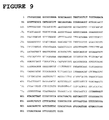
- Figure 9 shows the nucleic acid sequence of full-length AAP-5.
- the ATG start codon is at position 91-93 and the TAA stop codon is at position 793-795.
- Figure 10 shows the amino acid sequence deduced from the nucleic acid sequence of Figure 9.
Landscapes
- Chemical & Material Sciences (AREA)
- Health & Medical Sciences (AREA)
- Life Sciences & Earth Sciences (AREA)
- Organic Chemistry (AREA)
- General Health & Medical Sciences (AREA)
- Gastroenterology & Hepatology (AREA)
- Biochemistry (AREA)
- Biophysics (AREA)
- Zoology (AREA)
- Genetics & Genomics (AREA)
- Medicinal Chemistry (AREA)
- Molecular Biology (AREA)
- Proteomics, Peptides & Aminoacids (AREA)
- Toxicology (AREA)
- Peptides Or Proteins (AREA)
- Micro-Organisms Or Cultivation Processes Thereof (AREA)
- Medicines That Contain Protein Lipid Enzymes And Other Medicines (AREA)
Priority Applications (1)
| Application Number | Priority Date | Filing Date | Title |
|---|---|---|---|
| EP01201137A EP1138768A3 (de) | 2000-03-27 | 2001-03-27 | Apoptin-bindendes Protein AAP-5 |
Applications Claiming Priority (3)
| Application Number | Priority Date | Filing Date | Title |
|---|---|---|---|
| EP00201108A EP1138765A1 (de) | 2000-03-27 | 2000-03-27 | Apoptin verbundene Proteine AAP-5 und AAP-6 |
| EP00201108 | 2000-03-27 | ||
| EP01201137A EP1138768A3 (de) | 2000-03-27 | 2001-03-27 | Apoptin-bindendes Protein AAP-5 |
Publications (2)
| Publication Number | Publication Date |
|---|---|
| EP1138768A2 true EP1138768A2 (de) | 2001-10-04 |
| EP1138768A3 EP1138768A3 (de) | 2002-08-07 |
Family
ID=26072047
Family Applications (1)
| Application Number | Title | Priority Date | Filing Date |
|---|---|---|---|
| EP01201137A Withdrawn EP1138768A3 (de) | 2000-03-27 | 2001-03-27 | Apoptin-bindendes Protein AAP-5 |
Country Status (1)
| Country | Link |
|---|---|
| EP (1) | EP1138768A3 (de) |
Cited By (1)
| Publication number | Priority date | Publication date | Assignee | Title |
|---|---|---|---|---|
| EP1399460A4 (de) * | 2001-04-27 | 2005-04-13 | Sunnybrook & Women S College H | Mit brustkrebs assoziierte gene und deren verwendung |
Family Cites Families (4)
| Publication number | Priority date | Publication date | Assignee | Title |
|---|---|---|---|---|
| WO1999011293A1 (en) * | 1997-09-05 | 1999-03-11 | Human Genome Sciences, Inc. | 50 human secreted proteins |
| EP0921192A1 (de) * | 1997-12-03 | 1999-06-09 | Leadd B.V. | Molekulen interagierend mit Apoptin |
| EP0924296A3 (de) * | 1997-12-03 | 2001-08-01 | Leadd B.V. | Verfahren un Mittteln für die Induktion von Apoptosis durch Interferenz in RNS-Prozessierung |
| WO2000058473A2 (en) * | 1999-03-31 | 2000-10-05 | Curagen Corporation | Nucleic acids including open reading frames encoding polypeptides; 'orfx' |
-
2001
- 2001-03-27 EP EP01201137A patent/EP1138768A3/de not_active Withdrawn
Cited By (1)
| Publication number | Priority date | Publication date | Assignee | Title |
|---|---|---|---|---|
| EP1399460A4 (de) * | 2001-04-27 | 2005-04-13 | Sunnybrook & Women S College H | Mit brustkrebs assoziierte gene und deren verwendung |
Also Published As
| Publication number | Publication date |
|---|---|
| EP1138768A3 (de) | 2002-08-07 |
Similar Documents
| Publication | Publication Date | Title |
|---|---|---|
| AU763403B2 (en) | Methods and means for inducing apoptosis by interfering with bip-like proteins | |
| AU761037B2 (en) | Molecules interacting with apoptin | |
| EP1094110A1 (de) | Proteinsubstanz das Apoptose induziert | |
| US7741440B2 (en) | Apoptin-associating protein | |
| US6878692B2 (en) | Apoptin-associating protein | |
| US7767790B2 (en) | Apoptin-associating proteins | |
| EP1138768A2 (de) | Apoptin-bindendes Protein AAP-5 | |
| EP1111045A2 (de) | Apoptin-bindendes Protein | |
| US6809189B2 (en) | Apoptin-associating protein | |
| EP1106691A1 (de) | Apoptin verbundenes Protein | |
| EP1081226A1 (de) | Apoptin-bindendes Protein | |
| EP1118662A2 (de) | Apoptin bindendes Protein | |
| EP1108785A1 (de) | Apoptin-verbundenes Protein | |
| EP1120463A1 (de) | Apoptin verbundenes Protein | |
| CA2290031A1 (en) | Methods and means for inducing apoptosis by interference in rna processing |
Legal Events
| Date | Code | Title | Description |
|---|---|---|---|
| PUAI | Public reference made under article 153(3) epc to a published international application that has entered the european phase |
Free format text: ORIGINAL CODE: 0009012 |
|
| AK | Designated contracting states |
Kind code of ref document: A2 Designated state(s): AT BE CH CY DE DK ES FI FR GB GR IE IT LI LU MC NL PT SE TR |
|
| AX | Request for extension of the european patent |
Free format text: AL;LT;LV;MK;RO;SI |
|
| PUAL | Search report despatched |
Free format text: ORIGINAL CODE: 0009013 |
|
| RIC1 | Information provided on ipc code assigned before grant |
Free format text: 7C 12N 15/12 A, 7C 07K 14/47 B, 7C 07K 16/18 B, 7A 61K 38/17 B, 7A 61K 38/00 B |
|
| AK | Designated contracting states |
Kind code of ref document: A3 Designated state(s): AT BE CH CY DE DK ES FI FR GB GR IE IT LI LU MC NL PT SE TR |
|
| AX | Request for extension of the european patent |
Free format text: AL;LT;LV;MK;RO;SI |
|
| 111L | Licence recorded |
Free format text: 20021030 0100 SCHERING AG |
|
| 17P | Request for examination filed |
Effective date: 20030207 |
|
| AKX | Designation fees paid |
Designated state(s): AT BE CH CY DE DK ES FI FR GB GR IE IT LI LU MC NL PT SE TR |
|
| STAA | Information on the status of an ep patent application or granted ep patent |
Free format text: STATUS: THE APPLICATION IS DEEMED TO BE WITHDRAWN |
|
| 18D | Application deemed to be withdrawn |
Effective date: 20051003 |