EP1176152A2 - Ligandenbindedomäne des Ultraspiracle (USP)-Proteins - Google Patents
Ligandenbindedomäne des Ultraspiracle (USP)-Proteins Download PDFInfo
- Publication number
- EP1176152A2 EP1176152A2 EP01116617A EP01116617A EP1176152A2 EP 1176152 A2 EP1176152 A2 EP 1176152A2 EP 01116617 A EP01116617 A EP 01116617A EP 01116617 A EP01116617 A EP 01116617A EP 1176152 A2 EP1176152 A2 EP 1176152A2
- Authority
- EP
- European Patent Office
- Prior art keywords
- lbd
- usp
- computer
- conformation
- ligands
- Prior art date
- Legal status (The legal status is an assumption and is not a legal conclusion. Google has not performed a legal analysis and makes no representation as to the accuracy of the status listed.)
- Withdrawn
Links
Classifications
-
- C—CHEMISTRY; METALLURGY
- C07—ORGANIC CHEMISTRY
- C07K—PEPTIDES
- C07K14/00—Peptides having more than 20 amino acids; Gastrins; Somatostatins; Melanotropins; Derivatives thereof
- C07K14/435—Peptides having more than 20 amino acids; Gastrins; Somatostatins; Melanotropins; Derivatives thereof from animals; from humans
- C07K14/43504—Peptides having more than 20 amino acids; Gastrins; Somatostatins; Melanotropins; Derivatives thereof from animals; from humans from invertebrates
- C07K14/43563—Peptides having more than 20 amino acids; Gastrins; Somatostatins; Melanotropins; Derivatives thereof from animals; from humans from invertebrates from insects
-
- C—CHEMISTRY; METALLURGY
- C07—ORGANIC CHEMISTRY
- C07K—PEPTIDES
- C07K2299/00—Coordinates from 3D structures of peptides, e.g. proteins or enzymes
Definitions
- the invention relates to the spatial structure of the ligand binding domain of the ultraspiracle protein, the use of this structure to generate protein models this protein in different conformations and related proteins, as well as methods for finding ligands of the ultraspriracle protein and related proteins.
- the ultraspiracle protein (hereinafter referred to as USP) is the insect orthologist of the vertebrate retinoid X receptor (RXR). Like RXR, it belongs to Family of nuclear receptors (Nuclear Receptors, NR). These core receptors are located itself inside the cell. They bind to responsive as homo- or heterodimers Elements on the DNA and regulate the expression of genes. To be active they need special small, often hydrophobic ligands (e.g. steroids, retinoids, Bind vitamin D). Core receptors have a modular structure with functional ones Domains for transactivation, DNA binding and ligand binding. While the DNA binding domain of the nuclear receptors is highly conserved, the Ligand binding domains only moderate homologies with each other.
- RXR vertebrate retinoid X receptor
- the spatial Structures of different ligand binding domains have already been determined (Summary in 2) and provide an insight into the activation lying mechanism, the strong conformational changes of the ligand binding domains includes.
- the binding of agonists leads to displacement bound co-repressors and binding co-activators for activation, while binding antagonists interact with the co-activator prevented.
- the ecdysone receptor is an important insecticide target. If it is outside the activated in the time window provided for this in the insect development, so leads this leads to serious disturbances up to the death of the insect. On this Mechanism is based on the insecticidal activity of ecdysone agonists (10, 11). nonsteroidal Ligands of the EcR subunit that act specifically on lepidopters, are already used commercially as insecticides (12).
- USP is an orphan receptor for which no ligand has yet been used is known. It has been suspected on several occasions that USP is a receptor for Represents juvenile hormones, but has never been really experimental evidence of this found (9). It was even suspected that USP has no ligand at all, as described for some other nuclear receptors known from animals.
- the object of the present invention was therefore to determine the spatial structure of the Ligand binding domain (hereinafter referred to as LBD) of the USP and to describe the possible ligand binding pocket.
- LBD Ligand binding domain
- the crystalline LBD according to the invention is preferably an LBD of the USP from Heliothis virescens.
- the inventive method particularly preferably LBD an amino acid sequence according to SEQ ID NO: 1.
- the present invention also relates to a crystalline complex of a USP-LBD with a ligand.
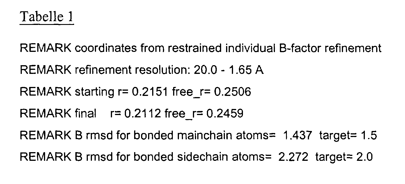
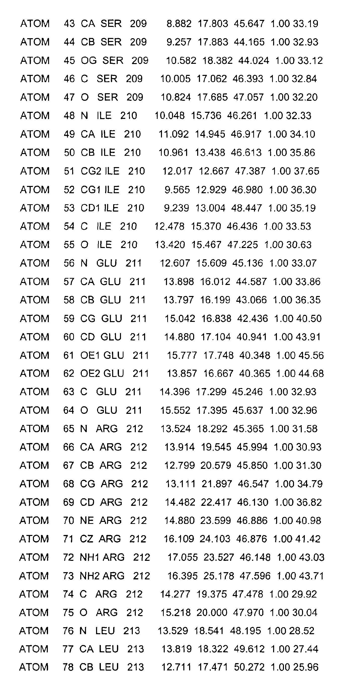
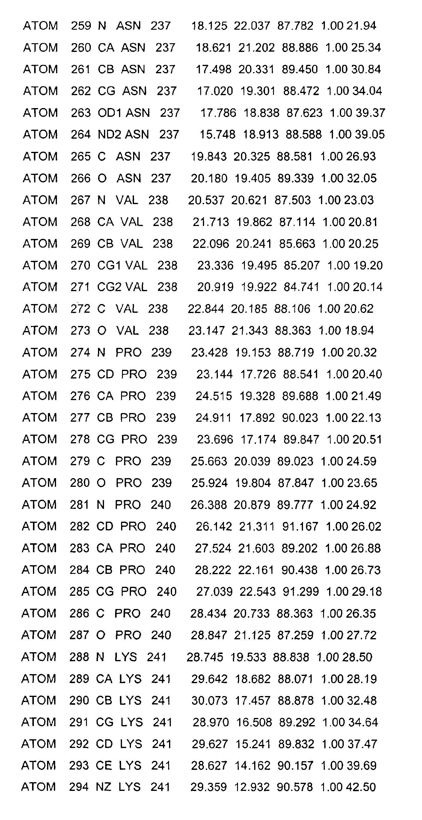
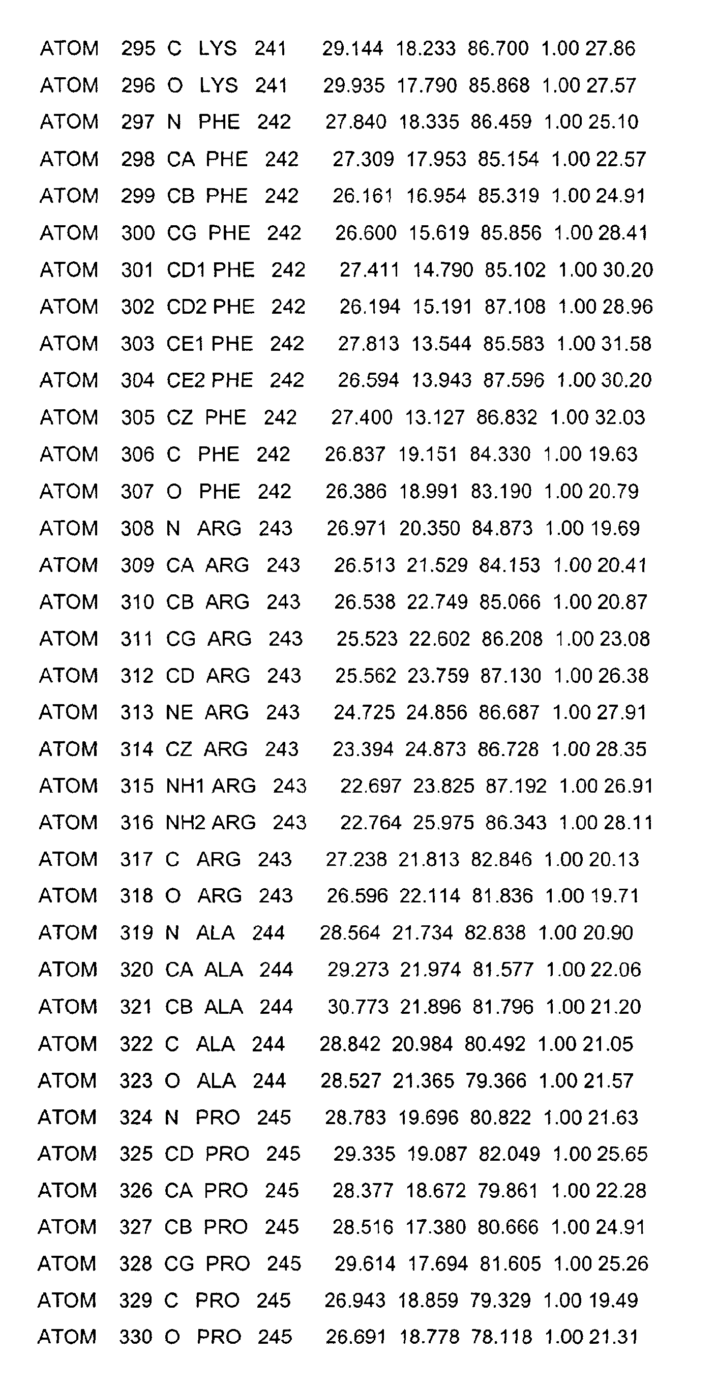
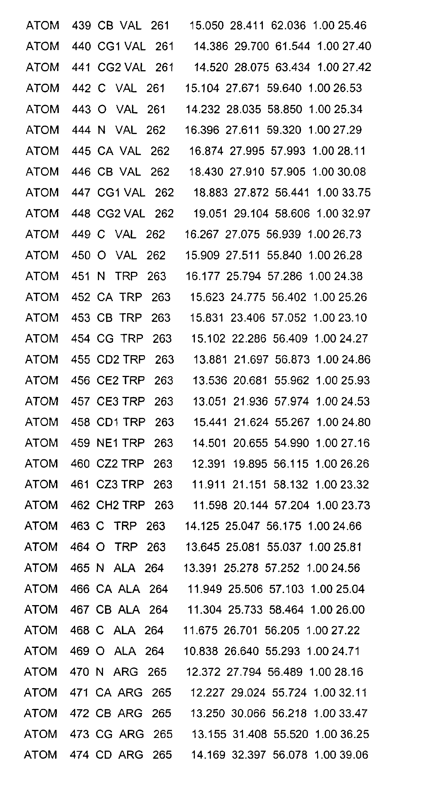
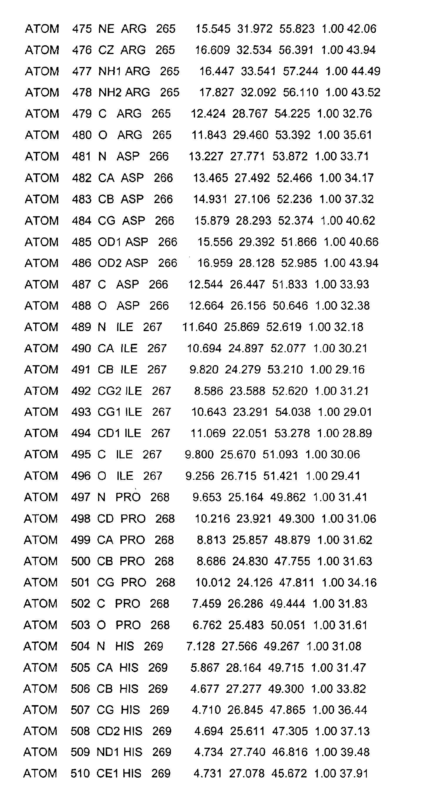
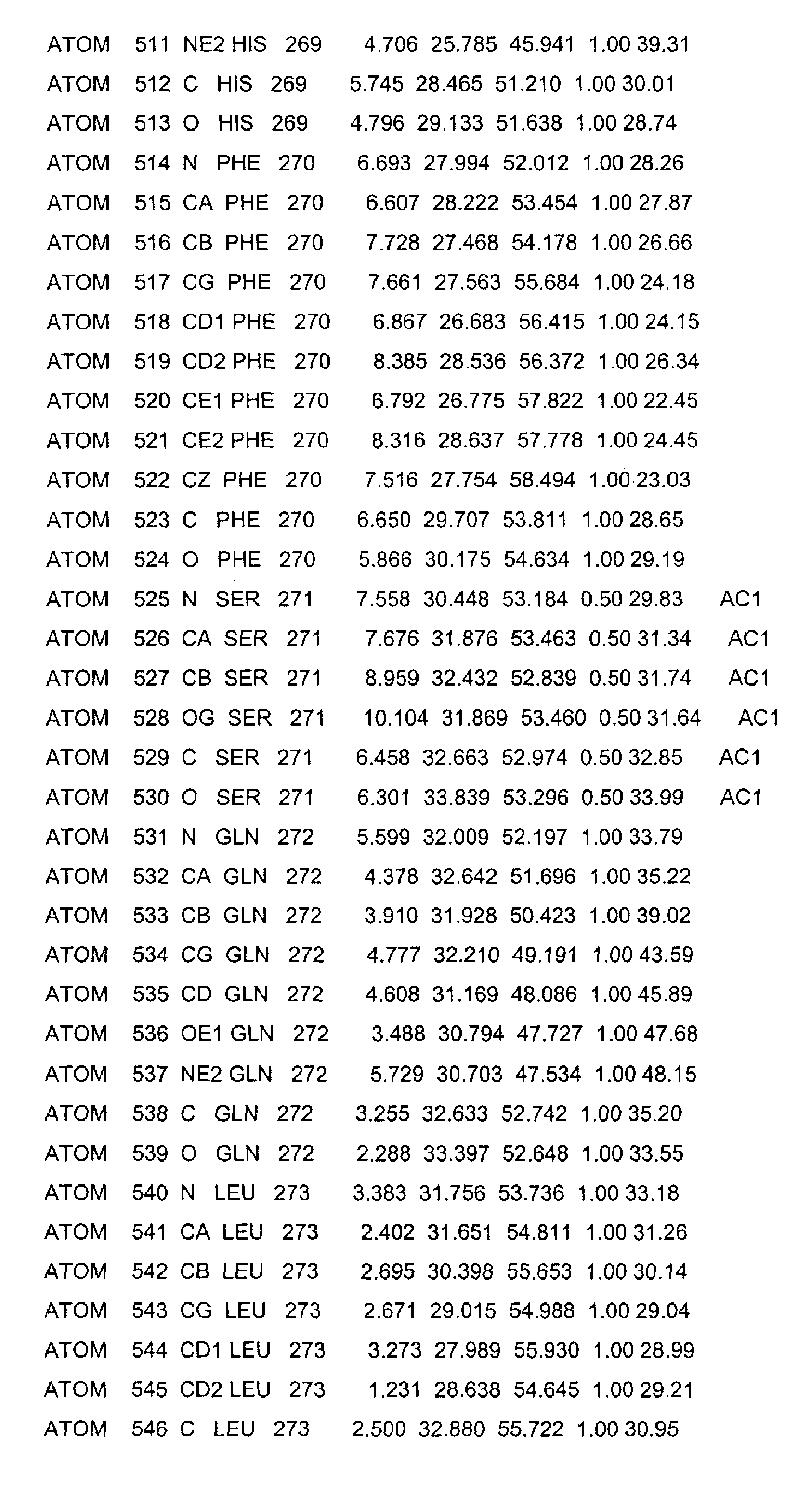
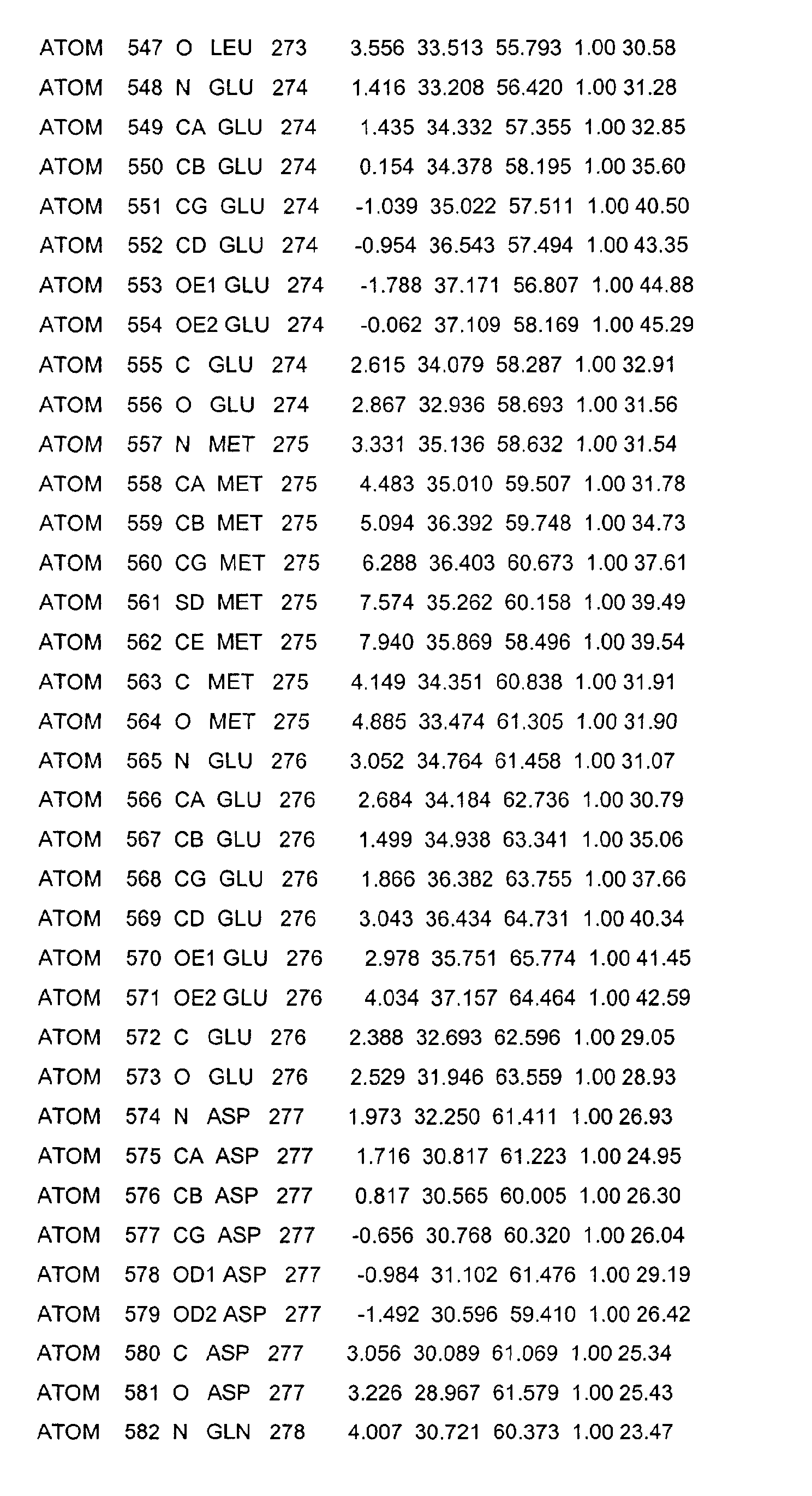
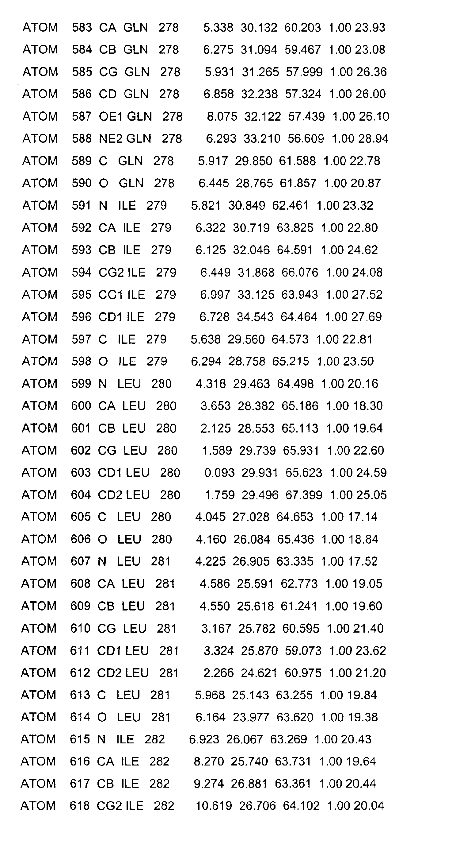
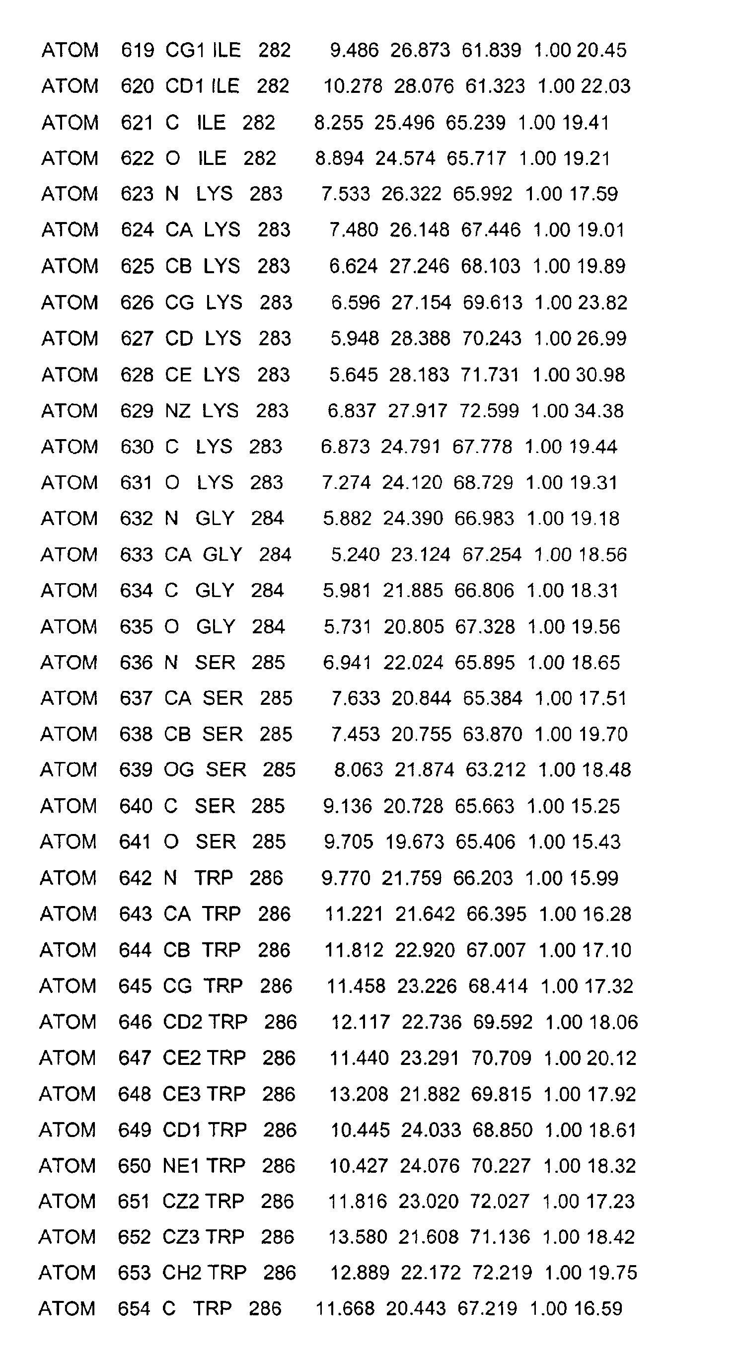
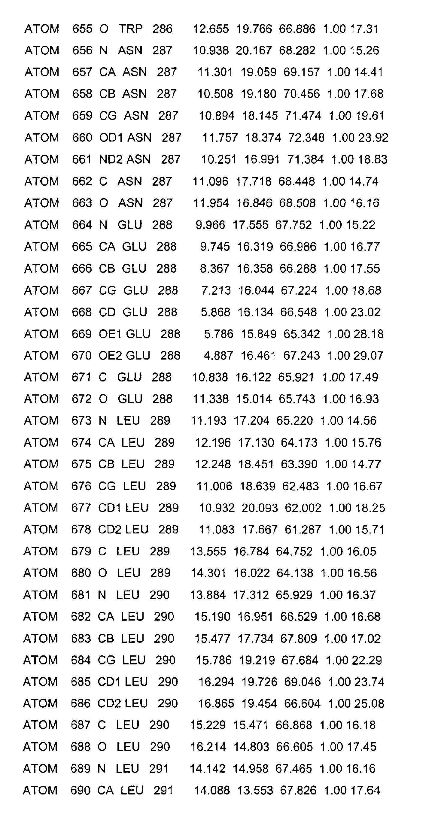
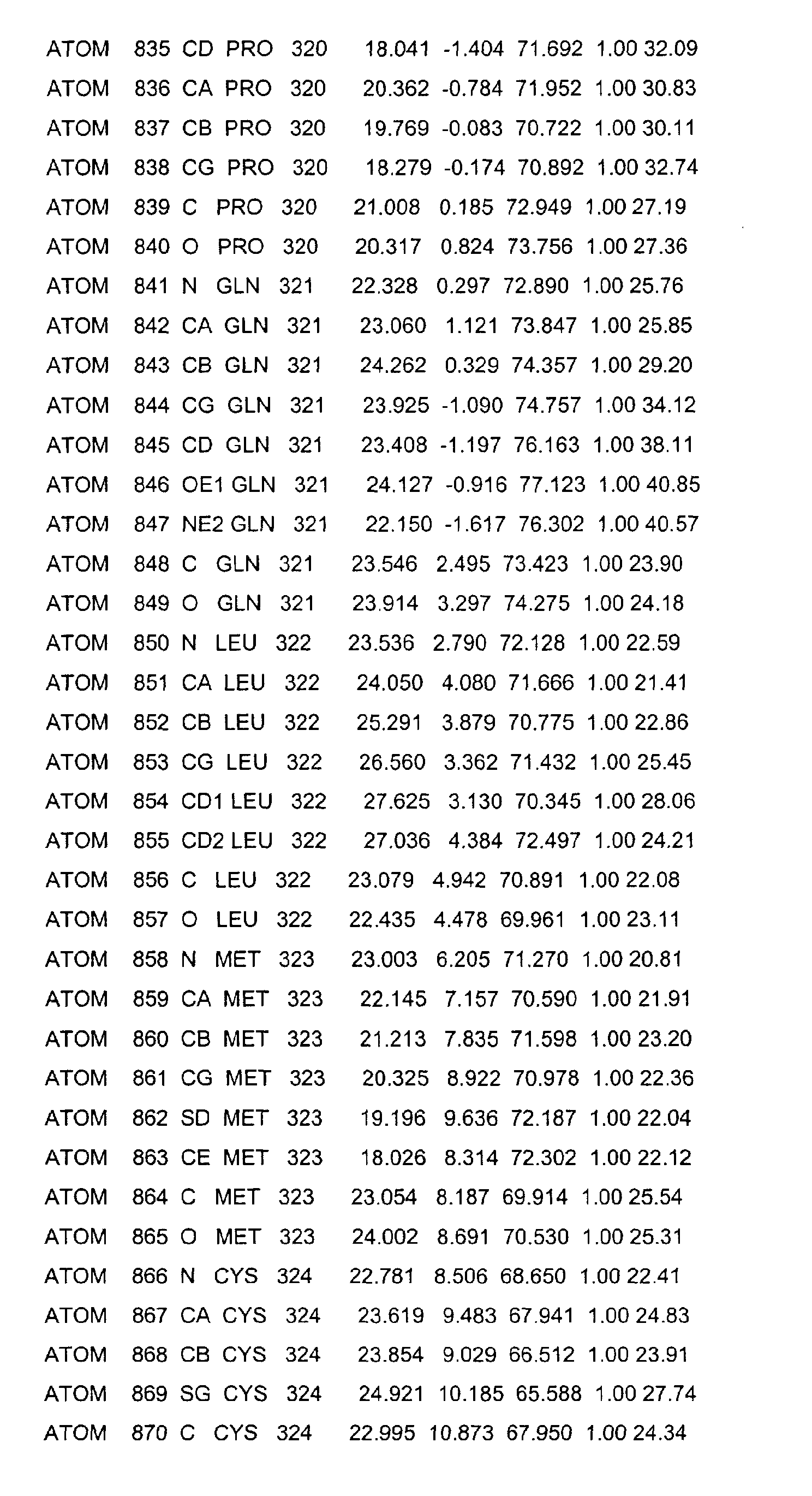
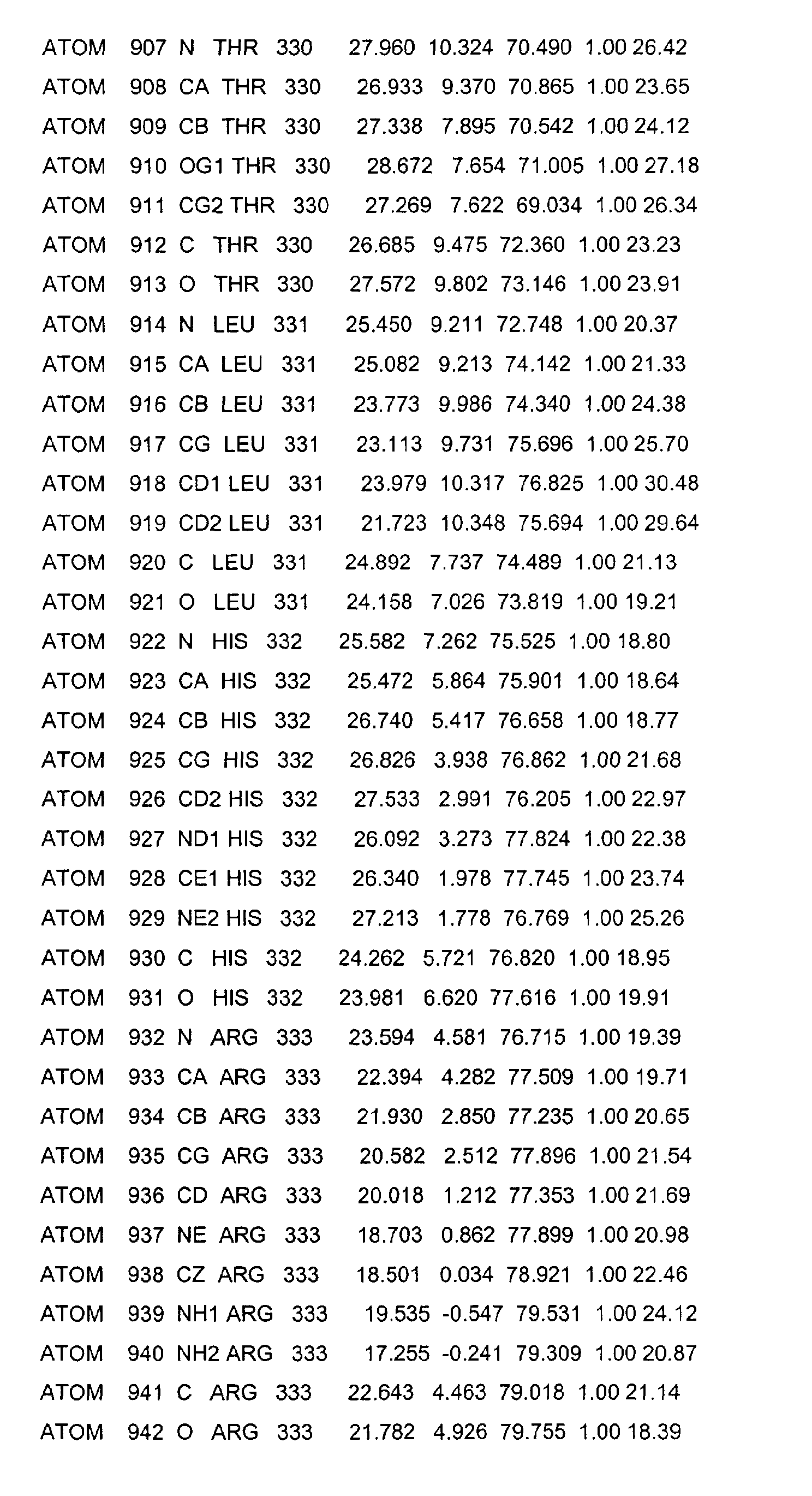
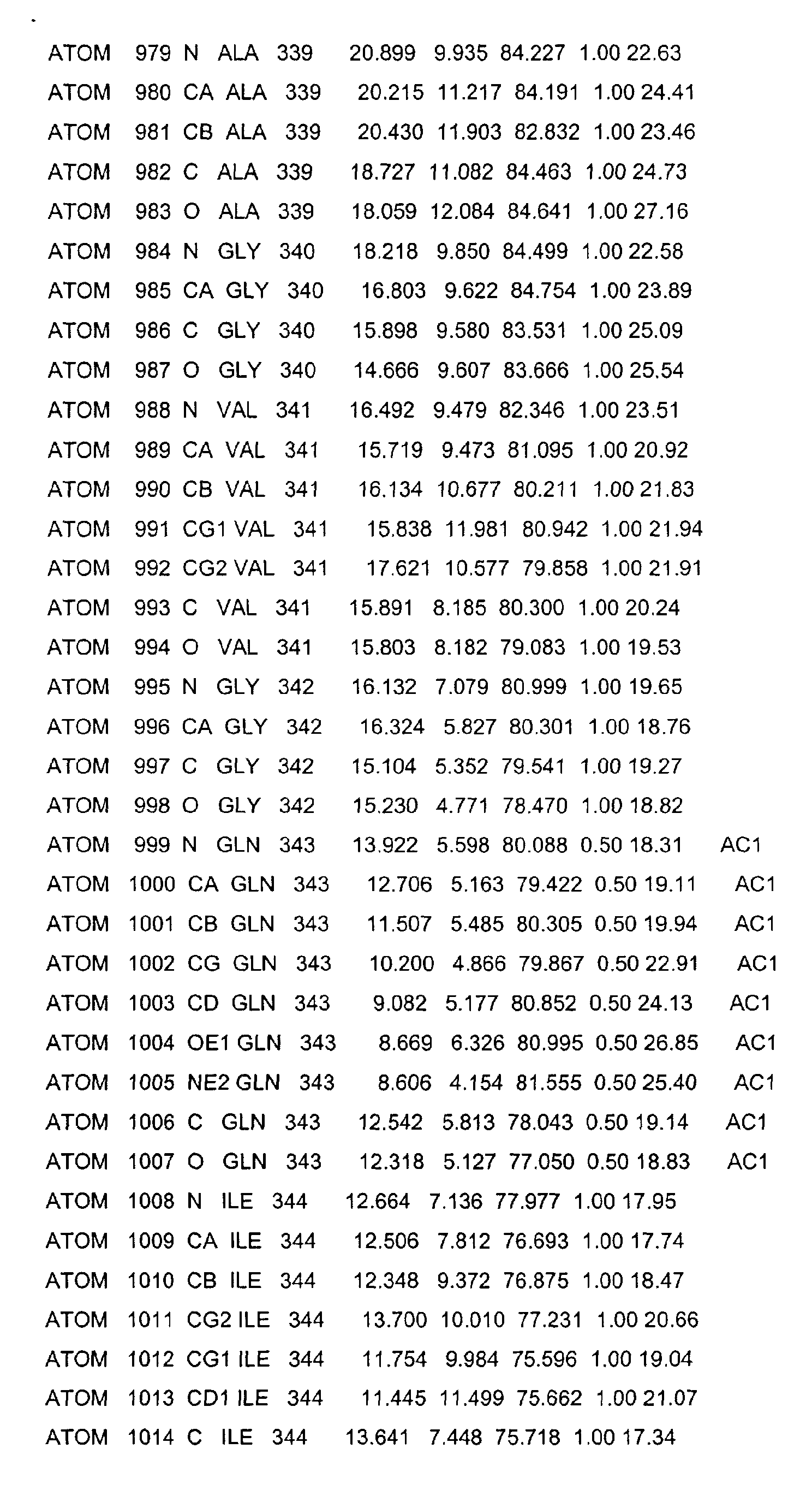
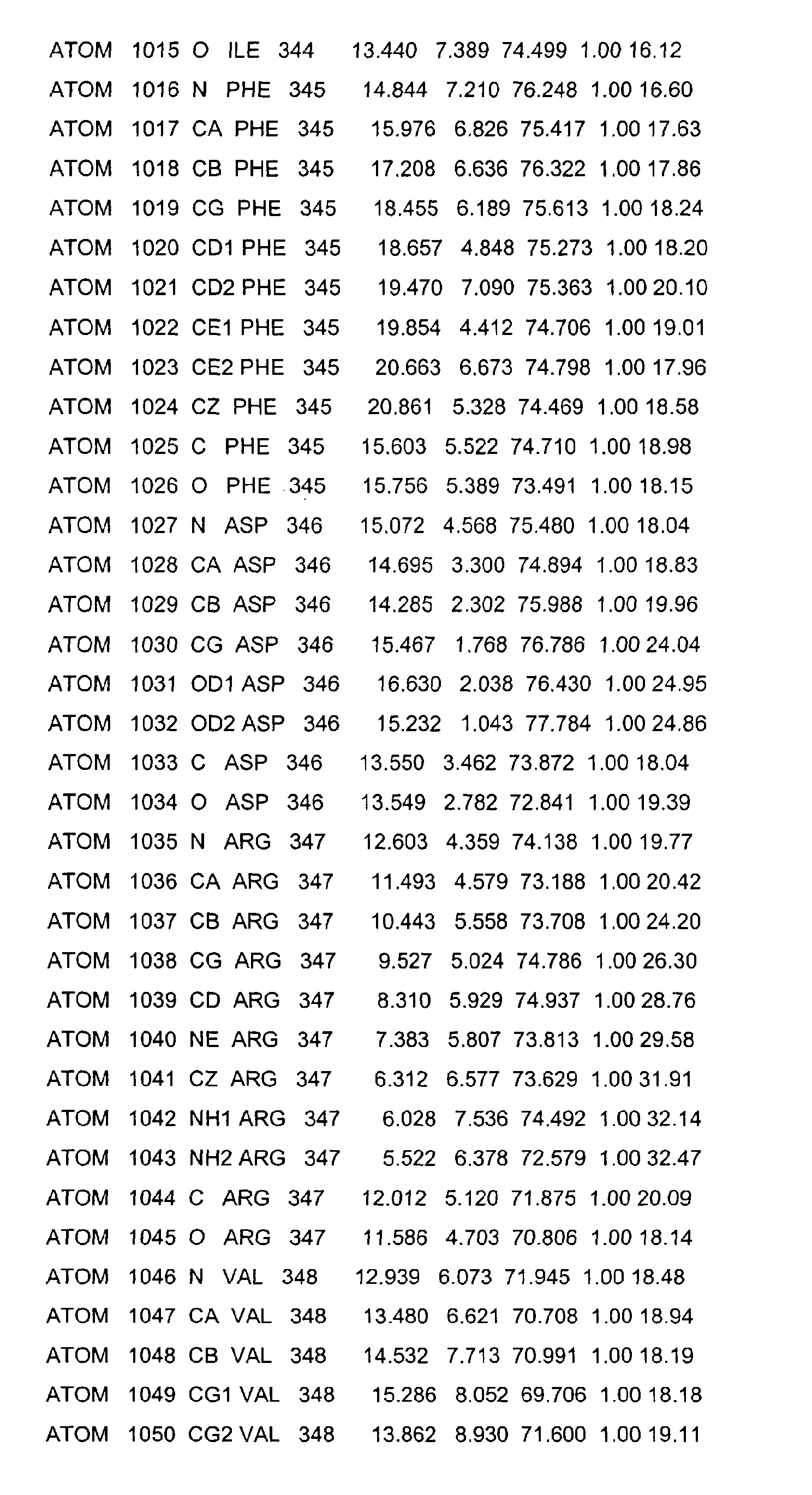
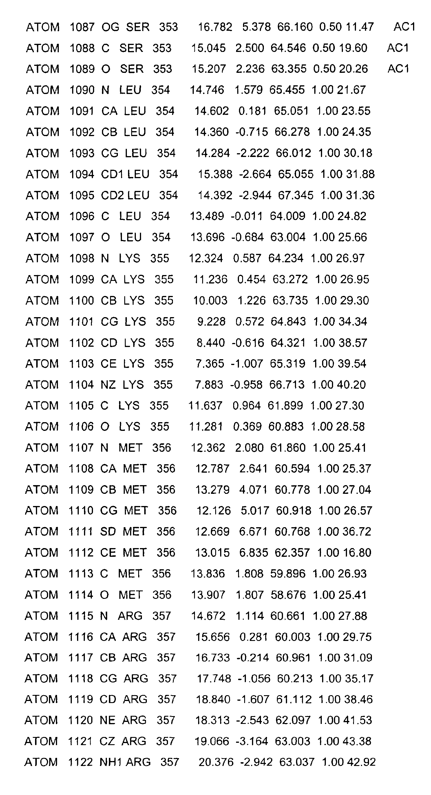
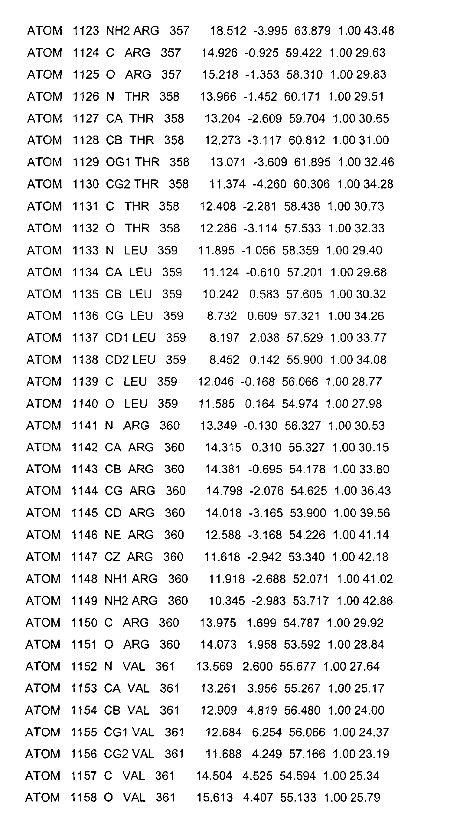
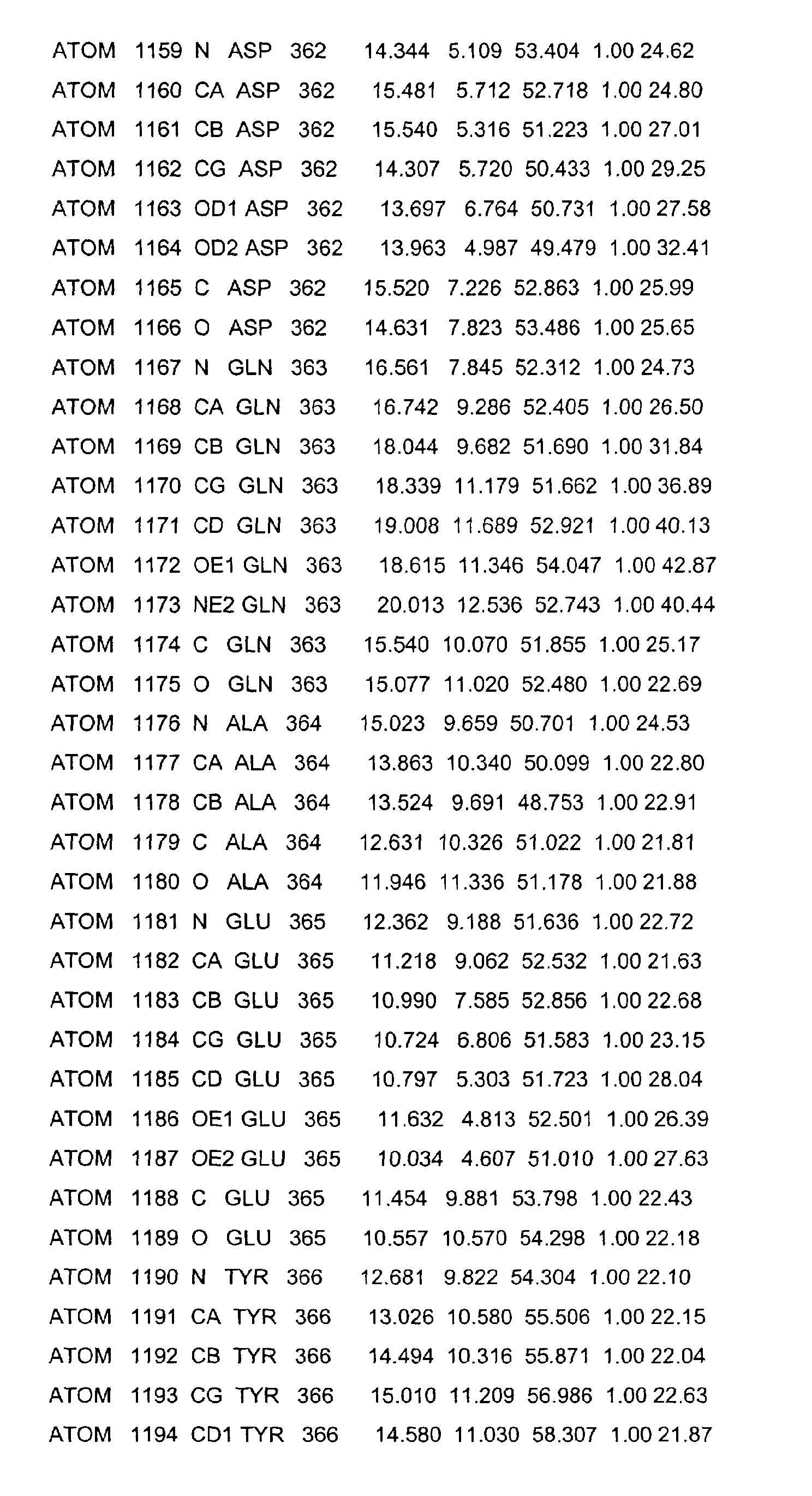
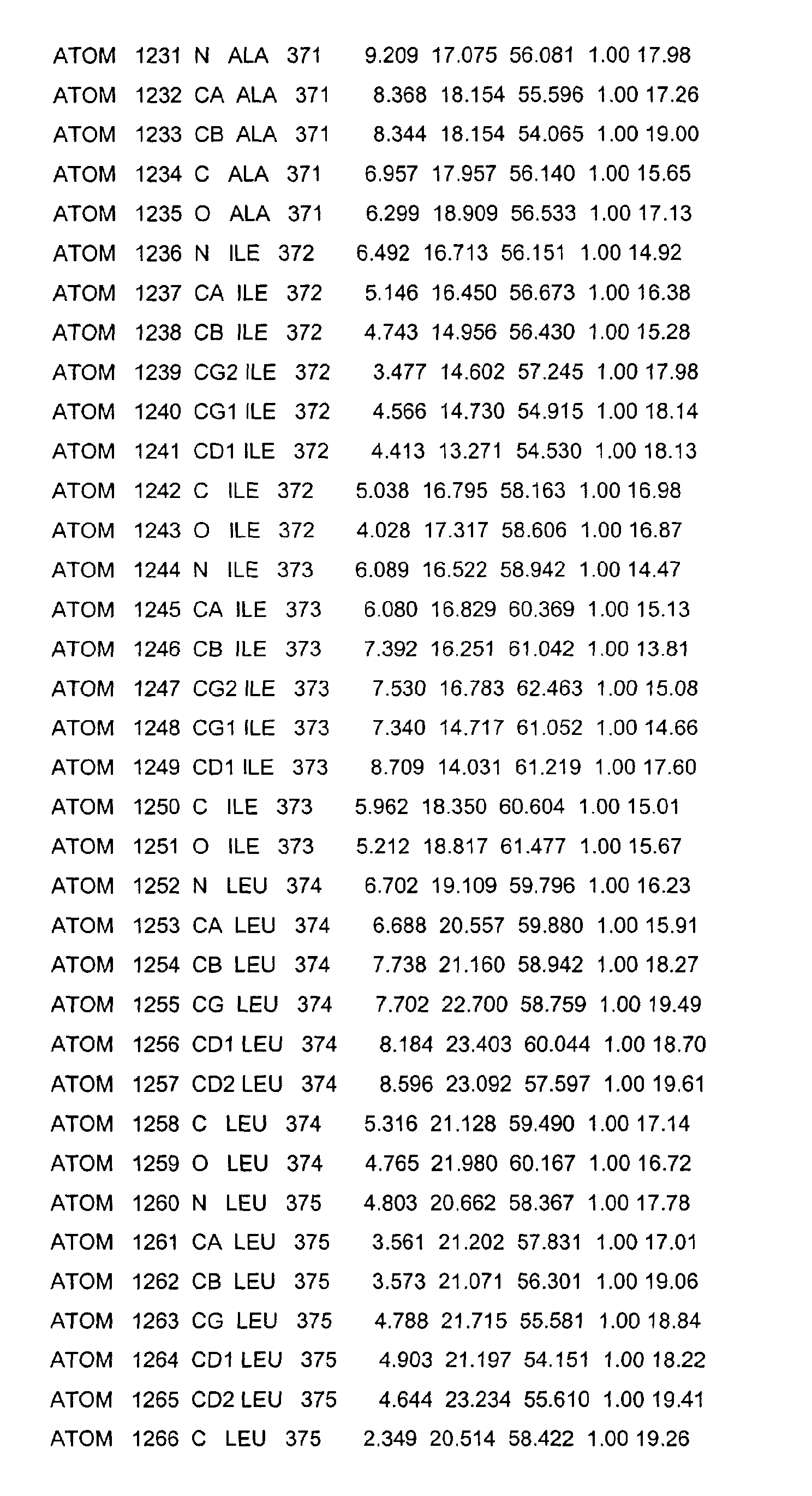
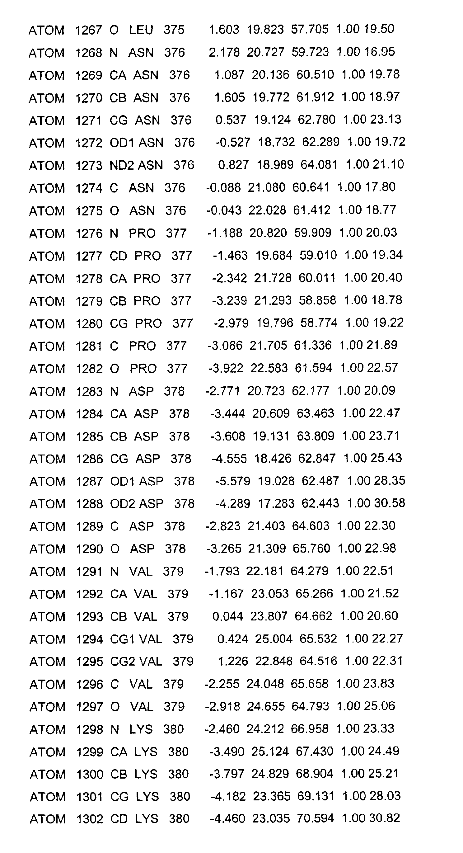
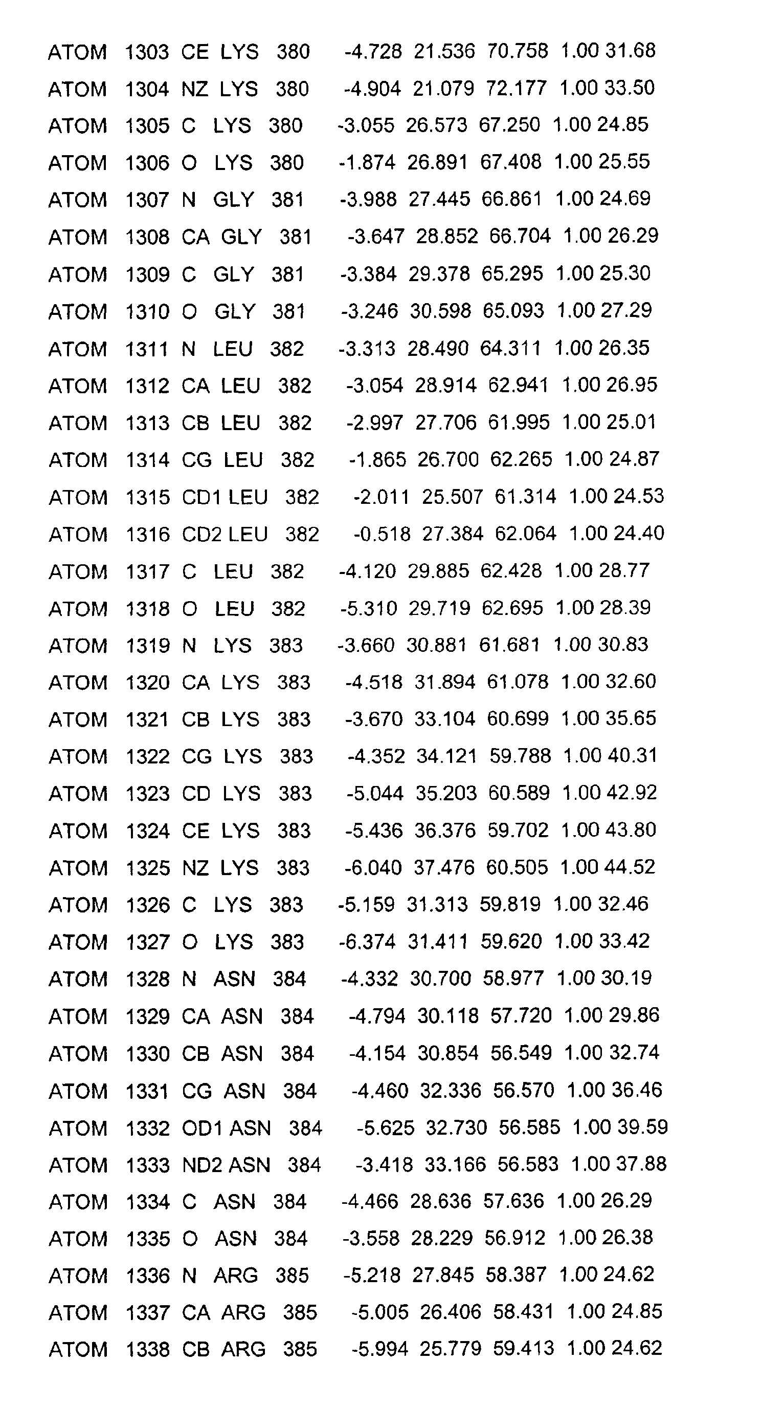
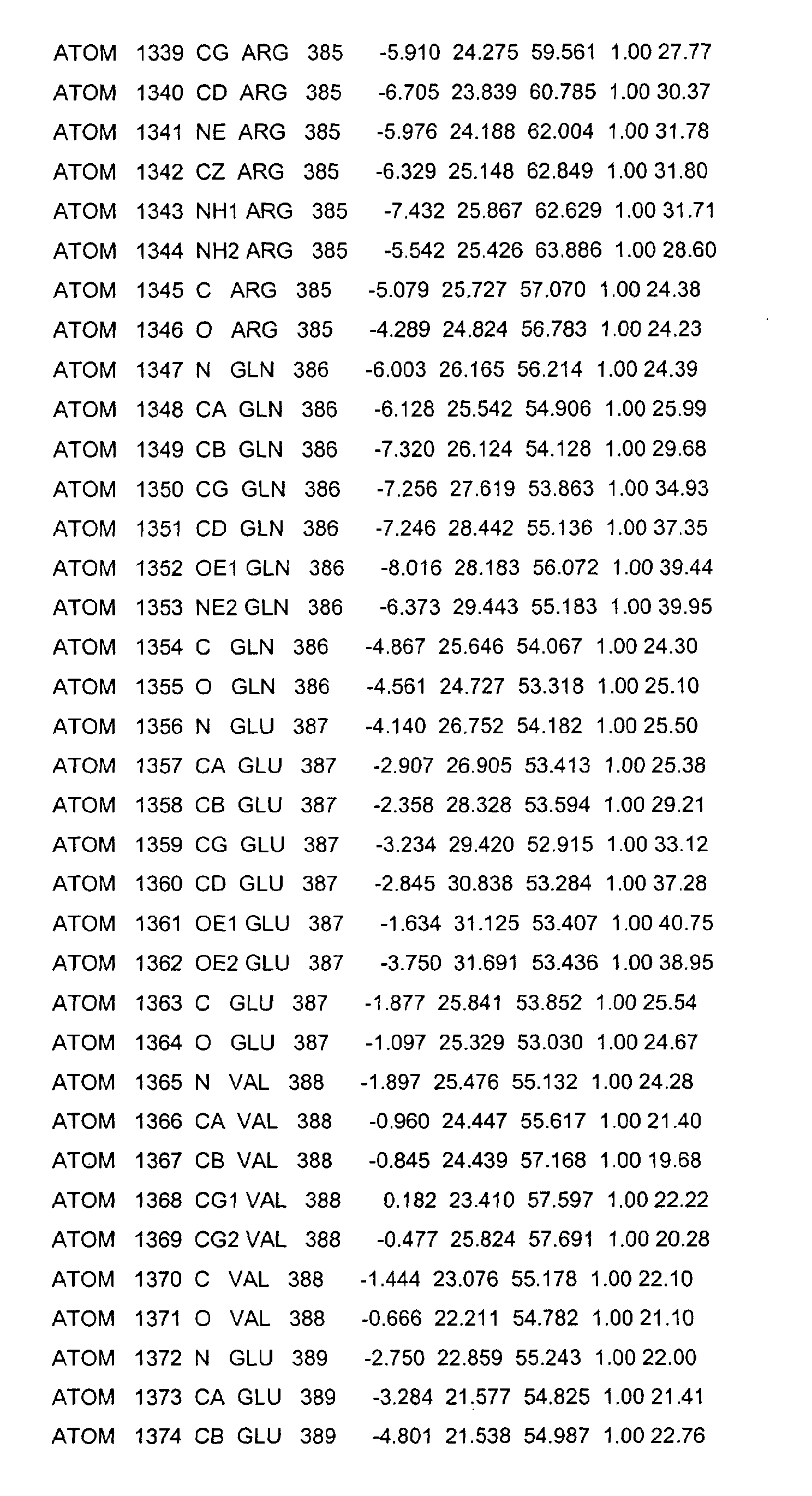
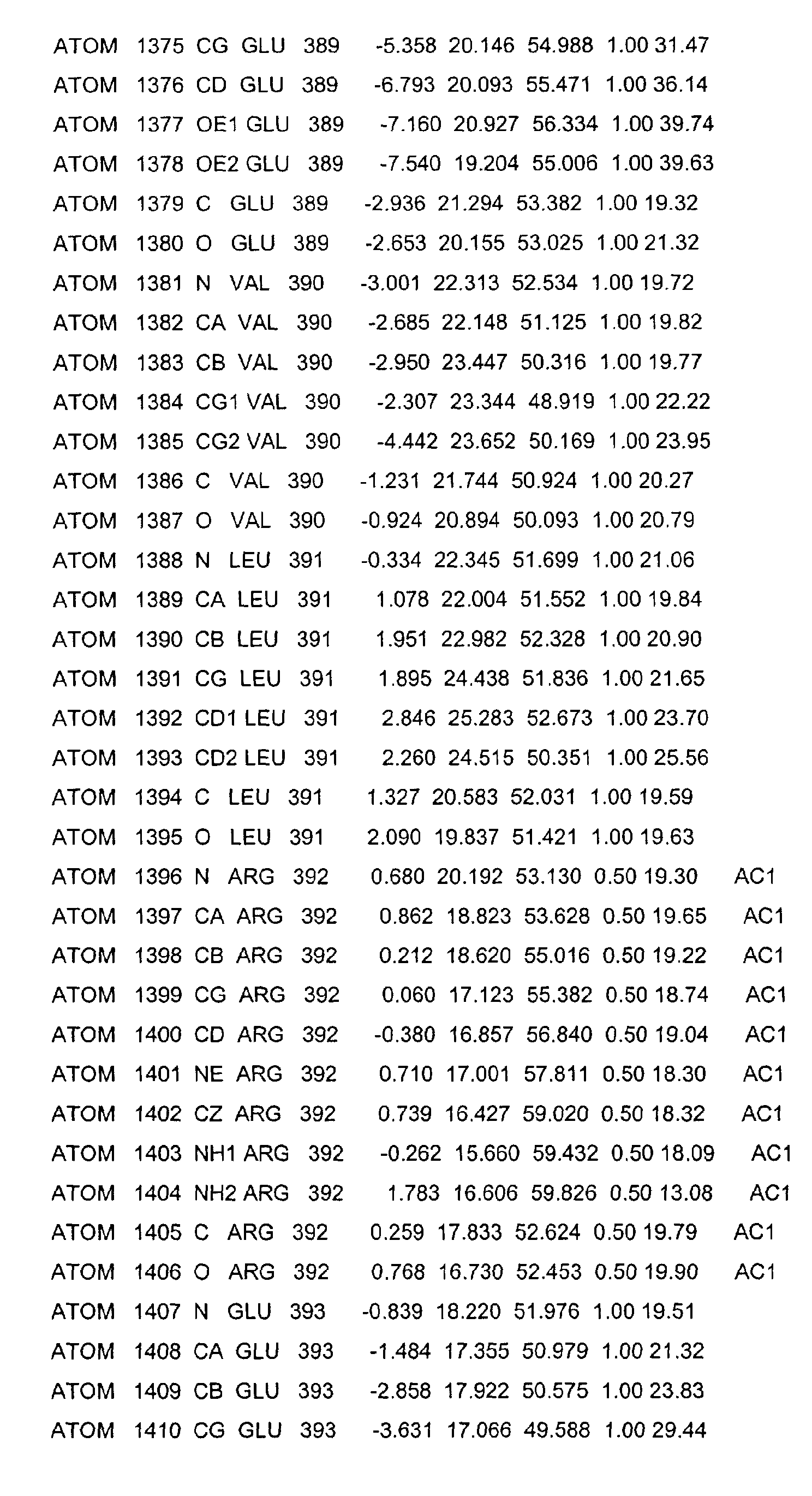
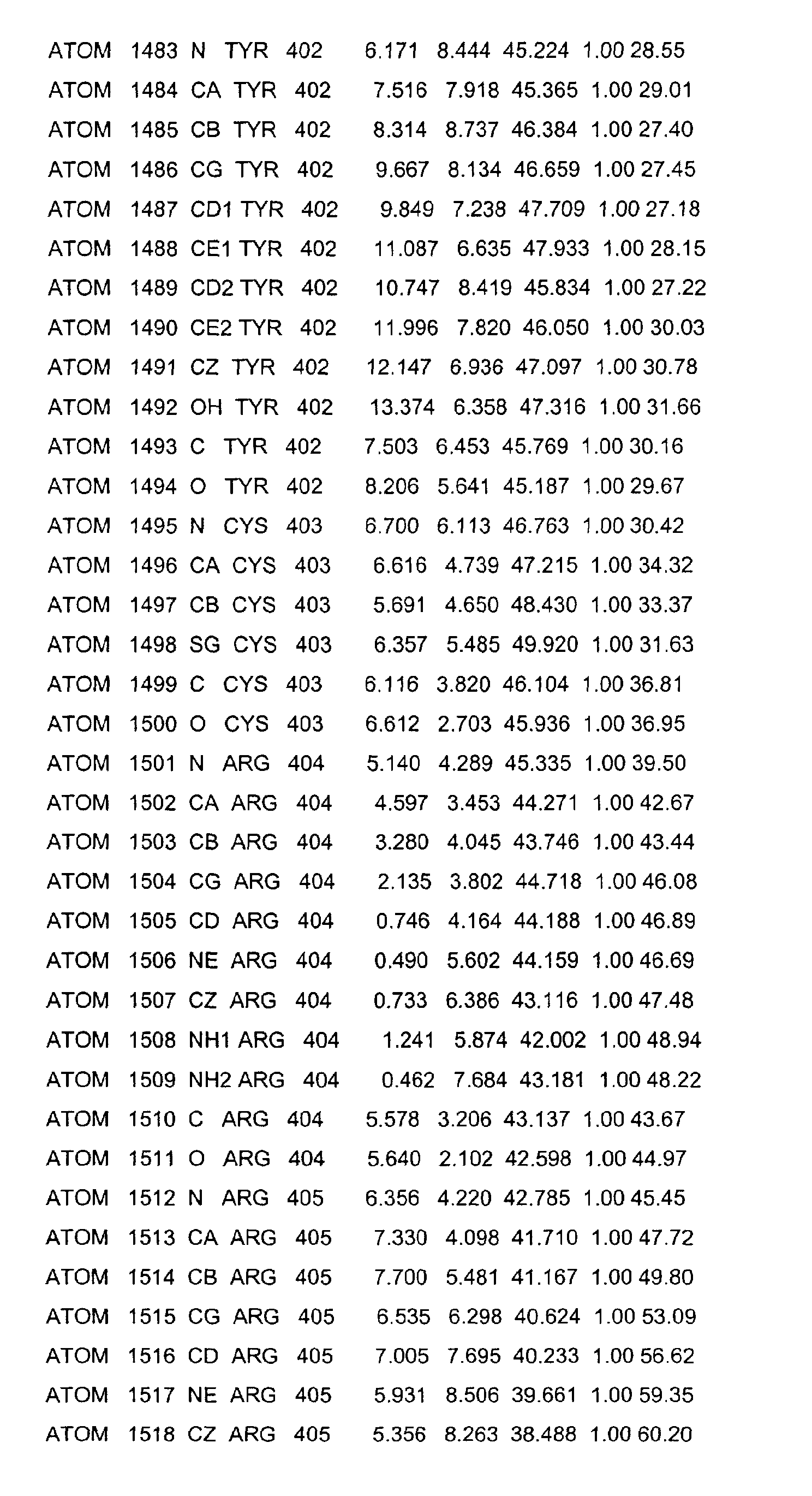
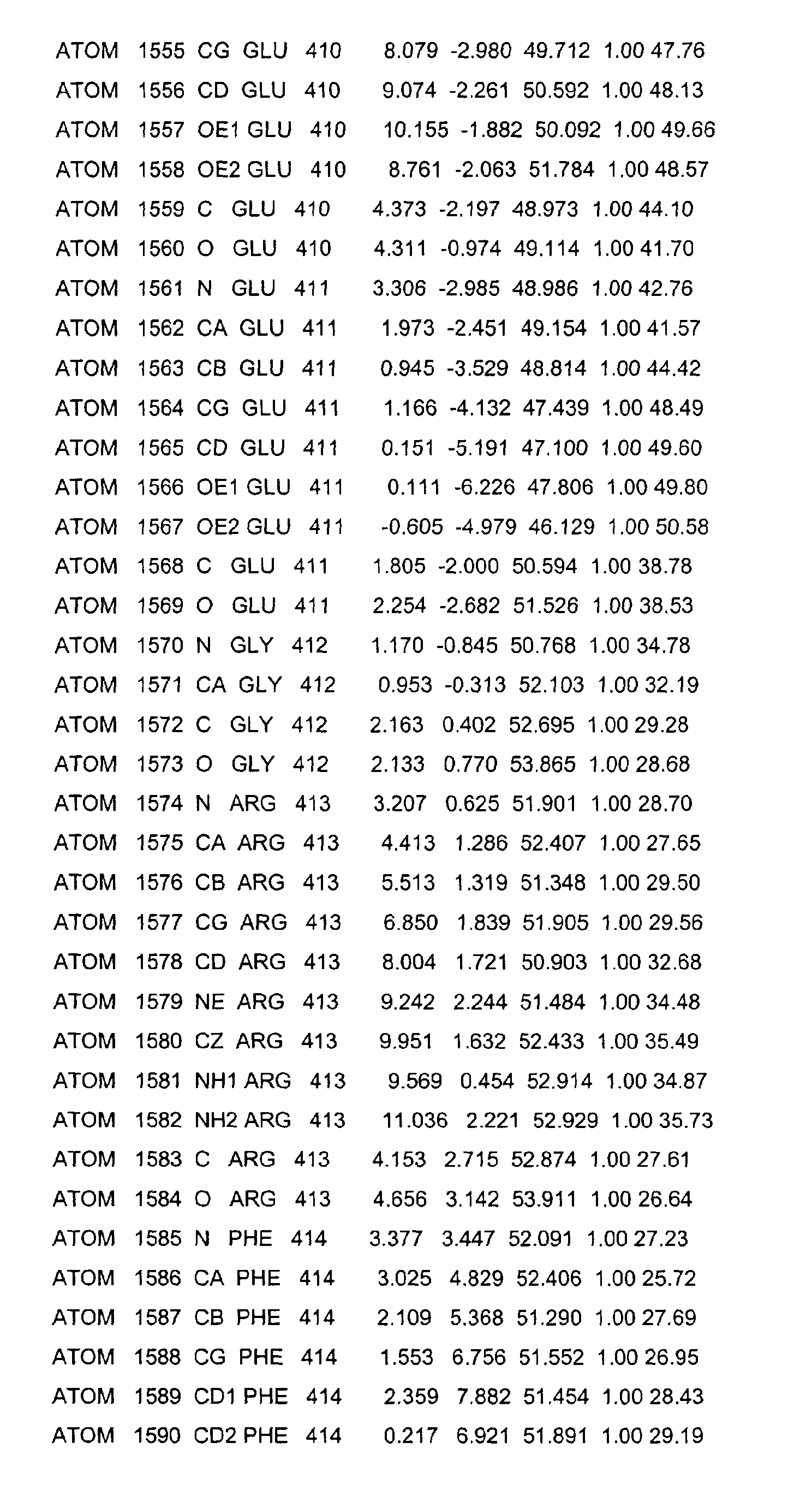
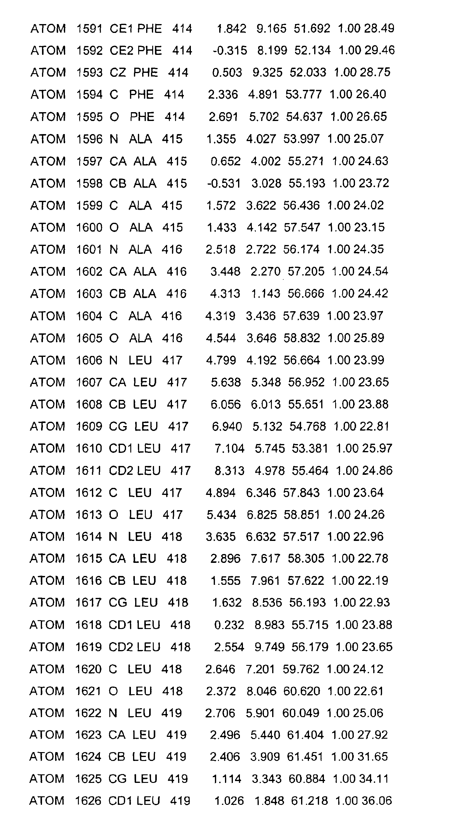
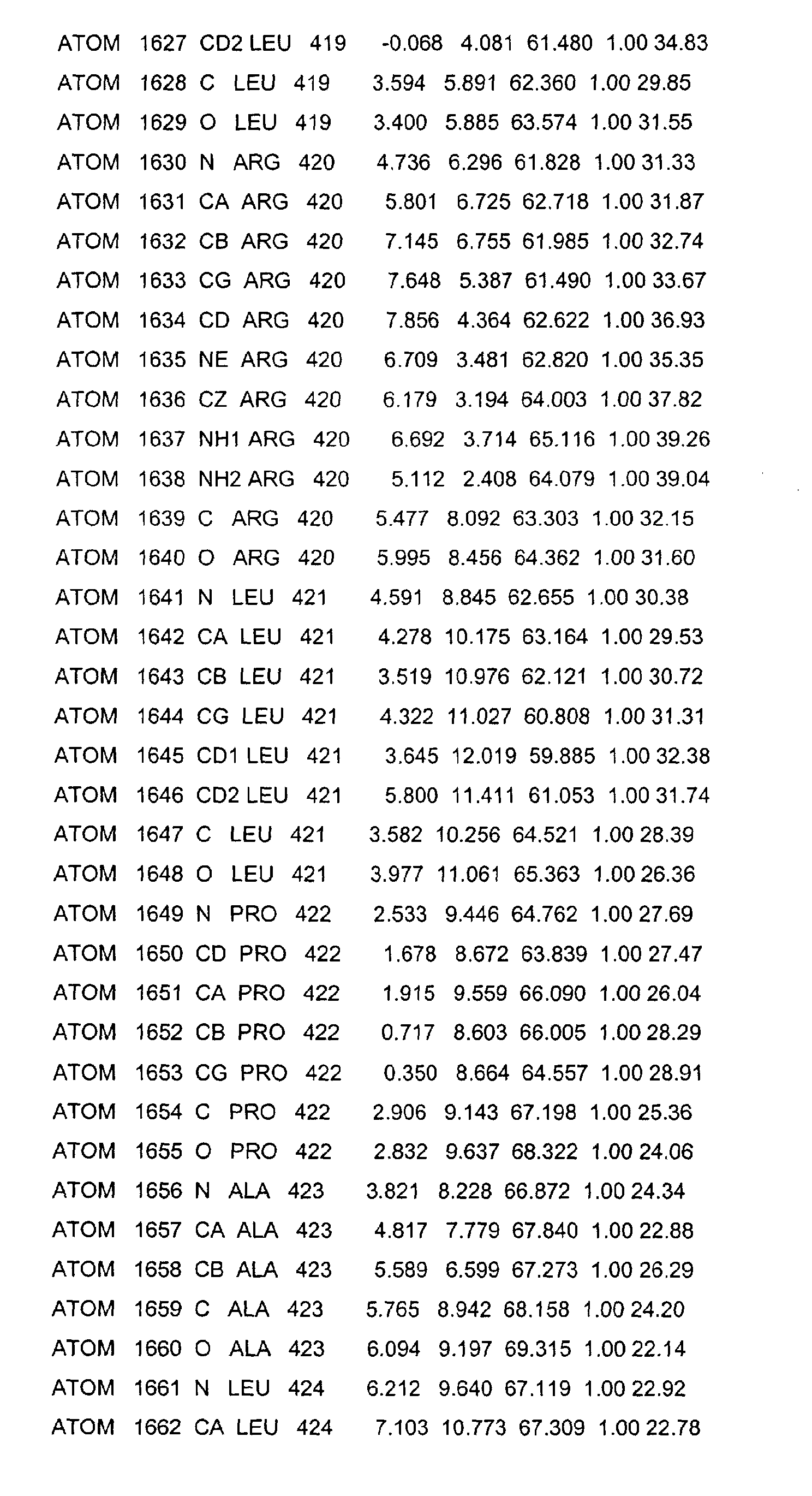
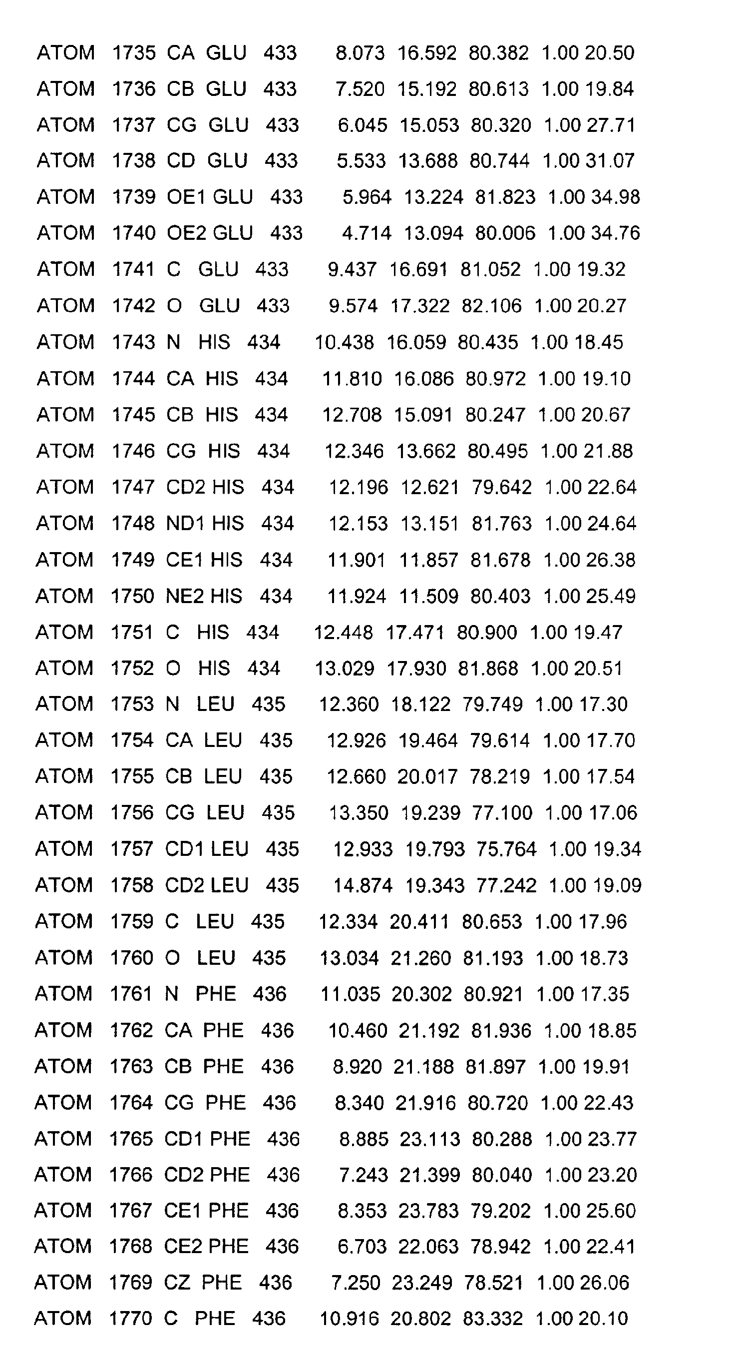
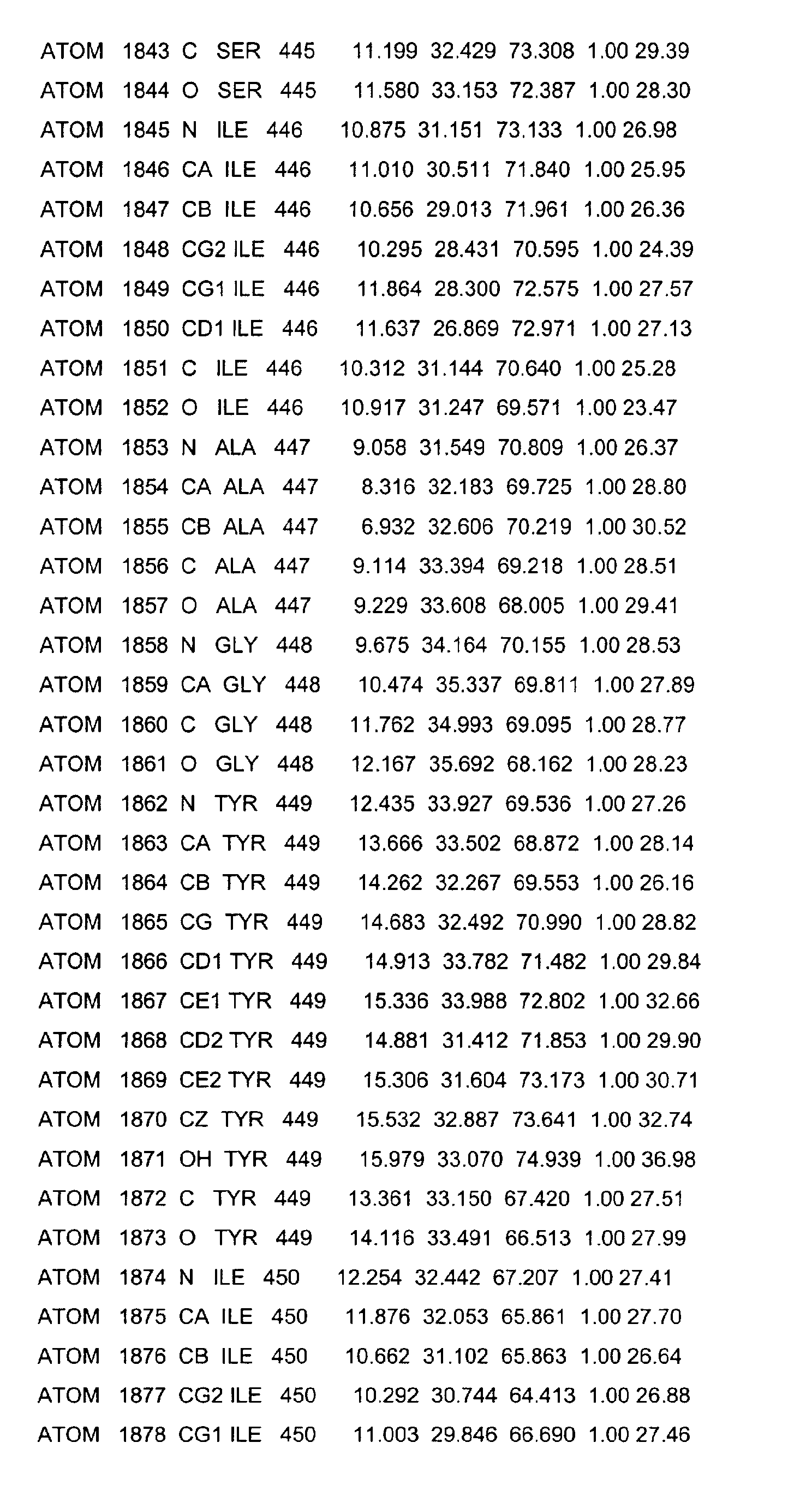
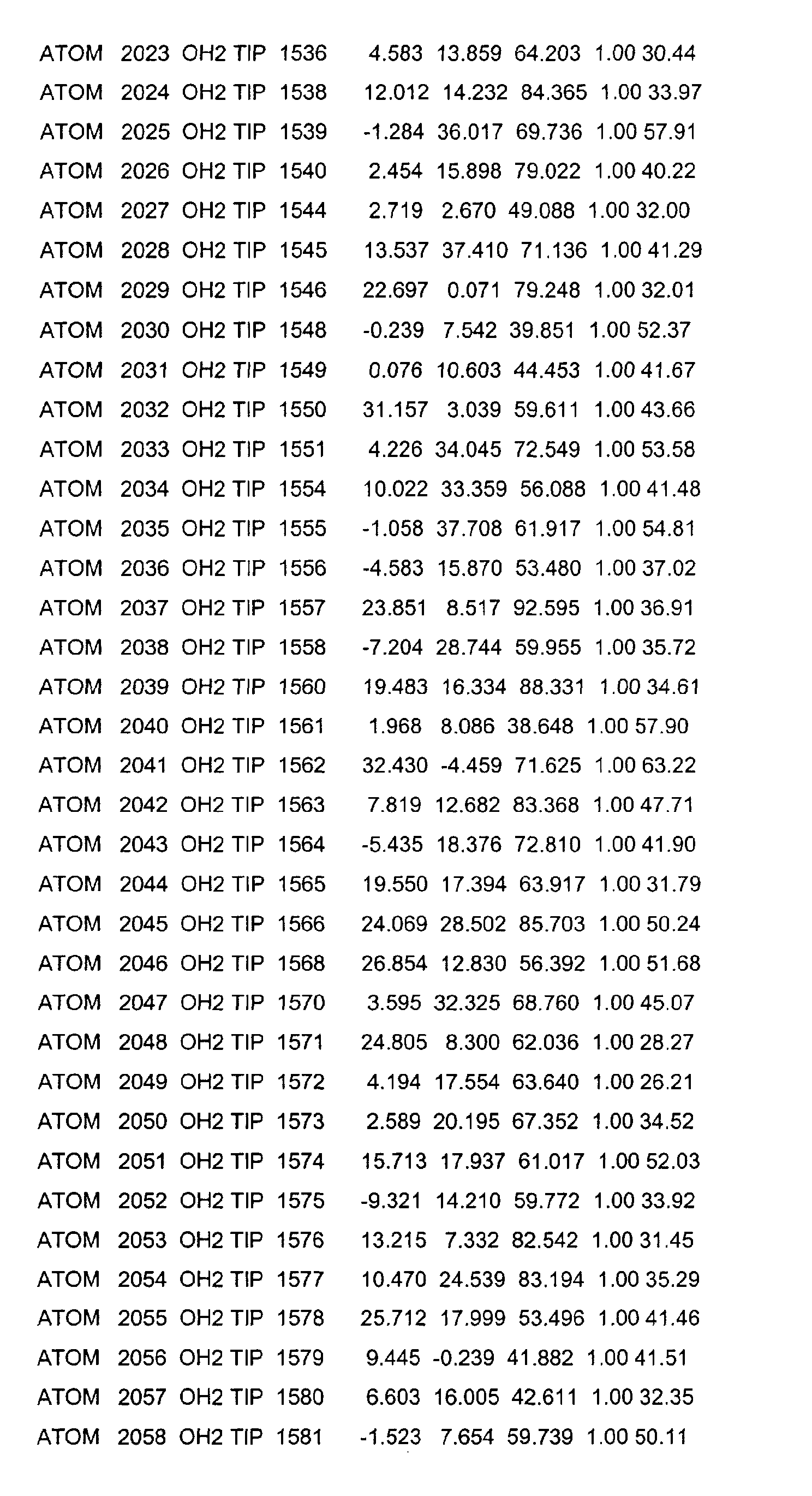
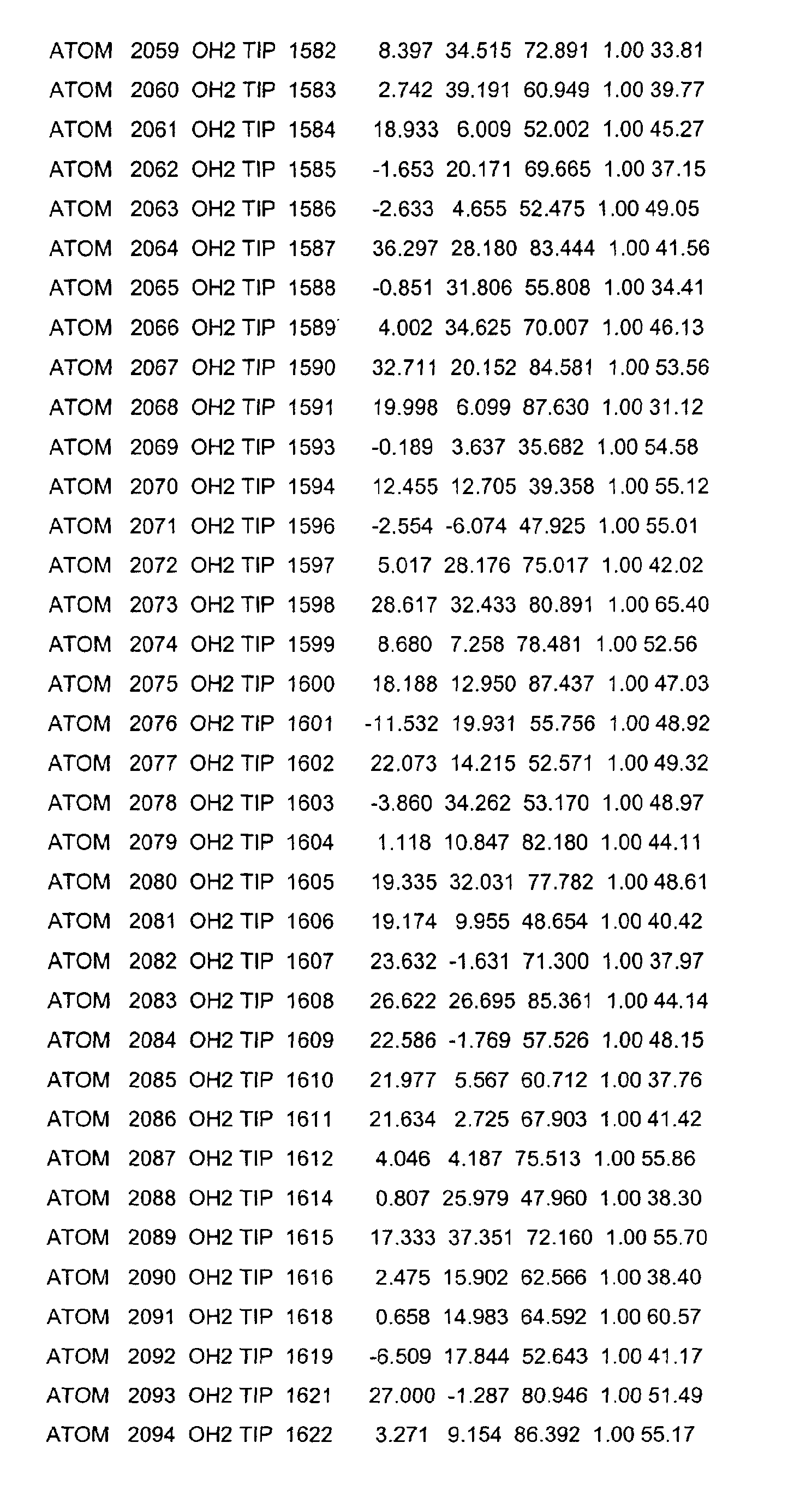
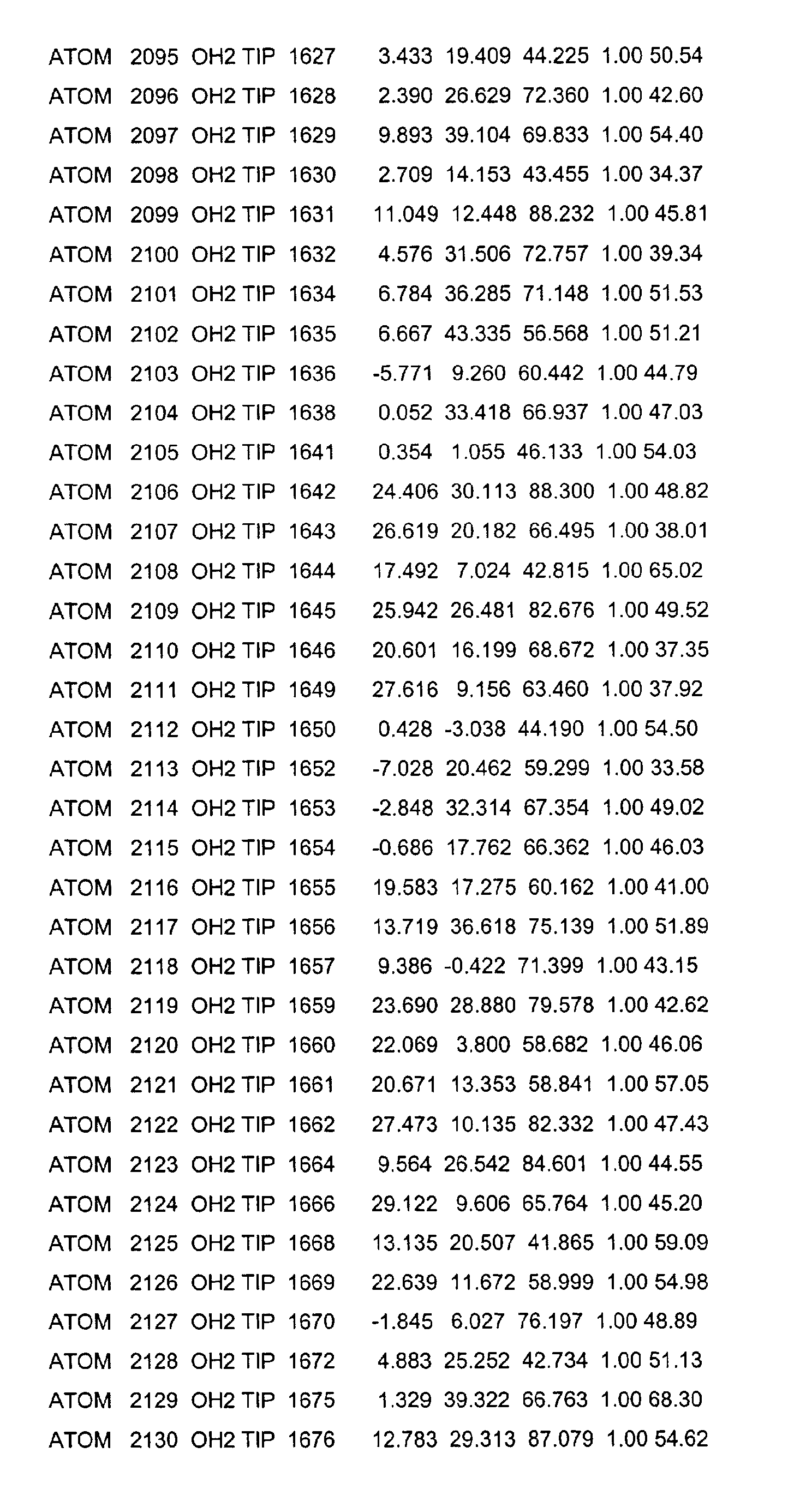
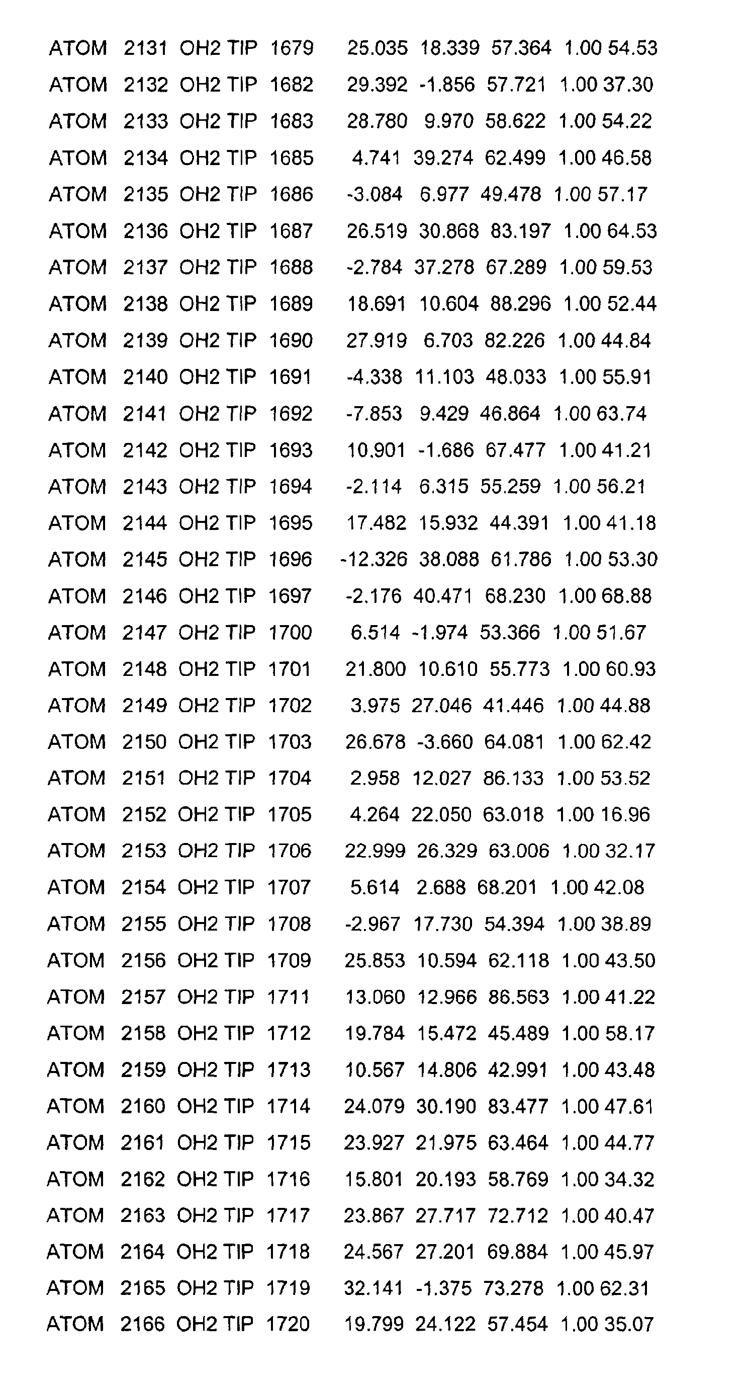
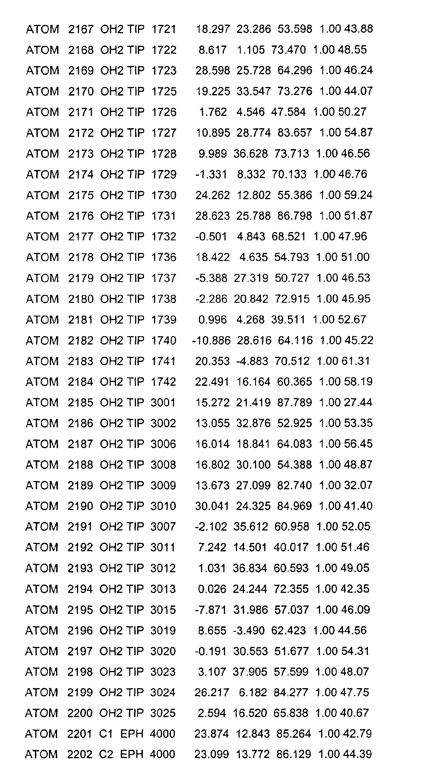
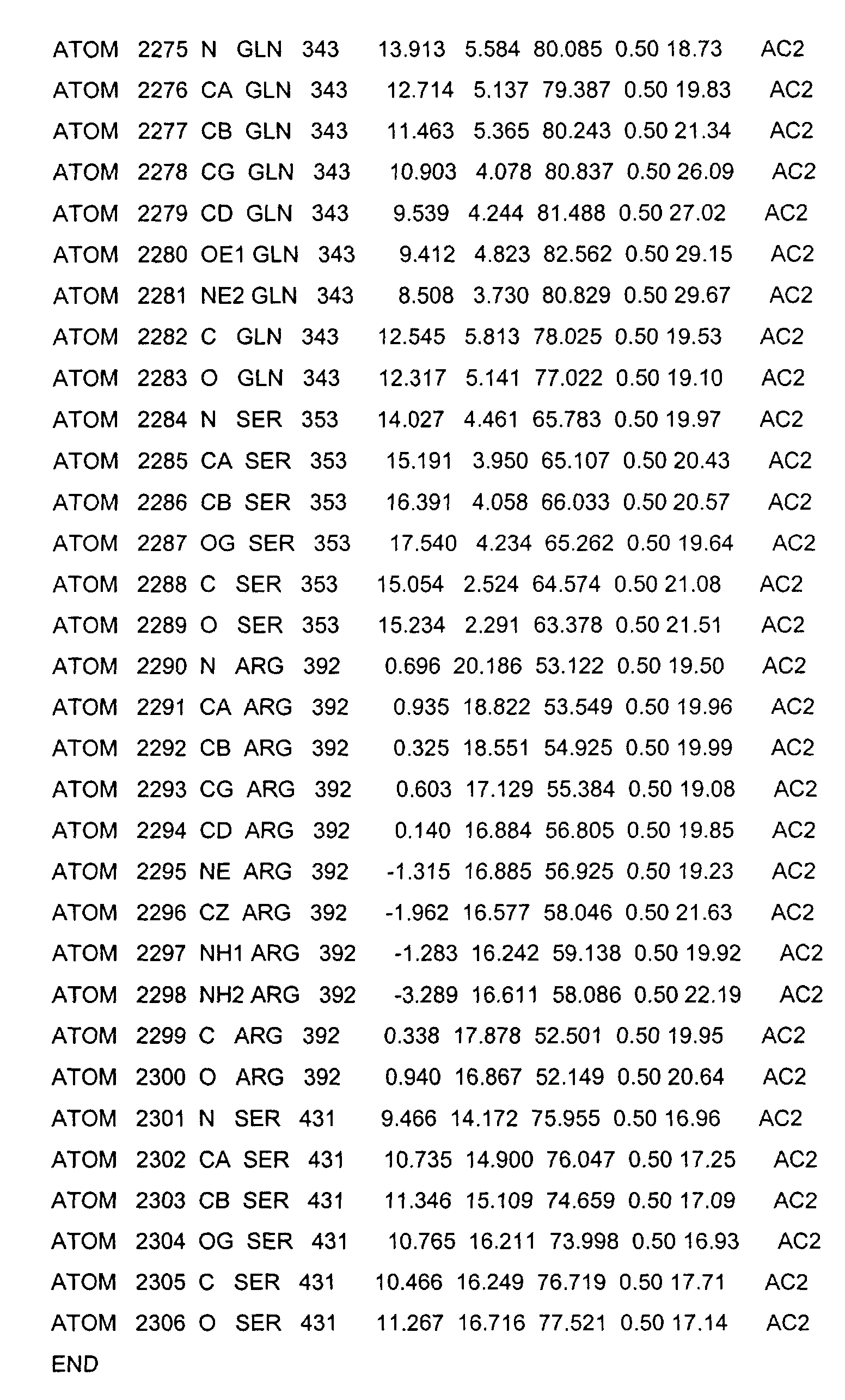
- the LBD according to the invention preferably has those defined in Table 1 Structural coordinates.
- the three-dimensional structure was created with the help of Protein crystals that are accessible for X-ray structure analysis at high Dissolution by means of molecular replacement solved and completely refined. object The present invention is therefore also based on these structural coordinates ascertainable three-dimensional structure of the USP-LBD.
- the present invention furthermore relates to a USP LBD which has a Ligand binding pocket, which is characterized by the amino acids LEU230, VAL238, PRO239, PHE242, LEU249, LEU291, ILE294, MET323, LEU331, GLN338, ALA339, VAL341, PHE345, SER431, HIS434, LEU435, PHE438 and LEU440 as defined in SEQ ID NO: 2 and Table 1.
- a USP-LBD is the subject of the present invention, the Ligand binding pocket, which is characterized by the amino acids LEU230, VAL238, PRO239, PHE242, PRO245, VAL246, LEU249, CYS250, GLY253, ASN287, LEU290, LEU291, ILE294, MET323, LEU325, LEU331, SER335, ALA336, GLN338, ALA339, VAL341, ILE344, PHE345, VAL348, SER431, HIS434, LEU435, PHE438 and LEU440 is defined according to SEQ ID NO: 2 and Table 1, includes.
- a USP-LBD is the subject of the present invention
- the Ligand binding pocket encompassed by the amino acids described above is defined, and in which one or more of these amino acids are mutated.
- these are conservative mutations in which an exchange by an amino acid with similar physical properties.
- Using the three-dimensional structure of the USP-LBD according to the invention can scan databases using established automated computer protocols that contain the structures of a large number of connections (Virtual screening). For virtual screening, algorithms such as FLEXX (13) or GOLD (14) can be used. By doing this, you can Compounds are identified whose three-dimensional structure allows in to reach the binding pocket and bind there, for example by forming Hydrogen bonds, through hydrophobic interaction, through electrostatic Interactions, through van der Waals interactions or through dipole interactions. The compounds identified in this way can be synthesized and then e.g. as insecticides or as effectors in expression systems (genes Switch) based on the USP.
- Genes Switch gene Switch
- USP-LBD Another application of the three-dimensional structure of the invention USP-LBD lies in the generation of new ligands.
- de novo design programs on the computer structural formulas generated for new ligands that can get into the binding pocket and there can bind, for example, through the formation of hydrogen bonds hydrophobic interaction, through electrostatic interactions, through van der Waals interactions or through dipole interactions.
- Possible de novo design programs are LUDI (15), LEGEND (16) or GROW (17).
- Compounds generated in this way can be synthesized and then also e.g. as insecticides or as effectors in expression systems (gene switch) based of the USP.
- the three-dimensional structure of the USP-LBD according to the invention also enables the three-dimensional structure of a USP-LBD from other organisms using modeling methods predict.
- Such protein models can be done in the same way can be used like the three-dimensional structure solved here.
- By comparing the Differences in the amino acid sequences it is possible to find differences in the Predict ligand binding pockets of different organisms. This is useful if specific ligands are searched for specific organisms, or if im Conversely, non-specific ligands are sought.
- the three-dimensional structure according to the invention for creating protein models serve other nuclear receptors related in the sequence.
- a computer readable data storage medium that is a data storage material on which the structural coordinates of an LBD according to the present Invention are stored.
- a computer-readable data storage medium in a form that enables one three-dimensional imaging of an LBD according to the present invention to generate a computer screen.
- a method for creating protein models of USP LBDs characterized by creating a three-dimensional image of a computer LBD according to the present invention.
- a process for building protein models of USP LBDs in one agonistic conformation characterized by computer-assisted creation a three-dimensional image of an LBD according to the present invention in an agonistic conformation.
- a process for creating protein models of nuclear receptors that Show homologies to USP-LBDs characterized by computer-aided Creation of a three-dimensional image of an LBD according to the present Invention with a mutated amino acid sequence.
- a process for creating protein models of nuclear receptors that Have homologies to USP-LBDs in an agonistic conformation characterized by computer-assisted creation of a three-dimensional Image of an LBD according to the present invention with a mutant Amino acid sequence in an agonistic conformation.
- the USP-LBD from Heliothis virescens was identified as the N-terminal Fusion protein with a His tag in a pET-15b expression vector cloned and overexpressed in E. coli strain BL21 (DE3).
- Cells were grown in 2x LB Medium cultured at 37 ° C and 2 hours with 0.8 mM isopropyl- ⁇ -D-thiogalactopyranoside induced at 24 ° C.
- the protein extract was on a cobalt chelate column with subsequent gel filtration on a Superdex 200 16/60 column purified. Then His day was removed by thrombin digestion and that Protein separated by gel filtration.
- a homogeneous, monomeric protein species lay in the solution and was by means of SDS and native polyacrylamide gel electrophoresis as well as denaturing and native electrospray ionization mass spectrometry approved.
- Crystallization was carried out by gas diffusion on hanging drops.
- the protein concentration used was 3-9 mg / ml.
- Crystals with a size of 200 x 200 x 400 mm 3 formed within 10 days from a solution containing 10% polyethylene glycol (PEG) 4000, 50 mM Tris (pH 7.5), 100 mM NaCl and 5 mM dithiothreitol and was equilibrated against a solution of 20% polyethylene glycol (PEG) 4000 and 100 mM Tris (pH 7.5) in the reservoir.
- the crystals belong to the tetragonal P4 3 22 space group with one monomer per asymmetric unit.
- the solvent content is 32% and the B factor estimated in the Wilson plot is 27 ⁇ 2 .
- Crystals were briefly immersed in a 10% glycerin solution and snap frozen in liquid nitrogen.
- the native data set was created with a single crystal at test stand ID14-EH2 at the ESRF (Grenoble, France).
- the data were processed with the help of HVAC programs (18).
- the crystal structure was solved by the molecular replacement method (19) using a partial hRXR ⁇ structure (20) as a search model.
- the phasing power of the model was low and required numerous manual assembly cycles with O (21).
- the wARP method (22) was used to check the correctness of the partially built structures.
- the refinement was carried out using CNS (23) using a maximum likelihood target function and solvent correction. Cycles of manual modeling and least-square minimization with subsequent simulated annealing and individual anisotropic B-factor refinement led to the final model. Solvent molecules were contoured in a F o - F c map with a surface of 3 ⁇ .
- the final model, refined to a resolution of 1.65 ⁇ , contains 246 amino acid residues, 259 water molecules and one ligand molecule. A large part of the connection loop between Helix H5 and the beginning of the ⁇ -sheet (amino acid residues 306-315) and the C-terminal extension of H12 (amino acid residues 459-466) could not be shown due to the poor electron density in these regions.
- the quality of the final model was checked with Procheck (24).
- a phosphatidylglycerin or a phosphatidylethanolamine or a phosphatidylcholine would match the crystallographic data and are consistent with the Results from mass spectroscopy and chemical analysis.
- These amphiphiles Molecules have a head group consisting of a phosphorylglycerol or a Phosphorylethanolamine group and a tail from two different Fatty acids which are bound to the glycerol-3-phosphate by ester bonds.
- a detailed description of the ligand and its interactions with the Remains of the USP-LBD are given below.
- the architecture of the USP-LBD shows a canonical NR folding with 11 ⁇ helices (H1, H3-H12) and two short ⁇ -strands (s1-s2).
- This structure was created with compared two other crystal structures, the essential properties of NRs own and are closely related to the USP of Heliothis virescens: the Binding pockets of agonist-bound RXR ⁇ (hRXR ⁇ / 9-cis RA) and antagonist-bound Mouse RXR ⁇ (msRXR ⁇ / oleic acid).
- the overlay of the USP-LBD with The structure of the Holo-RXR ⁇ -LBD was determined using a least square fit [method least squares; LSQ].
- the secondary structure elements the USP-LBD with those of the Holo-RXR ⁇ -LBD quite well overlap.
- the root mean square deviation, r.m.s.d.) is 1.22 ⁇ for 183 of 246 superimposed C ⁇ atoms. Seven helices can be overlaid quite well (r.m.s.d. 1.13, 0.88, 0.57, 1.18, 0.67, 0.69, 0.75 ⁇ for H4, H5, H7-H11).
- the C-terminus of H1 is around 2 ⁇ to the helix H3 bent and be r.m.s.d. is 1.63 ⁇ . H3, H6 and the ⁇ -sheet show larger ones Deviations.
- the structure of the USP-LBD shows that the activation helix H12 adopts a conformation similar to that of the antagonist RXR ⁇ .
- the antagonistic AF-2 conformation of the USP-LBD is discussed below.
- the connecting loop L1-3 of most NRs normally behaves like one very flexible region.
- the crystal structures show both the apo and also the holo conformations substantial differences in the areas that the Connect helices H1 and H3.
- L1-3 consists of one extended loop that spans the ⁇ sheet and an ⁇ loop.
- the Apo form contains an additional helix in this area, which is in the Holo form unfolded.
- the ⁇ loop is oriented towards the opposite Side of the protein center.
- L1-3 could act as a molecular spring that the accompanied conformational changes associated with ligand binding are.
- the conformation of L1-3 is similar for the ligand-bound RAR ⁇ -LBD to that of the Holo-RXR ⁇ .
- L1-3 follows one for ER LBDs way different from the Holo-RXR ⁇ . It runs between Helix H3 and the ⁇ -sheet, tightly packed to the protein center.
- L1-3 does not adopt any of the conformations that are otherwise found in other NRs. Its course (Val-220 to Pro-239) was unambiguously derived from the electron density maps. Only a few residues at the beginning of the loop, Asp-222, Pro-223 and Ser-224, were treated as alanines due to the poor electron density of the side chains. The temperature factors of these residues are therefore higher (60-64 ⁇ 2 ) than those of the other amino acids of L1-3 (36 ⁇ 2 on average over L1-3). The first residues of L1-3 form a path that crosses helix H3 in the range from Gln-256 to Val-262.
- L1-3 assumes a rather tense conformation, which makes it possible to make direct contacts with the remains of the helices H3, H11 and H12 and to stabilize their current positions. This is important in that these helices are the structural elements that undergo the greatest conformational changes due to ligand binding.
- L1-3 is not based on crystal packing effects.
- Helix H3 differs in both Length as well as in the position of the N- and C-terminal part of their counterparts in RXR.
- H3 starts at Pro-240 and is therefore one Turn longer than H3 in the ligand-bound RXR ⁇ (start at RXR ⁇ -Pro-264).
- the remains of H3 in the central part of the helix take almost identical positions compared to the positions of the corresponding residues in the Apo and Holo RXR ⁇ LBDs.
- both N- and C-terminal areas are for Curved outside of the protein center.
- the N-terminal region of H3 (Pro-240 to Cys-250) has shifted substantially towards H11.
- the ligand binding pocket of the USP from Heliothis virescens is formed from residues from loop L1-3, helices H3, H5, H6 and H7, the ⁇ -sheet and loop L11-12.
- the N-terminal part of the Helix H3 is clearly shifted outwards compared to its counterpart in RXR ⁇ .
- Two other secondary structures that contribute to the binding pocket also differ from those in RXR ⁇ : 1) Helix 6 has moved inward by approximately 1.9 ⁇ , and 2) the curvature of the ⁇ -sheet points to H1.
- the associated shift of the three structural elements leads to an expansion of the ligand binding pocket compared to that of the RXR ⁇ -LBD.
- the edge of the binding pocket is formed by the ⁇ loop of L1-3, the N-terminus of H3 and H6, whereas in RXR ⁇ the loop L11-12 and H6 form the entrance of the pocket.
- the binding pocket is approximately 13.5 ⁇ wide at its entrance (distance between Lys-241 in H3 and Gln-338 in H6). This input is much wider than RXR ⁇ (7.1 ⁇ from Pro-264 in H3 to Ala-340 in H6).
- the topology of the ligand binding pocket is quite unusual with a gap between H3 and H6. In RXR ⁇ and other NRs, this area forms fixed contacts to the connecting loop L1-3.
- the volume of the cavity of the USP-LBD is 2.5 times larger than that of the hRXR ⁇ -LBD (1256 ⁇ 3 for USP compared to 489 ⁇ 3 for hRXR ⁇ ).
- the USP ligand binding pocket from Heliothis virescens unexpectedly contains a molecule that is co-purified and co-crystallized with the USP-LBD has been.
- the fit of the electron density fits well with that of mass spectroscopy and analytical-chemical characterization of the molecules.
- this molecule is not the is natural ligand of vertebrate NR, it induces and stabilizes one antagonistic AF-2 conformation, most likely very similar to that actual antagonist-bound RXR ⁇ is.
- the best fit of the electron density was assumed of a phospholipid, the first tail of which consists of a fatty acid with a There is a length of 18 carbon atoms at C1, and one 16 carbon atom long second chain on C2. The longer of the two fatty acids is right twisted shape with two larger creases, whereas the other fatty acid a more normal shape inside the bag. The tail of the Phospholipids is hidden within the ligand pocket.
- the glycerin part and the two fatty acids form van der Waals contacts with the residues in L1-3 (Leu-230, Val-238), H3 (Phe-242, Leu-249), H5 (Leu-291), L6-7 (Ala-339), H7 (Phe-345), H11 (Ser-431, His-434, Phe-438) and L11-12 (Leu-440).
- the head group The phospholipid is located at the front of the pocket between H3 and H6.
- At the Phosphatidylglycerol forms the carbonyl group of phosphorylglycerol and at Phosphatidylethanolamine the amino group of ethanolamine has a strong hydrogen bond with Gln-338 (H6).
- an oxygen Phosphate group via a hydrogen bridge with a residue L1-3 (C ⁇ of Pro-239) bound.
- the phospholipid found here probably does not represent a natural ligand of USP. However, it has been clearly shown that USP ligands exist.
- the tail of the longer fatty acid lies approximately at atom C9 of 9- cis RA in the hRXR ⁇ -LBD, whereas the tail of the other fatty acid extends almost to the ⁇ -ionone ring of 9- cis RA.
- Arg-297 does not participate in anchoring the ligand as is intended in the agonistic RXR ⁇ , RAR ⁇ and other NR LBDs. Nevertheless, it occupies almost the same position as Arg-316 of the holo-RXR ⁇ and not the position of the Apo-RXR ⁇ conformation exposed to the solvent.
- Arg-297 forms hydrogen bonds to the backbone carbonyl group of Leu-325 ( ⁇ -sheet) and participates in a hydrogen bond network with Leu-290 (H5) and the side chain with water participation from Gln-256 (H3).
- Leu-290 H5
- Gln-256 H3
- two water molecules are involved in these interactions, which spatially lie approximately with the two oxygen atoms of the carboxylate group of 9- cis RA.
- the AF-2 domain in the structure of the USP-LBD has one through the ligand in antagonistic conformation generated in the ligand binding pocket.
- H12 takes that same conformation as in the case of other antagonist-bound nuclear receptors such as RXR ⁇ / oleic acid, RAR ⁇ / BMS614 and ER was found. In all In these cases it was observed that the furrow in which H12 lies is the binding site for corresponds to the helical core receptor box of core receptor coactivators. This helical core receptor box is characterized by the consensus sequence LXXLL, as shown for the ligand binding domain of PPAR ⁇ , TR ⁇ and ER ⁇ .
- H12 in the USP of Heliothis virescens are approximately Ile-450, Ala-453 and Leu-454 of H12 the same position as the first, second and third leucine residues of the LXXLL Binding motifs (IXXAL instead of LXXLL).
- IXXAL instead of LXXLL.
- H12 is in a groove from residues of H3 and H4 and L3-4 (Val-261, Arg-265, Met-275, Glu-276, Ile-279, Ile-282, Lys-283) packed.
- L1-3 is also involved in the furrow topology and has contacts with the residues Phe-227, Gln-228 and Phe-229 van-der-Waals H12.
- H12 in the USP-LBD is identical to that of H12 in the Antagonist-bound form of the RXR ⁇ -LBD.
- H11 winds up and so H12 allowed to adhere to the binding groove of the core receptor-coactivator binding motif Tie LXXLL.
- H11 is in the extension of H10 and overlaid deal very well with H11 in the holo-RXR ⁇ -LBD structure, except that the H11 of USP-LBD is two residues shorter. A region of 6 residues follows, the H11 and H12 connects (His-439 to Thr-444).
- H11-12 loop span a 12 ⁇ strand in a stretched conformation.
- the C-terminus of H11 contains three phenylalanines, which can also be found in RXR ⁇ .
- Apo-RXR ⁇ show the first two phenylalanines for the hydrophobic ligand binding pocket and the third phenylalanine faces the solvent.
- the phenylalanines switch roles.
- Phe-436 and Phe-437 are turned to the solvent, while Phe-438 to the ligand pocket contributes.
- the side chain of Phe-438 is compared to its counterpart slightly rotated in RXR ⁇ and touches the ligand at the level of its shorter one Fatty acid.
- the first residue corresponds of the phenylalanine triplet at the end of H11. This rest is roughly in the position of the C- ⁇ atom of Phe-437.
- the other two phenylalanine residues that are already part of L11-12, are in the ligand binding pocket inwards to the Protein-oriented.
- these two residues collide with the Phospholipidliganden.
- connection region L1-3 interacts with H3 and L11-12 and prevents one agonistic conformation
- the loop L1-3 interacts with H3, H11, L11-12 and H12. These structural elements are most affected by ligand binding.
- L1-3 stabilizes the N-terminus of H3 through a hydrogen bond network with Arg-243 and Asn-254 from H3.
- the guanidinium portion of the Arg-243 is on with strong hydrogen bonds the backbone carbonyls of Gly-233, Ser-236 and Val-238 are anchored (distances of 2.61, 2.97 and 2.78 ⁇ ) and shows a van der Waals contact to the side chain from Val-232.
- the backbone amide group of the Arg-243 is through a Hydrogen bond to the carbonyl group of Pro-239 (3.20 ⁇ ).
- the Side chain of Asn-254 forms hydrogen bonds to the carbonyl group of Leu-230 (2.83 ⁇ ), to the amide group of Phe-229 (3.10 ⁇ ) and via a water molecule to the side chain from Gln-228. It is also in van der Waals contact Phe-227 carbonyl group.
- the backbone carbonyl group of Asn-254 forms a strong hydrogen bond to the side chain of Glu-226 (2.74 ⁇ ).
- L1-3 (Gln-228 to Arg-231, Asp-235 and Ser-236) is also in contact with the N-terminal Range from H11 and to L11-12.
- the backbone carbonyl group of Gln-228 forms a hydrogen bond to Ala-442 (3.20 ⁇ ) and the backbone carbonyl group of Phe-229 forms a strong hydrogen bond to the amide group from Ala-442 (2.88 ⁇ ).
- Arg-231 stabilizes with strong interactions loop L11-12: the backbone amide group forms a strong one Hydrogen bond to the carbonyl group of Leu-440 (2.90 ⁇ ), while the A strong hydrogen bond to the carbonyl group of His-439 (3.00 ⁇ ) forms and shows van der Waals contacts with Val-441 and Ala-442.
- Other Interactions affect the backbone carbonyl of Asp-235 with the Side chain of His-439 and a water-mediated interaction with Val-441.
- the hydroxyl group of Ser-236 forms a van der Waals contact with the Leu-440 side chain.
- Residues (L1-3: Gln-228 to Arg-231, Asp-235 and Ser-236; L11-12: His-439 to Ala-442) Strictly preserved in all Lepidopteran USPs except Phe-229 and Asp-235.
- the steric prevention of the agonistic position of H12 is one here constitutive part of the receptor structure and not, as with other nuclear receptor LBDs, which are filled with fully antagonistic ligands, the consequence of Bulkyness of the ligand.
- the L1-3 region was relaxed by a Powell minimization of the Charmm program (1000 optimization steps, dielectric constant: 4, gradient tolerance: 10 -6 , step size 0.02, cutoff for non-binding interactions: 15 ⁇ ).
- This optimized structure was used as a model for the homology model of the USP-LBD used by Heliothis virescens.
- the biggest differences between these two structures are in the position the activation helix (H12) and the course of the loop between the helices H1 and H3.
- the activation helix H12 is located in the experimental structure in the antagonistic position while being an agonistic in the model structure Conformation that closes the ligand binding niche.
- the structure of the loop L1-3 lies above the helix H3 and stabilizes it antagonistic position of H12 through hydrophobic contacts. In contrast to that this loop in the agonistic homology model is quite far from the central one AF2-AD helix removed. Loop L1-3 is from helix H3 through the ⁇ sheet Cut.
- the size of the ligand binding niche differs between the two Structures considerable.
- the presence of the large fatty acid residue in the USP-LBD crystal structure causes a large cavity by shifting the helices H3, H6 and H11. These helices are tightly packed in the USP agonistic conformation and create a smaller ligand binding niche.
- H3, H4, H5, H8 and H9 are rigid and can be superimposed very well in the two structures. In contrast, they are Loop L1-3 and the C-termini of H3, H6 and H11 in both structures against each other postponed. These segments form the most mobile region of the ligand binding domain of nuclear receptors. This movement is probably for everyone Receptor and the shift generated by a ligand specific.
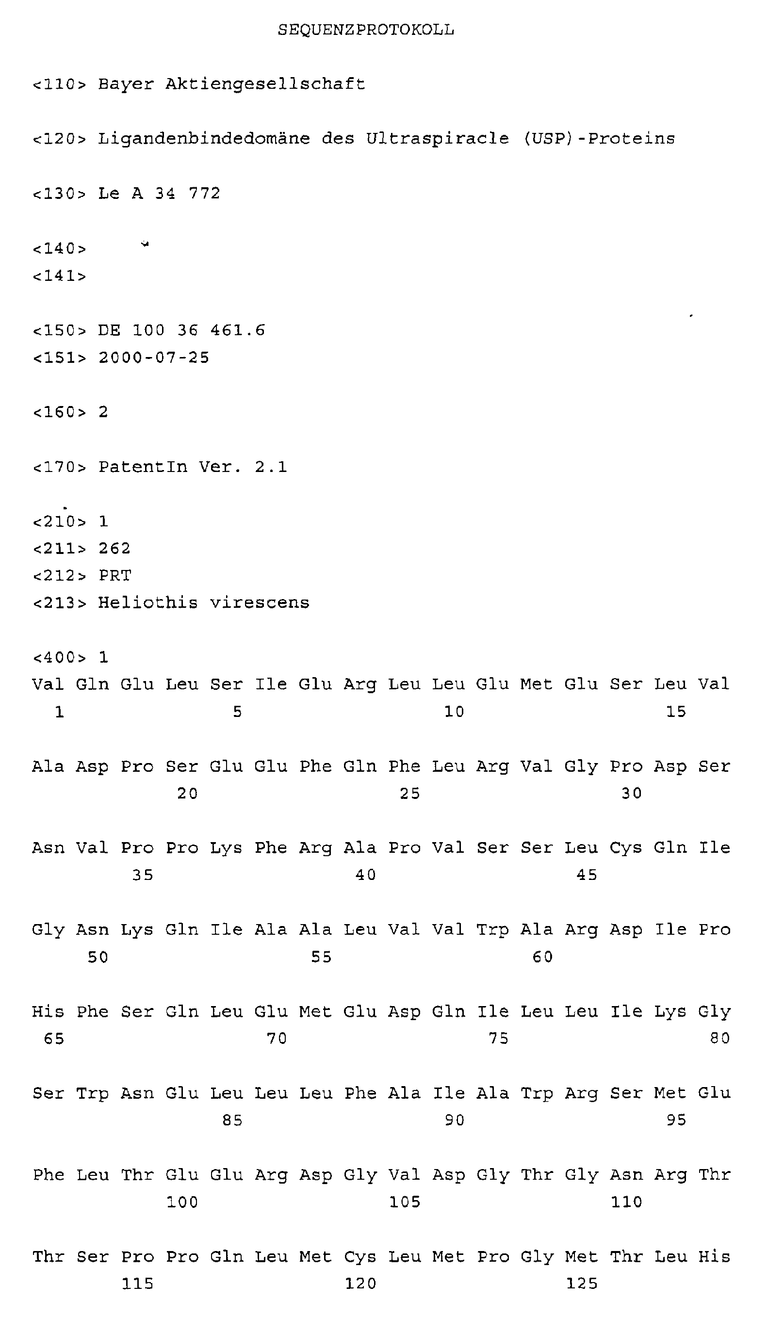
- SEQ ID NO: 1 shows the amino acid sequence of the USP-LBD from Heliothis virescens.
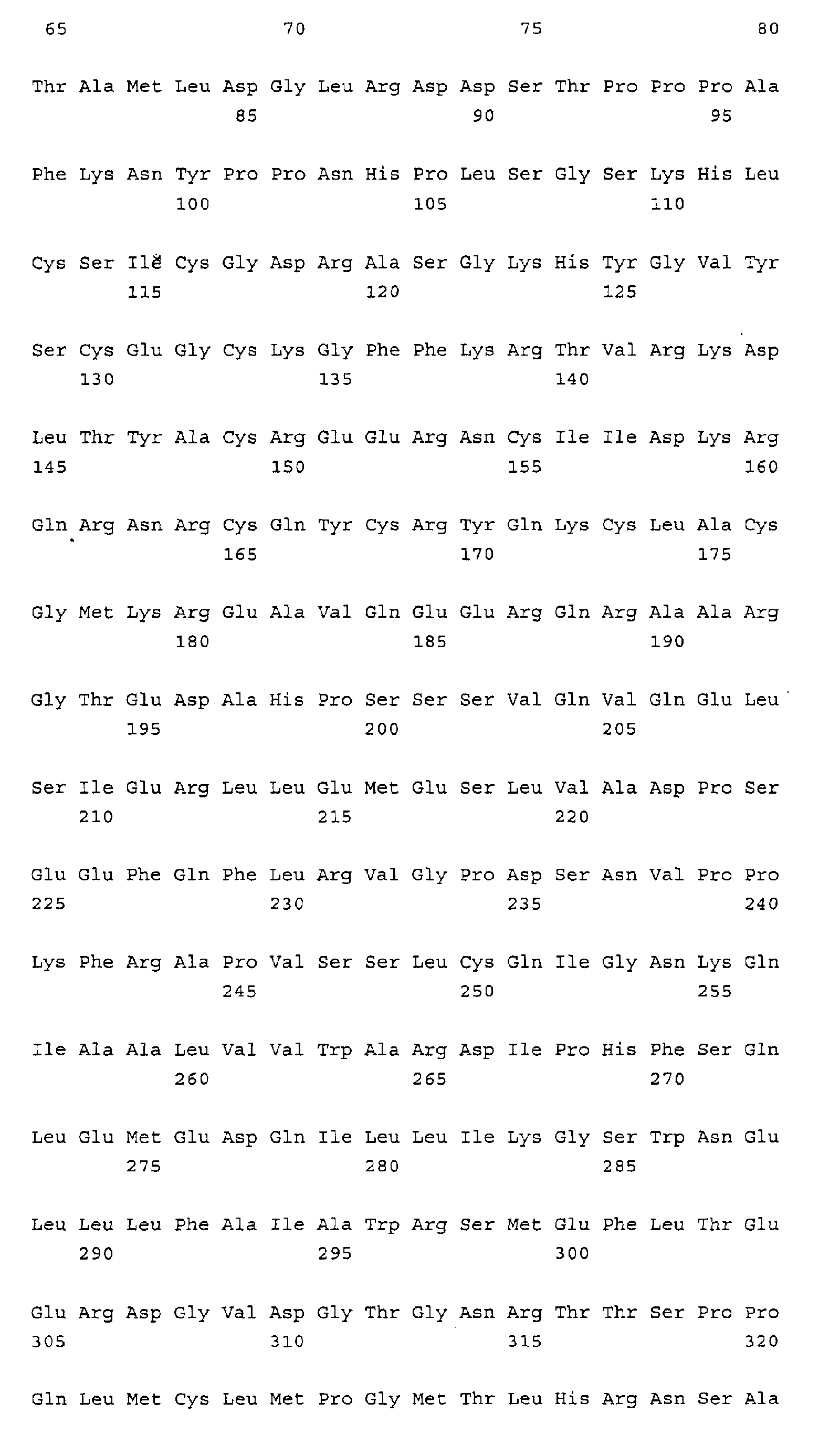
- SEQ ID NO: 2 shows the amino acid sequence of the USP from Heliothis virescens.
- Table 1 shows the structural coordinates of the LBD of the USP from Heliothis virescens.
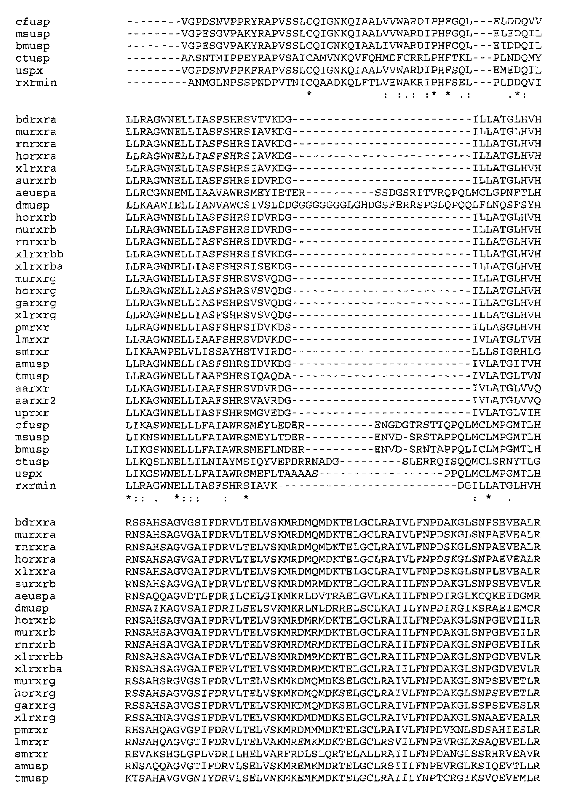
- Table 2 shows the amino acid sequence assignment for hRXR ⁇ and USP from Heliothis virescens as well as other core receptor LBDs for the formation of a homology model of the agonistic conformation of USP.
Landscapes
- Health & Medical Sciences (AREA)
- Chemical & Material Sciences (AREA)
- Life Sciences & Earth Sciences (AREA)
- Organic Chemistry (AREA)
- Zoology (AREA)
- Biochemistry (AREA)
- Medicinal Chemistry (AREA)
- Toxicology (AREA)
- Tropical Medicine & Parasitology (AREA)
- Biophysics (AREA)
- General Health & Medical Sciences (AREA)
- Genetics & Genomics (AREA)
- Gastroenterology & Hepatology (AREA)
- Molecular Biology (AREA)
- Proteomics, Peptides & Aminoacids (AREA)
- Insects & Arthropods (AREA)
- Peptides Or Proteins (AREA)
- Investigating Or Analysing Biological Materials (AREA)
- Medicines That Contain Protein Lipid Enzymes And Other Medicines (AREA)
Abstract
Description
| Ursprünglicher Rest | Substitution |
| Ala | Gly, Ser |
| Arg | Lys |
| Asn | Gln, His |
| Asp | Glu |
| Cys | Ser |
| Gln | Asn |
| Glu | Asp |
| Gly | Ala, Pro |
| His | Asn, Gln |
| Ile | Leu, Val |
| Leu | Ile, Val |
| Lys | Arg, Gln, His |
| Met | Leu, Tyr, Ile |
| Phe | Met, Leu, Tyr |
| Ser | Thr |
| Thr | Ser |
| Trp | Tyr, Phe |
| Tyr | Trp, Phe |
| Val | Ile, Leu |


Claims (21)
- Ligandenbindedomäne (LBD) des Ultraspiracle-Proteins (USP) in kristalliner Form.
- LBD gemäß Anspruch 1, dadurch gekennzeichnet, dass es sich um die LBD des USP von Heliothis virescens handelt.
- LBD gemäß Anspruch 1 oder 2, dadurch gekennzeichnet, dass sie die Aminosäuresequenz gemäß SEQ ID NO: 1 umfasst.
- LBD gemäß einem der Ansprüche 1 bis 3, dadurch gekennzeichnet, dass sie im Komplex mit einem Liganden vorliegt.
- LBD gemäß einem der Ansprüche 1 bis 4, dadurch gekennzeichnet, dass sie die in Tabelle 1 definierten Strukturkoordinaten aufweist.
- LBD gemäß einem der Ansprüche 1 bis 5, umfassend eine Ligandenbindungstasche, die durch die Aminosäuren LEU230, VAL238, PRO239, PHE242, LEU249, LEU291, ILE294, MET323, LEU331, GLN338, ALA339, VAL341, PHE345, SER431, HIS434, LEU435, PHE438 und LEU440 gemäß SEQ ID NO: 2 und Tabelle 1 definiert ist.
- LBD gemäß einem der Ansprüche 1 bis 5, umfassend eine Ligandenbindungstasche, die durch die Aminosäuren LEU230, VAL238, PRO239, PHE242, PRO245, VAL246, LEU249, CYS250, GLY253, ASN287, LEU290, LEU291, ILE294, MET323, LEU325, LEU331, SER335, ALA336, GLN338, ALA339, VAL341, ILE344, PHE345, VAL348, SER431, HIS434, LEU435, PHE438, LEU440 gemäß SEQ ID NO: 2 und Tabelle 1 definiert ist.
- Computer-lesbares Datenspeichermedium umfassend ein Datenspeichermaterial, auf welchem die Strukturkoordinaten einer LBD gemäß einem der Ansprüche 1 bis 7 gespeichert sind.
- Computer-lesbares Datenspeichermedium gemäß Anspruch 8 in einer Form, die es ermöglicht, eine dreidimensionale Abbildung einer LBD gemäß einem der Ansprüche 1 bis 7 auf einem Computer-Bildschirm zu erzeugen.
- Verfahren zum Erstellen von Proteinmodellen von USP-LBDs, gekennzeichnet durch Computer-unterstütztes Erstellen einer dreidimensionalen Abbildung einer LBD gemäß einem der Ansprüche 1 bis 7.
- Verfahren zum Erstellen von Proteinmodellen von USP-LBDs in einer agonistischen Konformation, gekennzeichnet durch Computer-unterstütztes Erstellen einer dreidimensionalen Abbildung einer LBD gemäß einem der Ansprüche 1 bis 7 in einer agonistischen Konformation.
- Verfahren zum Erstellen von Proteinmodellen von Kernrezeptoren, die Homologien zu USP-LBDs aufweisen, gekennzeichnet durch Computer-unterstütztes Erstellen einer dreidimensionalen Abbildung einer LBD gemäß einem der Ansprüche 1 bis 7 mit einer mutierten Aminosäuresequenz.
- Verfahren zum Erstellen von Proteinmodellen von Kernrezeptoren, die Homologien zu USP-LBDs aufweisen, in einer agonistischen Konformation, gekennzeichnet durch Computer-unterstütztes Erstellen einer dreidimensionalen Abbildung einer LBD gemäß einem der Ansprüche 1 bis 7 mit einer mutierten Aminosäuresequenz in einer agonistischen Konformation.
- Verfahren zum Auffinden von Liganden des USP, gekennzeichnet durch die folgenden Schritte:(c) Computer-unterstütztes Erstellen einer dreidimensionalen Abbildung einer LBD gemäß einem der Ansprüche 1 bis 7, und(d) Computer-unterstütztes Durchsuchen (virtuelles Screenen) von Datenbanken, die Strukturdaten von chemischen Verbindungen enthalten, nach solchen Strukturen, die die Fähigkeit besitzen, spezifische Wechselwirkungen mit einer LBD gemäß einem der Ansprüche 1 bis 7 einzugehen.
- Verfahren zum Auffinden von Liganden des USP, gekennzeichnet durch die folgenden Schritte:(c) Computer-unterstütztes Erstellen einer dreidimensionalen Abbildung einer LBD gemäß einem der Ansprüche 1 bis 7, und(d) Computer-unterstütztes Modellieren von chemischen Verbindungen mit Strukturen, die die Fähigkeit besitzen, spezifische Wechselwirkungen mit einer LBD gemäß einem der Ansprüche 1 bis 7 einzugehen.
- Verfahren zum Auffinden von Liganden von USP-LBDs in einer agonistischen Konformation, gekennzeichnet durch die folgenden Schritte:(c) Computer-unterstütztes Erstellen einer dreidimensionalen Abbildung einer LBD gemäß einem der Ansprüche 1 bis 7 in einer agonistischen Konformation, und(d) Computer-unterstütztes Durchsuchen (virtuelles Screenen) von Datenbanken, die Strukturdaten von chemischen Verbindungen enthalten, nach solchen Strukturen, die die Fähigkeit besitzen, spezifische Wechselwirkungen mit einer LBD in einer agonistischen Konformation einzugehen.
- Verfahren zum Auffinden von Liganden von USP-LBDs in einer agonistischen Konformation, gekennzeichnet durch die folgenden Schritte:(c) Computer-unterstütztes Erstellen einer dreidimensionalen Abbildung einer LBD gemäß einem der Ansprüche 1 bis 7 in einer agonistischen Konformation, und(d) Computer-unterstütztes Modellieren von chemischen Verbindungen mit Strukturen, die die Fähigkeit besitzen, spezifische Wechselwirkungen mit einer LBD in einer agonistischen Konformation einzugehen.
- Verfahren zum Auffinden von Wirkstoffen für den Pflanzenschutz, insbesondere von chemischen Verbindungen, welche durch Bindung an eine LBD gemäß einem der Ansprüche 1 bis 7 zur Aktivierung oder Hemmung von USP führen, umfassend die folgenden Schritte:(d) Durchführen des Verfahrens gemäß Anspruch 14 oder 15,(e) Synthetisieren der als Liganden identifizierten Verbindung(en), und(f) Detektieren der biologischen Aktivität der im Schritt (b) synthetisierten Verbindung durch Transaktivierungstests, Verdrängungstests oder Biotests.
- Verfahren zum Auffinden von Wirkstoffen für den Pflanzenschutz, insbesondere von chemischen Verbindungen, welche durch Bindung an eine LBD gemäß einem der Ansprüche 1 bis 7 in einer agonistischen Konformation zur Aktivierung oder Hemmung von USP führen, umfassend die folgenden Schritte:(d) Durchführen des Verfahrens gemäß Anspruch 16 oder 17,(e) Synthetisieren der als Liganden identifizierten Verbindung(en), und(f) Detektieren der biologischen Aktivität der im Schritt (b) synthetisierten Verbindung durch Transaktivierungstests, Verdrängungstests oder Biotests.
- Verfahren zum Auffinden von Effektoren für Systeme zur induzierbaren Expression von Zielgenen mittels USP, umfassend die folgenden Schritte:(e) Durchführen des Verfahrens gemäß Anspruch 14 oder 15,(f) Synthetisieren der als Liganden identifizierten Verbindung(en),(g) Applizieren einer im Schritt (b) synthetisierten Verbindung auf Wirtszellen oder Wirtsorganismen, welche ein auf USP basierendes Expressionssystem enthalten, und(h) Detektieren einer Induktion oder Hemmung des Expressionssystems.
- Verwendung einer LBD gemäß einem der Ansprüche 1 bis 7 oder eines Computer-lesbares Datenspeichermediums gemäß Anspruch 8 oder 9 zum Auffinden von Wirkstoffen für den Pflanzenschutz oder von Effektoren zur kontrollierten Expression von Zielgenen in Wirtszellen oder ganzen Wirtsorganismen.
Applications Claiming Priority (2)
| Application Number | Priority Date | Filing Date | Title |
|---|---|---|---|
| DE10036461A DE10036461A1 (de) | 2000-07-25 | 2000-07-25 | Ligandenbindedomäne des Ultraspiracle (USP)-Proteins |
| DE10036461 | 2000-07-25 |
Publications (2)
| Publication Number | Publication Date |
|---|---|
| EP1176152A2 true EP1176152A2 (de) | 2002-01-30 |
| EP1176152A3 EP1176152A3 (de) | 2002-12-11 |
Family
ID=7650312
Family Applications (1)
| Application Number | Title | Priority Date | Filing Date |
|---|---|---|---|
| EP01116617A Withdrawn EP1176152A3 (de) | 2000-07-25 | 2001-07-12 | Ligandenbindedomäne des Ultraspiracle (USP)-Proteins |
Country Status (4)
| Country | Link |
|---|---|
| US (1) | US20030027984A1 (de) |
| EP (1) | EP1176152A3 (de) |
| JP (1) | JP2002363198A (de) |
| DE (1) | DE10036461A1 (de) |
Cited By (1)
| Publication number | Priority date | Publication date | Assignee | Title |
|---|---|---|---|---|
| WO2004070028A1 (ja) * | 2003-02-07 | 2004-08-19 | Kumiai Chemical Industry Co., Ltd. | 脱皮ホルモン受容体及び当該受容体に対するリガンドのスクリーニング方法 |
Family Cites Families (5)
| Publication number | Priority date | Publication date | Assignee | Title |
|---|---|---|---|---|
| US6110698A (en) * | 1996-05-31 | 2000-08-29 | American Cyanamid Company | Screen for ultraspiracle inhibitors |
| US6236496B1 (en) * | 1996-12-11 | 2001-05-22 | Nippon Telegraph And Telephone Corporation | Optical fiber amplifier and optical amplification method |
| KR20010042384A (ko) * | 1998-03-30 | 2001-05-25 | 린다 에스. 스티븐슨 | 핵 수용체 보조활성제 결합을 위한 방법 및 화합물 |
| JP2002516983A (ja) * | 1998-03-30 | 2002-06-11 | ザ・リージェンツ・オブ・ザ・ユニバーシティー・オブ・カリフォルニア | 核受容体活性を調節するための方法及び化合物 |
| US7057015B1 (en) * | 1999-10-20 | 2006-06-06 | The Salk Institute For Biological Studies | Hormone receptor functional dimers and methods of their use |
-
2000
- 2000-07-25 DE DE10036461A patent/DE10036461A1/de not_active Withdrawn
-
2001
- 2001-07-12 EP EP01116617A patent/EP1176152A3/de not_active Withdrawn
- 2001-07-20 US US09/909,556 patent/US20030027984A1/en not_active Abandoned
- 2001-07-25 JP JP2001223966A patent/JP2002363198A/ja active Pending
Cited By (4)
| Publication number | Priority date | Publication date | Assignee | Title |
|---|---|---|---|---|
| WO2004070028A1 (ja) * | 2003-02-07 | 2004-08-19 | Kumiai Chemical Industry Co., Ltd. | 脱皮ホルモン受容体及び当該受容体に対するリガンドのスクリーニング方法 |
| JPWO2004070028A1 (ja) * | 2003-02-07 | 2006-05-25 | クミアイ化学工業株式会社 | 脱皮ホルモン受容体及び当該受容体に対するリガンドのスクリーニング方法 |
| US7422863B2 (en) | 2003-02-07 | 2008-09-09 | Kumiai Chemical Industry Co., Ltd. | Molting hormone receptor and method for screening ligand to the receptor |
| JP4634931B2 (ja) * | 2003-02-07 | 2011-02-16 | クミアイ化学工業株式会社 | 脱皮ホルモン受容体及び当該受容体に対するリガンドのスクリーニング方法 |
Also Published As
| Publication number | Publication date |
|---|---|
| US20030027984A1 (en) | 2003-02-06 |
| EP1176152A3 (de) | 2002-12-11 |
| DE10036461A1 (de) | 2002-02-07 |
| JP2002363198A (ja) | 2002-12-18 |
Similar Documents
| Publication | Publication Date | Title |
|---|---|---|
| Wogulis et al. | The crystal structure of an odorant binding protein from Anopheles gambiae: evidence for a common ligand release mechanism | |
| Corzo et al. | Pharmacologically active spider peptide toxins | |
| Verma | Ditryptophan conjugation triggers conversion of biotin fibers into soft spherical structures | |
| Corzo et al. | Novel peptides from assassin bugs (Hemiptera: Reduviidae): isolation, chemical and biological characterization | |
| Pucca et al. | Electrophysiological characterization of the first Tityus serrulatus alpha-like toxin, Ts5: Evidence of a pro-inflammatory toxin on macrophages | |
| Hart et al. | Species differences in the neuromuscular activity of post-synaptic neurotoxins from two Australian black snakes (Pseudechis porphyriacus and Pseudechis colletti) | |
| Saucedo et al. | Solution structure of native and recombinant expressed toxin CssII from the venom of the scorpion Centruroides suffusus suffusus, and their effects on Nav1. 5 sodium channels | |
| Alford et al. | Assessment of neuropeptide binding sites and the impact of biostable kinin and CAP2b analogue treatment on aphid (Myzus persicae and Macrosiphum rosae) stress tolerance | |
| Teichert et al. | αA-Conotoxin OIVA defines a new αA-conotoxin subfamily of nicotinic acetylcholine receptor inhibitors | |
| DE102004051014A1 (de) | Chemisch modifizierte Peptidanaloga | |
| Nachman et al. | Enhanced oral availability/pheromonotropic activity of peptidase-resistant topical amphiphilic analogs of pyrokinin/PBAN insect neuropeptides | |
| Iyison et al. | In silico characterization of adipokinetic hormone receptor and screening for pesticide candidates against stick insect, Carausius morosus | |
| EP1280901A1 (de) | Raumform von tpr-strukturmotiv enthaltenden polypeptiden mit chaperon-bindungsfunktion, deren kristalle und verbindungen zur inhibierung derartiger polypeptide | |
| Ruiz-Sanchez et al. | Secretion of Na+, K+ and fluid by the Malpighian (renal) tubule of the larval cabbage looper Trichoplusia ni (Lepidoptera: Noctuidae) | |
| Tang et al. | The tarantula toxin jingzhaotoxin-XI (κ-theraphotoxin-Cj1a) regulates the activation and inactivation of the voltage-gated sodium channel Nav1. 5 | |
| EP1176152A2 (de) | Ligandenbindedomäne des Ultraspiracle (USP)-Proteins | |
| DE69630120T2 (de) | Kristalle von fragmenten von cd40-liganden und deren verwendung | |
| DE60226133T2 (de) | Fragmente von retinoic acid-related orphan-rezeptoren (ror) welche die ligandenbindedomaine (lbd) enthalten, kristallstruktur der lbd von ror-beta und deren verwendungen | |
| Jones et al. | Sg β1, a novel locust (Schistocerca gregaria) non-α nicotinic acetylcholine receptor-like subunit with homology to the Drosophila melanogaster Dβ1 subunit | |
| Deng et al. | Jingzhaotoxin-IX, a novel gating modifier of both sodium and potassium channels from Chinese tarantula Chilobrachys jingzhao | |
| DE60120410T2 (de) | Modulation der tetraspanin funktion | |
| Coast et al. | An antidiuretic peptide (Tenmo-ADFb) with kinin-like diuretic activity on Malpighian tubules of the house cricket, Acheta domesticus (L.) | |
| Gross et al. | G-protein-coupled receptors (GPCRs) as biopesticide targets: a focus on octopamine and tyramine receptors | |
| DE10324799A1 (de) | Ligandenbindedomäne des Ecdyson-Rezeptors | |
| Chen et al. | Isolation and identification of toxins inhibiting Shal potassium channels from the venom of Ornithoctonus hainana spider |
Legal Events
| Date | Code | Title | Description |
|---|---|---|---|
| PUAI | Public reference made under article 153(3) epc to a published international application that has entered the european phase |
Free format text: ORIGINAL CODE: 0009012 |
|
| AK | Designated contracting states |
Kind code of ref document: A2 Designated state(s): AT BE CH CY DE DK ES FI FR GB GR IE IT LI LU MC NL PT SE TR |
|
| AX | Request for extension of the european patent |
Free format text: AL;LT;LV;MK;RO;SI |
|
| PUAL | Search report despatched |
Free format text: ORIGINAL CODE: 0009013 |
|
| RAP1 | Party data changed (applicant data changed or rights of an application transferred) |
Owner name: BAYER CROPSCIENCE AG |
|
| AK | Designated contracting states |
Kind code of ref document: A3 Designated state(s): AT BE CH CY DE DK ES FI FR GB GR IE IT LI LU MC NL PT SE TR |
|
| AX | Request for extension of the european patent |
Free format text: AL;LT;LV;MK;RO;SI |
|
| 17P | Request for examination filed |
Effective date: 20030611 |
|
| AKX | Designation fees paid |
Designated state(s): AT BE CH CY DE DK ES FI FR GB GR IE IT LI LU MC NL PT SE TR |
|
| STAA | Information on the status of an ep patent application or granted ep patent |
Free format text: STATUS: THE APPLICATION IS DEEMED TO BE WITHDRAWN |
|
| 18D | Application deemed to be withdrawn |
Effective date: 20050201 |