EP1179588A1 - Carboxypeptidase b du cerveau humain - Google Patents
Carboxypeptidase b du cerveau humain Download PDFInfo
- Publication number
- EP1179588A1 EP1179588A1 EP00922936A EP00922936A EP1179588A1 EP 1179588 A1 EP1179588 A1 EP 1179588A1 EP 00922936 A EP00922936 A EP 00922936A EP 00922936 A EP00922936 A EP 00922936A EP 1179588 A1 EP1179588 A1 EP 1179588A1
- Authority
- EP
- European Patent Office
- Prior art keywords
- protein
- brain
- disease
- compound
- antibody
- Prior art date
- Legal status (The legal status is an assumption and is not a legal conclusion. Google has not performed a legal analysis and makes no representation as to the accuracy of the status listed.)
- Granted
Links
- 210000004556 brain Anatomy 0.000 title claims description 184
- 102000003670 Carboxypeptidase B Human genes 0.000 title description 7
- 108090000087 Carboxypeptidase B Proteins 0.000 title description 7
- 108090000623 proteins and genes Proteins 0.000 claims abstract description 237
- 102000004169 proteins and genes Human genes 0.000 claims abstract description 215
- 208000024827 Alzheimer disease Diseases 0.000 claims abstract description 61
- 230000000694 effects Effects 0.000 claims abstract description 54
- 238000012216 screening Methods 0.000 claims abstract description 29
- 108090000765 processed proteins & peptides Proteins 0.000 claims description 74
- 238000000034 method Methods 0.000 claims description 66
- 102000035195 Peptidases Human genes 0.000 claims description 61
- 108091005804 Peptidases Proteins 0.000 claims description 61
- 239000000758 substrate Substances 0.000 claims description 52
- 108020004414 DNA Proteins 0.000 claims description 50
- 150000001413 amino acids Chemical class 0.000 claims description 50
- 150000001875 compounds Chemical class 0.000 claims description 44
- 208000037265 diseases, disorders, signs and symptoms Diseases 0.000 claims description 37
- 201000010099 disease Diseases 0.000 claims description 35
- 238000012360 testing method Methods 0.000 claims description 35
- 230000036961 partial effect Effects 0.000 claims description 32
- 238000003776 cleavage reaction Methods 0.000 claims description 27
- 230000007017 scission Effects 0.000 claims description 27
- 125000003729 nucleotide group Chemical group 0.000 claims description 24
- 238000009825 accumulation Methods 0.000 claims description 23
- 239000002773 nucleotide Substances 0.000 claims description 21
- 229940079593 drug Drugs 0.000 claims description 19
- 239000003814 drug Substances 0.000 claims description 19
- 235000019833 protease Nutrition 0.000 claims description 19
- 238000004519 manufacturing process Methods 0.000 claims description 18
- 239000013598 vector Substances 0.000 claims description 16
- 230000027455 binding Effects 0.000 claims description 13
- 206010008111 Cerebral haemorrhage Diseases 0.000 claims description 10
- 208000034656 Contusions Diseases 0.000 claims description 10
- 201000010374 Down Syndrome Diseases 0.000 claims description 10
- 102000007056 Recombinant Fusion Proteins Human genes 0.000 claims description 10
- 108010008281 Recombinant Fusion Proteins Proteins 0.000 claims description 10
- 206010039966 Senile dementia Diseases 0.000 claims description 10
- 206010044688 Trisomy 21 Diseases 0.000 claims description 10
- 230000009519 contusion Effects 0.000 claims description 10
- 239000004480 active ingredient Substances 0.000 claims description 8
- 230000000295 complement effect Effects 0.000 claims description 8
- 239000012530 fluid Substances 0.000 claims description 7
- 239000003153 chemical reaction reagent Substances 0.000 claims description 6
- 108091033319 polynucleotide Proteins 0.000 claims description 6
- 102000040430 polynucleotide Human genes 0.000 claims description 6
- 239000002157 polynucleotide Substances 0.000 claims description 6
- 210000002966 serum Anatomy 0.000 claims description 6
- 239000012228 culture supernatant Substances 0.000 claims description 5
- 230000007423 decrease Effects 0.000 claims description 5
- 238000012258 culturing Methods 0.000 claims description 4
- 108091026890 Coding region Proteins 0.000 claims description 2
- 125000003275 alpha amino acid group Chemical group 0.000 claims 3
- 210000001320 hippocampus Anatomy 0.000 abstract description 29
- 102000005367 Carboxypeptidases Human genes 0.000 abstract description 7
- 108010006303 Carboxypeptidases Proteins 0.000 abstract description 7
- 239000000284 extract Substances 0.000 abstract description 6
- 235000018102 proteins Nutrition 0.000 description 188
- 239000002299 complementary DNA Substances 0.000 description 54
- 239000004365 Protease Substances 0.000 description 43
- 210000004027 cell Anatomy 0.000 description 38
- 235000001014 amino acid Nutrition 0.000 description 37
- 229940024606 amino acid Drugs 0.000 description 37
- 235000019419 proteases Nutrition 0.000 description 37
- 102000004196 processed proteins & peptides Human genes 0.000 description 36
- 239000000523 sample Substances 0.000 description 32
- 125000000539 amino acid group Chemical group 0.000 description 26
- 108091032973 (ribonucleotides)n+m Proteins 0.000 description 18
- 230000000971 hippocampal effect Effects 0.000 description 18
- 210000000274 microglia Anatomy 0.000 description 18
- 210000002569 neuron Anatomy 0.000 description 18
- 238000004458 analytical method Methods 0.000 description 17
- 210000004899 c-terminal region Anatomy 0.000 description 17
- 238000001262 western blot Methods 0.000 description 17
- 239000012634 fragment Substances 0.000 description 13
- 210000001176 projection neuron Anatomy 0.000 description 13
- 101710134681 40 kDa protein Proteins 0.000 description 12
- 108020004635 Complementary DNA Proteins 0.000 description 12
- 238000001426 native polyacrylamide gel electrophoresis Methods 0.000 description 12
- 230000002797 proteolythic effect Effects 0.000 description 12
- 238000002415 sodium dodecyl sulfate polyacrylamide gel electrophoresis Methods 0.000 description 12
- 108091034117 Oligonucleotide Proteins 0.000 description 11
- 102000001708 Protein Isoforms Human genes 0.000 description 11
- 108010029485 Protein Isoforms Proteins 0.000 description 11
- 238000009396 hybridization Methods 0.000 description 11
- 102000004190 Enzymes Human genes 0.000 description 10
- 108090000790 Enzymes Proteins 0.000 description 10
- 102000015636 Oligopeptides Human genes 0.000 description 10
- 108010038807 Oligopeptides Proteins 0.000 description 10
- 229940088598 enzyme Drugs 0.000 description 10
- 230000006870 function Effects 0.000 description 10
- 230000005012 migration Effects 0.000 description 10
- 238000013508 migration Methods 0.000 description 10
- 239000013615 primer Substances 0.000 description 10
- 239000002987 primer (paints) Substances 0.000 description 10
- 210000001519 tissue Anatomy 0.000 description 10
- 101710159910 Movement protein Proteins 0.000 description 9
- JLCPHMBAVCMARE-UHFFFAOYSA-N [3-[[3-[[3-[[3-[[3-[[3-[[3-[[3-[[3-[[3-[[3-[[5-(2-amino-6-oxo-1H-purin-9-yl)-3-[[3-[[3-[[3-[[3-[[3-[[5-(2-amino-6-oxo-1H-purin-9-yl)-3-[[5-(2-amino-6-oxo-1H-purin-9-yl)-3-hydroxyoxolan-2-yl]methoxy-hydroxyphosphoryl]oxyoxolan-2-yl]methoxy-hydroxyphosphoryl]oxy-5-(5-methyl-2,4-dioxopyrimidin-1-yl)oxolan-2-yl]methoxy-hydroxyphosphoryl]oxy-5-(6-aminopurin-9-yl)oxolan-2-yl]methoxy-hydroxyphosphoryl]oxy-5-(6-aminopurin-9-yl)oxolan-2-yl]methoxy-hydroxyphosphoryl]oxy-5-(6-aminopurin-9-yl)oxolan-2-yl]methoxy-hydroxyphosphoryl]oxy-5-(6-aminopurin-9-yl)oxolan-2-yl]methoxy-hydroxyphosphoryl]oxyoxolan-2-yl]methoxy-hydroxyphosphoryl]oxy-5-(5-methyl-2,4-dioxopyrimidin-1-yl)oxolan-2-yl]methoxy-hydroxyphosphoryl]oxy-5-(4-amino-2-oxopyrimidin-1-yl)oxolan-2-yl]methoxy-hydroxyphosphoryl]oxy-5-(5-methyl-2,4-dioxopyrimidin-1-yl)oxolan-2-yl]methoxy-hydroxyphosphoryl]oxy-5-(5-methyl-2,4-dioxopyrimidin-1-yl)oxolan-2-yl]methoxy-hydroxyphosphoryl]oxy-5-(6-aminopurin-9-yl)oxolan-2-yl]methoxy-hydroxyphosphoryl]oxy-5-(6-aminopurin-9-yl)oxolan-2-yl]methoxy-hydroxyphosphoryl]oxy-5-(4-amino-2-oxopyrimidin-1-yl)oxolan-2-yl]methoxy-hydroxyphosphoryl]oxy-5-(4-amino-2-oxopyrimidin-1-yl)oxolan-2-yl]methoxy-hydroxyphosphoryl]oxy-5-(4-amino-2-oxopyrimidin-1-yl)oxolan-2-yl]methoxy-hydroxyphosphoryl]oxy-5-(6-aminopurin-9-yl)oxolan-2-yl]methoxy-hydroxyphosphoryl]oxy-5-(4-amino-2-oxopyrimidin-1-yl)oxolan-2-yl]methyl [5-(6-aminopurin-9-yl)-2-(hydroxymethyl)oxolan-3-yl] hydrogen phosphate Polymers Cc1cn(C2CC(OP(O)(=O)OCC3OC(CC3OP(O)(=O)OCC3OC(CC3O)n3cnc4c3nc(N)[nH]c4=O)n3cnc4c3nc(N)[nH]c4=O)C(COP(O)(=O)OC3CC(OC3COP(O)(=O)OC3CC(OC3COP(O)(=O)OC3CC(OC3COP(O)(=O)OC3CC(OC3COP(O)(=O)OC3CC(OC3COP(O)(=O)OC3CC(OC3COP(O)(=O)OC3CC(OC3COP(O)(=O)OC3CC(OC3COP(O)(=O)OC3CC(OC3COP(O)(=O)OC3CC(OC3COP(O)(=O)OC3CC(OC3COP(O)(=O)OC3CC(OC3COP(O)(=O)OC3CC(OC3COP(O)(=O)OC3CC(OC3COP(O)(=O)OC3CC(OC3COP(O)(=O)OC3CC(OC3COP(O)(=O)OC3CC(OC3CO)n3cnc4c(N)ncnc34)n3ccc(N)nc3=O)n3cnc4c(N)ncnc34)n3ccc(N)nc3=O)n3ccc(N)nc3=O)n3ccc(N)nc3=O)n3cnc4c(N)ncnc34)n3cnc4c(N)ncnc34)n3cc(C)c(=O)[nH]c3=O)n3cc(C)c(=O)[nH]c3=O)n3ccc(N)nc3=O)n3cc(C)c(=O)[nH]c3=O)n3cnc4c3nc(N)[nH]c4=O)n3cnc4c(N)ncnc34)n3cnc4c(N)ncnc34)n3cnc4c(N)ncnc34)n3cnc4c(N)ncnc34)O2)c(=O)[nH]c1=O JLCPHMBAVCMARE-UHFFFAOYSA-N 0.000 description 9
- 108020004999 messenger RNA Proteins 0.000 description 9
- 210000000496 pancreas Anatomy 0.000 description 9
- 238000003752 polymerase chain reaction Methods 0.000 description 9
- QKNYBSVHEMOAJP-UHFFFAOYSA-N 2-amino-2-(hydroxymethyl)propane-1,3-diol;hydron;chloride Chemical compound Cl.OCC(N)(CO)CO QKNYBSVHEMOAJP-UHFFFAOYSA-N 0.000 description 8
- 108091034057 RNA (poly(A)) Proteins 0.000 description 8
- 229920002684 Sepharose Polymers 0.000 description 8
- 108010064539 amyloid beta-protein (1-42) Proteins 0.000 description 8
- 239000013256 coordination polymer Substances 0.000 description 8
- 238000002991 immunohistochemical analysis Methods 0.000 description 8
- 238000012163 sequencing technique Methods 0.000 description 8
- 125000001433 C-terminal amino-acid group Chemical group 0.000 description 7
- 239000000427 antigen Substances 0.000 description 7
- 102000036639 antigens Human genes 0.000 description 7
- 108091007433 antigens Proteins 0.000 description 7
- 238000006243 chemical reaction Methods 0.000 description 7
- 210000003630 histaminocyte Anatomy 0.000 description 7
- 230000001900 immune effect Effects 0.000 description 7
- 230000001537 neural effect Effects 0.000 description 7
- 238000002360 preparation method Methods 0.000 description 7
- 239000000047 product Substances 0.000 description 7
- WSFSSNUMVMOOMR-UHFFFAOYSA-N Formaldehyde Chemical compound O=C WSFSSNUMVMOOMR-UHFFFAOYSA-N 0.000 description 6
- 241000283973 Oryctolagus cuniculus Species 0.000 description 6
- FAPWRFPIFSIZLT-UHFFFAOYSA-M Sodium chloride Chemical compound [Na+].[Cl-] FAPWRFPIFSIZLT-UHFFFAOYSA-M 0.000 description 6
- 210000001642 activated microglia Anatomy 0.000 description 6
- 210000001175 cerebrospinal fluid Anatomy 0.000 description 6
- 210000000172 cytosol Anatomy 0.000 description 6
- 230000008021 deposition Effects 0.000 description 6
- 238000001514 detection method Methods 0.000 description 6
- 238000001415 gene therapy Methods 0.000 description 6
- NOESYZHRGYRDHS-UHFFFAOYSA-N insulin Chemical compound N1C(=O)C(NC(=O)C(CCC(N)=O)NC(=O)C(CCC(O)=O)NC(=O)C(C(C)C)NC(=O)C(NC(=O)CN)C(C)CC)CSSCC(C(NC(CO)C(=O)NC(CC(C)C)C(=O)NC(CC=2C=CC(O)=CC=2)C(=O)NC(CCC(N)=O)C(=O)NC(CC(C)C)C(=O)NC(CCC(O)=O)C(=O)NC(CC(N)=O)C(=O)NC(CC=2C=CC(O)=CC=2)C(=O)NC(CSSCC(NC(=O)C(C(C)C)NC(=O)C(CC(C)C)NC(=O)C(CC=2C=CC(O)=CC=2)NC(=O)C(CC(C)C)NC(=O)C(C)NC(=O)C(CCC(O)=O)NC(=O)C(C(C)C)NC(=O)C(CC(C)C)NC(=O)C(CC=2NC=NC=2)NC(=O)C(CO)NC(=O)CNC2=O)C(=O)NCC(=O)NC(CCC(O)=O)C(=O)NC(CCCNC(N)=N)C(=O)NCC(=O)NC(CC=3C=CC=CC=3)C(=O)NC(CC=3C=CC=CC=3)C(=O)NC(CC=3C=CC(O)=CC=3)C(=O)NC(C(C)O)C(=O)N3C(CCC3)C(=O)NC(CCCCN)C(=O)NC(C)C(O)=O)C(=O)NC(CC(N)=O)C(O)=O)=O)NC(=O)C(C(C)CC)NC(=O)C(CO)NC(=O)C(C(C)O)NC(=O)C1CSSCC2NC(=O)C(CC(C)C)NC(=O)C(NC(=O)C(CCC(N)=O)NC(=O)C(CC(N)=O)NC(=O)C(NC(=O)C(N)CC=1C=CC=CC=1)C(C)C)CC1=CN=CN1 NOESYZHRGYRDHS-UHFFFAOYSA-N 0.000 description 6
- 210000004185 liver Anatomy 0.000 description 6
- 238000000746 purification Methods 0.000 description 6
- 238000005406 washing Methods 0.000 description 6
- 208000037259 Amyloid Plaque Diseases 0.000 description 5
- KCXVZYZYPLLWCC-UHFFFAOYSA-N EDTA Chemical compound OC(=O)CN(CC(O)=O)CCN(CC(O)=O)CC(O)=O KCXVZYZYPLLWCC-UHFFFAOYSA-N 0.000 description 5
- 101000934372 Homo sapiens Macrosialin Proteins 0.000 description 5
- 102100025136 Macrosialin Human genes 0.000 description 5
- 108091028043 Nucleic acid sequence Proteins 0.000 description 5
- 230000002159 abnormal effect Effects 0.000 description 5
- 230000005856 abnormality Effects 0.000 description 5
- 210000002987 choroid plexus Anatomy 0.000 description 5
- 238000012217 deletion Methods 0.000 description 5
- 230000037430 deletion Effects 0.000 description 5
- 210000000320 geniculate body Anatomy 0.000 description 5
- 230000008595 infiltration Effects 0.000 description 5
- 238000001764 infiltration Methods 0.000 description 5
- 239000012528 membrane Substances 0.000 description 5
- 238000012545 processing Methods 0.000 description 5
- 241000894007 species Species 0.000 description 5
- 210000000952 spleen Anatomy 0.000 description 5
- 102000040650 (ribonucleotides)n+m Human genes 0.000 description 4
- 101800001401 Activation peptide Proteins 0.000 description 4
- 102400000069 Activation peptide Human genes 0.000 description 4
- 108091003079 Bovine Serum Albumin Proteins 0.000 description 4
- 229940121981 Carboxypeptidase inhibitor Drugs 0.000 description 4
- 101710127041 Carboxypeptidase inhibitor Proteins 0.000 description 4
- 102000037716 Chondroitin-sulfate-ABC endolyases Human genes 0.000 description 4
- 108090000819 Chondroitin-sulfate-ABC endolyases Proteins 0.000 description 4
- WHUUTDBJXJRKMK-UHFFFAOYSA-N Glutamic acid Natural products OC(=O)C(N)CCC(O)=O WHUUTDBJXJRKMK-UHFFFAOYSA-N 0.000 description 4
- 101710140999 Metallocarboxypeptidase inhibitor Proteins 0.000 description 4
- 241001465754 Metazoa Species 0.000 description 4
- 241000710961 Semliki Forest virus Species 0.000 description 4
- KZSNJWFQEVHDMF-UHFFFAOYSA-N Valine Chemical compound CC(C)C(N)C(O)=O KZSNJWFQEVHDMF-UHFFFAOYSA-N 0.000 description 4
- 230000004913 activation Effects 0.000 description 4
- 238000001042 affinity chromatography Methods 0.000 description 4
- 230000003321 amplification Effects 0.000 description 4
- 229940098773 bovine serum albumin Drugs 0.000 description 4
- 239000000872 buffer Substances 0.000 description 4
- 210000000805 cytoplasm Anatomy 0.000 description 4
- 230000003247 decreasing effect Effects 0.000 description 4
- 102000037865 fusion proteins Human genes 0.000 description 4
- 108020001507 fusion proteins Proteins 0.000 description 4
- RWSXRVCMGQZWBV-WDSKDSINSA-N glutathione Chemical compound OC(=O)[C@@H](N)CCC(=O)N[C@@H](CS)C(=O)NCC(O)=O RWSXRVCMGQZWBV-WDSKDSINSA-N 0.000 description 4
- 230000003053 immunization Effects 0.000 description 4
- 238000002649 immunization Methods 0.000 description 4
- 238000002372 labelling Methods 0.000 description 4
- 238000003199 nucleic acid amplification method Methods 0.000 description 4
- 210000000056 organ Anatomy 0.000 description 4
- 230000001575 pathological effect Effects 0.000 description 4
- 230000007310 pathophysiology Effects 0.000 description 4
- 230000008569 process Effects 0.000 description 4
- 230000017854 proteolysis Effects 0.000 description 4
- 210000001550 testis Anatomy 0.000 description 4
- DVLFYONBTKHTER-UHFFFAOYSA-N 3-(N-morpholino)propanesulfonic acid Chemical compound OS(=O)(=O)CCCN1CCOCC1 DVLFYONBTKHTER-UHFFFAOYSA-N 0.000 description 3
- SQDAZGGFXASXDW-UHFFFAOYSA-N 5-bromo-2-(trifluoromethoxy)pyridine Chemical group FC(F)(F)OC1=CC=C(Br)C=N1 SQDAZGGFXASXDW-UHFFFAOYSA-N 0.000 description 3
- 108010043324 Amyloid Precursor Protein Secretases Proteins 0.000 description 3
- 102000002659 Amyloid Precursor Protein Secretases Human genes 0.000 description 3
- 102100040038 Amyloid beta precursor like protein 2 Human genes 0.000 description 3
- 208000002109 Argyria Diseases 0.000 description 3
- 230000007082 Aβ accumulation Effects 0.000 description 3
- 102100039511 Chymotrypsin-C Human genes 0.000 description 3
- 241000196324 Embryophyta Species 0.000 description 3
- 241000588724 Escherichia coli Species 0.000 description 3
- 102000053171 Glial Fibrillary Acidic Human genes 0.000 description 3
- 101710193519 Glial fibrillary acidic protein Proteins 0.000 description 3
- 102000005720 Glutathione transferase Human genes 0.000 description 3
- 108010070675 Glutathione transferase Proteins 0.000 description 3
- DHMQDGOQFOQNFH-UHFFFAOYSA-N Glycine Chemical compound NCC(O)=O DHMQDGOQFOQNFH-UHFFFAOYSA-N 0.000 description 3
- 101000890401 Homo sapiens Amyloid beta precursor like protein 2 Proteins 0.000 description 3
- 102000004877 Insulin Human genes 0.000 description 3
- 108090001061 Insulin Proteins 0.000 description 3
- DCXYFEDJOCDNAF-REOHCLBHSA-N L-asparagine Chemical compound OC(=O)[C@@H](N)CC(N)=O DCXYFEDJOCDNAF-REOHCLBHSA-N 0.000 description 3
- CKLJMWTZIZZHCS-REOHCLBHSA-N L-aspartic acid Chemical compound OC(=O)[C@@H](N)CC(O)=O CKLJMWTZIZZHCS-REOHCLBHSA-N 0.000 description 3
- ROHFNLRQFUQHCH-YFKPBYRVSA-N L-leucine Chemical compound CC(C)C[C@H](N)C(O)=O ROHFNLRQFUQHCH-YFKPBYRVSA-N 0.000 description 3
- AYFVYJQAPQTCCC-GBXIJSLDSA-N L-threonine Chemical compound C[C@@H](O)[C@H](N)C(O)=O AYFVYJQAPQTCCC-GBXIJSLDSA-N 0.000 description 3
- KZSNJWFQEVHDMF-BYPYZUCNSA-N L-valine Chemical compound CC(C)[C@H](N)C(O)=O KZSNJWFQEVHDMF-BYPYZUCNSA-N 0.000 description 3
- KDXKERNSBIXSRK-UHFFFAOYSA-N Lysine Natural products NCCCCC(N)C(O)=O KDXKERNSBIXSRK-UHFFFAOYSA-N 0.000 description 3
- 108010088373 Neurofilament Proteins Proteins 0.000 description 3
- 102000008763 Neurofilament Proteins Human genes 0.000 description 3
- ONIBWKKTOPOVIA-UHFFFAOYSA-N Proline Natural products OC(=O)C1CCCN1 ONIBWKKTOPOVIA-UHFFFAOYSA-N 0.000 description 3
- 238000010240 RT-PCR analysis Methods 0.000 description 3
- 241000700159 Rattus Species 0.000 description 3
- 108010022999 Serine Proteases Proteins 0.000 description 3
- 102000012479 Serine Proteases Human genes 0.000 description 3
- 244000061456 Solanum tuberosum Species 0.000 description 3
- 235000002595 Solanum tuberosum Nutrition 0.000 description 3
- 241000700605 Viruses Species 0.000 description 3
- 108010087973 appican Proteins 0.000 description 3
- 238000011888 autopsy Methods 0.000 description 3
- 230000033228 biological regulation Effects 0.000 description 3
- 230000015572 biosynthetic process Effects 0.000 description 3
- 108010081033 chymotrypsinogen C Proteins 0.000 description 3
- 238000010367 cloning Methods 0.000 description 3
- 230000001419 dependent effect Effects 0.000 description 3
- 238000003745 diagnosis Methods 0.000 description 3
- 239000012149 elution buffer Substances 0.000 description 3
- 210000002472 endoplasmic reticulum Anatomy 0.000 description 3
- 239000006167 equilibration buffer Substances 0.000 description 3
- -1 etc.) is used Substances 0.000 description 3
- MHMNJMPURVTYEJ-UHFFFAOYSA-N fluorescein-5-isothiocyanate Chemical compound O1C(=O)C2=CC(N=C=S)=CC=C2C21C1=CC=C(O)C=C1OC1=CC(O)=CC=C21 MHMNJMPURVTYEJ-UHFFFAOYSA-N 0.000 description 3
- 230000002518 glial effect Effects 0.000 description 3
- 210000005046 glial fibrillary acidic protein Anatomy 0.000 description 3
- 210000002216 heart Anatomy 0.000 description 3
- 210000004408 hybridoma Anatomy 0.000 description 3
- 239000003112 inhibitor Substances 0.000 description 3
- 229940125396 insulin Drugs 0.000 description 3
- 238000004255 ion exchange chromatography Methods 0.000 description 3
- 210000003734 kidney Anatomy 0.000 description 3
- 230000009456 molecular mechanism Effects 0.000 description 3
- 210000005044 neurofilament Anatomy 0.000 description 3
- 230000007935 neutral effect Effects 0.000 description 3
- 210000005259 peripheral blood Anatomy 0.000 description 3
- 239000011886 peripheral blood Substances 0.000 description 3
- 238000002264 polyacrylamide gel electrophoresis Methods 0.000 description 3
- 229920001184 polypeptide Polymers 0.000 description 3
- 108010040392 pro-proteinase E Proteins 0.000 description 3
- 230000002285 radioactive effect Effects 0.000 description 3
- 239000011541 reaction mixture Substances 0.000 description 3
- 238000003757 reverse transcription PCR Methods 0.000 description 3
- 210000004739 secretory vesicle Anatomy 0.000 description 3
- 238000000926 separation method Methods 0.000 description 3
- 239000011780 sodium chloride Substances 0.000 description 3
- 238000010186 staining Methods 0.000 description 3
- 238000012916 structural analysis Methods 0.000 description 3
- 230000002194 synthesizing effect Effects 0.000 description 3
- MPLHNVLQVRSVEE-UHFFFAOYSA-N texas red Chemical compound [O-]S(=O)(=O)C1=CC(S(Cl)(=O)=O)=CC=C1C(C1=CC=2CCCN3CCCC(C=23)=C1O1)=C2C1=C(CCC1)C3=[N+]1CCCC3=C2 MPLHNVLQVRSVEE-UHFFFAOYSA-N 0.000 description 3
- 239000004474 valine Substances 0.000 description 3
- MTCFGRXMJLQNBG-REOHCLBHSA-N (2S)-2-Amino-3-hydroxypropansäure Chemical compound OC[C@H](N)C(O)=O MTCFGRXMJLQNBG-REOHCLBHSA-N 0.000 description 2
- FWMNVWWHGCHHJJ-SKKKGAJSSA-N 4-amino-1-[(2r)-6-amino-2-[[(2r)-2-[[(2r)-2-[[(2r)-2-amino-3-phenylpropanoyl]amino]-3-phenylpropanoyl]amino]-4-methylpentanoyl]amino]hexanoyl]piperidine-4-carboxylic acid Chemical compound C([C@H](C(=O)N[C@H](CC(C)C)C(=O)N[C@H](CCCCN)C(=O)N1CCC(N)(CC1)C(O)=O)NC(=O)[C@H](N)CC=1C=CC=CC=1)C1=CC=CC=C1 FWMNVWWHGCHHJJ-SKKKGAJSSA-N 0.000 description 2
- 102100040055 Amyloid beta precursor like protein 1 Human genes 0.000 description 2
- 108010090849 Amyloid beta-Peptides Proteins 0.000 description 2
- 102000013455 Amyloid beta-Peptides Human genes 0.000 description 2
- 239000004475 Arginine Substances 0.000 description 2
- DCXYFEDJOCDNAF-UHFFFAOYSA-N Asparagine Natural products OC(=O)C(N)CC(N)=O DCXYFEDJOCDNAF-UHFFFAOYSA-N 0.000 description 2
- IJGRMHOSHXDMSA-UHFFFAOYSA-N Atomic nitrogen Chemical compound N#N IJGRMHOSHXDMSA-UHFFFAOYSA-N 0.000 description 2
- 230000007324 Aβ metabolism Effects 0.000 description 2
- 241000283690 Bos taurus Species 0.000 description 2
- 241000283707 Capra Species 0.000 description 2
- 241000282693 Cercopithecidae Species 0.000 description 2
- 108010072742 Chloroplast Proton-Translocating ATPases Proteins 0.000 description 2
- 229920001287 Chondroitin sulfate Polymers 0.000 description 2
- 108020004705 Codon Proteins 0.000 description 2
- 108010024636 Glutathione Proteins 0.000 description 2
- 101000756632 Homo sapiens Actin, cytoplasmic 1 Proteins 0.000 description 2
- 101000890407 Homo sapiens Amyloid beta precursor like protein 1 Proteins 0.000 description 2
- 102100034343 Integrase Human genes 0.000 description 2
- 102000036770 Islet Amyloid Polypeptide Human genes 0.000 description 2
- 108010041872 Islet Amyloid Polypeptide Proteins 0.000 description 2
- ZDXPYRJPNDTMRX-VKHMYHEASA-N L-glutamine Chemical compound OC(=O)[C@@H](N)CCC(N)=O ZDXPYRJPNDTMRX-VKHMYHEASA-N 0.000 description 2
- AGPKZVBTJJNPAG-WHFBIAKZSA-N L-isoleucine Chemical compound CC[C@H](C)[C@H](N)C(O)=O AGPKZVBTJJNPAG-WHFBIAKZSA-N 0.000 description 2
- COLNVLDHVKWLRT-QMMMGPOBSA-N L-phenylalanine Chemical compound OC(=O)[C@@H](N)CC1=CC=CC=C1 COLNVLDHVKWLRT-QMMMGPOBSA-N 0.000 description 2
- OUYCCCASQSFEME-QMMMGPOBSA-N L-tyrosine Chemical compound OC(=O)[C@@H](N)CC1=CC=C(O)C=C1 OUYCCCASQSFEME-QMMMGPOBSA-N 0.000 description 2
- ROHFNLRQFUQHCH-UHFFFAOYSA-N Leucine Natural products CC(C)CC(N)C(O)=O ROHFNLRQFUQHCH-UHFFFAOYSA-N 0.000 description 2
- 239000007993 MOPS buffer Substances 0.000 description 2
- 125000001429 N-terminal alpha-amino-acid group Chemical group 0.000 description 2
- PXHVJJICTQNCMI-UHFFFAOYSA-N Nickel Chemical compound [Ni] PXHVJJICTQNCMI-UHFFFAOYSA-N 0.000 description 2
- 238000000636 Northern blotting Methods 0.000 description 2
- 108010058846 Ovalbumin Proteins 0.000 description 2
- 108010076504 Protein Sorting Signals Proteins 0.000 description 2
- 108010067787 Proteoglycans Proteins 0.000 description 2
- 102000016611 Proteoglycans Human genes 0.000 description 2
- 239000013614 RNA sample Substances 0.000 description 2
- 108010092799 RNA-directed DNA polymerase Proteins 0.000 description 2
- PXIPVTKHYLBLMZ-UHFFFAOYSA-N Sodium azide Chemical compound [Na+].[N-]=[N+]=[N-] PXIPVTKHYLBLMZ-UHFFFAOYSA-N 0.000 description 2
- 238000002105 Southern blotting Methods 0.000 description 2
- AYFVYJQAPQTCCC-UHFFFAOYSA-N Threonine Natural products CC(O)C(N)C(O)=O AYFVYJQAPQTCCC-UHFFFAOYSA-N 0.000 description 2
- 239000004473 Threonine Substances 0.000 description 2
- 229940122618 Trypsin inhibitor Drugs 0.000 description 2
- 101710162629 Trypsin inhibitor Proteins 0.000 description 2
- 239000011543 agarose gel Substances 0.000 description 2
- VREFGVBLTWBCJP-UHFFFAOYSA-N alprazolam Chemical compound C12=CC(Cl)=CC=C2N2C(C)=NN=C2CN=C1C1=CC=CC=C1 VREFGVBLTWBCJP-UHFFFAOYSA-N 0.000 description 2
- 230000004075 alteration Effects 0.000 description 2
- 238000012870 ammonium sulfate precipitation Methods 0.000 description 2
- 238000005571 anion exchange chromatography Methods 0.000 description 2
- ODKSFYDXXFIFQN-UHFFFAOYSA-N arginine Natural products OC(=O)C(N)CCCNC(N)=N ODKSFYDXXFIFQN-UHFFFAOYSA-N 0.000 description 2
- 235000009582 asparagine Nutrition 0.000 description 2
- 229960001230 asparagine Drugs 0.000 description 2
- 210000001130 astrocyte Anatomy 0.000 description 2
- 238000000211 autoradiogram Methods 0.000 description 2
- 210000000227 basophil cell of anterior lobe of hypophysis Anatomy 0.000 description 2
- 230000004071 biological effect Effects 0.000 description 2
- 210000004369 blood Anatomy 0.000 description 2
- 239000008280 blood Substances 0.000 description 2
- 230000037396 body weight Effects 0.000 description 2
- 210000005013 brain tissue Anatomy 0.000 description 2
- 125000003178 carboxy group Chemical group [H]OC(*)=O 0.000 description 2
- 230000015556 catabolic process Effects 0.000 description 2
- 239000001913 cellulose Substances 0.000 description 2
- 229920002678 cellulose Polymers 0.000 description 2
- 238000012512 characterization method Methods 0.000 description 2
- 239000007795 chemical reaction product Substances 0.000 description 2
- 239000003795 chemical substances by application Substances 0.000 description 2
- 210000003763 chloroplast Anatomy 0.000 description 2
- 229940059329 chondroitin sulfate Drugs 0.000 description 2
- 238000010276 construction Methods 0.000 description 2
- NKLPQNGYXWVELD-UHFFFAOYSA-M coomassie brilliant blue Chemical compound [Na+].C1=CC(OCC)=CC=C1NC1=CC=C(C(=C2C=CC(C=C2)=[N+](CC)CC=2C=C(C=CC=2)S([O-])(=O)=O)C=2C=CC(=CC=2)N(CC)CC=2C=C(C=CC=2)S([O-])(=O)=O)C=C1 NKLPQNGYXWVELD-UHFFFAOYSA-M 0.000 description 2
- 230000001086 cytosolic effect Effects 0.000 description 2
- 238000000354 decomposition reaction Methods 0.000 description 2
- 238000006731 degradation reaction Methods 0.000 description 2
- 230000029087 digestion Effects 0.000 description 2
- 230000001079 digestive effect Effects 0.000 description 2
- LOKCTEFSRHRXRJ-UHFFFAOYSA-I dipotassium trisodium dihydrogen phosphate hydrogen phosphate dichloride Chemical compound P(=O)(O)(O)[O-].[K+].P(=O)(O)([O-])[O-].[Na+].[Na+].[Cl-].[K+].[Cl-].[Na+] LOKCTEFSRHRXRJ-UHFFFAOYSA-I 0.000 description 2
- 208000035475 disorder Diseases 0.000 description 2
- 238000001962 electrophoresis Methods 0.000 description 2
- 230000037433 frameshift Effects 0.000 description 2
- 230000004927 fusion Effects 0.000 description 2
- 239000000499 gel Substances 0.000 description 2
- 235000013922 glutamic acid Nutrition 0.000 description 2
- 239000004220 glutamic acid Substances 0.000 description 2
- 229960003180 glutathione Drugs 0.000 description 2
- 239000008187 granular material Substances 0.000 description 2
- 239000011539 homogenization buffer Substances 0.000 description 2
- 230000002209 hydrophobic effect Effects 0.000 description 2
- 230000002055 immunohistochemical effect Effects 0.000 description 2
- 238000001114 immunoprecipitation Methods 0.000 description 2
- 230000006698 induction Effects 0.000 description 2
- 230000002401 inhibitory effect Effects 0.000 description 2
- 230000003834 intracellular effect Effects 0.000 description 2
- 239000007791 liquid phase Substances 0.000 description 2
- 230000004807 localization Effects 0.000 description 2
- 210000001165 lymph node Anatomy 0.000 description 2
- 238000000386 microscopy Methods 0.000 description 2
- 230000004048 modification Effects 0.000 description 2
- 238000012986 modification Methods 0.000 description 2
- 238000010369 molecular cloning Methods 0.000 description 2
- 229930014626 natural product Natural products 0.000 description 2
- 210000004498 neuroglial cell Anatomy 0.000 description 2
- 230000002981 neuropathic effect Effects 0.000 description 2
- 230000003287 optical effect Effects 0.000 description 2
- 229940092253 ovalbumin Drugs 0.000 description 2
- 239000002953 phosphate buffered saline Substances 0.000 description 2
- 230000035790 physiological processes and functions Effects 0.000 description 2
- 210000002826 placenta Anatomy 0.000 description 2
- 230000002265 prevention Effects 0.000 description 2
- 230000001737 promoting effect Effects 0.000 description 2
- 235000004252 protein component Nutrition 0.000 description 2
- 108091008146 restriction endonucleases Proteins 0.000 description 2
- 238000007894 restriction fragment length polymorphism technique Methods 0.000 description 2
- 230000028327 secretion Effects 0.000 description 2
- 230000035945 sensitivity Effects 0.000 description 2
- 150000003384 small molecules Chemical class 0.000 description 2
- 239000000243 solution Substances 0.000 description 2
- 238000006467 substitution reaction Methods 0.000 description 2
- 238000004809 thin layer chromatography Methods 0.000 description 2
- 230000032258 transport Effects 0.000 description 2
- 239000003656 tris buffered saline Substances 0.000 description 2
- 239000002753 trypsin inhibitor Substances 0.000 description 2
- 238000010396 two-hybrid screening Methods 0.000 description 2
- 108700026220 vif Genes Proteins 0.000 description 2
- 230000004572 zinc-binding Effects 0.000 description 2
- JKMHFZQWWAIEOD-UHFFFAOYSA-N 2-[4-(2-hydroxyethyl)piperazin-1-yl]ethanesulfonic acid Chemical compound OCC[NH+]1CCN(CCS([O-])(=O)=O)CC1 JKMHFZQWWAIEOD-UHFFFAOYSA-N 0.000 description 1
- CFBILACNYSPRPM-UHFFFAOYSA-N 2-amino-2-(hydroxymethyl)propane-1,3-diol;2-[[1,3-dihydroxy-2-(hydroxymethyl)propan-2-yl]amino]acetic acid Chemical compound OCC(N)(CO)CO.OCC(CO)(CO)NCC(O)=O CFBILACNYSPRPM-UHFFFAOYSA-N 0.000 description 1
- HSTOKWSFWGCZMH-UHFFFAOYSA-N 3,3'-diaminobenzidine Chemical compound C1=C(N)C(N)=CC=C1C1=CC=C(N)C(N)=C1 HSTOKWSFWGCZMH-UHFFFAOYSA-N 0.000 description 1
- ZJGBFJBMTKEFNQ-UHFFFAOYSA-N 3-(2,5-dioxopyrrol-1-yl)benzoic acid Chemical compound OC(=O)C1=CC=CC(N2C(C=CC2=O)=O)=C1 ZJGBFJBMTKEFNQ-UHFFFAOYSA-N 0.000 description 1
- DGZSVBBLLGZHSF-UHFFFAOYSA-N 4,4-diethylpiperidine Chemical compound CCC1(CC)CCNCC1 DGZSVBBLLGZHSF-UHFFFAOYSA-N 0.000 description 1
- 239000005725 8-Hydroxyquinoline Substances 0.000 description 1
- 108091061431 APP family Proteins 0.000 description 1
- 102000041248 APP family Human genes 0.000 description 1
- 108010022579 ATP dependent 26S protease Proteins 0.000 description 1
- 102000007566 ATP-Dependent Proteases Human genes 0.000 description 1
- 108010071550 ATP-Dependent Proteases Proteins 0.000 description 1
- 102000007469 Actins Human genes 0.000 description 1
- 108010085238 Actins Proteins 0.000 description 1
- 108010048112 Amyloidogenic Proteins Proteins 0.000 description 1
- 102000009091 Amyloidogenic Proteins Human genes 0.000 description 1
- 206010059245 Angiopathy Diseases 0.000 description 1
- 108020004491 Antisense DNA Proteins 0.000 description 1
- 108020000948 Antisense Oligonucleotides Proteins 0.000 description 1
- 208000035404 Autolysis Diseases 0.000 description 1
- 241000894006 Bacteria Species 0.000 description 1
- 208000024806 Brain atrophy Diseases 0.000 description 1
- 241000282472 Canis lupus familiaris Species 0.000 description 1
- 101710132601 Capsid protein Proteins 0.000 description 1
- 102000003846 Carbonic anhydrases Human genes 0.000 description 1
- 108090000209 Carbonic anhydrases Proteins 0.000 description 1
- 102000000496 Carboxypeptidases A Human genes 0.000 description 1
- 108010080937 Carboxypeptidases A Proteins 0.000 description 1
- 102000053642 Catalytic RNA Human genes 0.000 description 1
- 108090000994 Catalytic RNA Proteins 0.000 description 1
- 241000700199 Cavia porcellus Species 0.000 description 1
- 206010057248 Cell death Diseases 0.000 description 1
- 208000018152 Cerebral disease Diseases 0.000 description 1
- 241000283153 Cetacea Species 0.000 description 1
- 102000011413 Chondroitinases and Chondroitin Lyases Human genes 0.000 description 1
- 108010023736 Chondroitinases and Chondroitin Lyases Proteins 0.000 description 1
- 102100023336 Chymotrypsin-like elastase family member 3B Human genes 0.000 description 1
- 206010053567 Coagulopathies Diseases 0.000 description 1
- 101000851141 Crotalus molossus nigrescens Snake venom metalloproteinase Proteins 0.000 description 1
- 102000012410 DNA Ligases Human genes 0.000 description 1
- 108010061982 DNA Ligases Proteins 0.000 description 1
- 230000004544 DNA amplification Effects 0.000 description 1
- 108010066072 DNA modification methylase EcoRI Proteins 0.000 description 1
- 206010012289 Dementia Diseases 0.000 description 1
- 238000002965 ELISA Methods 0.000 description 1
- 101900234631 Escherichia coli DNA polymerase I Proteins 0.000 description 1
- 101900242680 Escherichia coli Ribonuclease H Proteins 0.000 description 1
- 108700024394 Exon Proteins 0.000 description 1
- 102000018389 Exopeptidases Human genes 0.000 description 1
- 108010091443 Exopeptidases Proteins 0.000 description 1
- 108010037362 Extracellular Matrix Proteins Proteins 0.000 description 1
- 102000010834 Extracellular Matrix Proteins Human genes 0.000 description 1
- 108010074860 Factor Xa Proteins 0.000 description 1
- 102000009123 Fibrin Human genes 0.000 description 1
- 108010073385 Fibrin Proteins 0.000 description 1
- BWGVNKXGVNDBDI-UHFFFAOYSA-N Fibrin monomer Chemical compound CNC(=O)CNC(=O)CN BWGVNKXGVNDBDI-UHFFFAOYSA-N 0.000 description 1
- 101150066516 GST gene Proteins 0.000 description 1
- 108010010803 Gelatin Proteins 0.000 description 1
- 239000004471 Glycine Substances 0.000 description 1
- 239000007995 HEPES buffer Substances 0.000 description 1
- 241000238631 Hexapoda Species 0.000 description 1
- 241000282412 Homo Species 0.000 description 1
- 101000907951 Homo sapiens Chymotrypsin-like elastase family member 3B Proteins 0.000 description 1
- 101000888419 Homo sapiens Glial fibrillary acidic protein Proteins 0.000 description 1
- 108010001336 Horseradish Peroxidase Proteins 0.000 description 1
- MHAJPDPJQMAIIY-UHFFFAOYSA-N Hydrogen peroxide Chemical compound OO MHAJPDPJQMAIIY-UHFFFAOYSA-N 0.000 description 1
- 102000004310 Ion Channels Human genes 0.000 description 1
- 108090000862 Ion Channels Proteins 0.000 description 1
- 238000012218 Kunkel's method Methods 0.000 description 1
- XUJNEKJLAYXESH-REOHCLBHSA-N L-Cysteine Chemical compound SC[C@H](N)C(O)=O XUJNEKJLAYXESH-REOHCLBHSA-N 0.000 description 1
- ONIBWKKTOPOVIA-BYPYZUCNSA-N L-Proline Chemical compound OC(=O)[C@@H]1CCCN1 ONIBWKKTOPOVIA-BYPYZUCNSA-N 0.000 description 1
- QNAYBMKLOCPYGJ-REOHCLBHSA-N L-alanine Chemical compound C[C@H](N)C(O)=O QNAYBMKLOCPYGJ-REOHCLBHSA-N 0.000 description 1
- 125000000998 L-alanino group Chemical group [H]N([*])[C@](C([H])([H])[H])([H])C(=O)O[H] 0.000 description 1
- FFEARJCKVFRZRR-BYPYZUCNSA-N L-methionine Chemical compound CSCC[C@H](N)C(O)=O FFEARJCKVFRZRR-BYPYZUCNSA-N 0.000 description 1
- 125000000393 L-methionino group Chemical group [H]OC(=O)[C@@]([H])(N([H])[*])C([H])([H])C(SC([H])([H])[H])([H])[H] 0.000 description 1
- 125000000174 L-prolyl group Chemical group [H]N1C([H])([H])C([H])([H])C([H])([H])[C@@]1([H])C(*)=O 0.000 description 1
- QIVBCDIJIAJPQS-VIFPVBQESA-N L-tryptophane Chemical compound C1=CC=C2C(C[C@H](N)C(O)=O)=CNC2=C1 QIVBCDIJIAJPQS-VIFPVBQESA-N 0.000 description 1
- 125000000510 L-tryptophano group Chemical group [H]C1=C([H])C([H])=C2N([H])C([H])=C(C([H])([H])[C@@]([H])(C(O[H])=O)N([H])[*])C2=C1[H] 0.000 description 1
- 101000886871 Liolophura japonica Globin-1 Proteins 0.000 description 1
- 239000004472 Lysine Substances 0.000 description 1
- 241000713869 Moloney murine leukemia virus Species 0.000 description 1
- 241000699660 Mus musculus Species 0.000 description 1
- 241000699670 Mus sp. Species 0.000 description 1
- 102000036675 Myoglobin Human genes 0.000 description 1
- 108010062374 Myoglobin Proteins 0.000 description 1
- 102000003505 Myosin Human genes 0.000 description 1
- 108060008487 Myosin Proteins 0.000 description 1
- 230000004988 N-glycosylation Effects 0.000 description 1
- 206010028980 Neoplasm Diseases 0.000 description 1
- 208000012902 Nervous system disease Diseases 0.000 description 1
- 208000025966 Neurological disease Diseases 0.000 description 1
- 206010029350 Neurotoxicity Diseases 0.000 description 1
- 239000000020 Nitrocellulose Substances 0.000 description 1
- 108091092724 Noncoding DNA Proteins 0.000 description 1
- 239000004677 Nylon Substances 0.000 description 1
- 108700026244 Open Reading Frames Proteins 0.000 description 1
- 241001494479 Pecora Species 0.000 description 1
- 206010057249 Phagocytosis Diseases 0.000 description 1
- 241001092074 Philadelphus lewisii Species 0.000 description 1
- 108010065081 Phosphorylase b Proteins 0.000 description 1
- 206010035226 Plasma cell myeloma Diseases 0.000 description 1
- 102100038124 Plasminogen Human genes 0.000 description 1
- 108010051456 Plasminogen Proteins 0.000 description 1
- 239000002202 Polyethylene glycol Substances 0.000 description 1
- 229920001213 Polysorbate 20 Polymers 0.000 description 1
- 102000012412 Presenilin-1 Human genes 0.000 description 1
- 108010036933 Presenilin-1 Proteins 0.000 description 1
- 102000012419 Presenilin-2 Human genes 0.000 description 1
- 108010036908 Presenilin-2 Proteins 0.000 description 1
- 241000288906 Primates Species 0.000 description 1
- 229940124158 Protease/peptidase inhibitor Drugs 0.000 description 1
- 240000004808 Saccharomyces cerevisiae Species 0.000 description 1
- 241001067913 Sagittaria australis Species 0.000 description 1
- 238000012300 Sequence Analysis Methods 0.000 description 1
- MTCFGRXMJLQNBG-UHFFFAOYSA-N Serine Natural products OCC(N)C(O)=O MTCFGRXMJLQNBG-UHFFFAOYSA-N 0.000 description 1
- VMHLLURERBWHNL-UHFFFAOYSA-M Sodium acetate Chemical compound [Na+].CC([O-])=O VMHLLURERBWHNL-UHFFFAOYSA-M 0.000 description 1
- 241000282887 Suidae Species 0.000 description 1
- 101710137500 T7 RNA polymerase Proteins 0.000 description 1
- 108010006785 Taq Polymerase Proteins 0.000 description 1
- 108090000190 Thrombin Proteins 0.000 description 1
- 206010044221 Toxic encephalopathy Diseases 0.000 description 1
- 108090000631 Trypsin Proteins 0.000 description 1
- 102000004142 Trypsin Human genes 0.000 description 1
- QIVBCDIJIAJPQS-UHFFFAOYSA-N Tryptophan Natural products C1=CC=C2C(CC(N)C(O)=O)=CNC2=C1 QIVBCDIJIAJPQS-UHFFFAOYSA-N 0.000 description 1
- DGEZNRSVGBDHLK-UHFFFAOYSA-N [1,10]phenanthroline Chemical compound C1=CN=C2C3=NC=CC=C3C=CC2=C1 DGEZNRSVGBDHLK-UHFFFAOYSA-N 0.000 description 1
- 230000009102 absorption Effects 0.000 description 1
- 238000010521 absorption reaction Methods 0.000 description 1
- QPMSXSBEVQLBIL-CZRHPSIPSA-N ac1mix0p Chemical compound C1=CC=C2N(C[C@H](C)CN(C)C)C3=CC(OC)=CC=C3SC2=C1.O([C@H]1[C@]2(OC)C=CC34C[C@@H]2[C@](C)(O)CCC)C2=C5[C@]41CCN(C)[C@@H]3CC5=CC=C2O QPMSXSBEVQLBIL-CZRHPSIPSA-N 0.000 description 1
- 239000002253 acid Substances 0.000 description 1
- 230000002378 acidificating effect Effects 0.000 description 1
- 150000007513 acids Chemical class 0.000 description 1
- 230000009471 action Effects 0.000 description 1
- 239000012190 activator Substances 0.000 description 1
- 238000007792 addition Methods 0.000 description 1
- 239000002671 adjuvant Substances 0.000 description 1
- 238000001261 affinity purification Methods 0.000 description 1
- 230000002776 aggregation Effects 0.000 description 1
- 238000004220 aggregation Methods 0.000 description 1
- 230000032683 aging Effects 0.000 description 1
- 235000004279 alanine Nutrition 0.000 description 1
- BFNBIHQBYMNNAN-UHFFFAOYSA-N ammonium sulfate Chemical compound N.N.OS(O)(=O)=O BFNBIHQBYMNNAN-UHFFFAOYSA-N 0.000 description 1
- 229910052921 ammonium sulfate Inorganic materials 0.000 description 1
- 235000011130 ammonium sulphate Nutrition 0.000 description 1
- 108010064397 amyloid beta-protein (1-40) Proteins 0.000 description 1
- 230000003942 amyloidogenic effect Effects 0.000 description 1
- 238000000137 annealing Methods 0.000 description 1
- 230000001567 anti-fibrinolytic effect Effects 0.000 description 1
- 230000000692 anti-sense effect Effects 0.000 description 1
- 210000000628 antibody-producing cell Anatomy 0.000 description 1
- 239000003816 antisense DNA Substances 0.000 description 1
- 239000000074 antisense oligonucleotide Substances 0.000 description 1
- 238000012230 antisense oligonucleotides Methods 0.000 description 1
- 230000009925 apoptotic mechanism Effects 0.000 description 1
- 125000000637 arginyl group Chemical group N[C@@H](CCCNC(N)=N)C(=O)* 0.000 description 1
- 125000000613 asparagine group Chemical group N[C@@H](CC(N)=O)C(=O)* 0.000 description 1
- 235000003704 aspartic acid Nutrition 0.000 description 1
- 238000003556 assay Methods 0.000 description 1
- 230000004888 barrier function Effects 0.000 description 1
- 210000004227 basal ganglia Anatomy 0.000 description 1
- OQFSQFPPLPISGP-UHFFFAOYSA-N beta-carboxyaspartic acid Natural products OC(=O)C(N)C(C(O)=O)C(O)=O OQFSQFPPLPISGP-UHFFFAOYSA-N 0.000 description 1
- 210000004958 brain cell Anatomy 0.000 description 1
- 239000007853 buffer solution Substances 0.000 description 1
- 201000011510 cancer Diseases 0.000 description 1
- 239000000969 carrier Substances 0.000 description 1
- 230000030833 cell death Effects 0.000 description 1
- 230000001413 cellular effect Effects 0.000 description 1
- 210000003169 central nervous system Anatomy 0.000 description 1
- 208000015114 central nervous system disease Diseases 0.000 description 1
- 210000003710 cerebral cortex Anatomy 0.000 description 1
- 230000008859 change Effects 0.000 description 1
- 238000001311 chemical methods and process Methods 0.000 description 1
- 238000002038 chemiluminescence detection Methods 0.000 description 1
- 230000035602 clotting Effects 0.000 description 1
- 230000008045 co-localization Effects 0.000 description 1
- 230000000052 comparative effect Effects 0.000 description 1
- 239000000470 constituent Substances 0.000 description 1
- 230000009089 cytolysis Effects 0.000 description 1
- RGWHQCVHVJXOKC-SHYZEUOFSA-J dCTP(4-) Chemical compound O=C1N=C(N)C=CN1[C@@H]1O[C@H](COP([O-])(=O)OP([O-])(=O)OP([O-])([O-])=O)[C@@H](O)C1 RGWHQCVHVJXOKC-SHYZEUOFSA-J 0.000 description 1
- 230000034994 death Effects 0.000 description 1
- 230000006735 deficit Effects 0.000 description 1
- 239000007857 degradation product Substances 0.000 description 1
- 230000000593 degrading effect Effects 0.000 description 1
- 206010012601 diabetes mellitus Diseases 0.000 description 1
- 102000038379 digestive enzymes Human genes 0.000 description 1
- 108091007734 digestive enzymes Proteins 0.000 description 1
- 231100000673 dose–response relationship Toxicity 0.000 description 1
- 239000003937 drug carrier Substances 0.000 description 1
- 230000009977 dual effect Effects 0.000 description 1
- 238000010828 elution Methods 0.000 description 1
- 239000003995 emulsifying agent Substances 0.000 description 1
- 210000003890 endocrine cell Anatomy 0.000 description 1
- 230000003241 endoproteolytic effect Effects 0.000 description 1
- 150000002148 esters Chemical class 0.000 description 1
- 238000002474 experimental method Methods 0.000 description 1
- 239000013604 expression vector Substances 0.000 description 1
- 210000002744 extracellular matrix Anatomy 0.000 description 1
- 210000001723 extracellular space Anatomy 0.000 description 1
- 230000003619 fibrillary effect Effects 0.000 description 1
- 229950003499 fibrin Drugs 0.000 description 1
- 238000005194 fractionation Methods 0.000 description 1
- 239000012520 frozen sample Substances 0.000 description 1
- 239000008273 gelatin Substances 0.000 description 1
- 229920000159 gelatin Polymers 0.000 description 1
- 235000019322 gelatine Nutrition 0.000 description 1
- 235000011852 gelatine desserts Nutrition 0.000 description 1
- 230000002068 genetic effect Effects 0.000 description 1
- 238000010353 genetic engineering Methods 0.000 description 1
- ZDXPYRJPNDTMRX-UHFFFAOYSA-N glutamine Natural products OC(=O)C(N)CCC(N)=O ZDXPYRJPNDTMRX-UHFFFAOYSA-N 0.000 description 1
- 230000013595 glycosylation Effects 0.000 description 1
- 238000006206 glycosylation reaction Methods 0.000 description 1
- 210000002288 golgi apparatus Anatomy 0.000 description 1
- 239000001963 growth medium Substances 0.000 description 1
- 238000004128 high performance liquid chromatography Methods 0.000 description 1
- 238000013537 high throughput screening Methods 0.000 description 1
- HNDVDQJCIGZPNO-UHFFFAOYSA-N histidine Natural products OC(=O)C(N)CC1=CN=CN1 HNDVDQJCIGZPNO-UHFFFAOYSA-N 0.000 description 1
- 125000000487 histidyl group Chemical group [H]N([H])C(C(=O)O*)C([H])([H])C1=C([H])N([H])C([H])=N1 0.000 description 1
- 229940088597 hormone Drugs 0.000 description 1
- 239000005556 hormone Substances 0.000 description 1
- 102000051520 human GFAP Human genes 0.000 description 1
- 238000007654 immersion Methods 0.000 description 1
- 230000000984 immunochemical effect Effects 0.000 description 1
- 230000002621 immunoprecipitating effect Effects 0.000 description 1
- 238000012744 immunostaining Methods 0.000 description 1
- 238000000338 in vitro Methods 0.000 description 1
- 238000001727 in vivo Methods 0.000 description 1
- 238000011534 incubation Methods 0.000 description 1
- 230000005764 inhibitory process Effects 0.000 description 1
- 238000003780 insertion Methods 0.000 description 1
- 230000037431 insertion Effects 0.000 description 1
- 238000001361 intraarterial administration Methods 0.000 description 1
- 238000001990 intravenous administration Methods 0.000 description 1
- 238000005342 ion exchange Methods 0.000 description 1
- AGPKZVBTJJNPAG-UHFFFAOYSA-N isoleucine Natural products CCC(C)C(N)C(O)=O AGPKZVBTJJNPAG-UHFFFAOYSA-N 0.000 description 1
- 229960000310 isoleucine Drugs 0.000 description 1
- 108010045069 keyhole-limpet hemocyanin Proteins 0.000 description 1
- 230000003902 lesion Effects 0.000 description 1
- 231100001231 less toxic Toxicity 0.000 description 1
- 239000007788 liquid Substances 0.000 description 1
- 210000005229 liver cell Anatomy 0.000 description 1
- 238000011068 loading method Methods 0.000 description 1
- 230000007774 longterm Effects 0.000 description 1
- 208000018883 loss of balance Diseases 0.000 description 1
- 125000003588 lysine group Chemical group [H]N([H])C([H])([H])C([H])([H])C([H])([H])C([H])([H])C([H])(N([H])[H])C(*)=O 0.000 description 1
- 210000004962 mammalian cell Anatomy 0.000 description 1
- 239000003550 marker Substances 0.000 description 1
- 230000007246 mechanism Effects 0.000 description 1
- 230000001404 mediated effect Effects 0.000 description 1
- 230000004060 metabolic process Effects 0.000 description 1
- 229930182817 methionine Natural products 0.000 description 1
- 108091005601 modified peptides Proteins 0.000 description 1
- 238000012544 monitoring process Methods 0.000 description 1
- 239000000178 monomer Substances 0.000 description 1
- 210000002161 motor neuron Anatomy 0.000 description 1
- 238000002703 mutagenesis Methods 0.000 description 1
- 231100000350 mutagenesis Toxicity 0.000 description 1
- 230000035772 mutation Effects 0.000 description 1
- 201000000050 myeloid neoplasm Diseases 0.000 description 1
- 210000001020 neural plate Anatomy 0.000 description 1
- 230000004770 neurodegeneration Effects 0.000 description 1
- 208000015122 neurodegenerative disease Diseases 0.000 description 1
- 210000002682 neurofibrillary tangle Anatomy 0.000 description 1
- 230000016273 neuron death Effects 0.000 description 1
- 230000007135 neurotoxicity Effects 0.000 description 1
- 231100000228 neurotoxicity Toxicity 0.000 description 1
- 238000011587 new zealand white rabbit Methods 0.000 description 1
- 229910052759 nickel Inorganic materials 0.000 description 1
- 229920001220 nitrocellulos Polymers 0.000 description 1
- 229910052757 nitrogen Inorganic materials 0.000 description 1
- 230000009871 nonspecific binding Effects 0.000 description 1
- 229920001778 nylon Polymers 0.000 description 1
- 229920001542 oligosaccharide Polymers 0.000 description 1
- 150000002482 oligosaccharides Chemical class 0.000 description 1
- 238000005457 optimization Methods 0.000 description 1
- 210000004798 organs belonging to the digestive system Anatomy 0.000 description 1
- 229960003540 oxyquinoline Drugs 0.000 description 1
- 239000006174 pH buffer Substances 0.000 description 1
- 239000012188 paraffin wax Substances 0.000 description 1
- 230000008506 pathogenesis Effects 0.000 description 1
- 230000004963 pathophysiological condition Effects 0.000 description 1
- 239000000137 peptide hydrolase inhibitor Substances 0.000 description 1
- 102000013415 peroxidase activity proteins Human genes 0.000 description 1
- 108040007629 peroxidase activity proteins Proteins 0.000 description 1
- 230000008782 phagocytosis Effects 0.000 description 1
- 239000000825 pharmaceutical preparation Substances 0.000 description 1
- COLNVLDHVKWLRT-UHFFFAOYSA-N phenylalanine Natural products OC(=O)C(N)CC1=CC=CC=C1 COLNVLDHVKWLRT-UHFFFAOYSA-N 0.000 description 1
- 239000008363 phosphate buffer Substances 0.000 description 1
- 230000004962 physiological condition Effects 0.000 description 1
- 230000035479 physiological effects, processes and functions Effects 0.000 description 1
- 239000002504 physiological saline solution Substances 0.000 description 1
- 230000007505 plaque formation Effects 0.000 description 1
- 210000004180 plasmocyte Anatomy 0.000 description 1
- 230000008488 polyadenylation Effects 0.000 description 1
- 229920001223 polyethylene glycol Polymers 0.000 description 1
- 239000000256 polyoxyethylene sorbitan monolaurate Substances 0.000 description 1
- 235000010486 polyoxyethylene sorbitan monolaurate Nutrition 0.000 description 1
- 239000002244 precipitate Substances 0.000 description 1
- 239000003755 preservative agent Substances 0.000 description 1
- 230000000750 progressive effect Effects 0.000 description 1
- 238000010791 quenching Methods 0.000 description 1
- 230000000171 quenching effect Effects 0.000 description 1
- MCJGNVYPOGVAJF-UHFFFAOYSA-N quinolin-8-ol Chemical compound C1=CN=C2C(O)=CC=CC2=C1 MCJGNVYPOGVAJF-UHFFFAOYSA-N 0.000 description 1
- 150000003254 radicals Chemical class 0.000 description 1
- 230000006798 recombination Effects 0.000 description 1
- 238000005215 recombination Methods 0.000 description 1
- 238000011084 recovery Methods 0.000 description 1
- 230000001105 regulatory effect Effects 0.000 description 1
- 238000011160 research Methods 0.000 description 1
- 230000002441 reversible effect Effects 0.000 description 1
- 108091092562 ribozyme Proteins 0.000 description 1
- 150000003839 salts Chemical class 0.000 description 1
- 230000028043 self proteolysis Effects 0.000 description 1
- 230000007507 senile plaque formation Effects 0.000 description 1
- 208000022288 senile plaque formation Diseases 0.000 description 1
- 230000019491 signal transduction Effects 0.000 description 1
- 239000001509 sodium citrate Substances 0.000 description 1
- NLJMYIDDQXHKNR-UHFFFAOYSA-K sodium citrate Chemical compound O.O.[Na+].[Na+].[Na+].[O-]C(=O)CC(O)(CC([O-])=O)C([O-])=O NLJMYIDDQXHKNR-UHFFFAOYSA-K 0.000 description 1
- 230000002269 spontaneous effect Effects 0.000 description 1
- 239000003381 stabilizer Substances 0.000 description 1
- 238000003860 storage Methods 0.000 description 1
- 238000010254 subcutaneous injection Methods 0.000 description 1
- 239000007929 subcutaneous injection Substances 0.000 description 1
- 239000006228 supernatant Substances 0.000 description 1
- 239000013589 supplement Substances 0.000 description 1
- 239000004094 surface-active agent Substances 0.000 description 1
- 239000000375 suspending agent Substances 0.000 description 1
- 208000024891 symptom Diseases 0.000 description 1
- 238000003786 synthesis reaction Methods 0.000 description 1
- 238000010189 synthetic method Methods 0.000 description 1
- 210000003478 temporal lobe Anatomy 0.000 description 1
- 230000000542 thalamic effect Effects 0.000 description 1
- 229940126585 therapeutic drug Drugs 0.000 description 1
- 229960004072 thrombin Drugs 0.000 description 1
- 210000003412 trans-golgi network Anatomy 0.000 description 1
- 238000012546 transfer Methods 0.000 description 1
- 230000001131 transforming effect Effects 0.000 description 1
- 238000011830 transgenic mouse model Methods 0.000 description 1
- 238000013519 translation Methods 0.000 description 1
- 102000035160 transmembrane proteins Human genes 0.000 description 1
- 108091005703 transmembrane proteins Proteins 0.000 description 1
- 239000012588 trypsin Substances 0.000 description 1
- OUYCCCASQSFEME-UHFFFAOYSA-N tyrosine Natural products OC(=O)C(N)CC1=CC=C(O)C=C1 OUYCCCASQSFEME-UHFFFAOYSA-N 0.000 description 1
- 241000701161 unidentified adenovirus Species 0.000 description 1
- 235000015112 vegetable and seed oil Nutrition 0.000 description 1
- 239000008158 vegetable oil Substances 0.000 description 1
- XLYOFNOQVPJJNP-UHFFFAOYSA-N water Substances O XLYOFNOQVPJJNP-UHFFFAOYSA-N 0.000 description 1
- 238000001086 yeast two-hybrid system Methods 0.000 description 1
Images



Classifications
-
- C—CHEMISTRY; METALLURGY
- C07—ORGANIC CHEMISTRY
- C07K—PEPTIDES
- C07K14/00—Peptides having more than 20 amino acids; Gastrins; Somatostatins; Melanotropins; Derivatives thereof
- C07K14/435—Peptides having more than 20 amino acids; Gastrins; Somatostatins; Melanotropins; Derivatives thereof from animals; from humans
- C07K14/46—Peptides having more than 20 amino acids; Gastrins; Somatostatins; Melanotropins; Derivatives thereof from animals; from humans from vertebrates
- C07K14/47—Peptides having more than 20 amino acids; Gastrins; Somatostatins; Melanotropins; Derivatives thereof from animals; from humans from vertebrates from mammals
- C07K14/4701—Peptides having more than 20 amino acids; Gastrins; Somatostatins; Melanotropins; Derivatives thereof from animals; from humans from vertebrates from mammals not used
- C07K14/4711—Alzheimer's disease; Amyloid plaque core protein
-
- C—CHEMISTRY; METALLURGY
- C12—BIOCHEMISTRY; BEER; SPIRITS; WINE; VINEGAR; MICROBIOLOGY; ENZYMOLOGY; MUTATION OR GENETIC ENGINEERING
- C12N—MICROORGANISMS OR ENZYMES; COMPOSITIONS THEREOF; PROPAGATING, PRESERVING, OR MAINTAINING MICROORGANISMS; MUTATION OR GENETIC ENGINEERING; CULTURE MEDIA
- C12N9/00—Enzymes; Proenzymes; Compositions thereof; Processes for preparing, activating, inhibiting, separating or purifying enzymes
- C12N9/14—Hydrolases (3)
- C12N9/48—Hydrolases (3) acting on peptide bonds (3.4)
-
- A—HUMAN NECESSITIES
- A61—MEDICAL OR VETERINARY SCIENCE; HYGIENE
- A61K—PREPARATIONS FOR MEDICAL, DENTAL OR TOILETRY PURPOSES
- A61K38/00—Medicinal preparations containing peptides
-
- G—PHYSICS
- G01—MEASURING; TESTING
- G01N—INVESTIGATING OR ANALYSING MATERIALS BY DETERMINING THEIR CHEMICAL OR PHYSICAL PROPERTIES
- G01N2333/00—Assays involving biological materials from specific organisms or of a specific nature
- G01N2333/435—Assays involving biological materials from specific organisms or of a specific nature from animals; from humans
- G01N2333/46—Assays involving biological materials from specific organisms or of a specific nature from animals; from humans from vertebrates
- G01N2333/47—Assays involving proteins of known structure or function as defined in the subgroups
- G01N2333/4701—Details
- G01N2333/4709—Amyloid plaque core protein
-
- G—PHYSICS
- G01—MEASURING; TESTING
- G01N—INVESTIGATING OR ANALYSING MATERIALS BY DETERMINING THEIR CHEMICAL OR PHYSICAL PROPERTIES
- G01N2500/00—Screening for compounds of potential therapeutic value
-
- G—PHYSICS
- G01—MEASURING; TESTING
- G01N—INVESTIGATING OR ANALYSING MATERIALS BY DETERMINING THEIR CHEMICAL OR PHYSICAL PROPERTIES
- G01N2800/00—Detection or diagnosis of diseases
- G01N2800/28—Neurological disorders
- G01N2800/2814—Dementia; Cognitive disorders
- G01N2800/2821—Alzheimer
Definitions
- the present invention relates to a novel carboxypeptidase, and the gene, a method of production, and uses thereof.
- AD Alzheimer's disease
- a ⁇ a major, essential constituent in senile plaques and cerebrovascular amyloid
- a ⁇ -amyloid precursor protein APP
- a ⁇ 1-42 is highly amyloidogenic and an increase of this within neuronal cells may be directly associated with the pathophysiology of AD (Hardy, J., 1995, Trends. Neurosci. 20: 154-159; Beyreuther, K. and Masters, C.L., 1997, Nature 389: 677-678; Younkin, S.G., 1995, Ann. Neurol. 37: 287-288).
- AD Alzheimer's disease
- An objective of the present invention is to provide a novel carboxypeptidase expressed in brain and comprising a peptidase activity for brain APP, and the gene, a method of production; and uses thereof.
- the present inventors previously analyzed the processing of APP and A ⁇ -containing peptides in lymphoblastoid cells derived from patients with familial AD (Matsumoto, A. and Fujiwara, Y., 1993, Eur. J. Biochem. 217: 21-27; Matsumoto, A. and Matsumoto, R., 1994, Eur. J. Biochem. 225: 1055-1062).
- familial AD familial AD cells
- the inventors found a 68 kDa serine protease that shows ⁇ -secretase-like activity for synthetic substrates (Matsumoto, A. and Fujiwara, Y., 1994, Biochemistry 33: 3941-3948).
- This protease also cleaves natural APP prepared from lymphoblastoid cells (LAPP) and its A ⁇ -containing fragment at a site(s) in the vicinity of the A ⁇ -N-terminus (Matsumoto, A. et al., 1995, Neurosci. Lett. 195:171-174).
- LAPP lymphoblastoid cells
- This serine protease does not degrade natural brain APP as revealed by measuring this protease's activity using natural brain APP prepared from normal human brain as the substrate.
- natural APP as a substrate has tissue-specificity, and that it should be possible to identify a novel protease that uses natural brain APP as the substrate by screening proteases expressed in brains using natural brain APP-cleaving activity as an index.
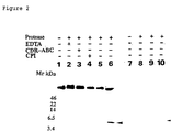
- the present inventors searched for protease activity towards natural brain APP in the homogenate fractions of the human hippocampus, and by analyzing the active fractions, succeeded in separating a 40-kDa protein that has the activity to produce A ⁇ -containing peptide by cleaving brain APP at multiple sites. Analysis of this protein revealed that it was a novel protease belonging to the carboxypeptidase (CP) family.
- This protease (brain carboxypeptidase B; brain CPB) is not an alternatively spliced isoform of plasma CPB, rather it has such specific features as 14 C-terminal amino acids.
- brain CPB isolated by the inventors has a physiological function in APP processing and may have significance in AD pathophysiology. Therefore, brain CPB itself, isolated by the present inventors, could be used as a drug for preventing and treating Alzheimer's disease, and for screening drugs for prevention and treatment. Furthermore, brain CPB isolated by the inventors may be used in tests for Alzheimer's disease.
- brain CPB isolated by the present inventors, and compounds that may regulate its activity may be applied not only for the Alzheimer's disease mentioned above, but also for the prevention, treatment, and diagnosis of various other diseases causing accumulation of A ⁇ .
- the present invention provides:
- the present invention relates to a novel brain carboxypeptidase B (brain CPB) protein expressed in the brain, which has peptidase activity towards brain APP.
- Human brain CPB protein included in the protein of this invention was isolated from an extract of human hippocampus using decomposition activity towards brain APP as an index.
- the gene encoding human brain CPB protein was cloned by immunological screening using antibodies against human brain CPB protein and by RT-PCR and RACE using a primer that was synthesized based on the partial amino acid sequence of human brain CPB protein.
- SEQ ID NO: 1 shows the nucleotide sequence of a cDNA encoding the human brain CPB isolated by the present inventors
- SEQ ID NO: 2 shows the amino acid sequence of human brain CPB protein (prepro-form) encoded by this cDNA.
- the human brain CPB of this invention is thought to be initially synthesized as a preproprotein, then a signal sequence is removed by cleavage to produce the proprotein, furthermore the activation peptide is removed by cleavage to produce the mature protein.
- SEQ ID NO: 3 shows the amino acid sequence of the pro-protein of human brain CPB of this invention
- SEQ ID NO: 4 shows the amino acid sequence of the mature protein.
- brain APP refers to APP molecular species expressed in the brain.
- Examples of molecular species of human brain APP are APP695 (Kang, J. et al. (1987) Nature 325, 733-736) and Appican (Shioi, J. et al. (1992) J. Biol. Chem. 267, 13819-13822), but are not limited to these examples.
- Human brain CPB has the activity to produce A ⁇ -containing peptides by cleaving APP, prepared from the human brain, at multiple sites. It is thought that the human brain CPB is synthesized as a 40-kDa preproprotein, and therefrom, a 30-kDa mature protein is produced. From the characteristics of the amino acid sequence, this protein was determined to be a novel protease belonging to the CPB family (brain carboxypeptidase B; brain CPB).
- This protease has high homology to a known human plasma CPB (HPCPB) belonging to the CPB family, but since it has specific characteristics, such as a unique set of 14 amino acids at its C terminus, this was presumed to be an independent gene, and not an isoform produced by alternative splicing of plasma CPB.
- HPCPB human plasma CPB
- HBCPB human brain CPB
- Antibody against C-terminal 14 amino acids of HBCPB anti C14 antibody
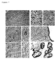
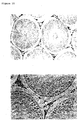
- Figure 7 the neuronal perikaryon and a portion of the microglia
- Figure 4C the RT-PCR analysis of brain mRNAs
- immunological screening done with anti prepro-HBCPB antibody gave three cDNA clones that had almost identical restriction enzyme patterns, indicating that HBCPB is the only CPB isoform expressed in human brain.
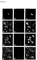
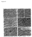
- HBCPB is expressed in various neuronal perikaria, particularly in that of the pyramidal neurons of the hippocampus, ependymal cells, choroid plexus cells, and in a portion of the microglia, but not in astrocytes.
- HBCPB seems to be expressed in a portion of the clustered microglia as parenchymal deposits with or without glial infiltration.
- protease immunoreactivity ranging from 10% to 60% of all the plaques in each brain.
- CSF cerebrospinal fluid
- ependymal cells are speculated to have secretion, absorption and transport functions and to provide a barrier between the brain and CSF (Del Bigio, M.R., 1995, Glia 14: 1-13).
- the expression of anti C14 immunoreactivity in these cells suggests that HBCPB is necessary for processing the peptides synthesized in brain cells, including the appropriate isoform of APP.
- target substrates in the neurons appear to have at least two chances to come across the protease, in the perikaria and in ependymal cells before it is released into the CSF.
- AD the presence of HBCPB in the extracellular space associated with enhanced activation of the microglia may represent a pathophysiological condition. .
- a ⁇ peptide is a substrate of the human brain CPB.
- the brain CPB protein of this invention is thought to be deeply involved with the onset of Alzheimer's disease and with the production of A ⁇ formed by decomposition of brain APP.
- the human brain CPB of this invention may have the function of reducing nerve cell death by decreasing and metabolizing the highly self-aggregating ⁇ -amyloid peptides (especially ⁇ -amyloid 1-42) accumulating in the brain.
- the brain CPB protein of this invention itself can be used as a drug.
- it could be possible to improve A ⁇ metabolism by continuously infusing the protein of this invention into the cerebrospinal fluid of an AD patient to elevate the concentration of human brain CPB in the cerebrospinal fluid, and thus promoting the uptake of the protein into nerve cells via ependymal cells to supplement the decreased or depleted human brain CPB.
- the brain CPB of this invention can be used as a tool for screening promoters or inhibitors of the protein of this invention useful as a drug.
- the protein of this invention or a compound that regulates the activity of the protein of this invention is useful as a drug for preventing or treating various diseases that involve pathological conditions of accumulation of A ⁇ peptide in the brain, and as an agent for testing and diagnosing such diseases. Examples of such diseases are senile dementia, Alzheimer's disease, Down's syndrome, hereditary cerebral hemorrhage (Holland type, etc.), and cephalic contusion (boxer's brain), and such.
- the present invention includes proteins that are structurally similar to the natural human brain CPB protein (SEQ ID NOs: 2, 3, and 4).
- Such structurally similar proteins include mutants of natural human brain CPB protein, and brain CPB proteins derived from other organisms.
- amino acid having properties similar to those of the amino acid to be substituted is preferably used for substitution. Since Ala, Val, Leu, Ile, Pro, Met, Phe and Trp are, for example, all classified into the non-polar amino acid, they are considered to have similar properties.
- Non-charged amino acids include Gly, Ser, Thr, Cys, Tyr, Asn, and Gln.
- Acidic amino acids include Asp and Glu, while basic amino acids include Lys, Arg and His.
- Proteins structurally similar to the human brain CPB protein having a peptidase activity against brain APP can be prepared using the known hybridization technique (2.9 Southern Blotting and Hybridization 2.9.1-2.9.10 (Selden, R.F.), 4.9 Analysis of RNA by Northern Hybridization 4.9.1-4.9.8 (Selden, R.F.), 6.3 Hybridization with Radioactive Probes Using DNA Fragments 6.3.1-6.3.6 (Straus, W.M:), 6.4 Hybridization with Radioactive Probes Using Oligonucleotides 6.4.1-6.4.5 (Duby, A.), in 'Current Protocols in Molecular Biology' (Ausubel, F.M., Brent, R., Kingston, R.E., Moore, D.D., Seidman, J.G., Smith, J.A., Struhl, K.
- PCR polymerase chain reaction
- Proteins encoded by DNAs hybridizing with the human brain CPB cDNA are included in this invention as long as they have a peptidase activity against brain APP.
- Other organisms used for isolating such proteins include, for example, monkeys, mice, rats, rabbits, goats, cattle, sheep, pigs, dogs, and so on, but are not limited thereto.
- cDNAs encoding such proteins can be isolated from such sources as brains of these organisms, for example, hippocampus.
- DNAs encoding the brain CPB protein derived from organisms other than human are usually highly homologous to human brain CPB. Being highly homologous means at least 40% or more, preferably 55% or more, and further preferably 70% or more (e.g. 85% or more, 90% or more, 95% or more) sequence identity at the amino acid level. Sequence homology can be determined by the homology search program of Data Bank of Japan: DDBJ (National Institute of Genetics), such as FASTA, BLAST, etc.
- hybridization conditions when the DNAs are isolated from a brain cDNA library using 32 P-labeled probe is as follows. After hybridization, washing is done under the low stringent conditions of 55°C, 2x SSC, and 0.1% SDS, more preferably, moderately stringent conditions of 55°C, 0.2x SSC, and 0.1% SDS, and still more preferably, highly stringent conditions of 68°C, 0.2x SSC, and 0.1% SDS.
- the protein of this invention can be prepared as either a natural protein or a recombinant protein utilizing gene recombination techniques.
- Natural proteins can be prepared by, for example, subjecting extracts from tissues that are supposed to contain the brain CPB protein (for example, hippocampus) to affinity chromatography using the antibody to the brain CPB protein as described below.
- a natural protein can be prepared by collecting the fraction that bound to a benzamidine-Sepharose column, and by appropriately combining preparative PAGE, etc.
- recombinant proteins may be prepared by culturing cells transformed with DNA encoding the brain CPB protein, allowing the transformants to express the protein, and recovering the protein as described below.
- the invention also includes partial peptides of the proteins of this invention.
- An example of partial peptides of the proteins of this invention may be a peptide containing the boundary portion of the activation peptide and the mature protein, among the proteins of this invention. Such a peptide is thought to act antagonistically in the production of the mature form of the protein of this invention by acting on the protease mediating the reaction that cleaves the mature protein from the proprotein.
- an example of the partial peptides of the proteins of this invention may be a peptide that corresponds to the substrate-binding site of the protein of this invention. This type of partial peptide may be utilized as inhibitors, and such, of the protein of this invention by biological administration.
- partial peptides are useful as an inhibitor or activator of signal transduction that is mediated by the protein of this invention.
- examples of the partial peptides of this invention may be a partial peptide of the C-terminal region of the protein of this invention (for example, SEQ ID NOs: 2, 3, and 4), and this peptide may be utilized to prepare an antibody.
- an activation peptide may be cited.
- Partial polypeptides comprising the amino acid sequence specific to the protein of this invention have at least 7, preferably at least 8, more preferably at least 9 amino acid residues. Partial peptides of this invention can be produced by, for example, genetic engineering techniques, known peptide synthetic methods, or by digestion of the protein of this invention with appropriate peptidases.
- This invention also relates to DNAs encoding the proteins of the invention.
- DNAs encoding the protein of this invention are not particularly limited as long as they can encode the proteins of this invention, including cDNAs, genomic DNAs, and synthetic DNAs.
- DNAs having any desired nucleotide sequences based on the degeneracy of genetic codes are also included in this invention as long as they can encode the proteins of this invention.
- cDNAs encoding the proteins of this invention can be screened, for example, by labeling cDNA of SEQ ID NO: 1 or segments thereof, RNAs complementary to them, or synthetic oligonucleotides comprising partial sequences of said cDNA with 32 P, etc., and hybridizing them with a cDNA library derived from tissues (e.g., hippocampus, etc.) expressing the proteins of this invention.
- cDNAs can be cloned by synthesizing oligonucleotides corresponding to nucleotide sequences of these cDNAs, and amplifying them by PCR with cDNA derived from suitable tissues (e.g. hippocampus, etc.) as a template.
- the genomic DNA can be screened, for example, by labeling cDNA of SEQ ID NO: 1 or segments thereof, RNAs complementary to them, or synthetic oligonucleotides comprising partial sequences of said cDNA with 32 P, etc., and hybridizing them with a genomic DNA library.
- the genomic DNA can be cloned by synthesizing oligonucleotides corresponding to nucleotide sequences of these cDNAs, and amplifying them by PCR with genomic DNA as a template.
- Synthetic DNAs can be prepared, for example, by chemically synthesizing oligonucleotides comprising partial sequences of cDNA of SEQ ID NO: 1, annealing them to form double strand, and ligating them with DNA ligase.
- the proteins of this invention can be prepared as recombinant proteins by inserting DNAs encoding the proteins of this invention (e.g. DNA of SEQ ID NO: 1) .into an appropriate expression vector, transforming suitable cells with the vector, culturing the transformants, and purifying expressed proteins from the transformants or culture supernatant of them.
- DNAs encoding the proteins of this invention e.g. DNA of SEQ ID NO: 1 .into an appropriate expression vector, transforming suitable cells with the vector, culturing the transformants, and purifying expressed proteins from the transformants or culture supernatant of them.
- examples of systems that can be used are: the pET System (Novagen) that allows induction of expression by T7 RNA polymerase and attachment of various tags; the pET CBD Fusion System 34b-38b (Novagen) that allows expression of the protein of this invention as a fusion protein with the cellulose binding domain followed by purification by affinity chromatography using a cellulose carrier; and, the GST Gene Fusion System (Pharmacia) that allows expression of the protein of this invention as a fusion protein with glutathione S-transferase followed by purification by glutathione Sepharose 4B.
- the pET System Novagen
- the pET CBD Fusion System 34b-38b Novagen
- GST Gene Fusion System Pharmacia
- SFV Semliki Forest Virus
- Recombinant proteins expressed in host cells can be purified by known methods.
- the protein of this invention expressed in the form of a fusion protein, for example, with a histidine residue tag or glutathione-S-transferase (GST) attached at the N-terminus can be purified by a nickel column or a glutathione sepharose column, etc.
- GST glutathione-S-transferase
- the portion comprising the protein of this invention can be recovered by inserting cleavage sites, such as Thrombin and Factor Xa, into the boundary area.
- the DNA encoding the protein of this invention or its antisense DNA, and such, may also be utilized for gene therapy of diseases caused by abnormalities in the protein of this invention (abnormalities in expression and function).
- abnormalities in expression and function There is no limitation on the vector to be used for gene therapy, as long as a therapeutically effective expression can be achieved by the vector, and examples of vectors are adenovirus vectors, Semliki Forest virus vectors, and such (Chen, J. et al. (1998) Cancer Res. 58, 3504-3507; Barkats, M. et al. (1998) Progress in Neurobiology 55, 333-341).
- gene therapy can be carried out after producing a vector in which prepro-human brain CPB-cDNA has been inserted by matching the frame with a Semliki Forest virus vector (pSFV-1), completing the genetically recombined virus using a helper virus, then selecting a patient appropriate for treatment by analyzing the general condition of the patient and virus antibody value, and conducting gene therapy on this patient.
- pSFV-1 Semliki Forest virus vector
- the present invention also relates to polynucleotides hybridizing with the DNA comprising the nucleotide sequence of SEQ ID NO: 1 or its complementary sequence, and containing at least 15 nucleotides.
- the polynucleotides specifically hybridize with the DNA comprising the nucleotide sequence of SEQ ID NO: 1 or its complementary sequence.
- “Specifically hybridize” means that a polynucleotide does not significantly cross-hybridize with DNAs encoding other proteins under usual hybridization conditions, preferably under stringent hybridization conditions as described above.
- Such polynucleotides include probes, primers, nucleotides or nucleotide derivatives (e.g. antisense oligonucleotides and ribozymes, etc.), whichcan specifically hybridize with DNAs encoding the proteins of this invention, or DNAs complementary to said DNAs.
- cDNAs encoding the proteins of this invention or oligonucleotides comprising partial sequences thereof can be used for cloning genes and cDNAs encoding the proteins of this invention, or amplifying them by PCR.
- the cDNAs and oligonucleotides can also be utilized for detecting polymorphism or abnormality (gene diagnosis, etc.) of the gene or cDNA by the restriction fragment length polymorphism (RFLP) method, single strand DNA conformation polymorphism (SSCP) method, etc.
- RFLP restriction fragment length polymorphism
- SSCP single strand DNA conformation polymorphism
- This invention also relates to antibodies binding to the proteins of the invention.
- the antibodies against the proteins of this invention can be prepared by known method (Scheidtmann, K.H., Immunological detection of known sequence, in "Protein structure", T.E. Creighton ed., IRL Press, Oxford University Press, pp. 93-115) .
- To prepare polyclonal antibodies for example, the naturally derived protein of this invention prepared from the hippocampus tissue or the recombinant protein in combination with an adjuvant are used to immunize animals such as rabbit, guinea pig, goat, and such. By performing the immunization several times, the antibody titer can be elevated.
- antiserum can be obtained by taking a blood sample from the immunized animal.
- This antiserum for example, can be fractionated by ammonium sulfate precipitation or anion-exchange chromatography, and purified by affinity chromatography using protein A or immobilized antigens, to prepare polyclonal antibodies.
- the partial peptide of the protein of this invention as the antigen.
- the protein of this invention or its partial peptide is used to immunize the immunization animal in the same manner as described above, and after the final immunization, the spleen or the lymph nodes are obtained from the immunized animal.
- Antibody producing cells contained within the spleen or lymph nodes are fused with myeloma cells using polyethylene glycol, and such, to prepare hybridomas.
- the hybridomas of interest are screened, then cultivated, and from this culture supernatant, monoclonal antibodies can be prepared.
- Monoclonal antibodies can be purified, for example, through fractionation by ammonium sulfate precipitation and anion-exchange chromatography, and by affinity chromatography purification using protein A or immobilized antigens.
- Antibodies thus prepared are used for the affinity purification of the proteins of this invention. They can also be used for the test and diagnosis of disorders caused by abnormal expression and structural abnormality of the proteins of this invention and for detection of the expression level of the protein, etc.
- the proteins of this invention are detected in the spinal fluid and sera, it is possible to test and diagnose diseases that cause accumulation of A ⁇ in the brain by detecting the proteins of this invention contained within these samples.
- the antibodies against the proteins of this invention are useful for this type of disease diagnostics.
- the test for diseases that cause accumulation of A ⁇ in the brain can be carried out by a method comprising the steps of, (a) preparing samples from the subject, and (b) detecting the amount of protein of the present invention contained within the sample using antibodies against the protein.
- Diseases targeted by test of this invention are not limited as long as they are diseases that cause accumulation of A ⁇ in the brain, and may be, for example, senile dementia, Alzheimer's disease, Down's syndrome, hereditary cerebral hemorrhage, and cephalic contusion.
- spinal fluid or serum is preferable.
- the protein of this invention included in the sample can be detected, for example, by Western blotting, immunoprecipitation, ELISA, and such.
- the antibody used for detection may be either monoclonal antibody or polyclonal antibody.
- pH buffer for example, phosphate buffer, HEPES buffer, etc.
- carriers for example bovine serum albumin or gelatin, etc.
- preservatives sodium azide
- Antibodies of this invention can be applied to the antibody treatment.
- they are preferably humanized or human antibodies.
- Such antibodies can be prepared by known methods (Parren, P.W. (1992) Hum. Antibodies Hybridomas 3, 137-145).
- the present invention also relates to a method for screening a compound binding to the protein of this invention.
- the screening method of this invention comprises: (a) contacting a test sample with the protein of this invention or partial peptide thereof, (b) detecting the binding activity between the test sample and the protein of this invention or the partial peptide thereof, and (c) selecting a compound that has an activity to bind to the protein of this invention or the partial peptide thereof.
- Test samples used for screening include, for example, cell extracts, expression products of a gene library, synthetic low molecular weight compounds, synthetic peptides, modified peptides, natural compounds, etc., but are not limited thereto. Those test samples used for screening may be labeled prior to use as the occasion demands. Labels include, for example, radioactive and fluorescent ones, etc., but are not limited to them.
- Screening of proteins that bind to the protein of this invention can be carried out, for example, by applying the culture supernatant or cell extract of cells that are thought to express proteins that bind to the protein of this invention, to an affinity column that has an oligopeptide, and such, consisting of the 14 C-terminal amino acids (C14) of the protein of this invention, and by purifying the protein that specifically binds to this column.
- the screening of proteins that bind to the protein of this invention, or genes thereof can be accomplished by utilizing, for example, West-western blot technique (Tarassishin, L.A. and Russell, W.C. (1997) Biochemistry (Mosc.) 62, 38-40; Matthews, D.A. and Russell, W.C. (1998) J.
- the present invention relates to a method to screen compounds that promote or inhibit peptidase activity of the protein of this invention.
- This method uses as an index, the substrate cleaving activity of the protein of this invention, and specifically comprises the steps of, (a) contacting the protein of this invention with its substrate in the presence of a test sample, (b) detecting the cleavage of the substrate, and (c) selecting a compound comprising the activity to increase or decrease substrate cleavage caused by the protein of this invention, in comparison to the cleavage in the absence of the test sample (control).
- Test samples used for this screening method include, for example, cell extracts, expression products of a gene library, synthetic low molecular weight compounds, proteins, natural or synthetic peptides, natural products, sera, etc., but are not limited to them.
- the test samples can be compounds isolated by the above-described screening method monitoring the binding activity of the compounds to the protein of this invention.
- the protein of this invention to be used for screening may be a natural protein or a recombinant protein.
- Brain APP is preferred as a substrate. Natural brain APP can be prepared by the method described in the Examples. In addition, as long as the protein of this invention can show a cleaving activity, partial peptides of brain APP, other APP isoforms and APP-like polypeptides , and oligopeptides synthesized to contain the cleavage site of the substrate may be used.
- Cleavage of the substrate can be detected, for example, by incubating the protein of this invention with a substrate, then performing SDS-PAGE, and finally performing Western blotting using an antibody against the substrate.
- detection can be carried out by Tris-tricine-type PAGE, or by thin-layer chromatography (TLC), and such, by labeling the N terminus, the C terminus, and such in advance.
- TLC thin-layer chromatography
- cleavage of the substrate by the protein of this invention is promoted when a test sample is present, compared to when the test sample is absent, that compound is determined to be the compound promoting substrate-cleaving activity of the protein of this invention.
- substrate cleavage by the protein of this invention is inhibited, when the test sample is present, that compound is determined to be the compound inhibiting substrate-cleaving activity of the protein of this invention.
- inhibiting substrate-cleaving activity includes cases where the cleaving activity is completely inhibited and cases where the inhibition is partial.
- the cleavage site is estimated by first incubating brain APP and the protein of this invention, then upon separating the cleaved brain APP by SDS-PAGE, Western blotting is carried out using domain-specific antibodies for APP (for example, for A ⁇ , A ⁇ 1-17 antibody, A ⁇ 17-24 antibody, A ⁇ 1-40 C-terminal antibody, A ⁇ 1-42 C terminal antibody, etc.).
- domain-specific antibodies for APP for example, for A ⁇ , A ⁇ 1-17 antibody, A ⁇ 17-24 antibody, A ⁇ 1-40 C-terminal antibody, A ⁇ 1-42 C terminal antibody, etc.
- the substrate fragments cleaved by the protein of this invention are purified, and by performing N-terminal or C-terminal amino acid sequencing, the cleavage site can be determined.
- the cleavage site can be estimated using a synthetic oligopeptide (for example, A ⁇ 1-42, etc.) as a substrate, and upon incubation with the protein of this invention, free amino acids that are digested from the C terminus can be measured qualitatively and quantitatively by an amino acid analyzer.
- a synthetic oligopeptide for example, A ⁇ 1-42, etc.
- the protein of this invention and the compounds isolated by the screening method of this invention may be applied, for example, as agents for regulating A ⁇ production, and as drugs for preventing and treating cerebral diseases in which the protein of the present invention is involved (for example, diseases causing A ⁇ accumulation, such as Alzheimer's disease).
- proteins of this invention or compounds isolated by the screening methods of this invention are used as drugs, they may be administered to patients as they are or as pharmaceutical preparations produced by known methods. They can be formulated together with, for example, pharmaceutically acceptable carriers or media such as sterilized water, physiological saline, vegetable oil, emulsifiers, suspending agents, surfactants, stabilizers, etc. They may be administered to patients by methods well known in the art, for example, by intra-arterial, intravenous, subcutaneous injection. They can also be administered intranasally, intrabronchially, intramuscularly, or orally. Doses may vary depending on the body weight and age of patients as well as administration method, and such, and can be suitably selected by those skilled in the art.
- gene therapy may be performed by inserting the DNA into a vector for gene therapy.
- Doses and method for its administration may vary depending on the body weight, age, symptoms, and so on of patients, but can be suitably selected by those skilled in the art.
- the present invention relates to a kit for screening a compound that promotes or inhibits peptidase activity of the protein of this invention, wherein said kit comprises the protein.
- the protein of this invention in the kit of this invention may be a purified or crudely purified protein, may be in a form expressed outside or inside a desired cell (including a transformant made to express the protein), or in a form bound to a support.
- the kit of this invention preferably comprises a substrate as another element in addition to the protein preparation mentioned above.
- substrate brain APP is preferred, although there is no limitation as long as the protein of this invention shows cleaving activity. Natural brain APP can be prepared by the method described in the Examples.
- partial peptides of brain APP, other APP isoforms and APP-like polypeptides , and oligopeptides synthesized to contain a cleavage site of a substrate may be included as substrates.
- the substrate may be labeled.
- buffer for the reaction between the protein of this invention and the substrate, washing solution , and a reagent for detecting cleavage of the substrate may be included.
- the homogenate was centrifuged at 39,000 g for 30 min. The supernatant was precipitated with 40% ammonium sulfate, and the precipitate dialyzed against ice-cold homogenization buffer. The dialysate was used as the hippocampal homogenate.
- the natural brain APP was prepared by immunoprecipitation of the homogenate using anti cAPP antibody (Matsumoto, A. and Fujiwara, Y. (1991) Biochem. Biophys. Res. Commun. 175, 361-365) as described (Matsumoto, A. and Fujiwara, Y., 1993, Eur. J. Biochem. 217: 21-27).
- the hippocampal homogenate (approximately 10 mg per preparation) was chromatographed in a benzamidine-SepharoseTM 6B affinity column (Amersham Pharmacia, UK), which captures a wide range of serine proteases, using the equilibration buffer 20 mM Tris-HCl (pH 7.5). After collecting the pass-through fraction, the column was vigorously washed with the equilibration buffer until the optical density of the eluant at 280 nm reached the base-line level.
- the fraction bound to the column was recovered using an elution buffer consisting of 20 mM Tris-HCl (pH 7.5) and 0.2 M NaCl, then it was dialyzed against ice-cold 10 mM Tris-HCl (pH 7.5), and lyophilized.
- the native-PAGE profile of the total bound fraction by Coomassie brilliant blue staining is shown in Figure 1A.
- a native polyacrylamide gel electrophoresis was conducted without SDS to separate the bound fraction without losing proteolytic activities.
- a preparative polyacrylamide gel electrophoresis apparatus with a liquid-phase collecting device (BiophoresisTM 3, Atto, Japan) and a Tris-glycine buffer system were used for native-PAGE (Smith, J.A. (1989) Electrophoretic separation of proteins. In Ausubel, F.M. et al. (eds.), Current Protocols in Molecular Biology., John Wiley and Sons, New York, USA, pp.10.2.1-10.2.9). Approximately 3 mg of the bound fraction was separated by the native-PAGE into 61 fractions ranging in molecular mass from 20 to 100 kDa. A portion of each fraction then was checked for its protein component by SDS-PAGE and the component made visible by silver staining (Figure 1B).
- the membrane was incubated at room temperature for 1 h with a horseradish peroxidase-labeled anti rabbit second antibody diluted 1:2000. After the washing, signals were detected on X-ray film using an ECL chemiluminescence detection system (Amersham Pharmacia, UK).
- fraction 19 with a molecular mass of about 40 kDa and fraction 11 with a molecular mass of about 30 kDa had similar proteolytic activities.
- Fraction 19 with the 40 kDa molecular mass with proteolytic activity therefore was further purified.
- fraction 19 was collected by preparative electrophoresis (totally five rounds) (Figure 1B). To homogenize fraction 19 (approximately 8 ⁇ g), it was run through a 1 ml Mono S ion-exchange column with 20 mM Tris-HCl (pH 7.5) as the equilibration buffer. After washing, the bound fraction was recovered with 20 mM Tris-HCl (pH 7.5)/0.2 M NaCl as the elution buffer. The 40 kDa proteolytic activity from fraction 19 was trapped in the column and recovered by elution, whereas the co-migrating protein passed through (Figure 1C). It then was dialyzed and lyophilized as above and used as HBCPB standard. Recovery of the HBCPB from the fraction bound to the benzamidine-Sepharose column (approximately 15 mg) was 2 ⁇ g (approximately 5 pmol).
- the brain APP substrate (approximately 0.1 pmol) was treated with the 40 kDa protease (0.005 to 0.1 pmol) at 37°C for 3 h in the presence of 1 mM Zn 2+ to analyze the isolated 40 kDa protease.
- the reaction product analyzed by Western blotting with anti A ⁇ 1/14 antibody ( Figure 2). Under these reaction conditions, the 40 kDa protein degraded natural brain APP, generating a 5.5 kDa fragment as the major component of the 3 to 11 kDa A ⁇ -containing peptides ( Figure 2, lanes 5 and 6).
- Each 40 and 30 kDa protein was immobilized on an Immobilon P SQ filter (Millipore, USA) then subjected to Edman degradation to isolate a cDNA clone by screening the hippocampal cDNA library with an oligonucleotide synthesized through N-terminal amino acid sequencing. Micro-sequencing analysis of the 40 kDa protein was unsuccessful, possibly owing to a blocked N-terminus. Results of sequencing of the 30 kDa protein are shown in Table 1. In this table, yields of respective amino acids in each cycle are shown by pmol. Most abundantly recovered amino acids are underlined.
- Each amino acid is abbreviated as follows: D: aspartic acid, E: glutamic acid, N: asparagine, S: serine, T: threonine, Q: glutamine, G: glycine, H: histidine, A: alanine, Y: tyrosine, R: arginine, P: proline, M: methionine, V: valine, W: tryptophan, K: lysine, F: phenylalanine, I: isoleucine, L: leucine.
- the 30 kDa protein is identical to the N-terminal sequence of the mature form of human plasma CPB.
- molecular cloning for the cDNA encoding the 40 kDa protein was done by two methods; RT-PCR analysis of hippocampal mRNA using amino acid sequencing results, and immunological screening of the human hippocampal cDNA library.
- Poly(A) + RNA was prepared from the frozen hippocampus blocks of the two brains using a quickPrepTM mRNA Purification Kit (Amersham Pharmacia, UK).
- the human hippocampal cDNA library was constructed by the method of Gubler and Hoffmann (Gubler, U. and Hoffman, B.J. (1983) Gene, 25, 263-269).
- First strand cDNA was synthesized from 1.5 ⁇ g poly(A) + RNA using cloned Moloney murine leukemia virus reverse transcriptase (FPLC-pureTM, Amersham Pharmacia, UK) which is primed with synthetic oligo(dT).
- FPLC-pureTM cloned Moloney murine leukemia virus reverse transcriptase
- Second strand cDNA then was synthesized from the first-strand DNA using cloned Escherichia coli DNA polymerase I (Takara, Japan) in the presence of cloned Escherichia coli ribonuclease H (Amersham Pharmacia, UK).
- the double-strand cDNA was blunt-ended by T4 phage polymerase (Takara, Japan), and its EcoRI site modified using Escherichia coli EcoRI methylase (Takara, Japan) then ligated with an EcoRI linker.
- the cDNA was ligated using a DNA ligation kit (Takara Japan) with the ⁇ gt11 phage vector EcoRI arm then packaged in vitro (PhageMakerTM, Takara, Japan).
- the estimated efficiency of this library construction was 9.0x 10 5 plaque forming units (pfu)/ ⁇ g poly(A) + RNA with Escherichia coli Y1090r - as the host bacteria.
- prepro-HPCPB prepro-HPCPB
- prepro-HBCPB human brain protease
- the difference is in the sequence of the mature enzyme: Firstly, the 37 amino acid residues in the middle of the mature plasma enzyme, that correspond to the 198 to 334th amino acid residues of prepro-HPCPB (the 633 to 743rd nucleotide residues of prepro-HPCPB-cDNA), are absent from HBCPB. Secondly, the absence of nucleotides corresponding to those of the 1190th to 1241st residues of prepro-HPCPB-cDNA induced the deletion of 40 amino acid residues, (the 384th to 423rd amino acids of prepro-HPCPB), and a frame-shift which generates a unique 14 amino acid residue in the C-terminus of prepro-HBCPB.
- the former alternation eventually induces an in-frame deletion, and the latter another deletion followed by an amino acid change due to translation of a different frame, including that of the 3' non-coding region (4 amino acids) of prepro-HPCPB-mRNA and a different stop codon.
- the fourth amino acid (proline), and the fifth (valine) and thirteenth (leucine) are replaced respectively by the functionally-identical (neutral as well as hydrophobic) amino acids valine, threonine and proline.
- RNA which differed from that used in the library construction, was hippocampal poly(A) + RNA prepared from three normal brains
- cDNA was synthesized by SUPERSCRIPTTM reverse transcriptase (GIBCO-BRL, USA) using the sense oligonucleotide (5'-GCCTCCGCATCGTACTATGAACAGTATCAC-3'/SEQ ID NO: 6) synthesized through the amino acid sequencing result (Table 1) and the adapter primer with oligo(dT) stretch as the antisense primer.
- sequence was identical to nucleotide 360 to 1436 of prepro-HBCPB-cDNA, corresponding to the mature form of HBCPB. That is, cDNA obtained by either of the two types of cloning procedures, were shown to be derived from the same mRNA in the hippocampus. In addition, from this result, it can be supposed that the 30-kDa protein is encoded by the same gene as the 40-kDa protein, and that the 30-kDa human brain CPB mature protein is produced by cleavage of the N-terminal portion of the 40-kDa protein.
- RNA samples from various organs were analyzed for the expression of prepro-HBCPB-mRNA or its homologue.
- Poly (A) + RNAs from human brain (hippocampus), heart, pancreas, kidney, liver and spleen were prepared from normal autopsy samples by loading 1 ⁇ g poly (A) + RNA on a 1% agarose gel cast in MOPS (3-[N-morpholino]propanesulfonic acid) buffer: 20 mM MOPS (pH 7.0), 5 mM NaOAc and 1 mM EDTA containing 6.6% formaldehyde.
- MOPS 3-[N-morpholino]propanesulfonic acid
- the gel was transferred onto a nylon membrane (Hybond N + , Amersham Pharmacia, UK) by capillary blotting and immobilized by baking. The blot was then hybridized at 68°C for 1 h in ExpressHybTM hybridization solution (Clontech, USA).
- the probe was the 538 bp C-terminal portion of prepro-HBCPB cDNA, synthesized by the PCR with 5'-GGTTCATAGGCCATAATCGAATGT-3' (SEQ ID NO: 7) as the sense-primer (corresponding to nucleotides 596 to 618 of the cDNA) and 5'-TCAGGGGCATTAAACATTCCTAAT-3' (SEQ ID NO: 8) as the antisense-primer (corresponding to nucleotides 1135 to 1112 of the cDNA), and TakaraTaqTM recombinant Taq polymerase (Takara, Japan) ( Figure 4C, lane 2).
- the human ⁇ -actin cDNA probe (Clontech, USA) was used as the control. These probes were radio-labeled with [ ⁇ - 32 P]dCTP using a Random Primer DNA Labeling Kit (Takara, Japan). The blot was washed twice at room temperature in 2X SSC for 20 min, then in 2X SSC, 0.1% SDS and twice at 65°C for 40 min in 0.1X SSC, 0.1% SDS.
- the fraction bound to the benzamidine-Sepharose column was separated by preparative SDS-PAGE and collected in a liquid-phase collecting device to prepare the 40 kDa protease in quantity sufficient (2 mg) to immunize two New Zealand White rabbits.
- the SDS-PAGE profile indicates that the 40 kDa protein was separated to homogeneity ( Figure 6A). Unlike native-PAGE, no further separation using ion-exchange chromatography was required to bring the denatured 40 kDa protein to homogeneity.
- anti prepro-HBCPB polyclonal antibody was raised against rabbit.
- Antiserum was obtained from rabbit, and the IgG fraction was purified by using EZ Prep (Pharmacia). A titer check of its specificity to the antigen indicated a 2000-fold sensitivity over preimmune sera (control antibody).
- Anti-C14 antibody was raised against the oligopeptide NH 2 -SNPPVEKLLPLSLK-COOH (SEQ ID NO: 9) corresponding to the unique C-terminal 14 amino acid residues of the protease.
- the oligopeptide was conjugated to keyhole limpet hemocyanin (Calbiochem, USA) using 3-maleimidobenzoic acid N-hydroxysuccimide ester (Sigma, USA) and used to immunize rabbits.
- the antiserum obtained was affinity-purified in an oligopeptide-conjugated Sepharose CL-4B column.
- a titer check of its specificity indicated 1500-fold sensitivity to the oligopeptide antigen over that of preimmune sera (control antibody).
- HBCPB human brain CPB
- Example 4 immunohistochemical analysis described below was performed. Immediately after autopsy, the samples used for the immunohistochemical study were fixed in 10% formaldehyde kept for at least 1 w and 5 ⁇ m-thick paraffin sections prepared. Immunohistochemical analysis was done by the Avidin-Biotin-Complex (ABC) method. After deparaffinization and quenching of endogenous peroxidase activity by immersion in 3% hydrogen peroxide solution for 5 min, the sections were placed in sodium citrate buffer then immersed in phosphate-buffered saline.
- ABSC Avidin-Biotin-Complex
- GFAP glial fibrillary acidic protein
- Immunofluorescent confocal laser microscopy was used to determine the precise intracellular neuronal localization of the protease (Figure 8).
- Antibodies used were anti human neurofilament antibody (Sanbio, USA), anti human CD68 monoclonal antibody (R&D Systems, USA), anti human GFAP monoclonal antibody (Advance Immunochemical, USA), anti A ⁇ 1/40 antibody (US Peptides, USA), anti prepro-HBCPB antibody and anti C14 antibody.
- An MRC-1024 confocal laser microscope Bio Rad, USA was used for observation.
- the immunoreactivity definitely is localized in the cytosol (perikaryon) of both hippocampal pyramidal neurons ( Figure 8A, B, C, G, H and I) and lateral geniculate body neurons (Figure 8D, E and F) in typical granular fashion and is detected by both the anti HBCPB ( Figure 8A through F) and anti C14 ( Figure 8G through I) antibodies.
- the hippocampal section was also analyzed using the anti human A ⁇ 1/40 and anti C14 antibodies to analyze the relationship of A ⁇ peptides and the protease localizations (Figure 8J, K and L). These two immunoreactivities are colocalized as granules in the perikarya of pyramidal neurons.
- C14 immunoreactivity also is present in the glia, possibly in the microglia, whereas A ⁇ 1/40 immunoreactivity mainly is present in neuronal perikarya.
- the present inventors analyzed brain tissue from five patients with sporadic AD.
- AD neurons especially the hippocampal pyramidal neurons, HBCPB is not ubiquitous as in normal samples, and a portion of the pyramidal neuron shows no immunoreactivity (Figure 9A and B).
- CPB Since the first purification and characterization of CPB (Folk, J.E. and Gladner, J.A. (1958) J. Biol.Chem., 231, 379-393), CPB has only been clearly identified in the pancreas (Pascual, R. et al. (1989) Eur. J. Biochem., 179, 606-616; Yamamoto, K.K. et al. (1992) J. Biol. Chem., 267, 2575-2581). This protease formerly was regarded as a digestive enzyme secreted only from digestive organs.
- CPB or its homologous enzyme has been found in non-digestive tissues such as mast cell secretory granules (Goldstein, S.M. et al. (1989) J. Clin. Invest., 83, 1630-1636; Reynolds, D.S. et al.(1992) J. Clin. Invest., 89, 273-282) and in plasma (Eaton, D.L. et al.(1991) J. Biol. Chem., 266, 21833-21838).
- non-digestive tissues such as mast cell secretory granules (Goldstein, S.M. et al. (1989) J. Clin. Invest., 83, 1630-1636; Reynolds, D.S. et al.(1992) J. Clin. Invest., 89, 273-282) and in plasma (Eaton, D.L. et al.(1991) J. Biol. Chem., 266, 21833-21838).
- CPs in human mast cell secretory granules are similar to those of tissue-type CPs: These 33 to 40 kDa Zn-metallopeptidases are active at a neutral to mild basic pH, are inhibited by potato carboxypeptidase inhibitor, EDTA, phenanthroline and 8-hydroxyquinoline, and are released from mast cells by immunological challenge (Goldstein, S.M. et al. (1989) J. Clin. Invest., 83, 1630-1636).
- HBCPB has a high homology with HPCPB (more than 70%) and moderate homology (approximately 25%) with human mast cell human pancreatic CPBs.
- HPCPB more than 70%
- moderate homology approximately 25%
- human mast cell human pancreatic CPBs three postulated glycosylation sites for asparagine-linked oligosaccharides are conserved in HBCPB, as in human plasma and mast cell CPBs ( Figure 3).
- the amino acid residues responsible for substrate binding Arg-183, Asn-204, Arg-205, Arg-220, Ser-274, Tyr-275, Tyr-326 and Asp-333
- those responsible for zinc binding His-181, Glu-184 and His-273 are well-conserved.
- prepro-HBCPB-mRNA is not a tissue-specific isoform derived from alternative RNA splicing, rather it is a distinct molecule recently evolved from the same ancestral gene of prepro-HPCPB.
- CPs Mature forms of CPs are, in general, highly basic with an overall +10 to 20 positive net charge [(Lys + Arg) - (Asp + Glu) ] at neutral pH. Structural analysis showed the mild basic nature of HBCPB, +7 positive net charges; whereas, both HPCPB synthesized in liver and mast cell CPBs have highly basic natures, +11 to 16 positive net charges. These structural characteristics indicate that HBCPB also is a member of the basic CPB family, and therefore has specificities for C-terminal arginine and lysine residues.
- the postulated function is antifibrinolytic activity during clotting, which retards plasma clot lysis by eliminating the plasminogen-binding sites on partially degraded fibrin (Sakharov, D.V. et al. (1997) J. Biol. Chem., 272, 14477-14482).
- a comparison of the structures of the C-terminal portions of the brain, plasma and pancreatic CPBs indicated that the 18 amino acid residues, with the most C-terminal residue (glutamic acid at the 385th residue of prepro-HPCPB) for substrate binding lack HBCPB.
- the C-terminal 14 amino acid residues unique to HBCPB have no homology to any portion of the other CPs identified ( Figure 4B).
- the peptide(s) with the highest homology to this sequence is located in the N-terminal adjacent to the light-dependent regulation domain of plant chloroplast ATP synthase ⁇ -subunits from various sources ( Figure 4B).
- ATP synthase catalyzed the synthesis of ATP coupled with an electrochemical gradient of protons produced by the photoelectron transfer chain. Its ⁇ -subunit, which is encoded by the nuclear rather than the chloroplast genome, is believed to be essential in the light-dependent regulation of ATP synthase (Inohara, N. et al. (1991) J. Biol.
- Pancreatic CPs exist not only as monomers but as binary or ternary complexes with one or two protease(s) that differ in their proteolytic properties (Kerfeleck, B. et al. (1985) Eur. J. Biochem., 151, 515-519).
- human pancreatic pro-CPA is a binary complex with proproteinase E in the secretory granules (Pascual, R. et al. (1989) Eur. J. Biochem., 179, 606-616).
- the protease in cattle pancreas is a ternary complex with chymotrypsinogen C/proproteinase E (Kobayashi, R. et al.
- the final proteolytic activity product is a single 40 kDa band made visible by sensitive silver staining. Furthermore, the inventors did not used SDS, which induces autolysis of human proteinase E (Sziegoleit, A. et al. (1985) Eur. J. Biochem., 151, 595-599), in the preparation process, which proteinase would have been detected if it was co-purified with HBCPB.
- Its core protein is the APP695 lacking 18 amino acid residues encoded by exon 15, and it carries chondroitin sulfate glycoconjugates bound to the recognition signal for glucosaminoglycan binding that is generated in the amino acid residues between exons 14 and 16 (Pangalos et al. 1995, J. Biol. Chem., 270, 10388-10391).
- APLP1 and APLP2 which share homologous sequences with APP may be co-precipitated; in fact, chondroitin sulfate is bound to a particular isoform of APLP2 as it is in APP (Thinakaran, G.
- the molecular diversity of brain APP also seems to be associated with a strict substrate-protease relationship, giving rise to the tissue-and species- specificities of AD pathophysiology.
- human brain CPB has the function of metabolizing amylin, which, similar to insulin, is produced in the ⁇ -cells and has ⁇ -amyloid-like properties .
- human brain CPB may possibly be related to the pathological physiology of a specific type of diabetes.
- human brain CPB was confirmed in the cytoplasm of human testis Leydig's cells. Therefore, human brain CPB may possibly be involved in the production, secretion, degradation, and such, of many proteinous hormones produced in these endocrine cells.
- the brain CPB protein of the present invention has the biological activity to cleave brain APP to produce A ⁇ -containing peptides. In addition, it has the biological activity to degrade A ⁇ peptide from the C terminus. Therefore, brain CPB protein may be a therapeutic drug that regulates pathological APP metabolisms and production of A ⁇ -containing peptides, and in addition, is a useful tool for elucidating the molecular mechanism of A ⁇ peptide production and the onset of diseases relating to A ⁇ peptide production (for example, Alzheimer's disease). Furthermore, brain CPB expressed in trace amounts in the spinal fluid and even in the peripheral blood is useful as a marker for CNS diseases to test and diagnose abnormalities in APP and A ⁇ metabolisms. In addition, brain CPB may be used to screen candidate compounds for drugs for diseases that cause A ⁇ accumulation in the brain. That is, the present invention has provided a novel and effective method for preventing, treating, and testing diseases that cause A ⁇ accumulation in the brain.
Landscapes
- Health & Medical Sciences (AREA)
- Chemical & Material Sciences (AREA)
- Life Sciences & Earth Sciences (AREA)
- Organic Chemistry (AREA)
- Engineering & Computer Science (AREA)
- Genetics & Genomics (AREA)
- Zoology (AREA)
- Wood Science & Technology (AREA)
- Bioinformatics & Cheminformatics (AREA)
- Biochemistry (AREA)
- Molecular Biology (AREA)
- General Health & Medical Sciences (AREA)
- Biomedical Technology (AREA)
- Medicinal Chemistry (AREA)
- Gastroenterology & Hepatology (AREA)
- Biophysics (AREA)
- Toxicology (AREA)
- Biotechnology (AREA)
- Microbiology (AREA)
- Neurology (AREA)
- General Engineering & Computer Science (AREA)
- Proteomics, Peptides & Aminoacids (AREA)
- Medicines That Contain Protein Lipid Enzymes And Other Medicines (AREA)
- Peptides Or Proteins (AREA)
- Enzymes And Modification Thereof (AREA)
- Pharmaceuticals Containing Other Organic And Inorganic Compounds (AREA)
- Investigating Or Analysing Biological Materials (AREA)
- Hydrogenated Pyridines (AREA)
- Measuring Or Testing Involving Enzymes Or Micro-Organisms (AREA)
- Preparation Of Compounds By Using Micro-Organisms (AREA)
- Micro-Organisms Or Cultivation Processes Thereof (AREA)
Applications Claiming Priority (3)
| Application Number | Priority Date | Filing Date | Title |
|---|---|---|---|
| JP12516999 | 1999-04-30 | ||
| JP12516999 | 1999-04-30 | ||
| PCT/JP2000/002878 WO2000066717A1 (fr) | 1999-04-30 | 2000-05-01 | Carboxypeptidase b du cerveau humain |
Publications (4)
| Publication Number | Publication Date |
|---|---|
| EP1179588A1 true EP1179588A1 (fr) | 2002-02-13 |
| EP1179588A4 EP1179588A4 (fr) | 2004-04-07 |
| EP1179588B1 EP1179588B1 (fr) | 2006-03-08 |
| EP1179588B9 EP1179588B9 (fr) | 2006-07-19 |
Family
ID=14903603
Family Applications (1)
| Application Number | Title | Priority Date | Filing Date |
|---|---|---|---|
| EP00922936A Expired - Lifetime EP1179588B9 (fr) | 1999-04-30 | 2000-05-01 | Carboxypeptidase b du cerveau humain |
Country Status (8)
| Country | Link |
|---|---|
| US (3) | US7060266B1 (fr) |
| EP (1) | EP1179588B9 (fr) |
| JP (1) | JP4579422B2 (fr) |
| AT (1) | ATE319819T1 (fr) |
| AU (1) | AU4316600A (fr) |
| DE (1) | DE60026501T2 (fr) |
| ES (1) | ES2260011T3 (fr) |
| WO (1) | WO2000066717A1 (fr) |
Cited By (4)
| Publication number | Priority date | Publication date | Assignee | Title |
|---|---|---|---|---|
| WO2004004698A3 (fr) * | 2002-07-09 | 2004-06-10 | Univ Medisch Centrum Utrecht | Structure beta-croisee contenant des proteines de liaison amyloide et procedes de detection de la structure beta-croisee en vue de moduler la formation de fibrille dans les structures beta-croisee ainsi que la toxicite induite par la structure beta-croisee |
| EP1395674A4 (fr) * | 2001-03-21 | 2005-04-20 | Applera Corp | Proteines proteases humaines isolees, molecules d'acide nucleique codant des proteines proteases humaines et leurs utilisations |
| US8067187B2 (en) | 2005-07-13 | 2011-11-29 | Crossbeta Biosciences B.V. | Cross-β structure binding compounds |
| US8114832B2 (en) | 2005-07-13 | 2012-02-14 | Crossbeta Biosciences B.V. | Method for detecting and/or removing a protein comprising a cross-beta structure from a pharmaceutical composition |
Families Citing this family (8)
| Publication number | Priority date | Publication date | Assignee | Title |
|---|---|---|---|---|
| US7060266B1 (en) | 1999-04-30 | 2006-06-13 | Akira Matsumoto | Human brain carboxypeptidase B |
| US7291587B2 (en) | 2001-11-09 | 2007-11-06 | Novo Nordisk Healthcare A/G | Pharmaceutical composition comprising factor VII polypeptides and TAFI polypeptides |
| EP1572906A4 (fr) | 2002-03-04 | 2008-05-28 | Bristol Myers Squibb Co | Nouvelles molecules d'acide nucleique et polypeptides codant pour des inhibiteurs de fibrolyse activables par la thrombine (tafi) de babouin |
| PT1794294E (pt) * | 2004-09-27 | 2011-10-13 | Sanofi Aventis Deutschland | Carboxipeptidase b recombinante |
| US7807789B2 (en) * | 2004-12-21 | 2010-10-05 | Cell Signaling Technology, Inc. | Reagents for the detection of protein phosphorylation in EGFR-signaling pathways |
| KR100851035B1 (ko) * | 2005-08-23 | 2008-08-11 | 대한민국 | GCP-Ⅱ를 유효성분으로 함유하는 β-아밀로이드의 뇌내축적 예방 및 치료용 약학적 조성물과 치료제 스크리닝용조성물 및 이를 이용한 스크리닝 방법 |
| JP5775356B2 (ja) * | 2011-05-09 | 2015-09-09 | 松本 明 | 神経保護作用を有するペプチド及びこれを含む薬剤 |
| US8877446B2 (en) * | 2011-08-26 | 2014-11-04 | Korea Research Institute Of Bioscience And Biotechnology | Method for detecting protein-protein interactions in cells |
Family Cites Families (3)
| Publication number | Priority date | Publication date | Assignee | Title |
|---|---|---|---|---|
| US5206161A (en) | 1991-02-01 | 1993-04-27 | Genentech, Inc. | Human plasma carboxypeptidase B |
| US5985562A (en) | 1997-06-03 | 1999-11-16 | Schering Aktiengesellschaft | Method of detecting thrombotic disease risk associated with plasma carboxypeptidase B polymorphisms |
| US7060266B1 (en) | 1999-04-30 | 2006-06-13 | Akira Matsumoto | Human brain carboxypeptidase B |
-
2000
- 2000-05-01 US US09/980,881 patent/US7060266B1/en not_active Expired - Lifetime
- 2000-05-01 DE DE60026501T patent/DE60026501T2/de not_active Expired - Lifetime
- 2000-05-01 AT AT00922936T patent/ATE319819T1/de not_active IP Right Cessation
- 2000-05-01 EP EP00922936A patent/EP1179588B9/fr not_active Expired - Lifetime
- 2000-05-01 JP JP2000615742A patent/JP4579422B2/ja not_active Expired - Lifetime
- 2000-05-01 WO PCT/JP2000/002878 patent/WO2000066717A1/fr not_active Ceased
- 2000-05-01 AU AU43166/00A patent/AU4316600A/en not_active Abandoned
- 2000-05-01 ES ES00922936T patent/ES2260011T3/es not_active Expired - Lifetime
-
2005
- 2005-09-22 US US11/233,958 patent/US7524934B2/en not_active Expired - Fee Related
-
2009
- 2009-03-18 US US12/406,925 patent/US7972771B2/en not_active Expired - Fee Related
Cited By (5)
| Publication number | Priority date | Publication date | Assignee | Title |
|---|---|---|---|---|
| EP1395674A4 (fr) * | 2001-03-21 | 2005-04-20 | Applera Corp | Proteines proteases humaines isolees, molecules d'acide nucleique codant des proteines proteases humaines et leurs utilisations |
| WO2004004698A3 (fr) * | 2002-07-09 | 2004-06-10 | Univ Medisch Centrum Utrecht | Structure beta-croisee contenant des proteines de liaison amyloide et procedes de detection de la structure beta-croisee en vue de moduler la formation de fibrille dans les structures beta-croisee ainsi que la toxicite induite par la structure beta-croisee |
| US8158585B2 (en) | 2002-07-09 | 2012-04-17 | Crossbeta Biosciences B.V. | Cross-β structure comprising amyloid-binding proteins and methods for detection of the cross-β structure, for modulating cross-β structures fiber formation and modulating cross-β structure-mediated toxicity |
| US8067187B2 (en) | 2005-07-13 | 2011-11-29 | Crossbeta Biosciences B.V. | Cross-β structure binding compounds |
| US8114832B2 (en) | 2005-07-13 | 2012-02-14 | Crossbeta Biosciences B.V. | Method for detecting and/or removing a protein comprising a cross-beta structure from a pharmaceutical composition |
Also Published As
| Publication number | Publication date |
|---|---|
| US20060024760A1 (en) | 2006-02-02 |
| DE60026501T2 (de) | 2006-08-10 |
| WO2000066717A1 (fr) | 2000-11-09 |
| AU4316600A (en) | 2000-11-17 |
| EP1179588B9 (fr) | 2006-07-19 |
| EP1179588B1 (fr) | 2006-03-08 |
| EP1179588A4 (fr) | 2004-04-07 |
| ATE319819T1 (de) | 2006-03-15 |
| ES2260011T3 (es) | 2006-11-01 |
| US7060266B1 (en) | 2006-06-13 |
| JP4579422B2 (ja) | 2010-11-10 |
| DE60026501D1 (en) | 2006-05-04 |
| US7972771B2 (en) | 2011-07-05 |
| US7524934B2 (en) | 2009-04-28 |
| US20100021951A1 (en) | 2010-01-28 |
Similar Documents
| Publication | Publication Date | Title |
|---|---|---|
| US7972771B2 (en) | Human brain carboxypeptidase B | |
| US11932908B2 (en) | Compositions and methods for diagnosis and treatment of epilepsy | |
| EP2224000B1 (fr) | Anticorps et son utilisation | |
| JP4454230B2 (ja) | β−セクレターゼ基質及びその使用 | |
| JP2006149394A (ja) | β−セクレターゼ、β−セクレターゼに対する抗体、及びβ−セクレターゼ阻害を検出するためのアッセイ | |
| JP2000032983A (ja) | アルツハイマ―病の診断および治療用の新規な手段 | |
| JPH04502364A (ja) | 神経系疾患または機能障害の検出法 | |
| HUT69771A (en) | Proteases causing abnormal degradation of amyloid beta-proteine precursors | |
| Li et al. | Identification and characterization of CPAMD8, a novel member of the complement 3/α2-macroglobulin family with a C-terminal Kazal domain | |
| JPH08509808A (ja) | 神経糸状タンパク質遺伝子発現およびアルツハイマー病の検出 | |
| Matsumoto et al. | A novel carboxypeptidase B that processes native β‐amyloid precursor protein is present in human hippocampus | |
| JPH09511332A (ja) | カルパイン活性化を検出する方法およびカルパイン阻害剤を同定する方法 | |
| JPWO2000066717A1 (ja) | ヒト脳カルボキシペプチダーゼb | |
| US20070199080A1 (en) | Diagnostic and therapeutic use of F-box proteins for Alzheimer's disease and related neurodegenerative disorders | |
| JP2007535959A (ja) | アルツハイマー病のためのkcnj6の診断および治療使用 | |
| US20080261302A1 (en) | Methods of diagnosing or treating neurological diseases and cell degeneration | |
| JP2000516087A (ja) | アルツハイマー病に関連する遺伝子配列およびタンパク質、ならびにその使用 | |
| Meckelein et al. | Identification of a novel serine protease-like molecule in human brain | |
| JP4937143B2 (ja) | 非拡散性球状Aβ(X−38..43)オリゴマー構造エピトープに対して特異性を有する自己抗体を検出する方法 | |
| US20060264610A1 (en) | Protein complexes of the tip60 transcriptional activator protein | |
| Savaglio | Confirmation and Characterization of a Functional APP/GAP-43 Protein Interaction | |
| JPWO2004001038A1 (ja) | 細胞または組織の神経化に関与する新規遺伝子およびタンパク質、並びにその利用 |
Legal Events
| Date | Code | Title | Description |
|---|---|---|---|
| PUAI | Public reference made under article 153(3) epc to a published international application that has entered the european phase |
Free format text: ORIGINAL CODE: 0009012 |
|
| 17P | Request for examination filed |
Effective date: 20011129 |
|
| AK | Designated contracting states |
Kind code of ref document: A1 Designated state(s): AT BE CH CY DE DK ES FI FR GB GR IE IT LI LU MC NL PT SE |
|
| AX | Request for extension of the european patent |
Free format text: AL;LT;LV;MK;RO;SI |
|
| RIC1 | Information provided on ipc code assigned before grant |
Ipc: 7C 07K 14/47 B Ipc: 7C 12N 15/57 B Ipc: 7C 12N 9/48 A |
|
| A4 | Supplementary search report drawn up and despatched |
Effective date: 20040223 |
|
| 17Q | First examination report despatched |
Effective date: 20040526 |
|
| GRAP | Despatch of communication of intention to grant a patent |
Free format text: ORIGINAL CODE: EPIDOSNIGR1 |
|
| GRAS | Grant fee paid |
Free format text: ORIGINAL CODE: EPIDOSNIGR3 |
|
| GRAA | (expected) grant |
Free format text: ORIGINAL CODE: 0009210 |
|
| AK | Designated contracting states |
Kind code of ref document: B1 Designated state(s): AT BE CH CY DE DK ES FI FR GB GR IE IT LI LU MC NL PT SE |
|
| PG25 | Lapsed in a contracting state [announced via postgrant information from national office to epo] |
Ref country code: LI Free format text: LAPSE BECAUSE OF FAILURE TO SUBMIT A TRANSLATION OF THE DESCRIPTION OR TO PAY THE FEE WITHIN THE PRESCRIBED TIME-LIMIT Effective date: 20060308 Ref country code: CH Free format text: LAPSE BECAUSE OF FAILURE TO SUBMIT A TRANSLATION OF THE DESCRIPTION OR TO PAY THE FEE WITHIN THE PRESCRIBED TIME-LIMIT Effective date: 20060308 Ref country code: AT Free format text: LAPSE BECAUSE OF FAILURE TO SUBMIT A TRANSLATION OF THE DESCRIPTION OR TO PAY THE FEE WITHIN THE PRESCRIBED TIME-LIMIT Effective date: 20060308 Ref country code: NL Free format text: LAPSE BECAUSE OF FAILURE TO SUBMIT A TRANSLATION OF THE DESCRIPTION OR TO PAY THE FEE WITHIN THE PRESCRIBED TIME-LIMIT Effective date: 20060308 Ref country code: BE Free format text: LAPSE BECAUSE OF FAILURE TO SUBMIT A TRANSLATION OF THE DESCRIPTION OR TO PAY THE FEE WITHIN THE PRESCRIBED TIME-LIMIT Effective date: 20060308 Ref country code: FI Free format text: LAPSE BECAUSE OF FAILURE TO SUBMIT A TRANSLATION OF THE DESCRIPTION OR TO PAY THE FEE WITHIN THE PRESCRIBED TIME-LIMIT Effective date: 20060308 |
|
| REG | Reference to a national code |
Ref country code: GB Ref legal event code: FG4D |
|
| REG | Reference to a national code |
Ref country code: CH Ref legal event code: EP |
|
| REG | Reference to a national code |
Ref country code: IE Ref legal event code: FG4D |
|
| PG25 | Lapsed in a contracting state [announced via postgrant information from national office to epo] |
Ref country code: IE Free format text: LAPSE BECAUSE OF NON-PAYMENT OF DUE FEES Effective date: 20060502 |
|
| REF | Corresponds to: |
Ref document number: 60026501 Country of ref document: DE Date of ref document: 20060504 Kind code of ref document: P |
|
| PG25 | Lapsed in a contracting state [announced via postgrant information from national office to epo] |
Ref country code: MC Free format text: LAPSE BECAUSE OF NON-PAYMENT OF DUE FEES Effective date: 20060531 |
|
| PG25 | Lapsed in a contracting state [announced via postgrant information from national office to epo] |
Ref country code: SE Free format text: LAPSE BECAUSE OF FAILURE TO SUBMIT A TRANSLATION OF THE DESCRIPTION OR TO PAY THE FEE WITHIN THE PRESCRIBED TIME-LIMIT Effective date: 20060608 Ref country code: DK Free format text: LAPSE BECAUSE OF FAILURE TO SUBMIT A TRANSLATION OF THE DESCRIPTION OR TO PAY THE FEE WITHIN THE PRESCRIBED TIME-LIMIT Effective date: 20060608 |
|
| PG25 | Lapsed in a contracting state [announced via postgrant information from national office to epo] |
Ref country code: PT Free format text: LAPSE BECAUSE OF FAILURE TO SUBMIT A TRANSLATION OF THE DESCRIPTION OR TO PAY THE FEE WITHIN THE PRESCRIBED TIME-LIMIT Effective date: 20060808 |
|
| NLV1 | Nl: lapsed or annulled due to failure to fulfill the requirements of art. 29p and 29m of the patents act | ||
| REG | Reference to a national code |
Ref country code: CH Ref legal event code: PL |
|
| REG | Reference to a national code |
Ref country code: ES Ref legal event code: FG2A Ref document number: 2260011 Country of ref document: ES Kind code of ref document: T3 |
|
| ET | Fr: translation filed | ||
| PLBE | No opposition filed within time limit |
Free format text: ORIGINAL CODE: 0009261 |
|
| STAA | Information on the status of an ep patent application or granted ep patent |
Free format text: STATUS: NO OPPOSITION FILED WITHIN TIME LIMIT |
|
| 26N | No opposition filed |
Effective date: 20061211 |
|
| PG25 | Lapsed in a contracting state [announced via postgrant information from national office to epo] |
Ref country code: GR Free format text: LAPSE BECAUSE OF FAILURE TO SUBMIT A TRANSLATION OF THE DESCRIPTION OR TO PAY THE FEE WITHIN THE PRESCRIBED TIME-LIMIT Effective date: 20060609 |
|
| PG25 | Lapsed in a contracting state [announced via postgrant information from national office to epo] |
Ref country code: LU Free format text: LAPSE BECAUSE OF NON-PAYMENT OF DUE FEES Effective date: 20060501 |
|
| PG25 | Lapsed in a contracting state [announced via postgrant information from national office to epo] |
Ref country code: CY Free format text: LAPSE BECAUSE OF FAILURE TO SUBMIT A TRANSLATION OF THE DESCRIPTION OR TO PAY THE FEE WITHIN THE PRESCRIBED TIME-LIMIT Effective date: 20060308 |
|
| PGFP | Annual fee paid to national office [announced via postgrant information from national office to epo] |
Ref country code: FR Payment date: 20110607 Year of fee payment: 12 Ref country code: ES Payment date: 20110525 Year of fee payment: 12 |
|
| PGFP | Annual fee paid to national office [announced via postgrant information from national office to epo] |
Ref country code: GB Payment date: 20110520 Year of fee payment: 12 |
|
| PGFP | Annual fee paid to national office [announced via postgrant information from national office to epo] |
Ref country code: IT Payment date: 20110525 Year of fee payment: 12 Ref country code: DE Payment date: 20110520 Year of fee payment: 12 |
|
| GBPC | Gb: european patent ceased through non-payment of renewal fee |
Effective date: 20120501 |
|
| PG25 | Lapsed in a contracting state [announced via postgrant information from national office to epo] |
Ref country code: IT Free format text: LAPSE BECAUSE OF NON-PAYMENT OF DUE FEES Effective date: 20120501 |
|
| REG | Reference to a national code |
Ref country code: FR Ref legal event code: ST Effective date: 20130131 |
|
| REG | Reference to a national code |
Ref country code: DE Ref legal event code: R119 Ref document number: 60026501 Country of ref document: DE Effective date: 20121201 |
|
| PG25 | Lapsed in a contracting state [announced via postgrant information from national office to epo] |
Ref country code: FR Free format text: LAPSE BECAUSE OF NON-PAYMENT OF DUE FEES Effective date: 20120531 Ref country code: GB Free format text: LAPSE BECAUSE OF NON-PAYMENT OF DUE FEES Effective date: 20120501 |
|
| PG25 | Lapsed in a contracting state [announced via postgrant information from national office to epo] |
Ref country code: DE Free format text: LAPSE BECAUSE OF NON-PAYMENT OF DUE FEES Effective date: 20121201 |
|
| REG | Reference to a national code |
Ref country code: ES Ref legal event code: FD2A Effective date: 20131030 |
|
| PG25 | Lapsed in a contracting state [announced via postgrant information from national office to epo] |
Ref country code: ES Free format text: LAPSE BECAUSE OF NON-PAYMENT OF DUE FEES Effective date: 20120502 |