EP1180036B1 - Zelltherapie für chronischen schlaganfall - Google Patents
Zelltherapie für chronischen schlaganfall Download PDFInfo
- Publication number
- EP1180036B1 EP1180036B1 EP00918005A EP00918005A EP1180036B1 EP 1180036 B1 EP1180036 B1 EP 1180036B1 EP 00918005 A EP00918005 A EP 00918005A EP 00918005 A EP00918005 A EP 00918005A EP 1180036 B1 EP1180036 B1 EP 1180036B1
- Authority
- EP
- European Patent Office
- Prior art keywords
- cells
- neuronal cells
- stroke
- neuronal
- brain
- Prior art date
- Legal status (The legal status is an assumption and is not a legal conclusion. Google has not performed a legal analysis and makes no representation as to the accuracy of the status listed.)
- Expired - Lifetime
Links
- 208000006011 Stroke Diseases 0.000 title description 53
- 238000002659 cell therapy Methods 0.000 title description 3
- 230000001684 chronic effect Effects 0.000 title description 3
- 210000004027 cell Anatomy 0.000 claims abstract description 128
- 210000002569 neuron Anatomy 0.000 claims abstract description 93
- 238000000034 method Methods 0.000 claims abstract description 44
- 210000004556 brain Anatomy 0.000 claims abstract description 25
- 230000006931 brain damage Effects 0.000 claims abstract description 22
- 231100000874 brain damage Toxicity 0.000 claims abstract description 22
- 208000029028 brain injury Diseases 0.000 claims abstract description 22
- 239000000203 mixture Substances 0.000 claims abstract description 17
- 210000001178 neural stem cell Anatomy 0.000 claims abstract description 14
- 230000006870 function Effects 0.000 claims abstract description 9
- 230000001605 fetal effect Effects 0.000 claims abstract description 6
- 230000003920 cognitive function Effects 0.000 claims abstract description 5
- 230000007659 motor function Effects 0.000 claims abstract description 4
- 230000037152 sensory function Effects 0.000 claims abstract description 4
- 210000003625 skull Anatomy 0.000 claims abstract description 4
- 210000001982 neural crest cell Anatomy 0.000 claims abstract description 3
- 230000033001 locomotion Effects 0.000 claims description 22
- 230000006378 damage Effects 0.000 claims description 16
- 210000000130 stem cell Anatomy 0.000 claims description 16
- 210000001161 mammalian embryo Anatomy 0.000 claims description 13
- 239000003814 drug Substances 0.000 claims description 6
- 210000003169 central nervous system Anatomy 0.000 claims description 5
- 230000019771 cognition Effects 0.000 claims description 4
- 238000004519 manufacturing process Methods 0.000 claims description 2
- 210000005036 nerve Anatomy 0.000 claims description 2
- 230000035807 sensation Effects 0.000 claims description 2
- IJGRMHOSHXDMSA-UHFFFAOYSA-N Atomic nitrogen Chemical compound N#N IJGRMHOSHXDMSA-UHFFFAOYSA-N 0.000 abstract description 4
- 208000014674 injury Diseases 0.000 abstract description 3
- 239000007788 liquid Substances 0.000 abstract description 2
- 230000004770 neurodegeneration Effects 0.000 abstract description 2
- 208000015122 neurodegenerative disease Diseases 0.000 abstract description 2
- 229910052757 nitrogen Inorganic materials 0.000 abstract description 2
- 239000008194 pharmaceutical composition Substances 0.000 abstract description 2
- 230000008733 trauma Effects 0.000 abstract description 2
- 208000005374 Poisoning Diseases 0.000 abstract 1
- 208000028867 ischemia Diseases 0.000 abstract 1
- 231100000572 poisoning Toxicity 0.000 abstract 1
- 230000000607 poisoning effect Effects 0.000 abstract 1
- 238000001356 surgical procedure Methods 0.000 description 15
- 238000002513 implantation Methods 0.000 description 14
- 239000007943 implant Substances 0.000 description 13
- 230000006735 deficit Effects 0.000 description 12
- 241000700159 Rattus Species 0.000 description 11
- 230000008859 change Effects 0.000 description 11
- 210000002414 leg Anatomy 0.000 description 10
- 230000001537 neural effect Effects 0.000 description 9
- 238000002054 transplantation Methods 0.000 description 9
- 230000002411 adverse Effects 0.000 description 8
- 238000011084 recovery Methods 0.000 description 8
- 102000003974 Fibroblast growth factor 2 Human genes 0.000 description 7
- 108090000379 Fibroblast growth factor 2 Proteins 0.000 description 7
- 230000006872 improvement Effects 0.000 description 7
- AOYNUTHNTBLRMT-SLPGGIOYSA-N 2-deoxy-2-fluoro-aldehydo-D-glucose Chemical compound OC[C@@H](O)[C@@H](O)[C@H](O)[C@@H](F)C=O AOYNUTHNTBLRMT-SLPGGIOYSA-N 0.000 description 6
- PMATZTZNYRCHOR-CGLBZJNRSA-N Cyclosporin A Chemical compound CC[C@@H]1NC(=O)[C@H]([C@H](O)[C@H](C)C\C=C\C)N(C)C(=O)[C@H](C(C)C)N(C)C(=O)[C@H](CC(C)C)N(C)C(=O)[C@H](CC(C)C)N(C)C(=O)[C@@H](C)NC(=O)[C@H](C)NC(=O)[C@H](CC(C)C)N(C)C(=O)[C@H](C(C)C)NC(=O)[C@H](CC(C)C)N(C)C(=O)CN(C)C1=O PMATZTZNYRCHOR-CGLBZJNRSA-N 0.000 description 6
- 108010036949 Cyclosporine Proteins 0.000 description 6
- 206010061216 Infarction Diseases 0.000 description 6
- 210000004227 basal ganglia Anatomy 0.000 description 6
- 230000001413 cellular effect Effects 0.000 description 6
- 230000000694 effects Effects 0.000 description 6
- 230000036541 health Effects 0.000 description 6
- 238000002600 positron emission tomography Methods 0.000 description 6
- 230000008569 process Effects 0.000 description 6
- 230000000638 stimulation Effects 0.000 description 6
- 210000001519 tissue Anatomy 0.000 description 6
- 210000005013 brain tissue Anatomy 0.000 description 5
- 206010008118 cerebral infarction Diseases 0.000 description 5
- 229960001265 ciclosporin Drugs 0.000 description 5
- 238000002591 computed tomography Methods 0.000 description 5
- 229930182912 cyclosporin Natural products 0.000 description 5
- 229940079593 drug Drugs 0.000 description 5
- 238000011156 evaluation Methods 0.000 description 5
- 210000003811 finger Anatomy 0.000 description 5
- 239000003102 growth factor Substances 0.000 description 5
- 238000003384 imaging method Methods 0.000 description 5
- 230000007574 infarction Effects 0.000 description 5
- 238000002347 injection Methods 0.000 description 5
- 239000007924 injection Substances 0.000 description 5
- 230000002829 reductive effect Effects 0.000 description 5
- 239000000725 suspension Substances 0.000 description 5
- 238000012360 testing method Methods 0.000 description 5
- 241001465754 Metazoa Species 0.000 description 4
- 208000007542 Paresis Diseases 0.000 description 4
- 230000001154 acute effect Effects 0.000 description 4
- DDRJAANPRJIHGJ-UHFFFAOYSA-N creatinine Chemical compound CN1CC(=O)NC1=N DDRJAANPRJIHGJ-UHFFFAOYSA-N 0.000 description 4
- 239000003112 inhibitor Substances 0.000 description 4
- 230000000302 ischemic effect Effects 0.000 description 4
- 210000002540 macrophage Anatomy 0.000 description 4
- 238000002595 magnetic resonance imaging Methods 0.000 description 4
- 230000007971 neurological deficit Effects 0.000 description 4
- 230000004083 survival effect Effects 0.000 description 4
- 206010010904 Convulsion Diseases 0.000 description 3
- 102000004127 Cytokines Human genes 0.000 description 3
- 108090000695 Cytokines Proteins 0.000 description 3
- IAZDPXIOMUYVGZ-UHFFFAOYSA-N Dimethylsulphoxide Chemical compound CS(C)=O IAZDPXIOMUYVGZ-UHFFFAOYSA-N 0.000 description 3
- 208000004552 Lacunar Stroke Diseases 0.000 description 3
- 108010025020 Nerve Growth Factor Proteins 0.000 description 3
- 102000015336 Nerve Growth Factor Human genes 0.000 description 3
- 208000008457 Neurologic Manifestations Diseases 0.000 description 3
- SHGAZHPCJJPHSC-YCNIQYBTSA-N all-trans-retinoic acid Chemical compound OC(=O)\C=C(/C)\C=C\C=C(/C)\C=C\C1=C(C)CCCC1(C)C SHGAZHPCJJPHSC-YCNIQYBTSA-N 0.000 description 3
- 238000010171 animal model Methods 0.000 description 3
- 239000003146 anticoagulant agent Substances 0.000 description 3
- 230000003542 behavioural effect Effects 0.000 description 3
- 230000008499 blood brain barrier function Effects 0.000 description 3
- 210000001218 blood-brain barrier Anatomy 0.000 description 3
- 230000002490 cerebral effect Effects 0.000 description 3
- 208000026106 cerebrovascular disease Diseases 0.000 description 3
- 208000037265 diseases, disorders, signs and symptoms Diseases 0.000 description 3
- 208000035475 disorder Diseases 0.000 description 3
- 230000005021 gait Effects 0.000 description 3
- 206010019465 hemiparesis Diseases 0.000 description 3
- 230000001506 immunosuppresive effect Effects 0.000 description 3
- 210000003657 middle cerebral artery Anatomy 0.000 description 3
- 229940053128 nerve growth factor Drugs 0.000 description 3
- 230000002980 postoperative effect Effects 0.000 description 3
- 238000011160 research Methods 0.000 description 3
- 229930002330 retinoic acid Natural products 0.000 description 3
- 210000002966 serum Anatomy 0.000 description 3
- 230000002459 sustained effect Effects 0.000 description 3
- 238000002560 therapeutic procedure Methods 0.000 description 3
- 229960001727 tretinoin Drugs 0.000 description 3
- ZCXUVYAZINUVJD-AHXZWLDOSA-N 2-deoxy-2-((18)F)fluoro-alpha-D-glucose Chemical compound OC[C@H]1O[C@H](O)[C@H]([18F])[C@@H](O)[C@@H]1O ZCXUVYAZINUVJD-AHXZWLDOSA-N 0.000 description 2
- 206010003591 Ataxia Diseases 0.000 description 2
- 206010071043 Basal ganglia stroke Diseases 0.000 description 2
- 201000006474 Brain Ischemia Diseases 0.000 description 2
- 201000009030 Carcinoma Diseases 0.000 description 2
- 206010008120 Cerebral ischaemia Diseases 0.000 description 2
- 108010071942 Colony-Stimulating Factors Proteins 0.000 description 2
- 102000007644 Colony-Stimulating Factors Human genes 0.000 description 2
- 102000053171 Glial Fibrillary Acidic Human genes 0.000 description 2
- 101710193519 Glial fibrillary acidic protein Proteins 0.000 description 2
- 206010019233 Headaches Diseases 0.000 description 2
- 206010019468 Hemiplegia Diseases 0.000 description 2
- 208000023105 Huntington disease Diseases 0.000 description 2
- 206010062016 Immunosuppression Diseases 0.000 description 2
- 206010061218 Inflammation Diseases 0.000 description 2
- 102000014150 Interferons Human genes 0.000 description 2
- 108010050904 Interferons Proteins 0.000 description 2
- 108010063738 Interleukins Proteins 0.000 description 2
- 102000015696 Interleukins Human genes 0.000 description 2
- 206010051078 Lacunar infarction Diseases 0.000 description 2
- 108090000542 Lymphotoxin-alpha Proteins 0.000 description 2
- 102000004083 Lymphotoxin-alpha Human genes 0.000 description 2
- 241001529936 Murinae Species 0.000 description 2
- 206010028813 Nausea Diseases 0.000 description 2
- 206010028980 Neoplasm Diseases 0.000 description 2
- 208000012902 Nervous system disease Diseases 0.000 description 2
- MWUXSHHQAYIFBG-UHFFFAOYSA-N Nitric oxide Chemical compound O=[N] MWUXSHHQAYIFBG-UHFFFAOYSA-N 0.000 description 2
- 208000018737 Parkinson disease Diseases 0.000 description 2
- 206010047700 Vomiting Diseases 0.000 description 2
- 208000027418 Wounds and injury Diseases 0.000 description 2
- 230000004075 alteration Effects 0.000 description 2
- 229940127219 anticoagulant drug Drugs 0.000 description 2
- 239000006285 cell suspension Substances 0.000 description 2
- 230000003833 cell viability Effects 0.000 description 2
- 208000020832 chronic kidney disease Diseases 0.000 description 2
- 208000022831 chronic renal failure syndrome Diseases 0.000 description 2
- 208000035850 clinical syndrome Diseases 0.000 description 2
- 229940047120 colony stimulating factors Drugs 0.000 description 2
- 230000002596 correlated effect Effects 0.000 description 2
- 229940109239 creatinine Drugs 0.000 description 2
- 238000011018 current good manufacturing practice Methods 0.000 description 2
- 230000034994 death Effects 0.000 description 2
- 231100000517 death Toxicity 0.000 description 2
- 238000011161 development Methods 0.000 description 2
- 230000018109 developmental process Effects 0.000 description 2
- 230000004069 differentiation Effects 0.000 description 2
- 231100000673 dose–response relationship Toxicity 0.000 description 2
- 230000005713 exacerbation Effects 0.000 description 2
- 210000003414 extremity Anatomy 0.000 description 2
- BTCSSZJGUNDROE-UHFFFAOYSA-N gamma-aminobutyric acid Chemical compound NCCCC(O)=O BTCSSZJGUNDROE-UHFFFAOYSA-N 0.000 description 2
- 210000005046 glial fibrillary acidic protein Anatomy 0.000 description 2
- 230000005484 gravity Effects 0.000 description 2
- 210000004247 hand Anatomy 0.000 description 2
- 231100000869 headache Toxicity 0.000 description 2
- 230000001900 immune effect Effects 0.000 description 2
- 238000000338 in vitro Methods 0.000 description 2
- 230000004054 inflammatory process Effects 0.000 description 2
- 229940047124 interferons Drugs 0.000 description 2
- 229940047122 interleukins Drugs 0.000 description 2
- 238000011835 investigation Methods 0.000 description 2
- 229940090993 isolyte s Drugs 0.000 description 2
- 210000003127 knee Anatomy 0.000 description 2
- 230000003902 lesion Effects 0.000 description 2
- GRPSNTXTTSBKGW-BVGHQBMWSA-J magnesium;potassium;sodium;(3r,4s,5s,6r)-6-(hydroxymethyl)oxane-2,3,4,5-tetrol;triacetate;chloride Chemical compound [Na+].[Mg+2].[Cl-].[K+].CC([O-])=O.CC([O-])=O.CC([O-])=O.OC[C@H]1OC(O)[C@H](O)[C@@H](O)[C@@H]1O GRPSNTXTTSBKGW-BVGHQBMWSA-J 0.000 description 2
- 238000002483 medication Methods 0.000 description 2
- 230000004060 metabolic process Effects 0.000 description 2
- 230000000394 mitotic effect Effects 0.000 description 2
- 230000008693 nausea Effects 0.000 description 2
- 210000003757 neuroblast Anatomy 0.000 description 2
- 239000004090 neuroprotective agent Substances 0.000 description 2
- 229940021182 non-steroidal anti-inflammatory drug Drugs 0.000 description 2
- 231100001221 nontumorigenic Toxicity 0.000 description 2
- 239000008188 pellet Substances 0.000 description 2
- 230000002062 proliferating effect Effects 0.000 description 2
- 108090000623 proteins and genes Proteins 0.000 description 2
- 239000000523 sample Substances 0.000 description 2
- 230000001953 sensory effect Effects 0.000 description 2
- 208000019206 urinary tract infection Diseases 0.000 description 2
- 230000035899 viability Effects 0.000 description 2
- 230000008673 vomiting Effects 0.000 description 2
- OGNSCSPNOLGXSM-UHFFFAOYSA-N (+/-)-DABA Natural products NCCC(N)C(O)=O OGNSCSPNOLGXSM-UHFFFAOYSA-N 0.000 description 1
- 238000010176 18-FDG-positron emission tomography Methods 0.000 description 1
- KISWVXRQTGLFGD-UHFFFAOYSA-N 2-[[2-[[6-amino-2-[[2-[[2-[[5-amino-2-[[2-[[1-[2-[[6-amino-2-[(2,5-diamino-5-oxopentanoyl)amino]hexanoyl]amino]-5-(diaminomethylideneamino)pentanoyl]pyrrolidine-2-carbonyl]amino]-3-hydroxypropanoyl]amino]-5-oxopentanoyl]amino]-5-(diaminomethylideneamino)p Chemical compound C1CCN(C(=O)C(CCCN=C(N)N)NC(=O)C(CCCCN)NC(=O)C(N)CCC(N)=O)C1C(=O)NC(CO)C(=O)NC(CCC(N)=O)C(=O)NC(CCCN=C(N)N)C(=O)NC(CO)C(=O)NC(CCCCN)C(=O)NC(C(=O)NC(CC(C)C)C(O)=O)CC1=CC=C(O)C=C1 KISWVXRQTGLFGD-UHFFFAOYSA-N 0.000 description 1
- APIXJSLKIYYUKG-UHFFFAOYSA-N 3 Isobutyl 1 methylxanthine Chemical compound O=C1N(C)C(=O)N(CC(C)C)C2=C1N=CN2 APIXJSLKIYYUKG-UHFFFAOYSA-N 0.000 description 1
- VHRSUDSXCMQTMA-PJHHCJLFSA-N 6alpha-methylprednisolone Chemical compound C([C@@]12C)=CC(=O)C=C1[C@@H](C)C[C@@H]1[C@@H]2[C@@H](O)C[C@]2(C)[C@@](O)(C(=O)CO)CC[C@H]21 VHRSUDSXCMQTMA-PJHHCJLFSA-N 0.000 description 1
- BSYNRYMUTXBXSQ-UHFFFAOYSA-N Aspirin Chemical compound CC(=O)OC1=CC=CC=C1C(O)=O BSYNRYMUTXBXSQ-UHFFFAOYSA-N 0.000 description 1
- 239000005528 B01AC05 - Ticlopidine Substances 0.000 description 1
- 206010069020 Basal ganglia infarction Diseases 0.000 description 1
- 208000014644 Brain disease Diseases 0.000 description 1
- 206010008111 Cerebral haemorrhage Diseases 0.000 description 1
- 206010010071 Coma Diseases 0.000 description 1
- 206010010774 Constipation Diseases 0.000 description 1
- 229930105110 Cyclosporin A Natural products 0.000 description 1
- UHDGCWIWMRVCDJ-CCXZUQQUSA-N Cytarabine Chemical compound O=C1N=C(N)C=CN1[C@H]1[C@@H](O)[C@H](O)[C@@H](CO)O1 UHDGCWIWMRVCDJ-CCXZUQQUSA-N 0.000 description 1
- 208000005156 Dehydration Diseases 0.000 description 1
- 241001269524 Dura Species 0.000 description 1
- 208000032320 Germ cell tumor of testis Diseases 0.000 description 1
- 102000018899 Glutamate Receptors Human genes 0.000 description 1
- 108010027915 Glutamate Receptors Proteins 0.000 description 1
- 108090000288 Glycoproteins Proteins 0.000 description 1
- 102000003886 Glycoproteins Human genes 0.000 description 1
- 206010019196 Head injury Diseases 0.000 description 1
- 208000032843 Hemorrhage Diseases 0.000 description 1
- 241000282412 Homo Species 0.000 description 1
- 108010002352 Interleukin-1 Proteins 0.000 description 1
- 108090000174 Interleukin-10 Proteins 0.000 description 1
- 108090000177 Interleukin-11 Proteins 0.000 description 1
- 108010002350 Interleukin-2 Proteins 0.000 description 1
- 108010002386 Interleukin-3 Proteins 0.000 description 1
- 108090000978 Interleukin-4 Proteins 0.000 description 1
- 108010002616 Interleukin-5 Proteins 0.000 description 1
- 108090001005 Interleukin-6 Proteins 0.000 description 1
- 108010002586 Interleukin-7 Proteins 0.000 description 1
- 108090001007 Interleukin-8 Proteins 0.000 description 1
- 208000032382 Ischaemic stroke Diseases 0.000 description 1
- 108010044467 Isoenzymes Proteins 0.000 description 1
- 208000000913 Kidney Calculi Diseases 0.000 description 1
- WHUUTDBJXJRKMK-VKHMYHEASA-N L-glutamic acid Chemical compound OC(=O)[C@@H](N)CCC(O)=O WHUUTDBJXJRKMK-VKHMYHEASA-N 0.000 description 1
- 101710128836 Large T antigen Proteins 0.000 description 1
- 206010024264 Lethargy Diseases 0.000 description 1
- 206010027476 Metastases Diseases 0.000 description 1
- 241000699670 Mus sp. Species 0.000 description 1
- 102000047918 Myelin Basic Human genes 0.000 description 1
- 101710107068 Myelin basic protein Proteins 0.000 description 1
- 206010028851 Necrosis Diseases 0.000 description 1
- 206010029148 Nephrolithiasis Diseases 0.000 description 1
- 108010088373 Neurofilament Proteins Proteins 0.000 description 1
- 102000008763 Neurofilament Proteins Human genes 0.000 description 1
- 206010030113 Oedema Diseases 0.000 description 1
- 208000002193 Pain Diseases 0.000 description 1
- 206010033799 Paralysis Diseases 0.000 description 1
- 102000012288 Phosphopyruvate Hydratase Human genes 0.000 description 1
- 108010022181 Phosphopyruvate Hydratase Proteins 0.000 description 1
- 241000288906 Primates Species 0.000 description 1
- 102000013674 S-100 Human genes 0.000 description 1
- 108700021018 S100 Proteins 0.000 description 1
- 206010039897 Sedation Diseases 0.000 description 1
- GLNADSQYFUSGOU-GPTZEZBUSA-J Trypan blue Chemical compound [Na+].[Na+].[Na+].[Na+].C1=C(S([O-])(=O)=O)C=C2C=C(S([O-])(=O)=O)C(/N=N/C3=CC=C(C=C3C)C=3C=C(C(=CC=3)\N=N\C=3C(=CC4=CC(=CC(N)=C4C=3O)S([O-])(=O)=O)S([O-])(=O)=O)C)=C(O)C2=C1N GLNADSQYFUSGOU-GPTZEZBUSA-J 0.000 description 1
- 108060008682 Tumor Necrosis Factor Proteins 0.000 description 1
- 102000000852 Tumor Necrosis Factor-alpha Human genes 0.000 description 1
- 108091000117 Tyrosine 3-Monooxygenase Proteins 0.000 description 1
- 102000048218 Tyrosine 3-monooxygenases Human genes 0.000 description 1
- CJGYSWNGNKCJSB-YVLZZHOMSA-M [(4ar,6r,7r,7ar)-6-[6-(butanoylamino)purin-9-yl]-2-oxido-2-oxo-4a,6,7,7a-tetrahydro-4h-furo[3,2-d][1,3,2]dioxaphosphinin-7-yl] butanoate Chemical compound C([C@H]1O2)OP([O-])(=O)O[C@H]1[C@@H](OC(=O)CCC)[C@@H]2N1C(N=CN=C2NC(=O)CCC)=C2N=C1 CJGYSWNGNKCJSB-YVLZZHOMSA-M 0.000 description 1
- 230000002159 abnormal effect Effects 0.000 description 1
- 230000005856 abnormality Effects 0.000 description 1
- 229960001138 acetylsalicylic acid Drugs 0.000 description 1
- 238000013019 agitation Methods 0.000 description 1
- 230000001668 ameliorated effect Effects 0.000 description 1
- 230000000702 anti-platelet effect Effects 0.000 description 1
- 239000000427 antigen Substances 0.000 description 1
- 102000036639 antigens Human genes 0.000 description 1
- 108091007433 antigens Proteins 0.000 description 1
- 201000007201 aphasia Diseases 0.000 description 1
- 210000003050 axon Anatomy 0.000 description 1
- 238000009227 behaviour therapy Methods 0.000 description 1
- 230000009286 beneficial effect Effects 0.000 description 1
- 230000015572 biosynthetic process Effects 0.000 description 1
- 210000004369 blood Anatomy 0.000 description 1
- 239000008280 blood Substances 0.000 description 1
- 230000003925 brain function Effects 0.000 description 1
- 210000000133 brain stem Anatomy 0.000 description 1
- 201000007293 brain stem infarction Diseases 0.000 description 1
- 230000015556 catabolic process Effects 0.000 description 1
- 230000003915 cell function Effects 0.000 description 1
- 210000003710 cerebral cortex Anatomy 0.000 description 1
- 238000006243 chemical reaction Methods 0.000 description 1
- 239000003795 chemical substances by application Substances 0.000 description 1
- 230000001713 cholinergic effect Effects 0.000 description 1
- 230000001149 cognitive effect Effects 0.000 description 1
- 150000001875 compounds Chemical class 0.000 description 1
- 230000001276 controlling effect Effects 0.000 description 1
- 230000002920 convulsive effect Effects 0.000 description 1
- 230000001054 cortical effect Effects 0.000 description 1
- 210000005257 cortical tissue Anatomy 0.000 description 1
- 230000003412 degenerative effect Effects 0.000 description 1
- 230000018044 dehydration Effects 0.000 description 1
- 238000006297 dehydration reaction Methods 0.000 description 1
- 210000001787 dendrite Anatomy 0.000 description 1
- 238000013461 design Methods 0.000 description 1
- 206010012601 diabetes mellitus Diseases 0.000 description 1
- 238000002405 diagnostic procedure Methods 0.000 description 1
- 230000035487 diastolic blood pressure Effects 0.000 description 1
- 238000002597 diffusion-weighted imaging Methods 0.000 description 1
- ZPWVASYFFYYZEW-UHFFFAOYSA-L dipotassium hydrogen phosphate Chemical compound [K+].[K+].OP([O-])([O-])=O ZPWVASYFFYYZEW-UHFFFAOYSA-L 0.000 description 1
- 239000003792 electrolyte Substances 0.000 description 1
- 230000002996 emotional effect Effects 0.000 description 1
- 238000005516 engineering process Methods 0.000 description 1
- 230000001973 epigenetic effect Effects 0.000 description 1
- 230000002461 excitatory amino acid Effects 0.000 description 1
- 239000003257 excitatory amino acid Substances 0.000 description 1
- 238000002474 experimental method Methods 0.000 description 1
- 230000001815 facial effect Effects 0.000 description 1
- 206010016256 fatigue Diseases 0.000 description 1
- 210000003754 fetus Anatomy 0.000 description 1
- ODKNJVUHOIMIIZ-RRKCRQDMSA-N floxuridine Chemical compound C1[C@H](O)[C@@H](CO)O[C@H]1N1C(=O)NC(=O)C(F)=C1 ODKNJVUHOIMIIZ-RRKCRQDMSA-N 0.000 description 1
- 210000000245 forearm Anatomy 0.000 description 1
- 210000005224 forefinger Anatomy 0.000 description 1
- 239000012595 freezing medium Substances 0.000 description 1
- 238000002599 functional magnetic resonance imaging Methods 0.000 description 1
- 229960003692 gamma aminobutyric acid Drugs 0.000 description 1
- 230000002068 genetic effect Effects 0.000 description 1
- 230000004153 glucose metabolism Effects 0.000 description 1
- 229930195712 glutamate Natural products 0.000 description 1
- 210000004884 grey matter Anatomy 0.000 description 1
- 230000012010 growth Effects 0.000 description 1
- 210000003128 head Anatomy 0.000 description 1
- 208000003119 hemimegalencephaly Diseases 0.000 description 1
- 238000002650 immunosuppressive therapy Methods 0.000 description 1
- 238000001727 in vivo Methods 0.000 description 1
- 230000028709 inflammatory response Effects 0.000 description 1
- 238000001802 infusion Methods 0.000 description 1
- 230000010354 integration Effects 0.000 description 1
- 210000002425 internal capsule Anatomy 0.000 description 1
- 238000007917 intracranial administration Methods 0.000 description 1
- 238000009533 lab test Methods 0.000 description 1
- 238000002690 local anesthesia Methods 0.000 description 1
- 210000004072 lung Anatomy 0.000 description 1
- 239000003550 marker Substances 0.000 description 1
- 230000035800 maturation Effects 0.000 description 1
- 238000005259 measurement Methods 0.000 description 1
- 230000001404 mediated effect Effects 0.000 description 1
- 239000002609 medium Substances 0.000 description 1
- 239000012528 membrane Substances 0.000 description 1
- ZMNSRFNUONFLSP-UHFFFAOYSA-N mephenoxalone Chemical compound COC1=CC=CC=C1OCC1OC(=O)NC1 ZMNSRFNUONFLSP-UHFFFAOYSA-N 0.000 description 1
- 229960001030 mephenoxalone Drugs 0.000 description 1
- 230000009401 metastasis Effects 0.000 description 1
- 229960004584 methylprednisolone Drugs 0.000 description 1
- 201000007309 middle cerebral artery infarction Diseases 0.000 description 1
- 239000003226 mitogen Substances 0.000 description 1
- 210000003205 muscle Anatomy 0.000 description 1
- 230000017074 necrotic cell death Effects 0.000 description 1
- 210000003061 neural cell Anatomy 0.000 description 1
- 210000005044 neurofilament Anatomy 0.000 description 1
- 230000037420 neurological improvement Effects 0.000 description 1
- 230000014511 neuron projection development Effects 0.000 description 1
- 230000005015 neuronal process Effects 0.000 description 1
- 239000002858 neurotransmitter agent Substances 0.000 description 1
- 238000010606 normalization Methods 0.000 description 1
- 108020004707 nucleic acids Proteins 0.000 description 1
- 102000039446 nucleic acids Human genes 0.000 description 1
- 150000007523 nucleic acids Chemical class 0.000 description 1
- 238000011580 nude mouse model Methods 0.000 description 1
- 210000001328 optic nerve Anatomy 0.000 description 1
- 230000036407 pain Effects 0.000 description 1
- 230000037361 pathway Effects 0.000 description 1
- 210000000578 peripheral nerve Anatomy 0.000 description 1
- 238000002135 phase contrast microscopy Methods 0.000 description 1
- 239000008055 phosphate buffer solution Substances 0.000 description 1
- 239000002243 precursor Substances 0.000 description 1
- 108090000765 processed proteins & peptides Proteins 0.000 description 1
- 238000004393 prognosis Methods 0.000 description 1
- 230000000644 propagated effect Effects 0.000 description 1
- 230000001902 propagating effect Effects 0.000 description 1
- 230000002685 pulmonary effect Effects 0.000 description 1
- 210000001747 pupil Anatomy 0.000 description 1
- 230000036647 reaction Effects 0.000 description 1
- 230000008929 regeneration Effects 0.000 description 1
- 238000011069 regeneration method Methods 0.000 description 1
- 230000008439 repair process Effects 0.000 description 1
- 230000010410 reperfusion Effects 0.000 description 1
- 238000002271 resection Methods 0.000 description 1
- 230000008886 response to retinoic acid Effects 0.000 description 1
- 230000000284 resting effect Effects 0.000 description 1
- 230000000717 retained effect Effects 0.000 description 1
- 230000002441 reversible effect Effects 0.000 description 1
- 231100000279 safety data Toxicity 0.000 description 1
- 210000002955 secretory cell Anatomy 0.000 description 1
- 230000036280 sedation Effects 0.000 description 1
- 210000000717 sertoli cell Anatomy 0.000 description 1
- 210000001082 somatic cell Anatomy 0.000 description 1
- 208000020431 spinal cord injury Diseases 0.000 description 1
- 230000000087 stabilizing effect Effects 0.000 description 1
- 239000000126 substance Substances 0.000 description 1
- 239000000758 substrate Substances 0.000 description 1
- 208000024891 symptom Diseases 0.000 description 1
- 210000000225 synapse Anatomy 0.000 description 1
- 230000002195 synergetic effect Effects 0.000 description 1
- 238000003786 synthesis reaction Methods 0.000 description 1
- 230000035488 systolic blood pressure Effects 0.000 description 1
- 230000008685 targeting Effects 0.000 description 1
- 208000001608 teratocarcinoma Diseases 0.000 description 1
- 230000002381 testicular Effects 0.000 description 1
- 208000002918 testicular germ cell tumor Diseases 0.000 description 1
- 238000012956 testing procedure Methods 0.000 description 1
- 210000001103 thalamus Anatomy 0.000 description 1
- 230000002537 thrombolytic effect Effects 0.000 description 1
- 210000003813 thumb Anatomy 0.000 description 1
- PHWBOXQYWZNQIN-UHFFFAOYSA-N ticlopidine Chemical compound ClC1=CC=CC=C1CN1CC(C=CS2)=C2CC1 PHWBOXQYWZNQIN-UHFFFAOYSA-N 0.000 description 1
- 229960005001 ticlopidine Drugs 0.000 description 1
- 230000008467 tissue growth Effects 0.000 description 1
- 231100000331 toxic Toxicity 0.000 description 1
- 230000002588 toxic effect Effects 0.000 description 1
- 241001430294 unidentified retrovirus Species 0.000 description 1
- 210000000689 upper leg Anatomy 0.000 description 1
- VBEQCZHXXJYVRD-GACYYNSASA-N uroanthelone Chemical compound C([C@@H](C(=O)N[C@H](C(=O)N[C@@H](CS)C(=O)N[C@@H](CC(N)=O)C(=O)N[C@@H](CS)C(=O)N[C@H](C(=O)N[C@@H]([C@@H](C)CC)C(=O)NCC(=O)N[C@@H](CC=1C=CC(O)=CC=1)C(=O)N[C@@H](CO)C(=O)NCC(=O)N[C@@H](CC(O)=O)C(=O)N[C@@H](CCCNC(N)=N)C(=O)N[C@@H](CS)C(=O)N[C@@H](CCC(N)=O)C(=O)N[C@@H]([C@@H](C)O)C(=O)N[C@@H](CCCNC(N)=N)C(=O)N[C@@H](CC(O)=O)C(=O)N[C@@H](CC(C)C)C(=O)N[C@@H](CCCNC(N)=N)C(=O)N[C@@H](CC=1C2=CC=CC=C2NC=1)C(=O)N[C@@H](CC=1C2=CC=CC=C2NC=1)C(=O)N[C@@H](CCC(O)=O)C(=O)N[C@@H](CC(C)C)C(=O)N[C@@H](CCCNC(N)=N)C(O)=O)C(C)C)[C@@H](C)O)NC(=O)[C@H](CO)NC(=O)[C@H](CC(O)=O)NC(=O)[C@H](CC(C)C)NC(=O)[C@H](CO)NC(=O)[C@H](CCC(O)=O)NC(=O)[C@@H](NC(=O)[C@H](CC=1NC=NC=1)NC(=O)[C@H](CCSC)NC(=O)[C@H](CS)NC(=O)[C@@H](NC(=O)CNC(=O)CNC(=O)[C@H](CC(N)=O)NC(=O)[C@H](CC(C)C)NC(=O)[C@H](CS)NC(=O)[C@H](CC=1C=CC(O)=CC=1)NC(=O)CNC(=O)[C@H](CC(O)=O)NC(=O)[C@H](CC=1C=CC(O)=CC=1)NC(=O)[C@H](CO)NC(=O)[C@H](CO)NC(=O)[C@H]1N(CCC1)C(=O)[C@H](CS)NC(=O)CNC(=O)[C@H]1N(CCC1)C(=O)[C@H](CC=1C=CC(O)=CC=1)NC(=O)[C@H](CO)NC(=O)[C@@H](N)CC(N)=O)C(C)C)[C@@H](C)CC)C1=CC=C(O)C=C1 VBEQCZHXXJYVRD-GACYYNSASA-N 0.000 description 1
- 230000000007 visual effect Effects 0.000 description 1
- 229960005080 warfarin Drugs 0.000 description 1
- PJVWKTKQMONHTI-UHFFFAOYSA-N warfarin Chemical compound OC=1C2=CC=CC=C2OC(=O)C=1C(CC(=O)C)C1=CC=CC=C1 PJVWKTKQMONHTI-UHFFFAOYSA-N 0.000 description 1
- XLYOFNOQVPJJNP-UHFFFAOYSA-N water Substances O XLYOFNOQVPJJNP-UHFFFAOYSA-N 0.000 description 1
- 210000000707 wrist Anatomy 0.000 description 1
Images
Classifications
-
- A—HUMAN NECESSITIES
- A61—MEDICAL OR VETERINARY SCIENCE; HYGIENE
- A61K—PREPARATIONS FOR MEDICAL, DENTAL OR TOILETRY PURPOSES
- A61K35/00—Medicinal preparations containing materials or reaction products thereof with undetermined constitution
- A61K35/12—Materials from mammals; Compositions comprising non-specified tissues or cells; Compositions comprising non-embryonic stem cells; Genetically modified cells
- A61K35/30—Nerves; Brain; Eyes; Corneal cells; Cerebrospinal fluid; Neuronal stem cells; Neuronal precursor cells; Glial cells; Oligodendrocytes; Schwann cells; Astroglia; Astrocytes; Choroid plexus; Spinal cord tissue
-
- A—HUMAN NECESSITIES
- A61—MEDICAL OR VETERINARY SCIENCE; HYGIENE
- A61P—SPECIFIC THERAPEUTIC ACTIVITY OF CHEMICAL COMPOUNDS OR MEDICINAL PREPARATIONS
- A61P21/00—Drugs for disorders of the muscular or neuromuscular system
-
- A—HUMAN NECESSITIES
- A61—MEDICAL OR VETERINARY SCIENCE; HYGIENE
- A61P—SPECIFIC THERAPEUTIC ACTIVITY OF CHEMICAL COMPOUNDS OR MEDICINAL PREPARATIONS
- A61P25/00—Drugs for disorders of the nervous system
-
- A—HUMAN NECESSITIES
- A61—MEDICAL OR VETERINARY SCIENCE; HYGIENE
- A61P—SPECIFIC THERAPEUTIC ACTIVITY OF CHEMICAL COMPOUNDS OR MEDICINAL PREPARATIONS
- A61P25/00—Drugs for disorders of the nervous system
- A61P25/28—Drugs for disorders of the nervous system for treating neurodegenerative disorders of the central nervous system, e.g. nootropic agents, cognition enhancers, drugs for treating Alzheimer's disease or other forms of dementia
-
- A—HUMAN NECESSITIES
- A61—MEDICAL OR VETERINARY SCIENCE; HYGIENE
- A61P—SPECIFIC THERAPEUTIC ACTIVITY OF CHEMICAL COMPOUNDS OR MEDICINAL PREPARATIONS
- A61P9/00—Drugs for disorders of the cardiovascular system
-
- A—HUMAN NECESSITIES
- A61—MEDICAL OR VETERINARY SCIENCE; HYGIENE
- A61K—PREPARATIONS FOR MEDICAL, DENTAL OR TOILETRY PURPOSES
- A61K35/00—Medicinal preparations containing materials or reaction products thereof with undetermined constitution
- A61K35/12—Materials from mammals; Compositions comprising non-specified tissues or cells; Compositions comprising non-embryonic stem cells; Genetically modified cells
Definitions
- This invention is in the medical treatment of neurological deficits resulting from stroke; more specifically, the invention applies cell therapy to restore lost cognitive, motor, sensory and speech function resulting from stroke.
- stroke is the third leading cause of death and the most common cause of adult disability. With an incidence of approximately 750,000 patients, approximately 30% (250,000) die, 30% (250,000) become severely and permanently disabled, and 30% (250,000) recover with little or no functional deficits. Currently four million Americans are living with the effects of stroke, and two thirds of those have moderate to severe impairments. In addition, improving diagnostic methods, such as diffusion-weighted imaging (showing dead brain tissue) and perfusion-weighted imaging (showing oxygen-starved but live brain tissue), are helping diagnose more new and old strokes.
- diffusion-weighted imaging shown dead brain tissue
- perfusion-weighted imaging shown oxygen-starved but live brain tissue
- Stroke is defined as a sudden, non-convulsive, focal neurologic deficit that is related either to cerebral ischemia or hemorrhage.
- the neurologic deficit created reflects the location and size of the cerebral infarction.
- Lacunar infarction is one type of ischemic stroke that is usually of small volume, and which may be typified by various clinical syndromes (e.g., hemiparesis with ataxia in the same limb, pure motor hemiplegia).
- lacunar stroke When located in a region of non-critical brain tissue, lacunar stroke is often not associated with symptoms.
- a critical structure such as the internal capsule, thalamus, basal ganglia or brain stem, significant neurologic disability can occur.
- treatment in the acute setting can consist of thrombolytic therapy, surgical resection of large strokes that cause major mass effect and coma, and rare reperfusion techniques such as extracranial-intracranial bypass.
- Neuroprotective agents such as glutamate receptor inhibitors or inhibitors of excitatory amino acid release were in clinical trials for treatment within the first six to twelve hours of stroke onset. To date, none of these trials has been successful since it is difficult for the stroke victim to reach the hospital within the narrow (3-6 hour) window during which the neuroprotective agents can rescue damaged neuronal cells. Agents that interfere with nitric oxide synthesis or generation of free radicals have also been tested.
- HNT neuronal cells were initially produced from a lung metastasis tumor removed from a 22-year-old patient with a testicular teratocarcinoma in 1972 at the Sloan Kettering Cancer Center in New York City. Dr. Peter Andrews at the Wistar Institute in Philadelphia was the first to observe that these cells exhibited the unique property of differentiating into embryonic neuronal cells upon treatment with retinoic acid. He published this observation in 1984. Dr. Virginia M.-Y. Lee, working at the University of Pennsylvania, then developed the process for producing large quantities of the human embryonic neuronal cells ( U.S. Patent No. 5,175,103 ).
- a number of degenerative brain disorders have been proposed for neurotransplantation. These include acute and chronic stroke, Parkinson's disease, Huntington's disease, head injury, spinal cord injury and others. No treatment now exists to restore lost brain function after stroke. We theorized that treating stroke patients by implanting suitable cells into the patients' stroked areas might lead to the cells' integration into the host brain, resulting in restoration of lost neural function.
- the method also includes the step of using a twist drill or a burr to form a hole in the skull through which the cells could be administered.
- Cells for administration in the method are selected from the group consisting of hNT neuronal cells, HCN-1 cells, fetal pig cells, neural crest cells or a combination thereof.
- compositions of 95% pure neuronal cells which is packaged in a vial with PBS.
- the vial is further encased in a container with liquid nitrogen, whereby the composition is kept at -170°C before use.
- neuronal cells for use in for improving speech in a person who has experienced brain damage which interferes with speech, wherein the brain damage results from stroke, by a method which comprises injecting a sterile composition of at least six million viable neuronal cells at a concentration of cells based on the total viable cell count into the damaged area, wherein if said cells are stem cells they have not been obtained by destruction of a human embryo.
- a method which comprises injecting a sterile composition of at least six million viable neuronal cells at a concentration of cells based on the total viable cell count into the damaged area, wherein if said cells are stem cells they have not been obtained by destruction of a human embryo.
- the injected neuronal cells may be human neuronal cells.
- neuronal cells for use in improving motor performance in a person who has experienced brain damage which interferes with movement, wherein the brain damage results from stroke, by a method which comprises injecting a sterile composition of at least six million viable neuronal cells at a concentration of cells based on the total viable cell count to the damaged area wherein if said cells are stem cells they have not been obtained by destruction of a human embryo.
- a method which comprises injecting a sterile composition of at least six million viable neuronal cells at a concentration of cells based on the total viable cell count to the damaged area wherein if said cells are stem cells they have not been obtained by destruction of a human embryo.
- the injected cells may be human neuronal cells.
- neuronal cells or neural stem cells for use in improving cognition in a person who has experienced brain damage which interferes with cognition, wherein the brain damage results from stroke, by a method which comprises delivering a sterile composition of at least six million viable neuronal cells at a concentration of cells based on the total viable cell count to the damaged area of the brain, wherein if said cells are stem cells they have not been obtained by destruction of a human embryo.
- a method which comprises delivering a sterile composition of at least six million viable neuronal cells at a concentration of cells based on the total viable cell count to the damaged area of the brain, wherein if said cells are stem cells they have not been obtained by destruction of a human embryo.
- the injected neuronal cells may be human neuronal cells or neural stem cells.
- neuronal cells for use in improving sensory function in a person who has experienced brain damage which interferes with sensation, wherein the brain damage results from stroke by a method which comprises delivering a sterile composition of at least six million viable neuronal cells at a concentration of cells based on the total viable cell count to the damaged area wherein if said cells are stem cells they have not been obtained by destruction of a human embryo.
- hNT neuronal cells licensed from the University of Pennsylvania, are human neuronal cells derived from a single cell line. Through eight years of in vitro and in vivo preclinical testing, the cells have been demonstrated to be human, fully post-mitotic, non-tumorigenic neuronal cells which demonstrate efficacy in animal models. After safety studies were performed in mice, rats and primates, implantation of human neurons into rats with basal ganglia stroke showed both motor and behavioral recovery in comparison to sham controls. A second experiment shows that the number of cells implanted correlated with the degree of recovery. The first clinical study evaluated the product as a somatic cell therapy that produced a novel way to restore lost cognitive and motor function. Further, early research is being planned in the use of hNT neuronal cells as a platform for the introduction and expression of specific human neuronal genes into the brain for the treatment of neurologic disorders.
- HNT neuronal cells were derived by treating the neuronal precursor cell line NT2/D 1 derived from an embryonic carcinoma with retinoic acid and mitotic inhibitors. Following treatment with retinoic acid, the NT2/D1 cells differentiate into non-proliferating, terminally differentiated neurons and proliferating non-neuronal accessory cells ( Andrews, P.W. Dev. Biol. 103:285-293, 1984 ).
- LBS-Neurons human neuronal cells After subsequent treatment with mitotic inhibitors (cytosine arabinoside and fluorodeoxyuridine), pure cultures of post-mitotic human neuronal cells result (Pleasure and Lee, 1993). These cells were then suspended in freezing medium (HAS, DMSO and PBS) and frozen in ampoules. The resultant product, when produced in compliance with current Good Manufacturing Practice (cGMP) guidelines, is called LBS-Neurons human neuronal cells.
- mitotic inhibitors cytosine arabinoside and fluorodeoxyuridine
- the NT2/D1 cell line was established in culture as a cell line by Dr. Peter Andrews at the Wistar Institute in Philadelphia during the early 1980s.
- Dr. Andrews received the original cells (known as Tera-2) from Dr. Jurgen Fögh of the Sloan Kettering Institute in New York City.
- the Tera-2 cells had been isolated from a pulmonary embryonic carcinoma of a 22-year-old Caucasian male with a metastasized primary testicular germ cell tumor.
- the post-mitotic human neuronal cells available as hNT neuronal cells resulted from the differentiation of NT2/D1 cells in response to retinoic acid. These human neuronal cells actively demonstrate neurite outgrowth, sending out numerous processes that assemble into neuronal networks. They also form polarized processes that have been identified functionally as axons and dendrites, and demonstrate the ability to form synapses upon maturation. These cells have retained their human characteristics as demonstrated by isoenzyme typing, expression of a variety of human antigens, and by karyotyping (Andrews et al., ibid., Miyazono et al., 1996, Layton Bioscience, Inc., 1996).
- hNT cells have been successfully implanted in various animal models where they histologically integrated with the neurons and sent processes into adjacent tissue.
- a recent report describes the results of transplanting hNT cells in rats with sustained ischemic damage.
- Transplants of 0, 5, 10, 20, 40, 80 or 160x10 3 neurons produced dose-dependent improvement in function and hNT survival.
- Animals receiving 40, 80 or 160x10 3 neurons produced a dose-dependent improvement in both passive avoidance and elevated body swing tests.
- Transplants of 80 or 160x10 3 hNT neurons demonstrated a 12-15% survival of hNT neurons in the graft, while transplants of 40x10 3 hNT neurons resulted in a 5% survival.
- the HCN-1 cell line is derived from parental cell lines from the cortical tissue of a patient with unilateral megalencephaly growth ( Ronnett G.V. et al. Science 248:603-5, 1990 ). HCN-1A cells have been induced to differentiate to a neuronal-like morphology and stain positively for neurofilament, neuron-specific enolase and p75NGFR, but not for myelin basic protein, S-100 or glial fibrillary acidic protein (GFAP). Because these cells also stain positively for ⁇ -amino butyric acid and glutamate, they appear to become neuro-transmitting bodies. Earlier Poltorak M et al.
- HCN-1 cells survived in the brain parenchyma and proposed that these cells may be suitable for intracerebral transplantation in humans.
- Ronnet GV et al. Neuroscience 63(4):1081-99, 1994 ) reported that HCN-1 cells grew processes resembling neurons when exposed to nerve growth factor, dibutyryl cyclic AMP and isobutylmethylxanthine.
- the nerve cells also can be administered with macrophages that have been activated by exposure to peripheral nerve cells.
- macrophages that have been activated by exposure to peripheral nerve cells.
- Such activated macrophages have been shown to clean up the site of CNS trauma, for example, a severed optic nerve, after which new nerve extensions started to grow across the lesion.
- Implanting macrophages exposed to CNS tissue (which secretes a chemical to inhibit macrophages) or nothing at all resulted in little or no regeneration ( Lazarov-Spiegler et al. FASEB J. 10:1, 1996 ).
- Sertoli cells have been disclosed in U.S. Patent No. 5,830,460 to University of South Florida as producing a sustained localized brain immunosuppressive effect on transplantation into the brain tissue.
- Hybrid Sertoli-secretory cells disclosed in U.S. Patent 5,827,736 also can be useful in the present invention, where the stroke destroys secretory cells.
- U.S. Patent No. 5,753,505 to Emory University discloses a cellular composition which is greater than about 90% mammalian, non-tumor-derived, neuronal progenitor cells which express a neuron-specific marker and which can give rise to progeny which can differentiate into neuronal cells. The cells are proposed for treatment of neuronal disorders.
- U.S. Patent No. 5,753,491 discloses human fetal neuro-derived cells lines as well as method of implanting the immortalized cells into a host.
- the cells are provided with a heterologous nucleic acid for a biologically active peptide, such as tyrosine hydroxylase.
- the cells may be delivered with other cells, such as hNT cells or PC12 cells.
- Gage et al. (U.S. Patent No. 5,766,948 and others) has disclosed methods for producing a neuroblast and a cellular composition which is an enriched population of neuroblast cells. These cells can be used to treat neuronal disorders.
- Fetal pig cells have been implanted into patients with neurodegenerative diseases,' such as Parkinson's disease and Huntington's chorea, and intractable seizures, in whom surgical removal of the excited area would otherwise have been performed.
- neurodegenerative diseases such as Parkinson's disease and Huntington's chorea
- intractable seizures in whom surgical removal of the excited area would otherwise have been performed.
- Such cells if properly screened for retroviruses, could also be used in the inventive method.
- Neural cells with stem cell properties have been isolated by Snyder et al., from the human fetal brain and propagated in vitro by a variety of equally effective and safe means - both epigenetic (e.g., with mitogens such as epidermal growth factor (EGF) or basic fibroblast growth factor (bFGF) or with membrane substrates) and genetic (e.g., with propagating genes such as vmyc or large T-antigen) ( Flax, JD et al., Nature Biotechnology 16:1033-39, 1998 ).
- epigenetic e.g., with mitogens such as epidermal growth factor (EGF) or basic fibroblast growth factor (bFGF) or with membrane substrates
- genetic e.g., with propagating genes such as vmyc or large T-antigen
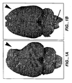
- FIG. 1A shows an infarcted rat brain, into which vehicle alone had been injected intracerebrally.
- the large infarct cavity (white arrowhead) represents significant tissue loss.
- Fig. 1B is a photo of a rat brain subjected to a similar infarct but treated three days later with a cisternal (region indicated by black arrow) infusion of a cellular suspension of murine NSCs plus basic fibroblast growth factor (bFGF), which is a significant distance from the region of infarction.
- bFGF basic fibroblast growth factor
- NSCs appear to have migrated to the region of damage and significantly ameliorated the cerebral volume loss (white arrowhead), appearing to have helped "fill in” the infarction cavity and reverse the tissue loss.
- animals treated in this manner showed a significant improvement in cortically mediated behavioral tasks. Therefore, these results indicate that NSC were drawn to stroke injuries in adult CNS.
- cytokines cytokines, growth factors and drugs
- Such moieties are optionally used or may be administered concomitantly with the transplant or later.
- cytokines include interleukins (IL) IL-1, IL-2, IL-3, IL-4, IL-5, IL-6, IL-7, IL-8, IL-10, and IL-11; tissue necrosis factors (TNF), TNF ⁇ and TNF ⁇ , also lymphotoxin (LT); interferons (IFN) IFN ⁇ , IFN ⁇ and IFN ⁇ ; tissue growth factor (TGF); and basic fibroblast growth factor (bFGF).
- TNF tissue necrosis factors
- IFN interferons
- TGF tissue growth factor
- bFGF basic fibroblast growth factor
- CSFs colony-stimulating factors
- Nerve growth factor has been shown to increase the rate of recovery in spatial alternation tasks after entorhinal lesions, possibly by acting on cholinergic pathways (Stein and Will, Brain Res. 261:127-31,1983).
- cyclosporine was used for at least part of the pre- and post-implant period and other similarly active compounds could be substituted. Cyclosporine was withdrawn in on patient because of seizures, and no marked diminution in function occurred thereafter. Therefore, immunosuppressive therapy may not be necessary, or perhaps could be confined only to the perioperative period.
- Observer-blinded determination of neurologic status was performed, including evaluation of the functional deficit, contrast-enhanced magnetic resonance image (MRI) scanning to measure the volume of blood-brain barrier alteration at the target site (as an indirect measure of inflammatory response), and positron emission tomography (PET) with fluorodeoxyglucose (FDG) scan for assessment of regional brain metabolism.
- MRI contrast-enhanced magnetic resonance image
- PET positron emission tomography
- FDG fluorodeoxyglucose
- a score of 10 is assigned alert, keenly responsive patients; a score of 8 to drowsy patients who can be aroused by minor stimulation to obey, answer or respond; a score of 6 to patients who require repeated stimulation to attend, or are lethargic or obtunded and require strong or painful stimulation to move; a score of 4 to patients who cannot be aroused by any stimulation but react purposefully to painful stimuli; a score of 2 to patients who cannot be aroused by any stimulation and react decerebrately to painful stimuli; and a score of 0 to patients who cannot be aroused by any stimulation and do not react to painful stimuli.
- the examiner without demonstrating, verbally gives the patient the following commands: 1. Stick out your tongue. 2. Put your finger (of the unaffected side) on your nose. 3. Close your eyes.
- the examiner has a conversation with the patient (how is the patient feeling, did he/she sleep well, how long has the patient been in the hospital%) and scores the patient as follows: normal speech (8), slight word-finding difficult but possible conversation (6), severe word-finding difficulties with difficult conversation (4), only yes or no (2), and mute (0).
- the examiner stands at arm's length and compares the patient's field of vision by advancing a moving finger from the periphery inward.
- the patient fixates on the examiner's pupil, first with one and then with the other eye closed. Normal is 8 and deficit is 0.
- the examiner steadies the patient's head and asks him/her to follow the examiner's moving finger.
- the examiner observes the resting eye position and subsequently the full range of movements by moving the index finger from the left to the right and back.
- Normal is 8, median eye position with impossible deviation to one side (4), lateral eye position with possible return to midline (2), and lateral eye position without return to midline (0).
- the examiner observes the patient as he/she talks and smiles, noting any asymmetrical elevation of one corner of the mouth or flattening of the nasolabial fold. Only the muscles of the lower half of the face are assessed. Normal is 8, paresis 4, and paralysis 0.
- the examiner asks the patient to close his/her eyes and actively lifts the patient's arms into position so that they are outstretched at 45° in relation to the horizontal plane with both hands in mid-position so that the palms face each other.
- the patient is asked to maintain this position for 5 seconds after the examiner releases the arms. Only the affected side is evaluated. Score is 4 for maintaining arm position for 5 sec; 3 is maintaining position for 5 sec with hand pronation; score is 2 if arm drifts before 5 sec and maintains a lower position; score is 1 if arm cannot maintain position but attempts to oppose gravity; and 0 if arm falls.
- the patient's arm is rested next to the leg with the hand in mid-position.
- the examiner asks the patient to raise the arm outstretched to 90° (4), if the arm is straight but movement is not full (3), flexed arm (2), trace movements (1), or no movement (0).
- the examiner asks the patient to form with both hands, as strongly as possible, a pinch grip with the thumb and forefinger on the same hand and to try to resist a weak pull.
- the examiner checks the strength of this grip by pulling the pinch with one finger. Equal strength is 8, reduced strength on the affected side is 4, and pinch grip impossible on affected side is 0.
- the examiner actively lifts the patient's affected leg into position so that the thigh forms an angle of 90° with the bed.
- the examiner asks the patient to close his/her eyes and to maintain this position for 5 seconds without support.
- Leg maintains position for 5 sec (4), leg drifts to intermediate position by 5 sec (2), leg drifts to bed within 5 sec but not immediately (1), and leg falls to bed immediately (0).
- the patient is supine with the legs outstretched.
- the examiner asks the patient to flex the hip and knee. Normal movement is 4, movement against resistance with reduced strength is 3, movement against gravity is 2, trace movement is 1, and no movement is 0.
- the patient is tested with the leg outstretched.
- the examiner asks the patient to dorsiflex the foot.
- Normal e.g., outstretched, full movement, normal strength
- leg outstretched with full movement but reduced strength is 6
- leg outstretched with less than full movement or flexed knee or supinated foot is 4
- trace movement is 2
- no movement is 0.
- a normal gait scores 10 gait with abnormal aspect and/or limited distance or speed is 8, walking with aid is 6, requiring the assistance of one or more persons is 4, no walking but standing supported is 2, and no walking or standing is 0.
- Inclusion criteria included major motor deficit from completed basal ganglia stroke defined on imaging.
- the permissible duration of stroke was six months to six years, with a required fixed deficit without substantial change for at least two months.
- Patient age could range from 40 to 75 years inclusive.
- the patient also had to be able to provide informed consent.
- Patients must have had a motor deficit such as hemiparesis following a completed basal ganglia infarction (4-15 mm) involving gray matter as defined on CT or MR imaging scan and by clinical syndromes of lacunar infarction (e.g., hemiparesis with ataxia in the same limb, pure motor hemiplegia).
- a substantial deficit was defined by a total score of 70 or less on the European Stroke Scale (see infra).
- Prohibited medications were all products with anticoagulant or anti-platelet activity, including warfarin, aspirin, nonsteroidal antiinflammatory drugs (NSAIDs), and ticlopidine. These medications were allowed to be restarted 24 hours after surgery.
- NSAIDs nonsteroidal antiinflammatory drugs
- the thawed cell suspension was transferred from the cryovials to sterile 15 mL centrifuge tubes containing Isolyte® S, pH 7.4 (multi-electrolyte injection, McGaw Inc., Irvine, CA), centrifuged at 200xg for 7 minutes at room temperature and the cell pellet gently resuspended in Isolyte S. This wash of the cells was repeated twice. For the final wash, all cells from different tubes were pooled together into one tube. Next a sample of the LBS-neuron suspension was diluted in 0.4% Trypan blue, and viable and dead cells counted using phase contrast microscopy.
- the cell concentration was calculated based on the total viable cell count.
- the pellet volume was measured, and the cells resuspended to a final concentration of 3.3x10 7 cells/mL in Isolyte S and aliquoted at 120 ⁇ L per sterile 1.0 mL vial.
- one or more vials were prepared. Vial(s) were loaded into a closed holder and carried by hand in an upright position to the operating room for immediate use.
- the cells were administered (in up to three tracts) by direct stereotactic injection.
- the first four patients received two million cells in three implants on one track, and the next eight patients where randomized to receive two or six million cells in three or nine implants, respectively.
- Aliquots of cells that were placed in culture and not implanted showed robust development of neuronal processes with 24 hours. Patients stopped all anticoagulant medications and started cyclosporine one week prior to surgery.
- Stereotactic instrumentation consisted of the following: Leksell Model G Stereotactic Coordinate Frame (Elekta Instruments, Atlanta, GA) and a 0.9 mm Outer Diameter Stereotactic Aspiration/Injection Cannula. Contrast-enhanced computed tomography (CT) stereotactic targeting of the stroke area was performed with 5-millimeter slices through the brain. Coronal and sagittal views were used to define a safe trajectory that entered a cortical gyrus and spared a sulcus. Stereotactic coordinates were obtained for each instrument placement.
- CT computed tomography
- Three points in the basal ganglia were a) inferior to the stroke, b) within the midportion of the stroke, and c) in the superior aspect of the basal ganglia either within or beyond the stroke.
- three trajectories were chosen in the same paramedian plane, spaced by 5-6 mm at the target.
- a twist drill or burr hole skull opening was made.
- the dura was opened and a 1.8-mm, 15-cm length stabilizing probe inserted to a point 4 cm proximal to the final target.
- a cannula with a 0.9-mm outer diameter was then inserted down to the deepest target point for the first implantation.
- the first inner cannula used had an internal volume of 100 ⁇ L; a second cannula designed later had a volume of 20 ⁇ L (Synergetics, St. Louis, MO).
- the cells were aspirated into a 250 ⁇ l syringe.
- the internal volume of the cannula was filled with the cell suspension, and then a 20 ⁇ l volume of cells was injected slowly at the first target site.
- the instrument was then withdrawn to the second and third sites for subsequent implants. After the three implants were made, the cannula was withdrawn from the brain.
- the wound was either closed or the next vial of cells prepared to inject implants 4-9 in those patients who received 6x10 6 cells. Following surgery, a post-operative CT scan confirmed the absence of hemorrhage.
- a postoperative CT scan confirmed the safety of the procedure. All patients were then observed overnight and discharged home the next morning. No new neurological deficits were identified acutely. All 12 patients were discharged within 24 hours.
- ESS European Stroke Scale
- NIHSS National Institutes of Health Stroke Scale
- BI Barthel Index
- SF-36 Short Form 36 Health Survey
- NIHSS scores reflected similar changes in functional performance as seen on the ESS.
- 8 patients had improved scores on the NIHSS (range: -1 to -4 points), 1 patient was unchanged and 3 patients deteriorated (range: 1 to 2 points) compared to their baseline scores.
- ESS-motor Motor elements of the ESS (ESS-motor) accounted for the majority of the change noted in patients treated with hNT neurons.
- PET scans performed at baseline and at week 24 showed that 6 of 11 patients had and improvement in cerebral glucose metabolism as indicated by fluorodeoxyglucose (FDG) uptake.
- FDG fluorodeoxyglucose
- One patient (#012) had not had a week-24 PET scan at the time of this report.
- the PET scan findings appeared to correlate with the clinical findings of neurologic improvement.
- 4 (67%) patients improved 3 points or more on the ESS from baseline to week 24, and 2 patients (33%) were essentially unchanged (0 and -1 point change).
- 4 of 5 (80%) did not improve on the ESS and 1 patient improved by 5 points.
- One patient with diabetes had an exacerbation of his chronic renal failure while on cyclosporine, one patient had a single seizure 5 months after implantation, and one patient at 6 months after implantation had a new right pontine infarction that was contralateral to the implantation site.
Landscapes
- Health & Medical Sciences (AREA)
- Life Sciences & Earth Sciences (AREA)
- Engineering & Computer Science (AREA)
- Veterinary Medicine (AREA)
- Animal Behavior & Ethology (AREA)
- Chemical & Material Sciences (AREA)
- Neurology (AREA)
- Biomedical Technology (AREA)
- Medicinal Chemistry (AREA)
- Bioinformatics & Cheminformatics (AREA)
- Public Health (AREA)
- Pharmacology & Pharmacy (AREA)
- General Health & Medical Sciences (AREA)
- Neurosurgery (AREA)
- Chemical Kinetics & Catalysis (AREA)
- Organic Chemistry (AREA)
- Nuclear Medicine, Radiotherapy & Molecular Imaging (AREA)
- General Chemical & Material Sciences (AREA)
- Developmental Biology & Embryology (AREA)
- Cell Biology (AREA)
- Ophthalmology & Optometry (AREA)
- Virology (AREA)
- Psychiatry (AREA)
- Heart & Thoracic Surgery (AREA)
- Cardiology (AREA)
- Biotechnology (AREA)
- Immunology (AREA)
- Hospice & Palliative Care (AREA)
- Zoology (AREA)
- Epidemiology (AREA)
- Orthopedic Medicine & Surgery (AREA)
- Physical Education & Sports Medicine (AREA)
- Medicines Containing Material From Animals Or Micro-Organisms (AREA)
- Micro-Organisms Or Cultivation Processes Thereof (AREA)
Claims (15)
- Neuronale Zellen zur Verwendung in einem Verfahren zur Behandlung eines Schlaganfalls bei einem Patienten, der mindestens drei Stunden zuvor einen Schlaganfall erlitten hat, wobei das Verfahren die Zuführung von mindestens sechs Millionen lebensfähiger neuronaler Zellen in einer auf der Gesamtzahl lebensfähiger Zellen basierenden Zellkonzentration in mindestens einen vom Schlaganfall betroffenen Hirnbereich umfasst, wobei, wenn die Zellen Stammzellen sind, diese nicht durch die Zerstörung eines menschlichen Embryos erhalten wurden.
- Neuronale Zellen nach Anspruch 1, wobei das Verfahren des Weiteren die Verwendung eines Spiralbohrers oder einer Fräse umfasst, um Zugang durch den Schädel zu erhalten, wodurch die Zellen zugeführt werden können.
- Neuronale Zellen nach Anspruch 1 oder 2, wobei die Zellen ausgewählt sind aus neuronalen hNT-Zellen, neuralen Stammzellen, HCN-1-Zellen, fötalen Schweinezellen, Zellen der Neuralleiste oder einer Kombination daraus.
- Neuronale Zellen nach einem der vorstehenden Ansprüche, wobei der Schlaganfall mindestens drei Monate zuvor stattgefunden hat.
- Neuronale Zellen zur Verwendung in einem Verfahren zur Verbesserung der Sprachfähigkeit eines Individuums, das einen Hirnschaden erlitten hat, der die Sprachfähigkeit beeinträchtigt, wobei der Hirnschaden das Ergebnis eines Schlaganfalls ist und das Verfahren das Injizieren einer sterilen Zusammensetzung von mindestens sechs Millionen lebensfähiger neuronaler Zellen in einer auf der Gesamtzahl lebensfähiger Zellen basierenden Zellkonzentration in den geschädigten Bereich umfasst, wobei, wenn die Zellen Stammzellen sind, diese nicht durch die Zerstörung eines menschlichen Embryos erhalten wurden.
- Neuronale Zellen nach Anspruch 5, wobei die injizierten neuronalen Zellen menschliche neuronale Zellen oder menschliche neurale Stammzellen sind.
- Neuronale Zellen zur Verwendung in einem Verfahren zur Verbesserung der motorischen Leistung bei einem Individuum, das einen Hirnschaden erlitten hat, der die Motorik beeinträchtigt, wobei der Hirnschaden das Ergebnis eines Schlaganfalls ist und das Verfahren das Injizieren einer sterilen Zusammensetzung von mindestens sechs Millionen lebensfähiger neuronaler Zellen in einer auf der Gesamtzahl lebensfähiger Zellen basierenden Zellkonzentration in den geschädigten Bereich umfasst, wobei, wenn die Zellen Stammzellen sind, diese nicht durch die Zerstörung eines menschlichen Embryos erhalten wurden.
- Neuronale Zellen nach Anspruch 7, wobei die injizierten neuronalen Zellen neuronale Zellen oder menschliche neurale Stammzellen sind.
- Neuronale Zellen zur Verwendung in einem Verfahren zur Verbesserung der kognitiven Leistung bei einem Individuum, das das einen Hirnschaden erlitten hat, der die kognitive Leistung beeinträchtigt, wobei der Hirnschaden das Ergebnis eines Schlaganfalls ist und das Verfahren das Zuführen einer sterilen Zusammensetzung von mindestens sechs Millionen lebensfähiger neuronaler Zellen in einer auf der Gesamtzahl lebensfähiger Zellen basierenden Zellkonzentration in den geschädigten Hirnbereich umfasst, wobei, wenn die Zellen Stammzellen sind, diese nicht durch die Zerstörung eines menschlichen Embryos erhalten wurden.
- Neuronale Zellen zur Verwendung in einem Verfahren zur Verbesserung der sensorischen Funktion bei einem Individuum, das das einen Hirnschaden erlitten hat, der die sensorische Funktion beeinträchtigt, wobei der Hirnschaden das Ergebnis eines Schlaganfalls ist und das Verfahren das Zuführen einer sterilen Zusammensetzung von mindestens sechs Millionen lebensfähiger neuronaler Zellen in einer auf der Gesamtzahl lebensfähiger Zellen basierenden Zellkonzentration in den geschädigten Bereich umfasst, wobei, wenn die Zellen Stammzellen sind, diese nicht durch die Zerstörung eines menschlichen Embryos erhalten wurden.
- Neuronale Zellen zur Verwendung in einem Verfahren zur Verbesserung der sensorischen, motorischen oder kognitiven Funktion bei einem Individuum, das einen Hirnschaden erlitten hat, der diese Funktionen beeinträchtigt, wobei der Hirnschaden das Ergebnis eines Schlaganfalls ist und das Verfahren das Zuführen einer sterilen Zusammensetzung von mindestens sechs Millionen lebensfähiger neuronaler Zellen in einer auf der Gesamtzahl lebensfähiger Zellen basierenden Zellkonzentration in einen Bereich umfasst, von dem aus die neuronalen Zellen zu dem geschädigten Bereich wandern, wobei, wenn die Zellen Stammzellen sind, diese nicht durch die Zerstörung eines menschlichen Embryos erhalten wurden.
- Neuronale Zellen nach Anspruch 11, wobei die Zusammensetzung der Cisterna zugeführt wird.
- Neuronale Zellen zur Verwendung in einem Verfahren, bei dem im Zentralnervensystem eines Individuums durch Schlaganfall verlorene Nerven ersetzt werden, wobei das Verfahren die Verabreichung einer sterilen Zusammensetzung von mindestens sechs Millionen lebensfähiger neuronaler Zellen in einer auf der Gesamtzahl lebensfähiger Zellen basierenden Zellkonzentration an das Individuum umfasst, wobei, wenn die Zellen Stammzellen sind, diese nicht durch die Zerstörung eines menschlichen Embryos erhalten wurden.
- Neuronale Zellen nach einem der Ansprüche 9 bis 13, wobei die neuronalen Zellen menschliche neuronale Zellen oder neurale Stammzellen sind.
- Verwendung von neuronalen Zellen für die Herstellung eines Medikaments zur Behandlung eines Schlaganfalls bei einem Patienten, der mindestens drei Stunden zuvor einen Schlaganfall erlitten hat, mithilfe eines Verfahrens, das die Zuführung von mindestens sechs Millionen lebensfähiger neuronaler Zellen in einer auf der Gesamtzahl lebensfähiger Zellen basierenden Zellkonzentration in mindestens einen vom Schlaganfall betroffenen Hirnbereich umfasst, wobei, wenn die Zellen Stammzellen sind, diese nicht durch die Zerstörung eines menschlichen Embryos erhalten wurden.
Priority Applications (1)
| Application Number | Priority Date | Filing Date | Title |
|---|---|---|---|
| EP08006165A EP1949904A3 (de) | 1999-04-27 | 2000-03-16 | Zelltherapie für chronische Apoplexie |
Applications Claiming Priority (5)
| Application Number | Priority Date | Filing Date | Title |
|---|---|---|---|
| US13123099P | 1999-04-27 | 1999-04-27 | |
| US131230P | 1999-04-27 | ||
| US14478599P | 1999-07-20 | 1999-07-20 | |
| US144785P | 1999-07-20 | ||
| PCT/US2000/006912 WO2000064459A1 (en) | 1999-04-27 | 2000-03-16 | Cell therapy for chronic stroke |
Related Child Applications (1)
| Application Number | Title | Priority Date | Filing Date |
|---|---|---|---|
| EP08006165A Division EP1949904A3 (de) | 1999-04-27 | 2000-03-16 | Zelltherapie für chronische Apoplexie |
Publications (3)
| Publication Number | Publication Date |
|---|---|
| EP1180036A1 EP1180036A1 (de) | 2002-02-20 |
| EP1180036A4 EP1180036A4 (de) | 2002-09-25 |
| EP1180036B1 true EP1180036B1 (de) | 2009-10-07 |
Family
ID=26829263
Family Applications (1)
| Application Number | Title | Priority Date | Filing Date |
|---|---|---|---|
| EP00918005A Expired - Lifetime EP1180036B1 (de) | 1999-04-27 | 2000-03-16 | Zelltherapie für chronischen schlaganfall |
Country Status (7)
| Country | Link |
|---|---|
| EP (1) | EP1180036B1 (de) |
| JP (1) | JP2003521474A (de) |
| AT (1) | ATE444758T1 (de) |
| AU (1) | AU780365B2 (de) |
| CA (1) | CA2371017A1 (de) |
| DE (1) | DE60043102D1 (de) |
| WO (1) | WO2000064459A1 (de) |
Families Citing this family (2)
| Publication number | Priority date | Publication date | Assignee | Title |
|---|---|---|---|---|
| US7560280B2 (en) | 2000-11-03 | 2009-07-14 | Kourion Therapeutics Gmbh | Human cord blood derived unrestricted somatic stem cells (USSC) |
| WO2007136042A1 (ja) * | 2006-05-19 | 2007-11-29 | Aichi Prefecture | 脳損傷改善剤 |
Citations (1)
| Publication number | Priority date | Publication date | Assignee | Title |
|---|---|---|---|---|
| WO1998057663A1 (en) * | 1997-06-17 | 1998-12-23 | University Technology Corporation | Neural transplantation for treating neurological disease |
Family Cites Families (2)
| Publication number | Priority date | Publication date | Assignee | Title |
|---|---|---|---|---|
| DK0594669T4 (da) * | 1991-07-08 | 2008-04-21 | Neurospheres Holdings Ltd | Neurale progenitorceller, som reagerer på vækstfaktorer og kan formeres in vitro |
| US5851832A (en) * | 1991-07-08 | 1998-12-22 | Neurospheres, Ltd. | In vitro growth and proliferation of multipotent neural stem cells and their progeny |
-
2000
- 2000-03-16 JP JP2000613449A patent/JP2003521474A/ja active Pending
- 2000-03-16 AT AT00918005T patent/ATE444758T1/de not_active IP Right Cessation
- 2000-03-16 EP EP00918005A patent/EP1180036B1/de not_active Expired - Lifetime
- 2000-03-16 CA CA002371017A patent/CA2371017A1/en not_active Abandoned
- 2000-03-16 DE DE60043102T patent/DE60043102D1/de not_active Expired - Lifetime
- 2000-03-16 WO PCT/US2000/006912 patent/WO2000064459A1/en not_active Ceased
- 2000-03-16 AU AU38889/00A patent/AU780365B2/en not_active Ceased
Patent Citations (1)
| Publication number | Priority date | Publication date | Assignee | Title |
|---|---|---|---|---|
| WO1998057663A1 (en) * | 1997-06-17 | 1998-12-23 | University Technology Corporation | Neural transplantation for treating neurological disease |
Non-Patent Citations (4)
| Title |
|---|
| BORLONGAN-CV: "Transplantation of cryopreserved human embryonal carcinoma-derived neurons (NT2N cells) promotes functional recovery in ischemic rats", EXPERIMENTAL NEUROLOGY, vol. 149, 1998, pages 310 - 321 * |
| KONDZIOLKA-D ET AL.: "Neural transplantation for stroke", JOURNAL OF CLINICAL NEUROSCIENCE, vol. 9, no. 3, 2002, pages 225-230 * |
| KONDZIOLKA-D ET AL.: "Transplantation of cultured human neuronal cells for patients with stroke", NEUROLOGY, vol. 55, August 2000 (2000-08-01), pages 565-569 * |
| WECHSLER-LR ET AL.: "Cell Therapy: Replacement", STROKE, vol. 34, 2003, pages 2081-2082 * |
Also Published As
| Publication number | Publication date |
|---|---|
| AU3888900A (en) | 2000-11-10 |
| AU780365B2 (en) | 2005-03-17 |
| DE60043102D1 (de) | 2009-11-19 |
| CA2371017A1 (en) | 2000-11-02 |
| ATE444758T1 (de) | 2009-10-15 |
| EP1180036A1 (de) | 2002-02-20 |
| EP1180036A4 (de) | 2002-09-25 |
| JP2003521474A (ja) | 2003-07-15 |
| WO2000064459A1 (en) | 2000-11-02 |
Similar Documents
| Publication | Publication Date | Title |
|---|---|---|
| US20140099286A1 (en) | Cell therapy for chronic stroke | |
| Olson et al. | Intraputaminal infusion of nerve growth factor to support adrenal medullary autografts in Parkinson's disease: one-year follow-up of first clinical trial | |
| JP5649786B2 (ja) | ヒト胚性幹細胞およびそれらの誘導体を含む組成物、使用方法、ならびに調製方法 | |
| JP5871865B2 (ja) | 神経前駆細胞又は神経幹細胞の神経細胞への分化及び増殖の誘導方法、分化及び増殖誘導用組成物、及び薬学的製剤 | |
| JP7473117B2 (ja) | 疼痛調節因子を含む幹細胞由来エクソソーム及びその用途 | |
| JP6401757B2 (ja) | 臍帯血細胞を用いた脳損傷の処置 | |
| US7247298B2 (en) | Treatment of brain damage | |
| EP1852500A1 (de) | Stammzellen aus dem Knochenmark für Regenerierung von Geweben | |
| KR20090055691A (ko) | 인간 제대혈 유래 간엽 줄기세포를 유효성분으로 포함하는,신경전구세포 또는 신경줄기세포의 신경세포로의 분화 및증식 유도용 조성물 | |
| US10137156B2 (en) | Composition comprising neural cell and elastin-like polypeptide for treating Parkinson's disease | |
| EP1180036B1 (de) | Zelltherapie für chronischen schlaganfall | |
| KR100959995B1 (ko) | 인간 제대혈 유래 간엽 줄기세포를 유효성분으로 포함하는,신경전구세포 또는 신경줄기세포의 신경세포로의 분화 및증식 유도용 조성물 | |
| US8277795B2 (en) | Methods and compositions for treating motor neuron diseases comprising mesenchymal stem cells | |
| Cooperative Group for Reassessment of Defibrase | Reassessment of defibrase in treatment of acute cerebral infarction: a multicenter, randomized, double-blind, placebo-controlled trial | |
| EP1949904A2 (de) | Zelltherapie für chronische Apoplexie | |
| RU2129427C1 (ru) | Способ лечения заболеваний центральной нервной системы | |
| AU2005202666A1 (en) | Cell therapy for chronic stroke | |
| RU2283121C2 (ru) | Способ лечения инсульта | |
| EP0981353B1 (de) | Verbesserter heilungsprozess von neuralverletzungen anhand von cm101/gbs-toxin | |
| RU2798554C2 (ru) | Биомедицинский клеточный продукт для лечения онкологических, нейродегенеративных, аутоимунных заболеваний и травм головного и спинного мозга | |
| KR20240090983A (ko) | 신경 복원 촉진에 사용되는 이소로이폴린 및 그 유도체의 용도 | |
| Meduna | The use of metrazol in the treatment of patients with mental diseases | |
| CN120837530A (zh) | 一种人脐带间充质干细胞在制备治疗自闭症谱系障碍中的用途 | |
| CN109758478A (zh) | 间充质干细胞在制备中风治疗剂中的用途 | |
| BR112021005060A2 (pt) | produto farmacêutico para prevenção ou tratamento de mal de alzheimer |
Legal Events
| Date | Code | Title | Description |
|---|---|---|---|
| PUAI | Public reference made under article 153(3) epc to a published international application that has entered the european phase |
Free format text: ORIGINAL CODE: 0009012 |
|
| 17P | Request for examination filed |
Effective date: 20011113 |
|
| AK | Designated contracting states |
Kind code of ref document: A1 Designated state(s): AT BE CH CY DE DK ES FI FR GB GR IE IT LI LU MC NL PT SE |
|
| AX | Request for extension of the european patent |
Free format text: AL;LT;LV;MK;RO;SI |
|
| RIC1 | Information provided on ipc code assigned before grant |
Free format text: 7A 61K 35/30 A, 7C 12N 5/06 B, 7C 12N 5/08 B, 7A 61P 9/00 B, 7A 61P 21/00 B, 7A 61P 25/00 B |
|
| A4 | Supplementary search report drawn up and despatched |
Effective date: 20020812 |
|
| AK | Designated contracting states |
Kind code of ref document: A4 Designated state(s): AT BE CH CY DE DK ES FI FR GB GR IE IT LI LU MC NL PT SE |
|
| 17Q | First examination report despatched |
Effective date: 20030806 |
|
| GRAP | Despatch of communication of intention to grant a patent |
Free format text: ORIGINAL CODE: EPIDOSNIGR1 |
|
| GRAS | Grant fee paid |
Free format text: ORIGINAL CODE: EPIDOSNIGR3 |
|
| GRAA | (expected) grant |
Free format text: ORIGINAL CODE: 0009210 |
|
| RAP1 | Party data changed (applicant data changed or rights of an application transferred) |
Owner name: LAYTON BIOSCIENCE, INC. Owner name: UNIVERSITY OF SOUTH FLORIDA |
|
| AK | Designated contracting states |
Kind code of ref document: B1 Designated state(s): AT BE CH CY DE DK ES FI FR GB GR IE IT LI LU MC NL PT SE |
|
| REG | Reference to a national code |
Ref country code: GB Ref legal event code: FG4D |
|
| REG | Reference to a national code |
Ref country code: CH Ref legal event code: EP |
|
| REG | Reference to a national code |
Ref country code: IE Ref legal event code: FG4D |
|
| REF | Corresponds to: |
Ref document number: 60043102 Country of ref document: DE Date of ref document: 20091119 Kind code of ref document: P |
|
| NLV1 | Nl: lapsed or annulled due to failure to fulfill the requirements of art. 29p and 29m of the patents act | ||
| PG25 | Lapsed in a contracting state [announced via postgrant information from national office to epo] |
Ref country code: FI Free format text: LAPSE BECAUSE OF FAILURE TO SUBMIT A TRANSLATION OF THE DESCRIPTION OR TO PAY THE FEE WITHIN THE PRESCRIBED TIME-LIMIT Effective date: 20091007 Ref country code: ES Free format text: LAPSE BECAUSE OF FAILURE TO SUBMIT A TRANSLATION OF THE DESCRIPTION OR TO PAY THE FEE WITHIN THE PRESCRIBED TIME-LIMIT Effective date: 20100118 Ref country code: SE Free format text: LAPSE BECAUSE OF FAILURE TO SUBMIT A TRANSLATION OF THE DESCRIPTION OR TO PAY THE FEE WITHIN THE PRESCRIBED TIME-LIMIT Effective date: 20091007 Ref country code: PT Free format text: LAPSE BECAUSE OF FAILURE TO SUBMIT A TRANSLATION OF THE DESCRIPTION OR TO PAY THE FEE WITHIN THE PRESCRIBED TIME-LIMIT Effective date: 20100208 |
|
| PG25 | Lapsed in a contracting state [announced via postgrant information from national office to epo] |
Ref country code: AT Free format text: LAPSE BECAUSE OF FAILURE TO SUBMIT A TRANSLATION OF THE DESCRIPTION OR TO PAY THE FEE WITHIN THE PRESCRIBED TIME-LIMIT Effective date: 20091007 Ref country code: BE Free format text: LAPSE BECAUSE OF FAILURE TO SUBMIT A TRANSLATION OF THE DESCRIPTION OR TO PAY THE FEE WITHIN THE PRESCRIBED TIME-LIMIT Effective date: 20091007 |
|
| PG25 | Lapsed in a contracting state [announced via postgrant information from national office to epo] |
Ref country code: NL Free format text: LAPSE BECAUSE OF FAILURE TO SUBMIT A TRANSLATION OF THE DESCRIPTION OR TO PAY THE FEE WITHIN THE PRESCRIBED TIME-LIMIT Effective date: 20091007 Ref country code: DK Free format text: LAPSE BECAUSE OF FAILURE TO SUBMIT A TRANSLATION OF THE DESCRIPTION OR TO PAY THE FEE WITHIN THE PRESCRIBED TIME-LIMIT Effective date: 20091007 |
|
| PLBE | No opposition filed within time limit |
Free format text: ORIGINAL CODE: 0009261 |
|
| STAA | Information on the status of an ep patent application or granted ep patent |
Free format text: STATUS: NO OPPOSITION FILED WITHIN TIME LIMIT |
|
| 26N | No opposition filed |
Effective date: 20100708 |
|
| PG25 | Lapsed in a contracting state [announced via postgrant information from national office to epo] |
Ref country code: GR Free format text: LAPSE BECAUSE OF FAILURE TO SUBMIT A TRANSLATION OF THE DESCRIPTION OR TO PAY THE FEE WITHIN THE PRESCRIBED TIME-LIMIT Effective date: 20100108 Ref country code: MC Free format text: LAPSE BECAUSE OF NON-PAYMENT OF DUE FEES Effective date: 20100331 |
|
| REG | Reference to a national code |
Ref country code: CH Ref legal event code: PL |
|
| PG25 | Lapsed in a contracting state [announced via postgrant information from national office to epo] |
Ref country code: IE Free format text: LAPSE BECAUSE OF NON-PAYMENT OF DUE FEES Effective date: 20100316 |
|
| PG25 | Lapsed in a contracting state [announced via postgrant information from national office to epo] |
Ref country code: LI Free format text: LAPSE BECAUSE OF NON-PAYMENT OF DUE FEES Effective date: 20100331 Ref country code: CH Free format text: LAPSE BECAUSE OF NON-PAYMENT OF DUE FEES Effective date: 20100331 |
|
| PG25 | Lapsed in a contracting state [announced via postgrant information from national office to epo] |
Ref country code: IT Free format text: LAPSE BECAUSE OF FAILURE TO SUBMIT A TRANSLATION OF THE DESCRIPTION OR TO PAY THE FEE WITHIN THE PRESCRIBED TIME-LIMIT Effective date: 20091007 |
|
| PG25 | Lapsed in a contracting state [announced via postgrant information from national office to epo] |
Ref country code: CY Free format text: LAPSE BECAUSE OF FAILURE TO SUBMIT A TRANSLATION OF THE DESCRIPTION OR TO PAY THE FEE WITHIN THE PRESCRIBED TIME-LIMIT Effective date: 20091007 |
|
| PG25 | Lapsed in a contracting state [announced via postgrant information from national office to epo] |
Ref country code: LU Free format text: LAPSE BECAUSE OF NON-PAYMENT OF DUE FEES Effective date: 20100316 |
|
| REG | Reference to a national code |
Ref country code: FR Ref legal event code: PLFP Year of fee payment: 17 |
|
| REG | Reference to a national code |
Ref country code: FR Ref legal event code: PLFP Year of fee payment: 18 |
|
| REG | Reference to a national code |
Ref country code: FR Ref legal event code: PLFP Year of fee payment: 19 |
|
| PGFP | Annual fee paid to national office [announced via postgrant information from national office to epo] |
Ref country code: DE Payment date: 20190328 Year of fee payment: 20 Ref country code: GB Payment date: 20190321 Year of fee payment: 20 |
|
| PGFP | Annual fee paid to national office [announced via postgrant information from national office to epo] |
Ref country code: FR Payment date: 20190227 Year of fee payment: 20 |
|
| REG | Reference to a national code |
Ref country code: DE Ref legal event code: R071 Ref document number: 60043102 Country of ref document: DE |
|
| REG | Reference to a national code |
Ref country code: GB Ref legal event code: PE20 Expiry date: 20200315 |
|
| PG25 | Lapsed in a contracting state [announced via postgrant information from national office to epo] |
Ref country code: GB Free format text: LAPSE BECAUSE OF EXPIRATION OF PROTECTION Effective date: 20200315 |