EP1182251A1 - Procédé pour l'identification de composés inhibant la protéolyse de IkB par l'ubiquitine - Google Patents
Procédé pour l'identification de composés inhibant la protéolyse de IkB par l'ubiquitine Download PDFInfo
- Publication number
- EP1182251A1 EP1182251A1 EP00117429A EP00117429A EP1182251A1 EP 1182251 A1 EP1182251 A1 EP 1182251A1 EP 00117429 A EP00117429 A EP 00117429A EP 00117429 A EP00117429 A EP 00117429A EP 1182251 A1 EP1182251 A1 EP 1182251A1
- Authority
- EP
- European Patent Office
- Prior art keywords
- hnrnp
- e3rs
- protein
- complex
- compound
- Prior art date
- Legal status (The legal status is an assumption and is not a legal conclusion. Google has not performed a legal analysis and makes no representation as to the accuracy of the status listed.)
- Withdrawn
Links
- 150000001875 compounds Chemical class 0.000 title claims abstract description 75
- 238000000034 method Methods 0.000 title claims abstract description 74
- 230000001404 mediated effect Effects 0.000 title claims abstract description 21
- 102000044159 Ubiquitin Human genes 0.000 title claims abstract description 14
- 108090000848 Ubiquitin Proteins 0.000 title claims abstract description 14
- 230000017854 proteolysis Effects 0.000 title claims abstract description 8
- 108010085697 Heterogeneous-Nuclear Ribonucleoprotein U Proteins 0.000 claims abstract description 195
- 108010080842 beta-Transducin Repeat-Containing Proteins Proteins 0.000 claims abstract description 30
- 102000000472 beta-Transducin Repeat-Containing Proteins Human genes 0.000 claims abstract description 30
- 102000007511 Heterogeneous-Nuclear Ribonucleoprotein U Human genes 0.000 claims abstract description 29
- 230000034190 positive regulation of NF-kappaB transcription factor activity Effects 0.000 claims abstract description 20
- 239000003814 drug Substances 0.000 claims abstract description 11
- 238000012544 monitoring process Methods 0.000 claims abstract description 8
- 108090000623 proteins and genes Proteins 0.000 claims description 93
- 102000004169 proteins and genes Human genes 0.000 claims description 88
- 210000004027 cell Anatomy 0.000 claims description 81
- 101000716102 Homo sapiens T-cell surface glycoprotein CD4 Proteins 0.000 claims description 49
- 102100036011 T-cell surface glycoprotein CD4 Human genes 0.000 claims description 49
- 238000006731 degradation reaction Methods 0.000 claims description 45
- 230000015556 catabolic process Effects 0.000 claims description 44
- 239000003112 inhibitor Substances 0.000 claims description 43
- 230000027455 binding Effects 0.000 claims description 39
- 102000000341 S-Phase Kinase-Associated Proteins Human genes 0.000 claims description 38
- 108010055623 S-Phase Kinase-Associated Proteins Proteins 0.000 claims description 38
- 238000010494 dissociation reaction Methods 0.000 claims description 34
- 230000005593 dissociations Effects 0.000 claims description 34
- 238000012360 testing method Methods 0.000 claims description 29
- 230000014509 gene expression Effects 0.000 claims description 26
- 239000005090 green fluorescent protein Substances 0.000 claims description 22
- 108010043121 Green Fluorescent Proteins Proteins 0.000 claims description 20
- 102000004144 Green Fluorescent Proteins Human genes 0.000 claims description 20
- 230000002401 inhibitory effect Effects 0.000 claims description 19
- 239000003795 chemical substances by application Substances 0.000 claims description 16
- 239000003550 marker Substances 0.000 claims description 15
- 239000013612 plasmid Substances 0.000 claims description 13
- 230000001939 inductive effect Effects 0.000 claims description 10
- 238000011282 treatment Methods 0.000 claims description 10
- 208000037265 diseases, disorders, signs and symptoms Diseases 0.000 claims description 9
- 230000001580 bacterial effect Effects 0.000 claims description 7
- 230000004186 co-expression Effects 0.000 claims description 7
- 208000035475 disorder Diseases 0.000 claims description 7
- 238000002866 fluorescence resonance energy transfer Methods 0.000 claims description 7
- 210000004962 mammalian cell Anatomy 0.000 claims description 7
- 238000005259 measurement Methods 0.000 claims description 7
- 239000003607 modifier Substances 0.000 claims description 7
- 102000004190 Enzymes Human genes 0.000 claims description 6
- 108090000790 Enzymes Proteins 0.000 claims description 6
- 238000002360 preparation method Methods 0.000 claims description 6
- 229930101283 tetracycline Natural products 0.000 claims description 6
- 229940079156 Proteasome inhibitor Drugs 0.000 claims description 5
- 240000004808 Saccharomyces cerevisiae Species 0.000 claims description 5
- 230000001419 dependent effect Effects 0.000 claims description 5
- 239000003207 proteasome inhibitor Substances 0.000 claims description 5
- 230000009467 reduction Effects 0.000 claims description 5
- 239000007787 solid Substances 0.000 claims description 5
- 241000238631 Hexapoda Species 0.000 claims description 4
- 101100005713 Homo sapiens CD4 gene Proteins 0.000 claims description 4
- 108010001441 Phosphopeptides Proteins 0.000 claims description 4
- 230000014759 maintenance of location Effects 0.000 claims description 4
- 239000013641 positive control Substances 0.000 claims description 4
- 108010076504 Protein Sorting Signals Proteins 0.000 claims description 3
- 108091005948 blue fluorescent proteins Proteins 0.000 claims description 3
- 239000013078 crystal Substances 0.000 claims description 3
- 238000009510 drug design Methods 0.000 claims description 3
- 239000007850 fluorescent dye Substances 0.000 claims description 3
- 102000034287 fluorescent proteins Human genes 0.000 claims description 3
- 108091006047 fluorescent proteins Proteins 0.000 claims description 3
- 230000000415 inactivating effect Effects 0.000 claims description 3
- 230000001225 therapeutic effect Effects 0.000 claims description 3
- 230000001010 compromised effect Effects 0.000 claims description 2
- 230000006698 induction Effects 0.000 claims description 2
- 238000004519 manufacturing process Methods 0.000 claims description 2
- 238000004321 preservation Methods 0.000 claims description 2
- 230000002285 radioactive effect Effects 0.000 claims description 2
- 230000006641 stabilisation Effects 0.000 claims description 2
- 238000011105 stabilization Methods 0.000 claims description 2
- 102000018745 NF-KappaB Inhibitor alpha Human genes 0.000 claims 3
- 108010052419 NF-KappaB Inhibitor alpha Proteins 0.000 claims 3
- 230000002596 correlated effect Effects 0.000 claims 3
- 238000002288 cocrystallisation Methods 0.000 claims 1
- 239000000470 constituent Substances 0.000 claims 1
- 238000003745 diagnosis Methods 0.000 claims 1
- JBIWCJUYHHGXTC-AKNGSSGZSA-N doxycycline Chemical group O=C1C2=C(O)C=CC=C2[C@H](C)[C@@H]2C1=C(O)[C@]1(O)C(=O)C(C(N)=O)=C(O)[C@@H](N(C)C)[C@@H]1[C@H]2O JBIWCJUYHHGXTC-AKNGSSGZSA-N 0.000 claims 1
- 230000002621 immunoprecipitating effect Effects 0.000 claims 1
- 230000000087 stabilizing effect Effects 0.000 claims 1
- OFVLGDICTFRJMM-WESIUVDSSA-N tetracycline Chemical compound C1=CC=C2[C@](O)(C)[C@H]3C[C@H]4[C@H](N(C)C)C(O)=C(C(N)=O)C(=O)[C@@]4(O)C(O)=C3C(=O)C2=C1O OFVLGDICTFRJMM-WESIUVDSSA-N 0.000 claims 1
- 230000001413 cellular effect Effects 0.000 abstract description 14
- 238000012216 screening Methods 0.000 abstract description 11
- 229940079593 drug Drugs 0.000 abstract description 8
- 230000002452 interceptive effect Effects 0.000 abstract description 3
- 102100024002 Heterogeneous nuclear ribonucleoprotein U Human genes 0.000 description 170
- 235000018102 proteins Nutrition 0.000 description 64
- 238000003556 assay Methods 0.000 description 51
- 108090000765 processed proteins & peptides Proteins 0.000 description 40
- 230000000694 effects Effects 0.000 description 34
- 230000003993 interaction Effects 0.000 description 30
- 239000012634 fragment Substances 0.000 description 28
- 239000000758 substrate Substances 0.000 description 28
- 230000034512 ubiquitination Effects 0.000 description 26
- 238000010798 ubiquitination Methods 0.000 description 26
- 108010057466 NF-kappa B Proteins 0.000 description 22
- 102000003945 NF-kappa B Human genes 0.000 description 22
- 230000006870 function Effects 0.000 description 19
- 210000004940 nucleus Anatomy 0.000 description 18
- 102000018700 F-Box Proteins Human genes 0.000 description 17
- 108010066805 F-Box Proteins Proteins 0.000 description 17
- 241000725303 Human immunodeficiency virus Species 0.000 description 15
- 241000283973 Oryctolagus cuniculus Species 0.000 description 15
- 150000001413 amino acids Chemical class 0.000 description 14
- 230000000937 inactivator Effects 0.000 description 13
- 238000001262 western blot Methods 0.000 description 13
- 102000006275 Ubiquitin-Protein Ligases Human genes 0.000 description 12
- 108010083111 Ubiquitin-Protein Ligases Proteins 0.000 description 12
- 238000000423 cell based assay Methods 0.000 description 12
- 239000002299 complementary DNA Substances 0.000 description 12
- 210000002472 endoplasmic reticulum Anatomy 0.000 description 12
- 238000002474 experimental method Methods 0.000 description 12
- 108020001507 fusion proteins Proteins 0.000 description 12
- 102000037865 fusion proteins Human genes 0.000 description 12
- 235000001014 amino acid Nutrition 0.000 description 11
- 239000002773 nucleotide Substances 0.000 description 11
- 125000003729 nucleotide group Chemical group 0.000 description 11
- 238000001114 immunoprecipitation Methods 0.000 description 10
- 102000004196 processed proteins & peptides Human genes 0.000 description 10
- SGKRLCUYIXIAHR-AKNGSSGZSA-N (4s,4ar,5s,5ar,6r,12ar)-4-(dimethylamino)-1,5,10,11,12a-pentahydroxy-6-methyl-3,12-dioxo-4a,5,5a,6-tetrahydro-4h-tetracene-2-carboxamide Chemical compound C1=CC=C2[C@H](C)[C@@H]([C@H](O)[C@@H]3[C@](C(O)=C(C(N)=O)C(=O)[C@H]3N(C)C)(O)C3=O)C3=C(O)C2=C1O SGKRLCUYIXIAHR-AKNGSSGZSA-N 0.000 description 9
- 230000004913 activation Effects 0.000 description 9
- 229960003722 doxycycline Drugs 0.000 description 9
- 239000013604 expression vector Substances 0.000 description 9
- 238000001890 transfection Methods 0.000 description 9
- 210000000805 cytoplasm Anatomy 0.000 description 8
- LOKCTEFSRHRXRJ-UHFFFAOYSA-I dipotassium trisodium dihydrogen phosphate hydrogen phosphate dichloride Chemical compound P(=O)(O)(O)[O-].[K+].P(=O)(O)([O-])[O-].[Na+].[Na+].[Cl-].[K+].[Cl-].[Na+] LOKCTEFSRHRXRJ-UHFFFAOYSA-I 0.000 description 8
- 239000002953 phosphate buffered saline Substances 0.000 description 8
- 241000283707 Capra Species 0.000 description 7
- 101001047854 Homo sapiens Heterogeneous nuclear ribonucleoprotein U Proteins 0.000 description 7
- 102000003960 Ligases Human genes 0.000 description 7
- 108090000364 Ligases Proteins 0.000 description 7
- 108010006519 Molecular Chaperones Proteins 0.000 description 7
- 238000004458 analytical method Methods 0.000 description 7
- 239000013613 expression plasmid Substances 0.000 description 7
- 239000000284 extract Substances 0.000 description 7
- 230000035772 mutation Effects 0.000 description 7
- 210000002966 serum Anatomy 0.000 description 7
- 238000002415 sodium dodecyl sulfate polyacrylamide gel electrophoresis Methods 0.000 description 7
- 239000013598 vector Substances 0.000 description 7
- 102000006479 Heterogeneous-Nuclear Ribonucleoproteins Human genes 0.000 description 6
- 108010019372 Heterogeneous-Nuclear Ribonucleoproteins Proteins 0.000 description 6
- 241001465754 Metazoa Species 0.000 description 6
- 108010077850 Nuclear Localization Signals Proteins 0.000 description 6
- 102000036366 SCF complex Human genes 0.000 description 6
- 108091007047 SCF complex Proteins 0.000 description 6
- 230000026731 phosphorylation Effects 0.000 description 6
- 238000006366 phosphorylation reaction Methods 0.000 description 6
- 239000002002 slurry Substances 0.000 description 6
- 238000010561 standard procedure Methods 0.000 description 6
- KDXKERNSBIXSRK-UHFFFAOYSA-N Lysine Natural products NCCCCC(N)C(O)=O KDXKERNSBIXSRK-UHFFFAOYSA-N 0.000 description 5
- 239000004098 Tetracycline Substances 0.000 description 5
- 102000040945 Transcription factor Human genes 0.000 description 5
- 108091023040 Transcription factor Proteins 0.000 description 5
- 230000000692 anti-sense effect Effects 0.000 description 5
- 230000008901 benefit Effects 0.000 description 5
- 230000001086 cytosolic effect Effects 0.000 description 5
- 238000001727 in vivo Methods 0.000 description 5
- 238000011534 incubation Methods 0.000 description 5
- 238000004949 mass spectrometry Methods 0.000 description 5
- 239000000203 mixture Substances 0.000 description 5
- 108010054624 red fluorescent protein Proteins 0.000 description 5
- 238000010186 staining Methods 0.000 description 5
- 229960002180 tetracycline Drugs 0.000 description 5
- 235000019364 tetracycline Nutrition 0.000 description 5
- 150000003522 tetracyclines Chemical class 0.000 description 5
- 108091032973 (ribonucleotides)n+m Proteins 0.000 description 4
- 229920000936 Agarose Polymers 0.000 description 4
- 101100016370 Danio rerio hsp90a.1 gene Proteins 0.000 description 4
- 101100285708 Dictyostelium discoideum hspD gene Proteins 0.000 description 4
- 108091006010 FLAG-tagged proteins Proteins 0.000 description 4
- 102100021854 Inhibitor of nuclear factor kappa-B kinase subunit beta Human genes 0.000 description 4
- 101710205525 Inhibitor of nuclear factor kappa-B kinase subunit beta Proteins 0.000 description 4
- 102000008300 Mutant Proteins Human genes 0.000 description 4
- 108010021466 Mutant Proteins Proteins 0.000 description 4
- 102100033104 NF-kappa-B inhibitor epsilon Human genes 0.000 description 4
- 101710093997 NF-kappa-B inhibitor epsilon Proteins 0.000 description 4
- 235000014680 Saccharomyces cerevisiae Nutrition 0.000 description 4
- 101100071627 Schizosaccharomyces pombe (strain 972 / ATCC 24843) swo1 gene Proteins 0.000 description 4
- FAPWRFPIFSIZLT-UHFFFAOYSA-M Sodium chloride Chemical compound [Na+].[Cl-] FAPWRFPIFSIZLT-UHFFFAOYSA-M 0.000 description 4
- 102000006612 Transducin Human genes 0.000 description 4
- 108010087042 Transducin Proteins 0.000 description 4
- 238000002050 diffraction method Methods 0.000 description 4
- 238000002635 electroconvulsive therapy Methods 0.000 description 4
- 239000000499 gel Substances 0.000 description 4
- RWSXRVCMGQZWBV-WDSKDSINSA-N glutathione Chemical compound OC(=O)[C@@H](N)CCC(=O)N[C@@H](CS)C(=O)NCC(O)=O RWSXRVCMGQZWBV-WDSKDSINSA-N 0.000 description 4
- 230000003301 hydrolyzing effect Effects 0.000 description 4
- 238000000338 in vitro Methods 0.000 description 4
- 230000005764 inhibitory process Effects 0.000 description 4
- 239000013642 negative control Substances 0.000 description 4
- 230000037361 pathway Effects 0.000 description 4
- 229920001184 polypeptide Polymers 0.000 description 4
- 230000008569 process Effects 0.000 description 4
- 230000000717 retained effect Effects 0.000 description 4
- 238000007423 screening assay Methods 0.000 description 4
- 230000035939 shock Effects 0.000 description 4
- 101150110885 CUL1 gene Proteins 0.000 description 3
- 108050001186 Chaperonin Cpn60 Proteins 0.000 description 3
- 102000052603 Chaperonins Human genes 0.000 description 3
- 102100023877 E3 ubiquitin-protein ligase RBX1 Human genes 0.000 description 3
- 102000008297 Nuclear Matrix-Associated Proteins Human genes 0.000 description 3
- 108010035916 Nuclear Matrix-Associated Proteins Proteins 0.000 description 3
- 108091000080 Phosphotransferase Proteins 0.000 description 3
- 239000007983 Tris buffer Substances 0.000 description 3
- 239000000427 antigen Substances 0.000 description 3
- 108091007433 antigens Proteins 0.000 description 3
- 102000036639 antigens Human genes 0.000 description 3
- 239000011324 bead Substances 0.000 description 3
- 230000007812 deficiency Effects 0.000 description 3
- VHJLVAABSRFDPM-QWWZWVQMSA-N dithiothreitol Chemical compound SC[C@@H](O)[C@H](O)CS VHJLVAABSRFDPM-QWWZWVQMSA-N 0.000 description 3
- 238000010828 elution Methods 0.000 description 3
- 238000011156 evaluation Methods 0.000 description 3
- 238000009472 formulation Methods 0.000 description 3
- 238000012744 immunostaining Methods 0.000 description 3
- 238000002347 injection Methods 0.000 description 3
- 239000007924 injection Substances 0.000 description 3
- 238000012423 maintenance Methods 0.000 description 3
- 230000007246 mechanism Effects 0.000 description 3
- 230000017128 negative regulation of NF-kappaB transcription factor activity Effects 0.000 description 3
- 210000000299 nuclear matrix Anatomy 0.000 description 3
- 230000030648 nucleus localization Effects 0.000 description 3
- 230000002018 overexpression Effects 0.000 description 3
- 102000020233 phosphotransferase Human genes 0.000 description 3
- 230000001105 regulatory effect Effects 0.000 description 3
- 230000007363 regulatory process Effects 0.000 description 3
- 230000004960 subcellular localization Effects 0.000 description 3
- LENZDBCJOHFCAS-UHFFFAOYSA-N tris Chemical compound OCC(N)(CO)CO LENZDBCJOHFCAS-UHFFFAOYSA-N 0.000 description 3
- 206010048998 Acute phase reaction Diseases 0.000 description 2
- 238000011537 Coomassie blue staining Methods 0.000 description 2
- 102000004127 Cytokines Human genes 0.000 description 2
- 108090000695 Cytokines Proteins 0.000 description 2
- 241000701022 Cytomegalovirus Species 0.000 description 2
- 101710095156 E3 ubiquitin-protein ligase RBX1 Proteins 0.000 description 2
- 108010010803 Gelatin Proteins 0.000 description 2
- JRZJKWGQFNTSRN-UHFFFAOYSA-N Geldanamycin Natural products C1C(C)CC(OC)C(O)C(C)C=C(C)C(OC(N)=O)C(OC)CCC=C(C)C(=O)NC2=CC(=O)C(OC)=C1C2=O JRZJKWGQFNTSRN-UHFFFAOYSA-N 0.000 description 2
- 108010024636 Glutathione Proteins 0.000 description 2
- 101001030696 Homo sapiens F-box/WD repeat-containing protein 11 Proteins 0.000 description 2
- 108010002352 Interleukin-1 Proteins 0.000 description 2
- 102000000589 Interleukin-1 Human genes 0.000 description 2
- 102000011781 Karyopherins Human genes 0.000 description 2
- 108010062228 Karyopherins Proteins 0.000 description 2
- 108010075654 MAP Kinase Kinase Kinase 1 Proteins 0.000 description 2
- 102100033115 Mitogen-activated protein kinase kinase kinase 1 Human genes 0.000 description 2
- 102100038895 Myc proto-oncogene protein Human genes 0.000 description 2
- 108010038807 Oligopeptides Proteins 0.000 description 2
- 102000015636 Oligopeptides Human genes 0.000 description 2
- 108700020796 Oncogene Proteins 0.000 description 2
- 229920001213 Polysorbate 20 Polymers 0.000 description 2
- 101710178916 RING-box protein 1 Proteins 0.000 description 2
- 206010040070 Septic Shock Diseases 0.000 description 2
- MTCFGRXMJLQNBG-UHFFFAOYSA-N Serine Natural products OCC(N)C(O)=O MTCFGRXMJLQNBG-UHFFFAOYSA-N 0.000 description 2
- 210000001744 T-lymphocyte Anatomy 0.000 description 2
- 108060008682 Tumor Necrosis Factor Proteins 0.000 description 2
- 102000000852 Tumor Necrosis Factor-alpha Human genes 0.000 description 2
- 241000700605 Viruses Species 0.000 description 2
- 230000004658 acute-phase response Effects 0.000 description 2
- 239000002671 adjuvant Substances 0.000 description 2
- 238000001042 affinity chromatography Methods 0.000 description 2
- 238000003450 affinity purification method Methods 0.000 description 2
- 230000004075 alteration Effects 0.000 description 2
- 230000015572 biosynthetic process Effects 0.000 description 2
- 230000000903 blocking effect Effects 0.000 description 2
- 239000013592 cell lysate Substances 0.000 description 2
- 210000004978 chinese hamster ovary cell Anatomy 0.000 description 2
- 230000008045 co-localization Effects 0.000 description 2
- 238000010276 construction Methods 0.000 description 2
- 238000012937 correction Methods 0.000 description 2
- YPHMISFOHDHNIV-FSZOTQKASA-N cycloheximide Chemical compound C1[C@@H](C)C[C@H](C)C(=O)[C@@H]1[C@H](O)CC1CC(=O)NC(=O)C1 YPHMISFOHDHNIV-FSZOTQKASA-N 0.000 description 2
- 230000007547 defect Effects 0.000 description 2
- 238000010790 dilution Methods 0.000 description 2
- 239000012895 dilution Substances 0.000 description 2
- 201000010099 disease Diseases 0.000 description 2
- 238000009826 distribution Methods 0.000 description 2
- 229940000406 drug candidate Drugs 0.000 description 2
- 230000004064 dysfunction Effects 0.000 description 2
- 238000013467 fragmentation Methods 0.000 description 2
- 238000006062 fragmentation reaction Methods 0.000 description 2
- 230000004927 fusion Effects 0.000 description 2
- 239000008273 gelatin Substances 0.000 description 2
- 229920000159 gelatin Polymers 0.000 description 2
- 235000019322 gelatine Nutrition 0.000 description 2
- 235000011852 gelatine desserts Nutrition 0.000 description 2
- QTQAWLPCGQOSGP-GBTDJJJQSA-N geldanamycin Chemical compound N1C(=O)\C(C)=C/C=C\[C@@H](OC)[C@H](OC(N)=O)\C(C)=C/[C@@H](C)[C@@H](O)[C@H](OC)C[C@@H](C)CC2=C(OC)C(=O)C=C1C2=O QTQAWLPCGQOSGP-GBTDJJJQSA-N 0.000 description 2
- 229960003180 glutathione Drugs 0.000 description 2
- 238000013537 high throughput screening Methods 0.000 description 2
- 102000050443 human FBXW11 Human genes 0.000 description 2
- 230000028993 immune response Effects 0.000 description 2
- 230000003053 immunization Effects 0.000 description 2
- 238000010166 immunofluorescence Methods 0.000 description 2
- 230000002779 inactivation Effects 0.000 description 2
- 230000004807 localization Effects 0.000 description 2
- 239000006166 lysate Substances 0.000 description 2
- 125000003588 lysine group Chemical group [H]N([H])C([H])([H])C([H])([H])C([H])([H])C([H])([H])C([H])(N([H])[H])C(*)=O 0.000 description 2
- 238000000816 matrix-assisted laser desorption--ionisation Methods 0.000 description 2
- 239000002609 medium Substances 0.000 description 2
- 239000012528 membrane Substances 0.000 description 2
- 108020004999 messenger RNA Proteins 0.000 description 2
- 230000002503 metabolic effect Effects 0.000 description 2
- 239000003226 mitogen Substances 0.000 description 2
- VYGYNVZNSSTDLJ-HKCOAVLJSA-N monorden Natural products CC1CC2OC2C=C/C=C/C(=O)CC3C(C(=CC(=C3Cl)O)O)C(=O)O1 VYGYNVZNSSTDLJ-HKCOAVLJSA-N 0.000 description 2
- 238000002703 mutagenesis Methods 0.000 description 2
- 231100000350 mutagenesis Toxicity 0.000 description 2
- YBYRMVIVWMBXKQ-UHFFFAOYSA-N phenylmethanesulfonyl fluoride Chemical compound FS(=O)(=O)CC1=CC=CC=C1 YBYRMVIVWMBXKQ-UHFFFAOYSA-N 0.000 description 2
- 239000000256 polyoxyethylene sorbitan monolaurate Substances 0.000 description 2
- 238000001556 precipitation Methods 0.000 description 2
- 239000000047 product Substances 0.000 description 2
- 235000004252 protein component Nutrition 0.000 description 2
- 230000004063 proteosomal degradation Effects 0.000 description 2
- 230000005855 radiation Effects 0.000 description 2
- AECPBJMOGBFQDN-YMYQVXQQSA-N radicicol Chemical compound C1CCCC(=O)C[C@H]2[C@H](Cl)C(=O)CC(=O)[C@H]2C(=O)O[C@H](C)C[C@H]2O[C@@H]21 AECPBJMOGBFQDN-YMYQVXQQSA-N 0.000 description 2
- 229930192524 radicicol Natural products 0.000 description 2
- 102000005962 receptors Human genes 0.000 description 2
- 108020003175 receptors Proteins 0.000 description 2
- 230000002829 reductive effect Effects 0.000 description 2
- 230000004044 response Effects 0.000 description 2
- 239000000523 sample Substances 0.000 description 2
- 230000009919 sequestration Effects 0.000 description 2
- 150000003384 small molecules Chemical class 0.000 description 2
- 239000011780 sodium chloride Substances 0.000 description 2
- 241000894007 species Species 0.000 description 2
- 238000001228 spectrum Methods 0.000 description 2
- 230000035882 stress Effects 0.000 description 2
- 238000006467 substitution reaction Methods 0.000 description 2
- 208000024891 symptom Diseases 0.000 description 2
- 238000004885 tandem mass spectrometry Methods 0.000 description 2
- 238000002560 therapeutic procedure Methods 0.000 description 2
- 210000001519 tissue Anatomy 0.000 description 2
- 231100000419 toxicity Toxicity 0.000 description 2
- 230000001988 toxicity Effects 0.000 description 2
- 238000013518 transcription Methods 0.000 description 2
- 230000035897 transcription Effects 0.000 description 2
- 230000032258 transport Effects 0.000 description 2
- MZOFCQQQCNRIBI-VMXHOPILSA-N (3s)-4-[[(2s)-1-[[(2s)-1-[[(1s)-1-carboxy-2-hydroxyethyl]amino]-4-methyl-1-oxopentan-2-yl]amino]-5-(diaminomethylideneamino)-1-oxopentan-2-yl]amino]-3-[[2-[[(2s)-2,6-diaminohexanoyl]amino]acetyl]amino]-4-oxobutanoic acid Chemical compound OC[C@@H](C(O)=O)NC(=O)[C@H](CC(C)C)NC(=O)[C@H](CCCN=C(N)N)NC(=O)[C@H](CC(O)=O)NC(=O)CNC(=O)[C@@H](N)CCCCN MZOFCQQQCNRIBI-VMXHOPILSA-N 0.000 description 1
- AZQWKYJCGOJGHM-UHFFFAOYSA-N 1,4-benzoquinone Chemical compound O=C1C=CC(=O)C=C1 AZQWKYJCGOJGHM-UHFFFAOYSA-N 0.000 description 1
- JKMHFZQWWAIEOD-UHFFFAOYSA-N 2-[4-(2-hydroxyethyl)piperazin-1-yl]ethanesulfonic acid Chemical compound OCC[NH+]1CCN(CCS([O-])(=O)=O)CC1 JKMHFZQWWAIEOD-UHFFFAOYSA-N 0.000 description 1
- ZCYVEMRRCGMTRW-UHFFFAOYSA-N 7553-56-2 Chemical compound [I] ZCYVEMRRCGMTRW-UHFFFAOYSA-N 0.000 description 1
- 208000030507 AIDS Diseases 0.000 description 1
- 108010022579 ATP dependent 26S protease Proteins 0.000 description 1
- 102000002260 Alkaline Phosphatase Human genes 0.000 description 1
- 108020004774 Alkaline Phosphatase Proteins 0.000 description 1
- 108020000948 Antisense Oligonucleotides Proteins 0.000 description 1
- 108010039627 Aprotinin Proteins 0.000 description 1
- 241000894006 Bacteria Species 0.000 description 1
- 108060000903 Beta-catenin Proteins 0.000 description 1
- 102000015735 Beta-catenin Human genes 0.000 description 1
- 108091003079 Bovine Serum Albumin Proteins 0.000 description 1
- 125000001433 C-terminal amino-acid group Chemical group 0.000 description 1
- 102000016289 Cell Adhesion Molecules Human genes 0.000 description 1
- 108010067225 Cell Adhesion Molecules Proteins 0.000 description 1
- 108020004414 DNA Proteins 0.000 description 1
- 238000012270 DNA recombination Methods 0.000 description 1
- 230000004568 DNA-binding Effects 0.000 description 1
- 239000006144 Dulbecco’s modified Eagle's medium Substances 0.000 description 1
- 101710201734 E3 protein Proteins 0.000 description 1
- YQYJSBFKSSDGFO-UHFFFAOYSA-N Epihygromycin Natural products OC1C(O)C(C(=O)C)OC1OC(C(=C1)O)=CC=C1C=C(C)C(=O)NC1C(O)C(O)C2OCOC2C1O YQYJSBFKSSDGFO-UHFFFAOYSA-N 0.000 description 1
- 229910052693 Europium Inorganic materials 0.000 description 1
- 102000055218 HECT-type E3 ubiquitin transferases Human genes 0.000 description 1
- 108030001237 HECT-type E3 ubiquitin transferases Proteins 0.000 description 1
- 108010036652 HSC70 Heat-Shock Proteins Proteins 0.000 description 1
- 102000012215 HSC70 Heat-Shock Proteins Human genes 0.000 description 1
- 101150007616 HSP90AB1 gene Proteins 0.000 description 1
- 102100040352 Heat shock 70 kDa protein 1A Human genes 0.000 description 1
- 102100027421 Heat shock cognate 71 kDa protein Human genes 0.000 description 1
- 102100032510 Heat shock protein HSP 90-beta Human genes 0.000 description 1
- 108010004889 Heat-Shock Proteins Proteins 0.000 description 1
- 102000002812 Heat-Shock Proteins Human genes 0.000 description 1
- 102100035621 Heterogeneous nuclear ribonucleoprotein A1 Human genes 0.000 description 1
- 101710141326 Heterogeneous nuclear ribonucleoprotein C Proteins 0.000 description 1
- 102100035616 Heterogeneous nuclear ribonucleoproteins A2/B1 Human genes 0.000 description 1
- 101001111722 Homo sapiens E3 ubiquitin-protein ligase RBX1 Proteins 0.000 description 1
- 101000574654 Homo sapiens GTP-binding protein Rit1 Proteins 0.000 description 1
- 101001037759 Homo sapiens Heat shock 70 kDa protein 1A Proteins 0.000 description 1
- 101001080568 Homo sapiens Heat shock cognate 71 kDa protein Proteins 0.000 description 1
- 101000854014 Homo sapiens Heterogeneous nuclear ribonucleoprotein A1 Proteins 0.000 description 1
- 101000854026 Homo sapiens Heterogeneous nuclear ribonucleoproteins A2/B1 Proteins 0.000 description 1
- 101000653567 Homo sapiens T-complex protein 1 subunit delta Proteins 0.000 description 1
- 101000713879 Homo sapiens T-complex protein 1 subunit eta Proteins 0.000 description 1
- 101000595467 Homo sapiens T-complex protein 1 subunit gamma Proteins 0.000 description 1
- 101000835696 Homo sapiens T-complex protein 1 subunit theta Proteins 0.000 description 1
- 101000653469 Homo sapiens T-complex protein 1 subunit zeta Proteins 0.000 description 1
- 101000761737 Homo sapiens Ubiquitin-conjugating enzyme E2 L3 Proteins 0.000 description 1
- 101800000120 Host translation inhibitor nsp1 Proteins 0.000 description 1
- 241000713772 Human immunodeficiency virus 1 Species 0.000 description 1
- VEXZGXHMUGYJMC-UHFFFAOYSA-N Hydrochloric acid Chemical compound Cl VEXZGXHMUGYJMC-UHFFFAOYSA-N 0.000 description 1
- UFHFLCQGNIYNRP-UHFFFAOYSA-N Hydrogen Chemical compound [H][H] UFHFLCQGNIYNRP-UHFFFAOYSA-N 0.000 description 1
- 239000003458 I kappa b kinase inhibitor Substances 0.000 description 1
- 102000001284 I-kappa-B kinase Human genes 0.000 description 1
- 108060006678 I-kappa-B kinase Proteins 0.000 description 1
- 108700002232 Immediate-Early Genes Proteins 0.000 description 1
- 108060003951 Immunoglobulin Proteins 0.000 description 1
- 101710125507 Integrase/recombinase Proteins 0.000 description 1
- 108010002350 Interleukin-2 Proteins 0.000 description 1
- 102000000588 Interleukin-2 Human genes 0.000 description 1
- 108090001007 Interleukin-8 Proteins 0.000 description 1
- 102000004890 Interleukin-8 Human genes 0.000 description 1
- QNAYBMKLOCPYGJ-REOHCLBHSA-N L-alanine Chemical compound C[C@H](N)C(O)=O QNAYBMKLOCPYGJ-REOHCLBHSA-N 0.000 description 1
- ROHFNLRQFUQHCH-YFKPBYRVSA-N L-leucine Chemical compound CC(C)C[C@H](N)C(O)=O ROHFNLRQFUQHCH-YFKPBYRVSA-N 0.000 description 1
- 101800000517 Leader protein Proteins 0.000 description 1
- ROHFNLRQFUQHCH-UHFFFAOYSA-N Leucine Natural products CC(C)CC(N)C(O)=O ROHFNLRQFUQHCH-UHFFFAOYSA-N 0.000 description 1
- 108060001084 Luciferase Proteins 0.000 description 1
- 239000005089 Luciferase Substances 0.000 description 1
- 102000004083 Lymphotoxin-alpha Human genes 0.000 description 1
- 108090000542 Lymphotoxin-alpha Proteins 0.000 description 1
- 102000001291 MAP Kinase Kinase Kinase Human genes 0.000 description 1
- 108060006687 MAP kinase kinase kinase Proteins 0.000 description 1
- 102000007474 Multiprotein Complexes Human genes 0.000 description 1
- 108010085220 Multiprotein Complexes Proteins 0.000 description 1
- 101001047859 Mus musculus Heterogeneous nuclear ribonucleoprotein U Proteins 0.000 description 1
- 101100400378 Mus musculus Marveld2 gene Proteins 0.000 description 1
- 101710135898 Myc proto-oncogene protein Proteins 0.000 description 1
- 241000244489 Navia Species 0.000 description 1
- 229930193140 Neomycin Natural products 0.000 description 1
- 206010028980 Neoplasm Diseases 0.000 description 1
- 101800000512 Non-structural protein 1 Proteins 0.000 description 1
- 102000007999 Nuclear Proteins Human genes 0.000 description 1
- 108010089610 Nuclear Proteins Proteins 0.000 description 1
- 108091028043 Nucleic acid sequence Proteins 0.000 description 1
- 102000043276 Oncogene Human genes 0.000 description 1
- 229930040373 Paraformaldehyde Natural products 0.000 description 1
- 108090000708 Proteasome Endopeptidase Complex Proteins 0.000 description 1
- 102000004245 Proteasome Endopeptidase Complex Human genes 0.000 description 1
- 229940123573 Protein synthesis inhibitor Drugs 0.000 description 1
- 230000004570 RNA-binding Effects 0.000 description 1
- 108091030071 RNAI Proteins 0.000 description 1
- 108091081024 Start codon Proteins 0.000 description 1
- OUUQCZGPVNCOIJ-UHFFFAOYSA-M Superoxide Chemical compound [O-][O] OUUQCZGPVNCOIJ-UHFFFAOYSA-M 0.000 description 1
- 102100029958 T-complex protein 1 subunit delta Human genes 0.000 description 1
- 102100036476 T-complex protein 1 subunit eta Human genes 0.000 description 1
- 102100036049 T-complex protein 1 subunit gamma Human genes 0.000 description 1
- 102100026311 T-complex protein 1 subunit theta Human genes 0.000 description 1
- 102100030664 T-complex protein 1 subunit zeta Human genes 0.000 description 1
- 206010044248 Toxic shock syndrome Diseases 0.000 description 1
- 231100000650 Toxic shock syndrome Toxicity 0.000 description 1
- 101710120037 Toxin CcdB Proteins 0.000 description 1
- 108700009124 Transcription Initiation Site Proteins 0.000 description 1
- 101710150448 Transcriptional regulator Myc Proteins 0.000 description 1
- 206010052779 Transplant rejections Diseases 0.000 description 1
- 239000013504 Triton X-100 Substances 0.000 description 1
- 229920004890 Triton X-100 Polymers 0.000 description 1
- 102000004142 Trypsin Human genes 0.000 description 1
- 108090000631 Trypsin Proteins 0.000 description 1
- 102000004243 Tubulin Human genes 0.000 description 1
- 108090000704 Tubulin Proteins 0.000 description 1
- 108010065158 Tumor Necrosis Factor Ligand Superfamily Member 14 Proteins 0.000 description 1
- 102000018478 Ubiquitin-Activating Enzymes Human genes 0.000 description 1
- 108010091546 Ubiquitin-Activating Enzymes Proteins 0.000 description 1
- 108060008747 Ubiquitin-Conjugating Enzyme Proteins 0.000 description 1
- 102000003431 Ubiquitin-Conjugating Enzyme Human genes 0.000 description 1
- 102100024861 Ubiquitin-conjugating enzyme E2 L3 Human genes 0.000 description 1
- 108010067390 Viral Proteins Proteins 0.000 description 1
- 241000282485 Vulpes vulpes Species 0.000 description 1
- 238000009825 accumulation Methods 0.000 description 1
- 238000006640 acetylation reaction Methods 0.000 description 1
- 230000009471 action Effects 0.000 description 1
- 230000003213 activating effect Effects 0.000 description 1
- 239000012190 activator Substances 0.000 description 1
- 239000004480 active ingredient Substances 0.000 description 1
- 239000000443 aerosol Substances 0.000 description 1
- 235000004279 alanine Nutrition 0.000 description 1
- 239000003708 ampul Substances 0.000 description 1
- 238000004873 anchoring Methods 0.000 description 1
- 238000010171 animal model Methods 0.000 description 1
- 229940124599 anti-inflammatory drug Drugs 0.000 description 1
- 239000002246 antineoplastic agent Substances 0.000 description 1
- 239000000074 antisense oligonucleotide Substances 0.000 description 1
- 238000012230 antisense oligonucleotides Methods 0.000 description 1
- 238000013459 approach Methods 0.000 description 1
- 229960004405 aprotinin Drugs 0.000 description 1
- 238000002820 assay format Methods 0.000 description 1
- 238000000429 assembly Methods 0.000 description 1
- 230000000712 assembly Effects 0.000 description 1
- 208000006673 asthma Diseases 0.000 description 1
- 210000003719 b-lymphocyte Anatomy 0.000 description 1
- 230000000721 bacterilogical effect Effects 0.000 description 1
- 208000036815 beta tubulin Diseases 0.000 description 1
- 238000010256 biochemical assay Methods 0.000 description 1
- 230000003851 biochemical process Effects 0.000 description 1
- 230000003115 biocidal effect Effects 0.000 description 1
- 230000008827 biological function Effects 0.000 description 1
- 210000004369 blood Anatomy 0.000 description 1
- 239000008280 blood Substances 0.000 description 1
- 229940098773 bovine serum albumin Drugs 0.000 description 1
- 239000000872 buffer Substances 0.000 description 1
- 210000004899 c-terminal region Anatomy 0.000 description 1
- 239000001506 calcium phosphate Substances 0.000 description 1
- 229910000389 calcium phosphate Inorganic materials 0.000 description 1
- 235000011010 calcium phosphates Nutrition 0.000 description 1
- 239000002775 capsule Substances 0.000 description 1
- 239000000969 carrier Substances 0.000 description 1
- 230000003197 catalytic effect Effects 0.000 description 1
- 238000004113 cell culture Methods 0.000 description 1
- 230000022131 cell cycle Effects 0.000 description 1
- 230000025084 cell cycle arrest Effects 0.000 description 1
- 230000006369 cell cycle progression Effects 0.000 description 1
- 230000006037 cell lysis Effects 0.000 description 1
- 230000009134 cell regulation Effects 0.000 description 1
- 230000004640 cellular pathway Effects 0.000 description 1
- 238000005119 centrifugation Methods 0.000 description 1
- 230000003196 chaotropic effect Effects 0.000 description 1
- 238000012512 characterization method Methods 0.000 description 1
- 238000007385 chemical modification Methods 0.000 description 1
- 238000002512 chemotherapy Methods 0.000 description 1
- 238000003776 cleavage reaction Methods 0.000 description 1
- 238000010367 cloning Methods 0.000 description 1
- 238000000749 co-immunoprecipitation Methods 0.000 description 1
- 238000000975 co-precipitation Methods 0.000 description 1
- 239000003086 colorant Substances 0.000 description 1
- 230000006957 competitive inhibition Effects 0.000 description 1
- 238000004590 computer program Methods 0.000 description 1
- 230000002153 concerted effect Effects 0.000 description 1
- 238000004624 confocal microscopy Methods 0.000 description 1
- NKLPQNGYXWVELD-UHFFFAOYSA-M coomassie brilliant blue Chemical compound [Na+].C1=CC(OCC)=CC=C1NC1=CC=C(C(=C2C=CC(C=C2)=[N+](CC)CC=2C=C(C=CC=2)S([O-])(=O)=O)C=2C=CC(=CC=2)N(CC)CC=2C=C(C=CC=2)S([O-])(=O)=O)C=C1 NKLPQNGYXWVELD-UHFFFAOYSA-M 0.000 description 1
- 229940127089 cytotoxic agent Drugs 0.000 description 1
- 230000006378 damage Effects 0.000 description 1
- 238000013461 design Methods 0.000 description 1
- 238000001514 detection method Methods 0.000 description 1
- 230000009025 developmental regulation Effects 0.000 description 1
- 230000029087 digestion Effects 0.000 description 1
- 239000002552 dosage form Substances 0.000 description 1
- 238000005516 engineering process Methods 0.000 description 1
- 108010048367 enhanced green fluorescent protein Proteins 0.000 description 1
- 210000003527 eukaryotic cell Anatomy 0.000 description 1
- OGPBJKLSAFTDLK-UHFFFAOYSA-N europium atom Chemical compound [Eu] OGPBJKLSAFTDLK-UHFFFAOYSA-N 0.000 description 1
- 230000001747 exhibiting effect Effects 0.000 description 1
- 238000002875 fluorescence polarization Methods 0.000 description 1
- 230000009368 gene silencing by RNA Effects 0.000 description 1
- 239000003481 heat shock protein 90 inhibitor Substances 0.000 description 1
- 229910052739 hydrogen Inorganic materials 0.000 description 1
- 239000001257 hydrogen Substances 0.000 description 1
- 230000007062 hydrolysis Effects 0.000 description 1
- 238000006460 hydrolysis reaction Methods 0.000 description 1
- 230000006028 immune-suppresssive effect Effects 0.000 description 1
- 102000018358 immunoglobulin Human genes 0.000 description 1
- 239000012133 immunoprecipitate Substances 0.000 description 1
- 230000001976 improved effect Effects 0.000 description 1
- 238000010348 incorporation Methods 0.000 description 1
- 239000012678 infectious agent Substances 0.000 description 1
- 208000015181 infectious disease Diseases 0.000 description 1
- 230000002458 infectious effect Effects 0.000 description 1
- 230000002757 inflammatory effect Effects 0.000 description 1
- 230000028709 inflammatory response Effects 0.000 description 1
- 239000004615 ingredient Substances 0.000 description 1
- 108091006086 inhibitor proteins Proteins 0.000 description 1
- ZPNFWUPYTFPOJU-LPYSRVMUSA-N iniprol Chemical compound C([C@H]1C(=O)NCC(=O)NCC(=O)N[C@H]2CSSC[C@H]3C(=O)N[C@@H](CCCCN)C(=O)N[C@@H](C)C(=O)N[C@@H](CCCNC(N)=N)C(=O)N[C@H](C(N[C@H](C(=O)N[C@@H](CCCNC(N)=N)C(=O)N[C@@H](CC=4C=CC(O)=CC=4)C(=O)N[C@@H](CC=4C=CC=CC=4)C(=O)N[C@@H](CC=4C=CC(O)=CC=4)C(=O)N[C@@H](CC(N)=O)C(=O)N[C@@H](C)C(=O)N[C@@H](CCCCN)C(=O)N[C@@H](C)C(=O)NCC(=O)N[C@@H](CC(C)C)C(=O)N[C@@H](CSSC[C@H](NC(=O)[C@H](CC(O)=O)NC(=O)[C@H](CCC(O)=O)NC(=O)[C@H](C)NC(=O)[C@H](CO)NC(=O)[C@H](CCCCN)NC(=O)[C@H](CC=4C=CC=CC=4)NC(=O)[C@H](CC(N)=O)NC(=O)[C@H](CC(N)=O)NC(=O)[C@H](CCCNC(N)=N)NC(=O)[C@H](CCCCN)NC(=O)[C@H](C)NC(=O)[C@H](CCCNC(N)=N)NC2=O)C(=O)N[C@@H](CCSC)C(=O)N[C@@H](CCCNC(N)=N)C(=O)N[C@@H]([C@@H](C)O)C(=O)N[C@@H](CSSC[C@H](NC(=O)[C@H](CC=2C=CC=CC=2)NC(=O)[C@H](CC(O)=O)NC(=O)[C@H]2N(CCC2)C(=O)[C@@H](N)CCCNC(N)=N)C(=O)N[C@@H](CC(C)C)C(=O)N[C@@H](CCC(O)=O)C(=O)N2[C@@H](CCC2)C(=O)N2[C@@H](CCC2)C(=O)N[C@@H](CC=2C=CC(O)=CC=2)C(=O)N[C@@H]([C@@H](C)O)C(=O)NCC(=O)N2[C@@H](CCC2)C(=O)N3)C(=O)NCC(=O)NCC(=O)N[C@@H](C)C(O)=O)C(=O)N[C@@H](CCC(N)=O)C(=O)N[C@H](C(=O)N[C@@H](CC=2C=CC=CC=2)C(=O)N[C@H](C(=O)N1)C(C)C)[C@@H](C)O)[C@@H](C)CC)=O)[C@@H](C)CC)C1=CC=C(O)C=C1 ZPNFWUPYTFPOJU-LPYSRVMUSA-N 0.000 description 1
- XKTZWUACRZHVAN-VADRZIEHSA-N interleukin-8 Chemical compound C([C@H](NC(=O)[C@H](CC(O)=O)NC(=O)[C@H](CC=1C2=CC=CC=C2NC=1)NC(=O)[C@@H](NC(C)=O)CCSC)C(=O)N[C@@H](CC(O)=O)C(=O)N[C@@H](CC(O)=O)C(=O)N[C@@H](CC(C)C)C(=O)N[C@@H](CC(N)=O)C(=O)N[C@@H](CC=1C=CC=CC=1)C(=O)N[C@@H]([C@@H](C)O)C(=O)NCC(=O)N[C@@H](CCSC)C(=O)N1[C@H](CCC1)C(=O)N1[C@H](CCC1)C(=O)N[C@@H](C)C(=O)N[C@H](CC(O)=O)C(=O)N[C@H](CCC(O)=O)C(=O)N[C@H](CC(O)=O)C(=O)N[C@H](CC=1C=CC(O)=CC=1)C(=O)N[C@H](CO)C(=O)N1[C@H](CCC1)C(N)=O)C1=CC=CC=C1 XKTZWUACRZHVAN-VADRZIEHSA-N 0.000 description 1
- 229940096397 interleukin-8 Drugs 0.000 description 1
- 229910052740 iodine Inorganic materials 0.000 description 1
- 239000011630 iodine Substances 0.000 description 1
- 150000002540 isothiocyanates Chemical class 0.000 description 1
- 108010045069 keyhole-limpet hemocyanin Proteins 0.000 description 1
- 238000002372 labelling Methods 0.000 description 1
- -1 lectines) Substances 0.000 description 1
- 239000003446 ligand Substances 0.000 description 1
- 230000004777 loss-of-function mutation Effects 0.000 description 1
- 210000002540 macrophage Anatomy 0.000 description 1
- 230000010874 maintenance of protein location Effects 0.000 description 1
- 239000000463 material Substances 0.000 description 1
- 239000011159 matrix material Substances 0.000 description 1
- 208000030159 metabolic disease Diseases 0.000 description 1
- 229910052751 metal Inorganic materials 0.000 description 1
- 239000002184 metal Substances 0.000 description 1
- 125000002496 methyl group Chemical group [H]C([H])([H])* 0.000 description 1
- 230000004048 modification Effects 0.000 description 1
- 238000012986 modification Methods 0.000 description 1
- 238000000302 molecular modelling Methods 0.000 description 1
- 239000003068 molecular probe Substances 0.000 description 1
- 229930014626 natural product Natural products 0.000 description 1
- 229960004927 neomycin Drugs 0.000 description 1
- 230000001537 neural effect Effects 0.000 description 1
- 230000004770 neurodegeneration Effects 0.000 description 1
- 208000015122 neurodegenerative disease Diseases 0.000 description 1
- 210000000633 nuclear envelope Anatomy 0.000 description 1
- 230000005937 nuclear translocation Effects 0.000 description 1
- 238000011275 oncology therapy Methods 0.000 description 1
- 102000027450 oncoproteins Human genes 0.000 description 1
- 108091008819 oncoproteins Proteins 0.000 description 1
- 210000000056 organ Anatomy 0.000 description 1
- 230000036542 oxidative stress Effects 0.000 description 1
- 229920002866 paraformaldehyde Polymers 0.000 description 1
- 244000045947 parasite Species 0.000 description 1
- 230000008506 pathogenesis Effects 0.000 description 1
- 230000001575 pathological effect Effects 0.000 description 1
- 239000000816 peptidomimetic Substances 0.000 description 1
- 239000008194 pharmaceutical composition Substances 0.000 description 1
- 239000000546 pharmaceutical excipient Substances 0.000 description 1
- 230000000144 pharmacologic effect Effects 0.000 description 1
- PHEDXBVPIONUQT-RGYGYFBISA-N phorbol 13-acetate 12-myristate Chemical compound C([C@]1(O)C(=O)C(C)=C[C@H]1[C@@]1(O)[C@H](C)[C@H]2OC(=O)CCCCCCCCCCCCC)C(CO)=C[C@H]1[C@H]1[C@]2(OC(C)=O)C1(C)C PHEDXBVPIONUQT-RGYGYFBISA-N 0.000 description 1
- 239000002644 phorbol ester Substances 0.000 description 1
- 238000003322 phosphorimaging Methods 0.000 description 1
- 125000005642 phosphothioate group Chemical group 0.000 description 1
- 230000004983 pleiotropic effect Effects 0.000 description 1
- 238000006116 polymerization reaction Methods 0.000 description 1
- 235000010486 polyoxyethylene sorbitan monolaurate Nutrition 0.000 description 1
- 230000003334 potential effect Effects 0.000 description 1
- 239000002244 precipitate Substances 0.000 description 1
- 230000002035 prolonged effect Effects 0.000 description 1
- 238000000159 protein binding assay Methods 0.000 description 1
- 230000012846 protein folding Effects 0.000 description 1
- 239000000007 protein synthesis inhibitor Substances 0.000 description 1
- 230000004850 protein–protein interaction Effects 0.000 description 1
- 238000000746 purification Methods 0.000 description 1
- 238000010791 quenching Methods 0.000 description 1
- 230000000171 quenching effect Effects 0.000 description 1
- 102000016914 ras Proteins Human genes 0.000 description 1
- 108010014186 ras Proteins Proteins 0.000 description 1
- 230000011514 reflex Effects 0.000 description 1
- 238000009877 rendering Methods 0.000 description 1
- 230000010076 replication Effects 0.000 description 1
- 238000011160 research Methods 0.000 description 1
- 230000002441 reversible effect Effects 0.000 description 1
- 206010039073 rheumatoid arthritis Diseases 0.000 description 1
- PYWVYCXTNDRMGF-UHFFFAOYSA-N rhodamine B Chemical compound [Cl-].C=12C=CC(=[N+](CC)CC)C=C2OC2=CC(N(CC)CC)=CC=C2C=1C1=CC=CC=C1C(O)=O PYWVYCXTNDRMGF-UHFFFAOYSA-N 0.000 description 1
- ATEBXHFBFRCZMA-VXTBVIBXSA-N rifabutin Chemical compound O([C@](C1=O)(C)O/C=C/[C@@H]([C@H]([C@@H](OC(C)=O)[C@H](C)[C@H](O)[C@H](C)[C@@H](O)[C@@H](C)\C=C\C=C(C)/C(=O)NC(=C2N3)C(=O)C=4C(O)=C5C)C)OC)C5=C1C=4C2=NC13CCN(CC(C)C)CC1 ATEBXHFBFRCZMA-VXTBVIBXSA-N 0.000 description 1
- 229960000885 rifabutin Drugs 0.000 description 1
- 230000007017 scission Effects 0.000 description 1
- 238000002805 secondary assay Methods 0.000 description 1
- 230000035945 sensitivity Effects 0.000 description 1
- 230000036303 septic shock Effects 0.000 description 1
- 238000012163 sequencing technique Methods 0.000 description 1
- 125000003607 serino group Chemical group [H]N([H])[C@]([H])(C(=O)[*])C(O[H])([H])[H] 0.000 description 1
- 230000019491 signal transduction Effects 0.000 description 1
- 230000011664 signaling Effects 0.000 description 1
- 238000002741 site-directed mutagenesis Methods 0.000 description 1
- 239000000243 solution Substances 0.000 description 1
- 210000000952 spleen Anatomy 0.000 description 1
- 239000007921 spray Substances 0.000 description 1
- 230000003068 static effect Effects 0.000 description 1
- 230000000638 stimulation Effects 0.000 description 1
- 239000000126 substance Substances 0.000 description 1
- 239000006228 supernatant Substances 0.000 description 1
- 230000008093 supporting effect Effects 0.000 description 1
- 239000000725 suspension Substances 0.000 description 1
- 239000003826 tablet Substances 0.000 description 1
- 230000008685 targeting Effects 0.000 description 1
- QYPNKSZPJQQLRK-UHFFFAOYSA-N tebufenozide Chemical compound C1=CC(CC)=CC=C1C(=O)NN(C(C)(C)C)C(=O)C1=CC(C)=CC(C)=C1 QYPNKSZPJQQLRK-UHFFFAOYSA-N 0.000 description 1
- 102000055501 telomere Human genes 0.000 description 1
- 108091035539 telomere Proteins 0.000 description 1
- 210000003411 telomere Anatomy 0.000 description 1
- 231100000331 toxic Toxicity 0.000 description 1
- 230000002588 toxic effect Effects 0.000 description 1
- 108091006106 transcriptional activators Proteins 0.000 description 1
- 238000012546 transfer Methods 0.000 description 1
- 230000001131 transforming effect Effects 0.000 description 1
- 230000005945 translocation Effects 0.000 description 1
- QORWJWZARLRLPR-UHFFFAOYSA-H tricalcium bis(phosphate) Chemical compound [Ca+2].[Ca+2].[Ca+2].[O-]P([O-])([O-])=O.[O-]P([O-])([O-])=O QORWJWZARLRLPR-UHFFFAOYSA-H 0.000 description 1
- 239000012588 trypsin Substances 0.000 description 1
- 238000010200 validation analysis Methods 0.000 description 1
- 230000035899 viability Effects 0.000 description 1
- 230000003612 virological effect Effects 0.000 description 1
- 238000003260 vortexing Methods 0.000 description 1
- 108700026222 vpu Genes Proteins 0.000 description 1
- 101150090490 vpu gene Proteins 0.000 description 1
Images


Classifications
-
- C—CHEMISTRY; METALLURGY
- C12—BIOCHEMISTRY; BEER; SPIRITS; WINE; VINEGAR; MICROBIOLOGY; ENZYMOLOGY; MUTATION OR GENETIC ENGINEERING
- C12N—MICROORGANISMS OR ENZYMES; COMPOSITIONS THEREOF; PROPAGATING, PRESERVING, OR MAINTAINING MICROORGANISMS; MUTATION OR GENETIC ENGINEERING; CULTURE MEDIA
- C12N9/00—Enzymes; Proenzymes; Compositions thereof; Processes for preparing, activating, inhibiting, separating or purifying enzymes
- C12N9/93—Ligases (6)
-
- C—CHEMISTRY; METALLURGY
- C07—ORGANIC CHEMISTRY
- C07K—PEPTIDES
- C07K14/00—Peptides having more than 20 amino acids; Gastrins; Somatostatins; Melanotropins; Derivatives thereof
- C07K14/435—Peptides having more than 20 amino acids; Gastrins; Somatostatins; Melanotropins; Derivatives thereof from animals; from humans
- C07K14/46—Peptides having more than 20 amino acids; Gastrins; Somatostatins; Melanotropins; Derivatives thereof from animals; from humans from vertebrates
- C07K14/47—Peptides having more than 20 amino acids; Gastrins; Somatostatins; Melanotropins; Derivatives thereof from animals; from humans from vertebrates from mammals
- C07K14/4701—Peptides having more than 20 amino acids; Gastrins; Somatostatins; Melanotropins; Derivatives thereof from animals; from humans from vertebrates from mammals not used
- C07K14/4738—Cell cycle regulated proteins, e.g. cyclin, CDC, INK-CCR
-
- C—CHEMISTRY; METALLURGY
- C12—BIOCHEMISTRY; BEER; SPIRITS; WINE; VINEGAR; MICROBIOLOGY; ENZYMOLOGY; MUTATION OR GENETIC ENGINEERING
- C12Q—MEASURING OR TESTING PROCESSES INVOLVING ENZYMES, NUCLEIC ACIDS OR MICROORGANISMS; COMPOSITIONS OR TEST PAPERS THEREFOR; PROCESSES OF PREPARING SUCH COMPOSITIONS; CONDITION-RESPONSIVE CONTROL IN MICROBIOLOGICAL OR ENZYMOLOGICAL PROCESSES
- C12Q1/00—Measuring or testing processes involving enzymes, nucleic acids or microorganisms; Compositions therefor; Processes of preparing such compositions
- C12Q1/25—Measuring or testing processes involving enzymes, nucleic acids or microorganisms; Compositions therefor; Processes of preparing such compositions involving enzymes not classifiable in groups C12Q1/26 - C12Q1/66
Definitions
- the present invention relates generally to methods for modulating, in particular inhibiting, the activation of nuclear factor kappaB (NF- ⁇ B).
- the invention is more particularly related to methods for identifying compounds that modulate ubiquitination of phosphorylated I ⁇ B ⁇ .
- NF- ⁇ B is a transcription factor that plays a pivotal role in the highly specific pattern of gene expression observed for immune, inflammatory and acute phase response genes, including interleukin 1, interleukin 8, tumor necrosis factor and certain cell adhesion molecules. Like other members of the Rel family of transcriptional activators, NF- ⁇ B is sequestered in an inactive form in the cytoplasm of most cell types.
- I ⁇ B inhibitor proteins
- NF- ⁇ B Activation and nuclear translocation of NF- ⁇ B occurs following signal-induced phosphorylation of I ⁇ B, which leads to proteolysis via the ubiquitin pathway.
- the NF- ⁇ B-associated I ⁇ B is phosphorylated, rendering it a target for degradation and thereby releasing and activating NF- ⁇ B.
- extracellular stimuli including mitogens, cytokines, antigens, stress inducing agents, UV light and viral proteins initiate a signal transduction pathway that ultimately leads to NF- ⁇ B release and activation.
- I ⁇ B degradation via the ubiquitin pathway has been suggested as a target mechanism for interfering with the activation of NF- ⁇ B.
- US patent 5,932,425 (WO 98/36070) describes a method for identifying agents that modulate the ubiquitination of phosphorylated of I ⁇ B ⁇ and/or I ⁇ B ⁇ .
- the method comprises incubating I ⁇ B with a cellular extract to allow phosphorylation of I ⁇ B and formation of a complex and assaying the ability of a test substance to modulate the ubiquitination of the formed complex.
- the method for identifying modulators of NF- ⁇ B activity described in WO 00/34447 relies on comparing the ability of an E3 (a ubiquitin ligase), to enhance ubiquitination of phosphorylated
- I ⁇ B in the presence and absence of a test compound. This method requires the use of I ⁇ B, phosphorylated by IKK.
- US patent 6,600,262 describes an alternative method for identifying compounds which modulate the ubiquitin-mediated proteolysis of a I ⁇ B polypeptide, which is based on determining the ability of a compound to modulate the ubiquitination of I ⁇ B by the HECT E3.
- HECT ligase has yet been implicated in the signal-induced degradation of I ⁇ B.
- Ubiquitin-mediated protein degradation is a highly selective process that is achieved through the concerted action of a versatile set of enzymes (Hershko and Ciechanover, 1998; Varshavsky, 1997).
- a single E1 enzyme (ubiquitin activating enzyme) is responsible for activation of the small protein ubiquitin, which is then passed on via trans-acetylation to several E2 enzymes (ubiquitin conjugating enzyme).
- Each E2 may collaborate with several different E3 proteins in creating a protein-ubiquitin conjugate.
- the E3s referred to as ubiquitin-protein ligases, confer specificity to the system and share a common property: substrate recognition and binding.
- the E2 proteins bear a significant homology to each other, the E3s many of which are associated with large multisubunit complexes, form a highly heterogenous group. Within these complexes the specific task of individual subunits is not always clear (Yamano et al., 1998; Zachariae and Nasmyth, 1999). Moreover, the composition of the complex is not necessarily static and may be subject to regulatory processes associated with the functional status of the cell (Fang et al., 1998; Zachariae et al., 1998). Only a few E3s have been characterized in detail and there is only scant information regarding mammalian E3s.
- E3s SCF ⁇ -TrCP/E3RS , a recently identified E3 complex that targets pI ⁇ B ⁇ and ⁇ -catenin for degradation (reviewed in (Karin and Ben-Neriah, 2000; Maniatis, 1999; Polakis, 1999)).
- SCF-type E3s are assemblies of several common ( S kp1, C ul1 and Roc1/Rbx1/Hrt1) and single variable ( F -box protein) protein components, which were discovered and have mainly been characterized in yeast (Deshaies, 1999; Patton et al., 1998). Genes encoding certain SCF subunits are essential to cell cycle progression and mutations in the different subunits result in a similar phenotype of cell cycle arrest, supporting the view that they are acting in concert. Many substrates of these E3s have a common feature, phosphorylation as a prerequisite for being recognized by the ligase.
- SCF ligases Having no apparent catalytic function of their own, SCF ligases rely on E2s for facilitating the covalent attachment of ubiquitin to the substrate. With the exception of the variable F-box proteins, the function of other SCF subunits is only partially resolved (Deshaies, 1999). At least one subunit, Skp1, is thought to serve as an adapter that links the F-box protein to the rest of the complex.
- the other subunits, Cul1 and the newly discovered subunit ROC1/Rbx1/Hrt1 may function in recruiting an E2 onto the substrate through a motif called the R-box (a RING finger, small metal-binding domain), and are involved in the polymerization of the ubiquitin chain (Ohta et al., 1999; Seol et al., 1999; Skowyra et al., 1999; Tan et al., 1999).
- Polyubiquitination is a signal for engaging the 26S proteasome and targeting substrates for rapid degradation.
- hnRNP-U has been identified as the dedicated chaperone of E3RS (Yaron et al., 1998), the receptor component of the I ⁇ B-E3.
- hnRNP-U was discovered as an hnRNP (Dreyfuss et al., 1984) and cloned (Kiledjian and Dreyfuss, 1992). It is an abundant protein, which similarly to many other hnRNPs, may participate in the maintenance of the internal nuclear architecture (Gohring et al., 1997). It adheres to the nuclear scaffold at A/T-rich regions through a specific DNA binding domain (therefore, also termed S caffold A ttachment F actor-A) and binds RNA through a separate domain, the RGG box (Kiledjian and Dreyfuss, 1992).
- hnRNPs represent a diverse group of proteins containing RNA-binding motifs, which participate in multiple regulatory processes that involve RNA and RNPs. Among the latter is mRNA splicing and transport, transcription and DNA recombination, the maintenance of telomere length and the control of RNA stability (Krecic and Swanson, 1999). Many of the 20 major hnRNP proteins shuttle in and out of the nucleus, yet are predominantly nuclear, possibly due to the presence of nuclear retention signals (Nakielny and Dreyfuss, 1999). Although several shuttling hnRNPs are involved in mRNA export, none have been implicated in protein transport in or out of the nucleus. Thus, the biological function of hnRNP-U has not been resolved (Krecic and Swanson, 1999).
- hnRNP-U interacts selectively with the WD repeat domain of E3RS, a feature indicated by its failure to associate with its most closely related F-box protein ⁇ -TrCP2/HOS. This selectivity is striking since ⁇ -TrCP2/HOS displays 85% overall similarity to E3RS and 93% identity throughout the WD40 repeat domain.
- Another mammalian F-box protein, Skp2 which contains a leucine-rich interaction domain, rather than the WD domain (Krek, 1998), is also incapable of associating with hnRNP-U. Therefore, hnRNP-U is not a common component of the SCF-type E3 complex.
- E3RS is stoichiometrically associated with hnRNP-U, an interaction that is supported by prior association of ⁇ -TrCP/E3RS with Skp1.
- the cellular I ⁇ B-E3 is composed of a ternary complex of ⁇ -TrCP/E3RS/Skp1/hnRNP-U alone or in association with other components of the SCF complex (Cull and Roc1) (Deshaies, 1999).
- hnRNP-U dissociates from the complex, allowing the binding and ubiquitination of the substrate.
- a specific mutation in WD domain of E3RS (K326A) abrogates both hnRNP-U and pI ⁇ B ⁇ binding, indicating that the interaction site of E3RS with the two proteins is identical or overlapping.
- the experiments of the invention have shown that the ubiquitination of pI ⁇ B ⁇ takes place both in the cytoplasm and in the nucleus, ensuring maximal activation of NF- ⁇ B.
- hnRNP-U was shown to have a chaperone function and to be essential for transporting the I ⁇ B ⁇ -E3 into the nucleus. It was shown that a mutant ⁇ -TrCP/E3RS, which is incapable of associating with hnRNP-U, cannot be transported into the nucleus.
- the present invention is based on the finding that maintenance of the functional competence and subcellular distribution of both the subunit ⁇ -TrCP/E3RS and the entire E3 complex (the SCF ⁇ -TrCP/E3RS ) depend on the interaction hnRNP-U with E3RS and that any interference with this interaction would abrogate the function of the E3.
- the present invention provides screening methods for identifying agents that interfere, either directly or indirectly, as described below, with the capacity of E3RS to engage in protein-protein association involving hnRNP-U, thus compromising I ⁇ B ⁇ -E3 activation and, consequently, NF- ⁇ B activation.
- E3RS inhibitors have the potential of being used as drugs or being developed into drugs for the treatment of disorders associated with NF- ⁇ B activation.
- NF- ⁇ B inhibitors are distinct from proteasome inhibitors, which also affect pI ⁇ B ⁇ degradation, yet have pleiotropic effects in many other cellular pathways, e.g. the cell cycle.
- the invention relates to a method for identifying compounds with the ability to disrupt the association of ⁇ -TrCP/E3RS and hnRNP-U, either directly or by compromising the ability of another protein, e.g. Skp1, to stabilize the interaction of ⁇ -TrCP/E3RS and hnRNP-U.
- another protein e.g. Skp1
- a compounds ability to affect the association of ⁇ -TrCP/E3RS and hnRNP-U indirecty by inhibiting Skp1 may be due to affecting E3RS-Skpl association or inhibiting the Skp1 protein per se.
- the screening method of the invention is based on a non-cellular biochemical assay.
- a complex containing the interacting proteins hnRNP-U and E3RS is used as the major assay component.
- This complex in the following termed "subject complex”, is incubated with the test compound, while one of the interacting proteins carries a detectable marker and the other one is immobilized onto a solid support.
- Preferred markers are those producing a signal that can be easily measured in a high throughput screen. Examples for markers useful in the present invention are known in the art, they are selected from radioactive labels, e.g. 125 Iodine, commercially available fluorescent markers for labelling proteins or peptides. e.g. Europium or the Green Fluorescent Protein (GFP), enzymes, e.g. luciferase, alkaline phosphatase etc.
- GFP Green Fluorescent Protein
- the non-labeled protein partner is immobilized onto a solid support, either directly or through a tag.
- Suitable tags are commercially available, e.g. the FLAG, HA, MYC, HIS tag, etc.
- solid supports useful in the invention are commercially available immunobeads or immunoplates, e.g. 96- well immunoplates, or microchips, which are coated with an antibody directed against one of the above-listed tags fused to the interacting protein.
- the subject complex is preferably obtained by stoichiometric co-expression of hnRNP-U and E3RS in a single host cell, e.g. a higher eukaryotic cell like a mammalian cell or an insect cell, or a yeast or a bacterial cell, according to routinely used expression methods (Current Protocols in.Molecular Biology, Asubel et al., John Wiley and Sons, Inc.).
- the expression vehicle may be composed of a combination of commercially available standard expression plasmids carrying the cDNA of either partner protein (for hnRNP-U: Kiledjian and Dreyfuss, 1992, for E3RS: Yaron et al., 1998), e. g.
- the cDNA may be cloned into an expression vector carrying the respective DNA sequence encoding the tag or marker, e.g. a commercially available FLAG or GST vector, as described in the Examles.
- the host cells are grown under standard conditions and the complex formed via co-expression of the two proteins is immunopurified according to known methods, e.g. affinity purification methods using FLAG immunobeads.
- the substrate complex may also be immobilized directly onto the immunoplates that are intended for use in the screening method, which obviates the need of a prior purification.
- the two interacting proteins may also be produced separately according to standard expression methods and then combined under conditions which allow the refolding that is necessary for stoichiometric interaction and thus formation of the subject complex, e.g. by using chaotropic agents such as guanidium isothiocyanate or guanidium hydrochloride.
- chaotropic agents such as guanidium isothiocyanate or guanidium hydrochloride.
- Test compounds are allowed to interact with the subject complex for a period of time sufficient to allow for dissociation of the complex, i.e. for approximately 20 - 30 min,.
- An agent that is known to induce dissociation of the complex serves as a positive control, e.g. a small molecule, pI ⁇ B ⁇ (see Example 4) or a synthetic phosphopeptide containing the I ⁇ B ⁇ degradation motif, e.g. the phosphopeptide pp10 (Yaron et al., 1998), while non-phosphorylated I ⁇ B ⁇ or a non-phoshorylated
- I ⁇ B ⁇ peptide e.g. p10 or p21, Yaron et al. 1997), or a modified I ⁇ B ⁇ peptide, e.g. a Ser-substituted peptide (p10S/E (Yaron et al., 1998) may be used as a negative control.
- p10S/E Ser-substituted peptide
- the complex partners Upon incubation of the subject complex with a compound that exhibits the desired effect, i.e. an E3RS inhibitor, which interferes with the interaction of ⁇ -TrCP/E3RS and hnRNP-U, the complex partners dissociate. Aliquots of the dissociated protein that carries the detectable label are collected, preferably at predetermined time intervals, and the rate of signal emission is a measured, e.g. after transferring the labeled protein to a measurement plate or a membrane. The signal intensity and the rate of signal emission reflect the ability of a test compound to disrupt the hnRNP-U/E3RS complex.
- a compound that exhibits the desired effect i.e. an E3RS inhibitor
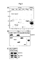
- Example 7 This feasibility of this type of assay is exemplified by the experiment described in Example 7, which demonstrates that the specific interaction that was shown to occur between a GFP-hnRNP-U fusion protein and ⁇ -TrCP/E3RS can be abrogated by a specific 10 amino-acid pI ⁇ B ⁇ peptide (pp10), but not by a Ser-substituted peptide (p10S/E) (Fig. 7).
- the subject complex used in the assay comprises three protein components, i.e. E3RS, hnRNP-U and Skp1. Since Skp1 facilitates the association of hnRNP-U with E3RS in vivo, incorporation of Skp1 into the subject complex more closely reflects the physiological situation. Therefore, obtaining a positive hit in such an assay is expected to more accurately predict the efficacy of the identified candidate inhibitor in therapy.
- This assay format has the potential to identify test compounds that either interfere directly with hnRNP-U/E3RS association, or indirectly by disrupting the Skp1/E3RS interaction.
- the three proteins are produced by co-expression in a single cell, as described above for co-expression of hnRNP-U and E3RS.
- the cDNA of Skp1 is available from Bai et al. 1996.
- Example 5 the preparation of a stoichometric three-component complex is exemplified.
- the disruption of the subject complex by an E3RS inhibitor is monitored by fluorescence measurements in solution.
- none of the interacting proteins needs to be immobilized onto a solid support.
- One or both proteins are fused to a fluorescent label that emits a signal of different intensity or quality (e.g., a different emission wave-length) upon association or dissociation from the partner protein.
- a useful example for monitoring the association of the partner proteins is FRET, Fluorescence Resonance Energy Transfer. (Pollok, et al., 1999; Bastiaens, 1999; Feriasamy, et al., 1999).
- both partner proteins are labeled by a different fluorescent probe (e.g., Green Fluorescent Protein and Red Fluorescent proteins, commercially available from Clontech) and upon association, the fluorescence of one fluorophores is quenched by intramolecular energy transfer.
- a different fluorescent probe e.g., Green Fluorescent Protein and Red Fluorescent proteins, commercially available from Clontech
- the association and dissociation of the subject complex is monitored by fluorescence polarization or fluorescence spin resonance, related techniques, based on quenching or quality changes of a fluorophore as a result of protein-protein association. Inclusion of an E3RS inhibitor into the assay will disrupt the subject complex, thereby affecting the emission of the fluorescence signal.
- the above-described assays have the following advantages as compared to the assay described in WO 00/33447: (i) they require fewer components than the described E3-substrate interruption assay or the pI ⁇ B ⁇ ubiquitination assay (i.e., there is no need for any substrate, ubiquitination enzymes etc,) and therefore, the assays are simpler and more accurate; (ii) they obviate the need to prepare an IKK-phosphorylated substrate; (iii) they assay a low-affinity complex (relatively to the high affinity E3-pI ⁇ B ⁇ complex), which is more amenable for interruption, thus allowing the identification of a broader range of inhibitors.
- the present invention provides a method for screening compounds that inactivate the hnRNP-U protein per se, i.e. its chaperone and E3RS transporting activity.
- inactivation results in compromising either the association of E3RS with hnRNP-U or the dissociation of the two proteins upon interaction with the substrate pI ⁇ B ⁇ , such dissociation being necessary for pI ⁇ B ⁇ ubiquitination and degradation.
- this type of E3RS inhibitors which target the chaperone and E3RS transporting activity of hnRNP-U, are termed "hnRNP-U inactivators".
- hn-RNP-U inactivators are expected to abolish or significantly diminish the inducible degradation of I ⁇ B ⁇ and thus NF- ⁇ B activation.
- hnRNP-U inactivators can be identified in an assay which is, in principle, set up in analogy to the above-described assays that employ the two-component or three-component subject complex to monitor E3RS/hnRNP-U dissociation.
- this assay variant is designed to identify compounds inhibiting dissociation of hnRNP-U from ⁇ -TrCP/E3RS.
- the subject complex (comprising E3RS, hnRNP-U and optionally Skp1) is incubated, in the presence of the test compound, with an agent capable of inducing dissociation of the complex, evident by the release of the labeled component which generates a detectable signal, e.g. GFP fluorescence or radioactivity.
- an agent capable of inducing dissociation of the complex, evident by the release of the labeled component which generates a detectable signal, e.g. GFP fluorescence or radioactivity.
- an agent may be a pI ⁇ B ⁇ peptide, e.g., as described above, the pp10 peptide that contains the pI ⁇ B ⁇ degradation motif, or it may be selected from inhibitors identified in the above assays that monitor dissociation of the subject complex.
- the effect of the test compound on the dissociation of the complex is monitored; reduction of the signal generated by the release of the labeled protein is indicative of an hnRNP-U inactivating
- the invention relates to cellular screening assay methods for identifying E3RS inhibitors, which, as described above, exert their effect either by inhibiting the association of hnRNP-U with E3RS or by inactivating hnRNP-U itself.
- a cellular screening assay may be set up as follows: mammalian cells, e.g. 293 cells, expressing a labeled E3RS (e.g. GFP-E3RS, obtained upon transfection of the cells with a plasmid carrying a GFP-E3RS fusion construct) are grown in the presence of the test compound for a period of time sufficient for the compound to penetrate the cell and to exert its potential effect, which may be any period of time from approximately 30 minutes up to 16 hours. Then the cells are subject to immunoprecipitation according to standard methods with an antibody that binds to the complex, preferably an anti-hnRNP-U antibody, in order to pull down the hnRNP-U/E3RS complex.
- mammalian cells e.g. 293 cells
- expressing a labeled E3RS e.g. GFP-E3RS, obtained upon transfection of the cells with a plasmid carrying a GFP-E3RS fusion construct
- the test compound for
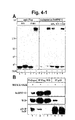
- Anti-hnRNP-U can be obtained according to standard methods, they may be either polyclonal or monoclonal. Polyclonal antibodies are conventionally obtained by immunizing animals, particularly rabbits, by injecting the antigen or fragments thereof and subsequently purifying the immunoglobulin. Monoclonal antibodies may be obtained by standard procedures following the principle described by Köhler and Milstein, 1975. In Examples 4 and 9, monoclonal or polyclonal anti-hnRNP-U antibodies were shown to precipitate a complex composed of hnRNP-U, E3RS and other SCF components.
- test compound identified in the assay is an E3RS inhibitor
- immunoprecipitation of hnRNP-U results in reduced coprecipitation of E3RS, leading to the absence or the reduction of a detectable E3RS signal.
- hnRNP-U may be used as the labeled complex partner and anti-E3RS antibodies are used to pull down the complex.
- the presence of an E3RS inhibitor will result in no detectable signal.
- a cellular screening assay that identifies hnRNP-U inactivators can be conducted according to this principle, with the modification that the assay is done in the presence of an agent inducing dissociation of the complex, as described above for the non-cellular assay.
- a cellular assay for monitoring the association of hnRNP-U and E3RS is based on the above-mentioned FRET technique.
- both partners are labeled by fluorescent labels, preferably through fusion with two different fluorescent proteins (e.g, GFP and Blue Fluorescent Protein (BFP)).
- BFP Blue Fluorescent Protein
- the invention relates to a cellular assay, which identifies E3RS inhibitors by identifying them as being compounds that have the ability of inhibiting Vpu-dependent CD4 degradation.
- Vpu-mediated CD4 degradation assay In this assay for identifying E3RS inhibitors, the mechanism of Vpu-mediated CD4 degradation is used as a surrogate for pI ⁇ B ⁇ degradation. In the following, is assay is termed "Vpu-mediated CD4 degradation assay", or simply "Vpu assay”.
- Vpu is a small polypeptide encoded by HIV that resides in the membrane of the endoplasmic reticulum in infected cells and shares with I ⁇ B the DSGXXS degradation motif (Karin and Ben-Neriah, 2000).
- ⁇ -TrCP/E3RS binds with Vpu via the WD40 domain of (Margottin et al., 1998) and requires phosphorylation at the two Ser residues of the shared motif.
- Vpu is directing the E3 or the proteasome to an associated host protein, CD4.
- the overexpression of Vpu results in competitive inhibition of the hnRNP-U/E3RS association, which provides the basis for the Vpu assay to serve as a surrogate assay for identifying E3RS inhibitors.
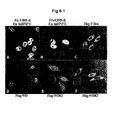
- the assay employs a mammalian cell line, e.g. 293 cells, that report CD4 degradation upon Vpu induction (Fig. 7).
- the reporter system comprises, as an essential feature, CD4 linked to a detectable label, e.g. in the form of a GFP-CD4 chimeric protein that fluoresces in the cell as long as it is stably expressed.
- the construction of a plasmid encoding a Vpu-mediated degradable CD4 is based on the following considerations:
- the HIV Vpu is an endoplasmic reticulum (ER)-associated protein, which normally binds to the portion of the cellular CD4 that is retained in ER through a complex with the HIV gp160 protein.
- ER endoplasmic reticulum
- a CD4 plasmid is constructed to express CD4 modified by being fused to a marker protein, e.g. GFP (to this end, the cDNA sequence of CD4, which has been described by Maddon et al., 1985, is fused to the GFP sequence).
- GFP marker protein
- the human CD4 is truncated at its carboxy-terminal region, down to the amino-acid sequence KKTC, an ER retention signal.
- the N-terminal CD4 sequence including the first three Ig-like domains (but preserving the CD4 signal sequence), is replaced with the marker sequence, e.g. the human GFP sequence, for allowing the quantitative measurement of the fusion protein through the signal, e.g. GFP fluorescence signal.
- the test cell further contains a plasmid encoding the HIV Vpu polypeptide, the cDNA sequence of which is available (Terwilliger et al., 1989).
- Vpu is expressed under the control of a regulated, e.g. tetracycline-regulated, promoter.
- Vpu is expressed in the engineered cell line only when the expression modifier, e.g. tetracycline or doxycycline (DOX), which has, with respect to the so-called “tet-off" expression system, the function of a suppressor, is omitted from the medium.
- DOX doxycycline
- One of the advantages of this regulatable expression system is to avoid the toxicity caused by overexpression of Vpu.
- Fig. 7A illustrates the principle and function of this construct:
- Vpu In the presence of the expression modifier (e.g. DOX) Vpu is not produced, therefore the modified CD4 is not degraded and produces a detectable signal, e.g. GFP fluorescence (A).
- the modified CD4 protein Upon DOX withdrawal, Vpu is expressed, the modified CD4 protein is subject to E3RS-mediated degradation and the cell no longer emits a detectable signal, e.g. fluorescence (B).
- a proteasome inhibitor e.g one of the inhibitors described in WO 95/25333 or by Lee and Goldberg (1998) serves as a positive control for the inhibition of CD4 degradation (C).
- the Wallac Victor II instrument which is suitable for high throughput screening.
- the inhibitors can be additionally tested while Vpu expression is suppressed, e.g. in the presence of DOX. Under these conditions, a specific E3RS inhibitor should not affect the intensity of the emitted signal.
- a viability control may be additionally incorporated into the system, e.g. by expressing a red fluorescent protein (RFP).
- RFP red fluorescent protein
- an RFP expression vector commercially available, e.g. from Clontech
- Red fluorescence of the assay cell line should not be affected by a specific E3RS inhibitor. Therefore, the actual E3RS inhibitory effect of a compound can then be indicated by the GFP/RFP ratio, which must be smaller than one. Further validation of the E3RS inhibitory effect can be achieved in standard in vitro ubiquitination assays, as described e.g. by Yaron et al., 1998 (see also Example 4).
- Vpu-mediated CD4 degradation assay can be confirmed to have an effect on the association or dissociation of the hnRNP-U/E3RS complex by using the above-described non-cellular or cellular assays that detect this effect.
- E3RS inhibitors identified in the above-described assays according to their ability to affect hnRNP-U/E3RS association or dissociation, either directly or indirectly, as described above, or the hnRNP-U inactivators described below, have the potential to be drug candidates also for the treatment of HIV-mediated diseases.
- the HIV inhibitory effect of these compounds can be further supported by subjecting them to the Vpu-mediated CD4 degradation assay, which is highly relevant to the mechanism of HIV pathogenesis (Lama et al., 1999).
- the cellular assays of the invention can be used as primary screening assays to detect active compounds, or as a means of confirming the activity of an E3RS inhibitor identified in a non-cellular assay (in this case they have the function of a secondary assay).
- Such assays can also be used to test the effect of an hnRNP-U antisense molecule, or an hnRNP-U inactivator developed on the basis of a rational design according to the crystal structure of the hnRNP-U/E3RS complex.
- the cellular assays of the invention are unique in the sense that they report a distinct biochemical process within a cell: E3RS/hnRNP-U activity.
- the previously reported cellular assays (WO 98/36070 and WO 00/34447) are based on I ⁇ B ⁇ degradation, which requires signal-induced IKK activation for I ⁇ B ⁇ phosphorylation.
- the assays of the invention do not require IKK activation through cell stimulation.
- they provide the only kind of assay for identifying inhibitors of I ⁇ B ⁇ ubiquitination that can be adapted for high throughput screening, as ubiquitination is reflected by an externally-emitted signal, e.g. a fluorescence signal readily detectable by standard detection devices, e.g.
- the assay of the invention is distinct from any of the previously described assays for finding inhibitors of NF- ⁇ B activation, e.g. those based on a ⁇ B reporter, which reflect any process that affects NF- ⁇ B activation (e.g., kinase activation, transcription modulation etc.)
- Another advantage of the assays of the invention lies in their potential to be applied for identifying inhibitors of cellular targets of HIV. These inhibitors are expected to be superior over other NF- ⁇ B inhibitors (e.g. IKK inhibitors) by inhibiting the function of both NF- ⁇ B and Vpu, which are necessary for HIV replication (Lama et al., 1999).
- the invention relates to hnRNP-U inactivators, i.e. agents that exert their effect directly on the hnRNP-U protein or its expression.
- hnRNP-U Although the protein structure of hnRNP-U is unknown as yet, the sequence of the protein reveals features that allow for an assessment of the protein's structure-function relationship.
- One of these features is a putative nucleotide-binding site (aa 485-492 (Kiledjian and Dreyfuss, 1992)).
- This motif is often associated with chaperone activity, including that of hsp90, which serves, similarly to hnRNP-U, as the "dedicated chaperone" for several signaling molecules (Buchner, 1999). Its interaction with certain oncoproteins, such as src and erb2 is essential for their transforming activity and in common with hnRNP-U it promotes the activity of these kinases only following its dissociation.
- hsp90 and hnRNP-U Two natural compounds, the macrocyclic antibiotic Radicicol and the benzoquinone ansamycin Geldanamycin tether the hsp90 at its nucleotide-binding site.
- hsp90 and hnRNP-U have common functional features. It may be assumed that the hnRNP-U nucleotide-binding site has a role that is similar to the one in hsp90. Therefore, compounds structurally related to Radicicol and Geldanamycin are expected to be potential hn-RNP-U inactivators.
- the E3RS inhibitory effect of these agents or chemically modified derivatives thereof can be confirmed in one of the above-described cellular or non-cellular assays.
- hn-RNP-U inactivators may function similarly to hsp90 inhibitors of the benzoquinone ansamycin group by modifying the nucleotide-binding site.
- site-directed mutagenesis can be used to abolish the putative function of this site. If this property is confirmed, its relevance is further evaluated by testing a potential nucleotide hydrolyzing activity of hnRNP-U. This can be done by standard ATP hydrolysis assays (Rosser and Nichitta, 2000).
- the ATP hydrolyzing property can serve as the basis for another screening approach.
- hnRNP-U is tested for its ATP hydrolyzing activity, either on its own or in combination with one or more of its interacting partners, e.g. E3RS, optionally in conjunction with Skp1.
- the latter assay can also be conducted as a cellular assay, while the effect of a compound is tested on cells expressing tagged hnRNP-U, preferably also expressing the interacting partners.
- hnRNP-U is pulled down with an antibody as described above or with standard affinity matrix methods and assayed for nucleotide hydrolyzing activity.
- hn-RNP-U inactivators may also be obtained by rational drug design based on the crystal structure obtained upon co-crystallizing hnRNP-U and E3RS, preferably together with Skp1.
- attempts to generate a functional E3 from bacteria or other cellular sources, which are devoid of hnRNP-U, e.g. insect cells have generally been unsuccessful.
- One of the essential findings of the present invention is that hnRNP-U is crucial for the proper folding of E3RS.
- An important implication of this finding is that hnRNP-U may be required for expressing E3RS for crystallography analysis. Therefore, this finding represents the basis for providing a source for functional E3RS.
- E3RS is co-expressed with hnRNP-U, preferably together with Skp1, in the same cell, as described above for obtaining the subject complex, and the complex is obtained by one of the above-described affinity purification methods.
- Proper folding of E3RS is an absolute requirement for the crystallography analysis and is expected to be facilitated by interaction either with hnRNP-U. Since proper folding of E3RS may also be facilitated by one of its substrates, in the obtained complex, hnRNP-U can be exchanged by one of the E3RS substrates, e.g. pI ⁇ B ⁇ (see e.g.
- Example 4 or Vpu, or with a phosphorylated fragment of the substrates (e.g. the pp10, see Example 3), thereby preserving the correct folding of E3RS.
- An E3RS complex either containing hnRNP-U, or an E3RS substrate, or a fragment thereof, can be obtained by standard methods of crystallography. Specialized molecular modeling techniques, including computer programs, may further be employed for designing E3RS inhibitors. See, e.g. Cohen et al., 1990, Navia and Murcko, 1992. For example, where the structure of a test compounds is known, a model of the test compound may be superimposed over the model of the E3RS structure.
- hnRNP-U antisense oligoribodeoxynucleotides Another class of hnRNP-U inactivators is hnRNP-U antisense oligoribodeoxynucleotides. Based on the hnRNP-U cDNA sequence, hnRNP-U antisense oligonucleotides can be obtained and assessed with regard to their pharmacological properties according to methods known in the art. "Antisense Technology", Methods in Enzymology, 2000 provides ample teaching for the person skilled in the art for the design, preparation, chemical modification and evaluation of the efficacy of antisensense molecules, as well as their formulation and therapeutical application.
- an hn-RNP-U antisense molecule is the oligoribodeoxynucleotides AGGCGAGGAACTCATGGTGAG, which is directed against the sequence flanking the start codon of hnRNP-U.
- nucleotides of this molecule are modified, e.g by phosphothioate or methyl groups to protect the molecule against degradation in the cell.
- the efficacy of an hnRNP-U antisense molecule can be determined by pI ⁇ B ⁇ degradation assays (Yaron et al., 1997 and 1998) and/or by one of the above-described cellular assays for E3RS inhibitors.
- RNAi double stranded hnRNP-U cDNA molecules
- E3RS inhibitors are synthetic peptides or phosphopeptides derived from the protein sequence of hnRNP-U and E3RS (e.g. pp10, Yaron et al, 1998), peptidomimetic compounds resembling the hnRNP-U and E3RS inhibitory peptides and small molecules identified in this or the above mentioned screens.
- the present invention relates to a method for producing functional E3RS by co-expressing ⁇ -TrCP/E3RS with hnRNP-U, optionally together with Skp1, in bacterial, yeast or insect cells.
- the present invention is directed to screening methods for identifying compounds affecting the hnRNP-U/E3RS complex, which plays a major role in the signal-induced degradation of pI ⁇ B ⁇ and consequently NF- ⁇ B activation. Since the degradation motifs of I ⁇ B ⁇ and I ⁇ B ⁇ are identical to the one of I ⁇ B ⁇ (Yaron et al., 1997) and E3RS was shown to be responsible also for the ubiquitination of I ⁇ B ⁇ and I ⁇ B ⁇ (Hattori et al., 1999), the screening methods of the invention and the E3RS inhibitors which function by direct or indirect inactivation of hnRNP-U, may also be applied to inhibiting I ⁇ B ⁇ and I ⁇ B ⁇ degradation.
- the screening methods of the invention are generally useful for identifying modulators of ubiquitin-mediated proteolysis of phosphorylated I ⁇ B.
- the methods of the invention may be used for identifying compounds that upregulate it.
- the person skilled in the art can easily adapt the methods described above for this purpose by adapting the assay readout accordingly.
- Compounds that upregulate the E3RS pathway can be used for the treatment of disorders associated with E3RS dysfunction and thus NF- ⁇ B deficiency.
- the transcription factor NF- ⁇ B is, inter alia, activated by treating cells with bacteriological stimuli (such as LPS), viruses (e.g. HIV virus type 1), viruses and viral products, parasites, inflammatory cytokines (e.g. TNF- ⁇ , TNF- ⁇ , IL-1, IL-2), T-cell mitogens (e.g. lectines), protein synthesis inhibitors (e.g. cycloheximide), physical stress (UV-light, gamma radiation), oxidative stress (e.g. hydrogen super oxide), chemotherapy, oncogenes and tumour promoters (e.g. Ras and phorbol ester) (Mayo, et al., 2000; Mercurio, et al, 1999; Lewis and Manning, 1999; Ghosh, et al., 1998)
- NF- ⁇ B modulators in particular inhibitors, have been suggested as drugs for the treatment of various pathological conditions in which the activation of NF- ⁇ B is involved.
- E3RS inhibitors function as NF- ⁇ B inhibitors
- the present invention relates, in a further aspect, to the use of E3RS inhibitors for the preparation of a medicament for the treatment of disorders associated with NF- ⁇ B activation
- E3RS inhibitors are, inter alia, expected to be effective as anti-inflammatory drugs, e.g. in the treatment of rheumatoid arthritis or asthma, in cancer therapy in order to increase the patient's sensitivity to chemotherapeutic agents, in the therapy of disorders of the central neural system, e.g. neurodegenerative diseases like Alzheimer, and stroke due to artherosclerosis, and as immune suppressive drugs
- the E3RS inhibitors of the invention can be tested in animal models.
- the compound to be evaluated is applied to animal, e.g. by injection, which is subjected to an agent or another stimulus eliciting NF- ⁇ B activation in an organ or tissue of the animal, e.g. by applying LPS, which induces NF- ⁇ B in the spleen.
- an agent or another stimulus eliciting NF- ⁇ B activation in an organ or tissue of the animal, e.g. by applying LPS, which induces NF- ⁇ B in the spleen.
- LPS which induces NF- ⁇ B in the spleen.
- the effect of the inhibitor can be determined.
- the animal is monitored for symptoms that are typically associated with NF- ⁇ B activation, e.g. septic shock after LPS injection.
- Toxicity and therapeutic efficacy of an E3RS inhibitor identified as a drug candidate can be determined by standard pharmaceutical procedures, which include conducting cell culture and animal experiments to determine the IC 50 , LD 5 0, the ED 50 .
- the data obtained are used for determining the human dose range, which will also depend on the dosage form (tablets, capsules, aerosol sprays, ampules, etc.) and the administration route (oral, buccal, nasal, paterental or rectal).
- a pharmaceutical composition containing the compound as the active ingredient can be formulated in conventional manner using or more physiologically active carriers and excipients. Methods for making such formulations can be found in manuals, e.g. "Remington Pharmaceutical Sciences". Examples for ingredients that are useful for formulating the compounds identified according to the present invention are also found in WO 99/18193.
- a tissue or blood sample is obtained from the animal or from the treated patient and subjected to hnRNP-U antibody precipitation, as described above.
- the E3RS activity within such precipitates is suitable as a parameter to monitor the efficacy of the drug; a compound exhibiting the desired effect is expected to abrogate the E3RS activity in the samples.
- the E3RS activity can be determined in a test according to principle described in Example 4. The significance of this type of assay lies in its ability to adjust the treatment using the inhibitors, e.g. the dosage and frequency of application. In addition, it can be used for optimizing the structure efficacy and/or formulation of the drug.
- hnRNP-U antibodies can be used for diagnostic purposes, e.g. for identifying conditions in which the E3RS is compromised, e.g. by infectious agents or metabolic aberrations, which results NF- ⁇ B deficiency.
- a sample obtained from a patient suffering from such conditions will be examined with the aid of the hnRNP-U antibodies, as described above.
- the correction of an E3RS dysfunction e.g. by reversing the metabolic aberration, can alleviate those symptoms that are secondary to NF- ⁇ B deficiency.
- E3RS analysis with the aid of anti-hnRNP-U antibodies is then used to indicate a successful correction of the metabolic disorder.
- hnRNP-U was PCR-cloned from both human and mouse cDNA libraries into the pFLAG-CMV-2 expression vector (Kodak) at the Not1 site.
- the Not1 cleavage site was incorporated into both the forward primer, 5'tagcggccgcaatgagttcctcgcctgtt 3', and the reverse primer, 5' tagcggccgctcaataatatccttggtgata 3'.
- Flag-tagged E3RS/pTrCP FL-E3RS
- the WD fragment F-Box deleted E3RS
- the F-Box fragment Flag-F-Box
- HA-E3RS was prepared by subcloning human E3RS into the pCGN-HA vector. Mutagenesis of Lys 304 (K1) and Lys 326 (K2) residues to Ala in the Flag-WD construct was performed using the Quickchange kit (Stratagene). These were designated WDK1 and WDK2. The K2 residue in Flag-E3RS was similarly mutated to Ala (E3K2).
- Human HOS/ ⁇ -TrCP2 cDNA was obtained by PCR-cloning according to Genbank entry AB033281 into the pFLAG-CMV-2 expression vector (Flag-HOS/ ⁇ -TrCP2).
- HA-Cul1 and Myc-Skp1 expression plasmids were described by Lyapina et al., 1998).
- Flag-Smad 2 expression vector (Wu et al., 2000), used as a negative control.
- the MEKK1 and IKK2 mammalian expression plasmids are described before (Mercurio et al., 1997; Yin et al., 1998).
- Anti-GFP affinity-purified monoclonal antibody was purchased from CLONTECH ⁇ Living Colors A.v. Monoclonal Antibody (JL-8) (1 mg/ml) ⁇ , and was used at a dilution 1:1000 (1 ⁇ g/ml) for Western Blot.
- Agarose-conjugated (A-1205) and purified monoclonal anti-Flag M2 antibodies (F-3165) were purchased from Sigma.
- Monoclonal anti-hnRNP-U antibodies (3G6) were described by (Pinol-Roma et al., 1988).
- Sepharose-immobilized anti-p65 (anti-NF- ⁇ B) (sc-109 AC) and anti-HA (sc-805) were from Santa Cruz.
- Polyclonal anti-E3RS serum was obtained by immunizing rabbits with the E3RS N-terminal peptide (KALKFMNSSEREDCNNGEP; amino-acids 11-29).
- Rabbit polyclonal anti-I ⁇ B ⁇ was described in (Alkalay et al., 1995). Rabbit anti-HA serum (sc-805) was purchased from Santa Cruz. Mouse monoclonal anti-Myc antibodies, c-myc(Ab-1), were from Oncogene Research Products. The goat anti-mouse RedX fluorescent secondary antibody is from Jackson (115-295-062) and goat anti-rabbit Alexa-488 from Molecular Probes (A-11008).
- polyclonal rabbit antiserum to human hnRNP-U was prepared against a fragment of hnRNP-U fused to GST.
- the GST fusion protein containing the carboxy-terminal half of the human hnRNP-U protein (amino-acids 406-806,contained in a BamH1-Bg12 fragment of the human hnRNP-U) was prepared as follows: The fragment was inserted into the BamH1 site of a commercially available bacterial expression vector (pGEX1, Clontech). The fusion protein was purified from bacterial lysates using glutathione-agarose affinity chromatography and glutathione elution according to standard procedures. Rabbits were injected with the fusion protein in complete Freund's adjuvant and were bled following 3 boosts.
- Anti-Vpu Rabbit polyclonal antibodies were prepared as follows:
- oligopeptide corresponding to amino acids 73 - 81 of the Vpu protein ⁇ encoded by the vpu gene of BH10 substrain of the IIIB HIV-1 isolate (Ratner et al. (1985). was conjugated to keyhole limpet haemocyanin and used to raise antibodies in three rabbits. After multiple injections of the antigen, the rabbits were shown to produce antibodies that recognize the oligopeptide. Serum from one of these rabbits that was shown to recognize in vitro translated vpu (Cohen, A.E., Tewillinger, E. F., Sodroski, J. G. and Hasseltine, W. A. (1988). Nature 334, 532-534) was used (at a dilution of 1:1000) for Western Blot analysis.
- CHO or 293T cells were plated out onto gelatin-coated plates and transfections performed using FuGENE 6 (Roche) or calcium phosphate.
- FuGENE 6 FuGENE 6
- the culture was supplemented with 50mM Hepes in DMEM medium and heat shock performed for 1 h at 43°C.
- immunofluorescence cells were plated onto gelatin-coated cover slips and immunostaining was performed at 24h post transfection. The cells were washed with phosphate buffered saline (PBS), fixed with 3% paraformaldehyde in PBS for 30 min at 4°C, permeabilized for 6 min using 0.25% Triton X-100 in PBS, and washed again with PBS.
- PBS phosphate buffered saline
- Blocking was done for 45 min with 10% goat serum in PBS-Tween 20 (1%).
- the primary antibodies anti-Flag (1:600), anti-HA (1:100) and anti-hnRNP-U (1:1000) were diluted in 5% bovine serum albumin, 1% Tween-20, PBS and incubated on the cover slips for 60 min.
- Cells were washed 3X with PBS, and then incubated with secondary fluorescent antibodies, goat anti-mouse RedX (1:150) or goat anti-rabbit Alexa-488 (1:100) for 45 min. Following 3 washes with PBS, the cover slips were mounted onto slides and analyzed by coNF-ocal microscopy.
- Cells were harvested 24-48 h after transfection, and extracted by suspension in 50mM Tris pH 7.6, 1mM dithiothreitol (DTT), 0.1% Nonidet P-40 (NP-40), 1mM phenyl methyl sulfonyl fluoride, 1% Aprotinin and vortexing for 20 sec. Cell extracts were collected following centrifugation at 20,000g for 30 min at 4°C. Cell extract (500 ⁇ g protein) was adjusted to 150 mM NaCl prior to immunoprecipitation. Flag-tagged proteins were immunoprecipitated with 2 ⁇ l of the anti-Flag immunobeads.
- Immunoprecipitation was for 2 h at 4°C and the immunobeads were washed 4X in 300mM NaCl, 1mM DTT, 0.1% NP-40 and 50mM Tris, pH 7.6.
- Flag-tagged proteins were eluted from the Flag immunobeads with 1mg/ml of Flag peptide in 50mM Tris, pH 7.6 for 30 min at 25°C.
- hnRNP-U was immunoprecipitated using 0.2-0.5 ⁇ l of the 3G6 antibody and 2-5 ⁇ l of Protein G immunobeads.
- Flag-E3RS or WD/hnRNP-U complex was disrupted by adding the phosphorylated I ⁇ B peptide pp10 (Yaron et al., 1998) to the extract at a concentration of 1mg/ml during the immunoprecipitation.
- E3RS-associated proteins were eluted from the washed Flag immunobeads by incubating them with pp10 (1mg/ml) for 30 min at RT. Control elutions were performed with the serine substituted I ⁇ B ⁇ peptide p10 S/E at similar conditions.
- Flag-E3RS and endogenous hnRNP-U from 293T cells were used as an E3 source and Flag-WD was used as a negative control.
- 100 ⁇ g protein extract was immunoprecipitated using 2 ⁇ l anti-Flag beads or 0.2 ⁇ l anti-hnRNP-U antibodies.
- I ⁇ B was phosphorylated by the consitutively-active I ⁇ B kinase (IKK2; (Mercurio et al., 1997)) and subjected to binding and ubiquitination assays with each of the above E3 sources, as described previously by (Yaron et al., 1998).
- Mass spectrometric identification of proteins was done according to the strategy previously described (Shevchenko et al., 1996). Briefly, gel bands were excised from a one-dimensional gel stained with Coomassie colloidal blue and digested in-gel with trypsin. The recovered unseparated peptide mixture was analyzed by MALDI mass spectrometry, using a Bruker Reflex III MALDI time-of-flight mass spectrometer (Bruker Daltronics, Bremen, Germany). Samples for MS/MS analysis were prepared essentially as described (Shevchenko et al., 1996).
- the inducible system consists of the tetracycline (Tc)- responsive promoter system developed by Gossen and Bujard (1992). Vpu and GFP-CD4 expression were performed in either HeLa Tet -off gene expression system (CLONTECH). These stably transfected HeLa cells carry the pTet-Off regulator plasmid encoding a tetracycline-controlled transactivator (tTA), which also includes a neomycin-resistance gene. Alternatively 293 cells were transfected with the inducible constructs and the pTet-Off plasmid (Lavon et al., 2000).
- CMV cytomegalovirus
- This new fusion-vector was termed pCD41eader-EGFP-C1.
- a 400-bp CD4 sequence (encoding amino-acids 311 -425 of the protein; Maddon et al. 1985) was PCR-cloned into EcoRI-KpnI liniarized pCD4leader-EGFP-C1 vector.
- This CD4 fragment includes the C2 Immunoglobulin-like extracellular domain (amino acids 311-376), the transmembrane domain (amino acids 377-398), and a cytoplasmic domain of CD4, in which the cytoplasmic C-terminal 13 amino acids were truncated.
- E3RS resides predominantly in the nucleus.
- HeLa cells transfected with Flag-E3RS and HA-Cul1 were doubly stained with mouse monoclonal anti-Flag and rabbit polyclonal anti-HA as primary antibodies and goat anti-mouse Ig (Rhodamine) or goat anti-rabbit IgG (Alexa-488) as secondary antibodies (Fig. 1B).
- CHO cells transfected with Flag-HOS/ ⁇ -TrCP2 were stained with anti-Flag antibodies (Fig. 1C).
- E3RS staining was mainly detected in the nucleus, whether observing the endogenous protein, or an epitope-tagged exogenously expressed one (Fig 1A,B).
- the amino-acid sequence of E3RS has no apparent nuclear localization motif (NLS). Therefore, its presence in the nucleus could be secondary to an associated protein, such as a component of the SCF complex.
- Cull an SCF component that carries the NLS, was mainly detected in the cytoplasm (Fig 1B).
- a Flag-tagged E3RS was immunopurified from overexpressing 293 cells and analyzed by SDS-PAGE (Fig 2A) and mass-spectrometry.
- E3RS contains two protein-protein interaction modules, the F-box and the WD40 repeat domain (Margottin et al., 1998; Yaron et al., 1998). To determine which module was responsible for binding hnRNP-U, fragments composed of the F-box or the WD40 repeat (an F-box-deleted E3RS) were expressed separately, immunopurified and analyzed for hnRNP-U binding.
- 293T cells were transfected with Flag-E3RS (lane 1), Flag-WD (lane 2), Flag-HOS/ ⁇ -TrCP2 (lane 3), Flag-F-Box (lane 4), Flag-Smad-2 (lane 5) and HA-Skp2 (lane 6).
- the cells were lysed and the extracts immunoprecipitated, using agarose-conjugated anti-Flag (lanes 1-5) and anti-HA (lane 6) antibodies. Immunoprecipitated proteins were analyzed by SDS-PAGE and Coomassie Brilliant Blue staining. Molecular weight markers (kD) are indicated.
- Mass-spectrometry sequencing of the major E3RS-associated protein identified it as hnRNP-U (Kiledjian and Dreyfuss, 1992; Pinol-Roma et al., 1988), or scaffold attachment factor A (SAF-A) (Romig et al., 1992), an abundant nuclear protein.
- the immunoprecipitated proteins were also analyzed by Western blot, using anti-Flag, anti-HA and monoclonal anti-hnRNP-U antibodies(Fig. 2B):
- the Western blot coNF-irmed the specificity of hnRNP-U association: the protein signal was detected in association with E3RS, but not with ⁇ -TrCP2/HOS or Skp2.
- hnRNP-U co-purified with the WD40-repeat (Figs. 2A, lane 2 and 2B) but not with the F-box fragment (Fig. 2A, lane 4 and 2B).
- Flag-tagged human and mouse hnRNP-U were co-transfected with HA-E3RS into 293T cells.
- Cell extracts were immunoprecipitated with anti-Flag antibodies and precipitated proteins analyzed by Western blot using anti-HA and anti-Flag antibodies.
- the E3RS-hnRNP-U association resembles an E3-substrate interaction
- Flag-E3RS was immunoprecipitated with anti-Flag immunobeads in the absence (lane 1), or presence (lane 2) of a phosphorylated I ⁇ B ⁇ peptide (pp10, containing the I ⁇ B ⁇ degradation motif).
- Flag-WD was immunoprecipitated with anti-Flag immunobeads with no peptide (lane 3), in the presence of pp10 (lane 4), or in the presence of the serine/glutamic acid-substituted peptide p10 S/E (lane 5) and the immune-complexes were analyzed by SDS-PAGE and Coomassie-Blue staining.
- hnRNP-U association with E3RS was stable, both in vivo (Figs. 2,3) and in vitro.
- hnRNP-U delivers an active SCF ⁇ -TrCP/E3RS complex to the ubiquitination substrate.
- hnRNP-U The specific association of hnRNP-U with E3RS suggested the possibility of engaging an active pI ⁇ B ⁇ ligase through the hnRNP-U protein.
- 293T cells were transfected with Flag-WD and HA-hnRNP-U with or without constitutively active MEKK and IKK2 (a kinase combination that induces I ⁇ B ⁇ phosphorylation).
- Cell lysates (5% input is shown) were immunoprecipitated using anti-Flag (Fig. 4B, lanes 2,3) and anti-p65 (Fig. 4B, lanes 4,5) and the immune complexes were analyzed by Western blot with anti-Flag, anti-HA and anti-I ⁇ B ⁇ antibodies.A co-expressed WD fragment was found to associate with pI ⁇ B ⁇ (Fig. 4B, lane 4), but not with a non-phosphorylated I ⁇ B ⁇ (Fig. 4B, lane 5).
- hnRNP-U could play a role in delivering either E3RS or the whole SCF complex to the substrate towards ubiquitination.
- Immunoprecipitation analysis of the endogenous hnRNP-U from SCF transfected cells revealed several SCF components (Fig 4C):
- 293T cells were transfected with HA-Cul1 and Myc-Skp1 alone (lanes 1,4), or together with Flag-E3RS (lanes 2,5) or with Flag-E3RS alone (lanes 3,6).
- Cell lysates (5% input is shown) were immunoprecipitated with monoclonal anti-hnRNP-U antibodies and analyzed by Western blot with the relevant antibodies. Molecular weight markers (kD) are indicated.
- Skp1 facilitates hnRNP-U association with E3RS and reverses the binding defect imposed by a specific E3RS mutation.
- a K2 mutant was constructed in the context of full-length E3RS (designated E3K2) and tested for hnRNP-U and pI ⁇ B ⁇ binding.
- 293T cells were transfected with Flag-E3RS alone (lanes 1,2) or together with Myc-Cul1 (Fig. 5, lane 3) or Myc-Skp1 (Fig. 5, lane 4); with Flag-E3K2 alone (Fig. 5, lanes 5,6) or together with Myc-Cul1 (Fig. 5, lane 7), or Myc-Skp1 (Fig. 5, lane 8).
- the K2 mutant retained the capacity to bind pI ⁇ B ⁇ (data not shown) and hnRNP-U (Fig 5, lane 5), although at 20% efficiency, compared with WT E3RS (lane 1).
- the heat shock resulted in the inability of the mutant protein to bind hnRNP-U (lane 6), while the WT E3RS retained 30% hnRNP-U binding (lane 2).
- hnRNP-U which is a nuclear scaffold-associated protein (Romig et al., 1992), could play a role in the subcellular localization of E3RS.
- HeLa cells (Fig. 6A,B) were doubly immunostained for the endogenous hnRNP-U together with endogenous E3RS (Fig. 6, A) or transfected HA-E3RS (Fig. 6B).
- CHO cells (Fig. 6 C-L) were immunostained for the following Flag-tagged proteins: F-Box (Fig. 6C), WD (Fig. 6D), WDK1 (Fig. 6E) and WDK2 (Fig. 6F).
- Cells transfected with Flag-E3RS (Fig. 6G,J) or Flag-E3K2 (Fig. 6H,K) were incubated at 37°C (Fig. 6G,H) or at 43°C (Fig.
- E3RS inhibitors induce the dissociation of GFP-hnRNP-U.
- a complex of Flag- ⁇ -TrCP/E3RS and GFP-hnRNP-U or Flag- ⁇ -TrCP/E3RS with the endogenous hnRNP-U was immunoprecipitated from 293 transfectants with anti-Flag antibodies, eluted with a Flag peptide, with pp10 (an E3RS-blocking peptide), or with the p10S/E control peptide and the eluted fractions were separated by SDS-PAGE.
- GFP-hnRNP-U was detected by Western blotting using pp10, but not p10S/E, indicating the specific dissociation of GFP-hnRNP-U from E3RS, similarly to the dissociation of the endogenous hnRNP-U.
- GFP-hnRNP-U dissociation can, therefore, fatefully report the capacity of a peptide or similar E3RS-interacting molecule to competitively block the E3RS binding sites.
- the GFP-hnRNP-U dissociation assay or a similar assay, based on the fluorescence signal of the GFP, can indicate the capacity of NF- ⁇ B inhibition by specific compounds.
- the ubiquitin-ligase activity is measured as a function of Vpu-mediated GFP-CD4 degradation (Fig 7A).
- the HIV Vpu is an endoplasmic reticulum (ER)-associated protein, which normally associates with the portion of the cellular CD4 that is retained in ER through a complex with the HIV gp160 protein.
- the GFP-CD4 plasmid was constructed to express the GFP fusion protein in the ER in the absence of gp160.
- the human CD4 was truncated at its carboxy-terminal region, down to the amino-acid sequence KKTC, an ER retention signal.
- the N-terminal CD4 sequence including the first three Ig-like domains (but preserving the CD4 signal sequence), was replaced with the human EGFP sequence for allowing the quantitative measurement of the fusion protein through the GFP fluorescence signal.
- a GFP-CD4 expression plasmid was co-transfected into 293 cells together with expression plasmids for Doxycycline (DOX)-regulated HIV-Vpu and tTA (tetracycline transactivator) plasmid.
- DOX Doxycycline
- tTA tetracycline transactivator
- Vpu and GFP-CD4 expression were detected by Western blotting.
- Vpu was only expressed in the absence of DOX.
- the GFP-CD4 signal disappeared upon Vpu expression (Fig 7B).
- Pretreatment of the transfected cells with the proteasome inhibitor MG132 (Lee and Goldberg (1998), resulted in the preservation of the GFP-CD4 signal (Fig 7C).
- a GST fusion protein containing the carboxy-terminal half of the human hnRNP-U protein (amino-acids 406 - 806,contained in a BamH1-Bgl2 fragment of the human hnRNP-U) was prepared as follows: The fragment was inserted into the BamH1 site of a commercially available bacterial expression vector (pGEX1, Clontech). The fusion protein was purified from bacterial lysates using glutathione-agarose affinity chromatography and glutathione elution according to standard procedures. Rabbits were injected with the fusion protein in complete Freund's adjuvant and were bled following 3 boosts.
- the anti-hnRNP-U serum was used to immunoprecipitate Flag-E3RS protein from transfected 293 cells.
- the E3RS immunoprecipitation capacity of the polyclonal rabbit antiserum was comparable to that of the 3G6 monoclonal antibody (see example 4) and was specific, as no E3RS was precipitated with the pre-immune serum of the same rabbit.
- Precipitation of the endogenous hnRNP-U from the same transfected cells was verified with the 3G6 antibodies, using Western blotting.
- SKP1 connects cell cycle regulators to the ubiquitin proteolysis machinery through a novel motif, the F-box.
- Cell 86 263-74.
- VHL-elongin BC tumor suppressor complex Formation of the VHL-elongin BC tumor suppressor complex is mediated by the chaperonin TRiC. Mol Cell 4 , 1051-61.
- Feriasamy A Day RN. Visualizing protein interactions in living cells using digitized GFP imaging and FRET microscopy. Methods Cell Biol. 1999;58:293-314.
- HOS a human homolog of Slimb, forms an SCF complex with Skp1 and Cullin1 and targets the phosphorylation-dependent degradation of I ⁇ B and beta-catenin.
- SAF-A scaffold attachment factor A
- A-T-rich scaffold attachment regions flank the hematopoietic serine protease genes clustered on chromosome 14q11.2. Blood 79 , 610-8.
- hnRNP U inhibits carboxy-terminal domain phosphorylation by TFIIH and represses RNA polymerase II elongation. Mol Cell Biol 19 , 6833-44.
- hnRNP complexes composition, structure, and function. Curr Opin Cell Biol 11 , 363-71.
- MEKK1 activates both I ⁇ B kinase ⁇ and I ⁇ B kinase ⁇ . Proc. Natl. Acad. Sci.USA 95 , 9319-9324.
- IKK-1 and IKK-2 cytokine-activated I ⁇ B kinases essential for NF- ⁇ B activation. Science 278, 860-866.
- ROC1 a homolog of APC11, represents a family of cullin partners with an associated ubiquitin ligase activity. Mol Cell 3 , 535-41.
- Cdc53/cullin and the essential Hrt1 RING-H2 subunit of SCF define a ubiquitin ligase module that activates the E2 enzyme Cdc34. Genes Dev 13, 1614-1626.
- F-box proteins are receptors that recruit phosphorylated substrates to the SCF ubiquitin-ligase complex. Cell 91 , 209-19.
- the SCF ⁇ -TRCP-ubiquitin ligase complex associates specifically with phosphorylated destruction motifs in I ⁇ B ⁇ and ⁇ -catenin and stimulates I ⁇ B ⁇ ubiquitination in vitro. Genes Dev 13 , 270-283.
- HTLV-1 Tax protein binds to MEKK1 to stimulate I ⁇ B kinase activity and NF- ⁇ B activation.
Landscapes
- Chemical & Material Sciences (AREA)
- Life Sciences & Earth Sciences (AREA)
- Health & Medical Sciences (AREA)
- Organic Chemistry (AREA)
- Zoology (AREA)
- Engineering & Computer Science (AREA)
- Wood Science & Technology (AREA)
- Genetics & Genomics (AREA)
- Bioinformatics & Cheminformatics (AREA)
- Biochemistry (AREA)
- General Health & Medical Sciences (AREA)
- Molecular Biology (AREA)
- Proteomics, Peptides & Aminoacids (AREA)
- General Engineering & Computer Science (AREA)
- Microbiology (AREA)
- Biotechnology (AREA)
- Biophysics (AREA)
- Medicinal Chemistry (AREA)
- Immunology (AREA)
- Physics & Mathematics (AREA)
- Analytical Chemistry (AREA)
- Biomedical Technology (AREA)
- Cell Biology (AREA)
- Toxicology (AREA)
- Gastroenterology & Hepatology (AREA)
- Investigating Or Analysing Biological Materials (AREA)
- Medicines That Contain Protein Lipid Enzymes And Other Medicines (AREA)
- Measuring Or Testing Involving Enzymes Or Micro-Organisms (AREA)
Priority Applications (5)
| Application Number | Priority Date | Filing Date | Title |
|---|---|---|---|
| EP00117429A EP1182251A1 (fr) | 2000-08-11 | 2000-08-11 | Procédé pour l'identification de composés inhibant la protéolyse de IkB par l'ubiquitine |
| IL15434401A IL154344A0 (en) | 2000-08-11 | 2001-08-10 | METHODS FOR IDENTIFYING COMPOUNDS THAT INHIBIT UBIQUITIN-MEDIATED PROTEOLYSIS OF IkB |
| PCT/IB2001/002428 WO2002016633A2 (fr) | 2000-08-11 | 2001-08-10 | Procede d'identification de composes inhibant la proteolyse a mediation assuree par l'ubiquitine de ikb |
| AU2002222343A AU2002222343A1 (en) | 2000-08-11 | 2001-08-10 | Methods for identifying inhibitors of ubiquitin-mediated proteolysis of ikb |
| US10/361,394 US6905836B2 (en) | 2000-08-11 | 2003-02-10 | Methods for identifying compounds that inhibit ubiquitin-mediated proteolysis of iκb |
Applications Claiming Priority (1)
| Application Number | Priority Date | Filing Date | Title |
|---|---|---|---|
| EP00117429A EP1182251A1 (fr) | 2000-08-11 | 2000-08-11 | Procédé pour l'identification de composés inhibant la protéolyse de IkB par l'ubiquitine |
Publications (1)
| Publication Number | Publication Date |
|---|---|
| EP1182251A1 true EP1182251A1 (fr) | 2002-02-27 |
Family
ID=8169510
Family Applications (1)
| Application Number | Title | Priority Date | Filing Date |
|---|---|---|---|
| EP00117429A Withdrawn EP1182251A1 (fr) | 2000-08-11 | 2000-08-11 | Procédé pour l'identification de composés inhibant la protéolyse de IkB par l'ubiquitine |
Country Status (5)
| Country | Link |
|---|---|
| US (1) | US6905836B2 (fr) |
| EP (1) | EP1182251A1 (fr) |
| AU (1) | AU2002222343A1 (fr) |
| IL (1) | IL154344A0 (fr) |
| WO (1) | WO2002016633A2 (fr) |
Cited By (3)
| Publication number | Priority date | Publication date | Assignee | Title |
|---|---|---|---|---|
| US6905836B2 (en) * | 2000-08-11 | 2005-06-14 | Yissum Research Development Company Of The Hebrew University Of Jerusalem | Methods for identifying compounds that inhibit ubiquitin-mediated proteolysis of iκb |
| FR2867783A1 (fr) * | 2004-03-16 | 2005-09-23 | Cytomics Systems | Procede de criblage d'agent modulant l'ubiquitination de la proteine ikbalpha et moyens destines a la mise en oeuvre dudit procede |
| CN111534533A (zh) * | 2020-04-27 | 2020-08-14 | 北京化工大学 | 一种t7rna聚合酶和t7启动子的表达系统及使用其在真核生物中表达蛋白质的方法 |
Families Citing this family (12)
| Publication number | Priority date | Publication date | Assignee | Title |
|---|---|---|---|---|
| EP1512396A4 (fr) * | 2002-06-05 | 2008-12-31 | Inst Med Molecular Design Inc | Inhibiteurs de l'activation de ap-1 et de nfat |
| WO2003103648A1 (fr) * | 2002-06-05 | 2003-12-18 | 株式会社医薬分子設計研究所 | Medicaments therapeutiques destines au diabete |
| JP4743382B2 (ja) * | 2002-06-06 | 2011-08-10 | 株式会社医薬分子設計研究所 | O−置換ヒドロキシアリール誘導体 |
| CA2488367A1 (fr) * | 2002-06-06 | 2003-12-18 | Institute Of Medicinal Molecular Design, Inc. | Anti-allergique |
| EA010835B1 (ru) * | 2002-06-10 | 2008-12-30 | Инститьют Оф Медисинал Молекьюлар Дизайн. Инк. | Лекарственное средство для лечения рака |
| EP1535609A4 (fr) * | 2002-06-10 | 2009-01-07 | Inst Med Molecular Design Inc | Inhibiteur de l'activation de nf-kb |
| CA2488979A1 (fr) * | 2002-06-11 | 2003-12-18 | Institute Of Medicinal Molecular Design, Inc. | Medicament pour le traitement des maladies neurodegeneratives |
| DE10308353A1 (de) * | 2003-02-27 | 2004-12-02 | Aventis Pharma Deutschland Gmbh | Diarylcycloalkylderivate, Verfahren zu ihrer Herstellung und ihre Verwendung als Arzneimittel |
| DE10308355A1 (de) * | 2003-02-27 | 2004-12-23 | Aventis Pharma Deutschland Gmbh | Aryl-cycloalkyl substituierte Alkansäurederivate, Verfahren zu ihrer Herstellung und ihre Anwendung als Arzneimittel |
| DE10308351A1 (de) * | 2003-02-27 | 2004-11-25 | Aventis Pharma Deutschland Gmbh | 1,3-substituierte Cycloalkylderivate mit sauren, meist heterocyclischen Gruppen, Verfahren zu ihrer Herstellung und ihre Verwendung als Arzneimittel |
| DE10308354A1 (de) * | 2003-02-27 | 2004-12-23 | Aventis Pharma Deutschland Gmbh | Cycloalkylderivate mit biosteren Carbonsäure-Gruppen, Verfahren zu ihrer Herstellung und ihre Verwendung als Arzneimittel |
| US7671058B2 (en) | 2006-06-21 | 2010-03-02 | Institute Of Medicinal Molecular Design, Inc. | N-(3,4-disubstituted phenyl) salicylamide derivatives |
Citations (1)
| Publication number | Priority date | Publication date | Assignee | Title |
|---|---|---|---|---|
| WO2000034447A2 (fr) * | 1998-12-10 | 2000-06-15 | Signal Pharmaceuticals, Inc. | COMPOSES ET METHODES PERMETTANT DE MODULER L'ACTIVATION DE NF-λB |
Family Cites Families (4)
| Publication number | Priority date | Publication date | Assignee | Title |
|---|---|---|---|---|
| US5457026A (en) * | 1992-11-20 | 1995-10-10 | University Of Pennsylvania | Methods of promoting intermolecular interactions involving a nucleic acid |
| US6573094B1 (en) * | 1997-10-16 | 2003-06-03 | Baylor College Of Medicine | F-box genes and proteins |
| FR2774378A1 (fr) * | 1998-01-30 | 1999-07-30 | Inst Nat Sante Rech Med | PROTEINE HUMAINE BETA-TrCP DE CIBLAGE DES PROTEINES VERS LES VOIES DE DEGRADATION PAR LE PROTEASOME |
| EP1182251A1 (fr) * | 2000-08-11 | 2002-02-27 | Yissum Research Development Company of the Hebrew University of Jerusalem | Procédé pour l'identification de composés inhibant la protéolyse de IkB par l'ubiquitine |
-
2000
- 2000-08-11 EP EP00117429A patent/EP1182251A1/fr not_active Withdrawn
-
2001
- 2001-08-10 WO PCT/IB2001/002428 patent/WO2002016633A2/fr not_active Ceased
- 2001-08-10 AU AU2002222343A patent/AU2002222343A1/en not_active Abandoned
- 2001-08-10 IL IL15434401A patent/IL154344A0/xx unknown
-
2003
- 2003-02-10 US US10/361,394 patent/US6905836B2/en not_active Expired - Fee Related
Patent Citations (1)
| Publication number | Priority date | Publication date | Assignee | Title |
|---|---|---|---|---|
| WO2000034447A2 (fr) * | 1998-12-10 | 2000-06-15 | Signal Pharmaceuticals, Inc. | COMPOSES ET METHODES PERMETTANT DE MODULER L'ACTIVATION DE NF-λB |
Non-Patent Citations (3)
| Title |
|---|
| HATAKEYAMA E A: "Ubiquitin-dependent degradation of IkBa is mediated by a ubiquitin ligase Skp1/Cul-1/F-box protein FWD1", PROCEEDINGS OF THE NATIONAL ACADEMY OF SCIENCES OF USA,NATIONAL ACADEMY OF SCIENCE. WASHINGTON,US, vol. 96, March 1999 (1999-03-01), pages 3859 - 3863, XP002152297, ISSN: 0027-8424 * |
| JORDAN PETER ET AL: "Major Cell Surface-Located Protein Substrates of an Ecto-Protein Kinase Are Homologs of Known Nuclear Proteins.", BIOCHEMISTRY, vol. 33, no. 49, 1994, pages 14696 - 14706, XP002162187, ISSN: 0006-2960 * |
| YARON A ET AL: "Identification of the receptor component of the IkappaBalpha-ubiquitin ligase", NATURE,GB,MACMILLAN JOURNALS LTD. LONDON, vol. 396, no. 6711, 10 December 1998 (1998-12-10), pages 590 - 594, XP002101957, ISSN: 0028-0836 * |
Cited By (4)
| Publication number | Priority date | Publication date | Assignee | Title |
|---|---|---|---|---|
| US6905836B2 (en) * | 2000-08-11 | 2005-06-14 | Yissum Research Development Company Of The Hebrew University Of Jerusalem | Methods for identifying compounds that inhibit ubiquitin-mediated proteolysis of iκb |
| FR2867783A1 (fr) * | 2004-03-16 | 2005-09-23 | Cytomics Systems | Procede de criblage d'agent modulant l'ubiquitination de la proteine ikbalpha et moyens destines a la mise en oeuvre dudit procede |
| WO2005093086A1 (fr) * | 2004-03-16 | 2005-10-06 | Cytomics Systems | PROCEDE DE CRIBLAGE D'AGENTS MODULANT L'UBIQUITINATION DE LA PROTEINE IkBα ET MOYENS DESTINES A LA MISE EN OEUVRE DUDIT PROCEDE |
| CN111534533A (zh) * | 2020-04-27 | 2020-08-14 | 北京化工大学 | 一种t7rna聚合酶和t7启动子的表达系统及使用其在真核生物中表达蛋白质的方法 |
Also Published As
| Publication number | Publication date |
|---|---|
| AU2002222343A1 (en) | 2002-03-04 |
| US20040014026A1 (en) | 2004-01-22 |
| WO2002016633A3 (fr) | 2003-02-06 |
| WO2002016633A2 (fr) | 2002-02-28 |
| IL154344A0 (en) | 2003-09-17 |
| US6905836B2 (en) | 2005-06-14 |
Similar Documents
| Publication | Publication Date | Title |
|---|---|---|
| US6905836B2 (en) | Methods for identifying compounds that inhibit ubiquitin-mediated proteolysis of iκb | |
| Davis et al. | Pseudosubstrate regulation of the SCFβ-TrCP ubiquitin ligase by hnRNP-U | |
| Vlach et al. | Phosphorylation‐dependent degradation of the cyclin‐dependent kinase inhibitor p27Kip1 | |
| Crépieux et al. | IκBα physically interacts with a cytoskeleton-associated protein through its signal response domain | |
| Spencer et al. | Signal-induced ubiquitination of IκBα by the F-box protein Slimb/β-TrCP | |
| Huang et al. | Altered sumoylation of p63α contributes to the split-hand/foot malformation phenotype | |
| Min et al. | TIP120A associates with cullins and modulates ubiquitin ligase activity | |
| Llorian et al. | SIPP1, a novel pre-mRNA splicing factor and interactor of protein phosphatase-1 | |
| Mund et al. | Substrate clustering potently regulates the activity of WW-HECT domain–containing ubiquitin ligases | |
| WO2000075184A9 (fr) | Modulation de taux de proteines au moyen du complexe scf | |
| US7322810B2 (en) | Method for identifying compounds that inhibit the activity of Myc | |
| JP2010501827A (ja) | Mphosph1とprc1の結合を阻害する作用物質のスクリーニング方法 | |
| US7410774B1 (en) | Compounds modulating sister chromatid separation and method for identifying same | |
| EP2115159A2 (fr) | Dosages de modulateurs de l'activite de la parkine utlisant de nouveaux substrats | |
| AU2006236851A1 (en) | Methods to identify compounds useful for the treatment of proliferative and differentiative disorders | |
| Brooks et al. | RNF138/NARF is a cell cycle regulated E3 ligase that poly-ubiquitinates G2E3 | |
| Lyapina | Characterization of the human SCF ubiquitin ligases: Structure, function, and regulation | |
| Andrews | Cullin 7: A member of the cullin family of ubiquitin ligases capable of binding to and antagonizing the activity of the tumor suppressor p53 | |
| Reimann | The role of the newly identified early mitotic inhibitor, Emi1, in cell cycle regulation and the early embryo | |
| Wang | Silencing of the Mitotic Checkpoint | |
| Liu | Regulation of ROC-SCF ubiquitin ligases | |
| Uldrijan et al. | REGULATION OF ONCOPROTEIN MDM2 IN NORMAL AND CANCER CELLS | |
| Kim | Characterization of N-terminal Myc ubiquitylation and the novel oncogene CUL7 | |
| US20160122742A1 (en) | Protein involved in dna replication, and modulation of its activity | |
| Jin | Study of Ubiquitinating Enzymes Essential for Cell Proliferation |
Legal Events
| Date | Code | Title | Description |
|---|---|---|---|
| PUAI | Public reference made under article 153(3) epc to a published international application that has entered the european phase |
Free format text: ORIGINAL CODE: 0009012 |
|
| AK | Designated contracting states |
Kind code of ref document: A1 Designated state(s): AT BE CH CY DE DK ES FI FR GB GR IE IT LI LU MC NL PT SE Kind code of ref document: A1 Designated state(s): DE |
|
| AX | Request for extension of the european patent |
Free format text: AL;LT;LV;MK;RO;SI |
|
| 17P | Request for examination filed |
Effective date: 20020827 |
|
| AKX | Designation fees paid |
Free format text: DE |
|
| 17Q | First examination report despatched |
Effective date: 20030514 |
|
| STAA | Information on the status of an ep patent application or granted ep patent |
Free format text: STATUS: THE APPLICATION IS DEEMED TO BE WITHDRAWN |
|
| 18D | Application deemed to be withdrawn |
Effective date: 20041113 |