EP1201181A1 - Procédé et périmètre d'examen de fonctions visuelles d'un oeil humain - Google Patents
Procédé et périmètre d'examen de fonctions visuelles d'un oeil humain Download PDFInfo
- Publication number
- EP1201181A1 EP1201181A1 EP00810984A EP00810984A EP1201181A1 EP 1201181 A1 EP1201181 A1 EP 1201181A1 EP 00810984 A EP00810984 A EP 00810984A EP 00810984 A EP00810984 A EP 00810984A EP 1201181 A1 EP1201181 A1 EP 1201181A1
- Authority
- EP
- European Patent Office
- Prior art keywords
- eye
- perimeter
- camera
- deviation
- fixation mark
- Prior art date
- Legal status (The legal status is an assumption and is not a legal conclusion. Google has not performed a legal analysis and makes no representation as to the accuracy of the status listed.)
- Withdrawn
Links
- 238000000034 method Methods 0.000 title claims description 11
- 230000004382 visual function Effects 0.000 title claims description 3
- 230000000007 visual effect Effects 0.000 abstract description 3
- 230000003287 optical effect Effects 0.000 description 4
- 206010039729 Scotoma Diseases 0.000 description 3
- 210000001747 pupil Anatomy 0.000 description 3
- 238000011835 investigation Methods 0.000 description 2
- 230000003111 delayed effect Effects 0.000 description 1
- 230000004438 eyesight Effects 0.000 description 1
Images
Classifications
-
- A—HUMAN NECESSITIES
- A61—MEDICAL OR VETERINARY SCIENCE; HYGIENE
- A61B—DIAGNOSIS; SURGERY; IDENTIFICATION
- A61B3/00—Apparatus for testing the eyes; Instruments for examining the eyes
- A61B3/02—Subjective types, i.e. testing apparatus requiring the active assistance of the patient
- A61B3/024—Subjective types, i.e. testing apparatus requiring the active assistance of the patient for determining the visual field, e.g. perimeter types
Definitions
- the invention relates to a method for checking visual functions of a human eye using a perimeter attached to a specific observer position in the direction of an observation axis is oriented, associated with a light source Means for the staggered generation of stimuli on selectable Locations in the vicinity of one arranged on the observation axis Fixation mark, with a computer and with a camera Observation of the eye.
- a perimeter is known from the applicant's CH-A-677 599 in which the subject's eye position is measured using a Camera is captured. If the deviation from the the ideal viewing direction is determined, the examination process interrupted, the subject was made aware of the deviation and relocated. During such an investigation, several such Interruptions occur. Delayed by these interruptions the examination process significantly and the duration of the examination will be extended accordingly.
- the invention is based on the object of a method create in which the disadvantages mentioned are avoided and therefore the duration of the examination is not interrupted is extended.
- the object is achieved in the generic method in that that the eye position is recorded with the camera, that the deviation the eye position from the defined fixation mark is determined automatically and that based on the determined deviation an automatic correction is carried out.
- the coordinates of the test sites are moved corrected based on the determined deviations so that there is no need to reposition the subject. Before every performance A stimulus turns the viewing direction using the camera or the position of the pupil of the examined eye. If a discrepancy is found, the Stimulus the corresponding corrected test location is calculated. This makes the subject independent of the subject's line of sight Stimulus always presented in the same place relative to the visual field. As mentioned, there is no need to interrupt the Investigation of the repositioning of the subject. Is conceivable also an embodiment in which a correction is made only after the Performance of the stimuli is performed. In both cases the subject did not have one during the entire examination Interruption disturbed.
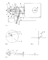
- Fig. 1 shows a perimeter 1 in which at a certain observer the eye 6 to be examined in an observation direction is fixed. The eye will be during an examination with suitable means, in timed staggering stimuli presented in the vicinity of an observer axis 9.
- the perimeter 1 is controlled by means of a computer 5. Becomes revelation here also referred to the applicant's CH-A-677 599 A5.
- Orientation around the observer eye 6 with reference to the observer axis 9 will facilitate the subject on this axis 9 a bright glowing mark (fixation mark) as a guide offered.
- This is done by an between an intermediate level 11 and partially transparent to the eyepiece 12, Deflecting mirror 28 directed towards the observer 10 Intersection between the deflecting mirror 28 and the observer axis 9 further intersects the optical axis 29 of two lenses 30, the axial with a light source 31 and an aperture 32nd is equipped.
- the aperture 32 determines the shape and size of the fixation mark.
- a further deflection mirror arranged in the optical axis 29 33 which is transparent for visible light and infrared Light reflecting is an IR sensitive CCD camera switched to the observer axis 9 with which the eye 6 can be viewed during an examination.
- the eye 6 with IR LEDs 38 becomes invisible to the test subject illuminated.
- the image captured by the camera 34 is in the Computer 5 evaluated.
- the position of the pupil 7 of the eye 6 determined and the deviations from the correct View direction and position are calculated. Such a possible Deviation in the viewing direction is shown schematically in Fig. 3.
- the pupil 7 of the eye 6 is located above the optical one Axis 9. Even if the eye 6 here shows the fixation mark 32 would be correctly fixed, for example a scotoma 3 according to FIG. 2 found in the wrong place, for example at 4.
- the position of the Stimulus test sites around the calculated values for example around corrected the value y according to FIG. 3.
- the correction will be made if necessary performed for each stimulus and in the x and y directions.
- the direction of view changes during an examination several times, the position of the person to be performed is correspondingly repeated several times Corrected stimulus.
- 4 schematically shows one such correction of a stimulus S.
- the fixation mark F is in the Zero point of the coordinate system.
- the eye 6 is correctly positioned and the line of sight is exactly on the optical axis 9, the camera 34 detects no deviation and accordingly a stimulus S presented. If the direction of view deviates, for example fix to point F 'according to Fig.
- this deviation captured by the camera 34 and in the computer 5 to a correction value converted. Because of this value it becomes a stimulus S 'presented at an appropriately corrected location.
- the stimulus S 'thus becomes relative despite the different viewing direction presented to the subject's field of vision at the correct location.
Landscapes
- Life Sciences & Earth Sciences (AREA)
- Health & Medical Sciences (AREA)
- Medical Informatics (AREA)
- Biophysics (AREA)
- Ophthalmology & Optometry (AREA)
- Engineering & Computer Science (AREA)
- Biomedical Technology (AREA)
- Heart & Thoracic Surgery (AREA)
- Physics & Mathematics (AREA)
- Molecular Biology (AREA)
- Surgery (AREA)
- Animal Behavior & Ethology (AREA)
- General Health & Medical Sciences (AREA)
- Public Health (AREA)
- Veterinary Medicine (AREA)
- Eye Examination Apparatus (AREA)
Priority Applications (3)
| Application Number | Priority Date | Filing Date | Title |
|---|---|---|---|
| EP00810984A EP1201181A1 (fr) | 2000-10-25 | 2000-10-25 | Procédé et périmètre d'examen de fonctions visuelles d'un oeil humain |
| JP2001287612A JP2002143091A (ja) | 2000-10-25 | 2001-09-20 | 人間の目の視野能力を検査する方法及びその方法を実施する視野計 |
| US10/000,974 US20020047991A1 (en) | 2000-10-25 | 2001-10-24 | Method for testing visual functions of a human eye and perimeter for carrying out the method |
Applications Claiming Priority (1)
| Application Number | Priority Date | Filing Date | Title |
|---|---|---|---|
| EP00810984A EP1201181A1 (fr) | 2000-10-25 | 2000-10-25 | Procédé et périmètre d'examen de fonctions visuelles d'un oeil humain |
Publications (1)
| Publication Number | Publication Date |
|---|---|
| EP1201181A1 true EP1201181A1 (fr) | 2002-05-02 |
Family
ID=8174988
Family Applications (1)
| Application Number | Title | Priority Date | Filing Date |
|---|---|---|---|
| EP00810984A Withdrawn EP1201181A1 (fr) | 2000-10-25 | 2000-10-25 | Procédé et périmètre d'examen de fonctions visuelles d'un oeil humain |
Country Status (3)
| Country | Link |
|---|---|
| US (1) | US20020047991A1 (fr) |
| EP (1) | EP1201181A1 (fr) |
| JP (1) | JP2002143091A (fr) |
Cited By (2)
| Publication number | Priority date | Publication date | Assignee | Title |
|---|---|---|---|---|
| DE102008053015B3 (de) * | 2008-10-21 | 2010-03-04 | Technische Universität Ilmenau | Verfahren und Vorrichtung zur farbkanalselektiven, blickgeführten Stimulation des visuellen Systems |
| DE102009010628A1 (de) * | 2009-02-20 | 2010-08-26 | Technische Universität Ilmenau | Verfahren und Vorrichtung zur farbkanalselektiven, funduskontrollierten Stimulation des visuellen Systems |
Families Citing this family (4)
| Publication number | Priority date | Publication date | Assignee | Title |
|---|---|---|---|---|
| DE102006011624A1 (de) * | 2006-03-10 | 2007-09-13 | Carl Zeiss Meditec Ag | Vorrichtung und Verfahren zur definierten Ausrichtung eines Auges |
| JP6062225B2 (ja) * | 2012-11-28 | 2017-01-18 | 株式会社クリュートメディカルシステムズ | 視覚機能計測装置 |
| JP6124330B2 (ja) * | 2012-12-28 | 2017-05-10 | スカラ株式会社 | 視野測定装置 |
| US11966511B2 (en) * | 2019-11-14 | 2024-04-23 | Rijksuniversiteit Groningen | Method, system and computer program product for mapping a visual field |
Citations (3)
| Publication number | Priority date | Publication date | Assignee | Title |
|---|---|---|---|---|
| US3883235A (en) * | 1971-09-17 | 1975-05-13 | John R Lynn | Automatic visual field examination including fixation monitoring compensation |
| CH677599A5 (fr) | 1988-09-22 | 1991-06-14 | Interzeag Ag | |
| US5550602A (en) * | 1994-11-09 | 1996-08-27 | Johannes Braeuning | Apparatus and method for examining visual functions |
Family Cites Families (1)
| Publication number | Priority date | Publication date | Assignee | Title |
|---|---|---|---|---|
| US4836670A (en) * | 1987-08-19 | 1989-06-06 | Center For Innovative Technology | Eye movement detector |
-
2000
- 2000-10-25 EP EP00810984A patent/EP1201181A1/fr not_active Withdrawn
-
2001
- 2001-09-20 JP JP2001287612A patent/JP2002143091A/ja active Pending
- 2001-10-24 US US10/000,974 patent/US20020047991A1/en not_active Abandoned
Patent Citations (3)
| Publication number | Priority date | Publication date | Assignee | Title |
|---|---|---|---|---|
| US3883235A (en) * | 1971-09-17 | 1975-05-13 | John R Lynn | Automatic visual field examination including fixation monitoring compensation |
| CH677599A5 (fr) | 1988-09-22 | 1991-06-14 | Interzeag Ag | |
| US5550602A (en) * | 1994-11-09 | 1996-08-27 | Johannes Braeuning | Apparatus and method for examining visual functions |
Non-Patent Citations (2)
| Title |
|---|
| BERTERA J H ET AL: "STABILIZED RETINAL MAPPING OF KNOWN RETINAL LOCI", PROCEEDINGS OF THE ANNUAL NORTHEAST BIOENGINEERING CONFERENCE,US,NEW YORK, IEEE, vol. CONF. 14, 10 March 1988 (1988-03-10), pages 136 - 137, XP000010509 * |
| SUASTE E ET AL: "PUPILLARY RESPONSE QUANTIFICATION WITH AN IMAGE-PROCESSING SYSTEM IN CLINICAL PERIMETRY", MIDWEST SYMPOSIUM ON CIRCUITS AND SYSTEMS: PROCEEDINGS,US,NEW YORK, IEEE, vol. SYMP. 38, 13 August 1995 (1995-08-13), pages 1381 - 1384, XP000825319, ISBN: 0-7803-2973-2 * |
Cited By (3)
| Publication number | Priority date | Publication date | Assignee | Title |
|---|---|---|---|---|
| DE102008053015B3 (de) * | 2008-10-21 | 2010-03-04 | Technische Universität Ilmenau | Verfahren und Vorrichtung zur farbkanalselektiven, blickgeführten Stimulation des visuellen Systems |
| DE102009010628A1 (de) * | 2009-02-20 | 2010-08-26 | Technische Universität Ilmenau | Verfahren und Vorrichtung zur farbkanalselektiven, funduskontrollierten Stimulation des visuellen Systems |
| DE102009010628B4 (de) * | 2009-02-20 | 2010-10-14 | Technische Universität Ilmenau | Verfahren und Vorrichtung zur farbkanalselektiven, funduskontrollierten Stimulation des visuellen Systems |
Also Published As
| Publication number | Publication date |
|---|---|
| US20020047991A1 (en) | 2002-04-25 |
| JP2002143091A (ja) | 2002-05-21 |
Similar Documents
| Publication | Publication Date | Title |
|---|---|---|
| DE69013756T2 (de) | Laserbehandlungsvorrichtung. | |
| CH696457A5 (de) | Ophthalmologisches Untersuchungsgerät. | |
| EP0495247B1 (fr) | Procédé de périmétrie du champ visuel de l'oeil | |
| EP0363610B1 (fr) | Dispositif d'examen de fonctions visuelles d'un oeil humain | |
| WO2018083323A1 (fr) | Procédé d'auto-examen d'un œil et dispositif d'auto-examen ophtalmologique | |
| DE69936214T2 (de) | Chirurgisches Augenfixationsgerät | |
| EP0825826A1 (fr) | Procede et dispositif de captage en parallele d'informations per ues visuellement | |
| EP1225454A2 (fr) | Procédé et dispositif de fixation d'une position | |
| EP3873322B1 (fr) | Quantification, basée sur la localisation, de l'impact d'une lumière halogène ou diffusée | |
| EP3192431B1 (fr) | Système d'examen optométrique et procédé d'examen des yeux | |
| DE19502337C2 (de) | Vorrichtung und Verfahren zur Prüfung von Sehfunktionen | |
| CH688304A5 (de) | Ophthalmologisches Geraet. | |
| EP1891890A1 (fr) | Appareil optique, utilisation d'un appareil optique selon l'invention tout comme procédé destiné à bloquer les reflets sur une trajectoire de faisceau d'observation d'un appareil optique | |
| EP1840627A2 (fr) | Dispositif et procédé destinés à la détermination de l'orientation d'un oeil | |
| EP1993431B1 (fr) | Dispositifs et procédé permettant d'aligner un il de manière définie | |
| EP0815789B1 (fr) | Appareil d'examen du champ visuel de l'oeil humain | |
| DE102014014093B4 (de) | Augenchirurgiesystem und Verfahren zum Betreiben eines Augenchirurgiesystems | |
| DE3731415A1 (de) | Verfahren fuer die gesichtsfelduntersuchung | |
| EP1903928B1 (fr) | Procede et dispositif pour verifier la vision scotopique | |
| DE3143882A1 (de) | "verfahren zur fixationssicherung bei ophthalmologischen untersuchungsgeraeten" | |
| DE4232280A1 (de) | Retinoskop mit Lichtleiterbeleuchtung | |
| DE102008010577A1 (de) | Verfahren und Einrichtung zur optisch-elektronischen Augeninspektion | |
| EP1224498A2 (fr) | Combinaison d'un dispositif de grossissement, notamment d'un microscope avec un dispositif de mesure | |
| DE102013226973B4 (de) | Verfahren und Vorrichtung zur gleichzeitigen Darstellung eines medizinischen Bildes und eines grafischen Bedienelementes | |
| DE102010021346B4 (de) | Ophthalmologisches Untersuchungsgerät |
Legal Events
| Date | Code | Title | Description |
|---|---|---|---|
| PUAI | Public reference made under article 153(3) epc to a published international application that has entered the european phase |
Free format text: ORIGINAL CODE: 0009012 |
|
| AK | Designated contracting states |
Kind code of ref document: A1 Designated state(s): AT BE CH CY DE DK ES FI FR GB GR IE IT LI LU MC NL PT SE Kind code of ref document: A1 Designated state(s): DE GB |
|
| AX | Request for extension of the european patent |
Free format text: AL;LT;LV;MK;RO;SI |
|
| 17P | Request for examination filed |
Effective date: 20020518 |
|
| AKX | Designation fees paid |
Free format text: DE GB |
|
| 17Q | First examination report despatched |
Effective date: 20031105 |
|
| STAA | Information on the status of an ep patent application or granted ep patent |
Free format text: STATUS: THE APPLICATION IS DEEMED TO BE WITHDRAWN |
|
| 18D | Application deemed to be withdrawn |
Effective date: 20040316 |