EP1205868A2 - Verfahren und Anlage zur Archivierung und Verteilung von verbesserten und unverbesserten digitalen medizinischen Bildern - Google Patents
Verfahren und Anlage zur Archivierung und Verteilung von verbesserten und unverbesserten digitalen medizinischen Bildern Download PDFInfo
- Publication number
- EP1205868A2 EP1205868A2 EP01204222A EP01204222A EP1205868A2 EP 1205868 A2 EP1205868 A2 EP 1205868A2 EP 01204222 A EP01204222 A EP 01204222A EP 01204222 A EP01204222 A EP 01204222A EP 1205868 A2 EP1205868 A2 EP 1205868A2
- Authority
- EP
- European Patent Office
- Prior art keywords
- image
- digital
- unenhanced
- digital medical
- storage
- Prior art date
- Legal status (The legal status is an assumption and is not a legal conclusion. Google has not performed a legal analysis and makes no representation as to the accuracy of the status listed.)
- Withdrawn
Links
Images

Classifications
-
- G—PHYSICS
- G16—INFORMATION AND COMMUNICATION TECHNOLOGY [ICT] SPECIALLY ADAPTED FOR SPECIFIC APPLICATION FIELDS
- G16H—HEALTHCARE INFORMATICS, i.e. INFORMATION AND COMMUNICATION TECHNOLOGY [ICT] SPECIALLY ADAPTED FOR THE HANDLING OR PROCESSING OF MEDICAL OR HEALTHCARE DATA
- G16H30/00—ICT specially adapted for the handling or processing of medical images
- G16H30/40—ICT specially adapted for the handling or processing of medical images for processing medical images, e.g. editing
-
- G—PHYSICS
- G16—INFORMATION AND COMMUNICATION TECHNOLOGY [ICT] SPECIALLY ADAPTED FOR SPECIFIC APPLICATION FIELDS
- G16H—HEALTHCARE INFORMATICS, i.e. INFORMATION AND COMMUNICATION TECHNOLOGY [ICT] SPECIALLY ADAPTED FOR THE HANDLING OR PROCESSING OF MEDICAL OR HEALTHCARE DATA
- G16H30/00—ICT specially adapted for the handling or processing of medical images
- G16H30/20—ICT specially adapted for the handling or processing of medical images for handling medical images, e.g. DICOM, HL7 or PACS
-
- G—PHYSICS
- G16—INFORMATION AND COMMUNICATION TECHNOLOGY [ICT] SPECIALLY ADAPTED FOR SPECIFIC APPLICATION FIELDS
- G16H—HEALTHCARE INFORMATICS, i.e. INFORMATION AND COMMUNICATION TECHNOLOGY [ICT] SPECIALLY ADAPTED FOR THE HANDLING OR PROCESSING OF MEDICAL OR HEALTHCARE DATA
- G16H40/00—ICT specially adapted for the management or administration of healthcare resources or facilities; ICT specially adapted for the management or operation of medical equipment or devices
- G16H40/60—ICT specially adapted for the management or administration of healthcare resources or facilities; ICT specially adapted for the management or operation of medical equipment or devices for the operation of medical equipment or devices
- G16H40/67—ICT specially adapted for the management or administration of healthcare resources or facilities; ICT specially adapted for the management or operation of medical equipment or devices for the operation of medical equipment or devices for remote operation
Definitions
- This invention relates in general to digital medical imaging and in particular to the storage and retrieval of images in a digital archive system.
- Picture Archiving and Communication Systems typically consist of digital acquisition modalities, digital image archives, diagnostic viewing workstations and laser film printers.
- Digital acquisition modalities include computed radiography (CR), direct digital radiography (DR), computed tomography (CT) and magnetic resonance (MR) among others.
- CR computed radiography
- DR direct digital radiography
- CT computed tomography
- MR magnetic resonance
- the typical workflow is for the image to be acquired at the modality and digital processing performed either on the modality console or on a dedicated quality control workstation prior to transmitting the image to the archive, workstation or printer.
- CR and DR in particular, improved image quality and utility can be achieved if the image that is used by the diagnostic workstation does not have the enhancement processing that is performed by the modality applied.
- few workstations can interactively perform this level of processing.
- DICOM Digital Imaging Communications in Medicine
- DICOM medical image archives provide storage of patient images that are ready for transmission and display on approved and validated DICOM display workstations.
- a workstation will query an archive for a list of images that meet certain selection criteria.
- the archive responds with a list of images, if any, resident on the archive which meet those criteria.
- the workstation may then request some or all of the available images to be transmitted to the workstation.
- the archive retrieves the selected images from storage and transmits them to the workstation.
- the archive since all images are display-ready, the archive may simply transmit the image data without adjustment to the processing capabilities of the requesting device.
- the present invention allows the storage and transmission of images that are not display-ready by applying enhancements prior to distribution to a workstation which cannot perform the required processing and transmitting the unenhanced version of the image to workstations capable of performing the enhancement processing.
- a recently adopted part of the DICOM standard (Digital Imaging and Communications in Medicine PS 3.3-1999, National Electrical Manufacturers Association, 1999) partially addresses the management of image data which is enhanced or unenhanced.
- the concept is presented in the DX Series Module by the introduction of Presentation Intent Type.
- This tag may have the value of FOR PRESENTATION or FOR PROCESSING. If the value of the tag is FOR PRESENTATION, the image has been enhanced such that the image may be displayed without additional processing required. If the value of the tag is FOR PROCESSING, the image must undergo additional enhancement processing prior to display and interpretation.
- FOR PROCESING image data is to allow more capable systems which can perform the required enhancement processing to do so while still using a DICOM image object while preserving the capability to provide image data that is display-ready by the use of FOR PRESENTATION.
- the standard does not however, indicate how these different types of image processing levels should be managed. While several methods are possible, the present invention discloses one method of managing image data that is unenhanced.
- U.S. Patent 5,179,651, issued January 12, 1993, inventors Taaffe, et al. describe a methodology whereby an image archive / processor combination can perform enhancement processing on an image prior to transmission to a display workstation if the workstation requests the processing to be performed by the image archive / processor. While this is similar to the present invention, the workstation initiates the request for processing given the complexity of the required processing task and the available workstation resources. In the present invention, the image server determines what processing should be performed prior to transmission. This removes the requirement of the workstation to request the level of processing desired. While this is a possible implementation to achieve a similar result, it imposes additional requirements on the workstation which are removed in the present invention.
- U.S. Patent 4,780,821 issued October 25, 1988, inventor Crossley, describes a method for a server computer to determine if a remote computer has the ability to run a requested application or gain access a particular type of data. Again, this is similar to the present invention except that if the server computer does not permit access to the application or data, the remote computer does not receive any data or application to satisfy the request.
- the image data is not denied if the requesting workstation does not have the ability to process the image data, instead the requested image is first enhanced, allowing the requesting workstation to properly display the image.
- a method for distributing digital medical images comprising:
- Fig. 1 is a block diagram of an image archive / image server system for distribution of enhanced and unenhanced imagery showing the architecture for the acquisition modalities, archive, server and diagnostic workstations.
- Fig. 2 is a flow chart for query and retrieval of images which shows the processing steps necessary for requesting and receiving enhanced or unenhanced imagery based on workstation capabilities.
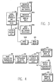
- Fig. 3 is a block diagram of an acquisition modality system for distribution of enhanced and unenhanced imagery showing the architecture for an acquisition modality to distribute imagery to workstations or archives.
- Fig. 4 is a flow chart for distribution of images from a modality which shows the steps necessary for a modality to send an enhanced or unenhanced image to a destination.
- DICOM network is used in the preferred embodiment of the invention and as such, DICOM terms are used to describe component relationships and transactions.
- image enhancement is not limited to a single algorithm performed on the image data. Multiple algorithms are typical in medical imaging to enhance an image including, but not limited to, tonescale generation and application, spatial processing, dynamic range adjustment and masking. The multiple individual algorithms should all be considered under the general term image enhancement.
- a digital image archive and server are used to manage the storage, processing and transmission of images among multiple modalities and medical imaging workstations.
- a modality stores images on the archive and a workstation retrieves an image from the image server.
- the image server acquires the requested image from the archive and performs any necessary processing prior to transmission to the requesting workstation.
- an imaging modality which produces enhanced imagery an imaging modality which produces unenhanced or enhanced imagery
- a viewing system which can process unenhanced imagery
- a viewing system which cannot process unenhanced imagery or alternatively can only display enhanced imagery.
- the enhanced image is transmitted to the archive by the modality.
- the image server retrieves the enhanced image from the archive and immediately transmits the image to the requesting workstation, regardless of the capabilities of the workstation. For the case of an imaging modality which can distribute unenhanced imagery, the unenhanced images are transmitted to the archive by the modality.
- the image server determines if the requesting workstation can properly process the unenhanced image. If the image server determines that the workstation can process the image, the image server retrieves the unenhanced image from the archive and immediately transmits the image to the requesting workstation.
- the image server determines that the workstation cannot process the image, the image server first retrieves the unenhanced image from the archive and then performs the enhancement processing on the retrieved image prior to transmitting the image to the requesting workstation. While DICOM does not specify this level of data management, this system can be implemented to be fully DICOM compliant
- Fig. 1 shows a block diagram of the preferred embodiment of the image archive / image server system according to the present invention.
- Digital modality 1-101A supplies image data that has not been enhanced.
- Digital modality 2-101B supplies image data that has already been enhanced.
- Both digital modality 1-101A and digital modality 2-101B transmit acquired images to the image archive 102 for storage.
- Digital modality 1-101A includes a special DICOM tag in the header, which denotes that the image is unenhanced. This special tag may be an existing element such as "Presentation Intent" in the Direct Radiography (DX) module or the special tag may be a private DICOM element.
- the image archive 102 is strictly an image storage device, no additional image processing occurs on the image archive 102.
- the image server 103 works with the image archive 102 such that the image server 103 performs enhancement processing on stored images as needed.
- the need for the image server 103 to perform enhancement processing is dependent on the capabilities of the requesting device.
- Each device that can request images from the image server 103 has an entry in the access control list 104.
- a field is included in the access control list 104 for each requesting device, which denotes if that particular device can process unenhanced imagery.
- Diagnostic workstation 1-105A has the capability to perform enhancement processing on images it receives.
- the image server 103 retrieves the image from the image archive 102 and checks the header for the special unenhanced image tag. If the image is enhanced, the image server 103 transmits the image to workstation 1-105A. If the image is unenhanced, the image server 103 checks the access control list 104 and determines that workstation 1-105A can receive unenhanced imagery. The image server 103 then transmits the unenhanced image to workstation 1-105A.
- Diagnostic workstation 2-105B does not have the capability to perform enhancement processing on images it receives.
- the image server 103 retrieves the image from the image archive 102 and checks the header for the special unenhanced image tag. If the image is enhanced, the image server 103 transmits the image to workstation 2-105B. If the image is unenhanced, the image server 103 checks the access control list 104 and determines that workstation 2-105B cannot receive unenhanced imagery. The image server 103 then applies enhancement processing on the image and transmits the enhanced image to workstation 2-105B.
- a new DICOM series unique identifier UID
- Fig. 2 This is a flow chart of typical image request and transmission steps in the preferred embodiment.
- the user of a workstation queries the image server for a list of images 201 based on a set of input criteria. Patient name, accession number and date are typical query parameters.
- the image server then provides the result of the query 202 to the requesting workstation.
- the user selects the image(s) to transfer 203 and the workstation then request the selected image(s) from the image server 204.
- the image server retrieves the image(s) from the image archive 205.
- the image server checks for the special tag in the image header which denotes if the image is enhanced 206.
- the image server sends the enhanced image to the workstation 207. If the tag is not present, it is assumed that the image is enhanced. If the tag specifies that the image is unenhanced, the image server then checks the capabilities of the requesting workstation in the access control list 208 and determines if the workstation accepts unenhanced data 209. If the workstation accepts unenhanced data, the image server transmits the unenhanced image to the workstation 210. If the workstation does not accept raw data, the image server applies the enhancement processing 211 and subsequently transmits the enhanced image to the workstation 212.
- Fig. 3 shows an acquisition modality system for distribution of enhanced and unenhanced images.
- Digital Modality 301 produces enhanced and unenhanced data.
- the access control list 302 provides information regarding enhancement capability for each image destination of the digital modality 301.
- the image destinations are as follows. Diagnostic Workstation 1-303A has enhancement processing and display capability. Diagnostic Workstation 2-303B has only display capability, or alternatively, no enhancement capability. Image Archive 1-304A has no enhancement processing. Image Archive 2-304B has image enhancement capability via the Image Server 304.
- the process for determining the image to distribute to each destination is described in Fig. 4. First, an image is selected for transmission to a destination 401. The modality checks destination in the access control list 402.
- the modality sends the unenhanced image to the destination 404. If the destination is determined 403 not to accept unenhanced data, the modality applies enhancement processing to the unenhanced image 405 and sends the enhanced image to the destination 406.
- Additional information about the image processing performed may be tracked, although it is not required for implementation. For example, an audit trail may be stored with the enhanced image including the image processing algorithms and parameters, the date and the individual selecting the enhancement. Also, information referencing the original unenhanced image may be kept with the enhanced version to facilitate retrieval of alternate versions. In the same manner, after an image has been enhanced, information may be stored with the unenhanced version indicating the enhanced version exists and where it may be located.
- the modality appends a special DICOM tag denoting an image is unenhanced.
- An alternative is for the image server to be programmed such that all images from a given modality are handled as unenhanced.
- the image server may also be programmed such that all images from a given modality are handled as enhanced.
- the access control list contains information regarding enhancement processing capability for each workstation. Alternatives to this include having the workstation send a message to the image server stating the ability to process unenhanced imagery. Also, the image server may be directly programmed with information regarding the ability of any workstation to receive unenhanced imagery.
- the ability to transmit unenhanced or enhanced imagery based on the capability of the receiving device is not limited to the image server.
- An acquisition modality itself may act as the image server of the present invention.
- the capabilities of the receiving system are determined from the access control list.
- the receiving system may be a diagnostic workstation or an image archive.
Landscapes
- Health & Medical Sciences (AREA)
- Engineering & Computer Science (AREA)
- Epidemiology (AREA)
- General Health & Medical Sciences (AREA)
- Medical Informatics (AREA)
- Primary Health Care (AREA)
- Public Health (AREA)
- Nuclear Medicine, Radiotherapy & Molecular Imaging (AREA)
- Biomedical Technology (AREA)
- Radiology & Medical Imaging (AREA)
- General Business, Economics & Management (AREA)
- Business, Economics & Management (AREA)
- Measuring And Recording Apparatus For Diagnosis (AREA)
- Medical Treatment And Welfare Office Work (AREA)
- Processing Or Creating Images (AREA)
- Image Processing (AREA)
- Image Analysis (AREA)
Applications Claiming Priority (2)
| Application Number | Priority Date | Filing Date | Title |
|---|---|---|---|
| US71242300A | 2000-11-14 | 2000-11-14 | |
| US712423 | 2000-11-14 |
Publications (2)
| Publication Number | Publication Date |
|---|---|
| EP1205868A2 true EP1205868A2 (de) | 2002-05-15 |
| EP1205868A3 EP1205868A3 (de) | 2005-02-23 |
Family
ID=24862044
Family Applications (1)
| Application Number | Title | Priority Date | Filing Date |
|---|---|---|---|
| EP01204222A Withdrawn EP1205868A3 (de) | 2000-11-14 | 2001-11-02 | Verfahren und Anlage zur Archivierung und Verteilung von verbesserten und unverbesserten digitalen medizinischen Bildern |
Country Status (2)
| Country | Link |
|---|---|
| EP (1) | EP1205868A3 (de) |
| JP (1) | JP2002230166A (de) |
Cited By (2)
| Publication number | Priority date | Publication date | Assignee | Title |
|---|---|---|---|---|
| EP1434157A3 (de) * | 2002-09-12 | 2004-10-20 | Konica Corporation | Bildinformationsverarbeitungsgerät und medizinisches Netzwerksystem |
| CN103577699A (zh) * | 2013-11-14 | 2014-02-12 | 哈尔滨工程大学 | 一种基于Android平台的DICIOM医学图像显示处理方法 |
Families Citing this family (2)
| Publication number | Priority date | Publication date | Assignee | Title |
|---|---|---|---|---|
| JP5972648B2 (ja) * | 2012-04-13 | 2016-08-17 | 東芝メディカルシステムズ株式会社 | 医用画像伝送システム及びこのシステムにおけるゲートサーバ |
| JP6513406B2 (ja) * | 2015-01-13 | 2019-05-15 | キヤノンメディカルシステムズ株式会社 | 医用画像処理装置 |
Family Cites Families (5)
| Publication number | Priority date | Publication date | Assignee | Title |
|---|---|---|---|---|
| US5179651A (en) * | 1988-11-08 | 1993-01-12 | Massachusetts General Hospital | Apparatus for retrieval and processing of selected archived images for display at workstation terminals |
| US5321520A (en) * | 1992-07-20 | 1994-06-14 | Automated Medical Access Corporation | Automated high definition/resolution image storage, retrieval and transmission system |
| AU687045B2 (en) * | 1993-03-31 | 1998-02-19 | Luma Corporation | Managing information in an endoscopy system |
| EP0889636B1 (de) * | 1997-06-30 | 2005-08-24 | Fuji Photo Film Co., Ltd. | System und Verfahren zur Bildübertragung |
| EP0892354B8 (de) * | 1997-07-16 | 2005-11-23 | Sony Deutschland Gmbh | Effiziente Darstellung und Uebertragung von Objekten mit Varianten |
-
2001
- 2001-11-02 EP EP01204222A patent/EP1205868A3/de not_active Withdrawn
- 2001-11-12 JP JP2001345563A patent/JP2002230166A/ja active Pending
Cited By (3)
| Publication number | Priority date | Publication date | Assignee | Title |
|---|---|---|---|---|
| EP1434157A3 (de) * | 2002-09-12 | 2004-10-20 | Konica Corporation | Bildinformationsverarbeitungsgerät und medizinisches Netzwerksystem |
| CN103577699A (zh) * | 2013-11-14 | 2014-02-12 | 哈尔滨工程大学 | 一种基于Android平台的DICIOM医学图像显示处理方法 |
| CN103577699B (zh) * | 2013-11-14 | 2017-02-08 | 哈尔滨工程大学 | 一种基于Android平台的DICOM医学图像显示处理方法 |
Also Published As
| Publication number | Publication date |
|---|---|
| EP1205868A3 (de) | 2005-02-23 |
| JP2002230166A (ja) | 2002-08-16 |
Similar Documents
| Publication | Publication Date | Title |
|---|---|---|
| US8583449B2 (en) | Method and apparatus for providing network based load balancing of medical image data | |
| US6954767B1 (en) | Server and method for searching for image using image prefetch, designating database and storage devices for searching, and setting retrieval and processing parameters for search | |
| US9684762B2 (en) | Rules-based approach to rendering medical imaging data | |
| US8965137B2 (en) | Image data transmission | |
| US20020091659A1 (en) | Portable viewing of medical images using handheld computers | |
| US20040193901A1 (en) | Dynamic configuration of patient tags and masking types while de-identifying patient data during image export from PACS diagnostic workstation | |
| US20050197860A1 (en) | Data management system | |
| CA3051767C (en) | Image viewer | |
| US20090028402A1 (en) | Medical Image System | |
| US20060106877A1 (en) | Image archiving system and method for handling new and legacy archives | |
| US20100114603A1 (en) | Systems and Methods for Obtaining Readings of Diagnostic Imaging Studies | |
| Robertson et al. | Hospital, radiology, and picture archiving and communication systems | |
| EP2435987B1 (de) | Darstellung und manipulation von bildern mit hoher tiefe in systemen zur anzeige von bildern mit niedriger tiefe | |
| EP2893474A1 (de) | System und verfahren zum abrufen und verarbeiten von metadaten | |
| EP1205868A2 (de) | Verfahren und Anlage zur Archivierung und Verteilung von verbesserten und unverbesserten digitalen medizinischen Bildern | |
| US20050187787A1 (en) | Method for payer access to medical image data | |
| US11823787B2 (en) | Systems and methods for transferring medical image records using a prefferred transfer protocol | |
| US20080123925A1 (en) | Medical Imaging System | |
| US20020082948A1 (en) | Computed radiography configuration and selection method and apparatus | |
| US8849068B2 (en) | Image management system, image management method, and program | |
| JP6881628B2 (ja) | 医療連携システム及び制御プログラム | |
| JP2001094744A (ja) | 画像管理装置、及び、ネットワーク上での画像ファイルの転送制御方法 | |
| Wimmer et al. | A distributed approach to integrated inquiry and display for radiology | |
| US20050222872A1 (en) | Information processing system, medical system and information processing method | |
| Toennies | Image Storage and Transfer |
Legal Events
| Date | Code | Title | Description |
|---|---|---|---|
| PUAI | Public reference made under article 153(3) epc to a published international application that has entered the european phase |
Free format text: ORIGINAL CODE: 0009012 |
|
| AK | Designated contracting states |
Kind code of ref document: A2 Designated state(s): AT BE CH CY DE DK ES FI FR GB GR IE IT LI LU MC NL PT SE TR |
|
| AX | Request for extension of the european patent |
Free format text: AL;LT;LV;MK;RO;SI |
|
| PUAL | Search report despatched |
Free format text: ORIGINAL CODE: 0009013 |
|
| AK | Designated contracting states |
Kind code of ref document: A3 Designated state(s): AT BE CH CY DE DK ES FI FR GB GR IE IT LI LU MC NL PT SE TR |
|
| AX | Request for extension of the european patent |
Extension state: AL LT LV MK RO SI |
|
| 17P | Request for examination filed |
Effective date: 20050714 |
|
| AKX | Designation fees paid |
Designated state(s): DE FR GB |
|
| 17Q | First examination report despatched |
Effective date: 20071109 |
|
| RAP1 | Party data changed (applicant data changed or rights of an application transferred) |
Owner name: CARESTREAM HEALTH, INC. |
|
| STAA | Information on the status of an ep patent application or granted ep patent |
Free format text: STATUS: THE APPLICATION IS DEEMED TO BE WITHDRAWN |
|
| 18D | Application deemed to be withdrawn |
Effective date: 20080320 |