EP1221342A2 - Verfahren zur Trennung von Zellen von einer Probe - Google Patents
Verfahren zur Trennung von Zellen von einer Probe Download PDFInfo
- Publication number
- EP1221342A2 EP1221342A2 EP01129402A EP01129402A EP1221342A2 EP 1221342 A2 EP1221342 A2 EP 1221342A2 EP 01129402 A EP01129402 A EP 01129402A EP 01129402 A EP01129402 A EP 01129402A EP 1221342 A2 EP1221342 A2 EP 1221342A2
- Authority
- EP
- European Patent Office
- Prior art keywords
- sample
- float
- tube
- antibody
- target component
- Prior art date
- Legal status (The legal status is an assumption and is not a legal conclusion. Google has not performed a legal analysis and makes no representation as to the accuracy of the status listed.)
- Granted
Links
- 238000000034 method Methods 0.000 title claims description 73
- 239000011325 microbead Substances 0.000 claims abstract description 55
- 238000003306 harvesting Methods 0.000 claims abstract description 31
- 239000000523 sample Substances 0.000 claims description 120
- 210000000265 leukocyte Anatomy 0.000 claims description 32
- 239000000470 constituent Substances 0.000 claims description 29
- 210000004369 blood Anatomy 0.000 claims description 25
- 239000008280 blood Substances 0.000 claims description 25
- 238000005070 sampling Methods 0.000 claims description 18
- 239000011324 bead Substances 0.000 claims description 12
- 238000002156 mixing Methods 0.000 claims description 11
- 239000011248 coating agent Substances 0.000 claims description 9
- 238000000576 coating method Methods 0.000 claims description 9
- 239000000126 substance Substances 0.000 claims description 3
- 238000000926 separation method Methods 0.000 abstract description 16
- 239000012530 fluid Substances 0.000 abstract description 13
- 210000004027 cell Anatomy 0.000 description 79
- 239000000306 component Substances 0.000 description 57
- 238000005119 centrifugation Methods 0.000 description 24
- 239000011230 binding agent Substances 0.000 description 17
- 239000012472 biological sample Substances 0.000 description 17
- 210000004881 tumor cell Anatomy 0.000 description 17
- 210000003743 erythrocyte Anatomy 0.000 description 15
- 239000002245 particle Substances 0.000 description 13
- 230000008569 process Effects 0.000 description 10
- 239000000463 material Substances 0.000 description 9
- 229920003023 plastic Polymers 0.000 description 8
- 239000004033 plastic Substances 0.000 description 7
- 238000004458 analytical method Methods 0.000 description 5
- 239000013060 biological fluid Substances 0.000 description 4
- 239000000203 mixture Substances 0.000 description 4
- 238000010187 selection method Methods 0.000 description 4
- 239000000427 antigen Substances 0.000 description 3
- 102000036639 antigens Human genes 0.000 description 3
- 108091007433 antigens Proteins 0.000 description 3
- 238000000386 microscopy Methods 0.000 description 3
- 210000005087 mononuclear cell Anatomy 0.000 description 3
- 239000011347 resin Substances 0.000 description 3
- 229920005989 resin Polymers 0.000 description 3
- 210000000130 stem cell Anatomy 0.000 description 3
- 102100024222 B-lymphocyte antigen CD19 Human genes 0.000 description 2
- 101000980825 Homo sapiens B-lymphocyte antigen CD19 Proteins 0.000 description 2
- 101000946889 Homo sapiens Monocyte differentiation antigen CD14 Proteins 0.000 description 2
- 101000738771 Homo sapiens Receptor-type tyrosine-protein phosphatase C Proteins 0.000 description 2
- 102100035877 Monocyte differentiation antigen CD14 Human genes 0.000 description 2
- 239000004793 Polystyrene Substances 0.000 description 2
- 102100037422 Receptor-type tyrosine-protein phosphatase C Human genes 0.000 description 2
- 238000001514 detection method Methods 0.000 description 2
- 238000005516 engineering process Methods 0.000 description 2
- 230000001605 fetal effect Effects 0.000 description 2
- 238000000684 flow cytometry Methods 0.000 description 2
- 239000011521 glass Substances 0.000 description 2
- 230000005484 gravity Effects 0.000 description 2
- -1 polyethylene Polymers 0.000 description 2
- 229920002223 polystyrene Polymers 0.000 description 2
- 229920000915 polyvinyl chloride Polymers 0.000 description 2
- 239000004800 polyvinyl chloride Substances 0.000 description 2
- 238000011084 recovery Methods 0.000 description 2
- 239000012780 transparent material Substances 0.000 description 2
- 102100035248 Alpha-(1,3)-fucosyltransferase 4 Human genes 0.000 description 1
- 102100022005 B-lymphocyte antigen CD20 Human genes 0.000 description 1
- 102000049320 CD36 Human genes 0.000 description 1
- 108010045374 CD36 Antigens Proteins 0.000 description 1
- 102100032912 CD44 antigen Human genes 0.000 description 1
- 201000008808 Fibrosarcoma Diseases 0.000 description 1
- 102100021260 Galactosylgalactosylxylosylprotein 3-beta-glucuronosyltransferase 1 Human genes 0.000 description 1
- 208000032612 Glial tumor Diseases 0.000 description 1
- 206010018338 Glioma Diseases 0.000 description 1
- 101001022185 Homo sapiens Alpha-(1,3)-fucosyltransferase 4 Proteins 0.000 description 1
- 101000897405 Homo sapiens B-lymphocyte antigen CD20 Proteins 0.000 description 1
- 101000868273 Homo sapiens CD44 antigen Proteins 0.000 description 1
- 101000894906 Homo sapiens Galactosylgalactosylxylosylprotein 3-beta-glucuronosyltransferase 1 Proteins 0.000 description 1
- 101001046686 Homo sapiens Integrin alpha-M Proteins 0.000 description 1
- 101001015004 Homo sapiens Integrin beta-3 Proteins 0.000 description 1
- 101000608935 Homo sapiens Leukosialin Proteins 0.000 description 1
- 101000917858 Homo sapiens Low affinity immunoglobulin gamma Fc region receptor III-A Proteins 0.000 description 1
- 101000917839 Homo sapiens Low affinity immunoglobulin gamma Fc region receptor III-B Proteins 0.000 description 1
- 101001071312 Homo sapiens Platelet glycoprotein IX Proteins 0.000 description 1
- 101000914514 Homo sapiens T-cell-specific surface glycoprotein CD28 Proteins 0.000 description 1
- 102100022338 Integrin alpha-M Human genes 0.000 description 1
- 102100022297 Integrin alpha-X Human genes 0.000 description 1
- 102100032999 Integrin beta-3 Human genes 0.000 description 1
- 102100039564 Leukosialin Human genes 0.000 description 1
- 102100029185 Low affinity immunoglobulin gamma Fc region receptor III-B Human genes 0.000 description 1
- 206010029260 Neuroblastoma Diseases 0.000 description 1
- 102100036851 Platelet glycoprotein IX Human genes 0.000 description 1
- 239000004698 Polyethylene Substances 0.000 description 1
- 239000004743 Polypropylene Substances 0.000 description 1
- 206010039491 Sarcoma Diseases 0.000 description 1
- 102100027213 T-cell-specific surface glycoprotein CD28 Human genes 0.000 description 1
- 238000007792 addition Methods 0.000 description 1
- 210000004381 amniotic fluid Anatomy 0.000 description 1
- 230000003321 amplification Effects 0.000 description 1
- 238000003556 assay Methods 0.000 description 1
- 230000015572 biosynthetic process Effects 0.000 description 1
- 239000012503 blood component Substances 0.000 description 1
- 210000001124 body fluid Anatomy 0.000 description 1
- 239000007767 bonding agent Substances 0.000 description 1
- 210000004556 brain Anatomy 0.000 description 1
- 210000000481 breast Anatomy 0.000 description 1
- 239000003153 chemical reaction reagent Substances 0.000 description 1
- 239000003795 chemical substances by application Substances 0.000 description 1
- 210000001072 colon Anatomy 0.000 description 1
- 230000000295 complement effect Effects 0.000 description 1
- 150000001875 compounds Chemical class 0.000 description 1
- 239000000356 contaminant Substances 0.000 description 1
- 238000011109 contamination Methods 0.000 description 1
- 239000013068 control sample Substances 0.000 description 1
- 239000007822 coupling agent Substances 0.000 description 1
- 239000000975 dye Substances 0.000 description 1
- 230000002708 enhancing effect Effects 0.000 description 1
- 239000000284 extract Substances 0.000 description 1
- 239000007789 gas Substances 0.000 description 1
- 210000003714 granulocyte Anatomy 0.000 description 1
- 208000019691 hematopoietic and lymphoid cell neoplasm Diseases 0.000 description 1
- 239000011261 inert gas Substances 0.000 description 1
- 230000002452 interceptive effect Effects 0.000 description 1
- 210000003734 kidney Anatomy 0.000 description 1
- 239000007788 liquid Substances 0.000 description 1
- 210000004185 liver Anatomy 0.000 description 1
- 239000000314 lubricant Substances 0.000 description 1
- 210000004072 lung Anatomy 0.000 description 1
- 210000004880 lymph fluid Anatomy 0.000 description 1
- 238000007885 magnetic separation Methods 0.000 description 1
- 238000002826 magnetic-activated cell sorting Methods 0.000 description 1
- 201000001441 melanoma Diseases 0.000 description 1
- 230000004048 modification Effects 0.000 description 1
- 238000012986 modification Methods 0.000 description 1
- 238000003199 nucleic acid amplification method Methods 0.000 description 1
- 150000007523 nucleic acids Chemical class 0.000 description 1
- 102000039446 nucleic acids Human genes 0.000 description 1
- 108020004707 nucleic acids Proteins 0.000 description 1
- 230000003287 optical effect Effects 0.000 description 1
- 210000001672 ovary Anatomy 0.000 description 1
- 210000000496 pancreas Anatomy 0.000 description 1
- 230000005298 paramagnetic effect Effects 0.000 description 1
- 210000004180 plasmocyte Anatomy 0.000 description 1
- 239000000088 plastic resin Substances 0.000 description 1
- 229920000779 poly(divinylbenzene) Polymers 0.000 description 1
- 229920002492 poly(sulfone) Polymers 0.000 description 1
- 229920002401 polyacrylamide Polymers 0.000 description 1
- 229920000515 polycarbonate Polymers 0.000 description 1
- 239000004417 polycarbonate Substances 0.000 description 1
- 229920000728 polyester Polymers 0.000 description 1
- 229920000573 polyethylene Polymers 0.000 description 1
- 229920001155 polypropylene Polymers 0.000 description 1
- 229920001296 polysiloxane Polymers 0.000 description 1
- 229920002635 polyurethane Polymers 0.000 description 1
- 239000004814 polyurethane Substances 0.000 description 1
- 210000002307 prostate Anatomy 0.000 description 1
- 208000023958 prostate neoplasm Diseases 0.000 description 1
- 210000000664 rectum Anatomy 0.000 description 1
- 230000004044 response Effects 0.000 description 1
- 230000000717 retained effect Effects 0.000 description 1
- 201000009410 rhabdomyosarcoma Diseases 0.000 description 1
- 210000003296 saliva Anatomy 0.000 description 1
- 210000000582 semen Anatomy 0.000 description 1
- 230000003068 static effect Effects 0.000 description 1
- 238000003756 stirring Methods 0.000 description 1
- 210000002784 stomach Anatomy 0.000 description 1
- 208000001608 teratocarcinoma Diseases 0.000 description 1
- 230000009974 thixotropic effect Effects 0.000 description 1
- 210000001519 tissue Anatomy 0.000 description 1
- 210000003932 urinary bladder Anatomy 0.000 description 1
- 210000002700 urine Anatomy 0.000 description 1
- 125000000391 vinyl group Chemical group [H]C([*])=C([H])[H] 0.000 description 1
- 229920002554 vinyl polymer Polymers 0.000 description 1
Images
Classifications
-
- B—PERFORMING OPERATIONS; TRANSPORTING
- B01—PHYSICAL OR CHEMICAL PROCESSES OR APPARATUS IN GENERAL
- B01L—CHEMICAL OR PHYSICAL LABORATORY APPARATUS FOR GENERAL USE
- B01L3/00—Containers or dishes for laboratory use, e.g. laboratory glassware; Droppers
- B01L3/50—Containers for the purpose of retaining a material to be analysed, e.g. test tubes
- B01L3/502—Containers for the purpose of retaining a material to be analysed, e.g. test tubes with fluid transport, e.g. in multi-compartment structures
- B01L3/5021—Test tubes specially adapted for centrifugation purposes
- B01L3/50215—Test tubes specially adapted for centrifugation purposes using a float to separate phases
-
- G—PHYSICS
- G01—MEASURING; TESTING
- G01N—INVESTIGATING OR ANALYSING MATERIALS BY DETERMINING THEIR CHEMICAL OR PHYSICAL PROPERTIES
- G01N1/00—Sampling; Preparing specimens for investigation
- G01N1/28—Preparing specimens for investigation including physical details of (bio-)chemical methods covered elsewhere, e.g. G01N33/50, C12Q
- G01N1/40—Concentrating samples
- G01N1/4077—Concentrating samples by other techniques involving separation of suspended solids
-
- G—PHYSICS
- G01—MEASURING; TESTING
- G01N—INVESTIGATING OR ANALYSING MATERIALS BY DETERMINING THEIR CHEMICAL OR PHYSICAL PROPERTIES
- G01N1/00—Sampling; Preparing specimens for investigation
- G01N1/28—Preparing specimens for investigation including physical details of (bio-)chemical methods covered elsewhere, e.g. G01N33/50, C12Q
- G01N1/2813—Producing thin layers of samples on a substrate, e.g. smearing, spinning-on
- G01N2001/2846—Cytocentrifuge method
-
- Y—GENERAL TAGGING OF NEW TECHNOLOGICAL DEVELOPMENTS; GENERAL TAGGING OF CROSS-SECTIONAL TECHNOLOGIES SPANNING OVER SEVERAL SECTIONS OF THE IPC; TECHNICAL SUBJECTS COVERED BY FORMER USPC CROSS-REFERENCE ART COLLECTIONS [XRACs] AND DIGESTS
- Y10—TECHNICAL SUBJECTS COVERED BY FORMER USPC
- Y10S—TECHNICAL SUBJECTS COVERED BY FORMER USPC CROSS-REFERENCE ART COLLECTIONS [XRACs] AND DIGESTS
- Y10S436/00—Chemistry: analytical and immunological testing
- Y10S436/805—Optical property
-
- Y—GENERAL TAGGING OF NEW TECHNOLOGICAL DEVELOPMENTS; GENERAL TAGGING OF CROSS-SECTIONAL TECHNOLOGIES SPANNING OVER SEVERAL SECTIONS OF THE IPC; TECHNICAL SUBJECTS COVERED BY FORMER USPC CROSS-REFERENCE ART COLLECTIONS [XRACs] AND DIGESTS
- Y10—TECHNICAL SUBJECTS COVERED BY FORMER USPC
- Y10S—TECHNICAL SUBJECTS COVERED BY FORMER USPC CROSS-REFERENCE ART COLLECTIONS [XRACs] AND DIGESTS
- Y10S436/00—Chemistry: analytical and immunological testing
- Y10S436/807—Apparatus included in process claim, e.g. physical support structures
- Y10S436/81—Tube, bottle, or dipstick
-
- Y—GENERAL TAGGING OF NEW TECHNOLOGICAL DEVELOPMENTS; GENERAL TAGGING OF CROSS-SECTIONAL TECHNOLOGIES SPANNING OVER SEVERAL SECTIONS OF THE IPC; TECHNICAL SUBJECTS COVERED BY FORMER USPC CROSS-REFERENCE ART COLLECTIONS [XRACs] AND DIGESTS
- Y10—TECHNICAL SUBJECTS COVERED BY FORMER USPC
- Y10S—TECHNICAL SUBJECTS COVERED BY FORMER USPC CROSS-REFERENCE ART COLLECTIONS [XRACs] AND DIGESTS
- Y10S436/00—Chemistry: analytical and immunological testing
- Y10S436/824—Immunological separation techniques
-
- Y—GENERAL TAGGING OF NEW TECHNOLOGICAL DEVELOPMENTS; GENERAL TAGGING OF CROSS-SECTIONAL TECHNOLOGIES SPANNING OVER SEVERAL SECTIONS OF THE IPC; TECHNICAL SUBJECTS COVERED BY FORMER USPC CROSS-REFERENCE ART COLLECTIONS [XRACs] AND DIGESTS
- Y10—TECHNICAL SUBJECTS COVERED BY FORMER USPC
- Y10S—TECHNICAL SUBJECTS COVERED BY FORMER USPC CROSS-REFERENCE ART COLLECTIONS [XRACs] AND DIGESTS
- Y10S436/00—Chemistry: analytical and immunological testing
- Y10S436/825—Pretreatment for removal of interfering factors from sample
-
- Y—GENERAL TAGGING OF NEW TECHNOLOGICAL DEVELOPMENTS; GENERAL TAGGING OF CROSS-SECTIONAL TECHNOLOGIES SPANNING OVER SEVERAL SECTIONS OF THE IPC; TECHNICAL SUBJECTS COVERED BY FORMER USPC CROSS-REFERENCE ART COLLECTIONS [XRACs] AND DIGESTS
- Y10—TECHNICAL SUBJECTS COVERED BY FORMER USPC
- Y10T—TECHNICAL SUBJECTS COVERED BY FORMER US CLASSIFICATION
- Y10T436/00—Chemistry: analytical and immunological testing
- Y10T436/25—Chemistry: analytical and immunological testing including sample preparation
- Y10T436/25125—Digestion or removing interfering materials
-
- Y—GENERAL TAGGING OF NEW TECHNOLOGICAL DEVELOPMENTS; GENERAL TAGGING OF CROSS-SECTIONAL TECHNOLOGIES SPANNING OVER SEVERAL SECTIONS OF THE IPC; TECHNICAL SUBJECTS COVERED BY FORMER USPC CROSS-REFERENCE ART COLLECTIONS [XRACs] AND DIGESTS
- Y10—TECHNICAL SUBJECTS COVERED BY FORMER USPC
- Y10T—TECHNICAL SUBJECTS COVERED BY FORMER US CLASSIFICATION
- Y10T436/00—Chemistry: analytical and immunological testing
- Y10T436/25—Chemistry: analytical and immunological testing including sample preparation
- Y10T436/25375—Liberation or purification of sample or separation of material from a sample [e.g., filtering, centrifuging, etc.]
Definitions
- the present invention is directed to a method of separating a target component and particularly target cells from a sample. More particularly, the invention is directed to a method of separating target cells from a biological sample by positive or negative separation and centrifugation.
- the analysis of blood components typically involves the centrifugation of anti-coagulated whole blood to separate the cells from plasma and to separate the various cells into layers according to the density of the cells. After centrifugation, the plasma fraction is removed from the sample. Blood collection is often performed in an evacuated tube and then cell separation is achieved by centrifugation of the collection tube.
- the tube can contain a separator body that is made of a plastic material with a specific gravity that will enable the separator to settle during the centrifugation step onto the top of the formed component layer in the blood sample.
- the separator prevents mixing of the formed and unformed component fractions in the centrifuged blood sample.
- the separator also stabilizes the centrifuged layers for separation and analysis.
- Another method of recovering cells from a blood sample uses a hollow insert placed in the centrifuge tube that contains the sample prior to centrifugation.
- the insert is made of a transparent plastic material and fits within the centrifuge tube.
- the insert slides within the tube when centrifuged to force the sample into the bore of the insert.
- the cells to be harvested from the sample collect in the bore of the insert thereby forming layers of constituents that separate according to the specific gravity of the constituents.
- the bore of the insert has a dimension to cause the layers to elongate in comparison to the thickness of the layer that would otherwise form in the tube without the insert.
- the resulting layers in the bore can be differentiated and removed from the bore using a hypodermic syringe or other cannula.
- An example of this process and device are disclosed in U.S. Patent No. 5,393,674 to Levine et al.
- U.S. Patent No. 5,707,876 to Levine Another method and apparatus for separating constituents from a sample are disclosed in U.S. Patent No. 5,707,876 to Levine.
- This device uses one or more boundary makers that are placed in the tube before centrifugation. The markers slide within the tube when centrifuged and identify boundaries of the constituent layers that gravimetrically separate during centrifugation.
- a cannula is inserted into the tube through an elastomeric cap for injecting a liquid or gas into the tube. The injected material displaces the centrifuged sample and the boundary markers to one end of the tube to express the centrifuged sample from the tube.
- the present invention is directed to a method for separating cells from a sample, and particularly a biological sample. Accordingly, a primary object of the invention is to provide a method for harvesting a specific constituent from a biological sample.
- Another object of the invention is to provide a method for the separation of a specific constituent from a biological fluid in higher concentrations than can be obtained by prior methods.
- a further object of the invention is to provide a method for harvesting selected cells from a biological fluid with low levels of contaminating constituents.
- Still another object of the invention is to provide a method for harvesting rare cells from a biological fluid where the harvested rare cells are substantially free of mononuclear cells.
- Another object of the invention is to provide a method for harvesting cells from a biological sample using a particulate carrier having a coating of an antibody having an affinity for a target cell in the sample.
- a further object of the invention is to provide a method for harvesting a target constituent from a biological sample using microbeads coated with an antibody having an affinity for white blood cells.
- Another object of the invention is to provide a method of separating a target component from a biological sample by centrifuging the sample in the presence of a float having an axial bore after combining the sample with a binding agent having an affinity for at least one component of the sample.
- Still another object of the invention is to provide a method for harvesting a target component from a biological sample by centrifuging the sample in the presence of a particulate carrier having a positive or negative selectivity for the target component.
- Another object of the invention is to provide a method of harvesting cells from a biological sample by mixing the sample with an amount of carrier particles containing an antibody having an affinity for white blood cells and where the particles have a density greater than the density of white blood cells for removing white blood cells from the sample.
- a further object of the invention is to provide a method of harvesting target cells from a biological sample by mixing the sample with an amount of carrier beads containing an antibody having an affinity for the target cells and the beads having a density less than the density of white blood cells for removing the target cells from the sample.
- Still another object of the invention is to provide a method for separating target cells from a sample and detecting the target cells in a tube, where the target cells are separated by mixing the sample with carrier beads having an affinity for either the target cells or contaminating cells.
- the objects and advantages of the invention are basically attained by providing a method of harvesting components from a sample material.
- the method comprises the steps of providing a sample material in a sampling container, the sampling container having a focusing device with a passage for receiving and elongating layers of sample components to be harvested from the sample, providing at least one antibody in the sampling receptacle, and mixing the antibody with the sample, wherein the antibody has an affinity for binding with at least one substance in the sample, and centrifuging the container and sample at sufficient G forces to separate components from the sample and to force a target component from the sample into the through passage.
- the objects of the invention are further attained by providing a method of harvesting a target component from a whole blood sample.
- the method comprises the steps of providing a whole blood sample in a sampling tube, the sampling tube containing a float dimensioned to fit within the sampling tube and having a through passage for receiving and elongating layers of blood constituents to be harvested from the sample, mixing the sample with at least one particulate carrier containing an antibody having a binding affinity for a specific sample constituent, centrifuging the tube and sample at sufficient G forces to move the float toward one end of the tube and to force a target component from the sample into the through passage, and removing the target component from the through passage.
- the objects of the invention are also attained by providing a method of harvesting a target component from a whole blood sample.
- the method comprises the steps of providing a whole blood sample in a sampling tube, the sampling tube containing a float dimensioned to fit within the sampling tube and having a through passage for receiving and elongating layers of blood constituents to be harvested from the sample, mixing the sample with an amount of first carrier beads having a coating of a first antibody that has a binding affinity for a target constituent in the sample and an amount of second carrier beads having a coating of the second antibody that has a binding affinity for white blood cells, centrifuging the tube and sample at sufficient G forces to move the float toward one end of the tube and to force the first carrier beads and target constituent into the through passage, and removing the first carrier beads and target constituent from the through passage.
- the present invention is directed to methods for harvesting a target component and particularly target cells from a biological sample. More particularly, the invention is directed to methods for harvesting rare cells from a biological sample with fewer contaminating cells present in the harvested rare cells.
- the method of harvesting a target component utilizes at least one binding agent capable of binding with a component of the sample to assist in isolating or enhancing the target component during centrifugal separation.
- the binding agent can be an antibody selected to have an affinity for either the target component or one or more contaminating components.
- the affinity binding agent such as an antibody, is provided as a coating on a particulate carrier having a particle size and density that is compatible with the component being harvested to enhance separation and recovery of the target component from the sample.
- the particulate carrier can have a density lighter or heavier than the density of the contaminating constituent of the sample.
- the method is directed to harvesting rare cells and particularly tumor cells from biological samples and particularly anticoagulated whole blood samples.
- the method can be used to recover a variety of other cell types, such as stem cells and fetal cells, from blood samples.
- the method of the invention in one embodiment subjects the biological sample to centrifugal separation, where the sample is mixed with a particulate carrier having an antibody or other affinity binding agents bound to the surface of the particulate carrier.
- the centrifugation step preferably uses a centrifuge harvesting device.
- the antibody is preferably provided as a coating on the surface of the particulate carrier.
- the particulate carrier in preferred embodiments is an amount of microbeads made of a suitable nonreactive plastic resin. Examples of suitable plastic resins include polystyrene, polydivinylbenzene and polyvinylchloride.
- microbeads for use in the method of the invention can be produced by various methods as known in the art.
- the microbeads have a particle size ranging from about 0.05 microns to about 7 microns, and typically about 4 microns to about 5 microns.
- the particulate carrier such as the plastic microbeads, have a density that complements the various components of the sample to enable enrichment of the target component and particularly to enable enrichment of rare cells.
- the particulate carrier includes an antibody having a binding affinity for the target component, such as tumor cells.
- other affinity binding agents can be used. It was generally believed that after centrifugation, tumor cells are concentrated at the interface of the platelet/plasma region and above the denser majority while cells. The tumor cells harvested generally have a high concentration of contaminating mononuclear cells. It has now been found that tumor cells are not always concentrated at the interface between the cell layers and are difficult to recover without significant contamination from other interfering cells.
- the particulate carrier has a density that is lower than the density of white blood cells so that the particulate carrier and the tumor cells that are bound to or captured on the carrier are concentrated above the layer of white blood cells.
- the method of harvesting and enriching a target component is a positive selection process comprising the steps of contacting the sample fluid with a binding agent that is able to bind with the target component, and centrifuging the sample with a device capable of expanding constituent layers and enriching the target component.
- the target component can then be harvested and further processed to identify and culture the component by known methods. Examples of suitable identifying processes include flow cytometry and molecular nucleic acid amplification.
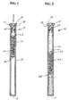
- the centrifugation in one embodiment is carried out using a centrifuge device 10 as shown in Figures 1 and 2.
- Device 10 in the embodiment illustrated includes a container, such as tube 12, that is preferably made of glass or other transparent material, such as plastic.
- Tube 12 has a closed bottom end 14 and an open top end 16.
- a stopper 18 is fitted in open top end 16 to close tube 12.
- stopper 18 is made of a suitable elastomeric or rubber-like material that can be pierced by a cannula, needle or other piercing device.
- Stopper 18 has a substantially cylindrical body portion 20 having an outer dimension to form a snug friction fit in top end 16 of tube 12.
- a shoulder 21 extends radially outward from an upper end of stopper 18 to engage top end 16 of tube 12.
- Tube 12 has a length and diameter suitable for centrifuging a sample fluid.
- tube 12 has a length of about 75 mm, an internal diameter of about 40 mm and a capacity of about 0.9 ml.
- a float 22 is disposed in tube 12 as shown in Figure 1.
- Float 22 is dimensioned to fit snugly in tube 12 and slide along the length of tube 12 during centrifuging of the sample.
- Float 22, in the embodiment illustrated includes an outer sleeve 24 having a cylindrical shape complementing the inner surface of tube 12.
- Outer sleeve 24 is preferably made from a pliable material, such as a vinyl resin, that is able to deform slightly during centrifugation. Outer sleeve 24 can expand and contract in response to the centrifugal force so that float 22 is able to slide within tube 12.
- the pliable material returns to its original shape and dimensions at static conditions so that outer sleeve 24 snugly contacts the inner surface of tube 12 and is able to slide within tube 12 under centrifugal force.
- a silicone lubricant can be applied to the inner surface of tube 12 to assist in the sliding movement of the float 22.
- Float 22 includes an inner sleeve 26 having an axial passage or bore 28 forming a through passage.
- Inner sleeve 26 is coupled to outer sleeve 24 by a bonding agent or other suitable method.
- Inner sleeve 26 is made of a rigid material that is dimensionally stable during centrifugation so substantially no distortion occurs during centrifugation.
- Axial bore 28 has a length and diameter suitable for expanding a fraction of the sample material during centrifugation. In one embodiment of the invention, axial bore 28 has an inner diameter of about 1.265 mm and a length of about 4.0 mm.
- Inner sleeve 26 is preferably made from a plastic such as polystyrene that does not interfere with the components of the sample.
- Float 22 is intended to be exemplary of a suitable centrifuge device capable of separating and expanding a cell fraction. It will be understood that there are other devices that can be used during centrifugation to separate rare cell fractions. Suitable devices typically include a passage or a constricted area for elongating constituent layers during centrifugation to enable separation of the constituent layers.
- Float 22 is dimensioned to fit in tube 12 and slide within the tube 12 while centrifuging to settle between selected density layers of the sample fluid and force the target component into the axial bore 28 of float 22.
- Axial bore 28 of float 22 has an internal volume suitable to collect a substantial portion of the target component. The internal volume and diameter of the float effectively expand the layer of the target component.
- Float 22 is selected to have a density to complement the target component and the sample so that the target component collects in the axial bore 28 by enabling float 22 to settle at a predetermined point in the sample.
- float 22 has a density to settle between the resulting plasma layer and the layer of red blood cells after centrifugation.
- the rare cells that normally collect at the interface between the plasma and red blood cell layers collect in the axial bore 28 where they can be recovered.
- the sample is centrifuged at a rate sufficient to separate the various constituents into layers.
- the centrifuge can be at a speed to produce a centrifugal force of about 400 G to about 800 G depending on the sample fluid. In embodiments, the centrifuge can produce a force of 1,000 G or more.
- the method of the invention can be a positive selection or a negative selection for harvesting components, and particularly rare cells.
- a sample fluid is mixed with at least one antibody having an affinity for the target component.
- a negative selection mixes the sample with an antibody having an affinity for the contaminating cells, such as leukocytes and/or red blood cells.
- Suitable negative selection antibody reagents in the form of specialized conjugates and complexes are available from StemCell Technologies Inc. and Miltenyi BioTech GmbH. Underivatized antibodies are available from other sources such as Pharmagen, Inc.
- the resulting mixture is then centrifuged using the harvesting float device to harvest the enriched target component. Prior to centrifuging, the sample can be combined with a density gradient media as known in the art to enhance the separation of the target component.
- the positive selection harvesting method in one embodiment of the invention utilizes a particulate carrier having a coating of antibody with an affinity toward the target component.
- the particulate carrier is an amount of plastic microbeads with a coating of an antibody with a binding affinity for the rare cells to be harvested, and particularly tumor cells.
- the density of the microbeads and the density of the float are coordinated to collect the microbeads in the axial bore of the float.
- the positive selection harvesting uses microbeads having a particle size of about 0.05 microns to about 7 microns, and typically about 4 to 5 microns, and a density less than white blood cells.
- the microbeads can have a density in the range of about 1.00 to about 1.05, and preferably about 1.02 to about 1.03.
- the microbeads and the captured rare cells have a density so that they are focused in the float when the sample and float are centrifuged.
- the float has an appropriate density so that the float settles in the sample after centrifuging where the rare cells normally settle.
- the microbeads are sufficiently light to float above the centrifuged layers of white and red blood cells.
- the float preferably has a density to float above the white and red blood cell layers to harvest the microbeads.
- the microbeads are made of a suitable material that is non-reactive with the target component and particularly non-reactive with the rare cells. Suitable materials include polyacrylamides, polyurethanes, polysulfones, fluorinated or chlorinated resins, such as polyvinylchloride, polyethylene, polypropylene, polycarbonates and polyesters.
- the particle is typically about 4 to 5 microns, although the particle size can vary depending on the target component and internal diameter of the float.
- a number of commercially available microbeads have an antigen bonded to the surface of the microbeads.
- the antibody can be bonded directly to the surface of the microbead or through an intermediate coupling agent. Suitable antibody coated microbeads are commercially available from Miltenyi Biotec GmbH. An example of a suitable microbead is available from Miltenyi Biotec under the tradename MACS CD 27.
- the antibody in the positive selection method has a binding affinity for the rare cells and is selected according to the target rare cells to be harvested from the sample.
- rare cells to be harvested include tumor cells, fetal cells and the like.
- tumor cells that can be bound to the particulate carrier can be of epithelial origin and can be localized or non-localized.
- the tumor cells can be of the bladder, brain, breast, colon, kidney, liver, lung, ovary, pancreas, prostate, rectum and stomach.
- Tumor cells can also be in the form of sarcoma, such as fibrosarcoma or rhabdosarcoma, hematopoietic tumor of the lymphoid or myeloid lineage, melanoma, teratocarcinoma, neuroblastoma, or glioma.
- sarcoma such as fibrosarcoma or rhabdosarcoma
- hematopoietic tumor of the lymphoid or myeloid lineage melanoma
- teratocarcinoma neuroblastoma
- glioma glioma
- the microbeads preferably have a surface area sufficient to contain an amount of the selected antibody to bind an effective amount of the rare cells being targeted.
- the amount of the microbeads combined with the sample can vary with the affinity of the antibody, concentration of the rare cells in the sample, the nature of the sample, and the volume of the sample.
- the method of the invention is suitable for use in harvesting rare cells from various bodily fluids, and particularly anticoagulated blood.
- Other fluids that can be analyzed for rare cell content include urine, saliva, lymph fluid, spinal fluid, semen, amniotic fluid, cavity fluids and tissue extracts.
- tube 12 is evacuated or filled with an inert gas at a subatmospheric internal pressure.
- the sample to be tested is transferred from a primary collecting tube by a transferring device having a double piercing needle or cannula.
- the needle extends from the transferring device to the tube by piercing the stopper in tube 12.
- the low pressure in tube 12 draws the fluid sample into tube 12.
- a thixotropic gel can be provided in tube 12 as known in the art to preserve band formation in the sample when centrifuged.
- Various other separation agents, dyes and the like can be added to tube 12 to promote separation and identification of components.
- the microbeads containing the antibody are provided in the tube 12 and are mixed with the fluid sample by gentle shaking or stirring. The sample is then incubated to bind the target component to the microbeads.
- the tube, float and sample are centrifuged at a sufficient speed and for a length of time necessary to separate the constituents of the sample into layers and force the microbeads and the trapped target component into the axial bore of the float.
- the sample can be centrifuged at a speed to provide sufficient centrifugation force to cause separation of the layers.
- the tube is slowly stopped and removed from the centrifuge.
- a needle or cannula then pierces the stopper and is inserted into the axial bore to remove the sample containing the microbeads.
- the harvested sample is further processed and analyzed by various processes as known in the art. In one embodiment, the harvested cells are analyzed using a flow cytometer.
- the rare cells or other target components can be washed and separated from the microbeads and the binding antibody by known methods.
- the resulting harvested rare cells are significantly enriched compared to many prior processes and have a substantially lower contaminant level of red and white blood cells.
- the method is a negative selection method for the enrichment of rare cells.
- the rare cells are enriched using a binding agent that is able to bind with the contaminating non-rare cells, such as red blood cells or white blood cells.
- the binding agent is able to bind to one or more white blood cell and/or red blood cell or that bind to surface antigens on the cells.
- the binding agents can be antibodies that are able to agglutinate the white blood cells or bind the white blood cells to red blood cells. The resulting larger and denser particles can be separated from the non--rare cells during centrifugation. Suitable antibodies that are able to bind with and capture the non-rare cells include antihuman antibodies.
- Suitable antibodies that can be used to bind with white blood cells include the leukocyte CD antibodies such as CD2, CD3, CD4, CD5, CD7, CD8, CD11a, CD11b, CD11c, CD14, CD15, CD16, CD19, CD20, CD28, CD36, CD42a, CD43, CD44, CD45, CD45R, CD45RA, CD45RB, CD45RO, CD57 and CD61.
- Other binding agents that can be used include a mixture of antihuman CD45, antihuman CD19, antihuman CD14 and antihuman CD3.
- the antibodies are bound to the surface of the microbeads as in the previous embodiments.
- the microbeads in the negative selection process have a particle size suitable for the sample and the target component. Generally, the particle size ranges from about 0.05 microns to about 7 microns, and preferably about 4 microns to about 5 microns.
- the beads preferably have a density greater than the density of white blood cells, and more preferably of about 1.07 to about 1.09 g/ml, and typically in the range of about 1.08 to about 1.09 g/ml.
- the microbeads sink during centrifugation and the rare cells settle above the red and white blood cell layers.
- the float has density to float on the non-rare cells layers so that the rare cells settle in the axial bore of the float where they can be removed.
- the method of the negative selection harvesting is similar to the positive selection discussed above.
- the sample fluid is provided in the tube and mixed with the microbeads containing the non-rare cell antibodies. After incubating, the tube containing the mixture is incubated and centrifuged for sufficient time to cause the layers to separate and the rare cells to collect in the axial bore of the float where the rare cells can be recovered.
- the method employs two microbeads having different affinity binding agents for capturing two different components.
- an amount of first microbeads having an affinity binding agent with an affinity for rare cells, such as tumor cells are mixed with the sample.
- the first microbeads have a density to separate from the white and red blood cells.
- the first microbeads have a particle size, density and affinity binding agent substantially the same as the microbeads of the positive selection of the previous embodiment.
- An amount of second microbeads having an affinity binding agent with an affinity for white blood cells is also mixed with the sample.
- the second microbeads have a density to separate the white blood cells, red blood cells, or a combination thereof from the rare cells.
- the resulting mixture is centrifuged with the float so that the first microbeads with the captured rare cells settle in the axial passage where they can be recovered.
- the second microbeads have a particle size, density and affinity binding agent substantially the same as the negative selection method of the previous embodiment. In this manner, the second microbeads separate from the first microbeads during centrifugation to separate the contaminating cells, such as the red and/or white blood cells from the target cells and the first microbeads.
- the first microbeads have a particle size and density to be collected in the axial passage of the float for recovering the target cells.
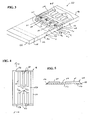
- Device 30 is particularly suitable for various cell manipulations after separation from a sample. For example, rare cells can be separated from a sample and subjected to various detection and assay processes with device 30.
- device 30 includes a hollow container 32 having a substantially rectangular shape.
- Container 32 includes a front wall 34, and an opposite rear wall 36 having a longitudinal length and a width.
- Opposite side walls 38 and a bottom wall 40 extend between front wall 34 and rear wall 36 to form an open cavity 42.
- Container 32 includes an open end 44 to receive a stopper 46 for closing cavity 42.
- container 32 is made of a transparent material such as glass or plastic.
- Container 32 is dimensioned to receive a volume of a biological sample suitable for analysis of a target component.
- Container 32 generally has a volume of about 8 ml to about 10 ml, and preferably about 9 ml.
- side walls 38 of container 32 have a dimension to define a thickness of cavity 42 that is sufficiently thin to visualize, detect and analyze a target component through front wall 34. Examples of suitable detection and analysis methods include microscopy to visualize cells in the sample.
- Container 32 is typically about 7 cm to about 8 cm in length, and about 3 cm to about 4 cm in width.
- Side walls 38 are dimensioned so that cavity 42 has a thickness of about 3 mm to about 6 mm, and preferably about 4 mm.
- Container 32 includes a movable float 48 that is able to slide within container 32 in the longitudinal dimension in a manner similar to the previous embodiment.
- Float 48 is dimensioned to fit within cavity 42 of container 32 and has an outer dimension corresponding to the inner dimension of container 32.
- float 48 has a base 50 with a substantially flat bottom surface 54.
- Base 50 includes an inclined leading end 56 and an inclined trailing end 58.
- a plurality of ribs 60 are coupled to top surface 54 of base 50.
- ribs 60 extend in a longitudinal direction with respect to the longitudinal dimension of base 50. Ribs 60 are aligned in pairs to form channels 62 extending the length of base 50 between adjacent ribs. Ribs 60 have a height to fit closely against the inner surface of container 32. Channels 62 are dimensioned to separate and elongate the layers during centrifugation.
- a biological sample such as a blood sample
- container 32 An amount of microbeads 64 having an affinity binding agent for a target component is mixed with the sample.
- Container 32 is then centrifuged as in the previous embodiment to collect the microbeads 64 with the captured target component in longitudinal channels 62 of float 48.
- Microbeads 52 and the captured target component can then be analyzed by visualizing the target component within container 32 by microscopy methods as known in the art.
- the blood sample after centrifuging separates into a layer of red blood cells 66, a granulocyte cell fraction layer 68, a mononuclear cell fraction 70, a plasma fraction 72 and a platelet/plasma interface 74.
- microbeads 64 have an affinity for the target compound and collect in channels 62.
- microbeads 64 can have an affinity for white and/or red blood cells in a negative selection process.
- Channels 62 are formed between ribs 60 and are enclosed by top wall 34 of container 32. Inclined leading edge 56 and inclined trailing edge 58 divert the sample through channels 62 as float 48 slides through container 32.
- Microbeads 64 are retained in a thin layer in channels 62 close to top wall 34 of container 32 so that the microbeads 64 can be visualized through top wall 34 by microscopy or other analytical methods as known in the art.
- front wall 34 of container 32 is substantially flat to prevent the optical distortion normally associated with cylindrical containers.
- This example compares the harvested tumor cells from a sample with and without a negative selection.
- the harvester separations were compared for 6.0 mls of freshly collected whole blood that were spiked with 0, 50 and 500 cultured prostate tumor PC-3 cells.
- An antibody cocktail obtained from StemCell Technologies, Inc. under the tradename RosetteSep was mixed with each of the blood samples and incubated for 20 minutes at room temperature.
- a control blood sample was prepared without the antibody treatment.
- the antibody cocktail provided a negative selection to remove the unwanted white blood cells.
- the blood samples were layered on top of a density media in a 16 x 100 PET Vacutainer tube obtained from Becton Dickinson containing a harvester float and 2 mls of POLYMORPHPREPTM density media.
- the samples were centrifuged in a swinging bucket centrifuge for 30 minutes at 20°C at a rate of about 650 g.
- the control sample showed the presence of white blood cells with the tumor cells in the harvester float.
- the antibody treatment demonstrated tumor cells collected in the harvester float with a greatly reduced white cell population. The tumor cells were removed from the harvester float. Flow cytometry demonstrated recovery of about 90% of the tumor cells.
Landscapes
- Health & Medical Sciences (AREA)
- Chemical & Material Sciences (AREA)
- Analytical Chemistry (AREA)
- General Health & Medical Sciences (AREA)
- Immunology (AREA)
- Biochemistry (AREA)
- Life Sciences & Earth Sciences (AREA)
- General Physics & Mathematics (AREA)
- Physics & Mathematics (AREA)
- Pathology (AREA)
- Hematology (AREA)
- Clinical Laboratory Science (AREA)
- Chemical Kinetics & Catalysis (AREA)
- Investigating Or Analysing Biological Materials (AREA)
- Apparatus Associated With Microorganisms And Enzymes (AREA)
- Micro-Organisms Or Cultivation Processes Thereof (AREA)
- Sampling And Sample Adjustment (AREA)
Applications Claiming Priority (2)
| Application Number | Priority Date | Filing Date | Title |
|---|---|---|---|
| US756590 | 2001-01-08 | ||
| US09/756,590 US7205157B2 (en) | 2001-01-08 | 2001-01-08 | Method of separating cells from a sample |
Publications (3)
| Publication Number | Publication Date |
|---|---|
| EP1221342A2 true EP1221342A2 (de) | 2002-07-10 |
| EP1221342A3 EP1221342A3 (de) | 2003-05-28 |
| EP1221342B1 EP1221342B1 (de) | 2016-05-25 |
Family
ID=25044157
Family Applications (1)
| Application Number | Title | Priority Date | Filing Date |
|---|---|---|---|
| EP01129402.2A Expired - Lifetime EP1221342B1 (de) | 2001-01-08 | 2001-12-10 | Verfahren zur trennung von zellen von einer probe |
Country Status (6)
| Country | Link |
|---|---|
| US (2) | US7205157B2 (de) |
| EP (1) | EP1221342B1 (de) |
| JP (2) | JP4290916B2 (de) |
| CA (1) | CA2365925C (de) |
| ES (1) | ES2587997T3 (de) |
| MX (1) | MXPA01013107A (de) |
Cited By (23)
| Publication number | Priority date | Publication date | Assignee | Title |
|---|---|---|---|---|
| US7276170B2 (en) | 2002-02-04 | 2007-10-02 | Colorado School Of Mines | Laminar flow-based separations of colloidal and cellular particles |
| GB2445073A (en) * | 2006-12-18 | 2008-06-25 | Ge Healthcare Bio Sciences Ab | Cell separation device |
| USRE41762E1 (en) | 2001-02-14 | 2010-09-28 | Stc.Unm | Nanostructured separation and analysis devices for biological membranes |
| CN101939645A (zh) * | 2007-12-05 | 2011-01-05 | 齐翁米克斯股份有限公司 | 细胞测定套件和方法 |
| US8119976B2 (en) | 2007-07-03 | 2012-02-21 | Colorado School Of Mines | Optical-based cell deformability |
| US8394342B2 (en) | 2008-07-21 | 2013-03-12 | Becton, Dickinson And Company | Density phase separation device |
| WO2013090189A1 (en) * | 2011-12-12 | 2013-06-20 | Rarecyte, Inc. | Tube and float systems and methods of using the same |
| US8585971B2 (en) | 2005-04-05 | 2013-11-19 | The General Hospital Corporation | Devices and method for enrichment and alteration of cells and other particles |
| US8747781B2 (en) | 2008-07-21 | 2014-06-10 | Becton, Dickinson And Company | Density phase separation device |
| US8794452B2 (en) | 2009-05-15 | 2014-08-05 | Becton, Dickinson And Company | Density phase separation device |
| US8895298B2 (en) | 2002-09-27 | 2014-11-25 | The General Hospital Corporation | Microfluidic device for cell separation and uses thereof |
| US8921102B2 (en) | 2005-07-29 | 2014-12-30 | Gpb Scientific, Llc | Devices and methods for enrichment and alteration of circulating tumor cells and other particles |
| US9017942B2 (en) | 2006-06-14 | 2015-04-28 | The General Hospital Corporation | Rare cell analysis using sample splitting and DNA tags |
| US9333445B2 (en) | 2008-07-21 | 2016-05-10 | Becton, Dickinson And Company | Density phase separation device |
| EP2397225B1 (de) * | 2010-06-18 | 2017-06-14 | F. Hoffmann-La Roche AG | Gravitationssensitive kennzeichnung und korrespondierendes probenbehältnis, verfahren und analytisches system |
| US9694359B2 (en) | 2014-11-13 | 2017-07-04 | Becton, Dickinson And Company | Mechanical separator for a biological fluid |
| US9878326B2 (en) | 2007-09-26 | 2018-01-30 | Colorado School Of Mines | Fiber-focused diode-bar optical trapping for microfluidic manipulation |
| US9885644B2 (en) | 2006-01-10 | 2018-02-06 | Colorado School Of Mines | Dynamic viscoelasticity as a rapid single-cell biomarker |
| US10557786B2 (en) | 2011-01-21 | 2020-02-11 | Theranos Ip Company, Llc | Systems and methods for sample use maximization |
| EP3440193A4 (de) * | 2016-04-07 | 2020-03-04 | Mesotex, Inc. | Verfahren zur isolierung von nukleierten zellen und von populationen von nukleierten zellen und verwendungen davon |
| US10634667B2 (en) | 2007-10-02 | 2020-04-28 | Theranos Ip Company, Llc | Modular point-of-care devices, systems, and uses thereof |
| US10722250B2 (en) | 2007-09-04 | 2020-07-28 | Colorado School Of Mines | Magnetic-field driven colloidal microbots, methods for forming and using the same |
| US11378498B2 (en) | 2006-06-14 | 2022-07-05 | Verinata Health, Inc. | Diagnosis of fetal abnormalities using polymorphisms including short tandem repeats |
Families Citing this family (43)
| Publication number | Priority date | Publication date | Assignee | Title |
|---|---|---|---|---|
| US7947236B2 (en) | 1999-12-03 | 2011-05-24 | Becton, Dickinson And Company | Device for separating components of a fluid sample |
| US20050266433A1 (en) * | 2004-03-03 | 2005-12-01 | Ravi Kapur | Magnetic device for isolation of cells and biomolecules in a microfluidic environment |
| US9487812B2 (en) | 2012-02-17 | 2016-11-08 | Colorado School Of Mines | Optical alignment deformation spectroscopy |
| US20090062828A1 (en) * | 2007-09-04 | 2009-03-05 | Colorado School Of Mines | Magnetic field-based colloidal atherectomy |
| EP2203746B1 (de) * | 2007-09-24 | 2013-03-06 | Technion Research & Development Foundation Ltd. | T-zellen-subpopulationen mit fähigkeit zur krebsbehandlung |
| WO2011050366A2 (en) * | 2009-10-23 | 2011-04-28 | Rarecyte, Inc. | Methods for changing densities on non-target particles of a suspension |
| CA2819260A1 (en) | 2010-12-15 | 2012-06-21 | Cytosed, Inc. | Antibody-linked immuno-sedimentation agent and method of isolating a target from a sample using same |
| US20120308447A1 (en) * | 2011-05-31 | 2012-12-06 | Timothy Alan Abrahamson | Tube and float systems for density-based fluid separation |
| US20150056649A1 (en) * | 2012-01-13 | 2015-02-26 | Konica Minolta, Inc. | Method for quantifying cell of interest in blood, and method for evaluating system for quantifying said cell |
| US9211512B2 (en) * | 2012-11-28 | 2015-12-15 | Samsung Electronics Co., Ltd. | Microfluidic apparatus and method of enriching target cells by using the same |
| US9956555B2 (en) | 2012-11-30 | 2018-05-01 | Rarecyte, Inc. | Apparatus, system, and method for collecting a target material |
| US9539570B2 (en) | 2012-11-30 | 2017-01-10 | Rarecyte, Inc. | Apparatus, system, and method for collecting a target material |
| US9945839B2 (en) | 2012-11-30 | 2018-04-17 | Rarecyte, Inc. | Apparatus, system, and method for collecting a target material |
| US9039999B2 (en) | 2012-11-30 | 2015-05-26 | Rarecyte, Inc. | Apparatus, system, and method for collecting a target material |
| US9533303B2 (en) | 2012-11-30 | 2017-01-03 | Rarecyte, Inc. | Apparatus, system, and method for collecting a target material |
| US10054524B2 (en) | 2012-11-30 | 2018-08-21 | Rarecyte, Inc. | Apparatus, system and method for collecting a target material |
| US9513291B2 (en) | 2012-11-30 | 2016-12-06 | Rarecyte, Inc. | Apparatus, system, and method for collecting a target material |
| US9417174B2 (en) | 2013-02-01 | 2016-08-16 | Rarecyte, Inc. | Tube and float system and methods of using the same |
| WO2014120678A2 (en) * | 2013-02-01 | 2014-08-07 | Rarecyte, Inc. | Tube and float systems and methods of using the same |
| US11493428B2 (en) | 2013-03-15 | 2022-11-08 | Gpb Scientific, Inc. | On-chip microfluidic processing of particles |
| US12590955B2 (en) | 2013-03-15 | 2026-03-31 | Zeon Corporation | Methods and systems for processing particles |
| EP3608022A1 (de) | 2013-03-15 | 2020-02-12 | The Trustees of Princeton University | Verfahren und vorrichtungen für reinigung mit hohem durchsatz |
| US20150064153A1 (en) | 2013-03-15 | 2015-03-05 | The Trustees Of Princeton University | High efficiency microfluidic purification of stem cells to improve transplants |
| KR101473557B1 (ko) * | 2013-03-29 | 2014-12-24 | 포항공과대학교 산학협력단 | 원심력을 이용한 세포유래 인공 마이크로베시클 제조 장치 |
| US20140309553A1 (en) * | 2013-04-11 | 2014-10-16 | Rarecyte, Inc. | Kits and methods for separating a target analyte from a suspension |
| US9937445B2 (en) * | 2014-03-27 | 2018-04-10 | Biomet Biologics, Llc | System and method for separating a fraction |
| NO337444B1 (no) * | 2014-06-19 | 2016-04-11 | Spinchip Diagnostics As | Analysemetode |
| US9885699B2 (en) * | 2014-07-31 | 2018-02-06 | Becton, Dickinson And Company | Methods for processing whole blood samples, and compositions for use in practicing the same |
| US11291931B2 (en) | 2014-12-15 | 2022-04-05 | Akadeum Life Sciences, Inc. | Method and system for buoyant separation |
| GB201502374D0 (en) | 2015-02-13 | 2015-04-01 | Prokyma Technologies Ltd | Method and apparatus relating to treatment of a blood sample for sequencing of circulating tumour cells |
| US10976232B2 (en) | 2015-08-24 | 2021-04-13 | Gpb Scientific, Inc. | Methods and devices for multi-step cell purification and concentration |
| WO2017065820A1 (en) * | 2015-10-14 | 2017-04-20 | Rarecyte, Inc. | Apparatus, system, and method for collecting a target material |
| CN105316277A (zh) * | 2015-10-22 | 2016-02-10 | 深圳华毓造血干细胞研究有限公司 | 贴壁性细胞的三维培养方法 |
| WO2019183334A1 (en) | 2018-03-21 | 2019-09-26 | Waters Technologies Corporation | Non-antibody high-affinity-based sample preparation, sorbents, devices and methods |
| EP3820588A4 (de) | 2018-07-09 | 2022-04-06 | Akadeum Life Sciences, Inc. | System und verfahren zur verarbeitung schwimmfähiger partikel |
| JP2021531842A (ja) * | 2018-07-09 | 2021-11-25 | ハヌマン ペリカン,インコーポレイテッド | 血液成分を分離するための装置および方法 |
| KR20220050150A (ko) * | 2019-08-13 | 2022-04-22 | 드롭웍스 인코포레이티드 | 샘플 여과를 위한 방법 및 조성물 |
| KR102298909B1 (ko) * | 2019-10-25 | 2021-09-06 | 주식회사 싸이토딕스 | 역류 원심분리를 이용한 ctc 분리방법 |
| KR102298910B1 (ko) * | 2019-10-25 | 2021-09-06 | 주식회사 싸이토딕스 | 역류 원심분리를 이용한 ctc 분리방법 |
| WO2023028329A1 (en) | 2021-08-26 | 2023-03-02 | Akadeum Life Sciences, Inc. | Method and system for buoyant separation |
| CN114636606A (zh) * | 2022-03-29 | 2022-06-17 | 中元汇吉生物技术股份有限公司 | 血样处理方法 |
| US12196754B2 (en) | 2022-04-01 | 2025-01-14 | Akadeum Life Sciences, Inc. | Method and system for buoyant-particle-assisted cell therapy |
| CN121511103A (zh) | 2023-02-14 | 2026-02-10 | 爱卡德姆生命科学公司 | 用于部分或完全自动化的浮力辅助分离的方法和系统 |
Family Cites Families (23)
| Publication number | Priority date | Publication date | Assignee | Title |
|---|---|---|---|---|
| US4027660A (en) * | 1976-04-02 | 1977-06-07 | Wardlaw Stephen C | Material layer volume determination |
| US4137755A (en) * | 1976-09-20 | 1979-02-06 | Wardlaw Stephen C | Material layer volume determination |
| DE2800934C2 (de) * | 1977-01-10 | 1986-09-18 | Robert Aaron Guilford Conn. Levine | Verfahren zur Durchführung von Volumenmessungen an der Zwischenschicht zwischen der Erythrozytenschicht und der Plasmaschicht einer zentrifugierten Blutprobe |
| US4567754A (en) * | 1985-03-29 | 1986-02-04 | Wardlaw Stephen C | Measurement of small heavy constituent layer in stratified mixture |
| US4927749A (en) * | 1986-04-09 | 1990-05-22 | Jeanette Simpson | Reagent for cell separation |
| JPH0774772B2 (ja) * | 1990-12-31 | 1995-08-09 | エイ. レビン ロバート | 血液サンプリング組立体、ターゲット細胞の採取方法およびターゲット成分の採取方法 |
| US5342790A (en) * | 1992-10-30 | 1994-08-30 | Becton Dickinson And Company | Apparatus for indirect fluorescent assay of blood samples |
| SE512416C2 (sv) * | 1993-10-12 | 2000-03-13 | Cma Microdialysis Ab | Sätt för uppsamling av små vätskeprovmängder, samt provbehållare för upptagning av små vätskemängder |
| US5840502A (en) * | 1994-08-31 | 1998-11-24 | Activated Cell Therapy, Inc. | Methods for enriching specific cell-types by density gradient centrifugation |
| US5646004A (en) * | 1994-08-31 | 1997-07-08 | Activated Cell Therapy, Inc. | Methods for enriching fetal cells from maternal body fluids |
| US5474687A (en) * | 1994-08-31 | 1995-12-12 | Activated Cell Therapy, Inc. | Methods for enriching CD34+ human hematopoietic progenitor cells |
| US5877299A (en) * | 1995-06-16 | 1999-03-02 | Stemcell Technologies Inc. | Methods for preparing enriched human hematopoietic cell preparations |
| US5866071A (en) * | 1996-03-06 | 1999-02-02 | National Science Council | Centrifuge tube with a built-in small tubing for separation following density gradients centrifugation |
| US5714125A (en) * | 1996-03-07 | 1998-02-03 | Medical Safety Products, Inc. | Device for collecting a blood sample from a plastic segment tube |
| US5707876A (en) * | 1996-03-25 | 1998-01-13 | Stephen C. Wardlaw | Method and apparatus for harvesting constituent layers from a centrifuged material mixture |
| CA2279474C (en) * | 1998-07-31 | 2011-01-04 | Stemcell Technologies Inc. | Novel antibody composition for debulking blood and bone marrow samples from cml patients |
| US6342344B1 (en) * | 1998-07-31 | 2002-01-29 | Stemcell Technologies Inc. | Antibody composition for isolating human cells from human-murine chimeric hematopoietic cell suspensions |
| US6153113A (en) * | 1999-02-22 | 2000-11-28 | Cobe Laboratories, Inc. | Method for using ligands in particle separation |
| US7135335B2 (en) * | 1999-05-28 | 2006-11-14 | Stemcell Technologies Inc. | Method for separating cells using immunorosettes |
| US6750326B2 (en) * | 1999-05-28 | 2004-06-15 | Stemcell Technologies Inc. | Method for separating cells using immunorosettes |
| CA2375115C (en) * | 1999-05-28 | 2011-05-24 | Stemcell Technologies Inc. | Method for separating cells using immunorosettes |
| CA2346362C (en) * | 2000-05-26 | 2010-06-15 | Stemcell Technologies Inc. | Novel antibody compositions for preparing enriched mesenchymal progenitor preparations |
| US20020164825A1 (en) * | 2000-09-09 | 2002-11-07 | Wen-Tien Chen | Cell separation matrix |
-
2001
- 2001-01-08 US US09/756,590 patent/US7205157B2/en not_active Expired - Lifetime
- 2001-12-10 ES ES01129402.2T patent/ES2587997T3/es not_active Expired - Lifetime
- 2001-12-10 EP EP01129402.2A patent/EP1221342B1/de not_active Expired - Lifetime
- 2001-12-17 CA CA2365925A patent/CA2365925C/en not_active Expired - Lifetime
- 2001-12-18 MX MXPA01013107A patent/MXPA01013107A/es unknown
-
2002
- 2002-01-08 JP JP2002001780A patent/JP4290916B2/ja not_active Expired - Lifetime
-
2007
- 2007-03-12 US US11/684,693 patent/US7524641B2/en not_active Expired - Lifetime
-
2008
- 2008-09-22 JP JP2008242860A patent/JP2008301835A/ja active Pending
Cited By (71)
| Publication number | Priority date | Publication date | Assignee | Title |
|---|---|---|---|---|
| USRE41762E1 (en) | 2001-02-14 | 2010-09-28 | Stc.Unm | Nanostructured separation and analysis devices for biological membranes |
| USRE42249E1 (en) | 2001-02-14 | 2011-03-29 | Stc.Unm | Nanostructured separation and analysis devices for biological membranes |
| USRE42315E1 (en) | 2001-02-14 | 2011-05-03 | Stc.Unm | Nanostructured separation and analysis devices for biological membranes |
| US7318902B2 (en) | 2002-02-04 | 2008-01-15 | Colorado School Of Mines | Laminar flow-based separations of colloidal and cellular particles |
| US7472794B2 (en) | 2002-02-04 | 2009-01-06 | Colorado School Of Mines | Cell sorting device and method of manufacturing the same |
| US7276170B2 (en) | 2002-02-04 | 2007-10-02 | Colorado School Of Mines | Laminar flow-based separations of colloidal and cellular particles |
| US11052392B2 (en) | 2002-09-27 | 2021-07-06 | The General Hospital Corporation | Microfluidic device for cell separation and uses thereof |
| US10081014B2 (en) | 2002-09-27 | 2018-09-25 | The General Hospital Corporation | Microfluidic device for cell separation and uses thereof |
| US8895298B2 (en) | 2002-09-27 | 2014-11-25 | The General Hospital Corporation | Microfluidic device for cell separation and uses thereof |
| US12409457B2 (en) | 2005-04-05 | 2025-09-09 | The General Hospital Corporation | Devices and method for enrichment and alteration of cells and other particles |
| US10786817B2 (en) | 2005-04-05 | 2020-09-29 | The General Hospital Corporation | Devices and method for enrichment and alteration of cells and other particles |
| US8585971B2 (en) | 2005-04-05 | 2013-11-19 | The General Hospital Corporation | Devices and method for enrichment and alteration of cells and other particles |
| US9956562B2 (en) | 2005-04-05 | 2018-05-01 | The General Hospital Corporation | Devices and method for enrichment and alteration of cells and other particles |
| US8921102B2 (en) | 2005-07-29 | 2014-12-30 | Gpb Scientific, Llc | Devices and methods for enrichment and alteration of circulating tumor cells and other particles |
| US9885644B2 (en) | 2006-01-10 | 2018-02-06 | Colorado School Of Mines | Dynamic viscoelasticity as a rapid single-cell biomarker |
| US9347100B2 (en) | 2006-06-14 | 2016-05-24 | Gpb Scientific, Llc | Rare cell analysis using sample splitting and DNA tags |
| US11378498B2 (en) | 2006-06-14 | 2022-07-05 | Verinata Health, Inc. | Diagnosis of fetal abnormalities using polymorphisms including short tandem repeats |
| US10155984B2 (en) | 2006-06-14 | 2018-12-18 | The General Hospital Corporation | Rare cell analysis using sample splitting and DNA tags |
| US9017942B2 (en) | 2006-06-14 | 2015-04-28 | The General Hospital Corporation | Rare cell analysis using sample splitting and DNA tags |
| US11781187B2 (en) | 2006-06-14 | 2023-10-10 | The General Hospital Corporation | Rare cell analysis using sample splitting and DNA tags |
| US9273355B2 (en) | 2006-06-14 | 2016-03-01 | The General Hospital Corporation | Rare cell analysis using sample splitting and DNA tags |
| GB2445073A (en) * | 2006-12-18 | 2008-06-25 | Ge Healthcare Bio Sciences Ab | Cell separation device |
| US8119976B2 (en) | 2007-07-03 | 2012-02-21 | Colorado School Of Mines | Optical-based cell deformability |
| US10722250B2 (en) | 2007-09-04 | 2020-07-28 | Colorado School Of Mines | Magnetic-field driven colloidal microbots, methods for forming and using the same |
| US9878326B2 (en) | 2007-09-26 | 2018-01-30 | Colorado School Of Mines | Fiber-focused diode-bar optical trapping for microfluidic manipulation |
| US11899010B2 (en) | 2007-10-02 | 2024-02-13 | Labrador Diagnostics Llc | Modular point-of-care devices, systems, and uses thereof |
| US10670588B2 (en) | 2007-10-02 | 2020-06-02 | Theranos Ip Company, Llc | Modular point-of-care devices, systems, and uses thereof |
| US11137391B2 (en) | 2007-10-02 | 2021-10-05 | Labrador Diagnostics Llc | Modular point-of-care devices, systems, and uses thereof |
| US10634667B2 (en) | 2007-10-02 | 2020-04-28 | Theranos Ip Company, Llc | Modular point-of-care devices, systems, and uses thereof |
| US10900958B2 (en) | 2007-10-02 | 2021-01-26 | Labrador Diagnostics Llc | Modular point-of-care devices, systems, and uses thereof |
| US11366106B2 (en) | 2007-10-02 | 2022-06-21 | Labrador Diagnostics Llc | Modular point-of-care devices, systems, and uses thereof |
| US11199538B2 (en) | 2007-10-02 | 2021-12-14 | Labrador Diagnostics Llc | Modular point-of-care devices, systems, and uses thereof |
| US11061022B2 (en) | 2007-10-02 | 2021-07-13 | Labrador Diagnostics Llc | Modular point-of-care devices, systems, and uses thereof |
| US11092593B2 (en) | 2007-10-02 | 2021-08-17 | Labrador Diagnostics Llc | Modular point-of-care devices, systems, and uses thereof |
| US11143647B2 (en) | 2007-10-02 | 2021-10-12 | Labrador Diagnostics, LLC | Modular point-of-care devices, systems, and uses thereof |
| CN101939645B (zh) * | 2007-12-05 | 2014-07-30 | 齐翁米克斯股份有限公司 | 细胞测定套件和方法 |
| CN101939645A (zh) * | 2007-12-05 | 2011-01-05 | 齐翁米克斯股份有限公司 | 细胞测定套件和方法 |
| US9700886B2 (en) | 2008-07-21 | 2017-07-11 | Becton, Dickinson And Company | Density phase separation device |
| US9933344B2 (en) | 2008-07-21 | 2018-04-03 | Becton, Dickinson And Company | Density phase separation device |
| US9339741B2 (en) | 2008-07-21 | 2016-05-17 | Becton, Dickinson And Company | Density phase separation device |
| US9333445B2 (en) | 2008-07-21 | 2016-05-10 | Becton, Dickinson And Company | Density phase separation device |
| US8747781B2 (en) | 2008-07-21 | 2014-06-10 | Becton, Dickinson And Company | Density phase separation device |
| US9714890B2 (en) | 2008-07-21 | 2017-07-25 | Becton, Dickinson And Company | Density phase separation device |
| US10350591B2 (en) | 2008-07-21 | 2019-07-16 | Becton, Dickinson And Company | Density phase separation device |
| US8394342B2 (en) | 2008-07-21 | 2013-03-12 | Becton, Dickinson And Company | Density phase separation device |
| US9452427B2 (en) | 2008-07-21 | 2016-09-27 | Becton, Dickinson And Company | Density phase separation device |
| US9919308B2 (en) | 2009-05-15 | 2018-03-20 | Becton, Dickinson And Company | Density phase separation device |
| US9919307B2 (en) | 2009-05-15 | 2018-03-20 | Becton, Dickinson And Company | Density phase separation device |
| US9364828B2 (en) | 2009-05-15 | 2016-06-14 | Becton, Dickinson And Company | Density phase separation device |
| US10456782B2 (en) | 2009-05-15 | 2019-10-29 | Becton, Dickinson And Company | Density phase separation device |
| US10413898B2 (en) | 2009-05-15 | 2019-09-17 | Becton, Dickinson And Company | Density phase separation device |
| US8794452B2 (en) | 2009-05-15 | 2014-08-05 | Becton, Dickinson And Company | Density phase separation device |
| US10376879B2 (en) | 2009-05-15 | 2019-08-13 | Becton, Dickinson And Company | Density phase separation device |
| US10807088B2 (en) | 2009-05-15 | 2020-10-20 | Becton, Dickinson And Company | Density phase separation device |
| US12090476B2 (en) | 2009-05-15 | 2024-09-17 | Becton, Dickinson And Company | Density phase separation device |
| US10343157B2 (en) | 2009-05-15 | 2019-07-09 | Becton, Dickinson And Company | Density phase separation device |
| US8998000B2 (en) | 2009-05-15 | 2015-04-07 | Becton, Dickinson And Company | Density phase separation device |
| US9079123B2 (en) | 2009-05-15 | 2015-07-14 | Becton, Dickinson And Company | Density phase separation device |
| EP2921233A1 (de) * | 2009-05-15 | 2015-09-23 | Becton Dickinson and Company | Dichtephasentrennvorrichtung |
| US11786895B2 (en) | 2009-05-15 | 2023-10-17 | Becton, Dickinson And Company | Density phase separation device |
| US9919309B2 (en) | 2009-05-15 | 2018-03-20 | Becton, Dickinson And Company | Density phase separation device |
| US9802189B2 (en) | 2009-05-15 | 2017-10-31 | Becton, Dickinson And Company | Density phase separation device |
| US9731290B2 (en) | 2009-05-15 | 2017-08-15 | Becton, Dickinson And Company | Density phase separation device |
| US11351535B2 (en) | 2009-05-15 | 2022-06-07 | Becton, Dickinson And Company | Density phase separation device |
| EP2397225B1 (de) * | 2010-06-18 | 2017-06-14 | F. Hoffmann-La Roche AG | Gravitationssensitive kennzeichnung und korrespondierendes probenbehältnis, verfahren und analytisches system |
| US11199489B2 (en) | 2011-01-20 | 2021-12-14 | Labrador Diagnostics Llc | Systems and methods for sample use maximization |
| US10557786B2 (en) | 2011-01-21 | 2020-02-11 | Theranos Ip Company, Llc | Systems and methods for sample use maximization |
| US10876956B2 (en) | 2011-01-21 | 2020-12-29 | Labrador Diagnostics Llc | Systems and methods for sample use maximization |
| WO2013090189A1 (en) * | 2011-12-12 | 2013-06-20 | Rarecyte, Inc. | Tube and float systems and methods of using the same |
| US9694359B2 (en) | 2014-11-13 | 2017-07-04 | Becton, Dickinson And Company | Mechanical separator for a biological fluid |
| EP3440193A4 (de) * | 2016-04-07 | 2020-03-04 | Mesotex, Inc. | Verfahren zur isolierung von nukleierten zellen und von populationen von nukleierten zellen und verwendungen davon |
Also Published As
| Publication number | Publication date |
|---|---|
| CA2365925C (en) | 2013-07-23 |
| US20020090741A1 (en) | 2002-07-11 |
| EP1221342B1 (de) | 2016-05-25 |
| MXPA01013107A (es) | 2004-05-21 |
| US7524641B2 (en) | 2009-04-28 |
| CA2365925A1 (en) | 2002-07-08 |
| US7205157B2 (en) | 2007-04-17 |
| US20070190584A1 (en) | 2007-08-16 |
| EP1221342A3 (de) | 2003-05-28 |
| JP4290916B2 (ja) | 2009-07-08 |
| JP2002253222A (ja) | 2002-09-10 |
| ES2587997T3 (es) | 2016-10-28 |
| JP2008301835A (ja) | 2008-12-18 |
Similar Documents
| Publication | Publication Date | Title |
|---|---|---|
| CA2365925C (en) | Method of separating cells from a sample | |
| US5646004A (en) | Methods for enriching fetal cells from maternal body fluids | |
| CA2515576C (en) | Devices for component removal during blood collection, and uses thereof | |
| US6291249B1 (en) | Method using an apparatus for separation of biological fluids | |
| JP3397795B2 (ja) | 細胞分離装置および方法 | |
| US4861477A (en) | Tubular container for centrifugal separation | |
| US9095798B2 (en) | Centrifuge separation method and apparatus using a medium density fluid | |
| US9101926B2 (en) | Method for separating a sample into density specific fractions | |
| US10054524B2 (en) | Apparatus, system and method for collecting a target material | |
| US9956555B2 (en) | Apparatus, system, and method for collecting a target material | |
| KR101533230B1 (ko) | 다단 미세유체 칩 및 이를 이용한 시료의 선택적 분리방법 | |
| US20150192506A1 (en) | Apparatus, system, and method for collecting a target material | |
| EP0553554B1 (de) | Verfahren und Vorrichtung zur Zentrifugaltrennung aufgrund des Dichtegradienten | |
| US9541481B2 (en) | Apparatus, system, and method for collecting a target material | |
| WO2005049168A2 (en) | Method and apparatus for pre-enrichment and recovery of cells from densified whole blood | |
| JPH11502106A (ja) | 少数細胞集団を濃縮する方法 | |
| WO2017209780A1 (en) | Apparatus, system, and method for collecting target material | |
| CN111139175B (zh) | 用于收集靶材料的仪器、系统和方法 | |
| WO2017065820A1 (en) | Apparatus, system, and method for collecting a target material |
Legal Events
| Date | Code | Title | Description |
|---|---|---|---|
| PUAI | Public reference made under article 153(3) epc to a published international application that has entered the european phase |
Free format text: ORIGINAL CODE: 0009012 |
|
| AK | Designated contracting states |
Kind code of ref document: A2 Designated state(s): AT BE CH CY DE DK ES FI FR GB GR IE IT LI LU MC NL PT SE TR |
|
| AX | Request for extension of the european patent |
Free format text: AL;LT;LV;MK;RO;SI |
|
| PUAL | Search report despatched |
Free format text: ORIGINAL CODE: 0009013 |
|
| AK | Designated contracting states |
Designated state(s): AT BE CH CY DE DK ES FI FR GB GR IE IT LI LU MC NL PT SE TR |
|
| AX | Request for extension of the european patent |
Extension state: AL LT LV MK RO SI |
|
| RIC1 | Information provided on ipc code assigned before grant |
Ipc: 7G 01N 33/49 B Ipc: 7G 01N 1/28 B Ipc: 7G 01N 33/569 B Ipc: 7B 01L 3/14 A |
|
| 17P | Request for examination filed |
Effective date: 20030701 |
|
| AKX | Designation fees paid |
Designated state(s): AT BE CH CY DE DK ES FI FR GB GR IE IT LI LU MC NL PT SE TR |
|
| 17Q | First examination report despatched |
Effective date: 20050621 |
|
| GRAP | Despatch of communication of intention to grant a patent |
Free format text: ORIGINAL CODE: EPIDOSNIGR1 |
|
| INTG | Intention to grant announced |
Effective date: 20151204 |
|
| GRAS | Grant fee paid |
Free format text: ORIGINAL CODE: EPIDOSNIGR3 |
|
| GRAA | (expected) grant |
Free format text: ORIGINAL CODE: 0009210 |
|
| AK | Designated contracting states |
Kind code of ref document: B1 Designated state(s): AT BE CH CY DE DK ES FI FR GB GR IE IT LI LU MC NL PT SE TR |
|
| REG | Reference to a national code |
Ref country code: GB Ref legal event code: FG4D |
|
| REG | Reference to a national code |
Ref country code: CH Ref legal event code: EP |
|
| REG | Reference to a national code |
Ref country code: IE Ref legal event code: FG4D Ref country code: AT Ref legal event code: REF Ref document number: 801805 Country of ref document: AT Kind code of ref document: T Effective date: 20160615 |
|
| REG | Reference to a national code |
Ref country code: DE Ref legal event code: R096 Ref document number: 60149956 Country of ref document: DE |
|
| REG | Reference to a national code |
Ref country code: NL Ref legal event code: MP Effective date: 20160525 |
|
| REG | Reference to a national code |
Ref country code: ES Ref legal event code: FG2A Ref document number: 2587997 Country of ref document: ES Kind code of ref document: T3 Effective date: 20161028 |
|
| PG25 | Lapsed in a contracting state [announced via postgrant information from national office to epo] |
Ref country code: NL Free format text: LAPSE BECAUSE OF FAILURE TO SUBMIT A TRANSLATION OF THE DESCRIPTION OR TO PAY THE FEE WITHIN THE PRESCRIBED TIME-LIMIT Effective date: 20160525 Ref country code: FI Free format text: LAPSE BECAUSE OF FAILURE TO SUBMIT A TRANSLATION OF THE DESCRIPTION OR TO PAY THE FEE WITHIN THE PRESCRIBED TIME-LIMIT Effective date: 20160525 |
|
| REG | Reference to a national code |
Ref country code: AT Ref legal event code: MK05 Ref document number: 801805 Country of ref document: AT Kind code of ref document: T Effective date: 20160525 |
|
| REG | Reference to a national code |
Ref country code: FR Ref legal event code: PLFP Year of fee payment: 16 |
|
| PG25 | Lapsed in a contracting state [announced via postgrant information from national office to epo] |
Ref country code: PT Free format text: LAPSE BECAUSE OF FAILURE TO SUBMIT A TRANSLATION OF THE DESCRIPTION OR TO PAY THE FEE WITHIN THE PRESCRIBED TIME-LIMIT Effective date: 20160926 Ref country code: GR Free format text: LAPSE BECAUSE OF FAILURE TO SUBMIT A TRANSLATION OF THE DESCRIPTION OR TO PAY THE FEE WITHIN THE PRESCRIBED TIME-LIMIT Effective date: 20160826 Ref country code: SE Free format text: LAPSE BECAUSE OF FAILURE TO SUBMIT A TRANSLATION OF THE DESCRIPTION OR TO PAY THE FEE WITHIN THE PRESCRIBED TIME-LIMIT Effective date: 20160525 |
|
| RAP2 | Party data changed (patent owner data changed or rights of a patent transferred) |
Owner name: BECTON, DICKINSON AND COMPANY |
|
| PG25 | Lapsed in a contracting state [announced via postgrant information from national office to epo] |
Ref country code: DK Free format text: LAPSE BECAUSE OF FAILURE TO SUBMIT A TRANSLATION OF THE DESCRIPTION OR TO PAY THE FEE WITHIN THE PRESCRIBED TIME-LIMIT Effective date: 20160525 |
|
| PG25 | Lapsed in a contracting state [announced via postgrant information from national office to epo] |
Ref country code: AT Free format text: LAPSE BECAUSE OF FAILURE TO SUBMIT A TRANSLATION OF THE DESCRIPTION OR TO PAY THE FEE WITHIN THE PRESCRIBED TIME-LIMIT Effective date: 20160525 Ref country code: BE Free format text: LAPSE BECAUSE OF FAILURE TO SUBMIT A TRANSLATION OF THE DESCRIPTION OR TO PAY THE FEE WITHIN THE PRESCRIBED TIME-LIMIT Effective date: 20160525 |
|
| REG | Reference to a national code |
Ref country code: DE Ref legal event code: R097 Ref document number: 60149956 Country of ref document: DE |
|
| PLBE | No opposition filed within time limit |
Free format text: ORIGINAL CODE: 0009261 |
|
| STAA | Information on the status of an ep patent application or granted ep patent |
Free format text: STATUS: NO OPPOSITION FILED WITHIN TIME LIMIT |
|
| 26N | No opposition filed |
Effective date: 20170228 |
|
| REG | Reference to a national code |
Ref country code: CH Ref legal event code: PL |
|
| PG25 | Lapsed in a contracting state [announced via postgrant information from national office to epo] |
Ref country code: MC Free format text: LAPSE BECAUSE OF FAILURE TO SUBMIT A TRANSLATION OF THE DESCRIPTION OR TO PAY THE FEE WITHIN THE PRESCRIBED TIME-LIMIT Effective date: 20160525 |
|
| REG | Reference to a national code |
Ref country code: IE Ref legal event code: MM4A |
|
| PG25 | Lapsed in a contracting state [announced via postgrant information from national office to epo] |
Ref country code: LI Free format text: LAPSE BECAUSE OF NON-PAYMENT OF DUE FEES Effective date: 20161231 Ref country code: CH Free format text: LAPSE BECAUSE OF NON-PAYMENT OF DUE FEES Effective date: 20161231 Ref country code: LU Free format text: LAPSE BECAUSE OF NON-PAYMENT OF DUE FEES Effective date: 20161210 |
|
| REG | Reference to a national code |
Ref country code: FR Ref legal event code: PLFP Year of fee payment: 17 |
|
| PG25 | Lapsed in a contracting state [announced via postgrant information from national office to epo] |
Ref country code: IE Free format text: LAPSE BECAUSE OF NON-PAYMENT OF DUE FEES Effective date: 20161210 |
|
| PG25 | Lapsed in a contracting state [announced via postgrant information from national office to epo] |
Ref country code: CY Free format text: LAPSE BECAUSE OF FAILURE TO SUBMIT A TRANSLATION OF THE DESCRIPTION OR TO PAY THE FEE WITHIN THE PRESCRIBED TIME-LIMIT Effective date: 20160525 |
|
| PG25 | Lapsed in a contracting state [announced via postgrant information from national office to epo] |
Ref country code: TR Free format text: LAPSE BECAUSE OF FAILURE TO SUBMIT A TRANSLATION OF THE DESCRIPTION OR TO PAY THE FEE WITHIN THE PRESCRIBED TIME-LIMIT Effective date: 20160525 |
|
| PGFP | Annual fee paid to national office [announced via postgrant information from national office to epo] |
Ref country code: GB Payment date: 20201123 Year of fee payment: 20 Ref country code: DE Payment date: 20201119 Year of fee payment: 20 Ref country code: FR Payment date: 20201120 Year of fee payment: 20 Ref country code: IT Payment date: 20201123 Year of fee payment: 20 |
|
| PGFP | Annual fee paid to national office [announced via postgrant information from national office to epo] |
Ref country code: ES Payment date: 20210107 Year of fee payment: 20 |
|
| REG | Reference to a national code |
Ref country code: DE Ref legal event code: R071 Ref document number: 60149956 Country of ref document: DE |
|
| REG | Reference to a national code |
Ref country code: GB Ref legal event code: PE20 Expiry date: 20211209 |
|
| PG25 | Lapsed in a contracting state [announced via postgrant information from national office to epo] |
Ref country code: GB Free format text: LAPSE BECAUSE OF EXPIRATION OF PROTECTION Effective date: 20211209 |
|
| REG | Reference to a national code |
Ref country code: ES Ref legal event code: FD2A Effective date: 20220405 |
|
| PG25 | Lapsed in a contracting state [announced via postgrant information from national office to epo] |
Ref country code: ES Free format text: LAPSE BECAUSE OF EXPIRATION OF PROTECTION Effective date: 20211211 |