EP1225228A2 - Verfahren zur Einbringung von Fremdmaterial in höhere Zellen unter Verwendung eines Laserstrahls - Google Patents
Verfahren zur Einbringung von Fremdmaterial in höhere Zellen unter Verwendung eines Laserstrahls Download PDFInfo
- Publication number
- EP1225228A2 EP1225228A2 EP01130275A EP01130275A EP1225228A2 EP 1225228 A2 EP1225228 A2 EP 1225228A2 EP 01130275 A EP01130275 A EP 01130275A EP 01130275 A EP01130275 A EP 01130275A EP 1225228 A2 EP1225228 A2 EP 1225228A2
- Authority
- EP
- European Patent Office
- Prior art keywords
- cell
- foreign matter
- laser
- living
- set forth
- Prior art date
- Legal status (The legal status is an assumption and is not a legal conclusion. Google has not performed a legal analysis and makes no representation as to the accuracy of the status listed.)
- Granted
Links
Images


Classifications
-
- C—CHEMISTRY; METALLURGY
- C12—BIOCHEMISTRY; BEER; SPIRITS; WINE; VINEGAR; MICROBIOLOGY; ENZYMOLOGY; MUTATION OR GENETIC ENGINEERING
- C12N—MICROORGANISMS OR ENZYMES; COMPOSITIONS THEREOF; PROPAGATING, PRESERVING, OR MAINTAINING MICROORGANISMS; MUTATION OR GENETIC ENGINEERING; CULTURE MEDIA
- C12N15/00—Mutation or genetic engineering; DNA or RNA concerning genetic engineering, vectors, e.g. plasmids, or their isolation, preparation or purification; Use of hosts therefor
- C12N15/09—Recombinant DNA-technology
- C12N15/87—Introduction of foreign genetic material using processes not otherwise provided for, e.g. co-transformation
-
- C—CHEMISTRY; METALLURGY
- C12—BIOCHEMISTRY; BEER; SPIRITS; WINE; VINEGAR; MICROBIOLOGY; ENZYMOLOGY; MUTATION OR GENETIC ENGINEERING
- C12M—APPARATUS FOR ENZYMOLOGY OR MICROBIOLOGY; APPARATUS FOR CULTURING MICROORGANISMS FOR PRODUCING BIOMASS, FOR GROWING CELLS OR FOR OBTAINING FERMENTATION OR METABOLIC PRODUCTS, i.e. BIOREACTORS OR FERMENTERS
- C12M35/00—Means for application of stress for stimulating the growth of microorganisms or the generation of fermentation or metabolic products; Means for electroporation or cell fusion
- C12M35/02—Electrical or electromagnetic means, e.g. for electroporation or for cell fusion
-
- C—CHEMISTRY; METALLURGY
- C12—BIOCHEMISTRY; BEER; SPIRITS; WINE; VINEGAR; MICROBIOLOGY; ENZYMOLOGY; MUTATION OR GENETIC ENGINEERING
- C12N—MICROORGANISMS OR ENZYMES; COMPOSITIONS THEREOF; PROPAGATING, PRESERVING, OR MAINTAINING MICROORGANISMS; MUTATION OR GENETIC ENGINEERING; CULTURE MEDIA
- C12N13/00—Treatment of microorganisms or enzymes with electrical or wave energy, e.g. magnetism, sonic waves
-
- C—CHEMISTRY; METALLURGY
- C12—BIOCHEMISTRY; BEER; SPIRITS; WINE; VINEGAR; MICROBIOLOGY; ENZYMOLOGY; MUTATION OR GENETIC ENGINEERING
- C12N—MICROORGANISMS OR ENZYMES; COMPOSITIONS THEREOF; PROPAGATING, PRESERVING, OR MAINTAINING MICROORGANISMS; MUTATION OR GENETIC ENGINEERING; CULTURE MEDIA
- C12N15/00—Mutation or genetic engineering; DNA or RNA concerning genetic engineering, vectors, e.g. plasmids, or their isolation, preparation or purification; Use of hosts therefor
- C12N15/09—Recombinant DNA-technology
- C12N15/63—Introduction of foreign genetic material using vectors; Vectors; Use of hosts therefor; Regulation of expression
- C12N15/79—Vectors or expression systems specially adapted for eukaryotic hosts
- C12N15/82—Vectors or expression systems specially adapted for eukaryotic hosts for plant cells, e.g. plant artificial chromosomes (PACs)
- C12N15/8201—Methods for introducing genetic material into plant cells, e.g. DNA, RNA, stable or transient incorporation, tissue culture methods adapted for transformation
- C12N15/8206—Methods for introducing genetic material into plant cells, e.g. DNA, RNA, stable or transient incorporation, tissue culture methods adapted for transformation by physical or chemical, i.e. non-biological, means, e.g. electroporation, PEG mediated
Definitions
- the present invention relates to a method for introducing a foreign matter into a living cell.
- the invention relates to such a foreign matter into a living cell with use of a laser.
- the methods for introducing foreign matters such as genetic substances into living cells there are known a method for directly introducing a foreign matter into a living cell with use of a fine glass tube (microinjection method), a method for applying a cell-fusing technique (a liposome fusion method and a protoplast fusion method), an introducing method using an infecting process (Agrobacterium method), a membrane permeability-promoting method (electropolation method), etc. when broadly classified.
- a fine micropipette is prepared with use of a glass tube, connecting it with a micromanipulator, and introducing a liquid of a foreign matter into a cell one by one under observation with a microscope.
- This method is a relatively excellent foreign matter-introducing method for animal cells having no cell walls.
- the liposome method there are the liposome method, the protoplast fusion method, etc.
- a colon bacterium Escherichia coli ) or the like having a coloned plasmide is converted to a protoplast, which is fused with a target cell with ethylene glycol or the like, thereby introducing a gene into the cell.
- a foreign matter is introduced into a target cell by using a liposome as a carrier.
- the infection process-utilizing introduction method there is known a method for introducing a gene by using an Agrobacterium carrying a Ti plasmid.
- a cell is suspended in a solution of a foreign matter, and DC pulses at a high voltage upon the resultant solution or suspension, and thereby the foreign matter is introduced into the cell.
- the microinjection method has a problem in that there is extremely difficult because skill is needed to handle a plant cell having a cell wall tougher than that of an animal cell.
- a laser method can introduce a foreign matter into a given target cell, but the conventional laser method has a problem in that there is difficulty in stably introducing foreign matters having extremely small physical strength, such as artificial chromosomes, or macrostructural bodies such as organella into cells.
- the foreign matter-introducing method comprising the steps of placing a small particle carrying a foreign matter at a cell surface of a living cell, boring a hole in a cell wall and/or a cell membrane by irradiating and treating said part of the cell surface with a laser beam, and introducing the foreign matter into the living cell.
- the living cell is a cell of a plant, and at least a part of the cell wall of the plant cell is removed.
- At least part of the cell wall is removed by irradiation with a laser beam or by irradiation with the laser beam and treatment with a hydrolysis enzyme in combination.
- the small particle is a fine particle having a particle diameter of 0.01 ⁇ m to 10 ⁇ m.
- the small particle is a liposome including the foreign matter.
- the small particle is a bead fixing the foreign matter.
- the foreign matter is fixed by adding an aqueous solution containing at least the foreign matter and a curing agent into a water-in-oil type emulsion having a curable raw material in water, and forming a cured reaction product.
- the curable raw material is sodium alginate
- the curing agent is calcium chloride
- the cured reaction product is calcium alginate
- the laser is at least one laser selected from the group consisting of a YAG laser, an exima laser, an Ar ion laser, a nitrogen laser and a nitrogen-excited color laser.
- the foreign matter is at least one material selected from the group consisting of a genetic material, a protein material, an organella, a physiologically active material and an indicating agent.
- the genetic material is at least one material selected from the group consisting of a DNA, a RNA, an oligonucleotide, a plasmid, a chromosome, an artificial chromosome, an organella DNA and a nucleic acid analogue.
- a living cell is irradiated with a laser beam, a part of a cell membrane and/or a cell wall of the living cell is bored, and the foreign matter is introduced into the living cell from a laser beam-irradiated site with use of a microinjector.
- at least one material is selected from the group consisting of a genetic material, a protein material, an organella, a physiologically active material and an indicating agent.
- At least one material is selected from the group consisting of a DNA, a RNA, an oligonucleotide, a plasmid, a chromosome, an artificial chromosome, an organella DNA and a nucleic acid analogue.
- a living cell is irradiated with a laser beam, a part of a cell wall of the living cell is bored, a part of the cell membrane is exposed, a liposome including a foreign matter is placed on the exposed cell membrane, and the exposed cell membrane is fused with the liposome, and thereby the foreign matter is introduced into the living cell.
- at least one material is selected from the group consisting of a genetic material, a protein material, an organella, a physiologically active material and an indicating agent.
- At least one material is selected from the group consisting of a DNA, a RNA, an oligonucleotide, a plasmid, a chromosome, an artificial chromosome, an organella DNA and a nucleic acid analogue.
- a fourth aspect of the present invention is directed to a method for preparing a spheroplast or a protoplast, comprising the steps of irradiating one or plural cells in a living tissue of a plant with a laser beam, removing a part of a cell wall of the cell or cell walls of the cells, treating the living tissue of the plant with a hydrolysis enzyme, and selectively removing only a cell wall or cell walls around a laser beam-irradiated site of the plant living tissue.
- the living tissue of the plant is at least material one selected from the group consisting of a leaf, a root, a stem, a shoot apex, a root apex, an embryo cell, a seed, a pollen, a callus, a suspended cell, an adventive embryo and a nairy root.
- a fifth aspect of the present invention is directed to a transformed body in which a genetic material is introduced into a living sell by using one of the foreign matter-introducing methods according to the present invention as mentioned above.
- the foreign matter-introducing method according to the present invention irradiates the living cell with the laser beam, and utilizes the hole bored formed at a part of the cell wall and/or the cell membrane of this living cell. That is, the foreign matter-introducing method according to the present invention can be realized by using any one of (1) placing the small particle carrying the foreign matter around a bored hole, (2) introducing the foreign matter into the living cell by using the microinjector through a bored hole, and (3) placing the liposome including the foreign matter on around the bored hole and fusing the cell membrane with the liposome.
- the living cells as targents of the present invention, animal cells, plant cells, microorganisms, etc. may be recited.
- the method (1) comprising the steps of placing a small particle carrying a foreign matter at a cell surface of a living cell, boring a hole in a cell wall and/or a cell membrane by irradiating and processing said part of the cell surface with a laser beam, and introducing the foreign matter into the living cell will be explained.
- the laser to be used in the present invention is not particularly limited.
- the laser is excellent in that it has extremely excellent light-condensing ability, while giving almost no thermal influence upon a portion other than a laser beam spot.
- the laser the YAG laser, the exima laser, the Ar ion laser, the nitrogen laser, the nitrogen-excited color laser, etc. may be recited.
- the exima laser is preferred from the standpoint of view that a heat damage of the processed cell is particularly small, the processing accuracy is high, and the processed depth can be easily controlled by the irradiating energy and the number of times of irradiations.
- the irradiating conditions of the laser upon the living cells can be appropriately changed depending upon the kinds of the living cells.
- the diameter of the laser spot is in a range of 1 to 100 ⁇ m, for example. Preferably, it is in a range of 5 to 30 ⁇ m. The reason for that range is that it is a size large enough to introduce the foreign matter after the irradiation and that a damage upon the cell is small.
- the energy density of the laser is not particularly limited, but is in a range of 1 to 100 mJ/cm 2 . Preferably, it is in a range of 30 to 80 mj/cm 2 . The reason for that range is that if it is less than 1 mj/cm 2 , the cell wall cannot be sufficiently processed, whereas if it is more than 100 mj/cm 2 , the laser penetrates the cell membrane and largely damages the cell.
- the laser may be applied in a range of the energy density higher than the above, particularly when the cell wall is to be completely removed with one irradiation.
- a preferred range of the energy density is 500 to 700 mj/cm 2 .
- the reason why the energy density is set in that range when the cell wall is to be completely removed with one irradiation is that if it is less than 500 mj/cm 2 , the laser cannot assuredly penetrate the cell wall, whereas if it is more than 700 mj/cm 2 , the laser can penetrate but the living percentage of the cell decreases thereafter.
- the output of the laser is preferably in a range of 1 to 1000 mj/cm 2 , and more preferably 10 to 100 mj/cm 2 .
- the size of the hole bored through the irradiation with the laser beam is not limited, although depending upon the size of the foreign matter to be introduced.
- the size of the bored hole is in a range of around 1 to 1000 ⁇ m 2 .
- the size of the hole is in a range of around 100 to 1000 ⁇ m.
- the size of the hole is in a range of around 1 to 100 ⁇ m 2 .
- the laser beam is irradiated under the above conditions, thereby boring a hole in a part of the cell membrane and/or the cell wall of the living cell.
- the small particle carrying the foreign matter is placed at a part of the cell membrane.
- the cell membrane In the animal cell, the cell membrane is located at an outer periphery of the cell, and can be easily exposed.
- the cell membrane of the plant cell is located inside the cell wall, the cell wall needs to be removed.
- the cell wall can be removed by using an ordinary protoplast-converting method.
- all or a part of the cell wall can be removed by using the spheroplast-converting method or the protplast-converting method in the present invention as mentioned later.
- All or a part of the cell wall can be removed by irradiating it with the laser, while its irradiation degree is freely set and varied, so that a hole can be bored particularly locally in the cell wall. Consequently, while a number of sound cells are maintained in the vicinity of the irradiated cell, the foreign matter can be accurately introduced into only the desired cell, which can excellently transform and multiply only the specified cell into which the foreign matter is introduced.
- a method using a micromanupulator, a pair of optical tweezers or the like can be recited, for example.
- the small particle means a fine particle which can carry (include, adsorb, attach, etc.) the foreign matter and release the carried foreign matter after being introduced into the cell.
- fine particles an alginic acid bead, a liposome, a metal particle such as a gold particle or a tungstein particle, a silicon carbide whisker, a mantle protein of a virus, etc. may be recited.
- beads are explained as the small particles by way of example, but the invention is not intended to be limited thereto.
- the material of the beads is not particularly limited, but a monovalent salt of alginic acid, an aqueous solution of ⁇ -carrageenan or the like, and watersoluble gelled polysaccharides such as agar and gellan gum may be recited, for example.
- the foreign matter can be carried on the small particle by adding an aqueous solution containing at least the foreign matter and a curing agent into a water-in-oil type emulsion having a curable raw material in water and forming a cured reaction product.
- a curable raw material sodium alginate may be recited, for example.
- the curing agent calcium chloride may be recited, for example.
- the cured reaction product is calcium alginate.
- calcium alginate is advantageously used from the point of view that calcium alginate satisfies the following conditions (1) to (9) required for the optical tweezers method.
- liposomes As the materials satisfying the above-mentioned conditions (1) to (9), liposomes, mantle proteins, etc. may be recited besides calcium alginate.
- the above-mentioned water-in-oil type emulsion is obtained by an aqueous solution of a monovalent salt of alginic acid to a foreign matter introduced into a bead with an organic solvent not miscible with water and suspending the bead by an ultrasonic wave treatment.
- the resulting water-in-oil type emulsion is gelled by adding and mixing an aqueous solution containing cations of divalency or higher valency and the foreign matter into the emulsion.
- the bead including the foreign matter inside and on the surface thereof can be formed in a particle diameter of 0.01 to 10 ⁇ m.
- the shape of the bead is not particularly limited, but preferably spherical.
- the bead used in the present invention is preferably a bead of calcium alginate.
- the calcium alginate is used not only for the above-mentioned reasons, but also alginic acid is gelled with divalent calcium ions.
- the bead can be obtained by emulsifying a solution of alginic acid in an organic solvent/water emulsion system, then mixing calcium chloride with it and stirring them.
- the organic solvent used in emulsifying is not particularly limited, but isoamyl alcohol, butanol, etc. may be recited, for example.
- the bead can be also prepared by converting the aluginic acid solution to a fine liquid drop by using a cell sower and dropping it in the calcium chloride solution.
- a gel which is semi-transparent and has a specific gravity higher than that of water can be prepared.
- the concentration of the sodium alginate aqueous solution is 0.5% to 3%, and that of calcium chloride is 50 mM to 1000 mM.
- concentration of the sodium alginate aqueous solution is not more than 0.25% or if the concentration of calciumchloride is not more than 25 mM, sodium alginate may not be gelled.
- the concentration of the sodium alginate is higher than 3%, the size of the liquid drop tends to increase on emulsifying.
- the concentration of the sodium alginate aqueous solution is 0.5% to 1.5%, and that of calcium chloride is 50 mM to 200 mM.
- the beads are preferably stored in an aqueous solution of 10 mM calcium chloride.
- the sol formation is prevented, because the beads rapidly become a sol state in a solution containing EDTA or EGTA which chelates divalent cations or a solution containing monovalent cations at a high concentration.
- the concentration of calcium is preferably less than 1 M. If it is more than 1 M, the beads are likely to be flocked, which may make it difficult to suspend again. If the centrifugal separation is effected at 7000 rpm or more for recovery, the beads are also likely to be flocked, which may make it difficult to suspend again.
- the foreign matter can be introduced into the living cell via the small particle.
- the foreign matter which can be introduced into the cell at least one material selected from the group consisting of a genetic material, a protein material, an organella, a physiologically active material and an indicating agent may be recited.
- At least one material selected from the group consisting of a DNA, a RNA, an oligonucleotide, a plasmid, a chromosome, an artificial chromosome, an organella DNA and a nucleic acid analogue may be recited.
- a protein an enzyme, a functional protein, a hormon or the like may be recited.
- a the physiologically active material, a low molecular weight organic material, a polypeptide, an inorganic material, a saccharide, etc. may be recited.
- a hole can bored in a cell membrane in a given time period by irradiation with laser.
- the small particle can be flown into the cell through the hole thus bored. After the small particle is taken into the cell, the hole is closed, so that the cell membrane restores its original state. In fact, the hole is bored in the membrane in an instant by irradiation with the laser, and almost simultaneously the small particle flows into the cell.
- the small particle containing the foreign matter has an appropriate bead size, it can be taken into the cell of the plant through endocytosis.
- the small particle thus taken into releases the foreign matter contained therein inside the plant cell.
- the size of the bead to be easily taken into is preferably 0.01 to 1 ⁇ m in particle diameter.
- the living cell is a cultured cell of an animal such as a human being or a Chinese hamster
- the small particle is taken into the cell through phagocytosis, and releases the foreign matter, when the cultured cell of the plant is mixed with the bead.
- the size of the bead to be easily taken into is preferably 0.1 to 0.5 ⁇ m in particle diameter.
- the bead containing the foreign matter is taken into the yeast through endocytosis, when the small particle is mixed with the yeast converted in the form of a spheroplast.
- the size of the bead to be easily taken into is preferably 0.01 to 1 ⁇ m in particle diameter.
- the same laser-irradiating conditions such as the kind, the irradiating condition, the spot diameter, etc. of the laser as in the cases of using the above-mentioned small particles can be applied in the method using the microinjector.
- the foreign matter when the foreign matter is to be introduced into the living cell through around the bored hole, it may be introduced into the cell directly or via the above small particle containing the foreign matter.
- the same small particle-preparing methed, the same conditions for the small particles, etc. as mentioned above can be employed.
- the small particle In case the small particle is to be introduced into the cell by the microinjection, it can be introduced while the foreign matter is included and thus protected in the small particle, if the foreign matter has small physical strength like an artificial chromosome, etc.
- the ordinary liposome method has poor efficiency, and needs complicated manipulation.
- such defects can be overcome by boring the hole with the laser beam.
- the liposome can be placed on around the hole by dropping a solution containing the liposome with use of a micro pipette, a glass capillary, a micromanipilator or the like.
- a solution containing the liposome a solution containing polyethylene glycol, mannitol, calcium chloride, etc. may be recited. Polyethylene glycol and calcium chloride are used to promote fusion between the liposome and the cell membrane. The concentration of each of them is appropriately adjusted depending upon the cell actually sampled. Mannitol is used to equalize the osmotic pressure between the exposed cell membrane of the cell and the solution to be dropped. The concentration of mannitol is also appropriately adjusted depending upon the cell actually sampled.
- the combination of the laser and the liposome method has the following advantages.
- the ordinary liposome method it is necessary that the entire tissue sample of a target plant is treated with an enzyme, and a cell converted completely in the form of a protoplast is used. Therefore, a cell into which a foreign matter is to be introduced cannot be specified.
- target plants to which the invention method is applicable are selected over a wide range, because the entire plant tissue needs not be converted to a protoplast, and (2) a physically weak foreign matter can be assuredly led into the cell.
- the same laser-irradiating conditions such as the kind, the irradiating condition, the spot diameter, etc. of the laser as in the cases of using the above-mentioned small particles can be applied in the invention method using the laser beam and the liposome method in combination.
- a living tissue of a plant is converted to a spheroplast or a protoplast.
- a part of a cell wall of a cell or a part of cell walls of plural cells is removed by irradiating the cell or cells with laser, and then a hydrolysis enzyme at a concentration lower than that employed in ordinarily converting the entire tissue to the protoplast is dropped on the living tissue of the plant.
- the living tissue of the plant at least one kind selected from the group consisting of a leaf, a root, a stem, a shoot apex, a root apex, an embryo cell, a seed, a pollen, a callus, a suspended cell, an adventive embryo and a nairy root can be recited.
- At least a part of the living tissue of the plant, more specifically, only a part of one cell can be converted to the protoplast.
- the plant cell can very rarely be multiplied by planting only one cell in a medium different from a cell of a microorganism and that of an animal.
- the multiplication of the plant cell requires the presence of a number of sound cells adjacent around it. These sound cells are generally called nurse cells. That is, in order to start the multiplication, one cell needs to be supplied with nutrition, a growth-requisite hormone or the like from other cells adjacent thereto.
- the invention method when used, only a part of one cell can be converted to a protoplast, and at the same time the cells around the one partially converted to the protoplast can be kept sound. Therefore, according to the present invention, the protoplast-converted cell can be mutiplied at a high probability even after the foreign matter is introduced into it. Particularly in case the foreign matter is a genetic material, it is expected that the transformation-realizing efficiency can be raised.
- cellulase cellulase, pectolyase, macerozyme, dorisynthesis, etc.
- concentration of the cell wall-decomposing enzyme is not particularly limited depending upon the kind, the size, the growing time period, etc. of the plant cell to be treated, but it may be set to a range of 0.1% to 2%, which is lower than the enzyme concentration ordinarily used for ordinary protoplast conversion. By so doing, only the vicinity of the cell processed with the laser can be converted to the protoplast.
- the same laser-irradiating conditions such as the kind, the irradiating condition, the spot diameter, etc. of the laser as in the cases of using the above-mentioned small particles can be applied in the spheroplast- or protoplast-converting method according to the present invention.
- a genetic material is introduced by using the above-mentioned foreign genetic material-introducing method.
- the foreign matter-introducing method of the present invention it is possible to introduce the organella such as a nucleus or a chromosome of the same or different organism or a huge DNA such as a artificially prepared chromosome into a living cell. Therefore, multigene, which has been difficult to introduce, can be introduced together. For example, a group of genes participating in biosynthesis of a useful physiologically active substance can be introduced together to effectively produce such a substance in a living thin growing fast.
- the foreign matter-introducing method according to the present invention can introduce a plant hormone into a plant. Therefore, when one wishes to control the growth of a tissue near an introducing site, the growth of a specified site of a specified plant can be controlled by introducing into an intended plant cell an auxin such as indoleacetic acid and naphthaleacetic acid, a cytokinin such as zeatin and kinetin, absccisic acid, gibberellin or peptide-based hormone according to the foreign matter-introducing method of the present invention.
- an auxin such as indoleacetic acid and naphthaleacetic acid
- a cytokinin such as zeatin and kinetin
- absccisic acid gibberellin or peptide-based hormone according to the foreign matter-introducing method of the present invention.
- a antibacterial substance such as phytoalexin, more specifically, pisatin, phaseolin, medicarpin, rishitin or rishitinol
- disease tolerance of a tissue likely to be infected with pathogenic bacteria can be improved or completely sterilized cells free from viruses and bacteria can be prepared.
- tolerance against stress such as UV or light in a light-receiving tissue of a leaf or the like and that against stress such as a heavy metal in a root portion can be improved by incorporating an active oxygen such as phytochelatin or glutathione.
- a substance was tried to be introduced into a cell at the time of irradiation with a laser.
- calcein which is a calcium ion indicator and often used for confirming the introduction of substances into cells, was employed as the material to be introduced.
- a 0.1 ⁇ 1 ⁇ drop of the substance to be introduced (a solution of calcein) was dropped on an epidermic cell of an onion.
- a hole of 10 ⁇ 10 ⁇ 30 ⁇ 30 ⁇ m was bored in the onion epidermic cell through the dropped solution with an exima laser at an energy density of 30 to 90 mJ/cm 2 .
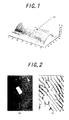
- Fig. 1 shows the shape of a pore of the cell wall of the onionepidermiccell treated with the laser beam. It can be clearly confirmed from Fig. 1 that the pore was formed by processing with the laser, while the cell swelled (Fig. 1(a)).
- FLUORESCEIN ISOTHIOCYANETE-D EXTRAN common name: FITC-Dextran
- FITC-Dextran commercially available from Sigma Co., Ltd.
- Aepidermiccell was cut out from an onion grown by a hydroponic method in two days, and placed on an MS solid culture. A part of a wall cell was removed by irradiating an introduction-targeted cell with an ArF exima laser beam (193 nm) in an irradiation area of 100 ⁇ m 2 at an energy density of 40 mJ/cm 2 . On this site was dropped, with a micromanipulator, about 0.1 ⁇ l of an aqueous solution in which FITC dextran-including small particles prepared above were suspended.
- an ArF eximer laser beam (193 nm) was irradiated upon the site through the dropped solution in an irradiation area of 100 ⁇ m 2 at an energy density of 10 mJ/cm 2 , thereby introducing a small particle into the cell.
- the small particle introduced was conformed in the following manner.
- the introduction of the small particle into the cell was confirmed by observing a fluorescent light of the FITC under a colarant-separating IB excited (U-MNIBA) filter with use of a fluorescent microscope in which an erected microscope of OLYMPUS BX 50 type was attached with an incident-light fluorescent device BX-FLA.
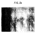
- Fig. 2(a) shows a fluorescent image obtained by observing the fluorescence light
- Fig. 2(b) does a bright-field image. As a result, living cell were sound with no abnormality observed.
- a calcein solution was introduced into the cell by using the microinjection through the hole bored in the laser processing. It was observed with the fluorescent microscope that the calcein solution was introduced into the cell.
- a cotyledons was used and laser processed with an exima laser.
- the energy density of the laser was 30 to 90 mJ/cm 2 , and a hole was bored in a size of 10 x 10 ⁇ 30 ⁇ 30 ⁇ m.
- a cell wall-digesting enzyme was dropped to the hole bored by the laser processing. Twenty four hours later, a protoplast cell of a mesophyll in which a cell wall near the hole was digested was isolated and observed.
- a buffer solution 10 mM Tris-HCl, pH 5.8 was added thereto, and left still for 15 minutes.
- the thin film of the lipid, the sample solution and the buffer solution were mixed under stirring with a vortex, thereby preparing a liposome including FITC-Dextran.
- An epidermic cell was cut out from an onion grown by hydroponic method for 2 days, and imbedded in a MS solid medium.
- An exima laser beam was irradiated upon the onion epidermic to process the epidermic.
- the irradiating area was 100 to 400 ⁇ m 2 , and the energy density was 20 to 60 mJ/cm 2 .
- An aqueous suspension (6% polyethylene glycol and 0.1 M mannitol contained) of the liposome including the FITC dextran as prepared above was dropped on the processed epidermic.
- An exima laser beam was irradiated upon the dropped aqueous suspension.
- a genetic material was tried to be introduced into a cell. More specifically, a genetic material absorbed on a gold particle or a magnetic particle was tried to be introduced.
- the plasmid DNA absorbed with the gold particles were used for the introduction.
- An edipermic cell of an onion was implanted in a 0.5 % agar medium containing 0.5 M mannitol, which was subjected to plasma separation.
- a cell wall was completely removed by processing a gap (space), which was formed between the cell wall and a cell membrane by the plasma separation, with laser in a square area of 10 ⁇ m ⁇ 10 ⁇ m at a laser energy density of 600 mJ/cm 2 .
- a foreign matter was introduced into the inner gap through a laser-formed pore by microinjection.
- the foreign matters used were the gold particles and magnetic particles absorbing the plasmid DNA, plasmid DNA-including alginic acid beads, liposome, etc.
- the onion epidermic was implanted in a 0.5% agar medium containing 0.2 M mannitol to recover a plasma. Then, after 24 hour culture, expression of the gene used for the introduction was observed.
- a chimera gene in which a gene (Green Fluprescense Protein (GFP)) encoding a fluorescent green light emission protein of the jellyfish ( Aequorea victoria ) is connected to a downstream of a 35S promoter of a cauliflower mosaic virus was used.
- This chimera gene is introduced into a plant cell, and expressed to produce a protein which emits a green fluorescence. Judgment was made based on whether the expression was effected or not through observing the light emission by using plasmid DNA including this chimera gene.
- the luminescent light was observed with the erected fluorescent microscope (BX-50) manufactured by Olympus Co., Ltd. (BX-50). More specifically, a bright-field image and a fluorescent image were observed with this microscope.
- a fluorescent filter used in this fluorescent observation was a U-MWIB2, U-MWIBA2 or U-MNIBA2.
- DNA plasmid absorbed on gold particles or the like were tried to be introduced in the same manner as in Example 5.
- An epidermic cell of an onion was implanted in a 0.5 % agar medium containing 0.5 M mannitol, which was subjected to plasma separation.
- a cell wall was partially or completely removed by processing a gap (space), which was formed between the cell wall and a cell membrane by the plasma separation, with laser in a square area of 10 ⁇ m x 10 ,u m at a laser energy density of 600 mJ/cm 2 .
- a solution containing a foreign matter such as the gold particles or magnetic particles absorbing the plasmid DNA, plasmid DNA-including alginic acid beads or liposome or the like was dropped on the pore formed by the laser processing according to the microinjection.
- the onion epidermic cell was implanted in a 0.5% agar medium containing 0.2 M mannitol to recover a plasma. Then, after 24 hour culture, expression of the gene used for the introduction was observed with the fluorescence microscope. The observation was effected by using the gene encoding the fluorescent green light emission protein of the jellyfish ( Aequorea victoria )_in the same manner as in Example 5.
- DNA plasmid absorbed on gold particles in the same manner as in Example 5 was used.
- a part of a cell wall was peeled off by laser processing a epidermic cell of an onion in a square area of 10 ⁇ m ⁇ 10 ⁇ m at a laser energy of 60 mJ/cm 2 .
- a solution containing gold particles or magnetic particles absorbing the plasmid DNA was dropped on the pore formed by the laser processing according to the microinjection.
- the foreign matter was introduced into the cell by effecting laser irradiation again. After the foreign matter was introduced, the laser treated cell was cultured for 24 hours, and expression of the gene was observed with the fluorescent microscope.
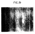
- Figs. 3(a) and 3(b) are photographs showing that gold particles are introduced into a cell by laser irradiation.
- Fig. 3(a) is a microphotograph obtained by focusing the surface of the cell. This microphotograph is a bright-field observed image obtained when a surface layer of the epidermic cell of the onion was peeled off by the laser processing, and the gold particles were placed on the pore processed portion by the micromanipulator. In this picture, the surface of the cell was focused.
- Fig. 3(b) is a microphotograph in which an interior of the cell is focused. With respect to the above sample, focusing was shifted to the interior of the cell. This reveals that the gold particles exist inside the cell.
- Exima laser was irradiated upon a epidermic cell of a stem of a torenia plant.
- the laser irradiation was effected in a square area (size) of 5 ⁇ 5 ⁇ 10 ⁇ 10 ⁇ m.
- the energy density was 60 to 80 ⁇ J/cm 2 .
- the cell wall-decomposing enzyme 0.1 % Pectolyase Y23, 1% cellulase Onozuka RS, 0.4M Mannitol, pH 5.5
- the cell was washed with sterilized distilled water (Decomposition of the cell was terminated by washing off the enzyme solution). It was confirmed with the erected microscope that the cell wall near the laser-processing site was decomposed and depressed. In order to confirm that the cell wall was completely removed, a 0.1% aqueous solution of Fluostain was directly dropped on the laser-processed site by using a microcapillary. After dying for 5 minutes, that site was observed with the fluorescent microscope, which confirmed that the laser-processed portion was not dyed. On the other hand, when a cell was merely processed with laser at a weak energy density and subjected to the same treatment as above, it was observed that the laser-processed site was dyed. Therefore, it is considered that the cell was completely removed through decomposition when the liquid containing the cell wall-decomposing enzyme was applied to the laser-processed site.
- a microcapillary was pierced into the above processed site by the microinjection to introduce a DNA solution.
- a DNA solution a plasmid DNA having GFP was employed.
- a plant was transferred to a MS plate medium, followed by culturing for 24 hours. Fluorescence of GFP was observed by using the fluorescent microscope (Olympus BX50).
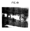
- Figs. 4(a) and 4(b) are bright-field images of the epidermic cell of the onion observed.
- Fig. 4(a) gives a bright-field image of the laser-processed onion epidermic cell at a left side and that of the cell laser-processed and then treated with the enzyme at a right side.
- Fig. 4(b) gives bright-field images of the above observed with the incident-light source.
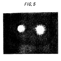
- Fig. 5 is an image obtained with a fluorescent light through observation of the above sample dyed with a cell wall-dying reagent, Fluostain, by using the fluorescent microscope.
- a center portion is not dyed with the reagent as seen by comparison with the left side image. Thus, it is considered that a cell membrane is exposed in the right image.
- Fig. 6 shows a transient expression of sGFP. That is, Fig. 6 is an image obtained by observing expression of sGFP with the fluorescent microscope after laser was irradiated upon the epidermic cell of a stem of a torenia plant, the cell was treated with the enzyme and then the sGFP plasmid solution was introduced by the microinjection method, and the cell was cultured for 24 hours. As is clear from this image, the fluorescent protein was expressed by the gene introduced.
- the microcapillary was used in the enzyme treatment, but needless to say such can be effected without use of the microcapillary.
- the laser-processed face is immersed in the enzyme solution, and reacted with it for a few or several minutes, and washed with sterilized distilled water so as to perform the enzyme treatment.
- a plasmid solution (containing sGFP) was introduced by using the same method as in the case of introducing the calcein solution as described in Example 2, and expression of the sGFP was confirmed with the fluorescent microscope after 24 hour culturing.
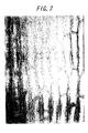
- Fig. 7 is a bright-field image of epidermic cell of an onion observed. That is, Fig. 7 gives the bright-field image of epidermic cell of the onion observed after 24-hours cultivation following the laser processing and the injection of the plasmide solution.
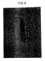
- Fig. 8 is a microphotograph showing that the sGFP plasmid solution was introduced into a cell and transiently expressed there. Fig. 8 is to see the expression of the sGFP through observation of the sGFP-containing plasmid in Fig. 7 with the fluorescent microscope.
- the foreign matter-introducing method of the present invention exhibits an advantageous effect that a single specified cell can be used as a target cell in producing a transformed plant by introducing the foreign matter or the gene therein.
- the foreign matter-introducing method of the present invention exhibits another advantageous effect that the target plant species to be irradiated with laser is not limited different from cases where plant pathogenic bacteria are used, and therefore the method is used for general purposes.
- the foreign matter-introducing method of the present invention exhibits a further advantage that a large-size foreign matter such as a nucleus or a chromosome can be introduced into a cell.
- the matter-introducing method of the present invention exhibits a still further advantage that plural kinds of foreign matters can be simultaneously introduced into a cell.
- the matter-introducing method of the present invention exhibits a still further advantage that a foreign matter having a low physical strength can be stably introduced into a cell.
Landscapes
- Health & Medical Sciences (AREA)
- Life Sciences & Earth Sciences (AREA)
- Genetics & Genomics (AREA)
- Engineering & Computer Science (AREA)
- Organic Chemistry (AREA)
- Biomedical Technology (AREA)
- Chemical & Material Sciences (AREA)
- Biotechnology (AREA)
- Zoology (AREA)
- Wood Science & Technology (AREA)
- Bioinformatics & Cheminformatics (AREA)
- General Engineering & Computer Science (AREA)
- Microbiology (AREA)
- Biochemistry (AREA)
- General Health & Medical Sciences (AREA)
- Molecular Biology (AREA)
- Physics & Mathematics (AREA)
- Cell Biology (AREA)
- Biophysics (AREA)
- Plant Pathology (AREA)
- Electromagnetism (AREA)
- Sustainable Development (AREA)
- Micro-Organisms Or Cultivation Processes Thereof (AREA)
- Apparatus Associated With Microorganisms And Enzymes (AREA)
- Breeding Of Plants And Reproduction By Means Of Culturing (AREA)
- Immobilizing And Processing Of Enzymes And Microorganisms (AREA)
Applications Claiming Priority (4)
| Application Number | Priority Date | Filing Date | Title |
|---|---|---|---|
| JP2000392167 | 2000-12-25 | ||
| JP2000392167 | 2000-12-25 | ||
| JP2001349559A JP2002325572A (ja) | 2000-12-25 | 2001-11-15 | 外来物質の導入方法 |
| JP2001349559 | 2001-11-15 |
Publications (3)
| Publication Number | Publication Date |
|---|---|
| EP1225228A2 true EP1225228A2 (de) | 2002-07-24 |
| EP1225228A3 EP1225228A3 (de) | 2002-11-13 |
| EP1225228B1 EP1225228B1 (de) | 2005-08-03 |
Family
ID=26606486
Family Applications (1)
| Application Number | Title | Priority Date | Filing Date |
|---|---|---|---|
| EP01130275A Expired - Lifetime EP1225228B1 (de) | 2000-12-25 | 2001-12-20 | Verfahren zur Einbringung von Fremdmaterial in höhere Zellen unter Verwendung eines Laserstrahls |
Country Status (6)
| Country | Link |
|---|---|
| US (1) | US7132289B2 (de) |
| EP (1) | EP1225228B1 (de) |
| JP (1) | JP2002325572A (de) |
| AU (1) | AU773342B2 (de) |
| DE (1) | DE60112398T2 (de) |
| DK (1) | DK1225228T3 (de) |
Cited By (18)
| Publication number | Priority date | Publication date | Assignee | Title |
|---|---|---|---|---|
| US9157066B2 (en) | 2005-12-13 | 2015-10-13 | The Trustees Of The University Of Pennsylvania | Transcriptome transfer produces cellular phenotype conversion |
| WO2017041051A1 (en) * | 2015-09-04 | 2017-03-09 | Sqz Biotechnologies Company | Intracellular delivery of biomolecules to cells comprising a cell wall |
| WO2017099787A1 (en) * | 2015-12-10 | 2017-06-15 | Gpd Technologies Llc | Systems and methods for using light energy to facilitate penetration of substances in plants |
| US10124336B2 (en) | 2013-08-16 | 2018-11-13 | Massachusetts Institute Of Technology | Selective delivery of material to cells |
| US10219443B2 (en) | 2014-04-07 | 2019-03-05 | Premier Citrus Apz, Llc | Systems and methods for using light energy to facilitate penetration of substances in plants |
| US10278334B2 (en) | 2014-04-07 | 2019-05-07 | Premier Citrus Apz, Llc | Systems and methods for delivering nucleic acids to a plant |
| US10526573B2 (en) | 2014-11-14 | 2020-01-07 | Massachusetts Institute Of Technology | Disruption and field enabled delivery of compounds and compositions into cells |
| US10583204B2 (en) | 2005-12-13 | 2020-03-10 | The Trustees Of The University Of Pennsylvania | Methods for transfecting nucleic acid into live cells |
| US10696944B2 (en) | 2011-10-17 | 2020-06-30 | Massachusetts Institute Of Technology | Intracellular delivery |
| US10883082B2 (en) | 2005-12-13 | 2021-01-05 | The Trustees Of The University Of Pennsylvania | Transcriptome transfer produces cellular phenotype conversion |
| US11111472B2 (en) | 2014-10-31 | 2021-09-07 | Massachusetts Institute Of Technology | Delivery of biomolecules to immune cells |
| US11125739B2 (en) | 2015-01-12 | 2021-09-21 | Massachusetts Institute Of Technology | Gene editing through microfluidic delivery |
| US11178823B2 (en) | 2014-04-07 | 2021-11-23 | Premier Citrus Apz, Llc | Systems and methods for using light energy to facilitate penetration of substances in plants |
| US11191278B2 (en) | 2016-03-25 | 2021-12-07 | Premier Citrus Apz, Llc | Systems and methods for delivering nucleic acids to a plant |
| EP3971296A1 (de) * | 2020-09-16 | 2022-03-23 | FRAUNHOFER-GESELLSCHAFT zur Förderung der angewandten Forschung e.V. | Verfahren zur erzeugung transformierter pflanzen |
| EP3971295A1 (de) * | 2020-09-16 | 2022-03-23 | FRAUNHOFER-GESELLSCHAFT zur Förderung der angewandten Forschung e.V. | Verfahren zur herstellung von genomeditierten pflanzen |
| US11299698B2 (en) | 2015-07-09 | 2022-04-12 | Massachusetts Institute Of Technology | Delivery of materials to anucleate cells |
| US12201652B2 (en) | 2016-05-03 | 2025-01-21 | Stemcell Technologies Canada Inc. | Intracellular delivery of biomolecules to induce tolerance |
Families Citing this family (37)
| Publication number | Priority date | Publication date | Assignee | Title |
|---|---|---|---|---|
| US6753161B2 (en) * | 1997-03-27 | 2004-06-22 | Oncosis Llc | Optoinjection methods |
| CA2392534A1 (en) | 1999-11-30 | 2001-06-07 | Oncosis | Method and apparatus for selectively targeting specific cells within a cell population |
| AU2001264317A1 (en) * | 2000-06-20 | 2002-01-02 | Koken Co., Ltd. | Preparations for oligonucleotide transfer |
| EP2363828A1 (de) | 2000-10-24 | 2011-09-07 | Cyntellect, Inc. | Vorrichtung zur gezielten Bestimmung von Zellen in einer dreidimensionalen Probe |
| EP1407787B1 (de) | 2001-06-20 | 2009-04-29 | Dainippon Sumitomo Pharma Co., Ltd. | Verfahren zur förderung des nukleinsäuretransfers |
| JP4022614B2 (ja) * | 2002-03-25 | 2007-12-19 | 国立大学法人大阪大学 | 新規なバイオビーズの作製方法 |
| US6863406B2 (en) * | 2002-08-01 | 2005-03-08 | The University Of Chicago | Apparatus and method for fabricating, sorting, and integrating materials with holographic optical traps |
| US7361171B2 (en) | 2003-05-20 | 2008-04-22 | Raydiance, Inc. | Man-portable optical ablation system |
| US8173929B1 (en) | 2003-08-11 | 2012-05-08 | Raydiance, Inc. | Methods and systems for trimming circuits |
| US8921733B2 (en) | 2003-08-11 | 2014-12-30 | Raydiance, Inc. | Methods and systems for trimming circuits |
| US9022037B2 (en) | 2003-08-11 | 2015-05-05 | Raydiance, Inc. | Laser ablation method and apparatus having a feedback loop and control unit |
| US20050095578A1 (en) * | 2003-10-31 | 2005-05-05 | Koller Manfred R. | Method and apparatus for cell permeabilization |
| WO2005061717A1 (ja) * | 2003-12-19 | 2005-07-07 | Dainippon Sumitomo Pharma Co., Ltd. | 新規な核酸導入法 |
| US7425426B2 (en) * | 2004-03-15 | 2008-09-16 | Cyntellect, Inc. | Methods for purification of cells based on product secretion |
| JP4504082B2 (ja) | 2004-04-28 | 2010-07-14 | 富士通株式会社 | 液体注入装置 |
| JP2006006223A (ja) * | 2004-06-25 | 2006-01-12 | Canon Inc | 微粒子の細胞への導入と回収装置、及びその導入と細胞あるいは細胞内の標的生体分子の回収方法 |
| US20090062184A1 (en) * | 2005-03-24 | 2009-03-05 | Dainippon Sumitomo Pharma Co., Ltd. | Fine particulate preparation comprising complex of nucleic acid molecule and collagen |
| US8135050B1 (en) | 2005-07-19 | 2012-03-13 | Raydiance, Inc. | Automated polarization correction |
| WO2007076458A1 (en) * | 2005-12-21 | 2007-07-05 | Primegen Biotech Llc | Microinjector chip |
| US7444049B1 (en) | 2006-01-23 | 2008-10-28 | Raydiance, Inc. | Pulse stretcher and compressor including a multi-pass Bragg grating |
| US8232687B2 (en) | 2006-04-26 | 2012-07-31 | Raydiance, Inc. | Intelligent laser interlock system |
| US8189971B1 (en) | 2006-01-23 | 2012-05-29 | Raydiance, Inc. | Dispersion compensation in a chirped pulse amplification system |
| US7822347B1 (en) | 2006-03-28 | 2010-10-26 | Raydiance, Inc. | Active tuning of temporal dispersion in an ultrashort pulse laser system |
| US8420891B2 (en) | 2007-06-21 | 2013-04-16 | National University Corporation Nagoya University | Method for introducing foreign substance into cell having cell wall |
| JP2009038988A (ja) * | 2007-08-06 | 2009-02-26 | Hiroshima Pref Gov | 辛味成分の少ないアリウム属植物の栽培方法 |
| WO2009140701A2 (en) * | 2008-05-16 | 2009-11-19 | Synthetic Genomics, Inc. | Delivery into cells using ultra-short pulse lasers |
| EP3124601A1 (de) * | 2008-08-14 | 2017-02-01 | Mesoblast International Sàrl | Gereinigte zusammensetzungen aus mesenchymalen stammzellen sowie verfahren zur reinigung von zusammensetzungen aus mesenchymalen stammzellen |
| US8125704B2 (en) | 2008-08-18 | 2012-02-28 | Raydiance, Inc. | Systems and methods for controlling a pulsed laser by combining laser signals |
| US20100055756A1 (en) * | 2008-08-28 | 2010-03-04 | Gregory Spooner | System and method for modifying biological cells using an ultra-short pulsed laser |
| JP2012514477A (ja) * | 2009-01-09 | 2012-06-28 | イントレクソン コーポレイション | 細胞の遺伝学的解析 |
| KR20110108390A (ko) | 2009-01-12 | 2011-10-05 | 신텔렉트 인코포레이티드 | 레이저로 매개된 박막 절개 및 세포 군체의 이동 |
| US8808733B2 (en) | 2009-03-31 | 2014-08-19 | The Board Of Trustees Of The University Of Arkansas | Method of controlled drug release from a liposome carrier |
| WO2012021748A1 (en) | 2010-08-12 | 2012-02-16 | Raydiance, Inc. | Polymer tubing laser micromachining |
| US9120181B2 (en) | 2010-09-16 | 2015-09-01 | Coherent, Inc. | Singulation of layered materials using selectively variable laser output |
| US9849087B2 (en) | 2011-11-08 | 2017-12-26 | The Board Of Trustees Of The University Of Arkansas | Methods and compositions for X-ray induced release from pH sensitive liposomes |
| US9485953B2 (en) * | 2012-07-19 | 2016-11-08 | R.J. Reynolds Tobacco Company | Method for treating tobacco plants with enzymes |
| CN111879434B (zh) * | 2020-07-24 | 2022-04-08 | 之江实验室 | 一种测量生物组织或细胞温度的方法及装置 |
Family Cites Families (10)
| Publication number | Priority date | Publication date | Assignee | Title |
|---|---|---|---|---|
| US495050A (en) * | 1893-04-11 | Multiple-boring device | ||
| JPS6083583A (ja) | 1983-10-13 | 1985-05-11 | Rikagaku Kenkyusho | 生細胞レ−ザ−穿孔装置 |
| JPS6083584A (ja) * | 1983-10-13 | 1985-05-11 | Rikagaku Kenkyusho | 生細胞内への物質移入法 |
| EP0137504B1 (de) | 1983-10-13 | 1991-01-16 | Rikagaku Kenkyusho | Verfahren und Apparat zum Einpflanzen eines Fremdstoffes in lebende Zelle |
| US4945050A (en) | 1984-11-13 | 1990-07-31 | Cornell Research Foundation, Inc. | Method for transporting substances into living cells and tissues and apparatus therefor |
| GB8806643D0 (en) | 1988-03-21 | 1988-04-20 | Ici Plc | Genetic manipulation |
| AU643563B2 (en) | 1990-11-01 | 1993-11-18 | Sapporo Breweries Limited | Method for preparing transformed plant |
| US6346101B1 (en) * | 1993-07-19 | 2002-02-12 | Research Foundation Of City College Of New York | Photon-mediated introduction of biological materials into cells and/or cellular components |
| US5916788A (en) * | 1995-10-12 | 1999-06-29 | Sony Corporation | Genetic recombination laser apparatus and genetic recombination method using the apparatus |
| JPH09163984A (ja) | 1995-10-12 | 1997-06-24 | Sony Corp | 遺伝子組替え用レーザ装置およびこれを用いた遺伝子組替え方法 |
-
2001
- 2001-11-15 JP JP2001349559A patent/JP2002325572A/ja active Pending
- 2001-12-17 US US10/015,607 patent/US7132289B2/en not_active Expired - Fee Related
- 2001-12-18 AU AU97290/01A patent/AU773342B2/en not_active Ceased
- 2001-12-20 EP EP01130275A patent/EP1225228B1/de not_active Expired - Lifetime
- 2001-12-20 DE DE60112398T patent/DE60112398T2/de not_active Expired - Fee Related
- 2001-12-20 DK DK01130275T patent/DK1225228T3/da active
Cited By (30)
| Publication number | Priority date | Publication date | Assignee | Title |
|---|---|---|---|---|
| US9845480B2 (en) | 2005-12-13 | 2017-12-19 | The Trustees Of The University Of Pennsylvania | Transcriptome transfer produces cellular phenotype conversion |
| US9157066B2 (en) | 2005-12-13 | 2015-10-13 | The Trustees Of The University Of Pennsylvania | Transcriptome transfer produces cellular phenotype conversion |
| US10883082B2 (en) | 2005-12-13 | 2021-01-05 | The Trustees Of The University Of Pennsylvania | Transcriptome transfer produces cellular phenotype conversion |
| US10583204B2 (en) | 2005-12-13 | 2020-03-10 | The Trustees Of The University Of Pennsylvania | Methods for transfecting nucleic acid into live cells |
| US10696944B2 (en) | 2011-10-17 | 2020-06-30 | Massachusetts Institute Of Technology | Intracellular delivery |
| US11806714B2 (en) | 2013-08-16 | 2023-11-07 | Massachusetts Institute Of Technology | Selective delivery of material to cells |
| US10124336B2 (en) | 2013-08-16 | 2018-11-13 | Massachusetts Institute Of Technology | Selective delivery of material to cells |
| US10870112B2 (en) | 2013-08-16 | 2020-12-22 | Massachusetts Institute Of Technology | Selective delivery of material to cells |
| US11178823B2 (en) | 2014-04-07 | 2021-11-23 | Premier Citrus Apz, Llc | Systems and methods for using light energy to facilitate penetration of substances in plants |
| US10219443B2 (en) | 2014-04-07 | 2019-03-05 | Premier Citrus Apz, Llc | Systems and methods for using light energy to facilitate penetration of substances in plants |
| US10278334B2 (en) | 2014-04-07 | 2019-05-07 | Premier Citrus Apz, Llc | Systems and methods for delivering nucleic acids to a plant |
| US11111472B2 (en) | 2014-10-31 | 2021-09-07 | Massachusetts Institute Of Technology | Delivery of biomolecules to immune cells |
| US12410392B2 (en) | 2014-10-31 | 2025-09-09 | Massachusetts Institute Of Technology | Delivery of biomolecules to immune cells |
| US10526573B2 (en) | 2014-11-14 | 2020-01-07 | Massachusetts Institute Of Technology | Disruption and field enabled delivery of compounds and compositions into cells |
| US11125739B2 (en) | 2015-01-12 | 2021-09-21 | Massachusetts Institute Of Technology | Gene editing through microfluidic delivery |
| US12130281B2 (en) | 2015-01-12 | 2024-10-29 | Massachusetts Institute Of Technology | Gene editing through microfluidic delivery |
| US11299698B2 (en) | 2015-07-09 | 2022-04-12 | Massachusetts Institute Of Technology | Delivery of materials to anucleate cells |
| US12441969B2 (en) | 2015-07-09 | 2025-10-14 | Massachusetts Institute Of Technology | Delivery of materials to anucleate cells |
| WO2017041051A1 (en) * | 2015-09-04 | 2017-03-09 | Sqz Biotechnologies Company | Intracellular delivery of biomolecules to cells comprising a cell wall |
| US11613759B2 (en) | 2015-09-04 | 2023-03-28 | Sqz Biotechnologies Company | Intracellular delivery of biomolecules to cells comprising a cell wall |
| WO2017099787A1 (en) * | 2015-12-10 | 2017-06-15 | Gpd Technologies Llc | Systems and methods for using light energy to facilitate penetration of substances in plants |
| CN108777945A (zh) * | 2015-12-10 | 2018-11-09 | 柑桔代理有限责任公司 | 利用光能促进植物中物质渗透的系统和方法 |
| AU2015417085B2 (en) * | 2015-12-10 | 2019-07-18 | Premier Citrus Apz, Llc | Systems and methods for using light energy to facilitate penetration of substances in plants |
| US11191278B2 (en) | 2016-03-25 | 2021-12-07 | Premier Citrus Apz, Llc | Systems and methods for delivering nucleic acids to a plant |
| US12201652B2 (en) | 2016-05-03 | 2025-01-21 | Stemcell Technologies Canada Inc. | Intracellular delivery of biomolecules to induce tolerance |
| WO2022058392A1 (en) * | 2020-09-16 | 2022-03-24 | Fraunhofer-Gesellschaft Zur Förderung Der Angewandten Forschung E.V.. | Methods for producing transformed plants |
| WO2022058388A1 (en) * | 2020-09-16 | 2022-03-24 | Fraunhofer-Gesellschaft Zur Förderung Der Angewandten Forschung E.V.. | Methods for the production of genome edited plants |
| EP3971295A1 (de) * | 2020-09-16 | 2022-03-23 | FRAUNHOFER-GESELLSCHAFT zur Förderung der angewandten Forschung e.V. | Verfahren zur herstellung von genomeditierten pflanzen |
| EP3971296A1 (de) * | 2020-09-16 | 2022-03-23 | FRAUNHOFER-GESELLSCHAFT zur Förderung der angewandten Forschung e.V. | Verfahren zur erzeugung transformierter pflanzen |
| US12559762B2 (en) | 2020-09-16 | 2026-02-24 | Fraunhofer-Gesellschaft zur Förderung der angewandten Forschung e.V. | Methods for producing transformed plants |
Also Published As
| Publication number | Publication date |
|---|---|
| DE60112398T2 (de) | 2005-12-29 |
| DE60112398D1 (de) | 2005-09-08 |
| EP1225228B1 (de) | 2005-08-03 |
| DK1225228T3 (da) | 2005-11-07 |
| AU9729001A (en) | 2002-06-27 |
| US7132289B2 (en) | 2006-11-07 |
| EP1225228A3 (de) | 2002-11-13 |
| JP2002325572A (ja) | 2002-11-12 |
| AU773342B2 (en) | 2004-05-20 |
| US20020115219A1 (en) | 2002-08-22 |
Similar Documents
| Publication | Publication Date | Title |
|---|---|---|
| US7132289B2 (en) | Method for introducing foreign matters into living cells | |
| JP4469959B2 (ja) | 細胞壁を備える細胞に外来物質を導入する方法 | |
| US20240384287A1 (en) | Compositions and methods for delivery of nucleic acid to plant cells | |
| EP3563673A1 (de) | Verfahren zum einbringen einer substanz in eine pflanze | |
| Tripathi et al. | Methods of genetic transformation: major emphasis to crop plants | |
| Miyamoto et al. | Advancing biomolecule delivery in plants: harnessing synthetic nanocarriers to overcome multiscale barriers for cutting-edge plant bioengineering | |
| Shivashakarappa et al. | DNA delivery into plant tissues using carbon dots made from citric acid and β-alanine | |
| Kumar et al. | Nano-assisted delivery tools for plant genetic engineering: a review on recent developments | |
| Finer | Plant nuclear transformation | |
| RU2663347C1 (ru) | Способ доставки биологически активных макромолекул в клетки растений | |
| JP4022614B2 (ja) | 新規なバイオビーズの作製方法 | |
| Shehzad et al. | Nanocarriers for delivering biomolecules in plants: mechanisms and applications | |
| JP3780333B2 (ja) | 外来性遺伝物質又は生理活性物質を細胞内へ導入する新規な方法 | |
| Singh et al. | Gene transformation using nano-technology and delivery methods of the bio-modifier-conjugated complex to plant cells | |
| US7663026B2 (en) | Transformation of seaweed utilizing biolistic gene transfer to spores | |
| AU769568B2 (en) | Method for processing cells | |
| EP2962552B1 (de) | Rekombinante pflanzenzelle, herstellungsverfahren dafür und verfahren zur herstellung eines zielproteins damit | |
| CN116463382A (zh) | 通过介孔二氧化硅纳米颗粒递送外源核酸到植物细胞 | |
| CN111349652B (zh) | 一种农杆菌介导的海带配子体遗传转化方法 | |
| Özkaya | Genetic transformation: current opinion and future prospect | |
| WO2021054407A1 (ja) | 導入装置、高分子の細胞導入方法、dnaナノ粒子結晶及びdnaナノ粒子結晶封入体の製造方法 | |
| Rahangdale et al. | Chapter 2: Gene Transfer Methods in Plants | |
| CN113136359B (zh) | 一种利用匍匐翦股颖原生质体观察亚细胞定位和蛋白互作的方法 | |
| Huang | High-Throughput Transient Gene Expression In Plant Cells | |
| Kumar et al. | Gene Transfer in Plants and Their Application |
Legal Events
| Date | Code | Title | Description |
|---|---|---|---|
| PUAI | Public reference made under article 153(3) epc to a published international application that has entered the european phase |
Free format text: ORIGINAL CODE: 0009012 |
|
| 17P | Request for examination filed |
Effective date: 20011220 |
|
| AK | Designated contracting states |
Kind code of ref document: A2 Designated state(s): AT BE CH CY DE DK ES FI FR GB GR IE IT LI LU MC NL PT SE TR |
|
| AX | Request for extension of the european patent |
Free format text: AL;LT;LV;MK;RO;SI |
|
| PUAL | Search report despatched |
Free format text: ORIGINAL CODE: 0009013 |
|
| AK | Designated contracting states |
Kind code of ref document: A3 Designated state(s): AT BE CH CY DE DK ES FI FR GB GR IE IT LI LU MC NL PT SE TR |
|
| AX | Request for extension of the european patent |
Free format text: AL;LT;LV;MK;RO;SI |
|
| AKX | Designation fees paid |
Designated state(s): DE DK FR GB NL |
|
| 17Q | First examination report despatched |
Effective date: 20031218 |
|
| GRAP | Despatch of communication of intention to grant a patent |
Free format text: ORIGINAL CODE: EPIDOSNIGR1 |
|
| RAP1 | Party data changed (applicant data changed or rights of an application transferred) |
Owner name: OSAKA UNIVERSITY |
|
| RIN1 | Information on inventor provided before grant (corrected) |
Inventor name: KOBAYASHI, AKIO Inventor name: HARAJIMA, SATOSHI Inventor name: SHOJI, TAKESHI Inventor name: KAJIYAMA, SHINICHIRO Inventor name: FUKUI, KIICHI Inventor name: OKUDA, SHINYA Inventor name: FUKUSAKI, EIICHIRO |
|
| GRAS | Grant fee paid |
Free format text: ORIGINAL CODE: EPIDOSNIGR3 |
|
| GRAA | (expected) grant |
Free format text: ORIGINAL CODE: 0009210 |
|
| AK | Designated contracting states |
Kind code of ref document: B1 Designated state(s): DE DK FR GB NL |
|
| REG | Reference to a national code |
Ref country code: GB Ref legal event code: FG4D |
|
| REF | Corresponds to: |
Ref document number: 60112398 Country of ref document: DE Date of ref document: 20050908 Kind code of ref document: P |
|
| REG | Reference to a national code |
Ref country code: DK Ref legal event code: T3 |
|
| ET | Fr: translation filed | ||
| PLBE | No opposition filed within time limit |
Free format text: ORIGINAL CODE: 0009261 |
|
| STAA | Information on the status of an ep patent application or granted ep patent |
Free format text: STATUS: NO OPPOSITION FILED WITHIN TIME LIMIT |
|
| 26N | No opposition filed |
Effective date: 20060504 |
|
| PGFP | Annual fee paid to national office [announced via postgrant information from national office to epo] |
Ref country code: FR Payment date: 20061128 Year of fee payment: 6 |
|
| PGFP | Annual fee paid to national office [announced via postgrant information from national office to epo] |
Ref country code: DK Payment date: 20061213 Year of fee payment: 6 |
|
| PGFP | Annual fee paid to national office [announced via postgrant information from national office to epo] |
Ref country code: NL Payment date: 20061215 Year of fee payment: 6 |
|
| PGFP | Annual fee paid to national office [announced via postgrant information from national office to epo] |
Ref country code: GB Payment date: 20061218 Year of fee payment: 6 |
|
| PGFP | Annual fee paid to national office [announced via postgrant information from national office to epo] |
Ref country code: DE Payment date: 20061228 Year of fee payment: 6 |
|
| REG | Reference to a national code |
Ref country code: DK Ref legal event code: EBP |
|
| GBPC | Gb: european patent ceased through non-payment of renewal fee |
Effective date: 20071220 |
|
| NLV4 | Nl: lapsed or anulled due to non-payment of the annual fee |
Effective date: 20080701 |
|
| PG25 | Lapsed in a contracting state [announced via postgrant information from national office to epo] |
Ref country code: DE Free format text: LAPSE BECAUSE OF NON-PAYMENT OF DUE FEES Effective date: 20080701 |
|
| REG | Reference to a national code |
Ref country code: FR Ref legal event code: ST Effective date: 20081020 |
|
| PG25 | Lapsed in a contracting state [announced via postgrant information from national office to epo] |
Ref country code: NL Free format text: LAPSE BECAUSE OF NON-PAYMENT OF DUE FEES Effective date: 20080701 |
|
| PG25 | Lapsed in a contracting state [announced via postgrant information from national office to epo] |
Ref country code: GB Free format text: LAPSE BECAUSE OF NON-PAYMENT OF DUE FEES Effective date: 20071220 |
|
| PG25 | Lapsed in a contracting state [announced via postgrant information from national office to epo] |
Ref country code: DK Free format text: LAPSE BECAUSE OF NON-PAYMENT OF DUE FEES Effective date: 20080102 |
|
| PG25 | Lapsed in a contracting state [announced via postgrant information from national office to epo] |
Ref country code: FR Free format text: LAPSE BECAUSE OF NON-PAYMENT OF DUE FEES Effective date: 20071231 |