EP1225542A2 - Segmentierung von tomographischen Daten - Google Patents
Segmentierung von tomographischen Daten Download PDFInfo
- Publication number
- EP1225542A2 EP1225542A2 EP01309786A EP01309786A EP1225542A2 EP 1225542 A2 EP1225542 A2 EP 1225542A2 EP 01309786 A EP01309786 A EP 01309786A EP 01309786 A EP01309786 A EP 01309786A EP 1225542 A2 EP1225542 A2 EP 1225542A2
- Authority
- EP
- European Patent Office
- Prior art keywords
- bone
- selected characteristic
- regions
- ray
- input volume
- Prior art date
- Legal status (The legal status is an assumption and is not a legal conclusion. Google has not performed a legal analysis and makes no representation as to the accuracy of the status listed.)
- Granted
Links
Images


Classifications
-
- G—PHYSICS
- G06—COMPUTING OR CALCULATING; COUNTING
- G06T—IMAGE DATA PROCESSING OR GENERATION, IN GENERAL
- G06T7/00—Image analysis
- G06T7/10—Segmentation; Edge detection
- G06T7/11—Region-based segmentation
-
- G—PHYSICS
- G06—COMPUTING OR CALCULATING; COUNTING
- G06T—IMAGE DATA PROCESSING OR GENERATION, IN GENERAL
- G06T2207/00—Indexing scheme for image analysis or image enhancement
- G06T2207/10—Image acquisition modality
- G06T2207/10072—Tomographic images
- G06T2207/10081—Computed x-ray tomography [CT]
-
- G—PHYSICS
- G06—COMPUTING OR CALCULATING; COUNTING
- G06T—IMAGE DATA PROCESSING OR GENERATION, IN GENERAL
- G06T2207/00—Indexing scheme for image analysis or image enhancement
- G06T2207/30—Subject of image; Context of image processing
- G06T2207/30004—Biomedical image processing
- G06T2207/30008—Bone
Definitions
- the present invention relates to image processing and more particularly segmenting and classifying a region or data type of interest based on selected characteristics.
- segmentation Identification of contiguous regions of the same material is known as segmentation.
- the automatic classification or segmentation of medical images into anatomical regions, for example bone, kidney or lung, is a difficult problem that is currently stalling the development of many clinical applications. While several techniques have been proposed over the years, very few techniques have achieved the high degree of automation and quality required by routine medical practice. Instead, most clinical applications rely on a large amount of user interaction. Thus, the process of segmentation is time-consuming, error prone, and subjective.
- the human skeletal system is mainly comprised of two types of bone - cortical bone and trabecular bone.
- Cortical bone is quite dense and typically located at the perimeter of a bone region.
- Trabecular bone is spongy and is typically located in interior bone regions.
- bone regions have several distinct characteristics that are nearly universal in the patient CT scan population.
- CT densities rapidly rise at bone boundaries (cortical) and then drop modestly within a few millimeters, thereby creating a "ring" effect in scan data.
- the structure of interior bone (trabecular) has a distinct spongy pattern that is characterized by constant change and relatively high frequency.
- the CT bone values range from 1200 to greater than 2000 Hounsfeld units for cortical bone, and trabecular ranging from 1000 to 1300 Hounsfeld units.
- CT computed tomography
- a method for segmenting an input volume in accordance with at least one selected characteristic comprising the steps of casting a plurality of rays along a plurality of views of the input volume, identifying regions corresponding to the selected characteristic within each ray, and processing the identified regions to generate an output volume indicating regions having the selected characteristic.
- a method of segmentation comprises the steps casting a plurality of rays through input data and classifying the resultant data in accordance with predetermined classification parameters or characteristics.
- Predetermined classification parameters such as bone-like profile characteristics.
- characteristics include high gradient, outer entrance (exterior to cortical peak), very high valued entrance (cortical peak), high gradient inner entrance (cortical peak to start of trabecular), total cortical thickness usually around 2mm, and fluctuating values in interior bone (trabecular).
- Figure 1 shows a block diagram illustrating an embodiment of the present invention. Referring to Figure 1, a method for segmenting input data for at least one selected characteristic is shown.
- input data S is acquired, for example a 2D scalar field.
- this embodiment has a two-dimensional (2D) Computed Tomography (CT) image as the input volume.
- CT Computed Tomography
- the method for segmenting can operate on a two- or three-dimensional scalar or vector field, such as found in other medical imaging applications (magnetic resonance (MR), ultrasound, X-ray) and other non-medical scanning or sensing applications.
- MR magnetic resonance
- X-ray X-ray
- a plurality of rays R are cast in the well-known technique of ray casting to sample the input volume and to produce a sampled one-dimensional (1D) representation of the input data suitable for array manipulation.
- Rays originate from one of many evenly rotated views V that are positioned on a circle that encloses S .
- the number of views V at an angle theta with respect to S are selected in a known manner based on considerations of image processing quality and speed requirements.
- classification performed by sampling many trajectories or rays, usually linear, through the input image S as shown in step 120.
- a plurality of evenly spaced rays is projected from a view.
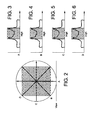
- Figure 2 illustrates four single rays (A-D) originating from four corresponding views (one is shown).
- step 130 probabilities that bone exists along the ray are computed, which will be discussed below in greater detail.
- step 140 the probabilities along each ray are then processed into the intermediate buffer P . Computations are repeated along each ray at step 150.
- the intermediate buffer P is converted into the final bone probability image B at step 160.
- step 170 the final image is converted through specialized region growing. The region grow step 170 searches for other pixels that must also be bone but which could not be detected as such by tomographic analysis.
- This region growing is an iterative process that begins with marking highly-probably bone pixels as bone, and testing adjacent pixels based on an additional set of criteria that can determine if the adjacent pixel is part of the same anatomical structure as the previous pixel, regardless of whether it originally obtained a high probability score by the tomographic analysis. Further in this embodiment, boundary logic is desirably employed to ensure that bone probabilities are not corrupted by the lack of information beyond the perimeter of the scanned image.
- the computation of bone probabilities for a ray is performed by analyzing the ray's scalar values and identifying the patterns indicative of bone density (cortical and trabecular). Alternatively, if the input data is other than medical image data, then the probability is calculated based on other predetermined classification values. For detecting bone, the probability computation processes each sample along the ray and takes note of when it appears the ray has entered bone and when it has exited. While many metrics can be used to calculate bone entrance and exit, one that showed early promise is to analyze the one-dimensional (1D) gradient (2D and 3D can be used and provide better information at the expense of processing time) along the ray.
- 1D one-dimensional
- Another desirable parameter or characteristic is the frequency of the signal along the ray. Combining all of this information enables the ability to assign a probability that a given position along a ray is in a bone region. For instance, if ray processing indicates a bone entrance previously, a high frequency area, and a bone exit further down the ray, then the likelihood that this pixel is bone is relatively high. Each characteristic increases the confidence that the pixel is within a bone region.
- Figures 2-6 illustrate the calculation of bone probability that occurs at a single pixel in the image.
- four rays (A-D) intersect the pixel, each with a scalar profile shown on the left.
- the gray region along each ray indicates that bone is highly likely in this region. Since all trajectories agree, the sum of the probabilities will be high in this bone region.
- Figures 3-6 show the trajectories
- Figures 7-9 illustrate representative images using the method of Figure 1.
- an original slice of CT angiography data is shown with a single ray 70.
- Region 71 represents high bone probability and region 72 represents low bone probability.
- Figure 8 shows the probability buffer calculated for ray 70.
- Figure 9 highlights bone regions in bright white. Note that contrast enhanced regions are not classified as bone.
- ray processing can analyze the original data as well as any other information derived from the image. For instance gradient information can be pre-calculated from the input image and analyzed during ray processing. It is also the case that ray trajectories can be non-linear, potentially following a blood vessel or some other structure.
- the method described here can be implemented on CT back-projection hardware.
- To do this with current back-projection hardware may require that the algorithm change slightly.
- synthetic view data might be generated that represents the probability of encountering bone along the entire ray. This value would then be attenuated as is done when simulating x-ray transport.
Landscapes
- Engineering & Computer Science (AREA)
- Computer Vision & Pattern Recognition (AREA)
- Physics & Mathematics (AREA)
- General Physics & Mathematics (AREA)
- Theoretical Computer Science (AREA)
- Apparatus For Radiation Diagnosis (AREA)
- Image Processing (AREA)
- Image Analysis (AREA)
- Magnetic Resonance Imaging Apparatus (AREA)
- Nuclear Medicine (AREA)
- Ultra Sonic Daignosis Equipment (AREA)
Applications Claiming Priority (2)
| Application Number | Priority Date | Filing Date | Title |
|---|---|---|---|
| US721194 | 1991-06-26 | ||
| US09/721,194 US6744911B1 (en) | 2000-11-22 | 2000-11-22 | Tomographic segmentation |
Publications (3)
| Publication Number | Publication Date |
|---|---|
| EP1225542A2 true EP1225542A2 (de) | 2002-07-24 |
| EP1225542A3 EP1225542A3 (de) | 2005-10-19 |
| EP1225542B1 EP1225542B1 (de) | 2011-09-28 |
Family
ID=24896934
Family Applications (1)
| Application Number | Title | Priority Date | Filing Date |
|---|---|---|---|
| EP01309786A Expired - Lifetime EP1225542B1 (de) | 2000-11-22 | 2001-11-21 | Segmentierung von tomographischen Daten |
Country Status (4)
| Country | Link |
|---|---|
| US (1) | US6744911B1 (de) |
| EP (1) | EP1225542B1 (de) |
| JP (1) | JP4236405B2 (de) |
| IL (1) | IL146452A (de) |
Cited By (9)
| Publication number | Priority date | Publication date | Assignee | Title |
|---|---|---|---|---|
| WO2006097911A1 (en) * | 2005-03-17 | 2006-09-21 | Algotec Systems Ltd. | Bone segmentation |
| US7616794B2 (en) * | 2004-01-26 | 2009-11-10 | Siemens Medical Solutions Usa, Inc. | System and method for automatic bone extraction from a medical image |
| US7860283B2 (en) | 2006-10-25 | 2010-12-28 | Rcadia Medical Imaging Ltd. | Method and system for the presentation of blood vessel structures and identified pathologies |
| US7873194B2 (en) | 2006-10-25 | 2011-01-18 | Rcadia Medical Imaging Ltd. | Method and system for automatic analysis of blood vessel structures and pathologies in support of a triple rule-out procedure |
| US7940977B2 (en) | 2006-10-25 | 2011-05-10 | Rcadia Medical Imaging Ltd. | Method and system for automatic analysis of blood vessel structures to identify calcium or soft plaque pathologies |
| US7940970B2 (en) | 2006-10-25 | 2011-05-10 | Rcadia Medical Imaging, Ltd | Method and system for automatic quality control used in computerized analysis of CT angiography |
| US8103074B2 (en) | 2006-10-25 | 2012-01-24 | Rcadia Medical Imaging Ltd. | Identifying aorta exit points from imaging data |
| RU2601212C2 (ru) * | 2011-10-11 | 2016-10-27 | Конинклейке Филипс Н.В. | Процесс интерактивной сегментации долей легкого, проводимой с учетом неоднозначности |
| CN110494188A (zh) * | 2017-03-30 | 2019-11-22 | 皇家飞利浦有限公司 | 确定用于在三维超声体积中可视化感兴趣物体的至少一幅最终二维图像 |
Families Citing this family (8)
| Publication number | Priority date | Publication date | Assignee | Title |
|---|---|---|---|---|
| US7349563B2 (en) * | 2003-06-25 | 2008-03-25 | Siemens Medical Solutions Usa, Inc. | System and method for polyp visualization |
| US7593762B2 (en) * | 2004-04-09 | 2009-09-22 | Siemens Medical Solutions Usa, Inc. | System and method for automatically segmenting bones in computed tomography angiography data |
| US7907761B2 (en) * | 2007-04-20 | 2011-03-15 | General Electric Company | Method for registration and navigation of volumetric scans using energy profiles |
| US9008394B2 (en) * | 2008-11-26 | 2015-04-14 | General Electric Company | Methods and apparatus for determining brain cortical thickness |
| US20100150418A1 (en) * | 2008-12-15 | 2010-06-17 | Fujifilm Corporation | Image processing method, image processing apparatus, and image processing program |
| US9064320B2 (en) | 2009-09-11 | 2015-06-23 | Straxcorp Pty Ltd | Method and system for image analysis of selected tissue structures |
| US8510048B2 (en) * | 2011-01-24 | 2013-08-13 | Raytheon Company | Method for detecting underground tunnels |
| US10438350B2 (en) | 2017-06-27 | 2019-10-08 | General Electric Company | Material segmentation in image volumes |
Family Cites Families (9)
| Publication number | Priority date | Publication date | Assignee | Title |
|---|---|---|---|---|
| US5226113A (en) | 1989-10-30 | 1993-07-06 | General Electric Company | Method and apparatus for volumetric projection rendering using reverse ray casting |
| JPH08138078A (ja) * | 1994-11-09 | 1996-05-31 | Toshiba Medical Eng Co Ltd | 画像処理装置 |
| US5959631A (en) * | 1996-08-28 | 1999-09-28 | Hewlett-Packard Company | Hardware and software for the visualization of three-dimensional data sets |
| US6343936B1 (en) * | 1996-09-16 | 2002-02-05 | The Research Foundation Of State University Of New York | System and method for performing a three-dimensional virtual examination, navigation and visualization |
| US5832134A (en) | 1996-11-27 | 1998-11-03 | General Electric Company | Data visualization enhancement through removal of dominating structures |
| US5891030A (en) * | 1997-01-24 | 1999-04-06 | Mayo Foundation For Medical Education And Research | System for two dimensional and three dimensional imaging of tubular structures in the human body |
| US5865750A (en) | 1997-05-07 | 1999-02-02 | General Electric Company | Method and apparatus for enhancing segmentation in three-dimensional ultrasound imaging |
| US6211884B1 (en) * | 1998-11-12 | 2001-04-03 | Mitsubishi Electric Research Laboratories, Inc | Incrementally calculated cut-plane region for viewing a portion of a volume data set in real-time |
| US6353677B1 (en) * | 1998-12-22 | 2002-03-05 | Mitsubishi Electric Research Laboratories, Inc. | Rendering objects having multiple volumes and embedded geometries using minimal depth information |
-
2000
- 2000-11-22 US US09/721,194 patent/US6744911B1/en not_active Expired - Lifetime
-
2001
- 2001-11-12 IL IL14645201A patent/IL146452A/xx unknown
- 2001-11-21 JP JP2001355434A patent/JP4236405B2/ja not_active Expired - Fee Related
- 2001-11-21 EP EP01309786A patent/EP1225542B1/de not_active Expired - Lifetime
Cited By (12)
| Publication number | Priority date | Publication date | Assignee | Title |
|---|---|---|---|---|
| US7616794B2 (en) * | 2004-01-26 | 2009-11-10 | Siemens Medical Solutions Usa, Inc. | System and method for automatic bone extraction from a medical image |
| WO2006097911A1 (en) * | 2005-03-17 | 2006-09-21 | Algotec Systems Ltd. | Bone segmentation |
| US8094906B2 (en) | 2005-03-17 | 2012-01-10 | Algotec Systems Ltd. | Bone segmentation |
| US8306305B2 (en) | 2005-03-17 | 2012-11-06 | Algotec Systems Ltd. | Bone segmentation |
| US7860283B2 (en) | 2006-10-25 | 2010-12-28 | Rcadia Medical Imaging Ltd. | Method and system for the presentation of blood vessel structures and identified pathologies |
| US7873194B2 (en) | 2006-10-25 | 2011-01-18 | Rcadia Medical Imaging Ltd. | Method and system for automatic analysis of blood vessel structures and pathologies in support of a triple rule-out procedure |
| US7940977B2 (en) | 2006-10-25 | 2011-05-10 | Rcadia Medical Imaging Ltd. | Method and system for automatic analysis of blood vessel structures to identify calcium or soft plaque pathologies |
| US7940970B2 (en) | 2006-10-25 | 2011-05-10 | Rcadia Medical Imaging, Ltd | Method and system for automatic quality control used in computerized analysis of CT angiography |
| US8103074B2 (en) | 2006-10-25 | 2012-01-24 | Rcadia Medical Imaging Ltd. | Identifying aorta exit points from imaging data |
| RU2601212C2 (ru) * | 2011-10-11 | 2016-10-27 | Конинклейке Филипс Н.В. | Процесс интерактивной сегментации долей легкого, проводимой с учетом неоднозначности |
| CN110494188A (zh) * | 2017-03-30 | 2019-11-22 | 皇家飞利浦有限公司 | 确定用于在三维超声体积中可视化感兴趣物体的至少一幅最终二维图像 |
| CN110494188B (zh) * | 2017-03-30 | 2021-09-10 | 皇家飞利浦有限公司 | 确定至少一幅最终二维图像的设备和方法 |
Also Published As
| Publication number | Publication date |
|---|---|
| JP2002301051A (ja) | 2002-10-15 |
| EP1225542A3 (de) | 2005-10-19 |
| EP1225542B1 (de) | 2011-09-28 |
| US6744911B1 (en) | 2004-06-01 |
| JP4236405B2 (ja) | 2009-03-11 |
| IL146452A0 (en) | 2002-07-25 |
| IL146452A (en) | 2005-05-17 |
Similar Documents
| Publication | Publication Date | Title |
|---|---|---|
| US6744911B1 (en) | Tomographic segmentation | |
| EP0653726B1 (de) | Verfahren zum Auffinden eines Histogrammgebietes von Interesse zur verbesserten Grauskalawiedergabe von Röntgenbildern | |
| US7499578B2 (en) | System, method and apparatus for small pulmonary nodule computer aided diagnosis from computed tomography scans | |
| US6760468B1 (en) | Method and system for the detection of lung nodule in radiological images using digital image processing and artificial neural network | |
| EP0576961B1 (de) | Automatische Detektion des Vorder- und Hintergrunds in digitalen Röntgenbildern | |
| EP0760624B1 (de) | Automatische erkennung von läsionen in der computertomographie | |
| US6208763B1 (en) | Method and apparatus for enhancing discrete pixel images | |
| US7756316B2 (en) | Method and system for automatic lung segmentation | |
| US6609021B1 (en) | Pulmonary nodule detection using cartwheel projection analysis | |
| US6173083B1 (en) | Method and apparatus for analyzing image structures | |
| EP2869261B1 (de) | Verfahren zur Verarbeitung von Daten, die ein dreidimensionales Volumen repräsentieren | |
| JP4708362B2 (ja) | コンピュータで実施される方法 | |
| JPH06339476A (ja) | 胸部x線画像における間質性浸潤の幾何学的パターンの特徴を決定するための方法及びシステム | |
| US20240169544A1 (en) | Methods and systems for biomedical image segmentation based on a combination of arterial and portal image information | |
| JP2011526508A (ja) | 医療用画像の領域分割 | |
| EP1997068B1 (de) | Verfahren und system zum unterscheiden zu bildrepräsentationen von klassen von objekten | |
| Antonelli et al. | Segmentation and reconstruction of the lung volume in CT images | |
| GB2451367A (en) | Nodule detection in computed tomography images | |
| Marar et al. | Mandible bone osteoporosis detection using cone-beam computed tomography | |
| CN119850525A (zh) | 基于深度学习的骨盆骨折区域判别方法、装置、介质、程序产品及终端 | |
| US20100049035A1 (en) | Brain image segmentation from ct data | |
| WO2002024075A1 (en) | Detect surface points of an ultrasound image | |
| Kinnard et al. | Automatic segmentation of mammographic masses using fuzzy shadow and maximum-likelihood analysis | |
| Kaftan et al. | Locally adaptive fuzzy pulmonary vessel segmentation in contrast enhanced CT data | |
| Prakash | Medical image processing methodology for liver tumour diagnosis |
Legal Events
| Date | Code | Title | Description |
|---|---|---|---|
| PUAI | Public reference made under article 153(3) epc to a published international application that has entered the european phase |
Free format text: ORIGINAL CODE: 0009012 |
|
| AK | Designated contracting states |
Kind code of ref document: A2 Designated state(s): AT BE CH CY DE DK ES FI FR GB GR IE IT LI LU MC NL PT SE TR |
|
| AX | Request for extension of the european patent |
Free format text: AL;LT;LV;MK;RO;SI |
|
| PUAL | Search report despatched |
Free format text: ORIGINAL CODE: 0009013 |
|
| AK | Designated contracting states |
Kind code of ref document: A3 Designated state(s): AT BE CH CY DE DK ES FI FR GB GR IE IT LI LU MC NL PT SE TR |
|
| AX | Request for extension of the european patent |
Extension state: AL LT LV MK RO SI |
|
| 17P | Request for examination filed |
Effective date: 20060419 |
|
| AKX | Designation fees paid |
Designated state(s): DE NL |
|
| 17Q | First examination report despatched |
Effective date: 20070130 |
|
| GRAP | Despatch of communication of intention to grant a patent |
Free format text: ORIGINAL CODE: EPIDOSNIGR1 |
|
| GRAS | Grant fee paid |
Free format text: ORIGINAL CODE: EPIDOSNIGR3 |
|
| GRAA | (expected) grant |
Free format text: ORIGINAL CODE: 0009210 |
|
| AK | Designated contracting states |
Kind code of ref document: B1 Designated state(s): DE NL |
|
| REG | Reference to a national code |
Ref country code: NL Ref legal event code: T3 |
|
| REG | Reference to a national code |
Ref country code: DE Ref legal event code: R096 Ref document number: 60145371 Country of ref document: DE Effective date: 20111208 |
|
| PLBE | No opposition filed within time limit |
Free format text: ORIGINAL CODE: 0009261 |
|
| STAA | Information on the status of an ep patent application or granted ep patent |
Free format text: STATUS: NO OPPOSITION FILED WITHIN TIME LIMIT |
|
| 26N | No opposition filed |
Effective date: 20120629 |
|
| REG | Reference to a national code |
Ref country code: DE Ref legal event code: R097 Ref document number: 60145371 Country of ref document: DE Effective date: 20120629 |
|
| PGFP | Annual fee paid to national office [announced via postgrant information from national office to epo] |
Ref country code: NL Payment date: 20131126 Year of fee payment: 13 |
|
| REG | Reference to a national code |
Ref country code: NL Ref legal event code: V1 Effective date: 20150601 |
|
| PG25 | Lapsed in a contracting state [announced via postgrant information from national office to epo] |
Ref country code: NL Free format text: LAPSE BECAUSE OF NON-PAYMENT OF DUE FEES Effective date: 20150601 |
|
| PGFP | Annual fee paid to national office [announced via postgrant information from national office to epo] |
Ref country code: DE Payment date: 20161123 Year of fee payment: 16 |
|
| REG | Reference to a national code |
Ref country code: DE Ref legal event code: R119 Ref document number: 60145371 Country of ref document: DE |
|
| PG25 | Lapsed in a contracting state [announced via postgrant information from national office to epo] |
Ref country code: DE Free format text: LAPSE BECAUSE OF NON-PAYMENT OF DUE FEES Effective date: 20180602 |