EP1231263A1 - Pluripotente O-2A Vorläuferzellen aus der Neurohypophyse - Google Patents
Pluripotente O-2A Vorläuferzellen aus der Neurohypophyse Download PDFInfo
- Publication number
- EP1231263A1 EP1231263A1 EP01400341A EP01400341A EP1231263A1 EP 1231263 A1 EP1231263 A1 EP 1231263A1 EP 01400341 A EP01400341 A EP 01400341A EP 01400341 A EP01400341 A EP 01400341A EP 1231263 A1 EP1231263 A1 EP 1231263A1
- Authority
- EP
- European Patent Office
- Prior art keywords
- explant
- cells
- progenitor cells
- population
- neurohypophysis
- Prior art date
- Legal status (The legal status is an assumption and is not a legal conclusion. Google has not performed a legal analysis and makes no representation as to the accuracy of the status listed.)
- Withdrawn
Links
- 210000004027 cell Anatomy 0.000 claims abstract description 128
- 210000000130 stem cell Anatomy 0.000 claims abstract description 78
- 238000000034 method Methods 0.000 claims abstract description 59
- 210000004248 oligodendroglia Anatomy 0.000 claims abstract description 40
- 210000001130 astrocyte Anatomy 0.000 claims abstract description 33
- 230000001537 neural effect Effects 0.000 claims abstract description 31
- 208000037265 diseases, disorders, signs and symptoms Diseases 0.000 claims abstract description 22
- 210000002569 neuron Anatomy 0.000 claims abstract description 21
- 230000001413 cellular effect Effects 0.000 claims abstract description 13
- 238000012258 culturing Methods 0.000 claims abstract description 13
- 201000010099 disease Diseases 0.000 claims abstract description 10
- 210000002894 multi-fate stem cell Anatomy 0.000 claims abstract description 7
- 239000003814 drug Substances 0.000 claims abstract description 5
- 210000004556 brain Anatomy 0.000 claims description 29
- 238000001727 in vivo Methods 0.000 claims description 21
- 230000035755 proliferation Effects 0.000 claims description 20
- 238000002372 labelling Methods 0.000 claims description 18
- 230000004069 differentiation Effects 0.000 claims description 17
- 238000002360 preparation method Methods 0.000 claims description 17
- 239000002609 medium Substances 0.000 claims description 16
- 241001465754 Metazoa Species 0.000 claims description 14
- 238000002955 isolation Methods 0.000 claims description 11
- 239000003795 chemical substances by application Substances 0.000 claims description 10
- 238000011282 treatment Methods 0.000 claims description 10
- 230000012010 growth Effects 0.000 claims description 9
- 150000001875 compounds Chemical class 0.000 claims description 8
- 239000000203 mixture Substances 0.000 claims description 8
- 230000014509 gene expression Effects 0.000 claims description 6
- 239000001963 growth medium Substances 0.000 claims description 6
- 150000007523 nucleic acids Chemical class 0.000 claims description 6
- 230000004044 response Effects 0.000 claims description 6
- 239000000725 suspension Substances 0.000 claims description 6
- 238000011161 development Methods 0.000 claims description 5
- 108020004707 nucleic acids Proteins 0.000 claims description 5
- 102000039446 nucleic acids Human genes 0.000 claims description 5
- 238000009826 distribution Methods 0.000 claims description 4
- 239000008194 pharmaceutical composition Substances 0.000 claims description 4
- 238000012216 screening Methods 0.000 claims description 4
- 208000024827 Alzheimer disease Diseases 0.000 claims description 3
- 208000018737 Parkinson disease Diseases 0.000 claims description 3
- 239000011324 bead Substances 0.000 claims description 3
- 239000003102 growth factor Substances 0.000 claims description 3
- 238000007885 magnetic separation Methods 0.000 claims description 3
- 238000001042 affinity chromatography Methods 0.000 claims description 2
- 230000000890 antigenic effect Effects 0.000 claims description 2
- 238000003556 assay Methods 0.000 claims description 2
- 230000003053 immunization Effects 0.000 claims description 2
- 238000002513 implantation Methods 0.000 claims description 2
- 210000005171 mammalian brain Anatomy 0.000 claims description 2
- 239000011159 matrix material Substances 0.000 claims description 2
- 230000004043 responsiveness Effects 0.000 claims description 2
- 241000700159 Rattus Species 0.000 description 37
- 108090000623 proteins and genes Proteins 0.000 description 26
- WOVKYSAHUYNSMH-RRKCRQDMSA-N 5-bromodeoxyuridine Chemical compound C1[C@H](O)[C@@H](CO)O[C@H]1N1C(=O)NC(=O)C(Br)=C1 WOVKYSAHUYNSMH-RRKCRQDMSA-N 0.000 description 24
- 241000699666 Mus <mouse, genus> Species 0.000 description 18
- 238000000338 in vitro Methods 0.000 description 18
- 239000003550 marker Substances 0.000 description 16
- 210000003169 central nervous system Anatomy 0.000 description 15
- 210000001519 tissue Anatomy 0.000 description 13
- 238000002054 transplantation Methods 0.000 description 13
- 210000000280 pituicyte Anatomy 0.000 description 11
- 102000053171 Glial Fibrillary Acidic Human genes 0.000 description 9
- 101000916489 Homo sapiens Chondroitin sulfate proteoglycan 4 Proteins 0.000 description 9
- LOKCTEFSRHRXRJ-UHFFFAOYSA-I dipotassium trisodium dihydrogen phosphate hydrogen phosphate dichloride Chemical compound P(=O)(O)(O)[O-].[K+].P(=O)(O)([O-])[O-].[Na+].[Na+].[Cl-].[K+].[Cl-].[Na+] LOKCTEFSRHRXRJ-UHFFFAOYSA-I 0.000 description 9
- 239000002953 phosphate buffered saline Substances 0.000 description 9
- 208000005156 Dehydration Diseases 0.000 description 8
- 108700005000 Glial Fibrillary Acidic Proteins 0.000 description 8
- 241000699670 Mus sp. Species 0.000 description 8
- 208000035475 disorder Diseases 0.000 description 8
- VYFYYTLLBUKUHU-UHFFFAOYSA-N dopamine Chemical compound NCCC1=CC=C(O)C(O)=C1 VYFYYTLLBUKUHU-UHFFFAOYSA-N 0.000 description 8
- 230000018044 dehydration Effects 0.000 description 7
- 238000006297 dehydration reaction Methods 0.000 description 7
- 230000001617 migratory effect Effects 0.000 description 7
- 239000002243 precursor Substances 0.000 description 7
- 108091003079 Bovine Serum Albumin Proteins 0.000 description 6
- 239000006144 Dulbecco’s modified Eagle's medium Substances 0.000 description 6
- 108010025020 Nerve Growth Factor Proteins 0.000 description 6
- 108700020796 Oncogene Proteins 0.000 description 6
- LEBBDRXHHNYZIA-LDUWYPJVSA-N [(2s,3r,4s,5r,6r)-3,4,5-trihydroxy-6-(hydroxymethyl)oxan-2-yl] n-[(z)-1,3-dihydroxyoctadec-4-en-2-yl]carbamate Chemical compound CCCCCCCCCCCCC\C=C/C(O)C(CO)NC(=O)O[C@@H]1O[C@H](CO)[C@H](O)[C@H](O)[C@H]1O LEBBDRXHHNYZIA-LDUWYPJVSA-N 0.000 description 6
- 210000000877 corpus callosum Anatomy 0.000 description 6
- 239000012894 fetal calf serum Substances 0.000 description 6
- 229930040373 Paraformaldehyde Natural products 0.000 description 5
- 238000012512 characterization method Methods 0.000 description 5
- 238000002474 experimental method Methods 0.000 description 5
- 229920002866 paraformaldehyde Polymers 0.000 description 5
- 238000002560 therapeutic procedure Methods 0.000 description 5
- 102100024785 Fibroblast growth factor 2 Human genes 0.000 description 4
- 108090000379 Fibroblast growth factor 2 Proteins 0.000 description 4
- 206010062767 Hypophysitis Diseases 0.000 description 4
- 102000007072 Nerve Growth Factors Human genes 0.000 description 4
- 108090000742 Neurotrophin 3 Proteins 0.000 description 4
- RJKFOVLPORLFTN-LEKSSAKUSA-N Progesterone Chemical compound C1CC2=CC(=O)CC[C@]2(C)[C@@H]2[C@@H]1[C@@H]1CC[C@H](C(=O)C)[C@@]1(C)CC2 RJKFOVLPORLFTN-LEKSSAKUSA-N 0.000 description 4
- 239000000427 antigen Substances 0.000 description 4
- 108091007433 antigens Proteins 0.000 description 4
- 102000036639 antigens Human genes 0.000 description 4
- 230000018109 developmental process Effects 0.000 description 4
- 229960003638 dopamine Drugs 0.000 description 4
- 238000011534 incubation Methods 0.000 description 4
- 238000002347 injection Methods 0.000 description 4
- 239000007924 injection Substances 0.000 description 4
- NOESYZHRGYRDHS-UHFFFAOYSA-N insulin Chemical compound N1C(=O)C(NC(=O)C(CCC(N)=O)NC(=O)C(CCC(O)=O)NC(=O)C(C(C)C)NC(=O)C(NC(=O)CN)C(C)CC)CSSCC(C(NC(CO)C(=O)NC(CC(C)C)C(=O)NC(CC=2C=CC(O)=CC=2)C(=O)NC(CCC(N)=O)C(=O)NC(CC(C)C)C(=O)NC(CCC(O)=O)C(=O)NC(CC(N)=O)C(=O)NC(CC=2C=CC(O)=CC=2)C(=O)NC(CSSCC(NC(=O)C(C(C)C)NC(=O)C(CC(C)C)NC(=O)C(CC=2C=CC(O)=CC=2)NC(=O)C(CC(C)C)NC(=O)C(C)NC(=O)C(CCC(O)=O)NC(=O)C(C(C)C)NC(=O)C(CC(C)C)NC(=O)C(CC=2NC=NC=2)NC(=O)C(CO)NC(=O)CNC2=O)C(=O)NCC(=O)NC(CCC(O)=O)C(=O)NC(CCCNC(N)=N)C(=O)NCC(=O)NC(CC=3C=CC=CC=3)C(=O)NC(CC=3C=CC=CC=3)C(=O)NC(CC=3C=CC(O)=CC=3)C(=O)NC(C(C)O)C(=O)N3C(CCC3)C(=O)NC(CCCCN)C(=O)NC(C)C(O)=O)C(=O)NC(CC(N)=O)C(O)=O)=O)NC(=O)C(C(C)CC)NC(=O)C(CO)NC(=O)C(C(C)O)NC(=O)C1CSSCC2NC(=O)C(CC(C)C)NC(=O)C(NC(=O)C(CCC(N)=O)NC(=O)C(CC(N)=O)NC(=O)C(NC(=O)C(N)CC=1C=CC=CC=1)C(C)C)CC1=CN=CN1 NOESYZHRGYRDHS-UHFFFAOYSA-N 0.000 description 4
- 230000005012 migration Effects 0.000 description 4
- 238000013508 migration Methods 0.000 description 4
- 239000003900 neurotrophic factor Substances 0.000 description 4
- 239000006228 supernatant Substances 0.000 description 4
- 230000004083 survival effect Effects 0.000 description 4
- 238000012546 transfer Methods 0.000 description 4
- 241001430294 unidentified retrovirus Species 0.000 description 4
- 239000013598 vector Substances 0.000 description 4
- 208000016192 Demyelinating disease Diseases 0.000 description 3
- 241000829100 Macaca mulatta polyomavirus 1 Species 0.000 description 3
- FAPWRFPIFSIZLT-UHFFFAOYSA-M Sodium chloride Chemical compound [Na+].[Cl-] FAPWRFPIFSIZLT-UHFFFAOYSA-M 0.000 description 3
- 241000700605 Viruses Species 0.000 description 3
- 238000004458 analytical method Methods 0.000 description 3
- 238000004113 cell culture Methods 0.000 description 3
- 230000002490 cerebral effect Effects 0.000 description 3
- 230000000694 effects Effects 0.000 description 3
- 210000002950 fibroblast Anatomy 0.000 description 3
- 239000012634 fragment Substances 0.000 description 3
- 230000002518 glial effect Effects 0.000 description 3
- 238000003125 immunofluorescent labeling Methods 0.000 description 3
- 238000003364 immunohistochemistry Methods 0.000 description 3
- 201000006417 multiple sclerosis Diseases 0.000 description 3
- 210000004498 neuroglial cell Anatomy 0.000 description 3
- 239000002858 neurotransmitter agent Substances 0.000 description 3
- 210000000956 olfactory bulb Anatomy 0.000 description 3
- 108010017843 platelet-derived growth factor A Proteins 0.000 description 3
- 230000008569 process Effects 0.000 description 3
- 230000001225 therapeutic effect Effects 0.000 description 3
- 238000001890 transfection Methods 0.000 description 3
- 241001529453 unidentified herpesvirus Species 0.000 description 3
- 102000012438 2',3'-Cyclic-Nucleotide Phosphodiesterases Human genes 0.000 description 2
- 108010022794 2',3'-Cyclic-Nucleotide Phosphodiesterases Proteins 0.000 description 2
- 241000713813 Gibbon ape leukemia virus Species 0.000 description 2
- 241000713858 Harvey murine sarcoma virus Species 0.000 description 2
- 102000004877 Insulin Human genes 0.000 description 2
- 108090001061 Insulin Proteins 0.000 description 2
- WTDRDQBEARUVNC-LURJTMIESA-N L-DOPA Chemical compound OC(=O)[C@@H](N)CC1=CC=C(O)C(O)=C1 WTDRDQBEARUVNC-LURJTMIESA-N 0.000 description 2
- WTDRDQBEARUVNC-UHFFFAOYSA-N L-Dopa Natural products OC(=O)C(N)CC1=CC=C(O)C(O)=C1 WTDRDQBEARUVNC-UHFFFAOYSA-N 0.000 description 2
- 101710128836 Large T antigen Proteins 0.000 description 2
- 241001529936 Murinae Species 0.000 description 2
- 108010083674 Myelin Proteins Proteins 0.000 description 2
- 102000006386 Myelin Proteins Human genes 0.000 description 2
- 229930193140 Neomycin Natural products 0.000 description 2
- 102000015336 Nerve Growth Factor Human genes 0.000 description 2
- 108090000189 Neuropeptides Proteins 0.000 description 2
- 241000283973 Oryctolagus cuniculus Species 0.000 description 2
- 241000714474 Rous sarcoma virus Species 0.000 description 2
- 229920004890 Triton X-100 Polymers 0.000 description 2
- 102000013127 Vimentin Human genes 0.000 description 2
- 108010065472 Vimentin Proteins 0.000 description 2
- UCTWMZQNUQWSLP-UHFFFAOYSA-N adrenaline Chemical compound CNCC(O)C1=CC=C(O)C(O)=C1 UCTWMZQNUQWSLP-UHFFFAOYSA-N 0.000 description 2
- 210000003050 axon Anatomy 0.000 description 2
- 239000007640 basal medium Substances 0.000 description 2
- 210000004204 blood vessel Anatomy 0.000 description 2
- 230000004663 cell proliferation Effects 0.000 description 2
- 208000015114 central nervous system disease Diseases 0.000 description 2
- 210000002932 cholinergic neuron Anatomy 0.000 description 2
- 230000007850 degeneration Effects 0.000 description 2
- BVTBRVFYZUCAKH-UHFFFAOYSA-L disodium selenite Chemical compound [Na+].[Na+].[O-][Se]([O-])=O BVTBRVFYZUCAKH-UHFFFAOYSA-L 0.000 description 2
- 238000012137 double-staining Methods 0.000 description 2
- 239000003651 drinking water Substances 0.000 description 2
- 235000020188 drinking water Nutrition 0.000 description 2
- 229940079593 drug Drugs 0.000 description 2
- 210000002889 endothelial cell Anatomy 0.000 description 2
- 210000001320 hippocampus Anatomy 0.000 description 2
- 238000010185 immunofluorescence analysis Methods 0.000 description 2
- 238000010348 incorporation Methods 0.000 description 2
- 230000002401 inhibitory effect Effects 0.000 description 2
- 208000014674 injury Diseases 0.000 description 2
- 229940125396 insulin Drugs 0.000 description 2
- 210000003140 lateral ventricle Anatomy 0.000 description 2
- 238000012423 maintenance Methods 0.000 description 2
- 230000004660 morphological change Effects 0.000 description 2
- 210000003078 multipolar neuron Anatomy 0.000 description 2
- 210000005012 myelin Anatomy 0.000 description 2
- 229960004927 neomycin Drugs 0.000 description 2
- 210000003061 neural cell Anatomy 0.000 description 2
- 230000000720 neurosecretory effect Effects 0.000 description 2
- 230000000508 neurotrophic effect Effects 0.000 description 2
- 210000001328 optic nerve Anatomy 0.000 description 2
- 210000000056 organ Anatomy 0.000 description 2
- 210000003635 pituitary gland Anatomy 0.000 description 2
- 229960003387 progesterone Drugs 0.000 description 2
- 239000000186 progesterone Substances 0.000 description 2
- 210000004129 prosencephalon Anatomy 0.000 description 2
- KIDHWZJUCRJVML-UHFFFAOYSA-N putrescine Chemical compound NCCCCN KIDHWZJUCRJVML-UHFFFAOYSA-N 0.000 description 2
- 230000001177 retroviral effect Effects 0.000 description 2
- 238000000926 separation method Methods 0.000 description 2
- QZAYGJVTTNCVMB-UHFFFAOYSA-N serotonin Chemical compound C1=C(O)C=C2C(CCN)=CNC2=C1 QZAYGJVTTNCVMB-UHFFFAOYSA-N 0.000 description 2
- 210000002966 serum Anatomy 0.000 description 2
- 239000002356 single layer Substances 0.000 description 2
- 239000011780 sodium chloride Substances 0.000 description 2
- 229960001471 sodium selenite Drugs 0.000 description 2
- 235000015921 sodium selenite Nutrition 0.000 description 2
- 239000011781 sodium selenite Substances 0.000 description 2
- 241000894007 species Species 0.000 description 2
- 210000000278 spinal cord Anatomy 0.000 description 2
- 238000010186 staining Methods 0.000 description 2
- 230000004936 stimulating effect Effects 0.000 description 2
- 230000001228 trophic effect Effects 0.000 description 2
- 241000701161 unidentified adenovirus Species 0.000 description 2
- 108700026220 vif Genes Proteins 0.000 description 2
- 210000005048 vimentin Anatomy 0.000 description 2
- 239000013603 viral vector Substances 0.000 description 2
- SFLSHLFXELFNJZ-QMMMGPOBSA-N (-)-norepinephrine Chemical compound NC[C@H](O)C1=CC=C(O)C(O)=C1 SFLSHLFXELFNJZ-QMMMGPOBSA-N 0.000 description 1
- PRDFBSVERLRRMY-UHFFFAOYSA-N 2'-(4-ethoxyphenyl)-5-(4-methylpiperazin-1-yl)-2,5'-bibenzimidazole Chemical compound C1=CC(OCC)=CC=C1C1=NC2=CC=C(C=3NC4=CC(=CC=C4N=3)N3CCN(C)CC3)C=C2N1 PRDFBSVERLRRMY-UHFFFAOYSA-N 0.000 description 1
- 241000283690 Bos taurus Species 0.000 description 1
- 241000701822 Bovine papillomavirus Species 0.000 description 1
- 238000012756 BrdU staining Methods 0.000 description 1
- 206010006187 Breast cancer Diseases 0.000 description 1
- 241000282465 Canis Species 0.000 description 1
- 108010059480 Chondroitin Sulfate Proteoglycans Proteins 0.000 description 1
- 102000005598 Chondroitin Sulfate Proteoglycans Human genes 0.000 description 1
- 230000006820 DNA synthesis Effects 0.000 description 1
- 206010012305 Demyelination Diseases 0.000 description 1
- 241000702421 Dependoparvovirus Species 0.000 description 1
- 241001269524 Dura Species 0.000 description 1
- 241000283073 Equus caballus Species 0.000 description 1
- 241000588724 Escherichia coli Species 0.000 description 1
- 241001661449 Espinosa Species 0.000 description 1
- 241000132179 Eurotium medium Species 0.000 description 1
- 241000282324 Felis Species 0.000 description 1
- 108010093031 Galactosidases Proteins 0.000 description 1
- 101710193519 Glial fibrillary acidic protein Proteins 0.000 description 1
- 101001092197 Homo sapiens RNA binding protein fox-1 homolog 3 Proteins 0.000 description 1
- 241000700588 Human alphaherpesvirus 1 Species 0.000 description 1
- 206010061598 Immunodeficiency Diseases 0.000 description 1
- 208000029462 Immunodeficiency disease Diseases 0.000 description 1
- WHUUTDBJXJRKMK-VKHMYHEASA-N L-glutamic acid Chemical compound OC(=O)[C@@H](N)CCC(O)=O WHUUTDBJXJRKMK-VKHMYHEASA-N 0.000 description 1
- 208000032420 Latent Infection Diseases 0.000 description 1
- 241000124008 Mammalia Species 0.000 description 1
- 201000005505 Measles Diseases 0.000 description 1
- 108091057508 Myc family Proteins 0.000 description 1
- 101710135898 Myc proto-oncogene protein Proteins 0.000 description 1
- 102100038895 Myc proto-oncogene protein Human genes 0.000 description 1
- 206010028980 Neoplasm Diseases 0.000 description 1
- 208000012902 Nervous system disease Diseases 0.000 description 1
- 102000001068 Neural Cell Adhesion Molecules Human genes 0.000 description 1
- 108010069196 Neural Cell Adhesion Molecules Proteins 0.000 description 1
- 208000025966 Neurological disease Diseases 0.000 description 1
- 102000043276 Oncogene Human genes 0.000 description 1
- 101800000989 Oxytocin Proteins 0.000 description 1
- XNOPRXBHLZRZKH-UHFFFAOYSA-N Oxytocin Natural products N1C(=O)C(N)CSSCC(C(=O)N2C(CCC2)C(=O)NC(CC(C)C)C(=O)NCC(N)=O)NC(=O)C(CC(N)=O)NC(=O)C(CCC(N)=O)NC(=O)C(C(C)CC)NC(=O)C1CC1=CC=C(O)C=C1 XNOPRXBHLZRZKH-UHFFFAOYSA-N 0.000 description 1
- 102100031951 Oxytocin-neurophysin 1 Human genes 0.000 description 1
- 102100035530 RNA binding protein fox-1 homolog 3 Human genes 0.000 description 1
- 241000711798 Rabies lyssavirus Species 0.000 description 1
- 241000283984 Rodentia Species 0.000 description 1
- 229930006000 Sucrose Natural products 0.000 description 1
- CZMRCDWAGMRECN-UGDNZRGBSA-N Sucrose Chemical compound O[C@H]1[C@H](O)[C@@H](CO)O[C@@]1(CO)O[C@@H]1[C@H](O)[C@@H](O)[C@H](O)[C@@H](CO)O1 CZMRCDWAGMRECN-UGDNZRGBSA-N 0.000 description 1
- 101710150448 Transcriptional regulator Myc Proteins 0.000 description 1
- 108090000704 Tubulin Proteins 0.000 description 1
- 102000004243 Tubulin Human genes 0.000 description 1
- 206010046865 Vaccinia virus infection Diseases 0.000 description 1
- GXBMIBRIOWHPDT-UHFFFAOYSA-N Vasopressin Natural products N1C(=O)C(CC=2C=C(O)C=CC=2)NC(=O)C(N)CSSCC(C(=O)N2C(CCC2)C(=O)NC(CCCN=C(N)N)C(=O)NCC(N)=O)NC(=O)C(CC(N)=O)NC(=O)C(CCC(N)=O)NC(=O)C1CC1=CC=CC=C1 GXBMIBRIOWHPDT-UHFFFAOYSA-N 0.000 description 1
- 108010004977 Vasopressins Proteins 0.000 description 1
- 102000002852 Vasopressins Human genes 0.000 description 1
- 101710101493 Viral myc transforming protein Proteins 0.000 description 1
- 208000027418 Wounds and injury Diseases 0.000 description 1
- 230000002159 abnormal effect Effects 0.000 description 1
- 230000007488 abnormal function Effects 0.000 description 1
- OIPILFWXSMYKGL-UHFFFAOYSA-N acetylcholine Chemical compound CC(=O)OCC[N+](C)(C)C OIPILFWXSMYKGL-UHFFFAOYSA-N 0.000 description 1
- 229960004373 acetylcholine Drugs 0.000 description 1
- 239000000556 agonist Substances 0.000 description 1
- 238000000540 analysis of variance Methods 0.000 description 1
- 210000003484 anatomy Anatomy 0.000 description 1
- 230000003042 antagnostic effect Effects 0.000 description 1
- 239000003242 anti bacterial agent Substances 0.000 description 1
- 230000000340 anti-metabolite Effects 0.000 description 1
- 229940088710 antibiotic agent Drugs 0.000 description 1
- 239000002256 antimetabolite Substances 0.000 description 1
- 229940100197 antimetabolite Drugs 0.000 description 1
- 238000013459 approach Methods 0.000 description 1
- KBZOIRJILGZLEJ-LGYYRGKSSA-N argipressin Chemical compound C([C@H]1C(=O)N[C@@H](CCC(N)=O)C(=O)N[C@@H](CC(N)=O)C(=O)N[C@@H](CSSC[C@@H](C(N[C@@H](CC=2C=CC(O)=CC=2)C(=O)N1)=O)N)C(=O)N1[C@@H](CCC1)C(=O)N[C@@H](CCCN=C(N)N)C(=O)NCC(N)=O)C1=CC=CC=C1 KBZOIRJILGZLEJ-LGYYRGKSSA-N 0.000 description 1
- 230000003376 axonal effect Effects 0.000 description 1
- 230000004888 barrier function Effects 0.000 description 1
- 230000008901 benefit Effects 0.000 description 1
- 102000005936 beta-Galactosidase Human genes 0.000 description 1
- 108010005774 beta-Galactosidase Proteins 0.000 description 1
- 230000015572 biosynthetic process Effects 0.000 description 1
- 229910021538 borax Inorganic materials 0.000 description 1
- 238000009395 breeding Methods 0.000 description 1
- 230000001488 breeding effect Effects 0.000 description 1
- 210000005056 cell body Anatomy 0.000 description 1
- 230000030833 cell death Effects 0.000 description 1
- 230000024245 cell differentiation Effects 0.000 description 1
- 230000010261 cell growth Effects 0.000 description 1
- 230000012292 cell migration Effects 0.000 description 1
- 210000003855 cell nucleus Anatomy 0.000 description 1
- 239000006285 cell suspension Substances 0.000 description 1
- 210000003710 cerebral cortex Anatomy 0.000 description 1
- 238000004587 chromatography analysis Methods 0.000 description 1
- 230000007278 cognition impairment Effects 0.000 description 1
- 239000003086 colorant Substances 0.000 description 1
- 238000004624 confocal microscopy Methods 0.000 description 1
- 210000001653 corpus striatum Anatomy 0.000 description 1
- 230000003436 cytoskeletal effect Effects 0.000 description 1
- 230000006378 damage Effects 0.000 description 1
- 230000034994 death Effects 0.000 description 1
- 230000001419 dependent effect Effects 0.000 description 1
- 230000008021 deposition Effects 0.000 description 1
- VLCINIKIVYNLPT-UHFFFAOYSA-J dicalcium;hydrogen phosphate Chemical compound [Ca+2].[Ca+2].OP(O)([O-])=O.[O-]P([O-])([O-])=O VLCINIKIVYNLPT-UHFFFAOYSA-J 0.000 description 1
- 238000010790 dilution Methods 0.000 description 1
- 239000012895 dilution Substances 0.000 description 1
- 208000037771 disease arising from reactivation of latent virus Diseases 0.000 description 1
- UQGFMSUEHSUPRD-UHFFFAOYSA-N disodium;3,7-dioxido-2,4,6,8,9-pentaoxa-1,3,5,7-tetraborabicyclo[3.3.1]nonane Chemical compound [Na+].[Na+].O1B([O-])OB2OB([O-])OB1O2 UQGFMSUEHSUPRD-UHFFFAOYSA-N 0.000 description 1
- 238000002224 dissection Methods 0.000 description 1
- 238000005553 drilling Methods 0.000 description 1
- 238000007876 drug discovery Methods 0.000 description 1
- 230000003511 endothelial effect Effects 0.000 description 1
- 230000006862 enzymatic digestion Effects 0.000 description 1
- 230000001747 exhibiting effect Effects 0.000 description 1
- 239000013604 expression vector Substances 0.000 description 1
- 238000001943 fluorescence-activated cell sorting Methods 0.000 description 1
- 229960003692 gamma aminobutyric acid Drugs 0.000 description 1
- BTCSSZJGUNDROE-UHFFFAOYSA-N gamma-aminobutyric acid Chemical compound NCCCC(O)=O BTCSSZJGUNDROE-UHFFFAOYSA-N 0.000 description 1
- 150000002270 gangliosides Chemical class 0.000 description 1
- 238000010353 genetic engineering Methods 0.000 description 1
- 210000004907 gland Anatomy 0.000 description 1
- 239000011521 glass Substances 0.000 description 1
- 210000005046 glial fibrillary acidic protein Anatomy 0.000 description 1
- 229930195712 glutamate Natural products 0.000 description 1
- 229940049906 glutamate Drugs 0.000 description 1
- 210000003016 hypothalamus Anatomy 0.000 description 1
- 230000007813 immunodeficiency Effects 0.000 description 1
- 238000010166 immunofluorescence Methods 0.000 description 1
- 238000012744 immunostaining Methods 0.000 description 1
- 238000000099 in vitro assay Methods 0.000 description 1
- 238000003780 insertion Methods 0.000 description 1
- 230000037431 insertion Effects 0.000 description 1
- 230000006651 lactation Effects 0.000 description 1
- 208000032839 leukemia Diseases 0.000 description 1
- 239000002502 liposome Substances 0.000 description 1
- 238000004519 manufacturing process Methods 0.000 description 1
- 230000001404 mediated effect Effects 0.000 description 1
- 239000012528 membrane Substances 0.000 description 1
- 210000002418 meninge Anatomy 0.000 description 1
- 210000001259 mesencephalon Anatomy 0.000 description 1
- 229920000609 methyl cellulose Polymers 0.000 description 1
- 239000001923 methylcellulose Substances 0.000 description 1
- 238000001531 micro-dissection Methods 0.000 description 1
- 238000000520 microinjection Methods 0.000 description 1
- 239000003068 molecular probe Substances 0.000 description 1
- 230000000877 morphologic effect Effects 0.000 description 1
- 210000000947 motile cell Anatomy 0.000 description 1
- 210000001577 neostriatum Anatomy 0.000 description 1
- 210000004126 nerve fiber Anatomy 0.000 description 1
- 229940053128 nerve growth factor Drugs 0.000 description 1
- 210000000933 neural crest Anatomy 0.000 description 1
- 210000002241 neurite Anatomy 0.000 description 1
- 230000004770 neurodegeneration Effects 0.000 description 1
- 208000015122 neurodegenerative disease Diseases 0.000 description 1
- 230000001272 neurogenic effect Effects 0.000 description 1
- 239000000712 neurohormone Substances 0.000 description 1
- SFLSHLFXELFNJZ-UHFFFAOYSA-N norepinephrine Natural products NCC(O)C1=CC=C(O)C(O)=C1 SFLSHLFXELFNJZ-UHFFFAOYSA-N 0.000 description 1
- 229960002748 norepinephrine Drugs 0.000 description 1
- 238000012758 nuclear staining Methods 0.000 description 1
- 210000004940 nucleus Anatomy 0.000 description 1
- XNOPRXBHLZRZKH-DSZYJQQASA-N oxytocin Chemical compound C([C@H]1C(=O)N[C@H](C(N[C@@H](CCC(N)=O)C(=O)N[C@@H](CC(N)=O)C(=O)N[C@@H](CSSC[C@H](N)C(=O)N1)C(=O)N1[C@@H](CCC1)C(=O)N[C@@H](CC(C)C)C(=O)NCC(N)=O)=O)[C@@H](C)CC)C1=CC=C(O)C=C1 XNOPRXBHLZRZKH-DSZYJQQASA-N 0.000 description 1
- 229960001723 oxytocin Drugs 0.000 description 1
- 230000032696 parturition Effects 0.000 description 1
- 230000001717 pathogenic effect Effects 0.000 description 1
- 230000009984 peri-natal effect Effects 0.000 description 1
- 230000008823 permeabilization Effects 0.000 description 1
- 238000000053 physical method Methods 0.000 description 1
- 239000013612 plasmid Substances 0.000 description 1
- 229920000729 poly(L-lysine) polymer Polymers 0.000 description 1
- 229920002451 polyvinyl alcohol Polymers 0.000 description 1
- 230000002028 premature Effects 0.000 description 1
- 230000001737 promoting effect Effects 0.000 description 1
- 238000000746 purification Methods 0.000 description 1
- 230000008929 regeneration Effects 0.000 description 1
- 238000011069 regeneration method Methods 0.000 description 1
- 230000000284 resting effect Effects 0.000 description 1
- 229940076279 serotonin Drugs 0.000 description 1
- 239000012679 serum free medium Substances 0.000 description 1
- 235000010339 sodium tetraborate Nutrition 0.000 description 1
- 239000004328 sodium tetraborate Substances 0.000 description 1
- 239000000243 solution Substances 0.000 description 1
- 230000001148 spastic effect Effects 0.000 description 1
- 238000012453 sprague-dawley rat model Methods 0.000 description 1
- 238000007619 statistical method Methods 0.000 description 1
- 230000000638 stimulation Effects 0.000 description 1
- 239000000126 substance Substances 0.000 description 1
- 210000003523 substantia nigra Anatomy 0.000 description 1
- 238000006467 substitution reaction Methods 0.000 description 1
- 239000005720 sucrose Substances 0.000 description 1
- 239000003053 toxin Substances 0.000 description 1
- 231100000765 toxin Toxicity 0.000 description 1
- 108700012359 toxins Proteins 0.000 description 1
- 230000009466 transformation Effects 0.000 description 1
- 230000008733 trauma Effects 0.000 description 1
- 208000007089 vaccinia Diseases 0.000 description 1
- 229960003726 vasopressin Drugs 0.000 description 1
- 239000003981 vehicle Substances 0.000 description 1
- 230000002861 ventricular Effects 0.000 description 1
- 210000004885 white matter Anatomy 0.000 description 1
Images



Classifications
-
- C—CHEMISTRY; METALLURGY
- C12—BIOCHEMISTRY; BEER; SPIRITS; WINE; VINEGAR; MICROBIOLOGY; ENZYMOLOGY; MUTATION OR GENETIC ENGINEERING
- C12N—MICROORGANISMS OR ENZYMES; COMPOSITIONS THEREOF; PROPAGATING, PRESERVING, OR MAINTAINING MICROORGANISMS; MUTATION OR GENETIC ENGINEERING; CULTURE MEDIA
- C12N5/00—Undifferentiated human, animal or plant cells, e.g. cell lines; Tissues; Cultivation or maintenance thereof; Culture media therefor
- C12N5/06—Animal cells or tissues; Human cells or tissues
- C12N5/0602—Vertebrate cells
- C12N5/0618—Cells of the nervous system
- C12N5/0622—Glial cells, e.g. astrocytes, oligodendrocytes; Schwann cells
-
- A—HUMAN NECESSITIES
- A61—MEDICAL OR VETERINARY SCIENCE; HYGIENE
- A61P—SPECIFIC THERAPEUTIC ACTIVITY OF CHEMICAL COMPOUNDS OR MEDICINAL PREPARATIONS
- A61P25/00—Drugs for disorders of the nervous system
- A61P25/14—Drugs for disorders of the nervous system for treating abnormal movements, e.g. chorea, dyskinesia
- A61P25/16—Anti-Parkinson drugs
-
- A—HUMAN NECESSITIES
- A61—MEDICAL OR VETERINARY SCIENCE; HYGIENE
- A61P—SPECIFIC THERAPEUTIC ACTIVITY OF CHEMICAL COMPOUNDS OR MEDICINAL PREPARATIONS
- A61P25/00—Drugs for disorders of the nervous system
- A61P25/28—Drugs for disorders of the nervous system for treating neurodegenerative disorders of the central nervous system, e.g. nootropic agents, cognition enhancers, drugs for treating Alzheimer's disease or other forms of dementia
-
- C—CHEMISTRY; METALLURGY
- C12—BIOCHEMISTRY; BEER; SPIRITS; WINE; VINEGAR; MICROBIOLOGY; ENZYMOLOGY; MUTATION OR GENETIC ENGINEERING
- C12N—MICROORGANISMS OR ENZYMES; COMPOSITIONS THEREOF; PROPAGATING, PRESERVING, OR MAINTAINING MICROORGANISMS; MUTATION OR GENETIC ENGINEERING; CULTURE MEDIA
- C12N5/00—Undifferentiated human, animal or plant cells, e.g. cell lines; Tissues; Cultivation or maintenance thereof; Culture media therefor
- C12N5/06—Animal cells or tissues; Human cells or tissues
- C12N5/0602—Vertebrate cells
- C12N5/0618—Cells of the nervous system
- C12N5/0623—Stem cells
-
- A—HUMAN NECESSITIES
- A61—MEDICAL OR VETERINARY SCIENCE; HYGIENE
- A61K—PREPARATIONS FOR MEDICAL, DENTAL OR TOILETRY PURPOSES
- A61K35/00—Medicinal preparations containing materials or reaction products thereof with undetermined constitution
- A61K35/12—Materials from mammals; Compositions comprising non-specified tissues or cells; Compositions comprising non-embryonic stem cells; Genetically modified cells
-
- C—CHEMISTRY; METALLURGY
- C12—BIOCHEMISTRY; BEER; SPIRITS; WINE; VINEGAR; MICROBIOLOGY; ENZYMOLOGY; MUTATION OR GENETIC ENGINEERING
- C12N—MICROORGANISMS OR ENZYMES; COMPOSITIONS THEREOF; PROPAGATING, PRESERVING, OR MAINTAINING MICROORGANISMS; MUTATION OR GENETIC ENGINEERING; CULTURE MEDIA
- C12N2503/00—Use of cells in diagnostics
- C12N2503/02—Drug screening
Definitions
- This invention relates to neurobiology and specifically to the isolation and use of population of multipotent stem cells isolated from the neurohypophysis of mammals. More specifically the invention relates to oligodendrocyte-type 2 astrocyte (O-2A) progenitor cells. The invention relates also to methods using this population in particular for regeneration and use of mammalian multipotent O-2A progenitor cells and their progeny for therapeutic treatments and cell cultures.
- O-2A oligodendrocyte-type 2 astrocyte
- multipotent progenitor cells could be very useful in the treatment of disorders associated to a lack of or a loss or abnormal activity of fully differentiated cells of an organ. There is thus a need for finding multipotent cells sources.
- tissues that can be investigated in order to isolate such progenitor cells, but the isolation methods are specific from each source of progenitor cells.
- oligodendrocyte - type II astrocyte progenitor cells O-2A
- This progenitor population has been shown to be a bifurcation point in cell lineage and cellular differentiation.
- Glial cells can be further divided into oligodendrocytes and astrocytes.
- Oligodendrocytes are the myelin producing cells of the CNS. Death of oligodendrocytes appears to induce the demyelination seen in multiple sclerosis (Waxman, S. G., New Engl. J Med., 306:1529, 1982), or periventricular white matter injury thought to underlie spastic motor and cognitive deficits frequently seen in premature infants (Oka, et al., J Neurosci., 13:1441, 1993).
- Therapeutic grafting has particular promise in demyelinating disease such as multiple sclerosis.
- demyelinating disease such as multiple sclerosis.
- oligodendrocyte progenitor cultures that have been grown and expanded in vitro can be engrafted back into the animal. Mice mutant for myelin production can serve as the recipients of these cells, and marked cells can be seen to migrate, engraft, differentiate and myelinate recipient nerve fibers (Espinosa de los Monteros, et al., Dev. Neurosci., 14:98, 1992).
- Such observations suggest of the use of human oligodendrocyte progenitors in grafting, as a therapy in demyelinating disease, and perhaps following trauma to the CNS.
- An object of the invention is to identify other mammalian sources of O-2A progenitors capable of being successfully grafted in order to give rise in vivo to cells allowing to compensate a neural disorder.
- Another object is to identify mammalian tissues capable of being successfully grafted in order to give rise in vivo to neurons.
- An other object is, considering that a transplantation approach can be limited by the availability of donor tissue, to provide a source of progenitor cells useful for treating neurological diseases of the CNS and the PNS in model animal systems and in human.
- a further object is to optimize the conditions of isolation and culture of such progenitor cells, considering the extreme complexity of the systems. Indeed, there are numerous types of cells in the CNS and many different neurotrophic factors which influence their growth and differentiation. Depending on the type of cell and the region of the brain in which the cell resides, a different neurotrophic factor or specific combination of factors affect the survival, proliferation and differentiation of the cell in vivo. Each type of cell responds to different combinations of neurotransmitters, neurotrophic factors, and other molecules in its natural environment.
- a further object is to provide genetically engineered O-2A progenitors and their progeny cells containing genes of interest.
- a further object is to provide methods for identifying markers in vivo characterizing O-2A progenitors and their progeny.
- the invention relates according to a first aspect to an isolated population of mammalian neurohypophysis (NH) cells comprising multipotent cells.
- Said multipotent cells are able to differentiate into at least oligodendrocytes and/or astrocytes and/or neurons.
- said population is derived from newborn neurohypophysis when this structure is still developing or from adult neurohypophysis.
- said population is derived from adult neurohypophysis after a physiological stimulus.
- said population is an explant of neurohypophysis, cultivated in an approriate culture medium.
- the invention relates also to a method for obtaining said cellular, comprising the steps of preparing a suspension from a neurohypophysis explant, culturing the suspension in an appropriate medium for growth and/or proliferation of said population.
- the invention relates also to a method for isolating said cellular population, comprising
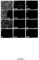
- Figure 1 shows that O-2A progenitors are NH-resident cells
- Bipolar cells migrating out of the adult NH explants in vitro are immunopositive for A2B5 (b) identifying them as O-2A progenitors.
- BrdU able to be incorporated by dividing cells
- c control
- d dehydrated
- d dehydrated
- E explant. Scale bar, 58 ⁇ m.
- Figure 3 shows the in vitro bipotentiality of O-2A progenitors from neonatal rat NH
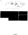
- Figure 4 shows the migration of NH derived cells in host mouse forebrain
- Pieces of newborn hypophysis were labeled with Hoechst to identify their nuclei and implanted in mouse.
- Figure 5 shows the differentiation potential of newborn mice NH derived cells in vivo
- Immunofluorescence analysis on sections of host brains was performed 21 days after transplantation of NH-derived cells- in the cerebral periventricular area of newborn mice to identify the phenotype. These sections contain Hoechst+ cells (in blue), cells labeled with the anti-CNPase (a-c in green), -GFAP (e-g in red) or -NeuN (i-k in green) antibodies. Examples were selected from the host brain number 9 (table I) in the corpus callosum (a-d) and in the fimbriae (e-h) and from the host brain number 7 (table I) in the corpus callosum (i-l). Arrow heads indicate double positive cells. Scale bars : a, e, i, 83 ⁇ m; b-d, f-h, j-l, 33 ⁇ m.
- progenitor cell refers to an undifferentiated cell which is able to proliferate and to give rise to more progenitor cells having the ability to generate a high number of cells that differentiate into fully differentiated cells, that are called their progeny, exhibiting specialized characters.
- a neurohypohysis O-2A progenitor cells refers to a progenitor cell population arising in the neural lobe of the neurohypophysis and giving rise to a differentiated progeny, namely mature functional oligodendrocytes and type 2 astrocytes. Indifferently « O-2A progenitors » or « O-2A progenitor cells » will be used.
- explant refers to a portin of an organ, the neurohypohisis, taken from the mammalian body and grown in an artificial medium.
- isolated population of cell means that the cell is extracted from the body. For instance dissection of neural lobe of neurohypophysis is an isolation technique. The step further to isolation is purification in order to obtain a preparation that contains at least 75 % of the cells.
- culture medium refers to a preparation for the culture of living cells.
- a tissue culture refers to the maintenance or growth of tissue, namely an explant, in vitro so as to preserve its structure and function.
- a cell culture refers to a growth of cells in vitro ; the cells proliferate and/or differentiate but do not get organized into tissue.
- the hypothalamo-neurohypophysial system secretes oxytocin and vasopressin, and shows remarkable plasticity in the adult in response to appropriate stimulation.
- the pituicytes are the principal cellular element of the neural lobe of the hypophysis (NH). They have the morphological characteristics of glial cells and are immunoreactive for glial fibrillary acid protein (GFAP) (Salm et al., 1982), and for the early glial markers vimentin (Marin et al., 1989) and S-100 (Cocchia and Miani, 1980).
- GFAP glial fibrillary acid protein
- pituicytes In the resting state pituicytes surround the neurosecretory axons and terminals of the magnocellular neurons from the hypothalamus and could form a physical barrier between the blood vessels and the neurosecretory terminals. When stimulated e.g. by parturition, lactation or dehydration, an increased juxtaposition between axonal terminals and blood vessels leads to a release of neuropeptides into the general circulation.
- the ability of pituicytes to undergo morphological changes in response to physiological stimuli is dependent on their expression of the polysialylated neural cell adhesion molecule (PSA-NCAM) (Theodosis et al, 1991 ; Theodosis and Poulain, 1999).
- PSA-NCAM polysialylated neural cell adhesion molecule
- NH does not contain myelinated axons and thus does not contain any mature oligodendrocytes.
- the applicant has identified a cell population additional to pituicytes, which express both in vitro and in vivo phenotypic markers of O-2A progenitors in the adult and developing rodent neurohypophysis. For this, the applicant has used the main following methods.
- mice Male rats (Sprague-Dawley) and mice (Swiss strain) which were raised in breeding colonies. Postnatal age was calculated counting postnatal (PN) day 0 as the day of birth. Rat and mouse pups from PN0 to PN3 and adult rats were used. All procedures involving the use of animals were performed in accordance with the European animal care guidelines and directives
- Glial fibrillary acidic protein (astrocyte cytoskeletal marker), galactocerebroside (GalC) (oligodendrocyte marker), 04 (oligodendrocyte marker) and A2B5 (oligodendrocyte surface marker) are neural cell specific surface markers able to identify the O-2A progenitors and their progeny.
- Rats were individually housed and maintained for 9 days either with normal drinking water or with 2% NaCl solution.
- Proliferation was estimated by counting BrdU-positive cells on 3 to 4 randomly chosen sections for each of the 3 control and 4 dehydrated rats. Surface area was calculated using Visiolab 2000 software (Biocom) and the data were expressed as number of BrdU + cells per ⁇ m2. The significance of the difference between control and other conditions was calculated with ANOVA using Stat View-Student software. The number of NG2-positive cells was counted on 2 randomly chosen sections from adult rat and compared to the total number of cells identified with the TOPRO3 nuclear marker.
- DMEM Dulbecco Modified Eagle Medium
- FCS fetal calf serum
- Pieces were then explanted on poly-L-lysine-treated glass coverslips in serum free medium (DMEM-F12 medium, supplemented with 100 ⁇ g/ml human transferin, 5 ⁇ g/ml insulin, 100 ⁇ M putrescin, 20 nM progesterone, 30 nM sodium selenite ; DMEM, GIBCO) supplemented with bFGF (10 ng/ml), PDGF-AA (10 ng/ml) and NT3 (10 ng/ml) in the presence of 0.4 % methylcellulose (Sigma, France). Cultures were incubated at 37°C in a 5 % CO2 and 95 % air atmosphere.
- serum free medium DMEM-F12 medium, supplemented with 100 ⁇ g/ml human transferin, 5 ⁇ g/ml insulin, 100 ⁇ M putrescin, 20 nM progesterone, 30 nM sodium selenite ; DMEM, GIBCO
- Brain transplantations of newborn mouse neurohypophyses are made as follows.
- Transplantations were performed as previously described (Vitry et al., 1999) incorporated by reference. Briefly, NH pieces from newborn mice were incubated in DMEM-10 %FCS containing 10 ⁇ g/ml Hoechst 33342 for 30 minutes at 37°C, washed in DMEM and injected into the brains of newborn mice, close to the subventricular zone of the left hemisphere. Mice were sacrificed at different times after transplantation (5, 15, 21 days) and brains were processed for cryostat sectioning. Serial sagital sections (14 ⁇ m) were collected and Hoechst-labeled cells were detected under UV light. Sections containing Hoechst-positive cells were used for immunofluorescence labeling as described by Vitry et al.
- NG2+ cells (approximately 9 % of the total cell number) were vimentin- (Fig. 1a) and S-100-(Fig. 1b) negative representing a cell population distinct from the pituicytes.
- the A2B5 antibody was used as a second marker for this population.
- motile cells have a bipolar morphology, round cell bodies, two long processes (Fig. 2a) and are A2B5+ (Fig. 2b). In control rat explants, A2B5+/BrdU+ cells are never observed among the A2B5+ migratory population (Fig. 2c).
- fibroblasts and endothelial cells around the explant were observed. Immunofluorescence labeling showed that the majority of cells on the top of this monolayer were GFAP+ (Fig. 3b).
- newborn NHs were cultured in defined medium supplemented with bFGF, PDGF and NT3. Fibroblasts, endothelial and bipolar cells (Wang et al., 1994) were observed among the migrating cells and A2B5 labeling was used to identify O-2A progenitors among these cells (Fig. 3c). Differentiation toward the oligodendrocyte lineage was monitored by analyzing expression of stage-specific markers.
- the migrating bipolar cells were both PSA-NCAM+ (Figs. 3d and 3e) and A2B5+ (Fig. 3f), whereas rare multipolar cells expressed the 04 antigen (Fig. 3g).
- PSA-NCAM+ cells Forty-eight hours after explantation in supplemented defined medium, PSA-NCAM+ cells were rarely found (not shown), while the majority of cells were multipolar, A2B5+ (Figs. 3h and 3i) and O4+ (Fig. 3j).
- cells had the typical morphology of mature oligodendrocytes, they expressed the 04 (Fig. 3k) and GalC (Fig. 31) antigens, and they showed MBP labeling (Fig. 3m).
- O-2A progenitors from newborn rat NH are able to give rise in vitro to either mature oligodendrocytes or to astrocytes.
- Newborn mouse NH explants were Hoechst-labeled and transplanted into the periventricular zone of newborn mouse brains as described in the methods. Three transplanted brains out of 14 were lacking Hoechst+ cells and were therefore excluded from the analysis. After different incubation period the other recipients were found to contain Hoechst+ cells (Table I) near the injection site i.e.
- the + and ++ symbols represent a semi-quantitative indication of the number of Hoechst+ cells found in each cerebral region.
- G location of the graft.
- Hoechst+ cells were found which expressed CNPase (Figs. 5a-5d) or GFAP (Figs. 5e-5h) in various cerebral locations. This result demonstrates that cells from the NH are able to give rise in vivo to both astrocytes and mature oligodendrocytes. Very surprisingly, using an anti-NeuN antibody (a specific neuronal marker) it was shown that some Hoechst+ cells also differentiated as neurons. These cells were localized in the neurogenic cerebral cortex and sometime in the corpus callosum migrating from the lateral wall to the cortex (Figs. 5i-5l).
- NHs grafted in newborn mouse brain could generate neurons in the complete absence of neuronal progenitors.
- Heterotypic transplantations of the neurohypophysis into neonatal brain revealed the presence of pluripotent cells able to generate neurons as well as astrocytes and oligodendrocytes. It is not clear whether these neurons are derived from O-2A progenitors or from the pituycites but in any case, the transplantation of the pieces of isolated neurohypophysis is efficient to generate neurons in vivo.
- both the neuroectodermal and focal origin of the NH is consistent with the presence of O-2A progenitors in this structure.
- O-2A progenitors are present in neonatal rats while the neural lobe is still developing, suggesting that these cells participate in the formation of the gland.
- Both the present studies in vivo and in vitro show that O-2A progenitors are resident cells of the NH, strongly suggesting that they could give rise to the major cell type of this structure, the pituicyte.
- the NH does not contain oligodendrocyte lineage cells other than the A2B5+/NG2+ O-2A progenitors.
- these cells differentiate into GFAP+ cells, a characteristic of pituicyte.
- the present invention can provide cellular populations of O-2A progenitors isolated from NH which may be cultured in appropriate conditions to regenerate and differenciate into namely oligodendrocytes, astrocytes.
- This culture will be made by appropriated, for instance described in US 5,693,482, the invention allowing to use a new source of O-2A progenitors, meaning the NH.
- the culture medium may be as follows.
- Basal medium consists in Dubelco Modified Eagle Medium (DMEM)/F12 (50/50), supplemented with 100 ⁇ g/ml human transferring, 5 ⁇ g/ml insulin, 100 ⁇ M putrescin, 20 nM progesterone, 30 nM sodium selenite ; (GIBCO, BRL). This medium is supplemented with bFGF (10 ng/ml), PDGF-AA (10 ng/ml) and NT3 (10 ng/ml) for the maintenance of O-2A progenitors in culture.
- DMEM Dubelco Modified Eagle Medium
- F12 50/50
- This medium is supplemented with bFGF (10 ng/ml), PDGF-AA (10 ng/ml) and NT3 (10 ng/ml) for the maintenance of O-2A progenitors in culture.
- the basal medium is supplemented with 15 % fetal calf serum (FCS) and PDGF-AA (10ng/ml).
- FCS fetal calf serum
- PDGF-AA 10ng/ml
- Differentiation of the 0-2A progenitors in oligodendrocytes is obtained in basam medium. Cultures are kept ay 37°C in a 5 % CO2 and 95 % air atmosphere.
- the invention is illustrated using O-2A progenitors issued from the rat.
- O2-A progenitors and their progeny may be isolated from NH from human and non-human primates, equines, canines, felines, bovines, porcines, etc.
- the invention provides cellular preparations comprising an enriched population of O-2A progenitors or their progeny obtained from the culture, differentiation and isolation of O-2A progenitors in appropriate medium.
- the preparation obtained contains a majority of or at least about 75 % of the population selected, and preferably 90 to 95 %. It is reminded that in order to isolate progenitor cells from the neurohypophysis explant, an agent which causes proliferation of the progenitor cells is useful. Different techniques may be used to assess the proliferation (such as DNA synthesis measuring, morphological changes), and to isolate the progenitor cells having proliferated in an explant (such as mechanical isolation, enzymatic digestion of the explant followed by the isolation of the activated progenitor cell population based on specific cell surface markers).
- procedure for separation may use magnetic separation, chromatography, fluorescence activated cell sorting, direct separation using markers such as magnetic beads reacting with a support. It may further be very useful to prepare genetically-engineered mammalian multipotent O-2A progenitors and their progeny, which are cultured in order to grow a sufficient number of cells for in vitro gene transfer followed by in vivo implantation. Nucleic acid sequences encoding genes of interest are introduced into multipotent O-2A progenitors where they are expressed. These genes can include neurotrophic or survival factors, immortalizing oncogenes, marker genes.
- the O-2A progenitors may be immortalized to maintain the cell at a defined developmental stage.
- the present techniques for immortalization typically involve the transfection of an oncogene to the cell. Transfection of the oncogene can be accomplished by appropriated, including using recombinant retroviruses, chemical or physical methods (calcium phosphate calcium-phosphate-mediated transfection, microinjection, insertion of a plasmid present in liposomes). For example, one method is to use an eukaryotic viral vector, such as simian virus 40 (SV40) or bovine papilloma virus, to transiently infect or transform the O-2A progenitors.
- SV40 simian virus 40
- bovine papilloma virus bovine papilloma virus
- Various viral vectors can be utilized for immortalization including adenovirus, adenoassociated virus, herpes virus, vaccinia, retrovirus.
- retroviral vectors in which a single foreign gene can be inserted include Moloney murine, leukemia virus (MoMuLV), Harvey murine sarcoma virus (HaMuSV), murine mammary tumor virus (MuMTV), gibbon ape leukemia virus (GaLV) and Rous Sarcoma Virus (RSV).
- a number of additional retroviral vectors can incorporate multiple genes. All of these vectors can transfer or incorporate a gene for a selectable marker so that transduced cells can be identified and generated.
- Herpes virus-based vectors may also be used to transfer genes into a O-2A progenitors : herpes viruses are capable of establishing a latent infection and an apparently non-pathogenic relationship with some neural cells, such vectorbased on HSV-1, for example, may be used. Similarly, it may be possible to use human and animal viruses that infect cells of the CNS efficiently, such as rabies virus, measles, and other paramyxoviruses and even the human immunodeficiency retrovirus (HIV), to develop useful delivery and expression vectors. When a recombinant retrovirus is engineered to contain an immortalizing oncogene, the oncogene can be any one of those known to immortalize.
- such commonly used immortalizing genes include genes of the myc family (c-myc and v-myc), adenovirus genes (E1a 12s and E1a 13s), the polyoma large T antigen and SV40 large T antigen.
- Marker genes such as the E.coli ⁇ .galactosidase gene, can be introduced into O-2A progenitors allowing the identification of these cells and their progeny.
- Selectable marker genes such as the neomycin phosphoribosyltransferase (neomycin-resistance) may be introduced to provide for a population of genetically-engineered O-2A progenitors which are identified by the ability to grow in the presence of selective pressure (i. e.
- O-2A progenitors and their progeny for identifying drugs, in particular using genes for a receptor molecule.
- neuronal receptors include the receptor which binds dopamine, GABA, adrenaline, noradrenaline, serotonin, glutamate, acetylcholine and various other neuropeptides. Transfer and expression of a particular receptor in O-2A progenitors of specific neural origin, would allow identification of neuroactive drugs and trophic factors which may be useful for the treatment of diseases involving that O-2A progenitors type and that receptor.
- a neuroactive compound which mimics a neurotransmitter and binds to a receptor and exhibits either an antagonistic or agonist effect, thereby inhibiting or stimulating a response in O-2A progenitors, can be identified.
- the introduction in a patient treated of O-2A progenitors and their progeny incorporating a gene unsufficiently expressed by the patient may be useful.
- a gene encoding a neurotrophic factor, such as nerve growth factor, (NGF) would prevent from a degeneration of cholinergic neurons, in particular for treatment of Alzheimer's disease which is characterized by degeneration of the cholinergic neurons of the basal forebrain.
- NGF nerve growth factor
- a gene encoding L-DOPA, the precursor to dopamine, would be useful for treatment of Parkinson's disease which is characterized by a loss of dopamine neurons in the substantia-nigra of the midbrain.
- the introduced gene may also be a gene capable of inhibating factors responsible for cell death in certain areas, for example in case of a stroke, or a gene efficient against a tumor.
- the present invention also provides a method of treating a subject with a cell disorder of the CNS which comprises administering to the subject a therapeutically effective amount of the O-2A progenitors or their progeny.
- the method of treating a subject with a CNS disorder comprises intracerebral grafting of O-2A progenitors, or oligodendrocytes or astrocytes or neurons which have been induced to differentiate from the O-2A progenitors, to the region of the CNS having the disorder.
- O-2A progenitors graft involves typically transplantation of cells into the CNS or into the ventricular cavities or subdurally onto the surface of a host brain. Such methods for grafting are described in Neural Grafting in the Mammalian CNS, Bjorklund and Stenevi, eds., (1985), incorporated by reference herein.
- Procedures include in particular intraparenchymal transplantation, (i.e., within the host brain) achieved by injection or deposition of tissue within the host brain so as to be apposed to the brain parenchyma at the time of transplantation.
- Administration of the O-2A progenitors into selected regions of the recipient subject's brain may be made by :
Landscapes
- Health & Medical Sciences (AREA)
- Engineering & Computer Science (AREA)
- Biomedical Technology (AREA)
- Life Sciences & Earth Sciences (AREA)
- Bioinformatics & Cheminformatics (AREA)
- Chemical & Material Sciences (AREA)
- Organic Chemistry (AREA)
- Genetics & Genomics (AREA)
- Biotechnology (AREA)
- Zoology (AREA)
- Wood Science & Technology (AREA)
- Neurosurgery (AREA)
- Neurology (AREA)
- General Health & Medical Sciences (AREA)
- General Engineering & Computer Science (AREA)
- Biochemistry (AREA)
- Microbiology (AREA)
- Cell Biology (AREA)
- Pharmacology & Pharmacy (AREA)
- Medicinal Chemistry (AREA)
- Chemical Kinetics & Catalysis (AREA)
- Developmental Biology & Embryology (AREA)
- Public Health (AREA)
- Animal Behavior & Ethology (AREA)
- General Chemical & Material Sciences (AREA)
- Veterinary Medicine (AREA)
- Nuclear Medicine, Radiotherapy & Molecular Imaging (AREA)
- Psychiatry (AREA)
- Hospice & Palliative Care (AREA)
- Psychology (AREA)
- Micro-Organisms Or Cultivation Processes Thereof (AREA)
- Medicines Containing Material From Animals Or Micro-Organisms (AREA)
- Investigating Or Analysing Biological Materials (AREA)
- Measuring Or Testing Involving Enzymes Or Micro-Organisms (AREA)
- Materials For Medical Uses (AREA)
Priority Applications (7)
| Application Number | Priority Date | Filing Date | Title |
|---|---|---|---|
| EP01400341A EP1231263A1 (de) | 2001-02-09 | 2001-02-09 | Pluripotente O-2A Vorläuferzellen aus der Neurohypophyse |
| AU2002253453A AU2002253453A1 (en) | 2001-02-09 | 2002-02-08 | Multipotent o-2a progenitors from the neurohypophysis |
| JP2002563304A JP2004528019A (ja) | 2001-02-09 | 2002-02-08 | 神経下垂体からのo−2a前駆多能性細胞 |
| CA002437648A CA2437648A1 (en) | 2001-02-09 | 2002-02-08 | Multipotent o-2a progenitors from the neurohypophysis |
| EP20020722574 EP1360277A2 (de) | 2001-02-09 | 2002-02-08 | Pluripotente o-2a vorläuferzellen aus der neurohypophyse |
| US10/467,470 US20040115807A1 (en) | 2001-02-09 | 2002-02-08 | O-2a progenitors multipotent cells from neurohypophysis |
| PCT/IB2002/001226 WO2002062967A2 (en) | 2001-02-09 | 2002-02-08 | Multipotent o-2a progenitors from the neurohypophysis |
Applications Claiming Priority (1)
| Application Number | Priority Date | Filing Date | Title |
|---|---|---|---|
| EP01400341A EP1231263A1 (de) | 2001-02-09 | 2001-02-09 | Pluripotente O-2A Vorläuferzellen aus der Neurohypophyse |
Publications (1)
| Publication Number | Publication Date |
|---|---|
| EP1231263A1 true EP1231263A1 (de) | 2002-08-14 |
Family
ID=8182618
Family Applications (2)
| Application Number | Title | Priority Date | Filing Date |
|---|---|---|---|
| EP01400341A Withdrawn EP1231263A1 (de) | 2001-02-09 | 2001-02-09 | Pluripotente O-2A Vorläuferzellen aus der Neurohypophyse |
| EP20020722574 Withdrawn EP1360277A2 (de) | 2001-02-09 | 2002-02-08 | Pluripotente o-2a vorläuferzellen aus der neurohypophyse |
Family Applications After (1)
| Application Number | Title | Priority Date | Filing Date |
|---|---|---|---|
| EP20020722574 Withdrawn EP1360277A2 (de) | 2001-02-09 | 2002-02-08 | Pluripotente o-2a vorläuferzellen aus der neurohypophyse |
Country Status (6)
| Country | Link |
|---|---|
| US (1) | US20040115807A1 (de) |
| EP (2) | EP1231263A1 (de) |
| JP (1) | JP2004528019A (de) |
| AU (1) | AU2002253453A1 (de) |
| CA (1) | CA2437648A1 (de) |
| WO (1) | WO2002062967A2 (de) |
Cited By (1)
| Publication number | Priority date | Publication date | Assignee | Title |
|---|---|---|---|---|
| CN111474358A (zh) * | 2020-04-16 | 2020-07-31 | 武汉巴菲尔生物技术服务有限公司 | 一种3d立体免疫荧光染色试剂盒及其应用 |
Citations (1)
| Publication number | Priority date | Publication date | Assignee | Title |
|---|---|---|---|---|
| US5693482A (en) * | 1992-07-27 | 1997-12-02 | California Institute Of Technology | Neural chest stem cell assay |
-
2001
- 2001-02-09 EP EP01400341A patent/EP1231263A1/de not_active Withdrawn
-
2002
- 2002-02-08 AU AU2002253453A patent/AU2002253453A1/en not_active Abandoned
- 2002-02-08 EP EP20020722574 patent/EP1360277A2/de not_active Withdrawn
- 2002-02-08 WO PCT/IB2002/001226 patent/WO2002062967A2/en not_active Ceased
- 2002-02-08 CA CA002437648A patent/CA2437648A1/en not_active Abandoned
- 2002-02-08 US US10/467,470 patent/US20040115807A1/en not_active Abandoned
- 2002-02-08 JP JP2002563304A patent/JP2004528019A/ja active Pending
Patent Citations (1)
| Publication number | Priority date | Publication date | Assignee | Title |
|---|---|---|---|---|
| US5693482A (en) * | 1992-07-27 | 1997-12-02 | California Institute Of Technology | Neural chest stem cell assay |
Non-Patent Citations (3)
| Title |
|---|
| KISS JOZEF ZOLTAN: "A role of adhesion molecules in neuroglial plasticity.", MOLECULAR AND CELLULAR ENDOCRINOLOGY, vol. 140, no. 1-2, 25 May 1998 (1998-05-25), pages 89 - 94, XP002165728, ISSN: 0303-7207 * |
| WANG CHENG ET AL: "Functional N-methyl-D-aspartate receptors in O-2A glial precursor cells: A critical role in regulating polysialic acid-neural cell adhesion molecule expression and cell migration.", JOURNAL OF CELL BIOLOGY, vol. 135, no. 6 PART 1, 1996, pages 1565 - 1581, XP002165729, ISSN: 0021-9525 * |
| WANG CHENG ET AL: "Requirement of polysialic acid for the migration of the O-2A glial progenitor cell from neurohypophyseal explants.", JOURNAL OF NEUROSCIENCE, vol. 14, no. 7, 1994, pages 4446 - 4457, XP002165730, ISSN: 0270-6474 * |
Cited By (2)
| Publication number | Priority date | Publication date | Assignee | Title |
|---|---|---|---|---|
| CN111474358A (zh) * | 2020-04-16 | 2020-07-31 | 武汉巴菲尔生物技术服务有限公司 | 一种3d立体免疫荧光染色试剂盒及其应用 |
| CN111474358B (zh) * | 2020-04-16 | 2023-08-18 | 伊莱瑞特(武汉)生物技术有限公司 | 一种3d立体免疫荧光染色试剂盒及其应用 |
Also Published As
| Publication number | Publication date |
|---|---|
| AU2002253453A1 (en) | 2002-08-19 |
| WO2002062967A3 (en) | 2002-12-12 |
| WO2002062967A2 (en) | 2002-08-15 |
| JP2004528019A (ja) | 2004-09-16 |
| EP1360277A2 (de) | 2003-11-12 |
| US20040115807A1 (en) | 2004-06-17 |
| CA2437648A1 (en) | 2002-08-15 |
Similar Documents
| Publication | Publication Date | Title |
|---|---|---|
| Takahashi et al. | Widespread integration and survival of adult-derived neural progenitor cells in the developing optic retina | |
| JP4848538B2 (ja) | ヒトcns神経幹細胞の培養 | |
| US8501467B2 (en) | Cultures of GFAP+ nestin+ cells that differentiate to neurons | |
| US20050003531A1 (en) | Lineage-restricted neuronal precursors | |
| US20140065227A1 (en) | Neural stem cells derived from induced pluripotent stem cells | |
| US20130017179A1 (en) | Lineage-Restricted Neuronal Precursors | |
| EP0681477A4 (de) | Genetische modifikation von neuralen stammzellen. | |
| KR20040084911A (ko) | 희소돌기아교세포 선조 세포를 이용한 선천적으로미엘린화되지 않은 전뇌의 미엘린화 | |
| CN1325637C (zh) | 室管膜神经干细胞及其分离方法 | |
| US20050214941A1 (en) | Expansion of neural stem cells with LIF | |
| JP3636474B2 (ja) | 正常神経上皮前駆細胞 | |
| US20040115807A1 (en) | O-2a progenitors multipotent cells from neurohypophysis | |
| US20040110288A1 (en) | Postnatal neural crest stem cells | |
| CA2213780C (en) | Pharmaceuticals containing multipotential precursor cells from tissues containing sensory receptors | |
| US20050186184A1 (en) | Mammalian pluripotent neural cells and uses thereof | |
| Hargus | Analysis of murine embryonic stem cells overexpressing the extracellular matrix molecule tenascin-R in vitro and after transplantation in a mouse model of Huntington's disease | |
| MXPA00000225A (en) | Lineage-restricted neuronal precursors | |
| MXPA97003493A (en) | In vitro induction of dopaminergi cells |
Legal Events
| Date | Code | Title | Description |
|---|---|---|---|
| PUAI | Public reference made under article 153(3) epc to a published international application that has entered the european phase |
Free format text: ORIGINAL CODE: 0009012 |
|
| AK | Designated contracting states |
Kind code of ref document: A1 Designated state(s): AT BE CH CY DE DK ES FI FR GB GR IE IT LI LU MC NL PT SE TR |
|
| AX | Request for extension of the european patent |
Free format text: AL;LT;LV;MK;RO;SI |
|
| 17P | Request for examination filed |
Effective date: 20030213 |
|
| AKX | Designation fees paid |
Designated state(s): AT BE CH CY DE DK ES FI FR GB GR IE IT LI LU MC NL PT SE TR |
|
| STAA | Information on the status of an ep patent application or granted ep patent |
Free format text: STATUS: THE APPLICATION HAS BEEN WITHDRAWN |
|
| 18W | Application withdrawn |
Effective date: 20030722 |