EP1237621B1 - Adaptive modulation von einem elektrischen feld für neuronale systeme - Google Patents
Adaptive modulation von einem elektrischen feld für neuronale systeme Download PDFInfo
- Publication number
- EP1237621B1 EP1237621B1 EP00983919A EP00983919A EP1237621B1 EP 1237621 B1 EP1237621 B1 EP 1237621B1 EP 00983919 A EP00983919 A EP 00983919A EP 00983919 A EP00983919 A EP 00983919A EP 1237621 B1 EP1237621 B1 EP 1237621B1
- Authority
- EP
- European Patent Office
- Prior art keywords
- activity
- field
- electric field
- neural system
- neuronal activity
- Prior art date
- Legal status (The legal status is an assumption and is not a legal conclusion. Google has not performed a legal analysis and makes no representation as to the accuracy of the status listed.)
- Expired - Lifetime
Links
- 230000001537 neural effect Effects 0.000 title claims abstract description 149
- 230000005684 electric field Effects 0.000 title claims abstract description 55
- 230000003044 adaptive effect Effects 0.000 title description 8
- 230000000694 effects Effects 0.000 claims abstract description 153
- 210000002569 neuron Anatomy 0.000 claims abstract description 25
- 210000004556 brain Anatomy 0.000 claims abstract description 19
- 230000004044 response Effects 0.000 claims abstract description 10
- 238000005259 measurement Methods 0.000 claims description 16
- 230000001787 epileptiform Effects 0.000 claims description 8
- 238000012544 monitoring process Methods 0.000 claims description 8
- 230000002269 spontaneous effect Effects 0.000 claims description 6
- 238000010304 firing Methods 0.000 claims description 4
- 230000008859 change Effects 0.000 claims description 3
- 206010013752 Drug withdrawal convulsions Diseases 0.000 claims description 2
- 238000000034 method Methods 0.000 abstract description 28
- 206010015037 epilepsy Diseases 0.000 abstract description 9
- 208000014644 Brain disease Diseases 0.000 abstract description 6
- 208000037265 diseases, disorders, signs and symptoms Diseases 0.000 abstract description 6
- 208000018737 Parkinson disease Diseases 0.000 abstract description 4
- 230000002159 abnormal effect Effects 0.000 abstract description 4
- 238000011065 in-situ storage Methods 0.000 abstract description 4
- 230000037023 motor activity Effects 0.000 abstract description 4
- 238000012360 testing method Methods 0.000 abstract description 3
- 230000003925 brain function Effects 0.000 abstract description 2
- 230000003955 neuronal function Effects 0.000 abstract description 2
- 239000002831 pharmacologic agent Substances 0.000 abstract description 2
- 230000035807 sensation Effects 0.000 abstract description 2
- 230000000392 somatic effect Effects 0.000 abstract description 2
- 239000003814 drug Substances 0.000 abstract 1
- 229940079593 drug Drugs 0.000 abstract 1
- 230000000007 visual effect Effects 0.000 abstract 1
- 206010010904 Convulsion Diseases 0.000 description 71
- 238000002474 experimental method Methods 0.000 description 31
- 230000001629 suppression Effects 0.000 description 13
- 210000001519 tissue Anatomy 0.000 description 13
- 238000000338 in vitro Methods 0.000 description 11
- 230000006399 behavior Effects 0.000 description 10
- 210000005013 brain tissue Anatomy 0.000 description 9
- 229910021607 Silver chloride Inorganic materials 0.000 description 8
- 238000001514 detection method Methods 0.000 description 8
- 230000003595 spectral effect Effects 0.000 description 8
- HTXDPTMKBJXEOW-UHFFFAOYSA-N dioxoiridium Chemical compound O=[Ir]=O HTXDPTMKBJXEOW-UHFFFAOYSA-N 0.000 description 7
- 230000001037 epileptic effect Effects 0.000 description 7
- 230000006870 function Effects 0.000 description 7
- 210000001320 hippocampus Anatomy 0.000 description 7
- 229910000457 iridium oxide Inorganic materials 0.000 description 7
- 230000033001 locomotion Effects 0.000 description 7
- 241000700159 Rattus Species 0.000 description 6
- 238000001228 spectrum Methods 0.000 description 6
- 230000000638 stimulation Effects 0.000 description 6
- CURLTUGMZLYLDI-UHFFFAOYSA-N Carbon dioxide Chemical compound O=C=O CURLTUGMZLYLDI-UHFFFAOYSA-N 0.000 description 5
- ZLMJMSJWJFRBEC-UHFFFAOYSA-N Potassium Chemical compound [K] ZLMJMSJWJFRBEC-UHFFFAOYSA-N 0.000 description 5
- 210000004027 cell Anatomy 0.000 description 5
- 201000010099 disease Diseases 0.000 description 5
- 238000009826 distribution Methods 0.000 description 5
- 238000001727 in vivo Methods 0.000 description 5
- 230000000977 initiatory effect Effects 0.000 description 5
- 230000010412 perfusion Effects 0.000 description 5
- 239000011591 potassium Substances 0.000 description 5
- 229910052700 potassium Inorganic materials 0.000 description 5
- 238000004458 analytical method Methods 0.000 description 4
- 230000003542 behavioural effect Effects 0.000 description 4
- 210000001175 cerebrospinal fluid Anatomy 0.000 description 4
- 239000003795 chemical substances by application Substances 0.000 description 4
- 230000003247 decreasing effect Effects 0.000 description 4
- VYFYYTLLBUKUHU-UHFFFAOYSA-N dopamine Chemical compound NCCC1=CC=C(O)C(O)=C1 VYFYYTLLBUKUHU-UHFFFAOYSA-N 0.000 description 4
- 230000000971 hippocampal effect Effects 0.000 description 4
- 230000008062 neuronal firing Effects 0.000 description 4
- 230000001360 synchronised effect Effects 0.000 description 4
- 210000001103 thalamus Anatomy 0.000 description 4
- RTZKZFJDLAIYFH-UHFFFAOYSA-N Diethyl ether Chemical compound CCOCC RTZKZFJDLAIYFH-UHFFFAOYSA-N 0.000 description 3
- 229910002092 carbon dioxide Inorganic materials 0.000 description 3
- 210000005056 cell body Anatomy 0.000 description 3
- 238000002635 electroconvulsive therapy Methods 0.000 description 3
- 238000000605 extraction Methods 0.000 description 3
- 238000001914 filtration Methods 0.000 description 3
- 238000002955 isolation Methods 0.000 description 3
- 239000000463 material Substances 0.000 description 3
- 230000000737 periodic effect Effects 0.000 description 3
- BASFCYQUMIYNBI-UHFFFAOYSA-N platinum Chemical compound [Pt] BASFCYQUMIYNBI-UHFFFAOYSA-N 0.000 description 3
- 230000010287 polarization Effects 0.000 description 3
- 230000008569 process Effects 0.000 description 3
- 230000001953 sensory effect Effects 0.000 description 3
- KWGRBVOPPLSCSI-WPRPVWTQSA-N (-)-ephedrine Chemical compound CN[C@@H](C)[C@H](O)C1=CC=CC=C1 KWGRBVOPPLSCSI-WPRPVWTQSA-N 0.000 description 2
- KWTSXDURSIMDCE-QMMMGPOBSA-N (S)-amphetamine Chemical compound C[C@H](N)CC1=CC=CC=C1 KWTSXDURSIMDCE-QMMMGPOBSA-N 0.000 description 2
- 108091006146 Channels Proteins 0.000 description 2
- 241000282412 Homo Species 0.000 description 2
- CSNNHWWHGAXBCP-UHFFFAOYSA-L Magnesium sulfate Chemical compound [Mg+2].[O-][S+2]([O-])([O-])[O-] CSNNHWWHGAXBCP-UHFFFAOYSA-L 0.000 description 2
- 241001465754 Metazoa Species 0.000 description 2
- 239000004677 Nylon Substances 0.000 description 2
- 208000021384 Obsessive-Compulsive disease Diseases 0.000 description 2
- 108010038988 Peptide Hormones Proteins 0.000 description 2
- 102000015731 Peptide Hormones Human genes 0.000 description 2
- 206010044565 Tremor Diseases 0.000 description 2
- 230000001594 aberrant effect Effects 0.000 description 2
- 230000036982 action potential Effects 0.000 description 2
- 238000013459 approach Methods 0.000 description 2
- QVGXLLKOCUKJST-UHFFFAOYSA-N atomic oxygen Chemical compound [O] QVGXLLKOCUKJST-UHFFFAOYSA-N 0.000 description 2
- 230000003190 augmentative effect Effects 0.000 description 2
- 210000004227 basal ganglia Anatomy 0.000 description 2
- 239000000872 buffer Substances 0.000 description 2
- 230000009172 bursting Effects 0.000 description 2
- 239000001569 carbon dioxide Substances 0.000 description 2
- 210000001638 cerebellum Anatomy 0.000 description 2
- 230000001186 cumulative effect Effects 0.000 description 2
- 229960003638 dopamine Drugs 0.000 description 2
- 230000002964 excitative effect Effects 0.000 description 2
- 230000001747 exhibiting effect Effects 0.000 description 2
- 239000012530 fluid Substances 0.000 description 2
- 239000007943 implant Substances 0.000 description 2
- 230000001965 increasing effect Effects 0.000 description 2
- 238000011534 incubation Methods 0.000 description 2
- 230000003834 intracellular effect Effects 0.000 description 2
- 150000002503 iridium Chemical class 0.000 description 2
- 230000007774 longterm Effects 0.000 description 2
- 230000000873 masking effect Effects 0.000 description 2
- 230000007246 mechanism Effects 0.000 description 2
- 239000012528 membrane Substances 0.000 description 2
- 230000028161 membrane depolarization Effects 0.000 description 2
- 229910052751 metal Inorganic materials 0.000 description 2
- 239000002184 metal Substances 0.000 description 2
- 210000000478 neocortex Anatomy 0.000 description 2
- 210000005036 nerve Anatomy 0.000 description 2
- 229920001778 nylon Polymers 0.000 description 2
- 230000003534 oscillatory effect Effects 0.000 description 2
- 229910052760 oxygen Inorganic materials 0.000 description 2
- 239000001301 oxygen Substances 0.000 description 2
- 229910052697 platinum Inorganic materials 0.000 description 2
- 238000002360 preparation method Methods 0.000 description 2
- 230000001902 propagating effect Effects 0.000 description 2
- 210000002763 pyramidal cell Anatomy 0.000 description 2
- 230000002829 reductive effect Effects 0.000 description 2
- QZAYGJVTTNCVMB-UHFFFAOYSA-N serotonin Chemical compound C1=C(O)C=C2C(CCN)=CNC2=C1 QZAYGJVTTNCVMB-UHFFFAOYSA-N 0.000 description 2
- 229920000260 silastic Polymers 0.000 description 2
- 239000000243 solution Substances 0.000 description 2
- 210000000130 stem cell Anatomy 0.000 description 2
- 239000000758 substrate Substances 0.000 description 2
- 208000024891 symptom Diseases 0.000 description 2
- 230000002123 temporal effect Effects 0.000 description 2
- 210000003478 temporal lobe Anatomy 0.000 description 2
- 230000001225 therapeutic effect Effects 0.000 description 2
- 238000002560 therapeutic procedure Methods 0.000 description 2
- 210000000857 visual cortex Anatomy 0.000 description 2
- 206010001497 Agitation Diseases 0.000 description 1
- 229910000851 Alloy steel Inorganic materials 0.000 description 1
- 208000019901 Anxiety disease Diseases 0.000 description 1
- 241000238421 Arthropoda Species 0.000 description 1
- 238000012935 Averaging Methods 0.000 description 1
- BVKZGUZCCUSVTD-UHFFFAOYSA-M Bicarbonate Chemical compound OC([O-])=O BVKZGUZCCUSVTD-UHFFFAOYSA-M 0.000 description 1
- 241000282472 Canis lupus familiaris Species 0.000 description 1
- 244000025254 Cannabis sativa Species 0.000 description 1
- 241000282693 Cercopithecidae Species 0.000 description 1
- VEXZGXHMUGYJMC-UHFFFAOYSA-M Chloride anion Chemical compound [Cl-] VEXZGXHMUGYJMC-UHFFFAOYSA-M 0.000 description 1
- CWYNVVGOOAEACU-UHFFFAOYSA-N Fe2+ Chemical compound [Fe+2] CWYNVVGOOAEACU-UHFFFAOYSA-N 0.000 description 1
- 241000282326 Felis catus Species 0.000 description 1
- 208000003098 Ganglion Cysts Diseases 0.000 description 1
- WQZGKKKJIJFFOK-GASJEMHNSA-N Glucose Natural products OC[C@H]1OC(O)[C@H](O)[C@@H](O)[C@@H]1O WQZGKKKJIJFFOK-GASJEMHNSA-N 0.000 description 1
- 241000238631 Hexapoda Species 0.000 description 1
- 241000282418 Hominidae Species 0.000 description 1
- 229910000575 Ir alloy Inorganic materials 0.000 description 1
- WHUUTDBJXJRKMK-VKHMYHEASA-N L-glutamic acid Chemical compound OC(=O)[C@@H](N)CCC(O)=O WHUUTDBJXJRKMK-VKHMYHEASA-N 0.000 description 1
- FYYHWMGAXLPEAU-UHFFFAOYSA-N Magnesium Chemical compound [Mg] FYYHWMGAXLPEAU-UHFFFAOYSA-N 0.000 description 1
- 241000124008 Mammalia Species 0.000 description 1
- FWJKNZONDWOGMI-UHFFFAOYSA-N Metharbital Chemical compound CCC1(CC)C(=O)NC(=O)N(C)C1=O FWJKNZONDWOGMI-UHFFFAOYSA-N 0.000 description 1
- AJXPJJZHWIXJCJ-UHFFFAOYSA-N Methsuximide Chemical compound O=C1N(C)C(=O)CC1(C)C1=CC=CC=C1 AJXPJJZHWIXJCJ-UHFFFAOYSA-N 0.000 description 1
- 241000237852 Mollusca Species 0.000 description 1
- 241000699670 Mus sp. Species 0.000 description 1
- 238000005481 NMR spectroscopy Methods 0.000 description 1
- 229940084576 Neurotransmitter agonist Drugs 0.000 description 1
- 229940123247 Neurotransmitter antagonist Drugs 0.000 description 1
- 241000282579 Pan Species 0.000 description 1
- 206010033799 Paralysis Diseases 0.000 description 1
- 229930182555 Penicillin Natural products 0.000 description 1
- JGSARLDLIJGVTE-MBNYWOFBSA-N Penicillin G Chemical compound N([C@H]1[C@H]2SC([C@@H](N2C1=O)C(O)=O)(C)C)C(=O)CC1=CC=CC=C1 JGSARLDLIJGVTE-MBNYWOFBSA-N 0.000 description 1
- WLWFNJKHKGIJNW-UHFFFAOYSA-N Phensuximide Chemical compound O=C1N(C)C(=O)CC1C1=CC=CC=C1 WLWFNJKHKGIJNW-UHFFFAOYSA-N 0.000 description 1
- CXOFVDLJLONNDW-UHFFFAOYSA-N Phenytoin Chemical compound N1C(=O)NC(=O)C1(C=1C=CC=CC=1)C1=CC=CC=C1 CXOFVDLJLONNDW-UHFFFAOYSA-N 0.000 description 1
- 206010040030 Sensory loss Diseases 0.000 description 1
- XUIMIQQOPSSXEZ-UHFFFAOYSA-N Silicon Chemical compound [Si] XUIMIQQOPSSXEZ-UHFFFAOYSA-N 0.000 description 1
- FAPWRFPIFSIZLT-UHFFFAOYSA-M Sodium chloride Chemical compound [Na+].[Cl-] FAPWRFPIFSIZLT-UHFFFAOYSA-M 0.000 description 1
- 229910000831 Steel Inorganic materials 0.000 description 1
- 238000000692 Student's t-test Methods 0.000 description 1
- 239000000150 Sympathomimetic Substances 0.000 description 1
- 208000005400 Synovial Cyst Diseases 0.000 description 1
- GWEVSGVZZGPLCZ-UHFFFAOYSA-N Titan oxide Chemical compound O=[Ti]=O GWEVSGVZZGPLCZ-UHFFFAOYSA-N 0.000 description 1
- RTAQQCXQSZGOHL-UHFFFAOYSA-N Titanium Chemical compound [Ti] RTAQQCXQSZGOHL-UHFFFAOYSA-N 0.000 description 1
- NIJJYAXOARWZEE-UHFFFAOYSA-N Valproic acid Chemical compound CCCC(C(O)=O)CCC NIJJYAXOARWZEE-UHFFFAOYSA-N 0.000 description 1
- 241000251539 Vertebrata <Metazoa> Species 0.000 description 1
- YJZATOSJMRIRIW-UHFFFAOYSA-N [Ir]=O Chemical class [Ir]=O YJZATOSJMRIRIW-UHFFFAOYSA-N 0.000 description 1
- BZKPWHYZMXOIDC-UHFFFAOYSA-N acetazolamide Chemical compound CC(=O)NC1=NN=C(S(N)(=O)=O)S1 BZKPWHYZMXOIDC-UHFFFAOYSA-N 0.000 description 1
- 229960000571 acetazolamide Drugs 0.000 description 1
- 238000001994 activation Methods 0.000 description 1
- 230000006978 adaptation Effects 0.000 description 1
- 230000001800 adrenalinergic effect Effects 0.000 description 1
- 239000000464 adrenergic agent Substances 0.000 description 1
- VLSMHEGGTFMBBZ-UHFFFAOYSA-N alpha-Kainic acid Natural products CC(=C)C1CNC(C(O)=O)C1CC(O)=O VLSMHEGGTFMBBZ-UHFFFAOYSA-N 0.000 description 1
- 229940025084 amphetamine Drugs 0.000 description 1
- 230000003321 amplification Effects 0.000 description 1
- 210000004727 amygdala Anatomy 0.000 description 1
- 210000003484 anatomy Anatomy 0.000 description 1
- 238000010171 animal model Methods 0.000 description 1
- 239000005557 antagonist Substances 0.000 description 1
- 230000003556 anti-epileptic effect Effects 0.000 description 1
- 230000000692 anti-sense effect Effects 0.000 description 1
- 230000002921 anti-spasmodic effect Effects 0.000 description 1
- 239000001961 anticonvulsive agent Substances 0.000 description 1
- 229960003965 antiepileptics Drugs 0.000 description 1
- 229940124575 antispasmodic agent Drugs 0.000 description 1
- 230000036506 anxiety Effects 0.000 description 1
- 238000003491 array Methods 0.000 description 1
- 239000012298 atmosphere Substances 0.000 description 1
- 230000002238 attenuated effect Effects 0.000 description 1
- 210000003926 auditory cortex Anatomy 0.000 description 1
- 230000015572 biosynthetic process Effects 0.000 description 1
- 230000007177 brain activity Effects 0.000 description 1
- 210000000133 brain stem Anatomy 0.000 description 1
- FFGPTBGBLSHEPO-UHFFFAOYSA-N carbamazepine Chemical compound C1=CC2=CC=CC=C2N(C(=O)N)C2=CC=CC=C21 FFGPTBGBLSHEPO-UHFFFAOYSA-N 0.000 description 1
- 229960000623 carbamazepine Drugs 0.000 description 1
- 239000000969 carrier Substances 0.000 description 1
- 238000006243 chemical reaction Methods 0.000 description 1
- ZPEIMTDSQAKGNT-UHFFFAOYSA-N chlorpromazine Chemical compound C1=C(Cl)C=C2N(CCCN(C)C)C3=CC=CC=C3SC2=C1 ZPEIMTDSQAKGNT-UHFFFAOYSA-N 0.000 description 1
- 230000001713 cholinergic effect Effects 0.000 description 1
- -1 cholinergics Substances 0.000 description 1
- 229960004362 clorazepate Drugs 0.000 description 1
- XDDJGVMJFWAHJX-UHFFFAOYSA-M clorazepic acid anion Chemical compound C12=CC(Cl)=CC=C2NC(=O)C(C(=O)[O-])N=C1C1=CC=CC=C1 XDDJGVMJFWAHJX-UHFFFAOYSA-M 0.000 description 1
- 239000011248 coating agent Substances 0.000 description 1
- 238000000576 coating method Methods 0.000 description 1
- 210000003477 cochlea Anatomy 0.000 description 1
- 150000001875 compounds Chemical class 0.000 description 1
- 238000002591 computed tomography Methods 0.000 description 1
- 230000001054 cortical effect Effects 0.000 description 1
- 210000003792 cranial nerve Anatomy 0.000 description 1
- 125000004122 cyclic group Chemical group 0.000 description 1
- KWGRBVOPPLSCSI-UHFFFAOYSA-N d-ephedrine Natural products CNC(C)C(O)C1=CC=CC=C1 KWGRBVOPPLSCSI-UHFFFAOYSA-N 0.000 description 1
- 230000007547 defect Effects 0.000 description 1
- 230000000994 depressogenic effect Effects 0.000 description 1
- 229960000632 dexamfetamine Drugs 0.000 description 1
- 239000008121 dextrose Substances 0.000 description 1
- 229960004132 diethyl ether Drugs 0.000 description 1
- MZDOIJOUFRQXHC-UHFFFAOYSA-N dimenhydrinate Chemical compound O=C1N(C)C(=O)N(C)C2=NC(Cl)=N[C]21.C=1C=CC=CC=1C(OCCN(C)C)C1=CC=CC=C1 MZDOIJOUFRQXHC-UHFFFAOYSA-N 0.000 description 1
- 229960004993 dimenhydrinate Drugs 0.000 description 1
- 230000003292 diminished effect Effects 0.000 description 1
- 230000000792 effect on seizure Effects 0.000 description 1
- 230000002996 emotional effect Effects 0.000 description 1
- 238000005516 engineering process Methods 0.000 description 1
- 230000002708 enhancing effect Effects 0.000 description 1
- 229960002179 ephedrine Drugs 0.000 description 1
- 208000028329 epileptic seizure Diseases 0.000 description 1
- 230000002397 epileptogenic effect Effects 0.000 description 1
- HAPOVYFOVVWLRS-UHFFFAOYSA-N ethosuximide Chemical compound CCC1(C)CC(=O)NC1=O HAPOVYFOVVWLRS-UHFFFAOYSA-N 0.000 description 1
- 229960002767 ethosuximide Drugs 0.000 description 1
- 230000000763 evoking effect Effects 0.000 description 1
- 230000005284 excitation Effects 0.000 description 1
- 210000001723 extracellular space Anatomy 0.000 description 1
- 210000005153 frontal cortex Anatomy 0.000 description 1
- 229960003692 gamma aminobutyric acid Drugs 0.000 description 1
- BTCSSZJGUNDROE-UHFFFAOYSA-N gamma-aminobutyric acid Chemical compound NCCCC(O)=O BTCSSZJGUNDROE-UHFFFAOYSA-N 0.000 description 1
- 210000000609 ganglia Anatomy 0.000 description 1
- 239000007789 gas Substances 0.000 description 1
- 230000002068 genetic effect Effects 0.000 description 1
- 238000010353 genetic engineering Methods 0.000 description 1
- 239000011521 glass Substances 0.000 description 1
- 229930195712 glutamate Natural products 0.000 description 1
- 229940049906 glutamate Drugs 0.000 description 1
- 229910052737 gold Inorganic materials 0.000 description 1
- 239000010931 gold Substances 0.000 description 1
- FDWREHZXQUYJFJ-UHFFFAOYSA-M gold monochloride Chemical compound [Cl-].[Au+] FDWREHZXQUYJFJ-UHFFFAOYSA-M 0.000 description 1
- 239000003102 growth factor Substances 0.000 description 1
- 239000005556 hormone Substances 0.000 description 1
- 229940088597 hormone Drugs 0.000 description 1
- 230000002102 hyperpolarization Effects 0.000 description 1
- 239000003326 hypnotic agent Substances 0.000 description 1
- 230000000147 hypnotic effect Effects 0.000 description 1
- 210000003016 hypothalamus Anatomy 0.000 description 1
- 230000006698 induction Effects 0.000 description 1
- 230000001939 inductive effect Effects 0.000 description 1
- 238000002347 injection Methods 0.000 description 1
- 239000007924 injection Substances 0.000 description 1
- 238000011835 investigation Methods 0.000 description 1
- 150000002500 ions Chemical class 0.000 description 1
- VLSMHEGGTFMBBZ-OOZYFLPDSA-N kainic acid Chemical compound CC(=C)[C@H]1CN[C@H](C(O)=O)[C@H]1CC(O)=O VLSMHEGGTFMBBZ-OOZYFLPDSA-N 0.000 description 1
- 229950006874 kainic acid Drugs 0.000 description 1
- 239000004922 lacquer Substances 0.000 description 1
- 210000003140 lateral ventricle Anatomy 0.000 description 1
- 230000003902 lesion Effects 0.000 description 1
- 210000003715 limbic system Anatomy 0.000 description 1
- 239000011777 magnesium Substances 0.000 description 1
- 229910052749 magnesium Inorganic materials 0.000 description 1
- 229910052943 magnesium sulfate Inorganic materials 0.000 description 1
- 229960003390 magnesium sulfate Drugs 0.000 description 1
- 235000019341 magnesium sulphate Nutrition 0.000 description 1
- 210000005171 mammalian brain Anatomy 0.000 description 1
- 210000002418 meninge Anatomy 0.000 description 1
- GMHKMTDVRCWUDX-UHFFFAOYSA-N mephenytoin Chemical compound C=1C=CC=CC=1C1(CC)NC(=O)N(C)C1=O GMHKMTDVRCWUDX-UHFFFAOYSA-N 0.000 description 1
- 229960000906 mephenytoin Drugs 0.000 description 1
- 229960003729 mesuximide Drugs 0.000 description 1
- 239000002207 metabolite Substances 0.000 description 1
- 229960002057 metharbital Drugs 0.000 description 1
- 238000004377 microelectronic Methods 0.000 description 1
- 238000012986 modification Methods 0.000 description 1
- 230000004048 modification Effects 0.000 description 1
- 230000000897 modulatory effect Effects 0.000 description 1
- 210000000337 motor cortex Anatomy 0.000 description 1
- 230000007659 motor function Effects 0.000 description 1
- 230000003551 muscarinic effect Effects 0.000 description 1
- 210000003205 muscle Anatomy 0.000 description 1
- 210000001577 neostriatum Anatomy 0.000 description 1
- 210000000653 nervous system Anatomy 0.000 description 1
- 230000036403 neuro physiology Effects 0.000 description 1
- 239000002858 neurotransmitter agent Substances 0.000 description 1
- 229910000510 noble metal Inorganic materials 0.000 description 1
- 231100000252 nontoxic Toxicity 0.000 description 1
- 230000003000 nontoxic effect Effects 0.000 description 1
- 238000003199 nucleic acid amplification method Methods 0.000 description 1
- 210000000869 occipital lobe Anatomy 0.000 description 1
- 230000002853 ongoing effect Effects 0.000 description 1
- ADIMAYPTOBDMTL-UHFFFAOYSA-N oxazepam Chemical compound C12=CC(Cl)=CC=C2NC(=O)C(O)N=C1C1=CC=CC=C1 ADIMAYPTOBDMTL-UHFFFAOYSA-N 0.000 description 1
- 229960004535 oxazepam Drugs 0.000 description 1
- SQYNKIJPMDEDEG-UHFFFAOYSA-N paraldehyde Chemical compound CC1OC(C)OC(C)O1 SQYNKIJPMDEDEG-UHFFFAOYSA-N 0.000 description 1
- 229960003868 paraldehyde Drugs 0.000 description 1
- 210000001152 parietal lobe Anatomy 0.000 description 1
- 230000037361 pathway Effects 0.000 description 1
- 229940049954 penicillin Drugs 0.000 description 1
- 230000002093 peripheral effect Effects 0.000 description 1
- 210000000578 peripheral nerve Anatomy 0.000 description 1
- 229960002695 phenobarbital Drugs 0.000 description 1
- DDBREPKUVSBGFI-UHFFFAOYSA-N phenobarbital Chemical compound C=1C=CC=CC=1C1(CC)C(=O)NC(=O)NC1=O DDBREPKUVSBGFI-UHFFFAOYSA-N 0.000 description 1
- 229960004227 phensuximide Drugs 0.000 description 1
- 229960002036 phenytoin Drugs 0.000 description 1
- HWLDNSXPUQTBOD-UHFFFAOYSA-N platinum-iridium alloy Chemical compound [Ir].[Pt] HWLDNSXPUQTBOD-UHFFFAOYSA-N 0.000 description 1
- 238000012805 post-processing Methods 0.000 description 1
- 229960002393 primidone Drugs 0.000 description 1
- DQMZLTXERSFNPB-UHFFFAOYSA-N primidone Chemical compound C=1C=CC=CC=1C1(CC)C(=O)NCNC1=O DQMZLTXERSFNPB-UHFFFAOYSA-N 0.000 description 1
- 238000012545 processing Methods 0.000 description 1
- 210000001176 projection neuron Anatomy 0.000 description 1
- 230000000069 prophylactic effect Effects 0.000 description 1
- 108090000623 proteins and genes Proteins 0.000 description 1
- 238000005546 reactive sputtering Methods 0.000 description 1
- 108020003175 receptors Proteins 0.000 description 1
- 238000011084 recovery Methods 0.000 description 1
- 230000000306 recurrent effect Effects 0.000 description 1
- 230000009467 reduction Effects 0.000 description 1
- 238000011160 research Methods 0.000 description 1
- 230000000717 retained effect Effects 0.000 description 1
- 210000001525 retina Anatomy 0.000 description 1
- 230000002207 retinal effect Effects 0.000 description 1
- 238000012552 review Methods 0.000 description 1
- 230000033764 rhythmic process Effects 0.000 description 1
- 239000000523 sample Substances 0.000 description 1
- 229920006395 saturated elastomer Polymers 0.000 description 1
- 238000009738 saturating Methods 0.000 description 1
- 210000004761 scalp Anatomy 0.000 description 1
- 201000000980 schizophrenia Diseases 0.000 description 1
- 229940125723 sedative agent Drugs 0.000 description 1
- 239000000932 sedative agent Substances 0.000 description 1
- 230000037152 sensory function Effects 0.000 description 1
- 238000000926 separation method Methods 0.000 description 1
- 229940076279 serotonin Drugs 0.000 description 1
- 229910052710 silicon Inorganic materials 0.000 description 1
- 239000010703 silicon Substances 0.000 description 1
- 229920002379 silicone rubber Polymers 0.000 description 1
- HKZLPVFGJNLROG-UHFFFAOYSA-M silver monochloride Chemical compound [Cl-].[Ag+] HKZLPVFGJNLROG-UHFFFAOYSA-M 0.000 description 1
- 238000004513 sizing Methods 0.000 description 1
- 230000035943 smell Effects 0.000 description 1
- 239000011780 sodium chloride Substances 0.000 description 1
- AEQFSUDEHCCHBT-UHFFFAOYSA-M sodium valproate Chemical compound [Na+].CCCC(C([O-])=O)CCC AEQFSUDEHCCHBT-UHFFFAOYSA-M 0.000 description 1
- 210000004092 somatosensory cortex Anatomy 0.000 description 1
- 210000000278 spinal cord Anatomy 0.000 description 1
- 238000012453 sprague-dawley rat model Methods 0.000 description 1
- 230000006641 stabilisation Effects 0.000 description 1
- 238000011105 stabilization Methods 0.000 description 1
- 239000010935 stainless steel Substances 0.000 description 1
- 229910001220 stainless steel Inorganic materials 0.000 description 1
- 239000010959 steel Substances 0.000 description 1
- 230000001975 sympathomimetic effect Effects 0.000 description 1
- 229940064707 sympathomimetics Drugs 0.000 description 1
- 238000005979 thermal decomposition reaction Methods 0.000 description 1
- 229910052719 titanium Inorganic materials 0.000 description 1
- 239000010936 titanium Substances 0.000 description 1
- OGIDPMRJRNCKJF-UHFFFAOYSA-N titanium oxide Inorganic materials [Ti]=O OGIDPMRJRNCKJF-UHFFFAOYSA-N 0.000 description 1
- 230000001052 transient effect Effects 0.000 description 1
- 238000011282 treatment Methods 0.000 description 1
- 229960004453 trimethadione Drugs 0.000 description 1
- IRYJRGCIQBGHIV-UHFFFAOYSA-N trimethadione Chemical compound CN1C(=O)OC(C)(C)C1=O IRYJRGCIQBGHIV-UHFFFAOYSA-N 0.000 description 1
- WFKWXMTUELFFGS-UHFFFAOYSA-N tungsten Chemical compound [W] WFKWXMTUELFFGS-UHFFFAOYSA-N 0.000 description 1
- 229910052721 tungsten Inorganic materials 0.000 description 1
- 239000010937 tungsten Substances 0.000 description 1
- 230000001515 vagal effect Effects 0.000 description 1
- 229940102566 valproate Drugs 0.000 description 1
- 229960000604 valproic acid Drugs 0.000 description 1
- 238000005303 weighing Methods 0.000 description 1
Images



Classifications
-
- A—HUMAN NECESSITIES
- A61—MEDICAL OR VETERINARY SCIENCE; HYGIENE
- A61N—ELECTROTHERAPY; MAGNETOTHERAPY; RADIATION THERAPY; ULTRASOUND THERAPY
- A61N1/00—Electrotherapy; Circuits therefor
- A61N1/18—Applying electric currents by contact electrodes
- A61N1/32—Applying electric currents by contact electrodes alternating or intermittent currents
- A61N1/36—Applying electric currents by contact electrodes alternating or intermittent currents for stimulation
- A61N1/3605—Implantable neurostimulators for stimulating central or peripheral nerve system
- A61N1/3606—Implantable neurostimulators for stimulating central or peripheral nerve system adapted for a particular treatment
- A61N1/36067—Movement disorders, e.g. tremor or Parkinson disease
-
- A—HUMAN NECESSITIES
- A61—MEDICAL OR VETERINARY SCIENCE; HYGIENE
- A61N—ELECTROTHERAPY; MAGNETOTHERAPY; RADIATION THERAPY; ULTRASOUND THERAPY
- A61N1/00—Electrotherapy; Circuits therefor
- A61N1/18—Applying electric currents by contact electrodes
- A61N1/32—Applying electric currents by contact electrodes alternating or intermittent currents
- A61N1/36—Applying electric currents by contact electrodes alternating or intermittent currents for stimulation
- A61N1/36014—External stimulators, e.g. with patch electrodes
- A61N1/3603—Control systems
- A61N1/36031—Control systems using physiological parameters for adjustment
-
- A—HUMAN NECESSITIES
- A61—MEDICAL OR VETERINARY SCIENCE; HYGIENE
- A61N—ELECTROTHERAPY; MAGNETOTHERAPY; RADIATION THERAPY; ULTRASOUND THERAPY
- A61N1/00—Electrotherapy; Circuits therefor
- A61N1/18—Applying electric currents by contact electrodes
- A61N1/32—Applying electric currents by contact electrodes alternating or intermittent currents
- A61N1/36—Applying electric currents by contact electrodes alternating or intermittent currents for stimulation
- A61N1/36014—External stimulators, e.g. with patch electrodes
- A61N1/3603—Control systems
- A61N1/36034—Control systems specified by the stimulation parameters
-
- A—HUMAN NECESSITIES
- A61—MEDICAL OR VETERINARY SCIENCE; HYGIENE
- A61N—ELECTROTHERAPY; MAGNETOTHERAPY; RADIATION THERAPY; ULTRASOUND THERAPY
- A61N1/00—Electrotherapy; Circuits therefor
- A61N1/18—Applying electric currents by contact electrodes
- A61N1/32—Applying electric currents by contact electrodes alternating or intermittent currents
- A61N1/36—Applying electric currents by contact electrodes alternating or intermittent currents for stimulation
- A61N1/3605—Implantable neurostimulators for stimulating central or peripheral nerve system
- A61N1/3606—Implantable neurostimulators for stimulating central or peripheral nerve system adapted for a particular treatment
- A61N1/36064—Epilepsy
-
- A—HUMAN NECESSITIES
- A61—MEDICAL OR VETERINARY SCIENCE; HYGIENE
- A61N—ELECTROTHERAPY; MAGNETOTHERAPY; RADIATION THERAPY; ULTRASOUND THERAPY
- A61N1/00—Electrotherapy; Circuits therefor
- A61N1/40—Applying electric fields by inductive or capacitive coupling ; Applying radio-frequency signals
-
- A—HUMAN NECESSITIES
- A61—MEDICAL OR VETERINARY SCIENCE; HYGIENE
- A61N—ELECTROTHERAPY; MAGNETOTHERAPY; RADIATION THERAPY; ULTRASOUND THERAPY
- A61N1/00—Electrotherapy; Circuits therefor
- A61N1/18—Applying electric currents by contact electrodes
- A61N1/32—Applying electric currents by contact electrodes alternating or intermittent currents
- A61N1/36—Applying electric currents by contact electrodes alternating or intermittent currents for stimulation
- A61N1/36014—External stimulators, e.g. with patch electrodes
- A61N1/36025—External stimulators, e.g. with patch electrodes for treating a mental or cerebral condition
Definitions
- the present invention relates to devices for modulating the neuronal activity of a neural system comprising neurons, such as a brain, brain regions, or any in vivo or in vitro collection of neurons.
- the present invention involves the use of applied electric fields to modulate the behavior of a target neural system.
- the polarity and magnitude of the applied electric field is varied according to information gathered from the modulated neural system to modulate the strength of the applied electric field.
- a sub-threshold stimulus is administered to modulate to the neural system.
- the devices of the present invention can be used to treat diseases of the nervous system, to restore neuronal function, paralysis, and motor and sensory deficits, to produce prosthetic devices that interact and modulate neuronal activity, to enhance or suppress neuronal activity and associated phenotypes, and the like.
- a neural system in accordance with the present invention can be any ensemble of one or more neurons, and/or other excitable cells, such as muscle, heart, retinal, cochlear, tissue culture cells, stem or progenitor cells, including cell-electrode interface devices and the like. Cells can be coupled electrically, chemically, or combinations thereof.
- the neural system can be an entire brain, ganglia, nerve, etc., or it can be a region or portion of it. Any animal source of material is suitable, including neural systems of invertebrates, such as mollusks, arthropods, insects, etc., vertebrates, such as mammals, humans, non-human mammals, great apes, monkeys, chimpanzees, dogs, cats, rats, mice, etc.
- a specific region of a mammalian brain is dissected out and placed in a chamber where its activity is modified.
- physical isolation of a target brain region is unnecessary; the activity modulation can be performed in situ, as well.
- Preferred target regions include, but are not limited to, neocortex, sensory cortex, motor cortex, frontal lobe, parietal lobe, occipital lobe, temporal lobe, thalamus, hypothalamus, limbic system, amygdala, septum, hippocampus, fornix, cerebellum, brain stem, medulla, pons, basal ganglia, globus pallidum, striatum, spinal cord, ganglion, cranial nerves, peripheral nerves, retina, cochlea, etc.
- the neuronal activity of the neural system is measured.
- neuronal activity it is meant any measurable physical behavior, output, or phenotype of the system.
- neurons typically display variations in their membrane potential, such as action potentials, depolarizations, and hyperpolarizations. These changes in the membrane potential can be utilized as a measure of neuronal activity, e.g., by monitoring intracellularly in a single neuron, or extracellularly, the electrical activity of a single neuron or the activity of an ensemble of neurons.
- Behaviors, or other products of a neural system can also be detected, and used as a feedback signal to determine the magnitude and strength of the modulating applied field. For instance, if a purpose is to elicit movement of a limb, then the neuronal activity can be limb motion.
- the neuronal activity which is measured or assessed can be a subset of the total activity observed in the system, e.g., a particular frequency band of the full neural signal. In the examples, hippocampus slices were monitored for neuronal activity.
- the measuring electrode detected various types of activity, including spontaneous neuronal firing, slow burst activity, and background noise, as well as fast frequency epileptic seizures, it was desired to modulate only the latter.
- the neuronal activity can be considered to be only the events of interest, e.g., the epileptic seizures.
- Methods for measuring and recording neuronal activity can be accomplished according to any suitable method.
- the neuronal activity is monitored extracellularly by measuring the extracellular electrical potential of a target population of neurons. Such measurements can reveal complex spikes or burst activity, sharp or slow waves, epileptiform spikes or seizures, arising from one or more neurons in the neural system.
- the neuronal activity can be measured by recording the neural system's electrical potential in the extracellular space.
- the electrodes used to measure the field potential produced by the neural system are referred to as “measuring electrodes” or “recording electrodes.”
- One or more electrodes can be used to measure the field potential. In preferred embodiments, two or more electrodes are utilized.
- the field potentials recorded at a given extracellular site will depend on a variety of factors, including the location of the electrode(s) with respect to the soma and dendritic layers, the architecture of the neural system, the perfusion solution, etc.
- the measuring electrodes can detect the field potential from the applied field as well as the activity generated by the neural system.
- a pair of differential electrodes aligned as closely as possible to the isopotential of the applied field, were used as measuring electrodes. They are "differential" in the sense that an active electrode is placed in the tissue, preferably near the cell body layer of the target neurons, while the reference electrode is placed preferably in the bath external to the tissue.
- the values obtained from each electrode can be electronically subtracted from each other, reducing background noise.
- the differential measuring electrodes can be placed at the same isopotential with respect to the applied field.
- the electrodes can be as close to the target population as possible, without damaging it.
- Other methods to reduce noise and the artifact from the applied field can be used as well, either alone, or in combination with the differential electrodes, including filtering and post-processing of the measured signal.
- Recording from the electrodes can be performed routinely. For instance, measurements can be made with an AC amplifier if the frequency and number of extracellular bursts are of interest. It can be equipped with filters to cut off frequencies below and above a particular range (band-pass filter) and amplify the signal in preferred ranges, e.g., 50-1000 Hz, preferably, 100-500 Hz. A DC amplifier can also be used, if slower potential changes are of interest.
- a method also involves applying an oriented electric field to the neural system effective to modify the neuronal activity of the neural system, preferably where the magnitude and polarity of said applied electric field is changed in response to the measured neuronal activity.
- the applied field is oriented in a particular direction with respect to the somatic-dendritic axis of the neurons in the neural system. Most preferably, the field is parallel to the somatic-dendritic axis.
- Changing the strength of the applied field in response to a measured activity of the neural system can also be referred to as "adaptive modulation" since the strength of the applied field is adjusted based on an activity value of the neural system (e.g., electrical activity, motor activity, such as limb motion, etc.).
- a function of the applied electric field is to modify the neuronal activity of the neural system.
- the electric field is thus applied to the neural system in an amount adequate to change the neuronal behavior of the neural system. Any amount of field which changes the neural system's behavior is an effective applied field. It is believed that a mechanism that underlies adaptive modulation is the ability of the applied field to alter the neuron's excitability by changing its threshold; however, the invention is not-bound nor limited to any theory, explanation, or mechanism of how it works.
- two pairs of electrodes can be used in the field application step.
- a pair of "field electrodes” can be used to produce the applied field.
- a second pair of electrodes, "sensing electrodes,” can be used to measure or sense the field generated by the "field electrodes.”
- the sensing and field electrodes can comprise the same materials described above for the measuring electrodes. In certain applications, however, such as in vivo applications, a field can be applied without sensing electrodes.
- the effective amount of applied field is sub-threshold with respect to the field potential experienced by the neural system.
- sub-threshold it is meant that the amount of applied field or current does not reliably, with 100% probability, initiate new action potentials within the neural system.
- the application of a supra-threshold stimulus reliably, with a high degree of probability, results in neuronal firing.
- a sub-threshold potential is, for example, less than 100 mV/mm, preferably 50 mV/min and less, more preferably, 25 mV/mm and less, such as 20 mV/mm, 15 mV/mm, or 10 mV/mm.
- the sub-threshold potential refers to the potential generated at the level of the target neurons.
- the amount of potential actually produced by the field electrodes is less important that the field perceived by the target neurons. It is the generated field sensed by the neurons that determines whether a stimulus is sub- or supra-threshold.
- the activity of the neural system can be modified in any desired manner, e.g., the activity can be suppressed, reduced, decreased, diminished, eliminated, counteracted, canceled out, etc., or it can be enhanced, increased, augmented, facilitated, etc.
- the same neuronal activity measured in the measurement step is remeasured. Most preferably, the measurement of the neuronal activity is performed simultaneously and continuously with the applied field.
- any effective electrodes can be used for the recording, sensing, and field electrodes, including, e.g., metal, steel, activated iridium, platinum, platinum-iridium, iridium oxide, titanium oxide, silver chloride, gold chloride, etc., where the electrode can be insulated by glass or lacquer, as well as silicon microelectronics, including tetrode or other multielectrode arrays or bundles, multichannel and ribbon devices.
- the electrodes can have relatively large tips with low resistance to detect activity from a number of neuronal elements within the neural system. Smaller tipped electrodes can be used for monitoring activity from single neurons or smaller populations. Activity can be measured from one or more electrodes, preferably two or more. In some cases, it may be desired to record from several regions of the neural system in order to characterize its activity. Recordings of intracellular, extracellular, or a combination thereof, can be analyzed separately, or together.
- the electrodes can be AC- or DC-coupled.
- iridium oxide type electrodes may be preferred since they are relatively nontoxic to cells, as well as being effective carriers of high current and charge densities.
- An activated iridium or iridium alloy wire can be used, or a metal substrate, such as noble metal (e.g., Au, Pt, or Ptlr), ferrous steel alloy, stainless steel, tungsten, titanium, Si microprobe, etc., or other suitable substrate, can be coated with a film of iridium oxide to produce an effective electrode.
- Any suitable method to prepare the coating can be used, including, but not limited to, an activation process (e.g., Loeb et al., J. Neuro. Sci.
- adaptive modulation of a neural system can be used to modify its neuronal activity. In preferred embodiments, this is achieved by characterizing the neuronal activity and then using a feedback algorithm to determine the field magnitude necessary to modulate its activity. Neuronal activity can be characterized by various measurements, depending upon the particular activity that is being assessed.
- measurements can include, e.g., local field polarity and magnitude (e.g., -10 mV), burst activity, burst amplitude, burst frequency, power in a predetermined frequency band of activity, non-burst activity, single or small population firing rate, amplitude or phase of periodic activity, such as theta rhythm, root-mean-square (RMS), variance, etc.
- RMS root-mean-square
- any suitable measure of neuronal activity can be used as the feedback stimulus for the applied field.
- the feedback stimulus can also be determined by multiple measurements, e.g., electrical activity, limb motion, cochlear activity, etc.
- the neuronal activity after appropriate filtering, was characterized by the RMS fluctuations of the measured signal, serving as the feedback stimulus.
- An electric field was subsequently applied in proportion to the RMS.
- the instantaneous RMS activity e.g., the last 0.25 sec of activity
- This value was compared with a threshold value, as determined by the long time average of the RMS (e.g., the last 30 seconds of activity).
- the magnitude of the applied field was then derived by calculating the difference between the A ⁇ and the threshold multiplied by a gain factor.
- Any suitable methods and/or algorithm for determining field strength and polarity can be used, e.g., linear and nonlinear proportional feedback, proportional - integral - differential feedback, etc.
- the values for instantaneous activity and threshold can be selected empirically, e.g., based on the activity characteristics of the system and the neuronal activity that is to be controlled.
- the goal is to choose a time scale that distinguishes the activity of interest from the baseline activity of the system.
- a timescale for the threshold e.g., the last 30 seconds of total activity
- instantaneous activity determinations e.g., last 0.25 sec of total activity
- a gain factor can be chosen such that the output of the applied field is adequate to modulate the neuronal activity that is being monitored. It can be empirically derived, based on previous performance of the neural system and various considerations, including, e.g., magnitude of the onset of the event which is being assessed, magnitude of the applied field necessary to modulate the neural system, characteristics of the field electrodes, characteristics of the neural system environment, etc. In the experiments described herein, a gain was chosen such that a typical difference between A ⁇ and the threshold yielded a field in the range of order of 10m V /mm. Successful control was achieved for the same experiment with gains differing by an order of magnitude indicating that the choice of gain was not critical.
- the applied field can utilize the full feedback signal ("full-wave control"), or, it can be half-wave rectified.
- full-wave control When half-wave rectification is used, a field is applied only when the instantaneous activity (or the calculated A ⁇ ) is above (or below) the threshold value. In the examples described below, a field was applied only when there was a positive difference between the instantaneous activity and the threshold. Thus, half-wave rectification indicates that the field is applied in only one direction.
- full-wave control a field is applied continuously when there is any difference between the instantaneous activity (or calculated A- ⁇ ) and the threshold value.
- half-wave rectification is the application of a field in only one direction, while full-wave control results in both negative and positive applied fields, depending upon the sign of the difference between instantaneous activity and threshold.
- full-wave control can involve the administration of both excitatory and suppressive signals, while half-wave rectification involves only one kind of signal, either excitatory or suppressive, depending upon the direction of the applied field.
- the experiments described below show that full-wave control was generally superior to half-wave rectification for seizure suppression, for reducing withdrawal seizures, and for obtaining a more regular baseline of neuronal activity.
- Full-wave control may also be desirable to avoid substantial electrode and tissue polarization which occurs when half-wave rectification is used.
- the electrodes may need to repolarized between field applications, e.g., by applying bias currents to the electrodes.
- the duration and intensity of the applied field can be determined by the measured activity. If the purpose is to eliminate neuronal activity, then preferably a field potential, or current, is applied until the activity level is reduced below a threshold level. At this point, the field can be discontinued until activity is observed again.
- the applied field is preferably not a stationary field, such as the fields described in Gluckman et al., J. Neurophys., 76:4202-4205, 1996; U.S. Pat. No. 5,800,459. See, also, U.S. Pat. Nos. 5,797,965 and 5,522,863.
- Activity can also be augmented, induced, or initiated.
- the feedback stimulus is positive feedback, where the applied field is used to enhance activity, e.g., by producing depolarization toward threshold and/or recruiting more neurons into the activity.
- the sign of the gain factor is switched so that a negative field is applied when the RMS activity goes above threshold, forcing the network to become more excitable.
- the ability to create activity in vitro and in vivo is useful in variety of ways. It can be used to create animal models for epilepsy or electroconvulsive therapy (ECT) and for testing agents which modulate these brain behaviors for therapeutic, prophylactic, and research purposes. It can also be used to induce ECT in humans for therapeutic purposes.
- a neural system will exhibit ongoing neuronal activity, such as spike activity varying in amplitude and frequency.
- This information can be processed in any suitable way to serve as a threshold stimulus for the applied field.
- the activity in a certain frequency band can be of particular interest because it indicates that certain state of the neural system has been reached, such as epilepsy. It therefore may be desired to apply the electric field only when the system becomes epileptic. This can be accomplished by processing the measured neuronal activity, and applying the field when a predetermined threshold of activity is reached.
- the long-term average of spontaneous or non-epileptic activity can be determined and used as the stimulus threshold, where no field is applied unless the long-term average, or a function of the average, is exceeded.
- a particular characteristic of neural activity can also be compared to a matched filter using a temporal, spectral, or wavelet filter, or a nonlinear filter, and its output compared with a threshold.
- the devices of the present invention are useful in any endeavor in which it is desired to modify the behavior of a neural system.
- an applied field in accordance with the present invention can be utilized to modulate any neural activity, including, e.g., synchronized firing, oscillatory firing, pulsating activity, and any in-phase activity of a neural system. Because of such ability to augment or reduce neuronal activity of a neural system, the invention is useful for modulating many kinds of output which arise from neural systems, including motor, sensory, emotional, behavioral, etc.
- the devices of the present invention are useful for treating brain diseases characterized by aberrant neuronal activity.
- Epilepsy for instance, is a brain disorder characterized by recurrent seizures, affecting 1-2% of the population. ln this disease, the pattern of neuronal discharge becomes transiently abnormal.
- an in vitro slice preparation is utilized to illustrate how epilepsy can be treated in accordance with the present invention. When perfused in a high potassium concentration, these networks show a broad range of interictal-like and epileptiform activity, from network wide synchronous events to local and propagating events. Application of the adaptive electric field can be used to suppress the epileptiform activity, effectively treating and controlling the brain disorder.
- a modulatory effect can be achieved analogously in situ.
- a device can be utilized which simulates the pair of field electrodes used in the in vitro method.
- the field electrodes can be positioned in any arrangement which is effective to produce a modulatory field. They can be in contact with brain tissue or associated meninges, e.g., by inserting, through an occipital entrance hole, one, or more, long flat electrode strips that contacts the long axis of the hippocampus surface in the temporal horn of the lateral ventricle.
- a round electrode (e.g., a single depth electrode with one or more suitable high current contacts) can also be utilized, e.g., by placing it within the long axis of the hippocampus in order to produce a radial electric field. Electrodes can also be external to the brain, e.g., on the scalp. The electrode strip preferably produces an effective electric field.
- Useful electrode strips include non-polarizing biocompatible electrodes embedded in silastic sheets with sealed electrode-lead connections, similar to those used for cochlear implants, e.g., a Clarion Cochlear Implant, comprising iridium oxide electrodes sealed within a curved silastic silicone elastomer sheath.
- a sheet comprising multiple electrodes can be placed over the neocortex in the subdural, subarachnoid, or epidural spaces, or within the sulci of the brain.
- Thin electrodes can also be inserted into brain tissue. In general, any types or combinations of electrodes, such as those mentioned above, can be used.
- any brain disorder that displays abnormal activity can be treated analogously.
- diseases include, schizophrenia, depression (unipolar or bipolar), Parkinson's disease, anxiety, obsessive-compulsive disorder (OCD), etc., where the electric field is applied to the particular brain region exhibiting the abnormal activity, e.g., cortex, hippocampus, thalamus, etc.
- Parkinson's disease is characterized by decreased activity in cells that produce dopamine.
- Patients with the disease experience tremors, rigidity, and difficulty in movement.
- Patients with Parkinson's disease can be treated by applying an electric field in an amount effective to ameliorate one or more symptoms of the disease.
- the applied field is sub-threshold.
- the field electrodes can be placed in any suitable region of the brain, such as the thalamus or basal ganglia.
- the electrodes can be of the same in situ type described above for treating epilepsy.
- the amount of applied field can be changed in response to an electrical activity in the brain, or in response to a manifestation of such electrical activity. For instance, the field can be applied until one or more symptoms are eliminated, such as tremors or difficulty in initiating movement. In such case, the field can be operated manually by the patient, or the behavior can be monitored automatically by feedback sensors either within the brain or placed strategically along the body to sense the behavioral output.
- a method also relates to restoring or repairing a brain function.
- These functions include, e.g., sensory functions, such as vision, hearing, smell, touch, and taste, motor activity and function, somatic activity and function, etc.
- the method can be useful to treat a condition where an animal (e.g., a human) has lost its vision due to a peripheral defect, such as the loss of an eye, but the visual cortex is largely intact.
- the present invention can be used to restore vision by creating patterned activity in the brain using an applied field.
- devices can be used to capture images (e.g., light intensity, wavelength, etc.), process the information, and use the information as a feedback stimulus to the visual cortex, or a subservient pathway, modulating the on-going cortical activity analogously to how epileptic activity was induced from non-epileptic activity as described above and below. Similar strategies can be applied to restoring other lost functions, e.g., hearing or touch to the auditory or somatosensory cortex, respectively.
- images e.g., light intensity, wavelength, etc.
- process the information e.g., a feedback stimulus to the visual cortex, or a subservient pathway, modulating the on-going cortical activity analogously to how epileptic activity was induced from non-epileptic activity as described above and below.
- Similar strategies can be applied to restoring other lost functions, e.g., hearing or touch to the auditory or somatosensory cortex, respectively.
- the present invention relates to a device as defined in claim 1.
- Fig. 1 illustrates an in vitro field-producing device.
- the (b) field application electronic means and (g) sensing electronic means are bundled together, along with an isolation stage.
- the (d) measuring electronic means is an amplifier of the type typically used to record extracellular and intracellular neuronal activity.
- the (e) feedback controller means in the example is a computer loaded with the appropriate software for taking data in from the recording electronics and outputting a signal, derived from feedback algorithm, to the field electronics.
- Fig. 1 also contains a computer ("user interface 7) for recording and displaying information from the various components of the device
- the device preferably is for applying a sub-threshold field. It can further comprise a power source for generating the applied field (e.g., a direct or inductive source); external feedback sensors for detecting behavioral output, etc.
- a power source for generating the applied field (e.g., a direct or inductive source); external feedback sensors for detecting behavioral output, etc.
- various methods can be used to place the electrodes the in target tissue, including, visually, stereotactically, endoscopically, ultrasonically, x-rays (such as CT scan), nuclear magnetic resonance, electrical activity, etc.
- an additional parameter that can be varied is the choice of the activity that is being measured.
- the feedback stimulus activity can be measured intracellularly from one or more neurons, or extracellularly, capturing field potential from single neurons or a neuronal population.
- the feedback stimulus can be remote or external to the neural system.
- the feedback stimulus can be recorded at the site of field application (e.g., using measuring electrodes placed in the tissue), at site remote from the field application, or using a behavioral feedback stimulus, such as movement of a limb when motor activity is modulated, or the ability to experience a sensation when sensory activity is modulated.
- the present invention also relates to methods of identifying pharmacological agents which modulate the neuronal activity of a neural system comprising neurons, comprising one or more of the following steps in any order, e.g., measuring the neuronal activity of a neural system; applying an oriented electric field to said neural system effective to modify the neuronal activity of the neural system, wherein the magnitude and polarity of said applied electric field is changed in response to the measured neuronal activity; and administering an agent which modulates the neuronal activity of the neural system.
- Such a method is especially useful for identifying agents that can be used therapeutically and/or prophylactically in brain disease.
- Any agent can be administered to the neural system, including, e.g., neurotransmitter agonists and antagonists (such as, serotonin, dopamine, GABA, glutamate), sympathomimetics, cholinergics, adrenergics, muscarinics, antispasmodics, hormones, peptides, genes (sense and antisense, including genetic therapy), metabolites, cells (e.g., where neural grafting is being used as a modulatory therapy), sedatives, hypnotics, anti-epileptics (e.g., acetazolamide, amphetamine, carbamazepine, chloropromazine, clorazepate, dextroamphetamine, dimenhydrinate, ephedrine, divalproex, ethosuximide, magnesium sulfate, mephenytoin, metharbital, methsuximide, oxazepam, paralde
- a slice of rat brain tissue obtained from the hippocampus of the temporal lobe is perfused with an oxygenated physiological perfusate fluid ("ACSF" or artificial cerebrospinal fluid) in an interface-type perfusion chamber (e.g., Hass-style) comprising an inlet 9 and outlet 10 for continuously replacing the perfusate.
- a heated oxygen/carbon dioxide gas (95% oxygen, 5% carbon dioxide at 35°C) is provided through inlet 11.
- the top of the chamber can be open, or covered.
- the anatomy of the brain tissue includes layers of pyramidal neurons of the Cornu Ammonis (CA) regions.
- CA Cornu Ammonis
- the ACSF perfusate is replaced through the inlet 9 with a high potassium solution, comprising 8.5 mM potassium and 141 mM chloride.
- the elevated potassium produces epileptic activity characterized by events in the form of spontaneous burst firings and seizure-like events within the two regions (CA3 and CA1 respectively) at opposite ends of the Comu Ammonis. Seizure-like activity can also be produced by other treatments, including, penicillin, low magnesium, kainic acid lesions, or any one of the epileptogenic compounds.
- naturally-occurring and induced mutants which result in aberrant brain activity including mutants produced by genetic-engineering, e.g., in channel genes and receptor genes, can be used as a source of brain tissue.
- the brain tissue slice labeled by reference numeral 1 in Fig. 1 is supported on a nylon mesh 2 submerged in artificial cerebrospinal fluid the perfusate within a chamber formed by an annular wall 3.
- a pair of parallel spaced Ag-AgCl field electrode plates 4 (Fl, F2) are placed on the floor of the chamber, positioned in such a manner to produce an electric field parallel to the soma-dendritic axis.
- the field electrodes 4 are spaced apart from each other, for example by 1.8 cm.
- An electric field is established between the electrodes 4 in the perfusion chamber within which the tissue slice 1 is submerged in the perfusate fluid.
- a pair of ground electrodes 10 (G) are positioned on the floor of the chamber.
- a pair of Ag-AgCl sensing electrodes 5 (S1, S2), placed 12 mm apart, are shown in Fig. 1 for sensing the field produced by the electrodes 4 and to feedback control the field in the chamber.
- Micropipette measuring electrodes 12 (above the chamber) are used to measure neuronal activity extracellularly.
- the electronics are set up so that the potential between S 1 and S2 is equal to a gain (of 1 or 0.1) times the program potential (from the computer or a waveform generator).
- the measuring electrodes 12 are adjacent to the pyramidal cell layer of the brain tissue slice 1 at a position along a field isopotential to minimize recording artifact by means of differential amplification. Such positional arrangement of the electrodes 12 allows for continuous recording of neuronal activity in the brain tissue slice 1 despite relatively substantial changes in the electric field established between the electrodes 4.
- the potential measured through the measuring electrodes 12 are filtered through the recording amplifier 6 and directed to the user interface for monitoring and parameter control 7 and the feedback controller 8.
- the monitoring and parameter control 7 can accept input from the recording electrode 6 and the feedback controller 8, and display and record such input.
- an electric field is externally imposed on the brain tissue slice 1 by applying a potential difference to the electrodes 4 through the field application electronics 9.
- the amount of generated field is determined by the feedback controller 8 which accepts information from the recording (measuring) electrode electronics 6 about the activity of the neural system, and using a selected algorithm (either as software, hardware, or a combination), generates a signal to the field electronics 9. This signal to the field electronics 9 results in the application of a field by the field electrode means 4.
- a gain 1 or 0.1.
- a layer of ground isolation stage that allow its potentials to float from those of the recording system.
- the electronics used to control the field can comprise an input stage A, a standard summing amplifier with a switchable gain of either 1.0 or 0.1 and a low pass frequency of 10kNz.
- the output of A is sent both to a monitoring stage B, and to an isolated output stage C.
- the monitoring stage B can be composed of a unity gain non-inverting amplifier which acts as a buffer to a monitoring channel for recording the summed input.
- the output stage C can be a circuit utilizing the Analog Devices AMP01 instrumentation amplifier and a OP37 op-amp which provides the feedback stabilized field via the Ag-AgCl electrode plates in a chamber D. This stage can be separately powered by rechargeable batteries in order to isolate this circuit from measurement ground.
- Unity gain buffers e.g., from an AD712 op-amp used to minimize the current through sensing plates S1 and S2.
- FIG. 1 A schematic of the experimental system is shown in Figure 1.
- a uniform electric field was introduced by passing current between a pair of large Ag-AgCl plates embedded in the chamber floor relatively far from the slice (17 mm plate separation).
- a 4 electrode technique was employed, where a separate pair of electrodes was used to sense the field in addition to the pair of field producing electrodes (Cole, 1972). This eliminated effects from the slow polarization known to occur even in "nonpolarizing" Ag-AgCl electrodes.
- Field application electronics were used that control the current between the field plates such that the potential difference between the sensing electronics equals an input voltage signal and such that the potential of the plates float with respect to signal ground (defined by a pair of Ag-AgCl plates near the chamber midline).
- the input voltage signal to the field electronics was computer-generated, and low pass filtered ( ⁇ 30 kHz) in order to eliminate artifacts from the digital to analog conversion.
- Electrophysiological recordings Synchronous neuronal population activity was monitored by measuring the extracellular potential in the cell body layer of the CA1 region. Extracellular recordings were made with paired saline filled micropipette electrodes (1-4 M) and a differential DC coupled amplifier (Grass Model P16). In order to produce a feedback system, measurement of neuronal activity must be performed simultaneously with the applied field. Two approaches to minimizing artifact from the field in the recordings were used. First, the micropipette electrodes were aligned as close as possible to an isopotential of the applied field. Alignment was achieved by applying a sinusoidal field and adjusting the position of the reference electrode so as to minimize the field artifact.
- the neuronal activity associated with seizures was characterized as the RMS of the recorded activity measured within a frequency band of 100-500 Hz, averaged over a time which varied from 0.1-1.5 s.
- the applied field was proportional to the positive difference between this RMS activity and a threshold value.
- the threshold was set by an average ( ⁇ 30-3000 s) of the measured RMS power.
- the frequency content of the applied field was restricted to less than 10 Hertz.
- a maximal (saturation) field amplitude was enforced.
- the output field was half-wave rectified (i.e. when the RMS was below threshold, no field was applied). Both the gain and the threshold were set empirically. In general, optimal control was found with a moderate gain which could be estimated by ⁇ (50 millivolts/mm) / (peak recorded power of a seizure).
- Field strengths are presented in units of mV/mm, with positive field correspondingly aligned with the primary dendrite-soma axis to produce a suppressive effect, as illustrated at the bottom of Figure 1.
- Gains are presented in arbitrary units, with positive gain corresponding to negative feedback mode.
- Seizure-like events in these slices are characterized from extracellular field potential recordings by an extended burst of high frequency (100-350 Hz) activity accompanied by a relatively large (0.2-5 mV) low frequency (0.01-1 Hz) negative potential shift which typically lasts many seconds.
- Three methods were used to characterize neuronal activity from the field potential recordings. First, events were detected from the high frequency activity in the field potentials. The RMS power in the frequency band 100-300 Hz was calculated from the field potential recordings with a time constant of 0.1-0.5 s, then analyzed with a simple threshold crossing event detection scheme. These "RMS events” were then characterized by their average and maximum power and duration. Second, events were detected from the low frequency deflection in the field potentials.
- the linear component of the stimulus artifact was calculated from the cross-correlation coefficient between the field-potential recordings and the stimulus.
- the stimulus artifact accounted for less than 5% of the RMS deviations in the field-potential recordings.
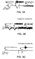
- Seizure Suppression In Figure 3A and 3B we show examples that illustrate how an electric field can be used to adaptively suppress seizure-like activity within the CA1. Suppression is achieved by using negative feedback. In both cases the high frequency activity, towards which the suppression algorithm is directed, is significantly attenuated. The DC shift was completely eliminated (3A) during suppression for some slices, while it was partially retained (3B) for others. During control, some non-zero level of network activity is still observed from the field potentials (third inset in each). We have documented successful suppression in 20 of 30 seizing slices with which we applied adaptive control.
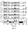
- Control can often be maintained for prolonged periods of time. To date, the longest we have maintained control is 16 minutes in a slice otherwise exhibiting seizures approximately every 40 seconds. Since the amplitude, duration and interval between of the events slowly change over the course of an hour (see Figure 4), 16 minutes is near the limit for reliable suppression testing in this system.
- Seizure Enhancement Positive feedback, set by changing the sign on the gain which reverses the applied field polarity, can be used to either enhance seizures or even create seizures where none were observed beforehand.
- Figure 3C we show an example of the characteristic population burst-firing events seen in high [K + ] hippocampal slices (Rutecki et al., 1985) in the uncontrolled state.
- the adaptively applied field With positive feedback control, the adaptively applied field now enhances the brief network bursts into large seizure-like events with the substantial low frequency potential shifts characteristic of seizures. We have documented seizure generation in all 4 non-sizing slices with which we applied positive feedback control.
- the type of stimulus applied is indicated in the lower trace, where the height of the blocks indicate either the gain (G, left axis) used in the proportional feedback routine, or the amplitude (A, right axis) of the waveform applied.
- the Greek letters indicate the type of stimulus applied, as indicated in the figure caption.
- Baseline recordings of 1-4 minutes were made between stimuli.
- the horizontal lines correspond to the trigger threshold for defining an event.
- the trigger threshold provides a logical dividing line between large and small events in the average deflection plots. In the duration plots, a horizontal line at 3 seconds is plotted as a rough threshold for distinguishing seizure-like episodes from smaller burst-like events.
- Negative (i.e. suppressive) feedback indicated by a negative gain, was applied with both full-wave ( ⁇ ) and half-wave ( ⁇ ) rectification. Even at the smallest gain used ( ⁇ 1 , ⁇ 1 ) , all six types of event characteristics are distinct from the baseline activity (black) for both detection schemes. At the intermediate gain used, no DC events were observed during the non-rectified control ( ⁇ 2 ), while only short, low power RMS events were observed. For half-wave rectified control at comparable gain ( ⁇ 2 ), short, small events were observed from both the DC and the RMS event extraction. At the highest gain used for non-rectified control ( ⁇ 3 , starting at time 3960 s). no DC or RMS events were detected throughout the 16 minutes of control application.
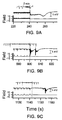
- the upper pair of traces (A) correspond to the measured field potential (lower) and applied field (upper) starting 2 minutes prior to the last application of non-rectified control ( ⁇ 3 ).
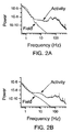
- the baseline activity, without control is characterized by large seizure-like events that start with a burst of high frequency activity, which are accompanied by a large low frequency potential shifts. Details of one of these events are shown in the trace of B at an expanded scale (15 s), high-pass filtered at 100 Hz, along with a spectrogram of the activity covering frequencies from 25-350 Hz.
- the power associated with these seizures can be observed in the spectrogram to start at high frequencies (near 120 Hz) and progress toward lower frequencies, a characteristic known as a 'spectral chirp'. Similar spectral chirps have been observed to be spectral signature of human seizures (Schiff, et. al., 2000). The neuronal activity following the seizure-like events in our experiments, as measured by the RMS power, is depressed across all frequencies.
- the full-wave random field (6B) did have the overall effect of breaking up the seizures in time and decreasing their duration as measured by the RMS event extraction (Top of 4). However, the maximum amplitude of those events as measured in the RMS was typically larger than baseline, and comparable findings were reflected in the low frequency deflections (DC events).
- the half-wave rectified field (6C) had little effect at either amplitude used.
- the distributions for all three conditions are populated primarily with windows of low power.
- the windows with high power are of great interest, since we associate high power in this frequency band with the first portion of the seizure-like events.
- the cumulative probability shown in Figure 8B.
- This distribution, C( p ) can be understood to be the fraction of windows with power greater than p . From it, we observe that the maximum power observed during baseline is roughly 4 times higher than observed during control. In addition, roughly 3% of the windows during baseline activity have higher power than the maximum observed during either type of control.
- the high-frequency burst of activity in the uncontrolled seizure-like events is usually followed by a quiet, refractory-like period.
- the objective of the control algorithm was to maintain a target level of activity by either suppressing or exciting the network.
- Half-wave rectified control (not shown) also decreased these excursions, but to a lesser extent.
- the window-to-window variance of the integrated power is plotted vs. the average power in Figure 8C for each of these conditions (baseline, control, and rectified control). We use the variance as a measure of the width of the distribution.
- the baseline activity is clearly differentiated statistically from both types of controlled activity using either the mean or variance as measures.
- the amount of intervention by the controller cycled on a longer time scale (of order a minute), often reaching a point at which no field would be applied for a few seconds.
- the activity when control was released depended on the phase of this cycle. If the controller was actively suppressing when shut off, then a seizure would progress (Fig. 9B). Otherwise, one would appear later, but within a few seconds of release.
Landscapes
- Health & Medical Sciences (AREA)
- Life Sciences & Earth Sciences (AREA)
- General Health & Medical Sciences (AREA)
- Public Health (AREA)
- Engineering & Computer Science (AREA)
- Biomedical Technology (AREA)
- Nuclear Medicine, Radiotherapy & Molecular Imaging (AREA)
- Radiology & Medical Imaging (AREA)
- Veterinary Medicine (AREA)
- Animal Behavior & Ethology (AREA)
- Neurosurgery (AREA)
- Neurology (AREA)
- Biophysics (AREA)
- Heart & Thoracic Surgery (AREA)
- Hospice & Palliative Care (AREA)
- Physiology (AREA)
- Measurement And Recording Of Electrical Phenomena And Electrical Characteristics Of The Living Body (AREA)
- Feedback Control In General (AREA)
- Medicines That Contain Protein Lipid Enzymes And Other Medicines (AREA)
- Electrotherapy Devices (AREA)
Claims (20)
- Feld-erzeugende Vorrichtung zum Modifizieren der neuronalen Aktivität eines Neuronen umfassenden Neuralsystems, wobei die Vorrichtung folgendes umfasst:die Feldelektrodeneinrichtung (4) zum Anlegen eines externen elektrischen Feldes an das Neuralsystem;die elektronische Feldanlegeeinrichtung (9) zum Erzeugen eines externen Feldes an dem Neuralsystem, welche funktionell mit der Feldelektrodeneinrichtung verbunden ist;die elektronische Messeinrichtung (6) zum Überwachen der neuronalen Aktivität des Neuralsystems;die elektronische Messungseinrichtung (7) zum Aufzeichnen der neuronalen Aktivität, welche funktionell mit der elektronischen Messeinrichtung verbunden ist;die Rückkopplungssteuereinrichtung (8) zum Bestimmen des Betrags des an das Neuralsystem anzulegenden externen Feldes, welche funktionell mit der Feldanlegeelektronikeinrichtung und der elektronischen Messeinrichtung verbunden ist,wobei die Vorrichtung so konfiguriert ist, dass sie die Größe und Polarität des angelegten elektrischen Feldes in Reaktion auf die gemessene neuronale Aktivität verändert, wobei das elektrische Feld ausgerichtet ist,
dadurch gekennzeichnet, dass die Vorrichtung so konfiguriert ist, dass sie sowohl negative als auch positive elektrische Felder abhängig von dem Vorzeichen der Differenz zwischen der gemessenen neuronalen Aktivität und einem Schwellenwert, und in einer Dauer und Intensität zum Reduzieren von Entzugsanfällen und zum Erzielen einer regelmäßigen Grundlinie neuronaler Aktivität anlegt. - Feld-erzeugende Vorrichtung nach Anspruch 1, welche ferner umfasst:eine Sensorelektrodeneinrichtung zum Detektieren des von der Feldelektrodeneinrichtung erzeugten externen Feldes;eine elektronische Sensoreinrichtung zum Aufzeichnen des von der Feldelektrodeneinrichtung erzeugten Feldes, welche funktionell mit der Sensorelektrodeneinrichtung und der elektronischen Feldanlegeeinrichtung verbunden ist.
- Vorrichtung nach Anspruch 1 oder 2, die so konfiguriert ist, dass sie eine Messung ausführt während gleichzeitig das ausgerichtete elektrische Feld angelegt wird.
- Vorrichtung nach einem der Ansprüche 1 bis 3, die so konfiguriert ist, dass sie das elektrische Feld proportional zu der neuronalen Aktivität anlegt.
- Vorrichtung nach einem der Ansprüche 1 bis 4, die so konfiguriert ist, dass sie das elektrische Feld proportional zu der Differenz zwischen dem quadratischen Mittelwert der gemessenen Aktivität und einem vorbestimmten Schwellenwertaktivitätswert in einem vorbestimmten Frequenzband anlegt.
- Vorrichtung nach einem der Ansprüche 1 bis 5, wobei das elektrische Feld unterschwellwertig ist.
- Vorrichtung nach einem der Ansprüche 1 bis 6, wobei die Messelektronikeinrichtung in der Lage ist, die gemessene elektrische Aktivität als den quadratischen Mittelwert innerhalb eines Frequenzbandes von 100 bis 500 Hz gemittelt über der Zeit γ darzustellen.
- Vorrichtung nach einem der Ansprüche 1 bis 7, wobei die Elektroden außerhalb der Neuronen in dem Neuralsystem positioniert werden können.
- Vorrichtung nach einem der Ansprüche 1 bis 8, die so konfiguriert ist, dass das elektrische Feld parallel zu der somatodendritischen Achse der Neuronen in dem Neuralsystem ausgerichtet ist.
- Vorrichtung nach einem der Ansprüche 1 bis 9, wobei die Feldelektroden in einer runden Elektrode umfasst sind.
- Vorrichtung nach einem der Ansprüche 1 bis 10, die so konfiguriert ist, dass sie das elektrische Feld adaptiv in Reaktion auf Veränderungen in der gemessenen neuronalen Aktivität des Neuralsystems anlegt.
- Vorrichtung nach einem der Ansprüche 1 bis 11, wobei die neuronale Aktivität elektrische Aktivität ist.
- Vorrichtung nach einem der Ansprüche 1 bis 12, wobei die Messelektroden auf einem Isopotential in Bezug auf das angelegte elektrische Feld positioniert werden können.
- Vorrichtung nach einem der Ansprüche 1 bis 13, wobei die neuronale Aktivität eine spontane epileptiforme elektrische Aktivität ist, und die Vorrichtung dafür konfiguriert ist, ein elektrisches Feld anzulegen, um sie zu unterdrücken.
- Vorrichtung nach einem der Ansprüche 1 bis 14, wobei die neuronale Aktivität eine spontane epileptiforme elektrische Aktivität ist, und die Vorrichtung dafür konfiguriert ist, sie zu induzieren oder zu verstärken.
- Vorrichtung nach einem der Ansprüche 1 bis 15, die so konfiguriert ist, dass sie ein elektrisches Feld anlegt, das die Feuerrate der Neuronen in dem Neuralsystem modifiziert.
- Vorrichtung nach einem der Ansprüche 1 bis 16, die so konfiguriert ist, dass sie das angelegte elektrische Feld erzeugt, indem Strom in das System injiziert wird.
- Vorrichtung nach einem der Ansprüche 1 bis 17, die so konfiguriert ist, dass sie Strom injiziert, bis ein vorbestimmtes Feldpotential erreicht ist.
- Vorrichtung nach einem der Ansprüche 1 bis 18, wobei das angelegte elektrische Feld kein kontinuierliches stationäres Feld ist.
- Vorrichtung nach Anspruch 19, die so konfiguriert ist, dass sie ein elektrisches Feld an das Gehirn anlegt, wenn die epileptiforme Aktivität durch Messelektroden detektiert wird.
Applications Claiming Priority (3)
| Application Number | Priority Date | Filing Date | Title |
|---|---|---|---|
| US16928099P | 1999-12-07 | 1999-12-07 | |
| US169280P | 1999-12-07 | ||
| PCT/US2000/032987 WO2001041867A1 (en) | 1999-12-07 | 2000-12-06 | Adaptive electric field modulation of neural systems |
Publications (2)
| Publication Number | Publication Date |
|---|---|
| EP1237621A1 EP1237621A1 (de) | 2002-09-11 |
| EP1237621B1 true EP1237621B1 (de) | 2006-04-19 |
Family
ID=22614990
Family Applications (1)
| Application Number | Title | Priority Date | Filing Date |
|---|---|---|---|
| EP00983919A Expired - Lifetime EP1237621B1 (de) | 1999-12-07 | 2000-12-06 | Adaptive modulation von einem elektrischen feld für neuronale systeme |
Country Status (9)
| Country | Link |
|---|---|
| US (2) | US6665562B2 (de) |
| EP (1) | EP1237621B1 (de) |
| JP (1) | JP2003516206A (de) |
| AT (1) | ATE323528T1 (de) |
| AU (1) | AU782860B2 (de) |
| CA (1) | CA2393535A1 (de) |
| DE (1) | DE60027459T2 (de) |
| TW (1) | TW483768B (de) |
| WO (1) | WO2001041867A1 (de) |
Cited By (1)
| Publication number | Priority date | Publication date | Assignee | Title |
|---|---|---|---|---|
| CN103211589A (zh) * | 2006-07-10 | 2013-07-24 | 瓦高希有限公司 | 用于测量周围神经系统内的活动的方法和装置 |
Families Citing this family (185)
| Publication number | Priority date | Publication date | Assignee | Title |
|---|---|---|---|---|
| US20020169485A1 (en) | 1995-10-16 | 2002-11-14 | Neuropace, Inc. | Differential neurostimulation therapy driven by physiological context |
| US7799337B2 (en) | 1997-07-21 | 2010-09-21 | Levin Bruce H | Method for directed intranasal administration of a composition |
| US9113801B2 (en) | 1998-08-05 | 2015-08-25 | Cyberonics, Inc. | Methods and systems for continuous EEG monitoring |
| US9415222B2 (en) | 1998-08-05 | 2016-08-16 | Cyberonics, Inc. | Monitoring an epilepsy disease state with a supervisory module |
| US7231254B2 (en) | 1998-08-05 | 2007-06-12 | Bioneuronics Corporation | Closed-loop feedback-driven neuromodulation |
| US7242984B2 (en) | 1998-08-05 | 2007-07-10 | Neurovista Corporation | Apparatus and method for closed-loop intracranial stimulation for optimal control of neurological disease |
| US9042988B2 (en) | 1998-08-05 | 2015-05-26 | Cyberonics, Inc. | Closed-loop vagus nerve stimulation |
| US9375573B2 (en) | 1998-08-05 | 2016-06-28 | Cyberonics, Inc. | Systems and methods for monitoring a patient's neurological disease state |
| US8762065B2 (en) | 1998-08-05 | 2014-06-24 | Cyberonics, Inc. | Closed-loop feedback-driven neuromodulation |
| US7209787B2 (en) | 1998-08-05 | 2007-04-24 | Bioneuronics Corporation | Apparatus and method for closed-loop intracranial stimulation for optimal control of neurological disease |
| US7747325B2 (en) | 1998-08-05 | 2010-06-29 | Neurovista Corporation | Systems and methods for monitoring a patient's neurological disease state |
| US7277758B2 (en) | 1998-08-05 | 2007-10-02 | Neurovista Corporation | Methods and systems for predicting future symptomatology in a patient suffering from a neurological or psychiatric disorder |
| US7974696B1 (en) | 1998-08-05 | 2011-07-05 | Dilorenzo Biomedical, Llc | Closed-loop autonomic neuromodulation for optimal control of neurological and metabolic disease |
| US7324851B1 (en) | 1998-08-05 | 2008-01-29 | Neurovista Corporation | Closed-loop feedback-driven neuromodulation |
| US7403820B2 (en) | 1998-08-05 | 2008-07-22 | Neurovista Corporation | Closed-loop feedback-driven neuromodulation |
| EP1237621B1 (de) * | 1999-12-07 | 2006-04-19 | George Mason University | Adaptive modulation von einem elektrischen feld für neuronale systeme |
| US6708064B2 (en) * | 2000-02-24 | 2004-03-16 | Ali R. Rezai | Modulation of the brain to affect psychiatric disorders |
| US7305268B2 (en) | 2000-07-13 | 2007-12-04 | Northstar Neurscience, Inc. | Systems and methods for automatically optimizing stimulus parameters and electrode configurations for neuro-stimulators |
| US7236831B2 (en) * | 2000-07-13 | 2007-06-26 | Northstar Neuroscience, Inc. | Methods and apparatus for effectuating a lasting change in a neural-function of a patient |
| US7010351B2 (en) | 2000-07-13 | 2006-03-07 | Northstar Neuroscience, Inc. | Methods and apparatus for effectuating a lasting change in a neural-function of a patient |
| US7756584B2 (en) | 2000-07-13 | 2010-07-13 | Advanced Neuromodulation Systems, Inc. | Methods and apparatus for effectuating a lasting change in a neural-function of a patient |
| US7024247B2 (en) | 2001-10-15 | 2006-04-04 | Northstar Neuroscience, Inc. | Systems and methods for reducing the likelihood of inducing collateral neural activity during neural stimulation threshold test procedures |
| US7672730B2 (en) | 2001-03-08 | 2010-03-02 | Advanced Neuromodulation Systems, Inc. | Methods and apparatus for effectuating a lasting change in a neural-function of a patient |
| US7146217B2 (en) | 2000-07-13 | 2006-12-05 | Northstar Neuroscience, Inc. | Methods and apparatus for effectuating a change in a neural-function of a patient |
| US7831305B2 (en) | 2001-10-15 | 2010-11-09 | Advanced Neuromodulation Systems, Inc. | Neural stimulation system and method responsive to collateral neural activity |
| US7299096B2 (en) | 2001-03-08 | 2007-11-20 | Northstar Neuroscience, Inc. | System and method for treating Parkinson's Disease and other movement disorders |
| US6685729B2 (en) * | 2001-06-29 | 2004-02-03 | George Gonzalez | Process for testing and treating aberrant sensory afferents and motors efferents |
| US7209788B2 (en) | 2001-10-29 | 2007-04-24 | Duke University | Closed loop brain machine interface |
| US7303578B2 (en) | 2001-11-01 | 2007-12-04 | Photothera, Inc. | Device and method for providing phototherapy to the brain |
| US9993659B2 (en) | 2001-11-01 | 2018-06-12 | Pthera, Llc | Low level light therapy for enhancement of neurologic function by altering axonal transport rate |
| US8308784B2 (en) * | 2006-08-24 | 2012-11-13 | Jackson Streeter | Low level light therapy for enhancement of neurologic function of a patient affected by Parkinson's disease |
| US10683494B2 (en) | 2001-11-01 | 2020-06-16 | Pthera LLC | Enhanced stem cell therapy and stem cell production through the administration of low level light energy |
| AUPR879201A0 (en) | 2001-11-09 | 2001-12-06 | Cochlear Limited | Subthreshold stimulation of a cochlea |
| US20100030130A1 (en) * | 2001-11-09 | 2010-02-04 | Cochlear Limited | Pharmaceutical intervention for modulation of neural plasticity |
| US20030105409A1 (en) | 2001-11-14 | 2003-06-05 | Donoghue John Philip | Neurological signal decoding |
| EP2316468A1 (de) * | 2002-02-22 | 2011-05-04 | Shire LLC | Abgabesystem und Verfahren zum Schutz und zur Verabreichung von Dextroamphetamin |
| US6993392B2 (en) * | 2002-03-14 | 2006-01-31 | Duke University | Miniaturized high-density multichannel electrode array for long-term neuronal recordings |
| US7221981B2 (en) | 2002-03-28 | 2007-05-22 | Northstar Neuroscience, Inc. | Electrode geometries for efficient neural stimulation |
| WO2003085484A2 (en) * | 2002-04-04 | 2003-10-16 | The Board Of Governors For Higher Education, State Of Rhode Island And Providence Plantations | Apparatus for neuromuscular measurement and control |
| US20080077192A1 (en) * | 2002-05-03 | 2008-03-27 | Afferent Corporation | System and method for neuro-stimulation |
| EP1513584A2 (de) | 2002-06-04 | 2005-03-16 | Cyberkinetics, Inc. | Optisch verbundene implantate, darauf bezogene systeme und deren verwendung |
| US7292890B2 (en) | 2002-06-20 | 2007-11-06 | Advanced Bionics Corporation | Vagus nerve stimulation via unidirectional propagation of action potentials |
| US20040015205A1 (en) | 2002-06-20 | 2004-01-22 | Whitehurst Todd K. | Implantable microstimulators with programmable multielectrode configuration and uses thereof |
| US7860570B2 (en) | 2002-06-20 | 2010-12-28 | Boston Scientific Neuromodulation Corporation | Implantable microstimulators and methods for unidirectional propagation of action potentials |
| US6717804B1 (en) * | 2002-09-30 | 2004-04-06 | Hewlett-Packard Development Company, L.P. | Light-emitting lock device control element and electronic device including the same |
| US7460904B2 (en) * | 2002-10-09 | 2008-12-02 | Wake Forest University Health Sciences | Wireless systems and methods for the detection of neural events using onboard processing |
| US20050049649A1 (en) * | 2002-10-21 | 2005-03-03 | The Cleveland Clinic Foundation | Electrical stimulation of the brain |
| US20050010261A1 (en) * | 2002-10-21 | 2005-01-13 | The Cleveland Clinic Foundation | Application of stimulus to white matter to induce a desired physiological response |
| US7212851B2 (en) | 2002-10-24 | 2007-05-01 | Brown University Research Foundation | Microstructured arrays for cortex interaction and related methods of manufacture and use |
| US7236830B2 (en) | 2002-12-10 | 2007-06-26 | Northstar Neuroscience, Inc. | Systems and methods for enhancing or optimizing neural stimulation therapy for treating symptoms of Parkinson's disease and/or other movement disorders |
| JP2006504494A (ja) * | 2002-11-01 | 2006-02-09 | ジョージメイソン、インテレクチュアル、プロパティーズ、インコーポレイテッド | 脳状態を決定するための方法および装置 |
| US7302298B2 (en) | 2002-11-27 | 2007-11-27 | Northstar Neuroscience, Inc | Methods and systems employing intracranial electrodes for neurostimulation and/or electroencephalography |
| US20050075680A1 (en) | 2003-04-18 | 2005-04-07 | Lowry David Warren | Methods and systems for intracranial neurostimulation and/or sensing |
| US6959215B2 (en) * | 2002-12-09 | 2005-10-25 | Northstar Neuroscience, Inc. | Methods for treating essential tremor |
| WO2004052449A1 (en) | 2002-12-09 | 2004-06-24 | Northstar Neuroscience, Inc. | Methods for treating neurological language disorders |
| CA2523540A1 (en) | 2003-04-24 | 2005-01-06 | Northstar Neuroscience, Inc. | Systems and methods for facilitating and/or effectuating development, rehabilitation, restoration, and/or recovery of visual function through neural stimulation |
| US7107104B2 (en) * | 2003-05-30 | 2006-09-12 | Medtronic, Inc. | Implantable cortical neural lead and method |
| EP1654032A2 (de) * | 2003-08-01 | 2006-05-10 | Northstar Neuroscience, Inc. | Gerät und verfahren für die neurale stimulation eines patienten |
| US20050143589A1 (en) * | 2003-11-09 | 2005-06-30 | Donoghue John P. | Calibration systems and methods for neural interface devices |
| US20050113744A1 (en) * | 2003-11-21 | 2005-05-26 | Cyberkinetics, Inc. | Agent delivery systems and related methods under control of biological electrical signals |
| US7751877B2 (en) * | 2003-11-25 | 2010-07-06 | Braingate Co., Llc | Neural interface system with embedded id |
| US20050131463A1 (en) * | 2003-12-12 | 2005-06-16 | Fedorov Nikolai B. | Perfusion chamber for recording evoked and spontaneous electrical activity from submerged acute brain slices |
| US20050137646A1 (en) | 2003-12-22 | 2005-06-23 | Scimed Life Systems, Inc. | Method of intravascularly delivering stimulation leads into brain |
| US8060207B2 (en) | 2003-12-22 | 2011-11-15 | Boston Scientific Scimed, Inc. | Method of intravascularly delivering stimulation leads into direct contact with tissue |
| US7647097B2 (en) * | 2003-12-29 | 2010-01-12 | Braingate Co., Llc | Transcutaneous implant |
| US7295875B2 (en) | 2004-02-20 | 2007-11-13 | Boston Scientific Scimed, Inc. | Method of stimulating/sensing brain with combination of intravascularly and non-vascularly delivered leads |
| US20050203366A1 (en) * | 2004-03-12 | 2005-09-15 | Donoghue John P. | Neurological event monitoring and therapy systems and related methods |
| US7590454B2 (en) | 2004-03-12 | 2009-09-15 | Boston Scientific Neuromodulation Corporation | Modular stimulation lead network |
| US20050203600A1 (en) | 2004-03-12 | 2005-09-15 | Scimed Life Systems, Inc. | Collapsible/expandable tubular electrode leads |
| US7177702B2 (en) | 2004-03-12 | 2007-02-13 | Scimed Life Systems, Inc. | Collapsible/expandable electrode leads |
| US8412348B2 (en) | 2004-05-06 | 2013-04-02 | Boston Scientific Neuromodulation Corporation | Intravascular self-anchoring integrated tubular electrode body |
| WO2005122887A2 (en) | 2004-06-15 | 2005-12-29 | Cochlear Americas | Automatic determination of the threshold of an evoked neural response |
| WO2006003662A2 (en) * | 2004-07-06 | 2006-01-12 | Technion Research & Development Foundation Ltd. | Low power hardware algorithms and architectures for spike sorting and detection |
| JP2008506464A (ja) | 2004-07-15 | 2008-03-06 | ノーススター ニューロサイエンス インコーポレイテッド | 神経刺激効率及び/又は効力の強化又はそれに影響を及ぼすためのシステム及び方法 |
| US7286879B2 (en) | 2004-07-16 | 2007-10-23 | Boston Scientific Scimed, Inc. | Method of stimulating fastigium nucleus to treat neurological disorders |
| US20060049957A1 (en) * | 2004-08-13 | 2006-03-09 | Surgenor Timothy R | Biological interface systems with controlled device selector and related methods |
| US20060058854A1 (en) * | 2004-09-14 | 2006-03-16 | Scimed Life Systems, Inc. | Method for stimulating neural tissue in response to a sensed physiological event |
| WO2006041738A2 (en) * | 2004-10-04 | 2006-04-20 | Cyberkinetics Neurotechnology Systems, Inc. | Biological interface system |
| WO2006057734A1 (en) * | 2004-10-21 | 2006-06-01 | Advanced Neuromodulation Systems, Inc. | New stimulation design for neuromodulation |
| US20100036452A1 (en) * | 2004-11-02 | 2010-02-11 | Bruce Gluckman | Modulation of neural traveling waves |
| US7565200B2 (en) | 2004-11-12 | 2009-07-21 | Advanced Neuromodulation Systems, Inc. | Systems and methods for selecting stimulation sites and applying treatment, including treatment of symptoms of Parkinson's disease, other movement disorders, and/or drug side effects |
| US7483746B2 (en) * | 2004-12-06 | 2009-01-27 | Boston Scientific Neuromodulation Corp. | Stimulation of the stomach in response to sensed parameters to treat obesity |
| US7937160B2 (en) | 2004-12-10 | 2011-05-03 | Boston Scientific Neuromodulation Corporation | Methods for delivering cortical electrode leads into patient's head |
| US20060161217A1 (en) * | 2004-12-21 | 2006-07-20 | Jaax Kristen N | Methods and systems for treating obesity |
| US20060136007A1 (en) * | 2004-12-21 | 2006-06-22 | Mickle Marlin H | Deep brain stimulation apparatus, and associated methods |
| US20060253166A1 (en) | 2005-01-06 | 2006-11-09 | Flaherty J C | Patient training routine for biological interface system |
| US8095209B2 (en) * | 2005-01-06 | 2012-01-10 | Braingate Co., Llc | Biological interface system with gated control signal |
| US7901368B2 (en) | 2005-01-06 | 2011-03-08 | Braingate Co., Llc | Neurally controlled patient ambulation system |
| US20060189899A1 (en) * | 2005-01-10 | 2006-08-24 | Flaherty J Christopher | Joint movement apparatus |
| US20060189901A1 (en) * | 2005-01-10 | 2006-08-24 | Flaherty J C | Biological interface system with surrogate controlled device |
| WO2006078432A2 (en) * | 2005-01-18 | 2006-07-27 | Cyberkinetics Neurotechnology Systems, Inc. | Biological interface system with automated configuration |
| US8788044B2 (en) * | 2005-01-21 | 2014-07-22 | Michael Sasha John | Systems and methods for tissue stimulation in medical treatment |
| US7627383B2 (en) | 2005-03-15 | 2009-12-01 | Boston Scientific Neuromodulation Corporation | Implantable stimulator |
| US8000783B2 (en) * | 2005-08-10 | 2011-08-16 | Board Of Governors For Higher Education, State Of Rhode Island And Providence Plantations | Processor controlled voltage-current analysis for nerve and muscle tissues |
| US8150522B2 (en) * | 2005-08-19 | 2012-04-03 | The Trustees Of The University Of Pennsylvania | Active control of epileptic seizures and diagnosis based on critical systems-like behavior |
| US20070073354A1 (en) * | 2005-09-26 | 2007-03-29 | Knudson Mark B | Neural blocking therapy |
| US7856264B2 (en) | 2005-10-19 | 2010-12-21 | Advanced Neuromodulation Systems, Inc. | Systems and methods for patient interactive neural stimulation and/or chemical substance delivery |
| US7729773B2 (en) | 2005-10-19 | 2010-06-01 | Advanced Neuromodualation Systems, Inc. | Neural stimulation and optical monitoring systems and methods |
| US8929991B2 (en) | 2005-10-19 | 2015-01-06 | Advanced Neuromodulation Systems, Inc. | Methods for establishing parameters for neural stimulation, including via performance of working memory tasks, and associated kits |
| ITMI20052061A1 (it) * | 2005-10-28 | 2007-04-29 | Fondazione Irccs | Apparecchiatura per il trattamento di disturbi neurologici mediante stimolazione cerebrale cronica adattativa retroazionata da biopotenziali locali |
| US20070106143A1 (en) * | 2005-11-08 | 2007-05-10 | Flaherty J C | Electrode arrays and related methods |
| US7729758B2 (en) | 2005-11-30 | 2010-06-01 | Boston Scientific Neuromodulation Corporation | Magnetically coupled microstimulators |
| US20070142872A1 (en) * | 2005-12-21 | 2007-06-21 | Mickle Marlin H | Deep brain stimulation apparatus, and associated methods |
| US20100023021A1 (en) * | 2005-12-27 | 2010-01-28 | Flaherty J Christopher | Biological Interface and Insertion |
| US20070156126A1 (en) * | 2005-12-29 | 2007-07-05 | Flaherty J C | Medical device insertion system and related methods |
| US8725243B2 (en) | 2005-12-28 | 2014-05-13 | Cyberonics, Inc. | Methods and systems for recommending an appropriate pharmacological treatment to a patient for managing epilepsy and other neurological disorders |
| US8868172B2 (en) | 2005-12-28 | 2014-10-21 | Cyberonics, Inc. | Methods and systems for recommending an appropriate action to a patient for managing epilepsy and other neurological disorders |
| JP5175218B2 (ja) * | 2005-12-28 | 2013-04-03 | ニューロサイエンシーズ リサーチ ファンデーション インコーポレイテッド | 予測的運動制御のための小脳モデルを有する頭脳ベース・デバイス |
| WO2007106692A2 (en) * | 2006-03-15 | 2007-09-20 | University Of Pittsbugh Of The Commonwealth System Of Higher Education | Vagus nerve stimulation apparatus, and associated methods |
| US8086316B2 (en) * | 2006-05-24 | 2011-12-27 | Drexel University | Wireless controlled neuromodulation system |
| EP2034885A4 (de) | 2006-06-23 | 2010-12-01 | Neurovista Corp | Minimal invasive überwachungssysteme und -verfahren |
| US8295934B2 (en) | 2006-11-14 | 2012-10-23 | Neurovista Corporation | Systems and methods of reducing artifact in neurological stimulation systems |
| US9173384B2 (en) * | 2006-12-12 | 2015-11-03 | Cornell University | Surgically implanted micro-platforms and microsystems in arthropods and methods based thereon |
| EP2126785A2 (de) | 2007-01-25 | 2009-12-02 | NeuroVista Corporation | Systeme und verfahren zur identifikation des kontra-iktalen zustands eines patienten |
| WO2008092133A2 (en) | 2007-01-25 | 2008-07-31 | Neurovista Corporation | Methods and systems for measuring a subject's susceptibility to a seizure |
| US8036736B2 (en) | 2007-03-21 | 2011-10-11 | Neuro Vista Corporation | Implantable systems and methods for identifying a contra-ictal condition in a subject |
| US9788744B2 (en) | 2007-07-27 | 2017-10-17 | Cyberonics, Inc. | Systems for monitoring brain activity and patient advisory device |
| US7877136B1 (en) | 2007-09-28 | 2011-01-25 | Boston Scientific Neuromodulation Corporation | Enhancement of neural signal transmission through damaged neural tissue via hyperpolarizing electrical stimulation current |
| US20090204173A1 (en) | 2007-11-05 | 2009-08-13 | Zi-Ping Fang | Multi-Frequency Neural Treatments and Associated Systems and Methods |
| US20090171168A1 (en) | 2007-12-28 | 2009-07-02 | Leyde Kent W | Systems and Method for Recording Clinical Manifestations of a Seizure |
| US9259591B2 (en) | 2007-12-28 | 2016-02-16 | Cyberonics, Inc. | Housing for an implantable medical device |
| US8195298B2 (en) * | 2008-02-15 | 2012-06-05 | Andres M Lozano | Method for treating neurological/psychiatric disorders with stimulation to the subcaudate area of the brain |
| US8473062B2 (en) | 2008-05-01 | 2013-06-25 | Autonomic Technologies, Inc. | Method and device for the treatment of headache |
| BRPI0913738A2 (pt) * | 2008-09-19 | 2015-10-13 | Terry William Burton Moore | "método e dispositivo para redução da tensão muscular através de manipulação elétrica" |
| KR101679088B1 (ko) | 2008-10-01 | 2016-12-06 | 셔윈 화 | 척추골의 전선-유도식 척추경 나사 고정 시스템 및 방법 |
| US8255057B2 (en) | 2009-01-29 | 2012-08-28 | Nevro Corporation | Systems and methods for producing asynchronous neural responses to treat pain and/or other patient conditions |
| US8412336B2 (en) | 2008-12-29 | 2013-04-02 | Autonomic Technologies, Inc. | Integrated delivery and visualization tool for a neuromodulation system |
| US8849390B2 (en) | 2008-12-29 | 2014-09-30 | Cyberonics, Inc. | Processing for multi-channel signals |
| US8588933B2 (en) | 2009-01-09 | 2013-11-19 | Cyberonics, Inc. | Medical lead termination sleeve for implantable medical devices |
| US9320908B2 (en) | 2009-01-15 | 2016-04-26 | Autonomic Technologies, Inc. | Approval per use implanted neurostimulator |
| US8494641B2 (en) | 2009-04-22 | 2013-07-23 | Autonomic Technologies, Inc. | Implantable neurostimulator with integral hermetic electronic enclosure, circuit substrate, monolithic feed-through, lead assembly and anchoring mechanism |
| EP3228350B1 (de) * | 2009-04-22 | 2025-09-24 | Nevro Corporation | Vorrichtrung zur selektiven hochfrequenzrückenmarksmodulation zur vermeidung von schmerzen mit weniger nebenwirkungen, sowie zugehörige systeme und verfahren |
| WO2010124128A1 (en) | 2009-04-22 | 2010-10-28 | Nevro Corporation | Spinal cord modulation for inducing paresthetic and anesthetic effects, and associated systems and methods |
| US8786624B2 (en) | 2009-06-02 | 2014-07-22 | Cyberonics, Inc. | Processing for multi-channel signals |
| US8498710B2 (en) | 2009-07-28 | 2013-07-30 | Nevro Corporation | Linked area parameter adjustment for spinal cord stimulation and associated systems and methods |
| US8779307B2 (en) * | 2009-10-05 | 2014-07-15 | Nokia Corporation | Generating perceptible touch stimulus |
| US9643019B2 (en) | 2010-02-12 | 2017-05-09 | Cyberonics, Inc. | Neurological monitoring and alerts |
| US9579690B2 (en) | 2010-05-20 | 2017-02-28 | Nokia Technologies Oy | Generating perceptible touch stimulus |
| WO2012075198A2 (en) | 2010-11-30 | 2012-06-07 | Nevro Corporation | Extended pain relief via high frequency spinal cord modulation, and associated systems and methods |
| WO2012145244A1 (en) | 2011-04-20 | 2012-10-26 | Medtronic, Inc. | Method and apparatus for assessing neural activation |
| US9173609B2 (en) | 2011-04-20 | 2015-11-03 | Medtronic, Inc. | Brain condition monitoring based on co-activation of neural networks |
| US8914119B2 (en) | 2011-04-20 | 2014-12-16 | Medtronic, Inc. | Electrical brain therapy parameter determination based on a bioelectrical resonance response |
| US8892207B2 (en) | 2011-04-20 | 2014-11-18 | Medtronic, Inc. | Electrical therapy for facilitating inter-area brain synchronization |
| US8812098B2 (en) | 2011-04-28 | 2014-08-19 | Medtronic, Inc. | Seizure probability metrics |
| US9878161B2 (en) | 2011-04-29 | 2018-01-30 | Medtronic, Inc. | Entrainment of bioelectrical brain signals |
| JP5535405B2 (ja) * | 2011-07-12 | 2014-07-02 | エクスケア・ジャパン株式会社 | 生体刺激用信号波形発生装置 |
| WO2013036880A1 (en) | 2011-09-08 | 2013-03-14 | Thacker James R | Selective high frequency spinal cord modulation for inhibiting pain, including cephalic and/or total body pain with reduced side effects, and associated systems and methods |
| WO2014007890A2 (en) * | 2012-03-29 | 2014-01-09 | California Institute Of Technology | Sensor probe for bio-sensing and chemical-sensing applications |
| US8676331B2 (en) | 2012-04-02 | 2014-03-18 | Nevro Corporation | Devices for controlling spinal cord modulation for inhibiting pain, and associated systems and methods, including controllers for automated parameter selection |
| US9833614B1 (en) | 2012-06-22 | 2017-12-05 | Nevro Corp. | Autonomic nervous system control via high frequency spinal cord modulation, and associated systems and methods |
| KR101583044B1 (ko) * | 2012-09-28 | 2016-01-07 | 한양대학교 산학협력단 | 뇌심부 자극을 위한 능동적 자극 패턴 조절 기능을 갖는 적응형 뇌심부 자극 시스템 |
| TWI622381B (zh) * | 2013-03-28 | 2018-05-01 | 台灣艾思特科技股份有限公司 | Brain wave analysis method |
| US9919146B2 (en) | 2013-05-01 | 2018-03-20 | Sherwin Hua | Methods and systems for intraventricular brain stimulation |
| US11083402B2 (en) | 2013-06-04 | 2021-08-10 | Medtronic, Inc. | Patient state determination based on one or more spectral characteristics of a bioelectrical brain signal |
| US9895539B1 (en) | 2013-06-10 | 2018-02-20 | Nevro Corp. | Methods and systems for disease treatment using electrical stimulation |
| US10149978B1 (en) | 2013-11-07 | 2018-12-11 | Nevro Corp. | Spinal cord modulation for inhibiting pain via short pulse width waveforms, and associated systems and methods |
| EP3259020B1 (de) | 2015-02-16 | 2020-11-11 | Newronika S.p.A. | Vorrichtung zur behandlung neurologischer erkrankungen |
| WO2016154091A1 (en) | 2015-03-20 | 2016-09-29 | Ricardo Vallejo | Method and apparatus for multimodal electrical modulation of pain |
| US10850102B2 (en) | 2015-03-20 | 2020-12-01 | Medtronic Sg, Llc | Method and apparatus for multimodal electrical modulation of pain |
| US11167139B2 (en) | 2015-03-20 | 2021-11-09 | Medtronic Sg, Llc | Method and apparatus for multi modal electrical modulation of pain using composite electromagnetic fields |
| US12311177B2 (en) | 2015-03-20 | 2025-05-27 | Medtronic Sg, Llc | Method and apparatus for multi modal electrical modulation of pain using composite electromagnetic fields |
| US11318310B1 (en) | 2015-10-26 | 2022-05-03 | Nevro Corp. | Neuromodulation for altering autonomic functions, and associated systems and methods |
| EP3407967B1 (de) | 2016-01-25 | 2021-05-19 | Nevro Corporation | Behandlung von kongestiver herzinsuffizienz mit elektrischer stimulation sowie entsprechende systeme |
| US10799701B2 (en) | 2016-03-30 | 2020-10-13 | Nevro Corp. | Systems and methods for identifying and treating patients with high-frequency electrical signals |
| US11446504B1 (en) | 2016-05-27 | 2022-09-20 | Nevro Corp. | High frequency electromagnetic stimulation for modulating cells, including spontaneously active and quiescent cells, and associated systems and methods |
| US11073508B2 (en) * | 2017-03-28 | 2021-07-27 | Arizona Board Of Regents On Behalf Of The University Of Arizona | Rapid conductance based ion channel analysis |
| EP3684463B1 (de) | 2017-09-19 | 2025-05-14 | Neuroenhancement Lab, LLC | Verfahren und vorrichtung für neuro-enhancement |
| US11717686B2 (en) | 2017-12-04 | 2023-08-08 | Neuroenhancement Lab, LLC | Method and apparatus for neuroenhancement to facilitate learning and performance |
| US12280219B2 (en) | 2017-12-31 | 2025-04-22 | NeuroLight, Inc. | Method and apparatus for neuroenhancement to enhance emotional response |
| US11318277B2 (en) | 2017-12-31 | 2022-05-03 | Neuroenhancement Lab, LLC | Method and apparatus for neuroenhancement to enhance emotional response |
| IT201800002962A1 (it) | 2018-02-22 | 2019-08-22 | Newronika Srl | Apparato per il trattamento di disordini neurologici mediante elettrostimolazione e metodo di elaborazione del segnale neurologico raccolto da detto apparato |
| US11364361B2 (en) | 2018-04-20 | 2022-06-21 | Neuroenhancement Lab, LLC | System and method for inducing sleep by transplanting mental states |
| US11452839B2 (en) | 2018-09-14 | 2022-09-27 | Neuroenhancement Lab, LLC | System and method of improving sleep |
| US11318309B2 (en) | 2018-12-13 | 2022-05-03 | Newronika S.P.A. | Method and apparatus for treating Tourette Syndrome by brain stimulation |
| US11602634B2 (en) | 2019-01-17 | 2023-03-14 | Nevro Corp. | Sensory threshold adaptation for neurological therapy screening and/or electrode selection, and associated systems and methods |
| US11590352B2 (en) | 2019-01-29 | 2023-02-28 | Nevro Corp. | Ramped therapeutic signals for modulating inhibitory interneurons, and associated systems and methods |
| US11918811B2 (en) | 2019-05-06 | 2024-03-05 | Medtronic Sg, Llc | Method and apparatus for multi modal or multiplexed electrical modulation of pain using composite electromagnetic fields |
| US11786694B2 (en) | 2019-05-24 | 2023-10-17 | NeuroLight, Inc. | Device, method, and app for facilitating sleep |
| WO2021092495A1 (en) | 2019-11-08 | 2021-05-14 | Spine 23, Inc. | Systems and methods for pedicle screw stabilization of spinal vertebrae |
| ES3058598T3 (en) | 2019-11-27 | 2026-03-11 | Spine23 Inc | Systems for treating a lateral curvature of a spine |
| CN111012341B (zh) * | 2020-01-08 | 2022-04-22 | 东南大学 | 基于可穿戴脑电设备的伪迹去除和脑电信号质量评估方法 |
| EP4445832B1 (de) | 2020-07-17 | 2026-07-15 | Newronika S.p.A. | Systeme zur adaptiven tiefenhirnstimulation |
| KR20240049213A (ko) | 2021-05-12 | 2024-04-16 | 스파인23 인코포레이티드 | 척추뼈의 척추경 나사를 안정화시키기 위한 시스템 및 방법 |
| WO2022251366A1 (en) | 2021-05-25 | 2022-12-01 | Nevro Corp. | Modified high frequency neuromodulation signals, and associated systems and methods |
| EP4537184A4 (de) * | 2022-06-07 | 2026-04-01 | Univ California | Bioelektrische neuromodulationsverfahren und systeme zur linderung neuropathischer schmerzen |
Family Cites Families (28)
| Publication number | Priority date | Publication date | Assignee | Title |
|---|---|---|---|---|
| US3850161A (en) | 1973-04-09 | 1974-11-26 | S Liss | Method and apparatus for monitoring and counteracting excess brain electrical energy to prevent epileptic seizures and the like |
| US5025807A (en) * | 1983-09-14 | 1991-06-25 | Jacob Zabara | Neurocybernetic prosthesis |
| GR890100705A (el) | 1989-10-31 | 1992-03-20 | Fotios Anninos | Ηλεκτρονικη συσκευη για την ομαλυνση λειτουργικων ανωμαλιων του κεντρικου νευρικου συστηματος (κνς) σε συνδυασμο με την χρηση του βιομαγνητομετρου. |
| JPH05505750A (ja) | 1990-08-24 | 1993-08-26 | アニノス,フォティオス | バイオ磁力計の使用に関連して中央神経系の機能不全を円滑にするための電子装置 |
| US5342401A (en) | 1992-08-19 | 1994-08-30 | The Regents Of The University Of California | Real time cardiac arrhythmia stabilizing system |
| US5797965A (en) | 1994-08-19 | 1998-08-25 | The United States Of America As Represented By The Secretary Of The Navy | Suppression of epileptiform activity |
| US6480743B1 (en) | 2000-04-05 | 2002-11-12 | Neuropace, Inc. | System and method for adaptive brain stimulation |
| US7167748B2 (en) * | 1996-01-08 | 2007-01-23 | Impulse Dynamics Nv | Electrical muscle controller |
| US5995868A (en) | 1996-01-23 | 1999-11-30 | University Of Kansas | System for the prediction, rapid detection, warning, prevention, or control of changes in activity states in the brain of a subject |
| US6066163A (en) * | 1996-02-02 | 2000-05-23 | John; Michael Sasha | Adaptive brain stimulation method and system |
| US5857978A (en) | 1996-03-20 | 1999-01-12 | Lockheed Martin Energy Systems, Inc. | Epileptic seizure prediction by non-linear methods |
| US5626145A (en) | 1996-03-20 | 1997-05-06 | Lockheed Martin Energy Systems, Inc. | Method and apparatus for extraction of low-frequency artifacts from brain waves for alertness detection |
| US5713923A (en) | 1996-05-13 | 1998-02-03 | Medtronic, Inc. | Techniques for treating epilepsy by brain stimulation and drug infusion |
| AU3304997A (en) * | 1996-05-31 | 1998-01-05 | Southern Illinois University | Methods of modulating aspects of brain neural plasticity by vagus nerve stimulation |
| US5752979A (en) | 1996-11-01 | 1998-05-19 | Medtronic, Inc. | Method of controlling epilepsy by brain stimulation |
| US5800459A (en) | 1996-12-24 | 1998-09-01 | The United States Of America As Represented By The Secretary Of The Navy | Electric field control of epiletiform activity |
| US6016449A (en) | 1997-10-27 | 2000-01-18 | Neuropace, Inc. | System for treatment of neurological disorders |
| US6018682A (en) | 1998-04-30 | 2000-01-25 | Medtronic, Inc. | Implantable seizure warning system |
| US6374140B1 (en) | 1998-04-30 | 2002-04-16 | Medtronic, Inc. | Method and apparatus for treating seizure disorders by stimulating the olfactory senses |
| US5938689A (en) | 1998-05-01 | 1999-08-17 | Neuropace, Inc. | Electrode configuration for a brain neuropacemaker |
| US6366813B1 (en) | 1998-08-05 | 2002-04-02 | Dilorenzo Daniel J. | Apparatus and method for closed-loop intracranical stimulation for optimal control of neurological disease |
| US5996868A (en) * | 1998-09-17 | 1999-12-07 | Paradis; Arthur C. | Utility chest |
| US6161045A (en) | 1999-06-01 | 2000-12-12 | Neuropace, Inc. | Method for determining stimulation parameters for the treatment of epileptic seizures |
| US6556872B2 (en) * | 1999-08-24 | 2003-04-29 | Ev Vascular, Inc. | Therapeutic device and method for treating diseases of cardiac muscle |
| US6304775B1 (en) | 1999-09-22 | 2001-10-16 | Leonidas D. Iasemidis | Seizure warning and prediction |
| EP1237621B1 (de) * | 1999-12-07 | 2006-04-19 | George Mason University | Adaptive modulation von einem elektrischen feld für neuronale systeme |
| US6873872B2 (en) * | 1999-12-07 | 2005-03-29 | George Mason University | Adaptive electric field modulation of neural systems |
| US6529774B1 (en) * | 2000-11-09 | 2003-03-04 | Neuropace, Inc. | Extradural leads, neurostimulator assemblies, and processes of using them for somatosensory and brain stimulation |
-
2000
- 2000-12-06 EP EP00983919A patent/EP1237621B1/de not_active Expired - Lifetime
- 2000-12-06 DE DE60027459T patent/DE60027459T2/de not_active Expired - Fee Related
- 2000-12-06 AU AU20613/01A patent/AU782860B2/en not_active Ceased
- 2000-12-06 AT AT00983919T patent/ATE323528T1/de not_active IP Right Cessation
- 2000-12-06 CA CA002393535A patent/CA2393535A1/en not_active Abandoned
- 2000-12-06 JP JP2001543211A patent/JP2003516206A/ja not_active Withdrawn
- 2000-12-06 WO PCT/US2000/032987 patent/WO2001041867A1/en not_active Ceased
- 2000-12-06 TW TW089126018A patent/TW483768B/zh not_active IP Right Cessation
- 2000-12-06 US US09/729,929 patent/US6665562B2/en not_active Expired - Fee Related
-
2003
- 2003-10-06 US US10/678,128 patent/US20040073273A1/en not_active Abandoned
Cited By (2)
| Publication number | Priority date | Publication date | Assignee | Title |
|---|---|---|---|---|
| CN103211589A (zh) * | 2006-07-10 | 2013-07-24 | 瓦高希有限公司 | 用于测量周围神经系统内的活动的方法和装置 |
| CN103211589B (zh) * | 2006-07-10 | 2015-11-18 | 瓦高希有限公司 | 用于测量周围神经系统内的活动的方法和装置 |
Also Published As
| Publication number | Publication date |
|---|---|
| EP1237621A1 (de) | 2002-09-11 |
| DE60027459D1 (de) | 2006-05-24 |
| US6665562B2 (en) | 2003-12-16 |
| AU2061301A (en) | 2001-06-18 |
| AU782860B2 (en) | 2005-09-01 |
| US20010029391A1 (en) | 2001-10-11 |
| DE60027459T2 (de) | 2006-12-21 |
| US20040073273A1 (en) | 2004-04-15 |
| TW483768B (en) | 2002-04-21 |
| WO2001041867A1 (en) | 2001-06-14 |
| ATE323528T1 (de) | 2006-05-15 |
| CA2393535A1 (en) | 2001-06-14 |
| JP2003516206A (ja) | 2003-05-13 |
Similar Documents
| Publication | Publication Date | Title |
|---|---|---|
| US6665562B2 (en) | Adaptive electric field modulation of neural systems | |
| US6873872B2 (en) | Adaptive electric field modulation of neural systems | |
| US8718755B2 (en) | Methods and devices for determining brain state | |
| Gluckman et al. | Adaptive electric field control of epileptic seizures | |
| Vitek et al. | External pallidal stimulation improves parkinsonian motor signs and modulates neuronal activity throughout the basal ganglia thalamic network | |
| Hashimoto et al. | A template subtraction method for stimulus artifact removal in high-frequency deep brain stimulation | |
| Wickelgren | Physiological and anatomical characteristics of reticulospinal neurones in lamprey | |
| US7353064B2 (en) | Systems and methods for enhancing or optimizing neural stimulation therapy for treating symptoms of movement disorders and/or other neurologic dysfunction | |
| US6819956B2 (en) | Optimal method and apparatus for neural modulation for the treatment of neurological disease, particularly movement disorders | |
| US6944501B1 (en) | Neurostimulator involving stimulation strategies and process for using it | |
| Richardson et al. | In vivo modulation of hippocampal epileptiform activity with radial electric fields | |
| Jost et al. | A new method to determine pudendal nerve motor latency and central motor conduction time to the external anal sphincter | |
| Rodenkirch et al. | Rapid and transient enhancement of thalamic information transmission induced by vagus nerve stimulation | |
| Testerman et al. | Electrical stimulation as therapy for neurological disorders | |
| Rolston et al. | Electrical stimulation for epilepsy: experimental approaches | |
| Rajdev et al. | Effect of stimulus parameters in the treatment of seizures by electrical stimulation in the kainate animal model | |
| Oulad Ben Taib et al. | Trains of epidural DC stimulation of the cerebellum tune corticomotor excitability | |
| Bushnell et al. | Electrical stimulation of peripheral and central pathways for the relief of musculoskeletal pain | |
| Nordstrom et al. | Motor cortical control of human masticatory muscles | |
| Rossini | The Anatomie and Physiologie Bases of Motor-Evoked Potentials | |
| Mathers et al. | Spontaneous hyperpolarizations at the membrane of cultured mouse dorsal root ganglion cells | |
| Makous et al. | Effects of a dorsal column lesion on temporal processing within the somatosensory system of primates | |
| US20100036452A1 (en) | Modulation of neural traveling waves | |
| Manjarrez et al. | Amplitude of somatosensory cortical evoked potentials is correlated with spontaneous activity of spinal neurones in the cat | |
| Andy et al. | Thalamic modulation of aggression |
Legal Events
| Date | Code | Title | Description |
|---|---|---|---|
| PUAI | Public reference made under article 153(3) epc to a published international application that has entered the european phase |
Free format text: ORIGINAL CODE: 0009012 |
|
| 17P | Request for examination filed |
Effective date: 20020703 |
|
| AK | Designated contracting states |
Kind code of ref document: A1 Designated state(s): AT BE CH CY DE DK ES FI FR GB GR IE IT LI LU MC NL PT SE TR |
|
| AX | Request for extension of the european patent |
Free format text: AL PAYMENT 20020703;LT PAYMENT 20020703;LV PAYMENT 20020703;MK PAYMENT 20020703;RO PAYMENT 20020703;SI PAYMENT 20020703 |
|
| RAP1 | Party data changed (applicant data changed or rights of an application transferred) |
Owner name: GEORGE MASON UNIVERSITY |
|
| 17Q | First examination report despatched |
Effective date: 20040302 |
|
| GRAP | Despatch of communication of intention to grant a patent |
Free format text: ORIGINAL CODE: EPIDOSNIGR1 |
|
| GRAS | Grant fee paid |
Free format text: ORIGINAL CODE: EPIDOSNIGR3 |
|
| GRAA | (expected) grant |
Free format text: ORIGINAL CODE: 0009210 |
|
| AK | Designated contracting states |
Kind code of ref document: B1 Designated state(s): AT BE CH CY DE DK ES FI FR GB GR IE IT LI LU MC NL PT SE TR |
|
| AX | Request for extension of the european patent |
Extension state: AL LT LV MK RO SI |
|
| PG25 | Lapsed in a contracting state [announced via postgrant information from national office to epo] |
Ref country code: IT Free format text: LAPSE BECAUSE OF FAILURE TO SUBMIT A TRANSLATION OF THE DESCRIPTION OR TO PAY THE FEE WITHIN THE PRESCRIBED TIME-LIMIT;WARNING: LAPSES OF ITALIAN PATENTS WITH EFFECTIVE DATE BEFORE 2007 MAY HAVE OCCURRED AT ANY TIME BEFORE 2007. THE CORRECT EFFECTIVE DATE MAY BE DIFFERENT FROM THE ONE RECORDED. Effective date: 20060419 Ref country code: NL Free format text: LAPSE BECAUSE OF FAILURE TO SUBMIT A TRANSLATION OF THE DESCRIPTION OR TO PAY THE FEE WITHIN THE PRESCRIBED TIME-LIMIT Effective date: 20060419 Ref country code: FI Free format text: LAPSE BECAUSE OF FAILURE TO SUBMIT A TRANSLATION OF THE DESCRIPTION OR TO PAY THE FEE WITHIN THE PRESCRIBED TIME-LIMIT Effective date: 20060419 |
|
| REG | Reference to a national code |
Ref country code: GB Ref legal event code: FG4D |
|
| REF | Corresponds to: |
Ref document number: 60027459 Country of ref document: DE Date of ref document: 20060524 Kind code of ref document: P |
|
| REG | Reference to a national code |
Ref country code: IE Ref legal event code: FG4D |
|
| PG25 | Lapsed in a contracting state [announced via postgrant information from national office to epo] |
Ref country code: SE Free format text: LAPSE BECAUSE OF FAILURE TO SUBMIT A TRANSLATION OF THE DESCRIPTION OR TO PAY THE FEE WITHIN THE PRESCRIBED TIME-LIMIT Effective date: 20060719 Ref country code: DK Free format text: LAPSE BECAUSE OF FAILURE TO SUBMIT A TRANSLATION OF THE DESCRIPTION OR TO PAY THE FEE WITHIN THE PRESCRIBED TIME-LIMIT Effective date: 20060719 |
|
| PG25 | Lapsed in a contracting state [announced via postgrant information from national office to epo] |
Ref country code: ES Free format text: LAPSE BECAUSE OF FAILURE TO SUBMIT A TRANSLATION OF THE DESCRIPTION OR TO PAY THE FEE WITHIN THE PRESCRIBED TIME-LIMIT Effective date: 20060730 |
|
| PG25 | Lapsed in a contracting state [announced via postgrant information from national office to epo] |
Ref country code: PT Free format text: LAPSE BECAUSE OF FAILURE TO SUBMIT A TRANSLATION OF THE DESCRIPTION OR TO PAY THE FEE WITHIN THE PRESCRIBED TIME-LIMIT Effective date: 20060919 |
|
| LTIE | Lt: invalidation of european patent or patent extension |
Effective date: 20060419 |
|
| NLV1 | Nl: lapsed or annulled due to failure to fulfill the requirements of art. 29p and 29m of the patents act | ||
| PG25 | Lapsed in a contracting state [announced via postgrant information from national office to epo] |
Ref country code: IE Free format text: LAPSE BECAUSE OF NON-PAYMENT OF DUE FEES Effective date: 20061206 |
|
| PGFP | Annual fee paid to national office [announced via postgrant information from national office to epo] |
Ref country code: GB Payment date: 20061206 Year of fee payment: 7 |
|
| PGFP | Annual fee paid to national office [announced via postgrant information from national office to epo] |
Ref country code: FR Payment date: 20061208 Year of fee payment: 7 |
|
| PGFP | Annual fee paid to national office [announced via postgrant information from national office to epo] |
Ref country code: AT Payment date: 20061213 Year of fee payment: 7 Ref country code: CH Payment date: 20061213 Year of fee payment: 7 |
|
| PGFP | Annual fee paid to national office [announced via postgrant information from national office to epo] |
Ref country code: DE Payment date: 20061214 Year of fee payment: 7 |
|
| ET | Fr: translation filed | ||
| PG25 | Lapsed in a contracting state [announced via postgrant information from national office to epo] |
Ref country code: MC Free format text: LAPSE BECAUSE OF NON-PAYMENT OF DUE FEES Effective date: 20061231 |
|
| REG | Reference to a national code |
Ref country code: CH Ref legal event code: NV Representative=s name: ING. MARCO ZARDI C/O M. ZARDI & CO. S.A. |
|
| PGFP | Annual fee paid to national office [announced via postgrant information from national office to epo] |
Ref country code: BE Payment date: 20070222 Year of fee payment: 7 |
|
| PLBE | No opposition filed within time limit |
Free format text: ORIGINAL CODE: 0009261 |
|
| STAA | Information on the status of an ep patent application or granted ep patent |
Free format text: STATUS: NO OPPOSITION FILED WITHIN TIME LIMIT |
|
| 26N | No opposition filed |
Effective date: 20070122 |
|
| PG25 | Lapsed in a contracting state [announced via postgrant information from national office to epo] |
Ref country code: GR Free format text: LAPSE BECAUSE OF FAILURE TO SUBMIT A TRANSLATION OF THE DESCRIPTION OR TO PAY THE FEE WITHIN THE PRESCRIBED TIME-LIMIT Effective date: 20060720 |
|
| BERE | Be: lapsed |
Owner name: *GEORGE MASON UNIVERSITY Effective date: 20071231 |
|
| PG25 | Lapsed in a contracting state [announced via postgrant information from national office to epo] |
Ref country code: LU Free format text: LAPSE BECAUSE OF NON-PAYMENT OF DUE FEES Effective date: 20061206 Ref country code: TR Free format text: LAPSE BECAUSE OF FAILURE TO SUBMIT A TRANSLATION OF THE DESCRIPTION OR TO PAY THE FEE WITHIN THE PRESCRIBED TIME-LIMIT Effective date: 20060419 |
|
| REG | Reference to a national code |
Ref country code: CH Ref legal event code: PL |
|
| GBPC | Gb: european patent ceased through non-payment of renewal fee |
Effective date: 20071206 |
|
| PG25 | Lapsed in a contracting state [announced via postgrant information from national office to epo] |
Ref country code: AT Free format text: LAPSE BECAUSE OF NON-PAYMENT OF DUE FEES Effective date: 20071206 |
|
| PG25 | Lapsed in a contracting state [announced via postgrant information from national office to epo] |
Ref country code: BE Free format text: LAPSE BECAUSE OF NON-PAYMENT OF DUE FEES Effective date: 20071231 |
|
| PG25 | Lapsed in a contracting state [announced via postgrant information from national office to epo] |
Ref country code: CH Free format text: LAPSE BECAUSE OF NON-PAYMENT OF DUE FEES Effective date: 20071231 Ref country code: LI Free format text: LAPSE BECAUSE OF NON-PAYMENT OF DUE FEES Effective date: 20071231 Ref country code: DE Free format text: LAPSE BECAUSE OF NON-PAYMENT OF DUE FEES Effective date: 20080701 |
|
| REG | Reference to a national code |
Ref country code: FR Ref legal event code: ST Effective date: 20081020 |
|
| PG25 | Lapsed in a contracting state [announced via postgrant information from national office to epo] |
Ref country code: CY Free format text: LAPSE BECAUSE OF FAILURE TO SUBMIT A TRANSLATION OF THE DESCRIPTION OR TO PAY THE FEE WITHIN THE PRESCRIBED TIME-LIMIT Effective date: 20060419 |
|
| PG25 | Lapsed in a contracting state [announced via postgrant information from national office to epo] |
Ref country code: GB Free format text: LAPSE BECAUSE OF NON-PAYMENT OF DUE FEES Effective date: 20071206 |
|
| PG25 | Lapsed in a contracting state [announced via postgrant information from national office to epo] |
Ref country code: FR Free format text: LAPSE BECAUSE OF NON-PAYMENT OF DUE FEES Effective date: 20071231 |