EP1255865B1 - Procédé pour la détection d'acides nucléiques utilisant des amorces universelles - Google Patents
Procédé pour la détection d'acides nucléiques utilisant des amorces universelles Download PDFInfo
- Publication number
- EP1255865B1 EP1255865B1 EP01910465A EP01910465A EP1255865B1 EP 1255865 B1 EP1255865 B1 EP 1255865B1 EP 01910465 A EP01910465 A EP 01910465A EP 01910465 A EP01910465 A EP 01910465A EP 1255865 B1 EP1255865 B1 EP 1255865B1
- Authority
- EP
- European Patent Office
- Prior art keywords
- target
- beads
- sequence
- probes
- probe
- Prior art date
- Legal status (The legal status is an assumption and is not a legal conclusion. Google has not performed a legal analysis and makes no representation as to the accuracy of the status listed.)
- Expired - Lifetime
Links
- 230000037452 priming Effects 0.000 title claims description 35
- 238000001514 detection method Methods 0.000 title abstract description 30
- 150000007523 nucleic acids Chemical class 0.000 title description 67
- 102000039446 nucleic acids Human genes 0.000 title description 63
- 108020004707 nucleic acids Proteins 0.000 title description 63
- 238000003556 assay Methods 0.000 claims abstract description 56
- 239000000523 sample Substances 0.000 claims description 284
- 239000011324 bead Substances 0.000 claims description 194
- 238000009396 hybridization Methods 0.000 claims description 62
- 238000000034 method Methods 0.000 claims description 59
- 239000000758 substrate Substances 0.000 claims description 53
- 239000004005 microsphere Substances 0.000 claims description 48
- 239000000835 fiber Substances 0.000 claims description 38
- 230000000295 complement effect Effects 0.000 claims description 22
- 108091093088 Amplicon Proteins 0.000 claims description 20
- YBJHBAHKTGYVGT-ZKWXMUAHSA-N (+)-Biotin Chemical compound N1C(=O)N[C@@H]2[C@H](CCCCC(=O)O)SC[C@@H]21 YBJHBAHKTGYVGT-ZKWXMUAHSA-N 0.000 claims description 16
- 108091034057 RNA (poly(A)) Proteins 0.000 claims description 12
- 229960002685 biotin Drugs 0.000 claims description 8
- 235000020958 biotin Nutrition 0.000 claims description 8
- 239000011616 biotin Substances 0.000 claims description 8
- 238000011144 upstream manufacturing Methods 0.000 claims description 8
- 239000007850 fluorescent dye Substances 0.000 claims description 7
- 230000005291 magnetic effect Effects 0.000 claims description 6
- 108090000623 proteins and genes Proteins 0.000 abstract description 32
- 230000003321 amplification Effects 0.000 abstract description 20
- 238000003199 nucleic acid amplification method Methods 0.000 abstract description 20
- 238000012544 monitoring process Methods 0.000 abstract description 6
- 238000011223 gene expression profiling Methods 0.000 abstract description 4
- 238000003491 array Methods 0.000 description 55
- 108020004999 messenger RNA Proteins 0.000 description 50
- 238000009739 binding Methods 0.000 description 49
- 230000027455 binding Effects 0.000 description 48
- 230000003287 optical effect Effects 0.000 description 38
- 239000000203 mixture Substances 0.000 description 31
- 239000000975 dye Substances 0.000 description 27
- 210000004027 cell Anatomy 0.000 description 26
- 239000003446 ligand Substances 0.000 description 22
- 125000003729 nucleotide group Chemical group 0.000 description 19
- 238000003752 polymerase chain reaction Methods 0.000 description 19
- 238000006243 chemical reaction Methods 0.000 description 17
- 239000002773 nucleotide Substances 0.000 description 17
- 230000004044 response Effects 0.000 description 17
- 238000004458 analytical method Methods 0.000 description 14
- 102000004169 proteins and genes Human genes 0.000 description 12
- 239000003795 chemical substances by application Substances 0.000 description 11
- 239000000126 substance Substances 0.000 description 11
- 230000015572 biosynthetic process Effects 0.000 description 10
- -1 carbocyclic sugars Chemical class 0.000 description 10
- 239000003153 chemical reaction reagent Substances 0.000 description 10
- 238000012360 testing method Methods 0.000 description 10
- 108091032973 (ribonucleotides)n+m Proteins 0.000 description 9
- 230000008859 change Effects 0.000 description 9
- 230000014509 gene expression Effects 0.000 description 9
- 239000002245 particle Substances 0.000 description 9
- 108091093037 Peptide nucleic acid Proteins 0.000 description 8
- 230000008901 benefit Effects 0.000 description 8
- 150000003839 salts Chemical class 0.000 description 8
- 238000003786 synthesis reaction Methods 0.000 description 8
- 101100232386 Arabidopsis thaliana IBL1 gene Proteins 0.000 description 7
- 102000001708 Protein Isoforms Human genes 0.000 description 7
- 108010029485 Protein Isoforms Proteins 0.000 description 7
- 239000012867 bioactive agent Substances 0.000 description 7
- 125000000524 functional group Chemical group 0.000 description 7
- 230000003993 interaction Effects 0.000 description 7
- 210000001519 tissue Anatomy 0.000 description 7
- 208000031261 Acute myeloid leukaemia Diseases 0.000 description 6
- ZHNUHDYFZUAESO-UHFFFAOYSA-N Formamide Chemical compound NC=O ZHNUHDYFZUAESO-UHFFFAOYSA-N 0.000 description 6
- 208000033776 Myeloid Acute Leukemia Diseases 0.000 description 6
- 108010090804 Streptavidin Proteins 0.000 description 6
- 239000000853 adhesive Substances 0.000 description 6
- 230000001070 adhesive effect Effects 0.000 description 6
- 125000003277 amino group Chemical group 0.000 description 6
- 238000011068 loading method Methods 0.000 description 6
- 239000004033 plastic Substances 0.000 description 6
- 229920003023 plastic Polymers 0.000 description 6
- 238000011160 research Methods 0.000 description 6
- 101000719121 Arabidopsis thaliana Protein MEI2-like 1 Proteins 0.000 description 5
- 102000004190 Enzymes Human genes 0.000 description 5
- 108090000790 Enzymes Proteins 0.000 description 5
- 101000868273 Homo sapiens CD44 antigen Proteins 0.000 description 5
- 101000857677 Homo sapiens Runt-related transcription factor 1 Proteins 0.000 description 5
- 206010028980 Neoplasm Diseases 0.000 description 5
- 102100025373 Runt-related transcription factor 1 Human genes 0.000 description 5
- 238000013019 agitation Methods 0.000 description 5
- 230000033228 biological regulation Effects 0.000 description 5
- 239000002131 composite material Substances 0.000 description 5
- 238000009826 distribution Methods 0.000 description 5
- 239000011521 glass Substances 0.000 description 5
- 230000002209 hydrophobic effect Effects 0.000 description 5
- 238000002372 labelling Methods 0.000 description 5
- 239000000463 material Substances 0.000 description 5
- 230000009871 nonspecific binding Effects 0.000 description 5
- 239000002777 nucleoside Substances 0.000 description 5
- 150000003833 nucleoside derivatives Chemical class 0.000 description 5
- 230000008569 process Effects 0.000 description 5
- 230000025915 regulation of apoptotic process Effects 0.000 description 5
- 102100032912 CD44 antigen Human genes 0.000 description 4
- 108010014303 DNA-directed DNA polymerase Proteins 0.000 description 4
- 102000016928 DNA-directed DNA polymerase Human genes 0.000 description 4
- 102000003972 Fibroblast growth factor 7 Human genes 0.000 description 4
- 108090000385 Fibroblast growth factor 7 Proteins 0.000 description 4
- VYPSYNLAJGMNEJ-UHFFFAOYSA-N Silicium dioxide Chemical compound O=[Si]=O VYPSYNLAJGMNEJ-UHFFFAOYSA-N 0.000 description 4
- 210000001744 T-lymphocyte Anatomy 0.000 description 4
- IQFYYKKMVGJFEH-XLPZGREQSA-N Thymidine Chemical compound O=C1NC(=O)C(C)=CN1[C@@H]1O[C@H](CO)[C@@H](O)C1 IQFYYKKMVGJFEH-XLPZGREQSA-N 0.000 description 4
- 230000004075 alteration Effects 0.000 description 4
- 150000001720 carbohydrates Chemical class 0.000 description 4
- 235000014633 carbohydrates Nutrition 0.000 description 4
- OPTASPLRGRRNAP-UHFFFAOYSA-N cytosine Chemical compound NC=1C=CNC(=O)N=1 OPTASPLRGRRNAP-UHFFFAOYSA-N 0.000 description 4
- 239000003112 inhibitor Substances 0.000 description 4
- DRAVOWXCEBXPTN-UHFFFAOYSA-N isoguanine Chemical compound NC1=NC(=O)NC2=C1NC=N2 DRAVOWXCEBXPTN-UHFFFAOYSA-N 0.000 description 4
- 230000004048 modification Effects 0.000 description 4
- 238000012986 modification Methods 0.000 description 4
- 230000004118 muscle contraction Effects 0.000 description 4
- 239000013307 optical fiber Substances 0.000 description 4
- 230000029279 positive regulation of transcription, DNA-dependent Effects 0.000 description 4
- 238000012545 processing Methods 0.000 description 4
- 230000035945 sensitivity Effects 0.000 description 4
- 230000019491 signal transduction Effects 0.000 description 4
- 239000007787 solid Substances 0.000 description 4
- 238000007619 statistical method Methods 0.000 description 4
- 125000003396 thiol group Chemical group [H]S* 0.000 description 4
- DEQPBRIACBATHE-FXQIFTODSA-N 5-[(3as,4s,6ar)-2-oxo-1,3,3a,4,6,6a-hexahydrothieno[3,4-d]imidazol-4-yl]-2-iminopentanoic acid Chemical compound N1C(=O)N[C@@H]2[C@H](CCCC(=N)C(=O)O)SC[C@@H]21 DEQPBRIACBATHE-FXQIFTODSA-N 0.000 description 3
- OKTJSMMVPCPJKN-UHFFFAOYSA-N Carbon Chemical compound [C] OKTJSMMVPCPJKN-UHFFFAOYSA-N 0.000 description 3
- 108700024394 Exon Proteins 0.000 description 3
- 108091092195 Intron Proteins 0.000 description 3
- 108020004711 Nucleic Acid Probes Proteins 0.000 description 3
- 108091034117 Oligonucleotide Proteins 0.000 description 3
- 239000004793 Polystyrene Substances 0.000 description 3
- 239000012491 analyte Substances 0.000 description 3
- 230000000692 anti-sense effect Effects 0.000 description 3
- 239000000872 buffer Substances 0.000 description 3
- 201000011510 cancer Diseases 0.000 description 3
- 125000003178 carboxy group Chemical group [H]OC(*)=O 0.000 description 3
- 230000021164 cell adhesion Effects 0.000 description 3
- 210000000349 chromosome Anatomy 0.000 description 3
- 230000002860 competitive effect Effects 0.000 description 3
- 150000001875 compounds Chemical class 0.000 description 3
- 238000004132 cross linking Methods 0.000 description 3
- 238000010494 dissociation reaction Methods 0.000 description 3
- 230000005593 dissociations Effects 0.000 description 3
- 239000003814 drug Substances 0.000 description 3
- 210000002919 epithelial cell Anatomy 0.000 description 3
- 230000001747 exhibiting effect Effects 0.000 description 3
- 239000012530 fluid Substances 0.000 description 3
- 238000011065 in-situ storage Methods 0.000 description 3
- 238000010348 incorporation Methods 0.000 description 3
- 238000002844 melting Methods 0.000 description 3
- 230000008018 melting Effects 0.000 description 3
- 230000007935 neutral effect Effects 0.000 description 3
- 239000002853 nucleic acid probe Substances 0.000 description 3
- 239000003960 organic solvent Substances 0.000 description 3
- 229920002223 polystyrene Polymers 0.000 description 3
- 108090000765 processed proteins & peptides Proteins 0.000 description 3
- 230000001105 regulatory effect Effects 0.000 description 3
- 238000000926 separation method Methods 0.000 description 3
- 230000037423 splicing regulation Effects 0.000 description 3
- XQCZBXHVTFVIFE-UHFFFAOYSA-N 2-amino-4-hydroxypyrimidine Chemical compound NC1=NC=CC(O)=N1 XQCZBXHVTFVIFE-UHFFFAOYSA-N 0.000 description 2
- 241000894006 Bacteria Species 0.000 description 2
- DWRXFEITVBNRMK-UHFFFAOYSA-N Beta-D-1-Arabinofuranosylthymine Natural products O=C1NC(=O)C(C)=CN1C1C(O)C(O)C(CO)O1 DWRXFEITVBNRMK-UHFFFAOYSA-N 0.000 description 2
- 108090000312 Calcium Channels Proteins 0.000 description 2
- 102000003922 Calcium Channels Human genes 0.000 description 2
- 201000009030 Carcinoma Diseases 0.000 description 2
- 102000004046 Caspase-2 Human genes 0.000 description 2
- 108090000552 Caspase-2 Proteins 0.000 description 2
- 108010017826 DNA Polymerase I Proteins 0.000 description 2
- 102000004594 DNA Polymerase I Human genes 0.000 description 2
- 108091060211 Expressed sequence tag Proteins 0.000 description 2
- 102000003971 Fibroblast Growth Factor 1 Human genes 0.000 description 2
- 108090000386 Fibroblast Growth Factor 1 Proteins 0.000 description 2
- 102100023600 Fibroblast growth factor receptor 2 Human genes 0.000 description 2
- 101710182389 Fibroblast growth factor receptor 2 Proteins 0.000 description 2
- 206010056740 Genital discharge Diseases 0.000 description 2
- NYHBQMYGNKIUIF-UUOKFMHZSA-N Guanosine Chemical compound C1=NC=2C(=O)NC(N)=NC=2N1[C@@H]1O[C@H](CO)[C@@H](O)[C@H]1O NYHBQMYGNKIUIF-UUOKFMHZSA-N 0.000 description 2
- 108010001336 Horseradish Peroxidase Proteins 0.000 description 2
- 101710163270 Nuclease Proteins 0.000 description 2
- 239000004677 Nylon Substances 0.000 description 2
- 108020005067 RNA Splice Sites Proteins 0.000 description 2
- FKNQFGJONOIPTF-UHFFFAOYSA-N Sodium cation Chemical compound [Na+] FKNQFGJONOIPTF-UHFFFAOYSA-N 0.000 description 2
- PPBRXRYQALVLMV-UHFFFAOYSA-N Styrene Chemical compound C=CC1=CC=CC=C1 PPBRXRYQALVLMV-UHFFFAOYSA-N 0.000 description 2
- 239000004809 Teflon Substances 0.000 description 2
- 229920006362 Teflon® Polymers 0.000 description 2
- GWEVSGVZZGPLCZ-UHFFFAOYSA-N Titan oxide Chemical compound O=[Ti]=O GWEVSGVZZGPLCZ-UHFFFAOYSA-N 0.000 description 2
- ISAKRJDGNUQOIC-UHFFFAOYSA-N Uracil Chemical compound O=C1C=CNC(=O)N1 ISAKRJDGNUQOIC-UHFFFAOYSA-N 0.000 description 2
- 229920006397 acrylic thermoplastic Polymers 0.000 description 2
- OIRDTQYFTABQOQ-KQYNXXCUSA-N adenosine Chemical compound C1=NC=2C(N)=NC=NC=2N1[C@@H]1O[C@H](CO)[C@@H](O)[C@H]1O OIRDTQYFTABQOQ-KQYNXXCUSA-N 0.000 description 2
- 238000000137 annealing Methods 0.000 description 2
- 239000000427 antigen Substances 0.000 description 2
- 102000036639 antigens Human genes 0.000 description 2
- 108091007433 antigens Proteins 0.000 description 2
- 238000003782 apoptosis assay Methods 0.000 description 2
- 238000013459 approach Methods 0.000 description 2
- 239000003125 aqueous solvent Substances 0.000 description 2
- 210000003719 b-lymphocyte Anatomy 0.000 description 2
- 108010058966 bacteriophage T7 induced DNA polymerase Proteins 0.000 description 2
- IQFYYKKMVGJFEH-UHFFFAOYSA-N beta-L-thymidine Natural products O=C1NC(=O)C(C)=CN1C1OC(CO)C(O)C1 IQFYYKKMVGJFEH-UHFFFAOYSA-N 0.000 description 2
- 210000004369 blood Anatomy 0.000 description 2
- 239000008280 blood Substances 0.000 description 2
- 229910052799 carbon Inorganic materials 0.000 description 2
- 238000007621 cluster analysis Methods 0.000 description 2
- 239000000356 contaminant Substances 0.000 description 2
- 239000003431 cross linking reagent Substances 0.000 description 2
- 239000002875 cyclin dependent kinase inhibitor Substances 0.000 description 2
- 229940043378 cyclin-dependent kinase inhibitor Drugs 0.000 description 2
- 229940104302 cytosine Drugs 0.000 description 2
- 238000004925 denaturation Methods 0.000 description 2
- 230000036425 denaturation Effects 0.000 description 2
- 238000011161 development Methods 0.000 description 2
- 230000018109 developmental process Effects 0.000 description 2
- 230000004069 differentiation Effects 0.000 description 2
- 201000010099 disease Diseases 0.000 description 2
- 208000037265 diseases, disorders, signs and symptoms Diseases 0.000 description 2
- 238000005516 engineering process Methods 0.000 description 2
- 238000002474 experimental method Methods 0.000 description 2
- 210000002950 fibroblast Anatomy 0.000 description 2
- GNBHRKFJIUUOQI-UHFFFAOYSA-N fluorescein Chemical compound O1C(=O)C2=CC=CC=C2C21C1=CC=C(O)C=C1OC1=CC(O)=CC=C21 GNBHRKFJIUUOQI-UHFFFAOYSA-N 0.000 description 2
- 239000012634 fragment Substances 0.000 description 2
- 230000006870 function Effects 0.000 description 2
- 238000007306 functionalization reaction Methods 0.000 description 2
- 210000003714 granulocyte Anatomy 0.000 description 2
- 230000012010 growth Effects 0.000 description 2
- UYTPUPDQBNUYGX-UHFFFAOYSA-N guanine Chemical compound O=C1NC(N)=NC2=C1N=CN2 UYTPUPDQBNUYGX-UHFFFAOYSA-N 0.000 description 2
- 239000005556 hormone Substances 0.000 description 2
- 229940088597 hormone Drugs 0.000 description 2
- 150000002500 ions Chemical class 0.000 description 2
- 230000000155 isotopic effect Effects 0.000 description 2
- 210000003734 kidney Anatomy 0.000 description 2
- 238000004020 luminiscence type Methods 0.000 description 2
- 210000004072 lung Anatomy 0.000 description 2
- 238000004949 mass spectrometry Methods 0.000 description 2
- 238000005259 measurement Methods 0.000 description 2
- 229910052751 metal Inorganic materials 0.000 description 2
- 239000002184 metal Substances 0.000 description 2
- 230000003957 neurotransmitter release Effects 0.000 description 2
- 238000007826 nucleic acid assay Methods 0.000 description 2
- 102000044158 nucleic acid binding protein Human genes 0.000 description 2
- 108700020942 nucleic acid binding protein Proteins 0.000 description 2
- 229920001778 nylon Polymers 0.000 description 2
- 125000004043 oxo group Chemical group O=* 0.000 description 2
- 238000000206 photolithography Methods 0.000 description 2
- 229920003229 poly(methyl methacrylate) Polymers 0.000 description 2
- 229920000642 polymer Polymers 0.000 description 2
- 239000002243 precursor Substances 0.000 description 2
- 102000004196 processed proteins & peptides Human genes 0.000 description 2
- 239000000047 product Substances 0.000 description 2
- 230000005522 programmed cell death Effects 0.000 description 2
- BBEAQIROQSPTKN-UHFFFAOYSA-N pyrene Chemical compound C1=CC=C2C=CC3=CC=CC4=CC=C1C2=C43 BBEAQIROQSPTKN-UHFFFAOYSA-N 0.000 description 2
- 238000011002 quantification Methods 0.000 description 2
- 102000005962 receptors Human genes 0.000 description 2
- 108020003175 receptors Proteins 0.000 description 2
- 230000022983 regulation of cell cycle Effects 0.000 description 2
- 238000012552 review Methods 0.000 description 2
- 239000000377 silicon dioxide Substances 0.000 description 2
- 239000002002 slurry Substances 0.000 description 2
- 150000003384 small molecules Chemical class 0.000 description 2
- 229910001415 sodium ion Inorganic materials 0.000 description 2
- 239000002904 solvent Substances 0.000 description 2
- 238000000527 sonication Methods 0.000 description 2
- 210000000130 stem cell Anatomy 0.000 description 2
- 230000005062 synaptic transmission Effects 0.000 description 2
- 230000008685 targeting Effects 0.000 description 2
- 230000002123 temporal effect Effects 0.000 description 2
- ISXSCDLOGDJUNJ-UHFFFAOYSA-N tert-butyl prop-2-enoate Chemical compound CC(C)(C)OC(=O)C=C ISXSCDLOGDJUNJ-UHFFFAOYSA-N 0.000 description 2
- 238000005382 thermal cycling Methods 0.000 description 2
- 229940104230 thymidine Drugs 0.000 description 2
- RWQNBRDOKXIBIV-UHFFFAOYSA-N thymine Chemical compound CC1=CNC(=O)NC1=O RWQNBRDOKXIBIV-UHFFFAOYSA-N 0.000 description 2
- 230000005945 translocation Effects 0.000 description 2
- 239000000439 tumor marker Substances 0.000 description 2
- 238000005406 washing Methods 0.000 description 2
- LXJXRIRHZLFYRP-VKHMYHEASA-L (R)-2-Hydroxy-3-(phosphonooxy)-propanal Natural products O=C[C@H](O)COP([O-])([O-])=O LXJXRIRHZLFYRP-VKHMYHEASA-L 0.000 description 1
- JLBJTVDPSNHSKJ-UHFFFAOYSA-N 4-Methylstyrene Chemical compound CC1=CC=C(C=C)C=C1 JLBJTVDPSNHSKJ-UHFFFAOYSA-N 0.000 description 1
- 101150079978 AGRN gene Proteins 0.000 description 1
- 229930024421 Adenine Natural products 0.000 description 1
- GFFGJBXGBJISGV-UHFFFAOYSA-N Adenine Chemical compound NC1=NC=NC2=C1N=CN2 GFFGJBXGBJISGV-UHFFFAOYSA-N 0.000 description 1
- 102100040026 Agrin Human genes 0.000 description 1
- 108700019743 Agrin Proteins 0.000 description 1
- 102000009027 Albumins Human genes 0.000 description 1
- 108010088751 Albumins Proteins 0.000 description 1
- 102000002260 Alkaline Phosphatase Human genes 0.000 description 1
- 108020004774 Alkaline Phosphatase Proteins 0.000 description 1
- 108091023037 Aptamer Proteins 0.000 description 1
- 102000014654 Aromatase Human genes 0.000 description 1
- 108010078554 Aromatase Proteins 0.000 description 1
- 238000012935 Averaging Methods 0.000 description 1
- 108700020463 BRCA1 Proteins 0.000 description 1
- 102000036365 BRCA1 Human genes 0.000 description 1
- 101150072950 BRCA1 gene Proteins 0.000 description 1
- 239000002126 C01EB10 - Adenosine Substances 0.000 description 1
- 102100035904 Caspase-1 Human genes 0.000 description 1
- 108090000426 Caspase-1 Proteins 0.000 description 1
- 102000011727 Caspases Human genes 0.000 description 1
- 108010076667 Caspases Proteins 0.000 description 1
- 102100032559 Clathrin light chain B Human genes 0.000 description 1
- 101710093540 Clathrin light chain B Proteins 0.000 description 1
- 239000004971 Cross linker Substances 0.000 description 1
- MIKUYHXYGGJMLM-GIMIYPNGSA-N Crotonoside Natural products C1=NC2=C(N)NC(=O)N=C2N1[C@H]1O[C@@H](CO)[C@H](O)[C@@H]1O MIKUYHXYGGJMLM-GIMIYPNGSA-N 0.000 description 1
- 108010015742 Cytochrome P-450 Enzyme System Proteins 0.000 description 1
- 102000003849 Cytochrome P450 Human genes 0.000 description 1
- 102000004127 Cytokines Human genes 0.000 description 1
- 108090000695 Cytokines Proteins 0.000 description 1
- LXJXRIRHZLFYRP-VKHMYHEASA-N D-glyceraldehyde 3-phosphate Chemical compound O=C[C@H](O)COP(O)(O)=O LXJXRIRHZLFYRP-VKHMYHEASA-N 0.000 description 1
- NYHBQMYGNKIUIF-UHFFFAOYSA-N D-guanosine Natural products C1=2NC(N)=NC(=O)C=2N=CN1C1OC(CO)C(O)C1O NYHBQMYGNKIUIF-UHFFFAOYSA-N 0.000 description 1
- 108020004414 DNA Proteins 0.000 description 1
- 238000000018 DNA microarray Methods 0.000 description 1
- 230000004568 DNA-binding Effects 0.000 description 1
- 101710088194 Dehydrogenase Proteins 0.000 description 1
- 229920002307 Dextran Polymers 0.000 description 1
- SHIBSTMRCDJXLN-UHFFFAOYSA-N Digoxigenin Natural products C1CC(C2C(C3(C)CCC(O)CC3CC2)CC2O)(O)C2(C)C1C1=CC(=O)OC1 SHIBSTMRCDJXLN-UHFFFAOYSA-N 0.000 description 1
- 108010007005 Estrogen Receptor alpha Proteins 0.000 description 1
- 102000000509 Estrogen Receptor beta Human genes 0.000 description 1
- 108010041356 Estrogen Receptor beta Proteins 0.000 description 1
- 102100038595 Estrogen receptor Human genes 0.000 description 1
- QTANTQQOYSUMLC-UHFFFAOYSA-O Ethidium cation Chemical class C12=CC(N)=CC=C2C2=CC=C(N)C=C2[N+](CC)=C1C1=CC=CC=C1 QTANTQQOYSUMLC-UHFFFAOYSA-O 0.000 description 1
- 241000206602 Eukaryota Species 0.000 description 1
- 229910052693 Europium Inorganic materials 0.000 description 1
- 108091008794 FGF receptors Proteins 0.000 description 1
- 102100037362 Fibronectin Human genes 0.000 description 1
- 239000004606 Fillers/Extenders Substances 0.000 description 1
- 102100022645 Glutamate receptor ionotropic, NMDA 1 Human genes 0.000 description 1
- 108010009202 Growth Factor Receptors Proteins 0.000 description 1
- 102000009465 Growth Factor Receptors Human genes 0.000 description 1
- 101000859758 Homo sapiens Cartilage-associated protein Proteins 0.000 description 1
- 101000916686 Homo sapiens Cytohesin-interacting protein Proteins 0.000 description 1
- 101001027128 Homo sapiens Fibronectin Proteins 0.000 description 1
- 101000726740 Homo sapiens Homeobox protein cut-like 1 Proteins 0.000 description 1
- 101000950669 Homo sapiens Mitogen-activated protein kinase 9 Proteins 0.000 description 1
- 101001133056 Homo sapiens Mucin-1 Proteins 0.000 description 1
- 101000761460 Homo sapiens Protein CASP Proteins 0.000 description 1
- 101000738771 Homo sapiens Receptor-type tyrosine-protein phosphatase C Proteins 0.000 description 1
- 102000008394 Immunoglobulin Fragments Human genes 0.000 description 1
- 108010021625 Immunoglobulin Fragments Proteins 0.000 description 1
- 229930010555 Inosine Natural products 0.000 description 1
- UGQMRVRMYYASKQ-KQYNXXCUSA-N Inosine Chemical compound O[C@@H]1[C@H](O)[C@@H](CO)O[C@H]1N1C2=NC=NC(O)=C2N=C1 UGQMRVRMYYASKQ-KQYNXXCUSA-N 0.000 description 1
- 108010001127 Insulin Receptor Proteins 0.000 description 1
- 102100036721 Insulin receptor Human genes 0.000 description 1
- 101000761459 Mesocricetus auratus Calcium-dependent serine proteinase Proteins 0.000 description 1
- 206010027476 Metastases Diseases 0.000 description 1
- 102100037809 Mitogen-activated protein kinase 9 Human genes 0.000 description 1
- 102100034256 Mucin-1 Human genes 0.000 description 1
- 101000663223 Mus musculus Serine/arginine-rich splicing factor 1 Proteins 0.000 description 1
- 102000005604 Myosin Heavy Chains Human genes 0.000 description 1
- 108010084498 Myosin Heavy Chains Proteins 0.000 description 1
- 108010054235 NMDA receptor A1 Proteins 0.000 description 1
- 108010069196 Neural Cell Adhesion Molecules Proteins 0.000 description 1
- 102100027347 Neural cell adhesion molecule 1 Human genes 0.000 description 1
- 102000008299 Nitric Oxide Synthase Human genes 0.000 description 1
- 108010021487 Nitric Oxide Synthase Proteins 0.000 description 1
- 239000000020 Nitrocellulose Substances 0.000 description 1
- 108091028043 Nucleic acid sequence Proteins 0.000 description 1
- 108091005461 Nucleic proteins Proteins 0.000 description 1
- 108020005187 Oligonucleotide Probes Proteins 0.000 description 1
- 102000043276 Oncogene Human genes 0.000 description 1
- 108700020796 Oncogene Proteins 0.000 description 1
- 238000012408 PCR amplification Methods 0.000 description 1
- 239000004698 Polyethylene Substances 0.000 description 1
- 239000004743 Polypropylene Substances 0.000 description 1
- 102000012419 Presenilin-2 Human genes 0.000 description 1
- 108010036908 Presenilin-2 Proteins 0.000 description 1
- 102000007066 Prostate-Specific Antigen Human genes 0.000 description 1
- 108010072866 Prostate-Specific Antigen Proteins 0.000 description 1
- 102100024933 Protein CASP Human genes 0.000 description 1
- 102000015097 RNA Splicing Factors Human genes 0.000 description 1
- 108010039259 RNA Splicing Factors Proteins 0.000 description 1
- 102100037422 Receptor-type tyrosine-protein phosphatase C Human genes 0.000 description 1
- 102000006382 Ribonucleases Human genes 0.000 description 1
- 108010083644 Ribonucleases Proteins 0.000 description 1
- 108091028664 Ribonucleotide Proteins 0.000 description 1
- 102000001332 SRC Human genes 0.000 description 1
- 108060006706 SRC Proteins 0.000 description 1
- 229920002684 Sepharose Polymers 0.000 description 1
- XUIMIQQOPSSXEZ-UHFFFAOYSA-N Silicon Chemical compound [Si] XUIMIQQOPSSXEZ-UHFFFAOYSA-N 0.000 description 1
- PJANXHGTPQOBST-VAWYXSNFSA-N Stilbene Natural products C=1C=CC=CC=1/C=C/C1=CC=CC=C1 PJANXHGTPQOBST-VAWYXSNFSA-N 0.000 description 1
- 108010017842 Telomerase Proteins 0.000 description 1
- 229910052771 Terbium Inorganic materials 0.000 description 1
- 108091023040 Transcription factor Proteins 0.000 description 1
- 102000040945 Transcription factor Human genes 0.000 description 1
- 102100033632 Tropomyosin alpha-1 chain Human genes 0.000 description 1
- 101710128188 Tropomyosin alpha-1 chain Proteins 0.000 description 1
- 102100036471 Tropomyosin beta chain Human genes 0.000 description 1
- 101710186456 Tropomyosin beta chain Proteins 0.000 description 1
- 102000004987 Troponin T Human genes 0.000 description 1
- 108090001108 Troponin T Proteins 0.000 description 1
- 102100031988 Tumor necrosis factor ligand superfamily member 6 Human genes 0.000 description 1
- 108050002568 Tumor necrosis factor ligand superfamily member 6 Proteins 0.000 description 1
- 108010053096 Vascular Endothelial Growth Factor Receptor-1 Proteins 0.000 description 1
- 102100033178 Vascular endothelial growth factor receptor 1 Human genes 0.000 description 1
- 241000700605 Viruses Species 0.000 description 1
- 208000008383 Wilms tumor Diseases 0.000 description 1
- 208000026448 Wilms tumor 1 Diseases 0.000 description 1
- 102100022748 Wilms tumor protein Human genes 0.000 description 1
- 101710127857 Wilms tumor protein Proteins 0.000 description 1
- JLCPHMBAVCMARE-UHFFFAOYSA-N [3-[[3-[[3-[[3-[[3-[[3-[[3-[[3-[[3-[[3-[[3-[[5-(2-amino-6-oxo-1H-purin-9-yl)-3-[[3-[[3-[[3-[[3-[[3-[[5-(2-amino-6-oxo-1H-purin-9-yl)-3-[[5-(2-amino-6-oxo-1H-purin-9-yl)-3-hydroxyoxolan-2-yl]methoxy-hydroxyphosphoryl]oxyoxolan-2-yl]methoxy-hydroxyphosphoryl]oxy-5-(5-methyl-2,4-dioxopyrimidin-1-yl)oxolan-2-yl]methoxy-hydroxyphosphoryl]oxy-5-(6-aminopurin-9-yl)oxolan-2-yl]methoxy-hydroxyphosphoryl]oxy-5-(6-aminopurin-9-yl)oxolan-2-yl]methoxy-hydroxyphosphoryl]oxy-5-(6-aminopurin-9-yl)oxolan-2-yl]methoxy-hydroxyphosphoryl]oxy-5-(6-aminopurin-9-yl)oxolan-2-yl]methoxy-hydroxyphosphoryl]oxyoxolan-2-yl]methoxy-hydroxyphosphoryl]oxy-5-(5-methyl-2,4-dioxopyrimidin-1-yl)oxolan-2-yl]methoxy-hydroxyphosphoryl]oxy-5-(4-amino-2-oxopyrimidin-1-yl)oxolan-2-yl]methoxy-hydroxyphosphoryl]oxy-5-(5-methyl-2,4-dioxopyrimidin-1-yl)oxolan-2-yl]methoxy-hydroxyphosphoryl]oxy-5-(5-methyl-2,4-dioxopyrimidin-1-yl)oxolan-2-yl]methoxy-hydroxyphosphoryl]oxy-5-(6-aminopurin-9-yl)oxolan-2-yl]methoxy-hydroxyphosphoryl]oxy-5-(6-aminopurin-9-yl)oxolan-2-yl]methoxy-hydroxyphosphoryl]oxy-5-(4-amino-2-oxopyrimidin-1-yl)oxolan-2-yl]methoxy-hydroxyphosphoryl]oxy-5-(4-amino-2-oxopyrimidin-1-yl)oxolan-2-yl]methoxy-hydroxyphosphoryl]oxy-5-(4-amino-2-oxopyrimidin-1-yl)oxolan-2-yl]methoxy-hydroxyphosphoryl]oxy-5-(6-aminopurin-9-yl)oxolan-2-yl]methoxy-hydroxyphosphoryl]oxy-5-(4-amino-2-oxopyrimidin-1-yl)oxolan-2-yl]methyl [5-(6-aminopurin-9-yl)-2-(hydroxymethyl)oxolan-3-yl] hydrogen phosphate Polymers Cc1cn(C2CC(OP(O)(=O)OCC3OC(CC3OP(O)(=O)OCC3OC(CC3O)n3cnc4c3nc(N)[nH]c4=O)n3cnc4c3nc(N)[nH]c4=O)C(COP(O)(=O)OC3CC(OC3COP(O)(=O)OC3CC(OC3COP(O)(=O)OC3CC(OC3COP(O)(=O)OC3CC(OC3COP(O)(=O)OC3CC(OC3COP(O)(=O)OC3CC(OC3COP(O)(=O)OC3CC(OC3COP(O)(=O)OC3CC(OC3COP(O)(=O)OC3CC(OC3COP(O)(=O)OC3CC(OC3COP(O)(=O)OC3CC(OC3COP(O)(=O)OC3CC(OC3COP(O)(=O)OC3CC(OC3COP(O)(=O)OC3CC(OC3COP(O)(=O)OC3CC(OC3COP(O)(=O)OC3CC(OC3COP(O)(=O)OC3CC(OC3CO)n3cnc4c(N)ncnc34)n3ccc(N)nc3=O)n3cnc4c(N)ncnc34)n3ccc(N)nc3=O)n3ccc(N)nc3=O)n3ccc(N)nc3=O)n3cnc4c(N)ncnc34)n3cnc4c(N)ncnc34)n3cc(C)c(=O)[nH]c3=O)n3cc(C)c(=O)[nH]c3=O)n3ccc(N)nc3=O)n3cc(C)c(=O)[nH]c3=O)n3cnc4c3nc(N)[nH]c4=O)n3cnc4c(N)ncnc34)n3cnc4c(N)ncnc34)n3cnc4c(N)ncnc34)n3cnc4c(N)ncnc34)O2)c(=O)[nH]c1=O JLCPHMBAVCMARE-UHFFFAOYSA-N 0.000 description 1
- 102000034337 acetylcholine receptors Human genes 0.000 description 1
- 108020000715 acetylcholine receptors Proteins 0.000 description 1
- 229960000643 adenine Drugs 0.000 description 1
- 229960005305 adenosine Drugs 0.000 description 1
- 150000003838 adenosines Chemical class 0.000 description 1
- 210000001789 adipocyte Anatomy 0.000 description 1
- 150000001299 aldehydes Chemical class 0.000 description 1
- PPQRONHOSHZGFQ-LMVFSUKVSA-N aldehydo-D-ribose 5-phosphate Chemical group OP(=O)(O)OC[C@@H](O)[C@@H](O)[C@@H](O)C=O PPQRONHOSHZGFQ-LMVFSUKVSA-N 0.000 description 1
- 125000001931 aliphatic group Chemical group 0.000 description 1
- 150000001408 amides Chemical class 0.000 description 1
- 150000001412 amines Chemical group 0.000 description 1
- 230000033115 angiogenesis Effects 0.000 description 1
- 239000004599 antimicrobial Substances 0.000 description 1
- 230000001640 apoptogenic effect Effects 0.000 description 1
- 230000006907 apoptotic process Effects 0.000 description 1
- 239000012062 aqueous buffer Substances 0.000 description 1
- 150000004982 aromatic amines Chemical class 0.000 description 1
- 150000001491 aromatic compounds Chemical class 0.000 description 1
- 238000002820 assay format Methods 0.000 description 1
- QVGXLLKOCUKJST-UHFFFAOYSA-N atomic oxygen Chemical compound [O] QVGXLLKOCUKJST-UHFFFAOYSA-N 0.000 description 1
- 230000004888 barrier function Effects 0.000 description 1
- 210000000227 basophil cell of anterior lobe of hypophysis Anatomy 0.000 description 1
- 239000011230 binding agent Substances 0.000 description 1
- 230000008827 biological function Effects 0.000 description 1
- 210000001124 body fluid Anatomy 0.000 description 1
- 210000000481 breast Anatomy 0.000 description 1
- 210000004899 c-terminal region Anatomy 0.000 description 1
- 150000001735 carboxylic acids Chemical class 0.000 description 1
- 210000004413 cardiac myocyte Anatomy 0.000 description 1
- 101150112018 ced-4 gene Proteins 0.000 description 1
- 230000030833 cell death Effects 0.000 description 1
- 230000024245 cell differentiation Effects 0.000 description 1
- 230000011748 cell maturation Effects 0.000 description 1
- 230000001413 cellular effect Effects 0.000 description 1
- 229920002678 cellulose Polymers 0.000 description 1
- 239000001913 cellulose Substances 0.000 description 1
- 238000005119 centrifugation Methods 0.000 description 1
- 239000000919 ceramic Substances 0.000 description 1
- 210000003756 cervix mucus Anatomy 0.000 description 1
- 230000003196 chaotropic effect Effects 0.000 description 1
- 238000012512 characterization method Methods 0.000 description 1
- 239000002738 chelating agent Substances 0.000 description 1
- 238000010382 chemical cross-linking Methods 0.000 description 1
- 125000003636 chemical group Chemical group 0.000 description 1
- 238000001311 chemical methods and process Methods 0.000 description 1
- 239000007795 chemical reaction product Substances 0.000 description 1
- 125000004218 chloromethyl group Chemical group [H]C([H])(Cl)* 0.000 description 1
- 210000001612 chondrocyte Anatomy 0.000 description 1
- 230000002759 chromosomal effect Effects 0.000 description 1
- 238000005253 cladding Methods 0.000 description 1
- 210000001072 colon Anatomy 0.000 description 1
- 239000002299 complementary DNA Substances 0.000 description 1
- 239000012141 concentrate Substances 0.000 description 1
- 210000001608 connective tissue cell Anatomy 0.000 description 1
- 229920001577 copolymer Polymers 0.000 description 1
- 230000008878 coupling Effects 0.000 description 1
- 238000010168 coupling process Methods 0.000 description 1
- 238000005859 coupling reaction Methods 0.000 description 1
- DMSZORWOGDLWGN-UHFFFAOYSA-N ctk1a3526 Chemical compound NP(N)(N)=O DMSZORWOGDLWGN-UHFFFAOYSA-N 0.000 description 1
- SUYVUBYJARFZHO-RRKCRQDMSA-N dATP Chemical compound C1=NC=2C(N)=NC=NC=2N1[C@H]1C[C@H](O)[C@@H](COP(O)(=O)OP(O)(=O)OP(O)(O)=O)O1 SUYVUBYJARFZHO-RRKCRQDMSA-N 0.000 description 1
- SUYVUBYJARFZHO-UHFFFAOYSA-N dATP Natural products C1=NC=2C(N)=NC=NC=2N1C1CC(O)C(COP(O)(=O)OP(O)(=O)OP(O)(O)=O)O1 SUYVUBYJARFZHO-UHFFFAOYSA-N 0.000 description 1
- RGWHQCVHVJXOKC-SHYZEUOFSA-J dCTP(4-) Chemical compound O=C1N=C(N)C=CN1[C@@H]1O[C@H](COP([O-])(=O)OP([O-])(=O)OP([O-])([O-])=O)[C@@H](O)C1 RGWHQCVHVJXOKC-SHYZEUOFSA-J 0.000 description 1
- HAAZLUGHYHWQIW-KVQBGUIXSA-N dGTP Chemical compound C1=NC=2C(=O)NC(N)=NC=2N1[C@H]1C[C@H](O)[C@@H](COP(O)(=O)OP(O)(=O)OP(O)(O)=O)O1 HAAZLUGHYHWQIW-KVQBGUIXSA-N 0.000 description 1
- NHVNXKFIZYSCEB-XLPZGREQSA-N dTTP Chemical compound O=C1NC(=O)C(C)=CN1[C@@H]1O[C@H](COP(O)(=O)OP(O)(=O)OP(O)(O)=O)[C@@H](O)C1 NHVNXKFIZYSCEB-XLPZGREQSA-N 0.000 description 1
- 230000006378 damage Effects 0.000 description 1
- 230000034994 death Effects 0.000 description 1
- 239000003398 denaturant Substances 0.000 description 1
- 230000001419 dependent effect Effects 0.000 description 1
- 230000008021 deposition Effects 0.000 description 1
- 238000013461 design Methods 0.000 description 1
- 230000000368 destabilizing effect Effects 0.000 description 1
- 239000003599 detergent Substances 0.000 description 1
- QONQRTHLHBTMGP-UHFFFAOYSA-N digitoxigenin Natural products CC12CCC(C3(CCC(O)CC3CC3)C)C3C11OC1CC2C1=CC(=O)OC1 QONQRTHLHBTMGP-UHFFFAOYSA-N 0.000 description 1
- SHIBSTMRCDJXLN-KCZCNTNESA-N digoxigenin Chemical compound C1([C@@H]2[C@@]3([C@@](CC2)(O)[C@H]2[C@@H]([C@@]4(C)CC[C@H](O)C[C@H]4CC2)C[C@H]3O)C)=CC(=O)OC1 SHIBSTMRCDJXLN-KCZCNTNESA-N 0.000 description 1
- 238000006471 dimerization reaction Methods 0.000 description 1
- NAGJZTKCGNOGPW-UHFFFAOYSA-K dioxido-sulfanylidene-sulfido-$l^{5}-phosphane Chemical compound [O-]P([O-])([S-])=S NAGJZTKCGNOGPW-UHFFFAOYSA-K 0.000 description 1
- 229940042399 direct acting antivirals protease inhibitors Drugs 0.000 description 1
- 230000006806 disease prevention Effects 0.000 description 1
- BFMYDTVEBKDAKJ-UHFFFAOYSA-L disodium;(2',7'-dibromo-3',6'-dioxido-3-oxospiro[2-benzofuran-1,9'-xanthene]-4'-yl)mercury;hydrate Chemical compound O.[Na+].[Na+].O1C(=O)C2=CC=CC=C2C21C1=CC(Br)=C([O-])C([Hg])=C1OC1=C2C=C(Br)C([O-])=C1 BFMYDTVEBKDAKJ-UHFFFAOYSA-L 0.000 description 1
- 230000000694 effects Effects 0.000 description 1
- 238000004520 electroporation Methods 0.000 description 1
- 230000009881 electrostatic interaction Effects 0.000 description 1
- 210000002889 endothelial cell Anatomy 0.000 description 1
- 230000002255 enzymatic effect Effects 0.000 description 1
- YQGOJNYOYNNSMM-UHFFFAOYSA-N eosin Chemical compound [Na+].OC(=O)C1=CC=CC=C1C1=C2C=C(Br)C(=O)C(Br)=C2OC2=C(Br)C(O)=C(Br)C=C21 YQGOJNYOYNNSMM-UHFFFAOYSA-N 0.000 description 1
- 210000003979 eosinophil Anatomy 0.000 description 1
- WTOSNONTQZJEBC-UHFFFAOYSA-N erythrosin Chemical compound OC(=O)C1=CC=CC=C1C(C1C(C(=C(O)C(I)=C1)I)O1)=C2C1=C(I)C(=O)C(I)=C2 WTOSNONTQZJEBC-UHFFFAOYSA-N 0.000 description 1
- 210000003527 eukaryotic cell Anatomy 0.000 description 1
- OGPBJKLSAFTDLK-UHFFFAOYSA-N europium atom Chemical compound [Eu] OGPBJKLSAFTDLK-UHFFFAOYSA-N 0.000 description 1
- 230000007717 exclusion Effects 0.000 description 1
- 238000010195 expression analysis Methods 0.000 description 1
- 230000002349 favourable effect Effects 0.000 description 1
- GVEPBJHOBDJJJI-UHFFFAOYSA-N fluoranthrene Natural products C1=CC(C2=CC=CC=C22)=C3C2=CC=CC3=C1 GVEPBJHOBDJJJI-UHFFFAOYSA-N 0.000 description 1
- 125000002485 formyl group Chemical group [H]C(*)=O 0.000 description 1
- 239000007789 gas Substances 0.000 description 1
- 238000004817 gas chromatography Methods 0.000 description 1
- 150000004676 glycans Chemical class 0.000 description 1
- 108020004445 glyceraldehyde-3-phosphate dehydrogenase Proteins 0.000 description 1
- 102000006602 glyceraldehyde-3-phosphate dehydrogenase Human genes 0.000 description 1
- 239000010439 graphite Substances 0.000 description 1
- 229910002804 graphite Inorganic materials 0.000 description 1
- 230000005484 gravity Effects 0.000 description 1
- 239000003102 growth factor Substances 0.000 description 1
- ZRALSGWEFCBTJO-UHFFFAOYSA-O guanidinium Chemical compound NC(N)=[NH2+] ZRALSGWEFCBTJO-UHFFFAOYSA-O 0.000 description 1
- 229940029575 guanosine Drugs 0.000 description 1
- 230000002607 hemopoietic effect Effects 0.000 description 1
- 210000003494 hepatocyte Anatomy 0.000 description 1
- 210000003630 histaminocyte Anatomy 0.000 description 1
- 125000002887 hydroxy group Chemical group [H]O* 0.000 description 1
- 238000007654 immersion Methods 0.000 description 1
- 210000000987 immune system Anatomy 0.000 description 1
- 230000006872 improvement Effects 0.000 description 1
- 208000015181 infectious disease Diseases 0.000 description 1
- 230000002458 infectious effect Effects 0.000 description 1
- 229960003786 inosine Drugs 0.000 description 1
- 102000006495 integrins Human genes 0.000 description 1
- 108010044426 integrins Proteins 0.000 description 1
- 230000001788 irregular Effects 0.000 description 1
- 210000002510 keratinocyte Anatomy 0.000 description 1
- 210000003292 kidney cell Anatomy 0.000 description 1
- 229910052747 lanthanoid Inorganic materials 0.000 description 1
- 150000002602 lanthanoids Chemical class 0.000 description 1
- 239000004816 latex Substances 0.000 description 1
- 229920000126 latex Polymers 0.000 description 1
- 210000000265 leukocyte Anatomy 0.000 description 1
- 230000000670 limiting effect Effects 0.000 description 1
- 210000004185 liver Anatomy 0.000 description 1
- 210000005229 liver cell Anatomy 0.000 description 1
- DLBFLQKQABVKGT-UHFFFAOYSA-L lucifer yellow dye Chemical compound [Li+].[Li+].[O-]S(=O)(=O)C1=CC(C(N(C(=O)NN)C2=O)=O)=C3C2=CC(S([O-])(=O)=O)=CC3=C1N DLBFLQKQABVKGT-UHFFFAOYSA-L 0.000 description 1
- 210000002751 lymph Anatomy 0.000 description 1
- 210000004698 lymphocyte Anatomy 0.000 description 1
- 239000012139 lysis buffer Substances 0.000 description 1
- 210000002540 macrophage Anatomy 0.000 description 1
- 239000006249 magnetic particle Substances 0.000 description 1
- 230000014759 maintenance of location Effects 0.000 description 1
- 125000005439 maleimidyl group Chemical group C1(C=CC(N1*)=O)=O 0.000 description 1
- 230000007257 malfunction Effects 0.000 description 1
- 230000036210 malignancy Effects 0.000 description 1
- 239000003550 marker Substances 0.000 description 1
- 230000007246 mechanism Effects 0.000 description 1
- 210000002752 melanocyte Anatomy 0.000 description 1
- 201000001441 melanoma Diseases 0.000 description 1
- 239000012528 membrane Substances 0.000 description 1
- 229910021645 metal ion Inorganic materials 0.000 description 1
- 150000002739 metals Chemical class 0.000 description 1
- 230000009401 metastasis Effects 0.000 description 1
- 230000001394 metastastic effect Effects 0.000 description 1
- 206010061289 metastatic neoplasm Diseases 0.000 description 1
- 239000000693 micelle Substances 0.000 description 1
- 238000010208 microarray analysis Methods 0.000 description 1
- 230000003278 mimic effect Effects 0.000 description 1
- 238000002156 mixing Methods 0.000 description 1
- 238000010369 molecular cloning Methods 0.000 description 1
- 239000003068 molecular probe Substances 0.000 description 1
- 210000002433 mononuclear leukocyte Anatomy 0.000 description 1
- 230000000877 morphologic effect Effects 0.000 description 1
- 210000002161 motor neuron Anatomy 0.000 description 1
- 238000000465 moulding Methods 0.000 description 1
- 230000035772 mutation Effects 0.000 description 1
- 208000025113 myeloid leukemia Diseases 0.000 description 1
- 210000000107 myocyte Anatomy 0.000 description 1
- 239000002159 nanocrystal Substances 0.000 description 1
- 230000032405 negative regulation of neuron apoptotic process Effects 0.000 description 1
- 230000001537 neural effect Effects 0.000 description 1
- 230000009207 neuronal maturation Effects 0.000 description 1
- 229920001220 nitrocellulos Polymers 0.000 description 1
- 239000012454 non-polar solvent Substances 0.000 description 1
- 125000003835 nucleoside group Chemical group 0.000 description 1
- 239000002751 oligonucleotide probe Substances 0.000 description 1
- 230000005853 oncogenic activation Effects 0.000 description 1
- 210000002997 osteoclast Anatomy 0.000 description 1
- 210000001672 ovary Anatomy 0.000 description 1
- 229910052760 oxygen Inorganic materials 0.000 description 1
- 239000001301 oxygen Substances 0.000 description 1
- 210000000496 pancreas Anatomy 0.000 description 1
- 239000002907 paramagnetic material Substances 0.000 description 1
- 230000036961 partial effect Effects 0.000 description 1
- 239000013610 patient sample Substances 0.000 description 1
- 239000000137 peptide hydrolase inhibitor Substances 0.000 description 1
- 239000007793 ph indicator Substances 0.000 description 1
- 150000004713 phosphodiesters Chemical group 0.000 description 1
- 230000002186 photoactivation Effects 0.000 description 1
- 229920000058 polyacrylate Polymers 0.000 description 1
- 230000008488 polyadenylation Effects 0.000 description 1
- 229920001748 polybutylene Polymers 0.000 description 1
- 229920000573 polyethylene Polymers 0.000 description 1
- 229920001155 polypropylene Polymers 0.000 description 1
- 229920001282 polysaccharide Polymers 0.000 description 1
- 239000005017 polysaccharide Substances 0.000 description 1
- 229920002635 polyurethane Polymers 0.000 description 1
- 239000004814 polyurethane Substances 0.000 description 1
- 239000011148 porous material Substances 0.000 description 1
- 230000001124 posttranscriptional effect Effects 0.000 description 1
- 238000007639 printing Methods 0.000 description 1
- 210000002307 prostate Anatomy 0.000 description 1
- 125000006239 protecting group Chemical group 0.000 description 1
- 238000000746 purification Methods 0.000 description 1
- 238000012175 pyrosequencing Methods 0.000 description 1
- 238000003908 quality control method Methods 0.000 description 1
- 239000002096 quantum dot Substances 0.000 description 1
- 230000002285 radioactive effect Effects 0.000 description 1
- 230000010837 receptor-mediated endocytosis Effects 0.000 description 1
- 230000009467 reduction Effects 0.000 description 1
- 230000002829 reductive effect Effects 0.000 description 1
- 229920005989 resin Polymers 0.000 description 1
- 239000011347 resin Substances 0.000 description 1
- 238000003757 reverse transcription PCR Methods 0.000 description 1
- PYWVYCXTNDRMGF-UHFFFAOYSA-N rhodamine B Chemical compound [Cl-].C=12C=CC(=[N+](CC)CC)C=C2OC2=CC(N(CC)CC)=CC=C2C=1C1=CC=CC=C1C(O)=O PYWVYCXTNDRMGF-UHFFFAOYSA-N 0.000 description 1
- 239000002336 ribonucleotide Substances 0.000 description 1
- 125000002652 ribonucleotide group Chemical group 0.000 description 1
- 150000003290 ribose derivatives Chemical group 0.000 description 1
- 238000005096 rolling process Methods 0.000 description 1
- 210000003296 saliva Anatomy 0.000 description 1
- 238000005464 sample preparation method Methods 0.000 description 1
- 239000012488 sample solution Substances 0.000 description 1
- 229920006395 saturated elastomer Polymers 0.000 description 1
- 238000009738 saturating Methods 0.000 description 1
- 238000010187 selection method Methods 0.000 description 1
- 210000000582 semen Anatomy 0.000 description 1
- 210000002966 serum Anatomy 0.000 description 1
- 229910052710 silicon Inorganic materials 0.000 description 1
- 239000010703 silicon Substances 0.000 description 1
- 150000003376 silicon Chemical class 0.000 description 1
- 210000003491 skin Anatomy 0.000 description 1
- 239000008279 sol Substances 0.000 description 1
- 241000894007 species Species 0.000 description 1
- 230000003595 spectral effect Effects 0.000 description 1
- 230000003068 static effect Effects 0.000 description 1
- 238000010972 statistical evaluation Methods 0.000 description 1
- 239000003270 steroid hormone Substances 0.000 description 1
- PJANXHGTPQOBST-UHFFFAOYSA-N stilbene Chemical compound C=1C=CC=CC=1C=CC1=CC=CC=C1 PJANXHGTPQOBST-UHFFFAOYSA-N 0.000 description 1
- 235000021286 stilbenes Nutrition 0.000 description 1
- 238000003756 stirring Methods 0.000 description 1
- 235000000346 sugar Nutrition 0.000 description 1
- 150000003871 sulfonates Chemical class 0.000 description 1
- 150000003467 sulfuric acid derivatives Chemical class 0.000 description 1
- 210000000225 synapse Anatomy 0.000 description 1
- 230000027912 synapse maturation Effects 0.000 description 1
- 238000010189 synthetic method Methods 0.000 description 1
- GZCRRIHWUXGPOV-UHFFFAOYSA-N terbium atom Chemical compound [Tb] GZCRRIHWUXGPOV-UHFFFAOYSA-N 0.000 description 1
- 210000001550 testis Anatomy 0.000 description 1
- WGTODYJZXSJIAG-UHFFFAOYSA-N tetramethylrhodamine chloride Chemical compound [Cl-].C=12C=CC(N(C)C)=CC2=[O+]C2=CC(N(C)C)=CC=C2C=1C1=CC=CC=C1C(O)=O WGTODYJZXSJIAG-UHFFFAOYSA-N 0.000 description 1
- MPLHNVLQVRSVEE-UHFFFAOYSA-N texas red Chemical compound [O-]S(=O)(=O)C1=CC(S(Cl)(=O)=O)=CC=C1C(C1=CC=2CCCN3CCCC(C=23)=C1O1)=C2C1=C(CCC1)C3=[N+]1CCCC3=C2 MPLHNVLQVRSVEE-UHFFFAOYSA-N 0.000 description 1
- RYYWUUFWQRZTIU-UHFFFAOYSA-K thiophosphate Chemical compound [O-]P([O-])([O-])=S RYYWUUFWQRZTIU-UHFFFAOYSA-K 0.000 description 1
- ZCUFMDLYAMJYST-UHFFFAOYSA-N thorium dioxide Chemical compound O=[Th]=O ZCUFMDLYAMJYST-UHFFFAOYSA-N 0.000 description 1
- 229940113082 thymine Drugs 0.000 description 1
- 239000004408 titanium dioxide Substances 0.000 description 1
- 238000004448 titration Methods 0.000 description 1
- 238000002054 transplantation Methods 0.000 description 1
- 230000001960 triggered effect Effects 0.000 description 1
- 239000001226 triphosphate Substances 0.000 description 1
- 210000004881 tumor cell Anatomy 0.000 description 1
- 230000007306 turnover Effects 0.000 description 1
- 230000004222 uncontrolled growth Effects 0.000 description 1
- 229940035893 uracil Drugs 0.000 description 1
- 210000002700 urine Anatomy 0.000 description 1
- 230000002792 vascular Effects 0.000 description 1
- 230000008728 vascular permeability Effects 0.000 description 1
- 230000029663 wound healing Effects 0.000 description 1
Images

Classifications
-
- C—CHEMISTRY; METALLURGY
- C12—BIOCHEMISTRY; BEER; SPIRITS; WINE; VINEGAR; MICROBIOLOGY; ENZYMOLOGY; MUTATION OR GENETIC ENGINEERING
- C12Q—MEASURING OR TESTING PROCESSES INVOLVING ENZYMES, NUCLEIC ACIDS OR MICROORGANISMS; COMPOSITIONS OR TEST PAPERS THEREFOR; PROCESSES OF PREPARING SUCH COMPOSITIONS; CONDITION-RESPONSIVE CONTROL IN MICROBIOLOGICAL OR ENZYMOLOGICAL PROCESSES
- C12Q1/00—Measuring or testing processes involving enzymes, nucleic acids or microorganisms; Compositions therefor; Processes of preparing such compositions
- C12Q1/68—Measuring or testing processes involving enzymes, nucleic acids or microorganisms; Compositions therefor; Processes of preparing such compositions involving nucleic acids
- C12Q1/6844—Nucleic acid amplification reactions
- C12Q1/6858—Allele-specific amplification
-
- C—CHEMISTRY; METALLURGY
- C12—BIOCHEMISTRY; BEER; SPIRITS; WINE; VINEGAR; MICROBIOLOGY; ENZYMOLOGY; MUTATION OR GENETIC ENGINEERING
- C12Q—MEASURING OR TESTING PROCESSES INVOLVING ENZYMES, NUCLEIC ACIDS OR MICROORGANISMS; COMPOSITIONS OR TEST PAPERS THEREFOR; PROCESSES OF PREPARING SUCH COMPOSITIONS; CONDITION-RESPONSIVE CONTROL IN MICROBIOLOGICAL OR ENZYMOLOGICAL PROCESSES
- C12Q1/00—Measuring or testing processes involving enzymes, nucleic acids or microorganisms; Compositions therefor; Processes of preparing such compositions
- C12Q1/68—Measuring or testing processes involving enzymes, nucleic acids or microorganisms; Compositions therefor; Processes of preparing such compositions involving nucleic acids
- C12Q1/6813—Hybridisation assays
- C12Q1/6816—Hybridisation assays characterised by the detection means
-
- C—CHEMISTRY; METALLURGY
- C12—BIOCHEMISTRY; BEER; SPIRITS; WINE; VINEGAR; MICROBIOLOGY; ENZYMOLOGY; MUTATION OR GENETIC ENGINEERING
- C12Q—MEASURING OR TESTING PROCESSES INVOLVING ENZYMES, NUCLEIC ACIDS OR MICROORGANISMS; COMPOSITIONS OR TEST PAPERS THEREFOR; PROCESSES OF PREPARING SUCH COMPOSITIONS; CONDITION-RESPONSIVE CONTROL IN MICROBIOLOGICAL OR ENZYMOLOGICAL PROCESSES
- C12Q1/00—Measuring or testing processes involving enzymes, nucleic acids or microorganisms; Compositions therefor; Processes of preparing such compositions
- C12Q1/68—Measuring or testing processes involving enzymes, nucleic acids or microorganisms; Compositions therefor; Processes of preparing such compositions involving nucleic acids
- C12Q1/6813—Hybridisation assays
- C12Q1/6834—Enzymatic or biochemical coupling of nucleic acids to a solid phase
- C12Q1/6837—Enzymatic or biochemical coupling of nucleic acids to a solid phase using probe arrays or probe chips
-
- C—CHEMISTRY; METALLURGY
- C12—BIOCHEMISTRY; BEER; SPIRITS; WINE; VINEGAR; MICROBIOLOGY; ENZYMOLOGY; MUTATION OR GENETIC ENGINEERING
- C12Q—MEASURING OR TESTING PROCESSES INVOLVING ENZYMES, NUCLEIC ACIDS OR MICROORGANISMS; COMPOSITIONS OR TEST PAPERS THEREFOR; PROCESSES OF PREPARING SUCH COMPOSITIONS; CONDITION-RESPONSIVE CONTROL IN MICROBIOLOGICAL OR ENZYMOLOGICAL PROCESSES
- C12Q1/00—Measuring or testing processes involving enzymes, nucleic acids or microorganisms; Compositions therefor; Processes of preparing such compositions
- C12Q1/68—Measuring or testing processes involving enzymes, nucleic acids or microorganisms; Compositions therefor; Processes of preparing such compositions involving nucleic acids
- C12Q1/6844—Nucleic acid amplification reactions
- C12Q1/6853—Nucleic acid amplification reactions using modified primers or templates
-
- C—CHEMISTRY; METALLURGY
- C12—BIOCHEMISTRY; BEER; SPIRITS; WINE; VINEGAR; MICROBIOLOGY; ENZYMOLOGY; MUTATION OR GENETIC ENGINEERING
- C12Q—MEASURING OR TESTING PROCESSES INVOLVING ENZYMES, NUCLEIC ACIDS OR MICROORGANISMS; COMPOSITIONS OR TEST PAPERS THEREFOR; PROCESSES OF PREPARING SUCH COMPOSITIONS; CONDITION-RESPONSIVE CONTROL IN MICROBIOLOGICAL OR ENZYMOLOGICAL PROCESSES
- C12Q1/00—Measuring or testing processes involving enzymes, nucleic acids or microorganisms; Compositions therefor; Processes of preparing such compositions
- C12Q1/68—Measuring or testing processes involving enzymes, nucleic acids or microorganisms; Compositions therefor; Processes of preparing such compositions involving nucleic acids
- C12Q1/6844—Nucleic acid amplification reactions
- C12Q1/6862—Ligase chain reaction [LCR]
-
- Y—GENERAL TAGGING OF NEW TECHNOLOGICAL DEVELOPMENTS; GENERAL TAGGING OF CROSS-SECTIONAL TECHNOLOGIES SPANNING OVER SEVERAL SECTIONS OF THE IPC; TECHNICAL SUBJECTS COVERED BY FORMER USPC CROSS-REFERENCE ART COLLECTIONS [XRACs] AND DIGESTS
- Y10—TECHNICAL SUBJECTS COVERED BY FORMER USPC
- Y10T—TECHNICAL SUBJECTS COVERED BY FORMER US CLASSIFICATION
- Y10T436/00—Chemistry: analytical and immunological testing
- Y10T436/14—Heterocyclic carbon compound [i.e., O, S, N, Se, Te, as only ring hetero atom]
- Y10T436/142222—Hetero-O [e.g., ascorbic acid, etc.]
- Y10T436/143333—Saccharide [e.g., DNA, etc.]
Definitions
- the present invention is directed to providing sensitive and accurate assays for gene detection, genome-wide gene expression profiling and alternative splice monitoring, with a minimum or absence of target-specific amplification.
- Gene probe assays currently play roles in identifying infectious organisms such as bacteria and viruses, in probing the expression of normal and mutant genes and identifying mutant genes such as oncogenes, In typing tissue for compatibility preceding tissue transplantation, in matching tissue or blood samples for forensic medicine, and for exploring homology among genes from different species.
- a gene probe assay should be sensitive, specific and easily automatable (for a review, see Nickerson, Current Opinion in Biotechnology 4:48-51 (1993)).
- the requirement for sensitivity i.e. low detection limits
- PCR polymerase chain reaction
- other amplification technologies which allow researchers to amplify exponentially a specific nucleic acid sequence before analysis (for a review, see Abramson et al., Current Opinion in Biotechnology, 4:41-47 (1993)).
- Variations in the concentrations of probes, of targets and of salts in the hybridization medium, in the reaction temperature, and in the length of the probe may alter or influence the specificity of the probe/target interaction.
- Genes in higher eukaryotes contain introns, which are removed during RNA processing to generate mature functional mRNAs. In most cases, the removal of introns is efficient, and thus these splicing events are constitutive.
- many transcripts are alternatively processed to generate multiple mRNAs from a single mRNA precurson(pre-mRNA) through the use of different 5' or 3' splice sites, exon inclusion or exclusion, and intron retention.
- the complexity of gene expression is further increased in many cases by coupling alternative splicing with alternative promoters and the use of alternative polyadenylation sites. Based on comparison among expressed sequence tags (ESTs) in databases, it is estimated that as many as 30% of the genes in humans exhibit alternative splicing (Gelfand, M.
- ESTs expressed sequence tags
- alternatively spliced transcripts may encode protein isoforms that have distinct functions, it becomes a major challenge in functional genomics to relate a biological function not only to the expression of specific genes but also to their isoforms resulting from post-transcriptional processing. This is particularly relevant to cancer research as molecular alterations during malignancy may result from changes not only in gene expression but also in RNA processing.
- Epithelial cells secrete acidic Fibroblast Growth Factor (aFGF), which binds and activates its receptor FGFR2 on the cell surface of fibroblasts. Conversely, fibroblasts secrete Keratinocyte Growth Factor (KGF), which binds and activates KGFR on epithelial cells.
- FGFR2 and KGFR are generated from the same pre-mRNA by alternative splicing (Miki T., et al., (1992). Determination of Ligand-binding specificity by alternative splicing: two distinct growth factor receptors encoded by a single gene. Proc. Natl. Acad. Sci. USA 89:246-250).
- Such cell-specific alternative splicing must be tightly regulated because cells expressing both a growth factor and its specific receptor will be transformed to uncontrolled growth.
- a number of apoptotic regulators such as Bcl-x, Ced-4, and Caspase-2 have two isoforms generated by alternative splicing (reviewed by Jiang, Z. H., Zhang, W. J., Rao, Y., & Wu, J.Y. (1998) Regulation of Ich-I pre-mRNA alternative splicing and apoptosis by mammalian splicing factors. Proc. Natl. Acad. Sci., 95:9155-9160). In each case, one form promotes programmed cell death and the other prevents cell death. Thus, alternative splicing provides a life or death choice in determining and regulating the ratio of these isoforms.
- CD44 is an important cell surface molecule involved in tissue-specific targeting of T cells, B cells, and macrophages in the immune system as well as in cell adhesion and signal transduction.
- the transcript has 10 alternative exons, which are included/excluded in combination to generate numerous isoforms. Alterations in CD44 splicing are among the best tumor markers (reviewed by Goodison, S. & Tarin, D. (1998). Current status of CD44 variant isoforms as cancer diagnostic markers. Histopathology 32:1-6).
- CD44 alternative splicing appears to be regulated by cytokines and by oncogenic activation, and the inclusion of a specific exon (v6) was shown to cause tumor metastasis in a model system (Gunthert et al., 1992.
- a new variant of glycoprotein CD44 confers metastatic potential to rat carcinoma cells. Cell 65 :13-14).
- AML1 is a transcription factor required for granulocyte differentiation.
- the protein contains an N-terminal DNA binding and protein dimerization domain, and a C-terminal transcriptional activation domain.
- AML acute myelogenous leukemia
- the N-terminal sequence of AML1 is fused to sequences from other chromosomes via chromosome translocation.
- no chromosome translocation is detected, but a change in alternative splicing of AML1 pre-mRNA appears instead.
- Alternative splicing results in a truncated version of AML1, which was shown to suppress granulocyte differentiation (Tanaka, T. et al., (1995).
- AML1 An Acute myeloid leukemia gene, AML1, regulates hemopoietic myoloid cell differentiation and transcriptional activation antagonistically by two alternative spliced forms. EMBO J. 14:341-350.) Thus, some fraction of AML cases may be triggered by a malfunction in splicing control and regulation.
- alternative splicing is associated with important biological events, and in many cases, the pattern or alteration of alternative splicing may be markers for specific diseases and/or targets for disease prevention and intervention.
- RNA splicing is widespread in higher eukaryotic cells and plays a vital role in gene expression.
- detection and analysis of alternative splicing currently rely on RNase protection and RT-PCR assays, which are labor Intensive, inefficient and low scale, especially in the era of functional genomics.
- Carrino, et al., WO 96/15271 discloses a method at amplification featuring a plurality of split probe reagents, in which the 3' and 5' ends of the split probe reagents are ligated together after hybridisation and before amplification
- Mitsuhashi US 5, 976, 797, diseases a method for quantifying total mRNA in a sample, comprising labeling hybridized mRNA and measuring the amount of captured label
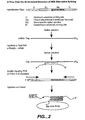
- the invention provides a method of detecting a first target sequence comprising a poly (A) sequence in a sample as defined in claim 1.
- the method includes hybridizing a first probe to the target sequence to form a first hybridization complex.
- the first probe comprises an upstream universal priming site (UUP), an adapter sequence, a first target-specific sequence, and a downstream universal priming site (DUP).
- UUP upstream universal priming site
- DUP downstream universal priming site
- the poly(A) sequence remains singte-stranded.
- the method further includes contacting the first hybridization complex with a support comprising a poly(T) sequence, such that the poly(A) sequence hybridizes with the pofy(T) sequence.
- the method includes removing unhybridized first probe sequences, denaturing the first hybridization complex, amplifying the first probe to generate a plurality of amplicons, contacting the amplicons with an array of capture probes to form assay complexes and detecting the assay complexes.
- the present invention is directed to the detection and quantification of a variety of nucleic acid reactions, particularly using microsphere arrays.
- the invention relates to gene detection, gene expression profiling and alternative splice monitoring, without prior amplification of the specific targets.
- the invention can be utilized with adapter sequences to create universal arrays.
- a plurality of probes are designed to have at least three different portions: a first portion that is target-specific to an mRNA target and two "universal priming" portions, an upstream and a downstream universal priming sequence.
- These target probes are hybridized to target mRNA sequences from a sample, without prior amplification, to form hybridization complexes.
- the hybridization complexes (and non-hybridized target mRNA sequences) are then removed. This is accomplished by using a polyA selection method such as the use of poly(T) sequences on a support that can specifically retain all mRNA including the hybrids.
- the hybrids are denatured. All the target probes can then be simultaneously amplified using universal primers that will hybridize to the upstream and downstream universal priming sequences.
- the resulting amplicons which can be directly or indirectly labeled, can then be detected on arrays, particularly microsphere arrays. This allows the detection and quantification of the mRNA target sequences.
- the system can take on a wide variety, of conformations, depending on the assay.
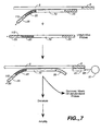
- a single target probe can be used.
- a single probe can be designed for any mRNA sequence, with an upstream and downstream universal primer.
- the detection of the mRNA sequence proceeds as outlined below.
- the target specific portion of the probe has a first domain that hybridizes to the first exon and a second domain that hybridizes to the second exon, and the assay is run under conditions whereby only if both domains hybridize to the target mRNA does the hybridization complex form.
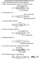
- any of the above embodiments can utilize one or more "adapter sequences” (sometimes referred to in the art as “zip codes”) to allow the use of "universal arrays". That is, arrays are generated that contain capture probes that are not target specific, but rather specific to individual artificial adapter sequences. One strand of the adapter sequences are added to the target probes (in the case of ligation probes, either probe may contain the adapter sequence), nested between the priming sequences, and thus are included in the amplicons. The adapters are then hybridized to the capture probes on the array, and detection proceeds. In some embodiments, as outlined below, there may be two adapter sequences used in the target probes.
- the present invention provides several significant advantages.
- the method can be used to detect gene expression or alternative splicing events from a single call or a few cells because of signal amplification of annealed probes. It also allows the direct hybridization of the probes to RNA targets, thus omitting a cDNA conversion step. Additionally, the hybridization reaction occurs in solution rather than on a surface, so that RNA hybridizes more predictably and with favorable kinetics according to their thermodynamic properties. The removal of excess probes and targets allows the isolated targets to reflect the level of individual gene expression level or splicing events in cells and the background signal (due to non-specific interactions) is reduced. Finally, the use of universal primers avoids biased signal amplification in PCR.
- the present invention provides methods for detecting, quantifying and/or identifying specific polyadenylated mRNA nucleic acid sequences in a sample.
- the sample solution may comprise any number of things, including, but not limited to, bodily fluids (including, but not limited to, blood, urine, serum, lymph, saliva, anal and vaginal secretions, perspiration and semen, of virtually any organism, with mammalian samples being preferred and human samples being particularly preferred).
- the sample may comprise individual cells, including primary cells (including bacteria), and cell lines, including, but not limited to, tumor cells of all types (particularly melanoma, myeloid leukemia, carcinomas of the lung, breast, ovaries, colon, kidney, prostate, pancreas and testes), cardiomyocytes, endothelial cells, epithelial cells, lymphocytes (T-cell and B cell), mast cells, eosinophils, vascular intimal cells, hepatocytes, leukocytes including mononuclear leukocytes, stem cells such as haemopoetic, neural, skin, lung, kidney, liver and myocyte stem cells; osteoclasts, chondrocytes and other connective tissue cells, keratinocytes, melanocytes, liver cells, kidney cells, and adipocytes.
- tumor cells of all types particularly melanoma, myeloid leukemia, carcinomas of the lung, breast, ovaries, colon, kidney, prostate, pancreas and
- Suitable cells also include known research cells, including, but not limited to, Jurkat T cells, NIH3T3 cells, CHO, Cos, 923, HeLa, WI-38, Weri-1, MG-63, etc. See the ATCC cell line catalog, hereby expressly incorporated by reference.
- the present invention provides compositions and methods for detecting the presence or absence of target mRNA nucleic acid sequences in a sample.
- "Target sequence” or grammatical equivalents as used herein means a polyadenylated mRNA sequence or a secondary target such as an amplicon.
- the target sequence may be a polyadenylated mRNA target sequence from a sample, or a secondary target such as an amplicon, which is the product of an amplification reaction such as PCR.
- a polyadenylated mRNA target sequence from a sample is amplified to produce an amplicon that is detected.
- the polyadenylated mRNA target sequence may be any length, with the understanding that longer sequences are more specific.
- probes are made to hybridize to polyadenylated mRNA target sequences to determine the presence, absence, quantity or sequence of a polyadenylated mRNA target sequence in a sample. Generally speaking, this term will be understood by those skilled in the art.
- the polyadenylated, mRNA target sequence may also be comprised of different target domains, that may be adjacent (i.e. contiguous) or separated.
- first and second are not meant to confer an orientation of the sequences with respect to the 5'-3' orientation of the target mRNA sequence.
- first target domain may be located either 5' to the second domain, or 3' to the second domain.
- the probes on the surface of the array e.g. attached to the microspheres
- the probes on the surface of the array may be attached in either orientation, either such that they have a free 3' end or a free 5' end; in some embodiments, the probes can be attached at one ore more internal positions, or at both ends.
- the polyadenylated mRNA target sequence is prepared using known techniques.
- the sample may be treated to lyse the cells, using known lysis buffers, sonication, electroporation, etc., with purification and amplification as outlined below occurring as needed, as will be appreciated by those in the art.
- the reactions outlined herein may be accomplished in a variety of ways, as will be appreciated by those in the art. Components of the reaction may be added simultaneously, or sequentially, in any order, with preferred embodiments outlined below.
- the reaction may include a variety of other reagents which may be included in the assays. These include reagents like salts, buffers, neutral proteins, e.g.
- albumin which may be used to facilitate optimal hybridization and detection, and/or reduce non-specific or background interactions.
- reagents that otherwise improve the efficiency of the assay such as protease inhibitors, nuclease inhibitors, anti-microbial agents, etc., may be used, depending on the sample preparation methods and purity of the target.
- a poly(T) support is used to remove unreacted target probes from the sample.
- a poly(T) support may be used to purify or concentrate poly(A) mRNA from a sample prior to running the assay. For example, total RNA may be isolated from a cell population, and then the poly(A) mRNA isolated from the total RNA and fed into the assay systems described below.
- double stranded target nucleic acids are denatured to render them single stranded so as to permit hybridization of the primers and other probes of the invention.
- a preferred embodiment utilizes a thermal step, generally by raising the temperature of the reaction to about 95°C, although pH changes and other techniques may also be used.
- nucleic acid or “oligonucleotide” or grammatical equivalents herein means at least two nucleotides covalently linked together.
- a nucleic acid of the present application will generally contain phosphodiester bonds, although in some cases, as outlined below, particularly for use with probes, nucleic acid analogs are included that may have alternate backbones, comprising, for example, phosphoramide (Beaucage et al., Tetrahedron 49(10):1925(1993) and references therein; Letsinger, J. Org. Chem. 35:3800 (1970); Sblui et al., Eur. J.
- nucleic acids include those with positive backbones (Denpoy et al., Proc. Natl. Acad. Sci. USA 92:6097 (1995); non-ionic backbones (U.S. Patent Nos. 5,386,023, 5,637,684,5,602,240, 5,216,141 and 4,469,863; Kiedrowshi et al., Angew. Chem. Intl. Ed. English 30:423 (1991); Letsinger et al., J. Am. Chem. Soc.
- nucleic acids containing one or more carbocyclic sugars are also included within the definition of nucleic adds (see Jenkins et al.; Chem. Soc. Rev. (1995) pp169-176). Several nucleic acid analogs are described in Rawls, C & E News June 2, 1997 page 35. The nucleic adds can also be "locked nucleic acids”.
- ribose-phosphate backbone may be done to facilitate the addition of labels, or to increase the stability and half-life of such molecules in physiological environments.
- nucleic acid analogs may find use as probes in the present invention.
- mixtures of naturally occurring nucleic acids and analogs can be made.
- mixtures of different nucleic acid analogs, and mixtures of naturally occuring nucleic acids and analogs may be made.
- PNA peptide nucleic acids
- These backbones are substantially non-ionic under neutral conditions, in contrast to the highly charged phosphodiester backbone of naturally occurring nucleic acids. This results in two advantages.
- the PNA backbone exhibits improved hybridization kinetics. PNAs have larger changes in the melting temperature (Tm) for mismatched versus perfectly matched basepairs. DNA and RNA typically exhibit a 2-4°C drop in Tm for an internal mismatch. With the non-ionic PNA backbone, the drop is closer to 7-9°C. Similarly, due to their non-ionic nature, hybridization of the bases attached to these backbones is relatively insensitive to salt concentration.
- the probe nucleic acids may be single stranded or double stranded, as specified, or contain portions of both double stranded or single stranded sequence.
- the hybridization complex comprising the target probe has a double stranded portion, where the target probe is hybridized, and one or more single stranded portions, including the poly(A) portion.
- the nucleic acid may contain any combination of deoxyribo- and ribo-nucleotides, and any combination of bases, including uracil, adenine, thymine, cytosine, guanine, inosine, xathanine hypoxathanine, isocytosine, isoguanine, etc.
- a preferred embodiment utilizes isocytosine and isoguanine in nucleic acids designed to be complementary to other probes, rather than target sequences, as this reduces non-specific hybridization, as is generally described in U.S. Patent No. 5,681,702.
- nucleoside includes nucleotides as well as nucleoside and nucleotide analogs, and modified nucleosides such as amino modified nucleosides.
- nucleoside includes non-naturally occuring analog structures.
- nucleoside includes individual units of a peptide nucleic acid, each containing a base, are referred to herein as a nucleoside.
- Probes and primers of the present application are designed to have at least a portion be complementary to a polyadenylated mRNA target sequence (either the polyadenylated mRNA target sequence of the sample or to other probe sequences, such as portions of amplicons, as is described below), such that hybridization of the polyadenylated mRNA target sequence and the probes of the present application occurs.
- a polyadenylated mRNA target sequence either the polyadenylated mRNA target sequence of the sample or to other probe sequences, such as portions of amplicons, as is described below
- this complementarity need not be perfect; there may be any number of base pair mismatches which will interfere with hybridization between the polyadenylated mRNA target sequence and the single stranded nucleic acids of the present invention.
- the sequence is not a complementary polyadenylated mRNA target sequence.
- substantially complementary herein is meant that the probes are sufficiently complementary to the polyadenylated mRNA target sequences to hybridize under normal reaction conditions, and preferably give the required specificity.
- hybridization conditions may be used in the present invention, including high, moderate and low stringency conditions; see for example Maniatis et al., Molecular Cloning: A Laboratory Manual, 2d Edition, 1989, and Short Protocols in Molecular Biology, ed. Ausubel, et al, hereby incorporated by reference. Stringent conditions are sequence-dependent and will be different in different circumstances. Longer sequences hybridize specifically at higher temperatures. An extensive guide to the hybridization of nucleic acids is found in Tijssen, Techniques in Biochemistry and Molecular Biology--Hybridization with Nucleic Acid Probes, "Overview of principles of hybridization and the strategy of nucleic acid assays" (1993).
- stringent conditions are selected to be about 5-10°C lower than the thermal melting point (Tm) for the specific sequence at a defined ionic strength and pH.
- Tm is the temperature (under defined ionic strength, pH and nucleic acid concentration) at which 50% of the probes complementary to the target hybridize to the polyadenylated mRNA target sequence at equilibrium (as the target sequences are present in excess, at Tm, 50% of the probes are occupied at equilibrium).
- Stringent conditions will be those in which the salt concentration is less than about 1.0 M sodium ion, typically about 0.01 to 1.0 M sodium ion concentration (or other salts) at pH 7.0 to 8.3 and the temperature is at least about 30°C for short probes (e.g.
- Stringent conditions may also be achieved with the addition of helix destabilizing agents such as formamide.
- the hybridization conditions may also vary when a non-ionic backbone, i.e. PNA is used, as is known in the art.
- cross-linking agents may be added after target binding to cross-link, i.e. covalently attach, the two strands of the hybridization complex.
- the assays are generally run under stringency conditions which allows formation of the first hybridization complex only in the presence of target.
- Stringency can be controlled by altering a step parameter that is a thermodynamic variable, including, but not limited to, temperature, formamide concentration, salt concentration, chaotropic salt concentration, pH, organic solvent concentration, etc.
- the size of the primer and probe nucleic acid may vary, as will be appreciated by those in the art with each portion of the probe and the total length of the probe in general varying from 5 to 500 nucleotides in length.
- Each portion is preferably between 10 and 100 being preferred, between 15 and 50 being particularly preferred, and from 10 to 35 being especially preferred, depending on the use and amplification technique.
- the universal priming sites of the probes are each preferably about 15-20 nucleotides in length, with 18 being especially preferred.
- the adapter sequences of the probes are preferably from 15-25 nucleotides in length, with 20 being especially preferred.
- the target specific portion of the probe is preferably from 15-50 nucleotides in length, with from 30 to 40 being especially preferred.
- probe set herein is meant a plurality of target probes that are used in a particular multiplexed assay.
- plurality means at least two, with more 10 than being preferred, depending on the assay, sample and purpose of the test.
- first target probe sets that comprise universal priming sites.
- universal priming site herein is meant a sequence of the probe that will bind a PCR primer for amplification.
- Each probe preferably comprises an upstream universal priming site (UUP) and a downstream universal priming site (DUP).
- UUP upstream universal priming site
- DUP downstream universal priming site
- upstream and downstream are not meant to convey a particular 5'- 3' orientation, and will depend on the orientation of the system.
- Preferably, only a single UUP sequence and a single DUP sequence is used in a probe set, although as will be appreciated by those in the art, different assays or different multiplexing analysis may utilize a plurality of universal priming sequences.
- the universal priming sites are preferably located at the 5' and 3' termini of the target probe (or the ligated probe), as only sequences flanked by priming sequences will be amplified.
- universal priming sequences are generally chosen to be as unique as possible given the particular assays and host genomes to ensure specificity of the assay.
- universal priming sequences range in size from about 5 to about 35 basepairs, with from about 15 to about 20 being particularly preferred.
- the orientation of the two priming sites is different. That is, one PCR primer will directly hybridize to the first priming site, while the other PCR primer will hybridize to the complement of the second priming site. Stated differently, the first priming site is in sense orientation, and the second priming site is in antisense orientation.
- the target probes comprise at least a first target-specific sequence, that is substantially complementary to the polyadenylated mRNA target sequence.
- ligation probes each comprise a target-specific sequence.
- the target-specific sequence may take on a wide variety of formats, depending on the use of probe. For example, for straight polyadenylated mRNA target sequence detection or gone expression monitoring or profiling, the target-specific probe sequence comprises a portion that will hybridize to all or part of the polyadenylated mRNA target sequence.
- a number of particular polyadenylated mRNA target sequences are preferred, Including, but not limited to, target sequences that span splice junctions.
- the target specific sequence spans a splice junction of interest.
- the target specific sequences are designed to be substantially complementary to sequences at the end of individual alternative exons.
- substantially complementary is meant that the probes are sufficiently complementary to the target sequences to hybridize under normal reaction conditions, and preferably give the required annealing specificity. Since exons are separated by introns, the detection sequences residue on different parts of the RNA molecule.
- a target specific sequence is composed of two parts: an upstream portion, complementary to the terminus of a first exon, and a downstream portion, complementary to the terminus of a second exon. Only if splicing has occurred and the intervening intron has been excised will the target specific sequence hybridize to the target sequence under the conditions of the assay.
- the target probes of the invention further comprise one or more adapter sequences.
- An "adapter sequence” is a sequence, generally exogeneous to the target sequences, e.g. artificial, that is designed to be substantially complementary (and preferably perfectly complementary) to a capture probe on the array.
- the use of adapter sequences allow the creation of more "universal" surfaces; that is, one standard array, comprising a finite set of capture probes can be made and used in any application.
- the end-user can customize the array by designing different soluble target probes, which, as will be appreciated by those in the art, is generally simpler and less costly.
- an array of different and usually artificial capture probes are made; that is, the capture probes do not have complementarity to known target sequences.
- the adapter sequences can then be incorporated In the target probes.
- adapter sequences will vary, depending on the desired strength of binding and the number of different adapters desired.
- adapter sequences range from about 6 to about 50 basepairs in length, with from about 8 to about 30 being preferred, and from about 15 to about 25 being particularly preferred.
- the placement and orientation of the adapter sequences can vary widely, depending on the configuration of the assay and the assay itself.
- the different components of the target probes can be placed in any order, just as long as the universal priming sites remain on the outermost ends of the probe, to allow all sequences between them to be amplified.
- the adapter sequences will have similar hybridization characteristics, e.g. same or similar melting temperature, similar (G+C) content.
- two adapter sequences per ligated target probe are used.
- one or both of the universal primers can be used as an adapter sequence. That is, one of the universal primers can be used to hybridize to a capture probe on the surface. However, in this embodiment, one of the universal primers must be target specific; e.g. one of the universal primers Is not really "universal", but rather one primer for each target must allow attachment to a different capture probe.
- the present application discloses target probes that comprise universal priming sequences, target specific sequence(s) and adapter sequences. These target probes are then added to the target sequences to form hybridization complexes.
- the hybridization complexes contain portions that are double stranded (the target-specific sequences of the target probes hybridized to a portion of the target mRNA sequence) and portions that are single stranded (the ends of the target probes comprising the universal priming sequences and the adapter sequences, and any unhybridized portion of the target sequence, such as poly(A) tails, as outlined herein).
- unhybridized probes are removed. This is important as all target probes may form some unpredictable structure which will complicate the amplification using the universal priming sequences. Thus to ensure specificity (e.g. that target probes directed to target sequences that are not present in the sample are not amplified and detected), it is important to remove all the nonhybridized probes.
- the separation of unhybridized target probes is done utilizing supports comprising poly(T) sequences.
- supports comprising poly(T) sequences are added to the mixture comprising the target sequences and the target probes.
- the first hybridization complexes comprise a single-stranded portion comprising a poly(A) sequence, generally ranging from 10 to 100s adenosines.
- the poly(T) support is then used to separate the unhybridized target probes from the hybridization complexes.
- magnetic beads when used, they may be removed from the mixture and washed; non-magnetic beads may bo removed via centrifugation and washed, etc.
- the poly(T) supports can be packed into a column and the assay mixture run through.
- the poly-A sequence is immobilized to the inside of a tube, i.e. a PCR tube, that is coated with poly(T).
- a tube i.e. a PCR tube
- the hybridization complexes are then released (and denatured) from the beads using a denaturation step such as a thermal step.
- the hybridization complexes are denatured and the target probes are amplified to form amplicons, which are then detected. This can be done In one of severe ways, including PCR amplification and rolling circle amplification. In addition, as outlined below, labels can be incorporated into the amplicons in a variety of ways.
- the target amplification technique is PCR.
- the polymerase chain reaction (PCR) is widely used and described, and involves the use of primer extension combined with thermal cycling to amplify a target sequence; see U.S. Patent Nos. 4,683,195 and 4,683,202, and PCR Essential Data, J. W. Wiley & sons, Ed. C.R. Newton, 1995,
- PCR may be briefly described as follows.
- the double stranded hybridization complex is denatured, generally by raising the temperature, and then cooled in the presence of an excess of a PCR primer, which then hybridizes to the first universal priming site.
- a DNA polymerase then acts to extend the primer with dNTPs, resulting in the synthesis of a new strand forming a hybridization complex.
- the sample is then heated again, to disassociate the hybridization complex, and the process is repeated.
- a second PCR primer for the complementary larget strand that hybridizes to the second universal priming site, rapid and exponential amplification occurs.
- PCR steps are denaturation, annealing and extension.
- PCR The particulars of PCR are well known, and include the use of a thermostable polymerase such as Taq I polymerase and thermal cycling.
- Suitable DNA polymerases include, but are not limited to the Klenow fragment of DNA polymerase I, SEQUENASE 1.0 and SEQUENASE 2.0 (U.S. Biochemical), T5 DNA polymerase and Phl29 DNA polymerase.
- nucleotide in this context herein is meant a deoxynucleoside-triphosphate (also called deoxynucleotides or dNTPs, e.g. dATP, dTTP, dCTP and dGTP).
- dNTPs deoxynucleotides
- one or more of the nucleotides may comprise a detectable label, which may be either a primary or a secondary label.
- the nucleotides may be nucleotide analogs, depending on the configuration of the system.
- the vasts may comprise a primary or secondary label.
- the PCR reaction requires at least one and preferably two PCR primer, a polymerase, and a set of dNTPs.
- the primers may comprise the label, or one or mere of the dNTPs may comprise a label.
- the present application discloses the generation of amplicons (sometimes referred to herein as secondary targets).
- the amplicons are labeled with a detection label.
- detection label or “detectable label” herein is meant a moiety that allows detection. This may be a primary label or a secondary label. Accordingly, detection labels may be primary labels (i.e directly detectable) or secondary labels (indirectly detectable).
- the detection label is a primary label.
- a primary label is one that can be directly detected, such as a fluorophore.
- labels fall into three classes: a) isotopic labels, which may be radioactive or heavy isotopes; b) magnetic, electrical, thermal labels; and c) colored or luminescent dyes. Labels can also include enzymes (horseradish peroxidase, etc.) and magnetic particles. Preferred labels include chromophores or phosphors but are preferably fluorescent dyes.
- Suitable dyes for use in the invention include, but are not limited to, fluorescent lanthanide complexes, Including those of Europium and Terbium, fluorescein, rhodamine, tetramethylrhodamine, eosin, erythrosin, coumarin, methyl-coumarins, quantum dots (also referred to as "nanocrystals"), pyrene, Malacite green, stilbene Lucifer Yellow, Cascade Blue TM , Texas Red, Cy dyes (Cy3, Cy5, etc.), alexa dyes, phycoerythin, bodipy, and others described in the 6th Edition of the Molecular Probes Handbook by Richard P. Haugland.
- a secondary detectable label is used.
- a secondary label is one that is indirectly detected; for example, a secondary label can bind or react with a primary label for detection, can act on an additional product to generate a primary label (e.g. enzymes), or may allow the separation of the compound comprising the secondary label from unlabeled materials, etc.
- Secondary labels include, but are not limited to, one of a binding partner pair such as biotin/streplavidin; chemically modifiable moieties, nuclease inhibitors, enzymes such as horseradish peroxidase, alkaline phosphatases, lucifierases, etc.
- the secondary label is a binding partner pair.
- the label may be a hapten or antigen, which will bind its binding partner.
- suitable binding partner pairs include, but are not limited to: antigens (such as proteins (including peptides)) and antibodies (including fragments thereof (FAbs, etc.)); proteins and small molecules, including biotin/streptavidin; enzymes and substrates or inhibitors; other protein-protein Interacting pairs receptor-ligands; and carbohydrates and their binding partners. Nucleic acid - nucleic acid binding proteins pairs are also useful. In general, the smaller of the pair is attached to the NTP for incorporation into the primer.
- Prefered binding partner pairs include, but are not limited to, biotin (or Imino-blotin) and streptavidin, digeoxinin and Abs, and Prolinx TM reagents (see www.prolinxinc.com/le4/home.hmtl).
- the binding partner pair comprises biotin or imino-biotin and a fluorescently labeled streptavidin.
- Imino-biotin is particularly preferred as imino-biotin disassociates from streptavidin in pH 4.0 buffer while biotin requires harsh denaturants (e.g. 6 M guanidinium HCl, pH 1.5 or 90% formamide at 95°C).
- the binding partner pair comprises a primary detection label (for example, attached to the NTP and therefore to the amplicon) and an antibody that will specifically bind to the primary detection label.
- a primary detection label for example, attached to the NTP and therefore to the amplicon
- an antibody that will specifically bind to the primary detection label.
- specifically bind herein is meant that the partners bind with specificity sufficient to differentiate between the pair and other components or contaminants of the system. The binding should be sufficient to remain bound under the conditions of the assay, including wash steps to remove non-specific binding.
- the dissociation constants of the pair will be less than about 10 -4 -10 -6 M -1 , with less than about 10 -5 to 10 -9 M -1 being preferred and less than about 10 -7 -10 -9 M -1 being particularly preferred.
- the secondary label is a chemically modifiable moiety.
- labels comprising reactive functional groups are incorporated into the nucleic acid.
- the functional group can then be subsequently labeled with a primary label.
- Suitable functional groups include, but are not limited to, amino groups, carboxy groups, maleimide groups, oxo groups and thiol groups, with amino groups and thiol groups being particularly preferred.
- primary labels containing amino groups can be attached to secondary labels comprising amino groups, for example using linkers as are known in the art; for example, homo-or hetero-bifunctional linkers as are well known (see 1994 Pierce Chemical Company catalog, technical section on cross-linkers, pages 155-200.
- labeling can occur in a variety of ways, as will be appreciated by those in the art. In general, labeling can occur In one of two ways: labels are incorporated into primers such that the amplification reaction results In amplicons that comprise the labels or labels are attached to dNTPs and incorporated by the polymerase into the amplicons.
- a preferred embodiment utilizes one primer comprising a biotin, that is used to bind a fluorescently labeled streptavidin.
- the present invention provides methods useful in the detection of nucleic acids, particularly the labeled amplicons outlined herein.
- preferred systems of the invention work as follows.
- Amplicons are attached (via hybridization) to an array site. This attachment can be either directly to a capture probe on the surface, through the use of adapters, or indirectly, using capture extender probes as outlined herein.
- the target sequence itself comprises the labels.
- a label probe is then added, forming an assay complex.
- the attachment of the label probe may be direct (i.e. hybridization to a portion of the target sequence), or indirect (i.e. hybridization to an amplifier probe that hybridizes to the target sequence), with all the required nucleic adds forming an assay complex.
- array compositions comprising at least a first substrate with a surface comprising individual sites.
- array or “biochip” herein is meant a plurality of nucleic acids in an array format, the size of the array will depend on the composition and end use of the array, Nucleic acids arrays are known in the art, and can be classified in a number of ways; both ordered arrays (e.g. the ability to resolve chemistries at discrete sites), and random arrays are included.
- Ordered arrays include, but are not limited to those made using photolithography techniques (Affymetrix GeneChip TM ), spotting techniques (Syntent and others), printing techniques (Hewlett Packard and Rosetta), three dimensional "gel pad” arrays, etc.
- a preferred embodiment utilizes microspheres on a variety of substrates including fiber optic bundles, as are outlined in WO99/18434, WO99/67641, WO98/40726, WO98150782;
- Arrays containing from about 2 different bioactive agents (e.g. different beads, when beads are used) to many millions can be made, with very large arrays being possible.
- the array will comprise from two to as many as a billion or more, depending on the size of the beads and the substrate, as well as the end use of the array, thus very high density, high density, moderate density, low density and very low density arrays may be made.
- Preferred ranges for very high density arrays are from about 10,000,000 to about 2,000,000,000, with from about 100,000,000 to about 1,000,000,000 being preferred (all numbers being in square cm).
- High density arrays range about 100,000 to about 10,000,000, with from about 1,000,000 to about 5,000,000 being particularly preferred.
- Moderate density arrays range from about 10,000 to about 100,000 being particularly preferred, and from about 20,000 to about 50,000 being especially preferred.
- Low density arrays are generally less than 10,000, with from about 1,000 to about 6,000 being preferred.
- Very low density arrays are less than 1,000, with from about 10 to about 1000 being preferred, and from about 100 to about 500 being particularly preferred.
- the compositions of the invention may not be in array format; that is, for some embodiments, compositions comprising a single bioactive agent may be made as well.
- multiple substrates may be used, either of different or identical compositions. Thus for example, large arrays may comprise a plurality of smaller substrates.
- one advantage of the disclosed compositions is that particularly through the use of fiber optic technology, extremely high density arrays can be made.
- beads of 200 ⁇ m or less with beads of 200 nm possible
- very small fibers are known
- substrate or “solid support or other grammatical equivalents herein is meant any material that can be modified to contain discrete individual sites appropriate for the attachment or association of beads and is amenable to at least one detection method. As will be appreciated by those in the art, the number of possible substrates is very large.
- Possible substrates include, but are not limited to, glass and modified or functionalized glass, plastics (Including acrylics, polystyrene and copolymers of styrene and other materials, polypropylene, polyethylene, polybutylene, polyurethanes, Teflon, etc.), polysaccharides, nylon or nitrocellulose, resins, silica or silica-based materials including silicon and modified silicon, carbon, metals, inorganic glasses, plastics, optical fiber bundles, and a variety of other polymers. In general, the substrates allow optical detection and do not themselves appreciably fluoresce.
- the substrate is flat (planar), although as will be appreciated by those in the art, other configurations of substrates may be used as well; for example, three dimensional configurations can be used, for example by embedding the beads in a porous block of plastic that allows sample access to the beads and using a confocal microscope for detection.
- the beads may be placed on the inside surface of a tube, for flow-through sample analysis to minimize sample volume;
- Preferred substrates include optical fiber bundles as discussed below, and flat planar substrates such as glass, polystyrene and other plastics and acrylics.
- the substrate is an optical fiber bundle or array, as is generally described in WO98/40726, WO98/50782.
- Preferred embodiments utilize preformed unitary fiber optic arrays.
- preformed unitary fiber optic array herein is meant an array of discrete individual fiber optic strands that are co-axially disposed and joined along their lengths. The fiber strands are generally individually clad.
- the fibers are not individually physically manipulatable; that is, one strand generally cannot be physically separated at any point along its length from another fiber strand.
- the array of array compositions can be configured in several ways.
- a "one component" system is used. That is, a first substrate comprising a plurality of assay locations (sometimes also referred to herein as "assay wells"), such as a microtiter plate, is configured such that each assay location contains an individual array. That is, the assay location and the array location are the same.
- the plastic material of the microtiter plate can be formed to contain a plurality of "bead wells" in the bottom of each of the assay wells. Beads containing the capture probes of the invention can then be loaded into the bead wells in each assay location as is more fully described below.
- a "two component" system can be used.
- the individual arrays are formed on a second substrate, which then can be fitted or “dipped” into the first microtiter plate substrate.
- a preferred embodiment utilizes fiber optic bundles as the individual arrays, generally with "bead wells” etched into one surface of each individual fiber, such that the beads containing the capture probes are loaded onto the end of the fiber optic bundle.
- the composite array thus comprises a number of individual arrays that are configured to fit within the wells of a microtiter plate.
- composite array or “combination array” or grammatical equivalents herein is meant a plurality of individual arrays, as outlined above. Generally the number of individual arrays is set by the size of the microtiter plate used; thus, 96 well, 384 well and 1536 well microtiter plates utilize composite arrays comprising 96, 384 and 1536 individual arrays, although as will be appreciated by those in the art, not each microtiter well need contain an individual array. It should be noted that the composite arrays can comprise individual arrays that are identical, similar or different. That is, in some embodiments, it may be desirable to do the same 2,000 assays on 96 different samples; alternatively, doing 192,000 experiments on the same sample (i.e.
- each row or column of the composite array could be the same, for redundancy/quality control.
- the random nature of the arrays may mean that the same population of beads may be added to two different surfaces, resulting in substantially similar but perhaps not identical arrays.
- At least one surface of the substrate is modified to contain discrete, individual sites for later association of microspheres. These sites may comprise physically altered sites, i.e. physical configurations such as wells or small depressions in the substrate that can retain the beads, such that a microsphere can rest in the well, or the use of other forces (magnetic or compressive), or chemically altered or active sites, such as chemically functionalized sites, electrostatically altered sites, hydrophobically/ hydrophilically functionalized sites, spots of adhesive, etc.
- sites may comprise physically altered sites, i.e. physical configurations such as wells or small depressions in the substrate that can retain the beads, such that a microsphere can rest in the well, or the use of other forces (magnetic or compressive), or chemically altered or active sites, such as chemically functionalized sites, electrostatically altered sites, hydrophobically/ hydrophilically functionalized sites, spots of adhesive, etc.
- the sites may be a pattern, i.e. a regular design or configuration, or randomly distributed.
- a preferred embodiment utilizes a regular pattern of sites such that the sites may be addressed in the X-Y coordinate plane.
- Pattern in this sense includes a repeating unit cell, preferably one that allows a high density of beads on the substrate.
- these sites may not be discrete sites. That is, it is possible to use a uniform surface of adhesive or chemical functionalities, for example, that allows the attachment of beads at any position. That Is, the surface of the substrate is modified to allow attachment of the microspheres at individual sites, whether or not those sites are contiguous or non-contiguous with other sites.
- the surface of the substrate may be modified such that discrete sites are formed that can only have a single associated bead, or alternatively, the surface of the substrate is modified and beads may go down anywhere, but they end up at discrete sites.
- the surface of the substrate is modified to contain wells, i.e. depressions in the surface of the substrate. This may be done as is generally known in the art using a variety of techniques, including, but not limited to, photolithography, stamping techniques, molding techniques and microetching techniques. As will be appreciated by those in the art, the technique used will depend on the composition and shape of the substrate.
- the substrate is a fiber optic bundle and the surface of the substrate is a terminal end of the fiber bundle, as is generally described in US Patent 6023540 (8. Feb. 2000).
- wells are made in a terminal or distal end of a fiber optic bundle comprising individual fibers.
- the cores of the individual fibers are etched, with respect to the cladding, such that small wells or depressions are formed at one end of the fibers. The required depth of the wells will depend on the size of the beads to be added to the wells.
- the microspheres are non-covalently associated in the wells, although the wells may additionally be chemically functionalized as is generally described below, cross-linking agents may be used, or a physical barrier may be used, i.e. a film or membrane over the beads.
- the surface of the substrate is modified to contain chemically modified sites, that can be used to attach, either covalently or non-covalently, the microspheres of the invention to the discrete sites or locations on the substrate.
- chemically modified sites in this context includes, but is not limited to, the addition of a pattern of chemical functional groups including amino groups, carboxy groups, oxo groups and thiol groups, that can be used to covalently attach microspheres, which generally also contain corresponding reactive functional groups; the addition of a pattern of adhesive that can be used to bind the microspheres (either by prior chemical functionalization for the addition of the adhesive or direct addition of the adhesive); the addition of a pattern of charged groups (similar to the chemical functionalities) for the electrostatic attachment of the microspheres, i.e.
- microspheres comprise charged groups opposite to the sites; the addition of a pattern of chemical functional groups that renders the sites differentially hydrophobic or hydrophilic, such that the addition of similarly hydrophobic or hydrophilic microspheres under suitable experimental conditions will result in association of the microspheres to the sites on the basis of hydroaffinity.
- pattern in this sense includes the use of a uniform treatment of the surface to allow attachment of the beads at discrete sites, as well as treatment of the surface resulting in discrete sites. As will be appreciated by those in the art, this may be accomplished in a variety of ways.
- the beads are not associated with a substrate. That is, the beads are in solution or are not distributed on a patterned substrate.
- compositions of the invention further comprise a population of microspheres.
- population herein is meant a plurality of beads as outlined above for arrays. Within the population are separate subpopulations, which can be a single microsphere or multiple identical microspheres. That is, in some embodiments, as is more fully outlined below, the array may contain only a single bead for each capture probe; preferred embodiments utilize a plurality of beads of each type.
- microspheres small discrete particles.
- the composition of the beads will vary, depending on the class of capture probe and the method of synthesis. Suitable bead compositions include those used in peptide, nucleic acid and organic moiety synthesis, including, but not limited to, plastics, ceramics, glass, polystyrene, methylstyrene, acrylic polymers, paramagnetic materials, thoria sol, carbon graphite, titanium dioxide, latex or cross-linked dextrans such as Sepharose, cellulose, nylon, cross-linked micelles and Teflon may all be used. "Microsphere Detection Guide” from Bangs Laboratories, Fishers IN is a helpful guide.
- the beads need not be spherical; irregular particles may be used.
- the beads may be porous, thus increasing the surface area of the bead available for either capture probe attachment or tag attachment.
- the bead sizes range from nanometers, i.e. 100 nm, to millimeters, i.e. 1 mm, with beads from about 0.2 micron to about 200 microns being preferred, and from about 0.5 to about 5 micron being particularly preferred, although in some embodiments smaller beads may be used.
- a key component of the invention is the use of a substrate/bead pairing that allows the association or attachment of the beads at discrete sites on the surface of the substrate, such that the beads do not move during the course of the assay.
- Each microsphere comprises a capture probe, although as will be appreciated by those in the art, there may be some microspheres which do not contain a capture probe, depending on the synthetic methods.
- Attachment of the nucleic acids may be done in a variety of ways, as will be appreciated by those in the art, including, but not limited to, chemical or affinity capture (for example, including the incorporation of derivatized nucleotides such as AminoLink or biotinylated nucleotides that can then be used to attach the nucleic acid to a surface, as well as affinity capture by hybridization), cross-linking, and electrostatic attachment, etc.
- affinity capture is used to attach the nucleic acids to the beads.
- nucleic acids can be derivatized, for example with one member of a binding pair, and the beads derivatized with the other member of a binding pair.
- Suitable binding pairs are as described herein for IBL/DBL pairs.
- the nucleic acids may be biotinylated (for example using enzymatic incorporate of biotinylated nucleotides, for by photoactivated cross-linking of biotin). Biotinylated nucleic acids can then be captured on streptavidin-coated beads, as is known in the art.
- other hapten-receptor combinations can be used, such as digoxigenin and anti-digoxigenin antibodies.
- chemical groups can be added in the form of derivatized nucleotides, that can them be used to add the nucleic acid to the surface.
- Preferred attachments are covalent, although even relatively weak interactions (i.e. non-covalent) can be sufficient to attach a nucleic acid to a surface, if there are multiple sites of attachment per each nucleic acid.
- electrostatic interactions can be used for attachment, for example by having beads carrying the opposite charge to the bioactive agent.
- affinity capture utilizing hybridization can be used to attach nucleic acids to beads.
- chemical crosslinking may be done, for example by photoactivated crosslinking of thymidine to reactive groups, as is known in the art.
- each bead comprises a single type of capture probe, although a plurality of individual capture probes are preferably attached to each bead.
- preferred embodiments utilize more than one microsphere containing a unique capture probe; that is, there is redundancy built into the system by the use of subpopulations of microspheres, each microsphere in the subpopulation containing the same capture probe.
- the capture probes may either be synthesized directly on the beads, or they may be made and then attached after synthesis.
- linkers are used to attach the capture probes to the beads, to allow both good attachment, sufficient flexibility to allow good interaction with the target molecule, and to avoid undesirable binding reactions.
- the capture probes are synthesized directly on the beads.
- many classes of chemical compounds are currently synthesized on solid supports, such as peptides, organic moieties, and nucleic acids. It is a relatively straightforward matter to adjust the current synthetic techniques to use beads.
- the capture probes are synthesized first, and then covalently attached to the beads. As will be appreciated by those in the art, this will be done depending on the composition of the capture probes and the beads.
- the functionalization of solid support surfaces such as certain polymers with chemically reactive groups such as thiols, amines, carboxyls, etc. is generally known in the art. Accordingly, "blank" microspheres may be used that have surface chemistries that facilitate the attachment of the desired functionality by the user.
- Some examples of these surface chemistries for blank microspheres include, but are not limited to, amino groups including aliphatic and aromatic amines, carboxylic acids, aldehydes, amides, chloromethyl groups, hydrazide, hydroxyl groups, sulfonates and sulfates.
- an encoding/decoding system When random arrays are used, an encoding/decoding system must be used.
- the beads are generally put onto the substrate randomly; as such there are several ways to correlate the functionality on the bead with its location, including the incorporation of unique optical signatures, generally fluorescent dyes, that could be used to identify the nucleic acid on any particular bead.
- unique optical signatures generally fluorescent dyes
- the beads may be randomly distributed on the array, a fast and inexpensive process as compared to either the in situ synthesis or spotting techniques of the prior art.
- the drawback to these methods is that for a large array, the system requires a large number of different optical signatures, which may be difficult or time-consuming to utilize. Accordingly, the present invention provides several improvements over these methods, generally directed to methods of coding and decoding the arrays. That is, as will be appreciated by those in the art, the placement of the capture probes is generally random, and thus a coding/decoding system is required to identify the probe at each location in the array.
- DBL decoding binding ligand
- IBLs identifier binding ligands
- this decoding may occur for all the beads, or only for those that bind a particular target sequence. Similarly, this may occur either prior to or after addition of a target sequence.
- the target sequences detected may be either a primary target sequence (e.g. a patient sample), or a reaction product from one of the methods described herein (e.g. an extended SBE probe, a ligated probe, a cleaved signal probe, etc.).
- the array is exposed to samples containing the target sequences, although as outlined below, this can be done prior to or during the analysis as well.
- the target sequences can hybridize (either directly or indirectly) to the capture probes as is more fully outlined below, and results in a change in the optical signal of a particular bead.
- decoding does not rely on the use of optical signatures, but rather on the use of decoding binding ligands that are added during a decoding step.
- the decoding binding ligands will bind either to a distinct identifier binding ligand partner that is placed on the beads, or to the capture probe itself.
- the decoding binding ligands are either directly or indirectly, labeled, and thus decoding occurs by detecting the presence of the label.
- the microspheres may additionally comprise identifier binding ligands for use in certain decoding systems.
- identifier binding ligands or “IBLs' herein is meant a compound that will specifically bind a corresponding decoder binding ligand (DBL) to facilitate the elucidation of the identity of the capture probe attached to the bead. That is, the IBL and the corresponding DBL form a binding partner pair.
- specifically bind herein Is meant that the IBL binds its DBL with specificity sufficient to differentiate between the corresponding DBL and other DBLs (that is, DBLs for other IBLs), or other components or contaminants of the system.
- the binding should be sufficient to remain bound under the conditions of the decoding step, including wash steps to remove non-specific binding.
- the dissociation constants of the IBL to its DBL will be less than about 10 -4 -10 -6 M -1 , with less than about 10 -5 to 10 -9 M -1 being preferred and less than about 10 -7 -10 -9 M -1 being particularly preferred.
- IBL-DBL binding pairs are known or can be readily found using known techniques.
- the DBLs include proteins (particularly including antibodies or fragments thereof (FAbs, etc.)) or small molecules, or vice versa (the IBL is an antibody and the DBL is a protein).
- Metal ion- metal ion ligands or chelators pairs are also useful.
- Antigen-antibody pairs enzymes and substrates or inhibitors, other protein-protein interacting pairs, receptor-ligands, complementary nucleic acid, and carbohydrates and their binding partners are also suitable binding pairs.
- Nucleic acid - nucleic acid binding proteins pairs are also useful. Similary, as is generally described in U.S.
- nucleic acid "aptamers" can be developed for binding to virtually any target; such an aptamer-target pair can be used as the IBL-DBL pair. Similary, there is a wide body of literature relating to the development of binding pairs based on combinatorial chemistry methods.
- the IBL is a molecule whose color or luminescence properties change in the presence of a selectively-binding DBL.
- the IBL may be a fluorescent pH indicator whose emission intensity changes with pH.
- the IBL may be a fluorescent ion indicator, whose emission properties change with ion concentration.
- the IBL is a molecule whose color or luminescence properties change in the presence of various solvents.
- the IBL may be a fluorescent molecule such as an ethidium salt whose fluorescence intensity increases in hydrophobic environments.
- the IBL may be a derivative of fluorescein whose color changes between aqueous and nonpolar solvents.
- the DBL may be attached to a bead, i.e. a "decoder bead", that may carry a label such as a fluorophore.
- the IBL-DBL pair comprise substantially complementary single-stranded nucleic acids.
- the binding ligands can be referred to as "identifier probes" and “decoder probes”.
- the identifier and decoder probes range from about 4 basepairs in length to about 1000, with from about 6 to about 100 being preferred, and from about 8 to about 40 being particularly preferred. What is important is that the probes are long enough to be specific, i.e. to distinguish between different IBL-DBL pairs, yet short enough to allow both a) dissociation, if necessary, under suitable experimental conditions, and b) efficient hybridization.
- the IBLs do not bind to DBLs. Rather, the IBLs are used as identifier moieties ("IMs") that are identified directly, for example through the use of mass spectroscopy.
- IMs identifier moieties
- the IBL and the capture probe are the same moiety; thus, for example, as outlined herein, particularly when no optical signatures are used, the capture probe can serve as both the identifier and the agent.
- the bead-bound probe (which serves as the capture probe) can also bind decoder probes, to identify the sequence of the probe on the bead.
- the DBLs bind to the capture probes.
- the microspheres may contain an optical signature. That is, as outlined in US Patent 6023540 (8 Feb. 2000) previous work had each subpopulation of microspheres comprising a unique optical signature or optical tag that is used to identify the unique capture probe of that subpopulation of microspheres; that is, decoding utilizes optical properties of the beads such that a bead comprising the unique optical signature may be distinguished from beads at other locations with different optical signatures. Thus the previous work assigned each capture probe a unique optical signature such that any microspheres comprising that capture probe are identifiable on the basis of the signature. These optical signatures comprised dyes, usually chromophores or fluorophores, that were entrapped or attached to the beads themselves. Diversity of optical signatures utilized different fluorochromes, different ratios of mixtures of fluorochromes, and different concentrations (intensities) of fluorochromes.
- the present invention does not rely solely on the use of optical properties to decode the arrays.
- optical signatures as an additional coding method, in conjunction with the present system.
- the size of the array may be effectively increased while using a single set of decoding moieties in several ways, one of which is the use of optical signatures one some beads.
- the use of two populations of beads, one with an optical signature and one without allows the effective doubling of the array size.
- the use of multiple optical signatures similarly increases the possible size of the array.
- each subpopulation of beads comprises a plurality of different IBLs.
- the number of possible unique codes is substantially increased. That is, by using one unique IBL per capture probe, the size of the array will be the number of unique IBLs (assuming no "reuse” occurs, as outlined below).
- the size of the array can be increased to 2 n , when the presence or absence of each IBL is used as the indicator. For example, the assignment of 10 IBLs per bead generates a 10 bit binary code, where each bit can be designated as "1" (IBL is present) or "0" (IBL is absent).
- each individual capture probe in the array is assigned a combination of IBLs, which can be added to the beads prior to the addition of the capture probe, after, or during the synthesis of the capture probe, i.e. simultaneous addition of IBLs and capture probe components.
- the combination of different IBLs can be used to elucidate the sequence of the nucleic acid.
- the first position of a nucleic acid can be elucidated: for example, adenosine can be represented by the presence of both IBL1 and IBL2; thymidine can be represented by the presence of IBL1 but not IBL2, cytosine can be represented by the presence of IBL2 but not IBL1, and guanosine can be represented by the absence of both.
- the second position of the nucleic acid can be done in a similar manner using IBL3 and IBL4; thus, the presence of IBL1, IBL2, IBL3 and IBL4 gives a sequence of AA; IBL1, IBL2, and IBL3 shows the sequence AT; IBL1, IBL3 and IBL4 gives the sequence TA, etc.
- the third position utilizes IBL5 and IBL6, etc. In this way, the use of 20 different identifiers can yield a unique code for every possible 10-mer.
- IBLs concentrations or densities of IBLs allows a "reuse" of sorts. If, for example, the bead comprising a first agent has a 1X concentration of IBL, and a second bead comprising a second agent has a 10X concentration of IBL, using saturating concentrations of the corresponding labelled DBL allows the user to distinguish between the two beads.
- the microspheres comprising the capture probes are generated, they are added to the substrate to form an array.
- the order of making, using and decoding the array can vary.
- the array can be made, decoded, and then the assay done.
- the array can be made, used in an assay, and then decoded; this may find particular use when only a few beads need be decoded.
- the beads can be added to the assay mixture, i.e. the sample containing the target sequences, prior to the addition of the beads to the substrate; after addition and assay, the array may be decoded.
- the sample comprising the beads is agitated or mixed; this can increase the amount of target sequence bound to the beads per unit time, and thus (in the case of nucleic acid assays) increase the hybridization kinetics. This may find particular use in cases where the concentration of target sequence in the sample is low; generally, for low concentrations, long binding times must be used.
- the methods of making the arrays and of decoding the arrays is done to maximize the number of different candidate agents that can be uniquely encoded.
- the compositions may be made in a variety of ways.
- the arrays are made by adding a solution or slurry comprising the beads to a surface containing the sites for attachment of the beads. This may be done in a variety of buffers, including aqueous and organic solvents, and mixtures. The solvent can evaporate, and excess beads are removed.
- a novel method of loading the beads onto the array comprises exposing the array to a solution of particles (including microspheres and cells) and then applying energy, e.g. agitating or vibrating the mixture. This results in an array comprising more tightly associated particles, as the agitation is done with sufficient energy to cause weakly-associated beads to fall off (or out, in the case of wells). These sites are then available to bind a different bead. In this way, beads that exhibit a high affinity for the sites are selected.
- arrays made in this way have two main advantages as compared to a more static loading: first of all, a higher percentage of the sites can be filled easily, and secondly, the arrays thus loaded show a substantial decrease in bead loss during assays.
- these methods are used to generate arrays that have at least about 50% of the sites filled, with at least about 75% being preferred, and at least about 90% being particularly preferred.
- arrays generated in this manner preferably lose less than about 20% of the beads during an assay, with less than about 10% being preferred and less than about 5% being particularly preferred.
- the substrate comprising the surface with the discrete sites is immersed into a solution comprising the particles (beads, cells, etc.).
- the surface may comprise wells, as is described herein, or other types of sites on a patterned surface such that there is a differential affinity for the sites. This differnetial affinity results in a competitive process, such that particles that will associate more tightly are selected.
- the entire surface to be "loaded” with beads is in fluid contact with the solution.
- This solution is generally a slurry ranging from about 10,000:1 beads:solution (vol:vol) to 1:1.
- the solution can comprise any number of reagents, including aqueous buffers, organic solvents, salts, other reagent components, etc.
- the solution preferably comprises an excess of beads; that is, there are more beads than sites on the array. Preferred embodiments utilize two-fold to billion-fold excess of beads.
- the immersion can mimic the assay conditions; for example, if the array is to be "dipped" from above into a microtiter plate comprising samples, this configuration can be repeated for the loading, thus minimizing the beads that are likely to fall out due to gravity.
- the substrate, the solution, or both are subjected to a competitive process, whereby the particles with lower affinity can be disassociated from the substrate and replaced by particles exhibiting a higher affinity to the site.
- This competitive process is done by the introduction of energy, in the form of heat, sonication, stirring or mixing, vibrating or agitating the solution or substrate, or both.
- a preferred embodiment utilizes agitation or vibration.
- the amount of manipulation of the substrate is minimized to prevent damage to the array; thus, preferred embodiments utilize the agitation of the solution rather than the array, although either will work.
- this agitation can take on any number of forms, with a preferred embodiment utilizing microtiter plates comprising bead solutions being agitated using microtiter plate shakers.
- the agitation proceeds for a period of time sufficient to load the array to a desired fill. Depending on the size and concentration of the beads and the size of the array, this time may range from about 1 second to days, with from about 1 minute to about 24 hours being preferred.
- sites of an array may comprise a bead; that is, there may be some sites on the substrate surface which are empty. In addition, there may be some sites that contain more than one bead, although this is not preferred.
- the beads in a non-random or ordered way.
- photoactivatible attachment linkers or photoactivatible adhesives or masks selected sites on the array may be sequentially rendered suitable for attachment, such that defined populations of beads are laid down.
- the arrays are constructed such that information about the identity of the capture probe is built into the array, such that the random deposition of the beads in the fiber wells can be "decoded” to allow identification of the capture probe at all positions. This may be done in a variety of ways, and either before, during or after the use of the array to detect target molecules.
- the array is "decoded” in order to identify the location of one or more of the capture probes, i.e. each subpopulation of beads, on the substrate surface.
- pyrosequencing techniques are used to decode the array.
- a selective decoding system is used. In this case, only those microspheres exhibiting a change In the optical signal as a result of the binding of a target sequence are decoded. This is commonly done when the number of "hits", i.e. the number of sites to decode, is generally low. That is, the array is first scanned under experimental conditions in the absence of the target sequences. The sample containing the target sequences is added, and only those locations exhibiting a change in the optical signal are decoded.
- the beads at either the positive or negative signal locations may be either selectively tagged or released from the array (for example through the use of photocleavable linkers), and subsequently sorted or enriched in a fluorescence-activated cell sorter (FACS). That Is, either all the negative beads are released, and then the positive beads are either released or analyzed in situ, or alternatively all the positives are released and analyzed.
- the labels may comprise halogenated aromatic compounds, and detection of the label is done using for example gas chromatography, chemical tags, isotopic tags mass spectral tags.
- each bead contains a fluorescent dye.
- the non-fluorescent active beads can be sorted from the fluorescent negative beads, Alternatively, when light is shown down the negative fibers, all the negatives are non-fluorescent and the the positives are fluorescent, and sorting can proceed.
- the characterization of the attached capture probe may be done directly, for example using mass spectroscopy.
- the identification may occur through the use of identifier moieties ("IMs"), which are similar to IBLs but need not necessarily bind to DBLs. That is, rather than elucidate the structure of the capture probe directly, the composition of the IMs may serve as the identifier.
- IMs identifier moieties
- a specific combination of IMs can serve to code the bead, and be used to identify the agent on the bead upon release from the bead followed by subsequent analysis, for example using a gas chromatograph or mass spectroscope.
- each bead comprises a non-fluorescent precursor to a fluorescent dye.
- photocleavable protecting groups such as certain ortho-nitrobenzyl groups
- photoactivation of the fluorochrome can be done. After the assay, light is shown down again either the "positive” or the "negative” fibers, to distinquish these populations.
- the illuminated precursors are then chemically converted to a fluorescent dye. All the beads are then released from the array, with sorting, to form populations of fluorescent and non-fluorescent beads (either the positives and the negatives or vice versa).
- the sites of attachment of the beads include a photopolymerizable reagent, or the photopolymerizable agent is added to the assembled array.
- the test assay After the test assay is run, light is shown down again either the "positive” or the "negative” fibers, to distinquish these populations.
- the irradiation either all the positives or all the negatives are polymerized and trapped or bound to the sites, while the other population of beads can be released from the array.
- the location of every capture probe is determined using decoder binding ligands (DBLs).
- DBLs are binding ligands that will either bind to identifier binding ligands, if present, or to the capture probes themselves, preferably when the capture probe is a nucleic acid or protein.
- the DBL binds to the IBL.
- the capture probes are single-stranded nucleic acids and the DBL is a substantially complementary single-stranded nucleic acid that binds (hybridizes) to the capture probe, termed a decoder probe herein.
- a decoder probe that is substantially complementary to each candidate probe is made and used to decode the array.
- the candidate probes and the decoder probes should be of sufficient length (and the decoding step run under suitable conditions) to allow specificity; i.e. each candidate probe binds to its corresponding decoder probe with sufficient specificity to allow the distinction of each candidate probe.
- the DBLs are either directly or indirectly labeled.
- the DBL is directly labeled, that is, the DBL comprises a label.
- the DBL is indirectly labeled; that is, a labeling binding ligand (LBL) that will bind to the DBL is used.
- LBL labeling binding ligand
- the labeling binding ligand-DBL pair can be as described above for IBL-DBL pairs.
- the identification of the location of the individual beads is done using one or more decoding steps comprising a binding between the labeled DBL and either the IBL or the capture probe (i.e. a hybridization between the candidate probe and the decoder probe when the capture probe is a nucleic acid).
- the DBLs can be removed and the array can be used; however, in some circumstances, for example when the DBL binds to an IBL and not to the capture probe, the removal of the DBL is not required (although it may be desirable in some circumstances).
- decoding may be done either before the array is used to in an assay, during the assay, or after the assay.
- each DBL is labeled with a unique label, such that the number of unique tags is equal to or greater than the number of capture probes (although in some cases, "reuse" of the unique labels can be done, as described herein; similarly, minor variants of candidate probes can share the same decoder, if the variants are encoded in another dimension, i.e. in the bead size or label).
- a DBL is made that will specifically bind to it and contains a unique tag, for example one or more fluorochromes.
- the location of each DBL can be elucidated. This allows the identification of the location of each capture probe; the random array has been decoded.
- the DBLs can then be removed, if necessary, and the target sample applied.
- the number of unique labels is less than the number of unique capture probes, and thus a sequential series of decoding steps are used.
- decoder probes are divided into n sets for decoding. The number of sets corresponds to the number of unique tags. Each decoder probe is labeled in n separate reactions with n distinct tags. All the decoder probes share the same n tags. The decoder probes are pooled so that each pool contains only one of the n tag versions of each decoder, and no two decoder probes have the same sequence of tags across all the pools. The number of pools required for this to be true is determined by the number of decoder probes and the n.
- Hybridization of each pool to the array generates a signal at every address.
- the sequential hybridization of each pool in turn will generate a unique, sequence-specific code for each candidate probe. This identifies the candidate probe at each address in the array. For example, if four tags are used, then 4 X n sequential hybridizations can ideally distinguish 4 n sequences, although in some cases more steps may be required.
- the hybrids are denatured and the decoder probes removed, so that the probes are rendered single-stranded for the next hybridization (although it is also possible to hybridize limiting amounts of target so that the available probe is not saturated. Sequential hybridizations can be carried out and analyzed by subtracting pre-existing signal from the previous hybridization).
- Decoder probes 1-16 are made that correspond to the probes on the beads.
- the first step is to label decoder probes 1-4 with tag A, decoder probes 5-8 with tag B, decoder probes 9-12 with tag C, and decoder probes 13-16 with tag D.
- the probes are mixed and the pool is contacted with the array containing the beads with the attached candidate probes.
- the location of each tag (and thus each decoder and candidate probe pair) is then determined.
- the first set of decoder probes are then removed.
- decoder probes 1, 5, 9 and 13 are labeled with tag A
- decoder probes 2, 6, 10 and 14 are labeled with tag B
- decoder probes 3, 7, 11 and 15 are labeled with tag C
- decoder probes 4, 8, 12 and 16 are labeled with tag D.
- those beads that contained tag A in both decoding steps contain candidate probe 1; tag A in the first decoding step and tag B in the second decoding step contain candidate probe 2; tag A in the first decoding step and tag C in the second step contain candidate probe 3; etc.
- the decoder probes are labeled in situ; that is, they need not be labeled prior to the decoding reaction.
- the incoming decoder probe is shorter than the candidate probe, creating a 5' "overhang" on the decoding probe.
- labeled ddNTPs each labeled with a unique tag
- a polymerase will allow the addition of the tags in a sequence specific manner, thus creating a sequence-specific pattern of signals.
- other modifications can be done, including ligation, etc.
- the size of the array will be set by the number of unique decoding binding ligands, it is possible to "reuse" a set of unique DBLs to allow for a greater number of test sites. This may be done in several ways; for example, by using some subpopulations that comprise optical signatures. Similarly, the use of a positional coding scheme within an array; different sub-bundles may reuse the set of DBLs. Similarly, one embodiment utilizes bead size as a coding modality, thus allowing the reuse of the set of unique DBLs for each bead size. Alternatively, sequential partial loading of arrays with beads can also allow the reuse of DBLs. Furthermore, "code sharing" can occur as well.
- the DBLs may be reused by having some subpopulations of beads comprise optical signatures.
- the optical signature is generally a mixture of reporter dyes, preferably fluoroscent.
- the composition of the mixture i.e. the ratio of one dye to another
- concentration of the dye leading to differences in signal intensity
- matrices of unique optical signatures may be generated. This may be done by covalently attaching the dyes to the surface of the beads, or alternatively, by entrapping the dye within the bead.
- the encoding can be accomplished in a ratio of at least two dyes, although more encoding dimensions may be added in the size of the beads, for example.
- the labels are distinguishable from one another; thus two different labels may comprise different molecules (i.e. two different fluors) or, alternatively, one label at two different concentrations or intensity.
- the dyes are covalently attached to the surface of the beads. This may be done as is generally outlined for the attachment of the capture probes, using functional groups on the surface of the beads. As will be appreciated by those in the art, these attachments are done to minimize the effect on the dye.
- the dyes are non-covalently associated with the beads, generally by entrapping the dyes in the pores of the beads.
- encoding in the ratios of the two or more dyes, rather than single dye concentrations is preferred since it provides insensitivity to the intensity of light used to interrogate the reporter dye's signature and detector sensitivity.
- a spatial or positional coding system is done.
- there are sub-bundles or subarrays i.e. portions of the total array
- each subarray is an "area code", that can have the same tags (i.e. telephone numbers) of other subarrays, that are separated by virtue of the location of the subarray.
- tags i.e. telephone numbers
- the same unique tags can be reused from bundle to bundle.
- the use of 50 unique tags in combination with 100 different subarrays can form an array of 5000 different capture probes.
- additional encoding parameters can be added, such as microsphere size.
- the use of different size beads may also allow the reuse of sets of DBLs; that is, it is possible to use microspheres of different sizes to expand the encoding dimensions of the microspheres.
- Optical fiber arrays can be fabricated containing pixels with different fiber diameters or cross-sections; alternatively, two or more fiber optic bundles, each with different cross-sections of the individual fibers, can be added together to form a larger bundle; or, fiber optic bundles with fiber of the same size cross-sections can be used, but just with different sized beads.
- the largest wells can be filled with the largest microspheres and then moving onto progressively smaller microspheres in the smaller wells until all size wells are then filled.
- the same dye ratio could be used to encode microspheres of different sizes thereby expanding the number of different oligonucleotide sequences or chemical functionalities present in the array.
- the coding and decoding is accomplished by sequential loading of the microspheres into the array.
- the optical signatures can be "reused".
- the library of microspheres each comprising a different capture probe (or the subpopulations each comprise a different capture probe), is divided into a plurality of sublibraries; for example, depending on the size of the desired array and the number of unique tags, 10 sublibraries each comprising roughly 10% of the total library may be made, with each sublibrary comprising roughly the same unique tags. Then, the first sublibrary is added to the fiber optic bundle comprising the wells, and the location of each capture probe is determined, generally through the use of DBLs.
- the second sublibrary is then added, and the location of each capture probe is again determined.
- the signal in this case will comprise the signal from the "first" DBL and the "second" DBL; by comparing the two matrices the location of each bead in each sublibrary can be determined. Similarly, adding the third, fourth, etc, sublibraries sequentially will allow the array to be filled.
- codes can be "shared" in several ways.
- a single code i.e. IBL/DBL pair
- two nucleic acid probes used in an mRNA quantitation assay can share the same code if the ranges of their hybridization signal intensities do not overlap. This can occur, for example, when one of the target sequences is always present at a much higher concentration than the other. Alternatively, the two target sequences might always be present at a similar concentration, but differ in hybridization efficiency.
- a single code can be assigned to multiple agents if the agents are functionally equivalent.
- the probes are functionally equivalent, even though they may differ In sequence.
- an array of this type could be used to detect homologs of known genes.
- each gene is represented by a heterologous set of probes, hybridizing to different regions of the gene (and therefore differing In sequence).
- the set of probes share a common code. If a homolog is present, it might hybridize to some but not all of the probes. The level of homology might be indicated by the fraction of probes hybridizing, as well as the average hybridization Intensity. Similarly, multiple antibodies to the same protein could all share the same code.
- decoding of self assembled random arrays is done on the bases of pH titration.
- the beads comprise optical signatures, wherein the optical signatures are generated by the use of pH-responsive dyes (sometimes referred to herein as "ph dyes") such as fluorophores.
- pH-responsive dyes sometimes referred to herein as "ph dyes”
- ph dyes pH-responsive dyes
- This embodiment is similar to that outlined in WO 98/40726, except that the dyes used in the present ivention exhibits changes in fluorescence intensity (or other properties) when the solution pH is adjusted from below the pKa to above the pKa (or vice versa).
- a set of pH dyes are used, each with a different pKa, preferably separated by at least 0.5 PH units.
- Preferred embodiments utilize a pH dye set of pKa's of 2.0, 2.5, 3.0, 3.5, 4.0, 4.5, 5.0, 5.5, 6.0, 6.5, 7.0, 7.5, 8.0, 8.5, 9.0, 9.5, 10.0, 10.5, 11, and 11.5.
- Each bead can contain any subset of the pH dyes, and in this way a unique code for the capture probe is generated.
- the decoding of an array is achieved by titrating the array from pH 1 to pH 13, and measuring the fluorescence signal from each bead as a function of solution pH.
- the present application discloses array compositions comprising a substrate with a surface comprising discrete sites.
- a population of microspheres is distributed on the sites, and the population comprises at least a first and a second subpopulation.
- Each subpopulation comprises a capture probe, and, in addition, at least one optical dye with a given pKa.
- the pKas of the different optical dyes are different.
- Target redundancy utilizes sensor elements with different probes to the same target: one probe may span the first 25 bases of the target, a second probe may span the second 25 bases of the target, etc.
- sensor redundancy is used.
- a plurality of sensor elements e.g. beads, comprising identical bioactive agents are used. That is, each subpopulation comprises a plurality of beads comprising identical bioactive agents (e.g. binding ligands).
- bioactive agents e.g. binding ligands.
- the optical signal from each sensor element can be combined and any number of statistical analyses run, as outlined below. This can be done for a variety of reasons. For example, in time varying measurements, redundancy can significantly reduce the noise in the system. For non-time based measurements, redundancy can significantly increase the confidence of the data.
- a plurality of identical sensor elements are used.
- the number of identical sensor elements will vary with the application and use of the sensor array. In general, anywhere from 2 to thousands may be used, with from 2 to 100 being preferred, 2 to 60 being particularly preferred and from 5 to 20 being especially preferred. In general, preliminary results indicate that roughly 10 beads gives a sufficient advantage, although for some sensor elements can be used.
- optical response signals from a plurality of sensor beads within each bead subpopulation can be manipulated and analyzed in a wide variety of ways, including baseline adjustment, averaging, standard deviation analysts, distribution and cluster analysis, confidence interval analysis, mean testing, etc.
- the first manipulation of the optical response signals is an optional baseline adjustment.
- the standardized optical responses are adjusted to start at a value of 0.0 by subtracting the integer 1.0 from all data points. Doing this allows the baseline-loop data to remain at zero even when summed together and the random response signal noise is canceled out.
- the fluidinstalle-loop temporal region frequently exhibits a characteristic change in response, either positive, negative or neutral, prior to the sample pulse and often requires a baseline adjustment to overcome noise associated with drift in the first few data points due to charge buildup in the CCD camera.
- the baseline from the first data point for each bead sensor is subtracted from all the response data for the same bead. If drift is observed, the averse baseline from the first ten data points for each bead sensor is substracted from the all the response data for the same bead.
- signal summing is done by simply adding the intensity values of all responses at each time point, generating a new temporal response comprised of the sum of all bead responses. These values can be baseline-adjusted or raw. As for all the analyses described herein, signal summing can be performed in real time or during post-data acquisition data reduction and analysis. In one embodiment, signal summing is performed with a commercial spreadsheet program (Excel, Microsoft, Redmond, WA) after optical response data is collected.
- a commercial spreadsheet program Excel, Microsoft, Redmond, WA
- cummulative response data is generated by simply adding all data points in successive time intervals.
- This final column comprised of the sum of all data points at a particular time interval, may then be compared or plotted with the individual bead responses to determine the extent of signal enhancement or improved signal-to-noise ratios.
- the mean of the subpopulation i.e. the plurality of identical beads
- Equation 1 ⁇ ⁇ ⁇ x i n
- the subpopulation may be redefined to exclude some beads if necessary (for example for obvious outliers, as discussed below).
- the subpopulation may be redefined to exclude some beads if necessary (for example for obvious outliers, as discussed below).
- the quality of the data is evaluated using confidence intervals, as is known in the art. Confidence intervals can be used to facilitate more comprehensive data processing to measure the statistical validity of a result.
- statistical parameters of a subpopulation of beads are used to do hypothesis testing.
- tests concerning means also called mean testing.
- statistical evaluation is done to determine whether two subpopulations are different. For example, one sample could be compared with another sample for each subpopulation within an array to determine if the variation is statistically significant.
- analyzing the distribution of individual members of a subpopulation of sensor elements may be done. For example, a subpopulation distribution can be evaluated to determine whether the distribution is binomial, Poisson, hypergeometric, etc.
- a preferred embodiment utilizes a plurality of sensor elements that are directed to a single target analyte but yet are not identical.
- a single target nucleic acid analyte may have two or more sensor elements each comprising a different probe. This adds a level of confidence as non-specific binding interactions can be statistically minimized.
- the redundant nucleic acid probes may be overlapping, adjacent, or spatially separated. However, it is preferred that two probes do not compete for a single binding site, so adjacent or separated probes are preferred.
- preferred embodiments utilize bioactive agent binding agents that bind to different parts of the target. For example, when antibodies (or antibody fragments) are used as bioactive agents for the binding of target proteins, preferred embodiments utilize antibodies to different epitopes.
- a plurality of different sensor elements may be used, with from about 2 to about 20 being preferred, and from about 2 to about 10 being especially preferred, and from 2 to about 5 being particularly preferred, including 2, 3, 4 or 5. However, as above, more may also be used, depending on the application.
- any number of statistical analyses may be run on the data from target redundant sensors.
- Bead summing One benefit of the sensor element summing (referred to herein as “bead summing" when beads are used), is the increase in sensitivity that can occur.
- the present invention finds use in gene expression monitoring and profiling in any mRNA sample, and in particular in the comparison of different cellular states, including the comparison of diseased tissue such as cancerous tissue with normal tissue.
- the present invention finds use in alternative splice analysis, including both discovery of splice junctions and detection of alternative splice junctions.
- the invention finds use in the correlation of alternative splicing patterns to morphological and clinical parameters of cancer.
- the mechanisms and regulation of constitutive and alternative splicing can be elucidated in a variety of cell types, as outlined above.
- Selected Splicing targets Gene Name Functional Significance Selected Reference 1 acetylcholinesteras synapse maturation 1 2 agrin AChR clustering at synapses 2 3 AML 1 transcriptional activation 3 4 ASF/SF2 mRNA splicing regulation 4 5 Bcl-x apoptosis regulation 5 6 BRCA1 transcriptional activation 6 7 c-src signal transduction regulation 7 8 calcium channel, alpha 1A neurotransmitterrelease 8 9 calcium channel, alpha 1B neurotransmitterrelease 9 10 caspase 1 (ICE) apoptotic regulation 10 11 caspase 2 (Ich-1) apoptotic regulation 11 12 CD45 T cell maturation 12 13 clathrin light chain B receptor-mediatedendocytosis 13 14 cytochrome P450, aromatase steroid hormone synthesis 14 15 estrogen receptor 1 hormone response 15 16 estrogen receptor 2 hormone response 16 17 fas
Landscapes
- Chemical & Material Sciences (AREA)
- Life Sciences & Earth Sciences (AREA)
- Organic Chemistry (AREA)
- Engineering & Computer Science (AREA)
- Zoology (AREA)
- Wood Science & Technology (AREA)
- Proteomics, Peptides & Aminoacids (AREA)
- Health & Medical Sciences (AREA)
- Biophysics (AREA)
- General Engineering & Computer Science (AREA)
- Immunology (AREA)
- Microbiology (AREA)
- Molecular Biology (AREA)
- Analytical Chemistry (AREA)
- Physics & Mathematics (AREA)
- Genetics & Genomics (AREA)
- Biochemistry (AREA)
- Bioinformatics & Cheminformatics (AREA)
- Biotechnology (AREA)
- General Health & Medical Sciences (AREA)
- Chemical Kinetics & Catalysis (AREA)
- Measuring Or Testing Involving Enzymes Or Micro-Organisms (AREA)
- Saccharide Compounds (AREA)
Claims (11)
- Procédé de détection d'une ou de plusieurs séquences cibles comprenant une séquence poly (A) dans un échantillon comprenant :(a) une hybridation d'une ou de plusieurs sondes cibles à une ou plusieurs séquences cibles afin de former un ou plusieurs complexes d'hybridation, ladite sonde cible comprenant :(i) un site d'initiation universel en amont (UUP);(ii) une séquence adaptatrice;(iii) une séquence cible spécifique; et(iv) un site d'initiation universel en aval (DUP);dans lequel ladite séquence poly(A) reste monobrin, et dans lequel ladite séquence adaptatrice est essentiellement complémentaire d'une sonde de capture, ladite séquence adaptatrice étant située entre lesdits sites d'initiation;(b) une mise en contact dudit ou desdits plusieurs complexes d'hybridation avec un support comprenant une séquence poly(T), de sorte que ladite séquence poly(A) s'hybride avec ladite séquence poly(T) ;(c) un enlèvement des séquences de sondes cibles non hybridées ;(d) une dénaturation dudit ou desdits complexes d'hybridation ;(e) une amplification de ladite ou desdites sondes cibles afin de générer une pluralité d'amplicons, lesdits amplicons comprenant ladite séquence adaptatrice ou lesdites séquences adaptatrices ;(f) une mise en contact desdits amplicons avec un réseau de sondes de capture afin de former des complexes d'essai ; et(g) une détection desdits complexes d'essai.
- Procédé selon la revendication 1, dans lequel ladite sonde cible comprend un marqueur.
- Procédé selon la revendication 2, dans lequel ledit marqueur est un marqueur pouvant être détecté directement.
- Procédé selon la revendication 3, dans lequel ledit marqueur primaire est un marqueur fluorescent.
- Procédé selon la revendication 2, dans lequel ledit marqueur peut être détecté indirectement.
- Procédé selon la revendication 2, dans lequel ledit marqueur est la biotine.
- Procédé selon l'une quelconque des revendications 1 à 6, dans lequel ladite amplification est réalisée en :(a) hybridant une première amorce universelle audit UUP ;(b) fournissant une polymérase et des dNTP de façon que ladite première amorce universelle soit étendue ;(c) hybridant une seconde amorce universelle audit DUP;(d) fournissant une polymérase et des dNTP de façon que ladite seconde amorce universelle soit étendue ; et(e) répétant les étapes (a) à (d).
- Procédé selon l'une quelconque des revendications 1 à 7, dans lequel ledit réseau comprend :(a) un substrat avec une surface à motifs comprenant des sites discrets ; et(b) une population de microsphères comprenant au moins une première sous-population comprenant une première sonde de capture et une seconde sous-population comprenant une seconde sonde de capture.
- Procédé selon la revendication 8, dans lequel lesdits sites discrets comprennent des puits.
- Procédé selon la revendication 8, dans lequel ledit substrat comprend un faisceau de fibre optique.
- Procédé selon l'une quelconque des revendications . 1 à 10, dans lequel ledit support comprenant une séquence poly(T) comprend des billes magnétiques.
Applications Claiming Priority (5)
| Application Number | Priority Date | Filing Date | Title |
|---|---|---|---|
| US18081000P | 2000-02-07 | 2000-02-07 | |
| US180810P | 2000-02-07 | ||
| US23473100P | 2000-09-22 | 2000-09-22 | |
| US234731P | 2000-09-22 | ||
| PCT/US2001/004055 WO2001057268A2 (fr) | 2000-02-07 | 2001-02-07 | Detection d'acides nucleiques et procedes utilisant l'amorçage universel |
Publications (2)
| Publication Number | Publication Date |
|---|---|
| EP1255865A2 EP1255865A2 (fr) | 2002-11-13 |
| EP1255865B1 true EP1255865B1 (fr) | 2007-04-18 |
Family
ID=26876657
Family Applications (1)
| Application Number | Title | Priority Date | Filing Date |
|---|---|---|---|
| EP01910465A Expired - Lifetime EP1255865B1 (fr) | 2000-02-07 | 2001-02-07 | Procédé pour la détection d'acides nucléiques utilisant des amorces universelles |
Country Status (7)
| Country | Link |
|---|---|
| US (1) | US6812005B2 (fr) |
| EP (1) | EP1255865B1 (fr) |
| AT (1) | ATE360095T1 (fr) |
| AU (2) | AU3806701A (fr) |
| CA (1) | CA2401962A1 (fr) |
| DE (1) | DE60127939T2 (fr) |
| WO (1) | WO2001057268A2 (fr) |
Cited By (1)
| Publication number | Priority date | Publication date | Assignee | Title |
|---|---|---|---|---|
| WO2019107893A3 (fr) * | 2017-11-29 | 2019-07-18 | 주식회사 파나진 | Procédé d'amplification d'acide nucléique cible et composition associée |
Families Citing this family (164)
| Publication number | Priority date | Publication date | Assignee | Title |
|---|---|---|---|---|
| CA2255599C (fr) | 1996-04-25 | 2006-09-05 | Bioarray Solutions, Llc | Assemblage electrocinetique de particules proches des surfaces regule par la lumiere |
| EP1090293B2 (fr) | 1998-06-24 | 2019-01-23 | Illumina, Inc. | Décodage de détecteurs matricels à microspheres |
| US20030207295A1 (en) * | 1999-04-20 | 2003-11-06 | Kevin Gunderson | Detection of nucleic acid reactions on bead arrays |
| US20060275782A1 (en) | 1999-04-20 | 2006-12-07 | Illumina, Inc. | Detection of nucleic acid reactions on bead arrays |
| US7244559B2 (en) | 1999-09-16 | 2007-07-17 | 454 Life Sciences Corporation | Method of sequencing a nucleic acid |
| US7211390B2 (en) | 1999-09-16 | 2007-05-01 | 454 Life Sciences Corporation | Method of sequencing a nucleic acid |
| US7611869B2 (en) | 2000-02-07 | 2009-11-03 | Illumina, Inc. | Multiplexed methylation detection methods |
| US7582420B2 (en) | 2001-07-12 | 2009-09-01 | Illumina, Inc. | Multiplex nucleic acid reactions |
| US7955794B2 (en) | 2000-09-21 | 2011-06-07 | Illumina, Inc. | Multiplex nucleic acid reactions |
| US8076063B2 (en) | 2000-02-07 | 2011-12-13 | Illumina, Inc. | Multiplexed methylation detection methods |
| US7361488B2 (en) * | 2000-02-07 | 2008-04-22 | Illumina, Inc. | Nucleic acid detection methods using universal priming |
| EP1130113A1 (fr) * | 2000-02-15 | 2001-09-05 | Johannes Petrus Schouten | Méthode d'amplification dépendant de ligatures multiples |
| EP1285089A4 (fr) * | 2000-04-25 | 2004-07-07 | Affymetrix Inc | Technique de surveillance de l'expression de genes alternativement episses |
| US9709559B2 (en) | 2000-06-21 | 2017-07-18 | Bioarray Solutions, Ltd. | Multianalyte molecular analysis using application-specific random particle arrays |
| DE60117556T2 (de) | 2000-06-21 | 2006-11-02 | Bioarray Solutions Ltd. | Multianalytische molekularanalyse durch verwendung anwendungsspezifischer zufallspartikelarrays |
| US7572642B2 (en) | 2001-04-18 | 2009-08-11 | Ambrigen, Llc | Assay based on particles, which specifically bind with targets in spatially distributed characteristic patterns |
| US7262063B2 (en) | 2001-06-21 | 2007-08-28 | Bio Array Solutions, Ltd. | Directed assembly of functional heterostructures |
| CA2497740C (fr) | 2001-10-15 | 2011-06-21 | Bioarray Solutions, Ltd. | Analyse multiplexee de loci polymorphes par une methode de detection basee sur le prolongement de la sonde |
| US6902921B2 (en) | 2001-10-30 | 2005-06-07 | 454 Corporation | Sulfurylase-luciferase fusion proteins and thermostable sulfurylase |
| US6956114B2 (en) | 2001-10-30 | 2005-10-18 | '454 Corporation | Sulfurylase-luciferase fusion proteins and thermostable sulfurylase |
| US8765484B2 (en) * | 2002-02-07 | 2014-07-01 | The Regents Of The University Of California | Optically encoded particles |
| US7108976B2 (en) * | 2002-06-17 | 2006-09-19 | Affymetrix, Inc. | Complexity management of genomic DNA by locus specific amplification |
| US9388459B2 (en) * | 2002-06-17 | 2016-07-12 | Affymetrix, Inc. | Methods for genotyping |
| US20050118616A1 (en) * | 2002-08-16 | 2005-06-02 | Kawashima Tadashi R. | Amplification of target nucleotide sequence without polymerase chain reaction |
| US7459273B2 (en) * | 2002-10-04 | 2008-12-02 | Affymetrix, Inc. | Methods for genotyping selected polymorphism |
| AU2003298655A1 (en) | 2002-11-15 | 2004-06-15 | Bioarray Solutions, Ltd. | Analysis, secure access to, and transmission of array images |
| US7575865B2 (en) | 2003-01-29 | 2009-08-18 | 454 Life Sciences Corporation | Methods of amplifying and sequencing nucleic acids |
| CA2513899C (fr) | 2003-01-29 | 2013-03-26 | 454 Corporation | Procede d'amplification et de sequencage d'acides nucleiques |
| CN103396933B (zh) | 2003-02-26 | 2016-04-20 | 凯利达基因组股份有限公司 | 通过杂交进行的随机阵列dna分析 |
| NZ545000A (en) * | 2003-07-03 | 2008-08-29 | Univ California | Genome mapping of functional DNA elements and cellular proteins |
| US8114978B2 (en) * | 2003-08-05 | 2012-02-14 | Affymetrix, Inc. | Methods for genotyping selected polymorphism |
| US7927796B2 (en) | 2003-09-18 | 2011-04-19 | Bioarray Solutions, Ltd. | Number coding for identification of subtypes of coded types of solid phase carriers |
| CA2539824C (fr) | 2003-09-22 | 2015-02-03 | Xinwen Wang | Polyelectrolyte immobilise en surface a plusieurs groupes fonctionnels pouvant se lier de maniere covalente a des biomolecules |
| CA2544041C (fr) | 2003-10-28 | 2015-12-08 | Bioarray Solutions Ltd. | Optimisation de l'analyse de l'expression genique a l'aide de sondes de capture immobilisees |
| CA2544202C (fr) | 2003-10-29 | 2012-07-24 | Bioarray Solutions Ltd. | Analyse multiplexee d'acide nucleique par fragmentation d'adn bicatenaire |
| EP1702414A4 (fr) * | 2003-12-22 | 2008-04-23 | Univ California Office Of The | Particules optiquement codees a spectres de niveaux de gris |
| AU2004308380A1 (en) * | 2003-12-22 | 2005-07-14 | The Regents Of The University Of California | Optically encoded particles, system and high-throughput screening |
| US7622281B2 (en) * | 2004-05-20 | 2009-11-24 | The Board Of Trustees Of The Leland Stanford Junior University | Methods and compositions for clonal amplification of nucleic acid |
| US7785843B2 (en) * | 2004-06-23 | 2010-08-31 | Sequenom, Inc. | Target-specific compomers and methods of use |
| WO2006012468A2 (fr) * | 2004-07-23 | 2006-02-02 | (Osi) Eyetech, Inc. | Detection d'oligonuleotides par hybridation double |
| US7848889B2 (en) | 2004-08-02 | 2010-12-07 | Bioarray Solutions, Ltd. | Automated analysis of multiplexed probe-target interaction patterns: pattern matching and allele identification |
| US20070108465A1 (en) * | 2005-03-10 | 2007-05-17 | The Regents Of The University Of California | Porous microstructure multi layer spectroscopy and biosensing |
| US7452671B2 (en) * | 2005-04-29 | 2008-11-18 | Affymetrix, Inc. | Methods for genotyping with selective adaptor ligation |
| US8486629B2 (en) * | 2005-06-01 | 2013-07-16 | Bioarray Solutions, Ltd. | Creation of functionalized microparticle libraries by oligonucleotide ligation or elongation |
| EP3257949A1 (fr) | 2005-06-15 | 2017-12-20 | Complete Genomics Inc. | Analyse d'acide nucléique par des mélanges aléatoires de fragments non chevauchants |
| AU2006287548A1 (en) * | 2005-09-06 | 2007-03-15 | Gen-Probe Incorporated | Methods, compositions and kits for isothermal amplification of nucleic acids |
| US7962291B2 (en) * | 2005-09-30 | 2011-06-14 | Affymetrix, Inc. | Methods and computer software for detecting splice variants |
| US7485425B2 (en) * | 2005-10-03 | 2009-02-03 | Applied Biosystems Inc. | Methods for amplification of nucleic acids using spanning primers |
| US7960104B2 (en) | 2005-10-07 | 2011-06-14 | Callida Genomics, Inc. | Self-assembled single molecule arrays and uses thereof |
| US11306351B2 (en) | 2005-12-21 | 2022-04-19 | Affymetrix, Inc. | Methods for genotyping |
| US20070168197A1 (en) * | 2006-01-18 | 2007-07-19 | Nokia Corporation | Audio coding |
| SG170028A1 (en) | 2006-02-24 | 2011-04-29 | Callida Genomics Inc | High throughput genome sequencing on dna arrays |
| CN101432439B (zh) | 2006-02-24 | 2013-07-24 | 考利达基因组股份有限公司 | Dna阵列上的高通量基因组测序 |
| US7897747B2 (en) * | 2006-05-25 | 2011-03-01 | The Board Of Trustees Of The Leland Stanford Junior University | Method to produce single stranded DNA of defined length and sequence and DNA probes produced thereby |
| US7833716B2 (en) | 2006-06-06 | 2010-11-16 | Gen-Probe Incorporated | Tagged oligonucleotides and their use in nucleic acid amplification methods |
| WO2007147079A2 (fr) | 2006-06-14 | 2007-12-21 | Living Microsystems, Inc. | analyse de celluleS rareS avec recours au fractionnement d'échantillons et à des marqueurs d'adn |
| EP2589668A1 (fr) | 2006-06-14 | 2013-05-08 | Verinata Health, Inc | Analyse de cellules rares utilisant la division d'échantillons et les marqueurs d'ADN |
| EP4170042A1 (fr) | 2006-06-14 | 2023-04-26 | Verinata Health, Inc. | Procédés pour le diagnostic d'anomalies f tales |
| US7910354B2 (en) | 2006-10-27 | 2011-03-22 | Complete Genomics, Inc. | Efficient arrays of amplified polynucleotides |
| US20090111706A1 (en) | 2006-11-09 | 2009-04-30 | Complete Genomics, Inc. | Selection of dna adaptor orientation by amplification |
| US8133701B2 (en) * | 2006-12-05 | 2012-03-13 | Sequenom, Inc. | Detection and quantification of biomolecules using mass spectrometry |
| US7902345B2 (en) * | 2006-12-05 | 2011-03-08 | Sequenom, Inc. | Detection and quantification of biomolecules using mass spectrometry |
| EP3095873B1 (fr) | 2006-12-21 | 2018-04-18 | Gen-Probe Incorporated | Procédés et compositions pour l'amplification d'acide nucléiques |
| US20080194416A1 (en) * | 2007-02-08 | 2008-08-14 | Sigma Aldrich | Detection of mature small rna molecules |
| US20080274458A1 (en) * | 2007-05-01 | 2008-11-06 | Latham Gary J | Nucleic acid quantitation methods |
| US7769548B2 (en) * | 2007-05-10 | 2010-08-03 | Illumina, Inc. | Microarray analytical data stitching system and method |
| WO2009032781A2 (fr) | 2007-08-29 | 2009-03-12 | Sequenom, Inc. | Méthodes et compositions s'appliquant à une amplification en chaîne par polymèrase universelle spécifique de la taille |
| US20090061424A1 (en) * | 2007-08-30 | 2009-03-05 | Sigma-Aldrich Company | Universal ligation array for analyzing gene expression or genomic variations |
| US9388457B2 (en) | 2007-09-14 | 2016-07-12 | Affymetrix, Inc. | Locus specific amplification using array probes |
| US8951731B2 (en) | 2007-10-15 | 2015-02-10 | Complete Genomics, Inc. | Sequence analysis using decorated nucleic acids |
| US7767441B2 (en) | 2007-10-25 | 2010-08-03 | Industrial Technology Research Institute | Bioassay system including optical detection apparatuses, and method for detecting biomolecules |
| US7871571B2 (en) * | 2007-10-25 | 2011-01-18 | Parker John A | Biomolecule analyzing system |
| US8518640B2 (en) | 2007-10-29 | 2013-08-27 | Complete Genomics, Inc. | Nucleic acid sequencing and process |
| US7897344B2 (en) | 2007-11-06 | 2011-03-01 | Complete Genomics, Inc. | Methods and oligonucleotide designs for insertion of multiple adaptors into library constructs |
| US8298768B2 (en) | 2007-11-29 | 2012-10-30 | Complete Genomics, Inc. | Efficient shotgun sequencing methods |
| US8415099B2 (en) | 2007-11-05 | 2013-04-09 | Complete Genomics, Inc. | Efficient base determination in sequencing reactions |
| WO2009061840A1 (fr) | 2007-11-05 | 2009-05-14 | Complete Genomics, Inc. | Procédés et conceptions d'oligonucléotide pour une insertion d'adaptateurs multiples en utilisant une méthylation sélective |
| US8592150B2 (en) | 2007-12-05 | 2013-11-26 | Complete Genomics, Inc. | Methods and compositions for long fragment read sequencing |
| US20090181390A1 (en) * | 2008-01-11 | 2009-07-16 | Signosis, Inc. A California Corporation | High throughput detection of micrornas and use for disease diagnosis |
| WO2009097368A2 (fr) * | 2008-01-28 | 2009-08-06 | Complete Genomics, Inc. | Procédés et compositions pour un appel de base efficace dans des réactions de séquençage |
| US9074244B2 (en) | 2008-03-11 | 2015-07-07 | Affymetrix, Inc. | Array-based translocation and rearrangement assays |
| US9524369B2 (en) | 2009-06-15 | 2016-12-20 | Complete Genomics, Inc. | Processing and analysis of complex nucleic acid sequence data |
| US8512955B2 (en) | 2009-07-01 | 2013-08-20 | Gen-Probe Incorporated | Methods and compositions for nucleic acid amplification |
| WO2011047087A2 (fr) | 2009-10-13 | 2011-04-21 | Nanostring Technologies, Inc. | Détection de protéine par l'intermédiaire de nanorapporteurs |
| JP2013509883A (ja) | 2009-11-06 | 2013-03-21 | ザ ボード オブ トラスティーズ オブ ザ リーランド スタンフォード ジュニア ユニバーシティ | 臓器移植患者における移植片拒絶反応の非侵襲的診断方法 |
| US20190300945A1 (en) | 2010-04-05 | 2019-10-03 | Prognosys Biosciences, Inc. | Spatially Encoded Biological Assays |
| US10787701B2 (en) | 2010-04-05 | 2020-09-29 | Prognosys Biosciences, Inc. | Spatially encoded biological assays |
| WO2011127099A1 (fr) | 2010-04-05 | 2011-10-13 | Prognosys Biosciences, Inc. | Tests biologiques à codage spatial |
| WO2012106385A2 (fr) | 2011-01-31 | 2012-08-09 | Apprise Bio, Inc. | Procédés d'identification de multiples épitopes dans des cellules |
| EP2683833B1 (fr) | 2011-03-10 | 2018-09-26 | Gen-Probe Incorporated | Procédés de sélection et d'optimisation de séquences de marquage oligonucléotidiques |
| GB201106254D0 (en) | 2011-04-13 | 2011-05-25 | Frisen Jonas | Method and product |
| CA3069089A1 (fr) | 2011-09-22 | 2013-03-28 | Lineage Biosciences, Inc. | Compositions et procedes pour analyser des echantillons heterogenes |
| EP2882868B1 (fr) | 2012-08-08 | 2019-07-31 | H. Hoffnabb-La Roche Ag | Accroissement de la plage dynamique pour identifier de multiples épitopes dans des cellules |
| EP4592400A3 (fr) | 2012-10-17 | 2025-10-29 | 10x Genomics Sweden AB | Procédés et produits pour optimiser la detection localisée ou spatiale de l'expression génique dans un échantillon de tissu |
| EP3611262B1 (fr) | 2013-03-15 | 2020-11-11 | Lineage Biosciences, Inc. | Méthode de séquençage du répertoire immunitaire |
| CA2916662C (fr) | 2013-06-25 | 2022-03-08 | Prognosys Biosciences, Inc. | Procedes et systemes pour determiner des motifs spatiales de cibles biologiques dans un echantillon |
| US10415083B2 (en) | 2013-10-28 | 2019-09-17 | The Translational Genomics Research Institute | Long insert-based whole genome sequencing |
| US9957550B2 (en) | 2014-09-08 | 2018-05-01 | BioSpyder Technologies, Inc. | Attenuators |
| US9938566B2 (en) | 2014-09-08 | 2018-04-10 | BioSpyder Technologies, Inc. | Profiling expression at transcriptome scale |
| EP3901282B1 (fr) | 2015-04-10 | 2023-06-28 | Spatial Transcriptomics AB | Analyse de plusieurs acides nucléiques spatialement différenciés de spécimens biologiques |
| US11519033B2 (en) | 2018-08-28 | 2022-12-06 | 10X Genomics, Inc. | Method for transposase-mediated spatial tagging and analyzing genomic DNA in a biological sample |
| JP2022513124A (ja) * | 2018-11-21 | 2022-02-07 | アヴィダ バイオメッド, インコーポレイテッド | 標的化された核酸ライブラリー形成のための方法 |
| US12529094B2 (en) | 2018-12-10 | 2026-01-20 | 10X Genomics, Inc. | Imaging system hardware |
| WO2020123316A2 (fr) | 2018-12-10 | 2020-06-18 | 10X Genomics, Inc. | Procédés de détermination d'un emplacement d'un analyte biologique dans un échantillon biologique |
| US11926867B2 (en) | 2019-01-06 | 2024-03-12 | 10X Genomics, Inc. | Generating capture probes for spatial analysis |
| US11649485B2 (en) | 2019-01-06 | 2023-05-16 | 10X Genomics, Inc. | Generating capture probes for spatial analysis |
| WO2020243579A1 (fr) | 2019-05-30 | 2020-12-03 | 10X Genomics, Inc. | Procédés de détection de l'hétérogénéité spatiale d'un échantillon biologique |
| US12157124B2 (en) | 2019-11-06 | 2024-12-03 | 10X Genomics, Inc. | Imaging system hardware |
| WO2021091611A1 (fr) | 2019-11-08 | 2021-05-14 | 10X Genomics, Inc. | Agents de capture d'analytes marqués spatialement pour le multiplexage d'analytes |
| WO2021092433A2 (fr) | 2019-11-08 | 2021-05-14 | 10X Genomics, Inc. | Amélioration de la spécificité de la liaison d'un analyte |
| ES2982420T3 (es) | 2019-12-23 | 2024-10-16 | 10X Genomics Inc | Métodos para el análisis espacial mediante el uso de la ligazón con plantilla de ARN |
| CN115135984B (zh) | 2019-12-23 | 2025-12-23 | 10X基因组学有限公司 | 可逆固定试剂及其使用方法 |
| EP4081656A1 (fr) | 2019-12-23 | 2022-11-02 | 10X Genomics, Inc. | Compositions et méthodes d'utilisation d'échantillons biologiques fixés dans des dosages basés sur des compartiments |
| US12365942B2 (en) | 2020-01-13 | 2025-07-22 | 10X Genomics, Inc. | Methods of decreasing background on a spatial array |
| US12405264B2 (en) | 2020-01-17 | 2025-09-02 | 10X Genomics, Inc. | Electrophoretic system and method for analyte capture |
| US11732299B2 (en) | 2020-01-21 | 2023-08-22 | 10X Genomics, Inc. | Spatial assays with perturbed cells |
| US11702693B2 (en) | 2020-01-21 | 2023-07-18 | 10X Genomics, Inc. | Methods for printing cells and generating arrays of barcoded cells |
| US20210230681A1 (en) | 2020-01-24 | 2021-07-29 | 10X Genomics, Inc. | Methods for spatial analysis using proximity ligation |
| US11821035B1 (en) | 2020-01-29 | 2023-11-21 | 10X Genomics, Inc. | Compositions and methods of making gene expression libraries |
| US12076701B2 (en) | 2020-01-31 | 2024-09-03 | 10X Genomics, Inc. | Capturing oligonucleotides in spatial transcriptomics |
| US11898205B2 (en) | 2020-02-03 | 2024-02-13 | 10X Genomics, Inc. | Increasing capture efficiency of spatial assays |
| US12110541B2 (en) | 2020-02-03 | 2024-10-08 | 10X Genomics, Inc. | Methods for preparing high-resolution spatial arrays |
| US11732300B2 (en) | 2020-02-05 | 2023-08-22 | 10X Genomics, Inc. | Increasing efficiency of spatial analysis in a biological sample |
| WO2021158925A1 (fr) | 2020-02-07 | 2021-08-12 | 10X Genomics, Inc. | Évaluation quantitative et automatisée de la performance de perméabilisation pour la transcriptomique spatiale |
| US11835462B2 (en) | 2020-02-11 | 2023-12-05 | 10X Genomics, Inc. | Methods and compositions for partitioning a biological sample |
| US12281357B1 (en) | 2020-02-14 | 2025-04-22 | 10X Genomics, Inc. | In situ spatial barcoding |
| US12399123B1 (en) | 2020-02-14 | 2025-08-26 | 10X Genomics, Inc. | Spatial targeting of analytes |
| US11891654B2 (en) | 2020-02-24 | 2024-02-06 | 10X Genomics, Inc. | Methods of making gene expression libraries |
| US11926863B1 (en) | 2020-02-27 | 2024-03-12 | 10X Genomics, Inc. | Solid state single cell method for analyzing fixed biological cells |
| US11768175B1 (en) | 2020-03-04 | 2023-09-26 | 10X Genomics, Inc. | Electrophoretic methods for spatial analysis |
| EP4139485B1 (fr) | 2020-04-22 | 2023-09-06 | 10X Genomics, Inc. | Procédés d'analyse spatiale utilisant un appauvrissement d'arn ciblée |
| US12416603B2 (en) | 2020-05-19 | 2025-09-16 | 10X Genomics, Inc. | Electrophoresis cassettes and instrumentation |
| EP4153776B1 (fr) | 2020-05-22 | 2025-03-05 | 10X Genomics, Inc. | Analyse spatiale pour détecter des variants de séquence |
| EP4414459B1 (fr) | 2020-05-22 | 2025-09-03 | 10X Genomics, Inc. | Mesure spatio-temporelle simultanée de l'expression génique et de l'activité cellulaire |
| WO2021242834A1 (fr) | 2020-05-26 | 2021-12-02 | 10X Genomics, Inc. | Procédé de réinitialisation d'un réseau |
| US12265079B1 (en) | 2020-06-02 | 2025-04-01 | 10X Genomics, Inc. | Systems and methods for detecting analytes from captured single biological particles |
| EP4600376A3 (fr) | 2020-06-02 | 2025-10-22 | 10X Genomics, Inc. | Transcriptome spatial pour récepteurs d'antigènes |
| AU2021283174A1 (en) | 2020-06-02 | 2023-01-05 | 10X Genomics, Inc. | Nucleic acid library methods |
| US12031177B1 (en) | 2020-06-04 | 2024-07-09 | 10X Genomics, Inc. | Methods of enhancing spatial resolution of transcripts |
| EP4421186B1 (fr) | 2020-06-08 | 2025-08-13 | 10X Genomics, Inc. | Procédés de détermination d'une marge chirurgicale et procédés d'utilisation de ceux-ci |
| US12435363B1 (en) | 2020-06-10 | 2025-10-07 | 10X Genomics, Inc. | Materials and methods for spatial transcriptomics |
| WO2021252747A1 (fr) | 2020-06-10 | 2021-12-16 | 1Ox Genomics, Inc. | Procédés de distribution de fluide |
| EP4165207B1 (fr) | 2020-06-10 | 2024-09-25 | 10X Genomics, Inc. | Methode pour determiner la position d'un analyte dans un echantillon biologique |
| ES2994976T3 (en) | 2020-06-25 | 2025-02-05 | 10X Genomics Inc | Spatial analysis of dna methylation |
| US11761038B1 (en) | 2020-07-06 | 2023-09-19 | 10X Genomics, Inc. | Methods for identifying a location of an RNA in a biological sample |
| US12209280B1 (en) | 2020-07-06 | 2025-01-28 | 10X Genomics, Inc. | Methods of identifying abundance and location of an analyte in a biological sample using second strand synthesis |
| US11981960B1 (en) | 2020-07-06 | 2024-05-14 | 10X Genomics, Inc. | Spatial analysis utilizing degradable hydrogels |
| US12553898B1 (en) | 2020-08-10 | 2026-02-17 | 10X Genomics, Inc. | Fluorescent hybridization of antibody-oligonucleotide for multiplexing and signal amplification |
| US11981958B1 (en) | 2020-08-20 | 2024-05-14 | 10X Genomics, Inc. | Methods for spatial analysis using DNA capture |
| WO2022061150A2 (fr) | 2020-09-18 | 2022-03-24 | 10X Geonomics, Inc. | Appareil de manipulation d'échantillons et procédés d'enregistrement d'images |
| US11926822B1 (en) | 2020-09-23 | 2024-03-12 | 10X Genomics, Inc. | Three-dimensional spatial analysis |
| US11827935B1 (en) | 2020-11-19 | 2023-11-28 | 10X Genomics, Inc. | Methods for spatial analysis using rolling circle amplification and detection probes |
| EP4729631A2 (fr) | 2020-12-21 | 2026-04-22 | 10X Genomics, Inc. | Procédés, compositions et systèmes de capture de sondes et/ou de codes à barres |
| EP4421491A3 (fr) | 2021-02-19 | 2024-11-27 | 10X Genomics, Inc. | Procédé d'utilisation d'un dispositif de support d'analyse modulaire |
| AU2022238446A1 (en) | 2021-03-18 | 2023-09-07 | 10X Genomics, Inc. | Multiplex capture of gene and protein expression from a biological sample |
| EP4305196B1 (fr) | 2021-04-14 | 2025-04-02 | 10X Genomics, Inc. | Procédés de mesure d'une mauvaise localisation d'un analyte |
| EP4320271B1 (fr) | 2021-05-06 | 2025-03-19 | 10X Genomics, Inc. | Procédés pour augmenter la résolution d'une analyse spatiale |
| WO2022256503A1 (fr) | 2021-06-03 | 2022-12-08 | 10X Genomics, Inc. | Procédés, compositions, kits et systèmes pour améliorer la capture d'analytes pour une analyse spatiale |
| US12553805B2 (en) | 2021-08-02 | 2026-02-17 | 10X Genomics, Inc. | Methods of preserving a biological sample |
| WO2023034489A1 (fr) | 2021-09-01 | 2023-03-09 | 10X Genomics, Inc. | Procédés, compositions et kits pour bloquer une sonde de capture sur un réseau spatial |
| WO2023086880A1 (fr) | 2021-11-10 | 2023-05-19 | 10X Genomics, Inc. | Procédés, compositions et kits pour déterminer l'emplacement d'un analyte dans un échantillon biologique |
| WO2023102118A2 (fr) | 2021-12-01 | 2023-06-08 | 10X Genomics, Inc. | Procédés, compositions et systèmes pour la détection améliorée d'analytes in situ et analyse spatiale |
| WO2023122033A1 (fr) | 2021-12-20 | 2023-06-29 | 10X Genomics, Inc. | Auto-test pour dispositif d'imagerie |
| EP4482979B1 (fr) | 2022-11-09 | 2025-10-01 | 10X Genomics, Inc. | Procédés, compositions et kits pour déterminer l'emplacement de plusieurs analytes dans un échantillon biologique |
Family Cites Families (96)
| Publication number | Priority date | Publication date | Assignee | Title |
|---|---|---|---|---|
| US4200110A (en) | 1977-11-28 | 1980-04-29 | United States Of America | Fiber optic pH probe |
| US4499052A (en) | 1982-08-30 | 1985-02-12 | Becton, Dickinson And Company | Apparatus for distinguishing multiple subpopulations of cells |
| US4682895A (en) | 1985-08-06 | 1987-07-28 | Texas A&M University | Fiber optic probe for quantification of colorimetric reactions |
| US4824789B1 (en) | 1986-10-10 | 1996-08-13 | Minnesota Mining & Mfg | Gas sensor |
| US4798738A (en) | 1986-10-10 | 1989-01-17 | Cardiovascular Devices, Inc. | Micro sensor |
| US4822746A (en) | 1986-06-25 | 1989-04-18 | Trustees Of Tufts College | Radiative and non-radiative energy transfer and absorbance modulated fluorescence detection methods and sensors |
| US5143853A (en) | 1986-06-25 | 1992-09-01 | Trustees Of Tufts College | Absorbance modulated fluorescence detection methods and sensors |
| US5252494A (en) | 1986-06-25 | 1993-10-12 | Trustees Of Tufts College | Fiber optic sensors, apparatus, and detection methods using controlled release polymers and reagent formulations held within a polymeric reaction matrix |
| US5254477A (en) | 1986-06-25 | 1993-10-19 | Trustees Of Tufts College | Flourescence intramolecular energy transfer conjugate compositions and detection methods |
| US5114864A (en) | 1986-06-25 | 1992-05-19 | Trustees Of Tufts College | Fiber optic sensors, apparatus, and detection methods using fluid erodible controlled release polymers for delivery of reagent formulations |
| ATE66695T1 (de) | 1986-12-01 | 1991-09-15 | Molecular Biosystems Inc | Verfahren zur erhoehung der empfindlichkeit von nukleinsaeure-hybridisierungstesten. |
| SE458968B (sv) | 1987-06-16 | 1989-05-22 | Wallac Oy | Biospecifikt analysfoerfarande foer flera analyter i vilket ingaar partikelraekning och maerkning med fluorescerande maerksubstanser |
| US5132242A (en) | 1987-07-15 | 1992-07-21 | Cheung Sau W | Fluorescent microspheres and methods of using them |
| US5194300A (en) | 1987-07-15 | 1993-03-16 | Cheung Sau W | Methods of making fluorescent microspheres |
| US4785814A (en) | 1987-08-11 | 1988-11-22 | Cordis Corporation | Optical probe for measuring pH and oxygen in blood and employing a composite membrane |
| US5002867A (en) | 1988-04-25 | 1991-03-26 | Macevicz Stephen C | Nucleic acid sequence determination by multiple mixed oligonucleotide probes |
| NO164622C (no) | 1988-05-11 | 1990-10-24 | Tore Lindmo | Binaer immunometrisk partikkelbasert metode for maaling av spesifikke serum-antigener ved hjelp av vaeskestroemsmikrofotometri og et ferdigpreparert maaloppsett derav. |
| US5575849A (en) | 1988-11-25 | 1996-11-19 | Canon Kabushiki Kaisha | Apparatus for producing a substrate having a surface with a plurality of spherical dimples for photoconductive members |
| JPH02299598A (ja) | 1989-04-14 | 1990-12-11 | Ro Inst For Molecular Genetics & Geneteic Res | 微視的サイズの別個の粒子と結合している核酸試料中のごく短い配列の全部または一部のオリゴヌクレチドプローブとのハイブリダイゼーションによる決定法 |
| US5302509A (en) | 1989-08-14 | 1994-04-12 | Beckman Instruments, Inc. | Method for sequencing polynucleotides |
| US5326692B1 (en) | 1992-05-13 | 1996-04-30 | Molecular Probes Inc | Fluorescent microparticles with controllable enhanced stokes shift |
| US5494810A (en) | 1990-05-03 | 1996-02-27 | Cornell Research Foundation, Inc. | Thermostable ligase-mediated DNA amplifications system for the detection of genetic disease |
| EP0478319B1 (fr) | 1990-09-28 | 1997-04-02 | Kabushiki Kaisha Toshiba | Méthode pour la détection de gènes |
| JPH06502766A (ja) | 1990-11-14 | 1994-03-31 | アクゾ・ノベル・ナムローゼ・フェンノートシャップ | ポリスチレン支持体ベース−サンドイッチハイブリダイゼーションアッセイ法を用いる核酸の非同位体検出およびそれに用いる組成物 |
| US5105305A (en) | 1991-01-10 | 1992-04-14 | At&T Bell Laboratories | Near-field scanning optical microscope using a fluorescent probe |
| US5244636A (en) | 1991-01-25 | 1993-09-14 | Trustees Of Tufts College | Imaging fiber optic array sensors, apparatus, and methods for concurrently detecting multiple analytes of interest in a fluid sample |
| US5320814A (en) | 1991-01-25 | 1994-06-14 | Trustees Of Tufts College | Fiber optic array sensors, apparatus, and methods for concurrently visualizing and chemically detecting multiple analytes of interest in a fluid sample |
| US5250264A (en) | 1991-01-25 | 1993-10-05 | Trustees Of Tufts College | Method of making imaging fiber optic sensors to concurrently detect multiple analytes of interest in a fluid sample |
| US5244813A (en) | 1991-01-25 | 1993-09-14 | Trustees Of Tufts College | Fiber optic sensor, apparatus, and methods for detecting an organic analyte in a fluid or vapor sample |
| US5380489A (en) | 1992-02-18 | 1995-01-10 | Eastman Kodak Company | Element and method for nucleic acid amplification and detection using adhered probes |
| WO1993002360A1 (fr) | 1991-07-16 | 1993-02-04 | Transmed Biotech Incorporated | Procedes et compositions d'analyse simultanee d'analytes multiples |
| US5639603A (en) | 1991-09-18 | 1997-06-17 | Affymax Technologies N.V. | Synthesizing and screening molecular diversity |
| AU2872092A (en) | 1991-10-15 | 1993-05-21 | Multilyte Limited | Binding assay employing labelled reagent |
| US5632957A (en) | 1993-11-01 | 1997-05-27 | Nanogen | Molecular biological diagnostic systems including electrodes |
| US6051380A (en) | 1993-11-01 | 2000-04-18 | Nanogen, Inc. | Methods and procedures for molecular biological analysis and diagnostics |
| US5888723A (en) | 1992-02-18 | 1999-03-30 | Johnson & Johnson Clinical Diagnostics, Inc. | Method for nucleic acid amplification and detection using adhered probes |
| EP0565999A2 (fr) | 1992-04-16 | 1993-10-20 | Siemens Aktiengesellschaft | Dispositif de couplage optique entre deux groupes de guides d'ondes |
| KR100249110B1 (ko) | 1992-05-06 | 2000-04-01 | 다니엘 엘. 캐시앙 | 핵산 서열 증폭 방법, 이에 이용되는 조성물 및 키트 |
| CA2115342C (fr) * | 1992-06-17 | 2003-08-26 | Robert B. Wallace | Methode de detection et de separation de sequences d'acide nucleique |
| WO1994002515A1 (fr) | 1992-07-21 | 1994-02-03 | Bunsen Rush Laboratories Inc. | Modeles de bibliotheques d'oligomeres et procedes s'y rapportant |
| DK0723146T3 (da) | 1992-09-14 | 2004-08-30 | Stanford Res Inst Int | Opkonverterende reportermolekyler til biologiske og andre assays ved anvendelse af laser-excitationsteknikker |
| US5565324A (en) | 1992-10-01 | 1996-10-15 | The Trustees Of Columbia University In The City Of New York | Complex combinatorial chemical libraries encoded with tags |
| US5795714A (en) | 1992-11-06 | 1998-08-18 | Trustees Of Boston University | Method for replicating an array of nucleic acid probes |
| US5541311A (en) | 1992-12-07 | 1996-07-30 | Third Wave Technologies, Inc. | Nucleic acid encoding synthesis-deficient thermostable DNA polymerase |
| US5298741A (en) | 1993-01-13 | 1994-03-29 | Trustees Of Tufts College | Thin film fiber optic sensor array and apparatus for concurrent viewing and chemical sensing of a sample |
| CA2102884A1 (fr) | 1993-03-04 | 1994-09-05 | James J. Wynne | Procedes et dispositifs dentaires utilisant les rayons ultraviolets |
| JP3302458B2 (ja) | 1993-08-31 | 2002-07-15 | 富士通株式会社 | 集積化光装置及び製造方法 |
| WO1995014106A2 (fr) | 1993-11-17 | 1995-05-26 | Id Biomedical Corporation | Detection cyclique par scission de sondes de sequences d'acides nucleiques |
| US5494798A (en) | 1993-12-09 | 1996-02-27 | Gerdt; David W. | Fiber optic evanscent wave sensor for immunoassay |
| AU7552394A (en) | 1993-12-15 | 1995-07-03 | Combichem, Inc. | Combinatorial libraries and methods for their use |
| US5496997A (en) | 1994-01-03 | 1996-03-05 | Pope; Edward J. A. | Sensor incorporating an optical fiber and a solid porous inorganic microsphere |
| WO1995021271A1 (fr) | 1994-02-07 | 1995-08-10 | Molecular Tool, Inc. | Analysetm d'elements genetiques induite par la ligase/polymerase de polymorphismes de mononucleotides et son utilisation dans des analyses genetiques |
| US5856083A (en) | 1994-05-06 | 1999-01-05 | Pharmacopeia, Inc. | Lawn assay for compounds that affect enzyme activity or bind to target molecules |
| US5942391A (en) | 1994-06-22 | 1999-08-24 | Mount Sinai School Of Medicine | Nucleic acid amplification method: ramification-extension amplification method (RAM) |
| WO1996003212A1 (fr) | 1994-07-26 | 1996-02-08 | Sydney Brenner | Dispositif multidimensionnel a canaux destine a la synthese d'une banque combinatoire |
| US5512490A (en) | 1994-08-11 | 1996-04-30 | Trustees Of Tufts College | Optical sensor, optical sensing apparatus, and methods for detecting an analyte of interest using spectral recognition patterns |
| US5604097A (en) | 1994-10-13 | 1997-02-18 | Spectragen, Inc. | Methods for sorting polynucleotides using oligonucleotide tags |
| US5846719A (en) | 1994-10-13 | 1998-12-08 | Lynx Therapeutics, Inc. | Oligonucleotide tags for sorting and identification |
| US5585069A (en) | 1994-11-10 | 1996-12-17 | David Sarnoff Research Center, Inc. | Partitioned microelectronic and fluidic device array for clinical diagnostics and chemical synthesis |
| WO1996015271A1 (fr) | 1994-11-16 | 1996-05-23 | Abbott Laboratories | Amplification multiplex dependant des ligatures |
| AU5110096A (en) | 1995-03-28 | 1996-10-16 | Novartis Ag | Process for the production of combinatorial compound libraries |
| ATE215993T1 (de) | 1995-05-19 | 2002-04-15 | Abbott Lab | Nachweis von nukleinsäuren mit breitem dynamikbereich unter verwendung von aggregat- primer-reihen |
| US5690894A (en) | 1995-05-23 | 1997-11-25 | The Regents Of The University Of California | High density array fabrication and readout method for a fiber optic biosensor |
| US5656241A (en) | 1995-09-07 | 1997-08-12 | Optical Sensors Incorporated | Method for manufacturing fiber optic sensors |
| WO1997014028A2 (fr) | 1995-10-11 | 1997-04-17 | Luminex Corporation | Procedes et appareil d'analyse multiplexee de specimens cliniques |
| GB9521159D0 (en) | 1995-10-16 | 1995-12-20 | Brint Norman T | A weapon |
| US5780231A (en) | 1995-11-17 | 1998-07-14 | Lynx Therapeutics, Inc. | DNA extension and analysis with rolling primers |
| US5763175A (en) | 1995-11-17 | 1998-06-09 | Lynx Therapeutics, Inc. | Simultaneous sequencing of tagged polynucleotides |
| US5854033A (en) | 1995-11-21 | 1998-12-29 | Yale University | Rolling circle replication reporter systems |
| US5633972A (en) | 1995-11-29 | 1997-05-27 | Trustees Of Tufts College | Superresolution imaging fiber for subwavelength light energy generation and near-field optical microscopy |
| US5814524A (en) | 1995-12-14 | 1998-09-29 | Trustees Of Tufts College | Optical sensor apparatus for far-field viewing and making optical analytical measurements at remote locations |
| EP2332957B1 (fr) | 1996-02-09 | 2015-04-08 | Cornell Research Foundation, Inc. | Détection de différences entre séquences d'acide nucléique faisant appel à la réaction de détection par ligase et à des réseaux adressables |
| US5840256A (en) | 1996-04-09 | 1998-11-24 | David Sarnoff Research Center Inc. | Plate for reaction system |
| CA2255599C (fr) | 1996-04-25 | 2006-09-05 | Bioarray Solutions, Llc | Assemblage electrocinetique de particules proches des surfaces regule par la lumiere |
| EP2369007B1 (fr) | 1996-05-29 | 2015-07-29 | Cornell Research Foundation, Inc. | Détection de différences entre des séquences d'acides nucléiques faisant appel à la réaction de détection par ligation en chaîne couplée à la réaction de polymérisation en chaîne |
| PL331513A1 (en) | 1996-06-06 | 1999-07-19 | Lynx Therapeutics | Method of sequencing, by a ligand effect, specific encoded adapters and composition containing double-string oligonucleotidic adapters |
| US5854684A (en) | 1996-09-26 | 1998-12-29 | Sarnoff Corporation | Massively parallel detection |
| GB9620209D0 (en) | 1996-09-27 | 1996-11-13 | Cemu Bioteknik Ab | Method of sequencing DNA |
| US5900481A (en) | 1996-11-06 | 1999-05-04 | Sequenom, Inc. | Bead linkers for immobilizing nucleic acids to solid supports |
| US5976797A (en) | 1996-12-20 | 1999-11-02 | Hitachi Chemical Co., Ltd. | Method for quantifying total mRNA with poly(A)-complementary oligonucleotide-immobilized microtiter plate |
| CA2276462C (fr) | 1996-12-31 | 2007-06-12 | High Throughput Genomics, Inc. | Procede et dispositif d'analyse moleculaire multiplexee |
| US20020042048A1 (en) | 1997-01-16 | 2002-04-11 | Radoje Drmanac | Methods and compositions for detection or quantification of nucleic acid species |
| US6023540A (en) | 1997-03-14 | 2000-02-08 | Trustees Of Tufts College | Fiber optic sensor with encoded microspheres |
| EP0975807B1 (fr) | 1997-05-02 | 2006-09-27 | Gen-Probe Incorporated | Hybridation en deux temps et capture d'un polynucleotide |
| US6406845B1 (en) | 1997-05-05 | 2002-06-18 | Trustees Of Tuft College | Fiber optic biosensor for selectively detecting oligonucleotide species in a mixed fluid sample |
| JP4294740B2 (ja) | 1997-05-23 | 2009-07-15 | ソレクサ・インコーポレイテッド | 分析物の系列的プロセシングのためのシステムおよび装置 |
| JP4302780B2 (ja) | 1997-05-23 | 2009-07-29 | バイオアレイ ソリューションズ エルエルシー | マトリックスで結合された化合物の色別コード化とインシトゥ探索 |
| US6096496A (en) | 1997-06-19 | 2000-08-01 | Frankel; Robert D. | Supports incorporating vertical cavity emitting lasers and tracking apparatus for use in combinatorial synthesis |
| DE69830854T2 (de) | 1997-12-04 | 2006-04-20 | Amersham Biosciences Uk Ltd., Amersham | Mehrfaches testverfahren |
| AU3027699A (en) | 1998-02-02 | 1999-08-16 | Amersham Pharmacia Biotech Ab | Nucleic acid analysis method |
| US5998175A (en) | 1998-07-24 | 1999-12-07 | Lumigen, Inc. | Methods of synthesizing and amplifying polynucleotides by ligation of multiple oligomers |
| JP2002539849A (ja) | 1999-03-26 | 2002-11-26 | ホワイトヘッド インスチチュート フォアー バイオメディカル リサーチ | ユニバーサルアレイ |
| US6284465B1 (en) | 1999-04-15 | 2001-09-04 | Agilent Technologies, Inc. | Apparatus, systems and method for locating nucleic acids bound to surfaces |
| AU4701200A (en) | 1999-05-07 | 2000-11-21 | Quantum Dot Corporation | A method of detecting an analyte using semiconductor nanocrystals |
| AU779835B2 (en) | 1999-05-20 | 2005-02-10 | Illumina, Inc. | Method and apparatus for retaining and presenting at least one microsphere array to solutions and/or to optical imaging systems |
| DK1218542T3 (da) | 1999-09-13 | 2004-08-02 | Nugen Technologies Inc | Fremgangsmåder og sammensætninger til lineær isotermisk amplifikation af polynukleotidsekvenser |
-
2001
- 2001-02-07 WO PCT/US2001/004055 patent/WO2001057268A2/fr not_active Ceased
- 2001-02-07 CA CA002401962A patent/CA2401962A1/fr not_active Abandoned
- 2001-02-07 AU AU3806701A patent/AU3806701A/xx active Pending
- 2001-02-07 AU AU2001238067A patent/AU2001238067B2/en not_active Ceased
- 2001-02-07 AT AT01910465T patent/ATE360095T1/de not_active IP Right Cessation
- 2001-02-07 US US09/779,202 patent/US6812005B2/en not_active Expired - Lifetime
- 2001-02-07 EP EP01910465A patent/EP1255865B1/fr not_active Expired - Lifetime
- 2001-02-07 DE DE60127939T patent/DE60127939T2/de not_active Expired - Fee Related
Cited By (2)
| Publication number | Priority date | Publication date | Assignee | Title |
|---|---|---|---|---|
| WO2019107893A3 (fr) * | 2017-11-29 | 2019-07-18 | 주식회사 파나진 | Procédé d'amplification d'acide nucléique cible et composition associée |
| US11634760B2 (en) | 2017-11-29 | 2023-04-25 | Panagene Inc. | Method for amplifying target nucleic acid and composition for amplifying target nucleic acid |
Also Published As
| Publication number | Publication date |
|---|---|
| WO2001057268A2 (fr) | 2001-08-09 |
| WO2001057268A3 (fr) | 2002-07-18 |
| DE60127939D1 (de) | 2007-05-31 |
| AU3806701A (en) | 2001-08-14 |
| DE60127939T2 (de) | 2008-01-24 |
| AU2001238067B2 (en) | 2007-01-25 |
| ATE360095T1 (de) | 2007-05-15 |
| EP1255865A2 (fr) | 2002-11-13 |
| US20020172946A1 (en) | 2002-11-21 |
| US6812005B2 (en) | 2004-11-02 |
| CA2401962A1 (fr) | 2001-08-09 |
Similar Documents
| Publication | Publication Date | Title |
|---|---|---|
| EP1255865B1 (fr) | Procédé pour la détection d'acides nucléiques utilisant des amorces universelles | |
| US7361488B2 (en) | Nucleic acid detection methods using universal priming | |
| AU2001238067A1 (en) | Nucleic acid detection methods using universal priming | |
| EP1259643B1 (fr) | Procedes de detection d'acide nucleique par amorcage universel | |
| US6355431B1 (en) | Detection of nucleic acid amplification reactions using bead arrays | |
| AU2001238389B2 (en) | Parallel genotyping of multiple patient samples | |
| EP1923472B1 (fr) | Détection de réactions d'acide nucléique sur les réseaux à perle | |
| US20110152111A1 (en) | Multiplex nucleic acid analysis using archived or fixed samples | |
| US20030108867A1 (en) | Nucleic acid sequencing using microsphere arrays | |
| US20050181440A1 (en) | Nucleic acid sequencing using microsphere arrays | |
| EP1395805A2 (fr) | Techniques de detection multiplexees | |
| AU2001238389A1 (en) | Parallel genotyping of multiple patient samples | |
| AU2007200845A1 (en) | Nucleic acid detection methods using universal priming | |
| AU2002345657A1 (en) | Multiplexed detection methods |
Legal Events
| Date | Code | Title | Description |
|---|---|---|---|
| PUAI | Public reference made under article 153(3) epc to a published international application that has entered the european phase |
Free format text: ORIGINAL CODE: 0009012 |
|
| 17P | Request for examination filed |
Effective date: 20020904 |
|
| AK | Designated contracting states |
Kind code of ref document: A2 Designated state(s): AT BE CH CY DE DK ES FI FR GB GR IE IT LI LU MC NL PT SE TR |
|
| AX | Request for extension of the european patent |
Free format text: AL;LT;LV;MK;RO;SI |
|
| 17Q | First examination report despatched |
Effective date: 20030217 |
|
| RTI1 | Title (correction) |
Free format text: NUCLEIC ACID DETECTION METHOD USING UNIVERSAL PRIMING |
|
| GRAP | Despatch of communication of intention to grant a patent |
Free format text: ORIGINAL CODE: EPIDOSNIGR1 |
|
| GRAS | Grant fee paid |
Free format text: ORIGINAL CODE: EPIDOSNIGR3 |
|
| GRAA | (expected) grant |
Free format text: ORIGINAL CODE: 0009210 |
|
| AK | Designated contracting states |
Kind code of ref document: B1 Designated state(s): AT BE CH CY DE DK ES FI FR GB GR IE IT LI LU MC NL PT SE TR |
|
| PG25 | Lapsed in a contracting state [announced via postgrant information from national office to epo] |
Ref country code: FI Free format text: LAPSE BECAUSE OF FAILURE TO SUBMIT A TRANSLATION OF THE DESCRIPTION OR TO PAY THE FEE WITHIN THE PRESCRIBED TIME-LIMIT Effective date: 20070418 |
|
| REG | Reference to a national code |
Ref country code: CH Ref legal event code: EP |
|
| REG | Reference to a national code |
Ref country code: IE Ref legal event code: FG4D |
|
| REF | Corresponds to: |
Ref document number: 60127939 Country of ref document: DE Date of ref document: 20070531 Kind code of ref document: P |
|
| PG25 | Lapsed in a contracting state [announced via postgrant information from national office to epo] |
Ref country code: SE Free format text: LAPSE BECAUSE OF FAILURE TO SUBMIT A TRANSLATION OF THE DESCRIPTION OR TO PAY THE FEE WITHIN THE PRESCRIBED TIME-LIMIT Effective date: 20070718 |
|
| PG25 | Lapsed in a contracting state [announced via postgrant information from national office to epo] |
Ref country code: ES Free format text: LAPSE BECAUSE OF FAILURE TO SUBMIT A TRANSLATION OF THE DESCRIPTION OR TO PAY THE FEE WITHIN THE PRESCRIBED TIME-LIMIT Effective date: 20070729 |
|
| PG25 | Lapsed in a contracting state [announced via postgrant information from national office to epo] |
Ref country code: PT Free format text: LAPSE BECAUSE OF FAILURE TO SUBMIT A TRANSLATION OF THE DESCRIPTION OR TO PAY THE FEE WITHIN THE PRESCRIBED TIME-LIMIT Effective date: 20070918 |
|
| NLV1 | Nl: lapsed or annulled due to failure to fulfill the requirements of art. 29p and 29m of the patents act | ||
| RAP2 | Party data changed (patent owner data changed or rights of a patent transferred) |
Owner name: ILLUMINA, INC. Owner name: THE REGENTS OF THE UNIVERSITY OF CALIFORNIA |
|
| PG25 | Lapsed in a contracting state [announced via postgrant information from national office to epo] |
Ref country code: AT Free format text: LAPSE BECAUSE OF FAILURE TO SUBMIT A TRANSLATION OF THE DESCRIPTION OR TO PAY THE FEE WITHIN THE PRESCRIBED TIME-LIMIT Effective date: 20070418 |
|
| EN | Fr: translation not filed | ||
| PG25 | Lapsed in a contracting state [announced via postgrant information from national office to epo] |
Ref country code: BE Free format text: LAPSE BECAUSE OF FAILURE TO SUBMIT A TRANSLATION OF THE DESCRIPTION OR TO PAY THE FEE WITHIN THE PRESCRIBED TIME-LIMIT Effective date: 20070418 |
|
| PG25 | Lapsed in a contracting state [announced via postgrant information from national office to epo] |
Ref country code: DK Free format text: LAPSE BECAUSE OF FAILURE TO SUBMIT A TRANSLATION OF THE DESCRIPTION OR TO PAY THE FEE WITHIN THE PRESCRIBED TIME-LIMIT Effective date: 20070418 Ref country code: NL Free format text: LAPSE BECAUSE OF FAILURE TO SUBMIT A TRANSLATION OF THE DESCRIPTION OR TO PAY THE FEE WITHIN THE PRESCRIBED TIME-LIMIT Effective date: 20070418 |
|
| PLBE | No opposition filed within time limit |
Free format text: ORIGINAL CODE: 0009261 |
|
| STAA | Information on the status of an ep patent application or granted ep patent |
Free format text: STATUS: NO OPPOSITION FILED WITHIN TIME LIMIT |
|
| 26N | No opposition filed |
Effective date: 20080121 |
|
| PG25 | Lapsed in a contracting state [announced via postgrant information from national office to epo] |
Ref country code: IT Free format text: LAPSE BECAUSE OF FAILURE TO SUBMIT A TRANSLATION OF THE DESCRIPTION OR TO PAY THE FEE WITHIN THE PRESCRIBED TIME-LIMIT Effective date: 20070418 Ref country code: GR Free format text: LAPSE BECAUSE OF FAILURE TO SUBMIT A TRANSLATION OF THE DESCRIPTION OR TO PAY THE FEE WITHIN THE PRESCRIBED TIME-LIMIT Effective date: 20070719 Ref country code: FR Free format text: LAPSE BECAUSE OF FAILURE TO SUBMIT A TRANSLATION OF THE DESCRIPTION OR TO PAY THE FEE WITHIN THE PRESCRIBED TIME-LIMIT Effective date: 20071214 |
|
| REG | Reference to a national code |
Ref country code: CH Ref legal event code: PUEA Owner name: ILLUMINA, INC. Free format text: ILLUMINA, INC.#9885 TOWNE CENTRE DRIVE#SAN DIEGO, CALIFORNIA 92121-1975 (US) -TRANSFER TO- ILLUMINA, INC.#9885 TOWNE CENTRE DRIVE#SAN DIEGO, CALIFORNIA 92121-1975 (US) $ THE REGENTS OF THE UNIVERSITY OF CALIFORNIA#1111 FRANKLIN STREET, 12TH FLOOR#OAKLAND, CA 94607 (US) Ref country code: CH Ref legal event code: NV Representative=s name: E. BLUM & CO. AG PATENT- UND MARKENANWAELTE VSP |
|
| PG25 | Lapsed in a contracting state [announced via postgrant information from national office to epo] |
Ref country code: MC Free format text: LAPSE BECAUSE OF NON-PAYMENT OF DUE FEES Effective date: 20080228 |
|
| PG25 | Lapsed in a contracting state [announced via postgrant information from national office to epo] |
Ref country code: FR Free format text: LAPSE BECAUSE OF FAILURE TO SUBMIT A TRANSLATION OF THE DESCRIPTION OR TO PAY THE FEE WITHIN THE PRESCRIBED TIME-LIMIT Effective date: 20070418 |
|
| REG | Reference to a national code |
Ref country code: GB Ref legal event code: 732E |
|
| PG25 | Lapsed in a contracting state [announced via postgrant information from national office to epo] |
Ref country code: IE Free format text: LAPSE BECAUSE OF NON-PAYMENT OF DUE FEES Effective date: 20080207 |
|
| PGFP | Annual fee paid to national office [announced via postgrant information from national office to epo] |
Ref country code: DE Payment date: 20090206 Year of fee payment: 9 |
|
| PGFP | Annual fee paid to national office [announced via postgrant information from national office to epo] |
Ref country code: CH Payment date: 20090213 Year of fee payment: 9 Ref country code: GB Payment date: 20090204 Year of fee payment: 9 |
|
| PG25 | Lapsed in a contracting state [announced via postgrant information from national office to epo] |
Ref country code: CY Free format text: LAPSE BECAUSE OF FAILURE TO SUBMIT A TRANSLATION OF THE DESCRIPTION OR TO PAY THE FEE WITHIN THE PRESCRIBED TIME-LIMIT Effective date: 20070418 |
|
| PG25 | Lapsed in a contracting state [announced via postgrant information from national office to epo] |
Ref country code: LU Free format text: LAPSE BECAUSE OF NON-PAYMENT OF DUE FEES Effective date: 20080207 |
|
| PG25 | Lapsed in a contracting state [announced via postgrant information from national office to epo] |
Ref country code: TR Free format text: LAPSE BECAUSE OF FAILURE TO SUBMIT A TRANSLATION OF THE DESCRIPTION OR TO PAY THE FEE WITHIN THE PRESCRIBED TIME-LIMIT Effective date: 20070418 |
|
| REG | Reference to a national code |
Ref country code: CH Ref legal event code: PL |
|
| GBPC | Gb: european patent ceased through non-payment of renewal fee |
Effective date: 20100207 |
|
| PG25 | Lapsed in a contracting state [announced via postgrant information from national office to epo] |
Ref country code: LI Free format text: LAPSE BECAUSE OF NON-PAYMENT OF DUE FEES Effective date: 20100228 Ref country code: CH Free format text: LAPSE BECAUSE OF NON-PAYMENT OF DUE FEES Effective date: 20100228 |
|
| PG25 | Lapsed in a contracting state [announced via postgrant information from national office to epo] |
Ref country code: DE Free format text: LAPSE BECAUSE OF NON-PAYMENT OF DUE FEES Effective date: 20100901 |
|
| PG25 | Lapsed in a contracting state [announced via postgrant information from national office to epo] |
Ref country code: GB Free format text: LAPSE BECAUSE OF NON-PAYMENT OF DUE FEES Effective date: 20100207 |