EP1255871B1 - Amplification de sonde multiplex pouvant etre ligaturee - Google Patents
Amplification de sonde multiplex pouvant etre ligaturee Download PDFInfo
- Publication number
- EP1255871B1 EP1255871B1 EP01919289.7A EP01919289A EP1255871B1 EP 1255871 B1 EP1255871 B1 EP 1255871B1 EP 01919289 A EP01919289 A EP 01919289A EP 1255871 B1 EP1255871 B1 EP 1255871B1
- Authority
- EP
- European Patent Office
- Prior art keywords
- probe
- dna
- probes
- nucleic acid
- sequence
- Prior art date
- Legal status (The legal status is an assumption and is not a legal conclusion. Google has not performed a legal analysis and makes no representation as to the accuracy of the status listed.)
- Expired - Lifetime
Links
- 239000000523 sample Substances 0.000 title claims description 730
- 238000003199 nucleic acid amplification method Methods 0.000 title claims description 247
- 230000003321 amplification Effects 0.000 title claims description 244
- 108020004414 DNA Proteins 0.000 claims description 428
- 150000007523 nucleic acids Chemical class 0.000 claims description 210
- 108091034117 Oligonucleotide Proteins 0.000 claims description 197
- 238000006243 chemical reaction Methods 0.000 claims description 170
- 102000039446 nucleic acids Human genes 0.000 claims description 168
- 108020004707 nucleic acids Proteins 0.000 claims description 168
- JLCPHMBAVCMARE-UHFFFAOYSA-N [3-[[3-[[3-[[3-[[3-[[3-[[3-[[3-[[3-[[3-[[3-[[5-(2-amino-6-oxo-1H-purin-9-yl)-3-[[3-[[3-[[3-[[3-[[3-[[5-(2-amino-6-oxo-1H-purin-9-yl)-3-[[5-(2-amino-6-oxo-1H-purin-9-yl)-3-hydroxyoxolan-2-yl]methoxy-hydroxyphosphoryl]oxyoxolan-2-yl]methoxy-hydroxyphosphoryl]oxy-5-(5-methyl-2,4-dioxopyrimidin-1-yl)oxolan-2-yl]methoxy-hydroxyphosphoryl]oxy-5-(6-aminopurin-9-yl)oxolan-2-yl]methoxy-hydroxyphosphoryl]oxy-5-(6-aminopurin-9-yl)oxolan-2-yl]methoxy-hydroxyphosphoryl]oxy-5-(6-aminopurin-9-yl)oxolan-2-yl]methoxy-hydroxyphosphoryl]oxy-5-(6-aminopurin-9-yl)oxolan-2-yl]methoxy-hydroxyphosphoryl]oxyoxolan-2-yl]methoxy-hydroxyphosphoryl]oxy-5-(5-methyl-2,4-dioxopyrimidin-1-yl)oxolan-2-yl]methoxy-hydroxyphosphoryl]oxy-5-(4-amino-2-oxopyrimidin-1-yl)oxolan-2-yl]methoxy-hydroxyphosphoryl]oxy-5-(5-methyl-2,4-dioxopyrimidin-1-yl)oxolan-2-yl]methoxy-hydroxyphosphoryl]oxy-5-(5-methyl-2,4-dioxopyrimidin-1-yl)oxolan-2-yl]methoxy-hydroxyphosphoryl]oxy-5-(6-aminopurin-9-yl)oxolan-2-yl]methoxy-hydroxyphosphoryl]oxy-5-(6-aminopurin-9-yl)oxolan-2-yl]methoxy-hydroxyphosphoryl]oxy-5-(4-amino-2-oxopyrimidin-1-yl)oxolan-2-yl]methoxy-hydroxyphosphoryl]oxy-5-(4-amino-2-oxopyrimidin-1-yl)oxolan-2-yl]methoxy-hydroxyphosphoryl]oxy-5-(4-amino-2-oxopyrimidin-1-yl)oxolan-2-yl]methoxy-hydroxyphosphoryl]oxy-5-(6-aminopurin-9-yl)oxolan-2-yl]methoxy-hydroxyphosphoryl]oxy-5-(4-amino-2-oxopyrimidin-1-yl)oxolan-2-yl]methyl [5-(6-aminopurin-9-yl)-2-(hydroxymethyl)oxolan-3-yl] hydrogen phosphate Polymers Cc1cn(C2CC(OP(O)(=O)OCC3OC(CC3OP(O)(=O)OCC3OC(CC3O)n3cnc4c3nc(N)[nH]c4=O)n3cnc4c3nc(N)[nH]c4=O)C(COP(O)(=O)OC3CC(OC3COP(O)(=O)OC3CC(OC3COP(O)(=O)OC3CC(OC3COP(O)(=O)OC3CC(OC3COP(O)(=O)OC3CC(OC3COP(O)(=O)OC3CC(OC3COP(O)(=O)OC3CC(OC3COP(O)(=O)OC3CC(OC3COP(O)(=O)OC3CC(OC3COP(O)(=O)OC3CC(OC3COP(O)(=O)OC3CC(OC3COP(O)(=O)OC3CC(OC3COP(O)(=O)OC3CC(OC3COP(O)(=O)OC3CC(OC3COP(O)(=O)OC3CC(OC3COP(O)(=O)OC3CC(OC3COP(O)(=O)OC3CC(OC3CO)n3cnc4c(N)ncnc34)n3ccc(N)nc3=O)n3cnc4c(N)ncnc34)n3ccc(N)nc3=O)n3ccc(N)nc3=O)n3ccc(N)nc3=O)n3cnc4c(N)ncnc34)n3cnc4c(N)ncnc34)n3cc(C)c(=O)[nH]c3=O)n3cc(C)c(=O)[nH]c3=O)n3ccc(N)nc3=O)n3cc(C)c(=O)[nH]c3=O)n3cnc4c3nc(N)[nH]c4=O)n3cnc4c(N)ncnc34)n3cnc4c(N)ncnc34)n3cnc4c(N)ncnc34)n3cnc4c(N)ncnc34)O2)c(=O)[nH]c1=O JLCPHMBAVCMARE-UHFFFAOYSA-N 0.000 claims description 111
- 238000000034 method Methods 0.000 claims description 107
- 230000000295 complement effect Effects 0.000 claims description 95
- 239000002773 nucleotide Substances 0.000 claims description 77
- 125000003729 nucleotide group Chemical group 0.000 claims description 74
- 238000001514 detection method Methods 0.000 claims description 73
- 108091093088 Amplicon Proteins 0.000 claims description 70
- 238000009396 hybridization Methods 0.000 claims description 53
- 108091028043 Nucleic acid sequence Proteins 0.000 claims description 49
- 108020004999 messenger RNA Proteins 0.000 claims description 49
- 238000000137 annealing Methods 0.000 claims description 46
- 239000000203 mixture Substances 0.000 claims description 46
- 102000053602 DNA Human genes 0.000 claims description 37
- 230000002255 enzymatic effect Effects 0.000 claims description 36
- 238000011534 incubation Methods 0.000 claims description 28
- 108091008146 restriction endonucleases Proteins 0.000 claims description 28
- 230000000694 effects Effects 0.000 claims description 25
- 238000011002 quantification Methods 0.000 claims description 23
- 102000003960 Ligases Human genes 0.000 claims description 22
- 108090000364 Ligases Proteins 0.000 claims description 22
- 230000029087 digestion Effects 0.000 claims description 22
- 239000011541 reaction mixture Substances 0.000 claims description 21
- 108020004682 Single-Stranded DNA Proteins 0.000 claims description 19
- 102000004190 Enzymes Human genes 0.000 claims description 18
- 108090000790 Enzymes Proteins 0.000 claims description 18
- 108020004711 Nucleic Acid Probes Proteins 0.000 claims description 17
- 239000002853 nucleic acid probe Substances 0.000 claims description 17
- 239000013611 chromosomal DNA Substances 0.000 claims description 15
- 238000000429 assembly Methods 0.000 claims description 4
- 230000000712 assembly Effects 0.000 claims description 4
- 238000005520 cutting process Methods 0.000 claims description 2
- 239000013615 primer Substances 0.000 description 190
- 238000003752 polymerase chain reaction Methods 0.000 description 134
- 239000000047 product Substances 0.000 description 106
- 239000012634 fragment Substances 0.000 description 91
- 108091032973 (ribonucleotides)n+m Proteins 0.000 description 61
- 230000035772 mutation Effects 0.000 description 60
- 108090000623 proteins and genes Proteins 0.000 description 46
- 239000002299 complementary DNA Substances 0.000 description 40
- 238000003556 assay Methods 0.000 description 36
- 210000004027 cell Anatomy 0.000 description 29
- TWRXJAOTZQYOKJ-UHFFFAOYSA-L Magnesium chloride Chemical compound [Mg+2].[Cl-].[Cl-] TWRXJAOTZQYOKJ-UHFFFAOYSA-L 0.000 description 24
- 238000004458 analytical method Methods 0.000 description 24
- 101000656669 Homo sapiens 40S ribosomal protein S24 Proteins 0.000 description 23
- 230000002759 chromosomal effect Effects 0.000 description 22
- 101150029409 CFTR gene Proteins 0.000 description 21
- 230000015572 biosynthetic process Effects 0.000 description 21
- 239000000872 buffer Substances 0.000 description 19
- 102000012605 Cystic Fibrosis Transmembrane Conductance Regulator Human genes 0.000 description 18
- 108010079245 Cystic Fibrosis Transmembrane Conductance Regulator Proteins 0.000 description 18
- 238000004925 denaturation Methods 0.000 description 18
- 230000036425 denaturation Effects 0.000 description 18
- 239000000499 gel Substances 0.000 description 17
- QKNYBSVHEMOAJP-UHFFFAOYSA-N 2-amino-2-(hydroxymethyl)propane-1,3-diol;hydron;chloride Chemical compound Cl.OCC(N)(CO)CO QKNYBSVHEMOAJP-UHFFFAOYSA-N 0.000 description 15
- 102100031780 Endonuclease Human genes 0.000 description 15
- 108010072866 Prostate-Specific Antigen Proteins 0.000 description 15
- 102100038358 Prostate-specific antigen Human genes 0.000 description 15
- 108010092799 RNA-directed DNA polymerase Proteins 0.000 description 15
- 239000000539 dimer Substances 0.000 description 15
- 210000001519 tissue Anatomy 0.000 description 15
- 206010028980 Neoplasm Diseases 0.000 description 14
- 230000000875 corresponding effect Effects 0.000 description 14
- 230000002829 reductive effect Effects 0.000 description 14
- 238000012360 testing method Methods 0.000 description 14
- 210000000349 chromosome Anatomy 0.000 description 13
- YBJHBAHKTGYVGT-ZKWXMUAHSA-N (+)-Biotin Chemical compound N1C(=O)N[C@@H]2[C@H](CCCCC(=O)O)SC[C@@H]21 YBJHBAHKTGYVGT-ZKWXMUAHSA-N 0.000 description 12
- 238000002474 experimental method Methods 0.000 description 12
- 229910001629 magnesium chloride Inorganic materials 0.000 description 12
- 230000008901 benefit Effects 0.000 description 11
- 201000011510 cancer Diseases 0.000 description 11
- 238000012217 deletion Methods 0.000 description 11
- 238000010438 heat treatment Methods 0.000 description 11
- 238000010839 reverse transcription Methods 0.000 description 11
- 108010006785 Taq Polymerase Proteins 0.000 description 10
- 230000037430 deletion Effects 0.000 description 10
- 239000000126 substance Substances 0.000 description 10
- 238000011282 treatment Methods 0.000 description 10
- 108700028369 Alleles Proteins 0.000 description 9
- 102100033270 Cyclin-dependent kinase inhibitor 1 Human genes 0.000 description 9
- 102000012410 DNA Ligases Human genes 0.000 description 9
- 108010061982 DNA Ligases Proteins 0.000 description 9
- 101100166894 Homo sapiens CFTR gene Proteins 0.000 description 9
- BAWFJGJZGIEFAR-NNYOXOHSSA-O NAD(+) Chemical compound NC(=O)C1=CC=C[N+]([C@H]2[C@@H]([C@H](O)[C@@H](COP(O)(=O)OP(O)(=O)OC[C@@H]3[C@H]([C@@H](O)[C@@H](O3)N3C4=NC=NC(N)=C4N=C3)O)O2)O)=C1 BAWFJGJZGIEFAR-NNYOXOHSSA-O 0.000 description 9
- 239000013614 RNA sample Substances 0.000 description 9
- 244000052769 pathogen Species 0.000 description 9
- 210000002307 prostate Anatomy 0.000 description 9
- 239000013598 vector Substances 0.000 description 9
- HRPVXLWXLXDGHG-UHFFFAOYSA-N Acrylamide Chemical compound NC(=O)C=C HRPVXLWXLXDGHG-UHFFFAOYSA-N 0.000 description 8
- 241000701867 Enterobacteria phage T7 Species 0.000 description 8
- 101001012157 Homo sapiens Receptor tyrosine-protein kinase erbB-2 Proteins 0.000 description 8
- 102100030086 Receptor tyrosine-protein kinase erbB-2 Human genes 0.000 description 8
- 229960002685 biotin Drugs 0.000 description 8
- 239000011616 biotin Substances 0.000 description 8
- 238000007834 ligase chain reaction Methods 0.000 description 8
- 239000013612 plasmid Substances 0.000 description 8
- 108020004418 ribosomal RNA Proteins 0.000 description 8
- 238000000926 separation method Methods 0.000 description 8
- 108010046075 Thymosin Proteins 0.000 description 7
- 102000007501 Thymosin Human genes 0.000 description 7
- 208000005652 acute fatty liver of pregnancy Diseases 0.000 description 7
- 238000013459 approach Methods 0.000 description 7
- 238000001502 gel electrophoresis Methods 0.000 description 7
- 239000002245 particle Substances 0.000 description 7
- 238000002360 preparation method Methods 0.000 description 7
- 230000008569 process Effects 0.000 description 7
- LCJVIYPJPCBWKS-NXPQJCNCSA-N thymosin Chemical compound SC[C@@H](N)C(=O)N[C@H](CO)C(=O)N[C@H](CC(O)=O)C(=O)N[C@@H](C)C(=O)N[C@@H](C)C(=O)N[C@H](C(C)C)C(=O)N[C@H](CC(O)=O)C(=O)N[C@H](C(C)C)C(=O)N[C@H](CO)C(=O)N[C@H](CO)C(=O)N[C@H](CCC(O)=O)C(=O)N[C@H]([C@@H](C)CC)C(=O)N[C@H]([C@H](C)O)C(=O)N[C@H](C(C)C)C(=O)N[C@H](CCCCN)C(=O)N[C@H](CC(O)=O)C(=O)N[C@H](CC(C)C)C(=O)N[C@H](CCCCN)C(=O)N[C@H](CCC(O)=O)C(=O)N[C@H](CCCCN)C(=O)N[C@H](CCCCN)C(=O)N[C@H](CCC(O)=O)C(=O)N[C@H](C(C)C)C(=O)N[C@H](C(C)C)C(=O)N[C@H](CCC(O)=O)C(=O)N[C@H](CCC(O)=O)C(=O)N[C@@H](C)C(=O)N[C@H](CCC(O)=O)C(O)=O LCJVIYPJPCBWKS-NXPQJCNCSA-N 0.000 description 7
- 241000589158 Agrobacterium Species 0.000 description 6
- 108010017826 DNA Polymerase I Proteins 0.000 description 6
- 102000004594 DNA Polymerase I Human genes 0.000 description 6
- 108010014303 DNA-directed DNA polymerase Proteins 0.000 description 6
- 102000016928 DNA-directed DNA polymerase Human genes 0.000 description 6
- KCXVZYZYPLLWCC-UHFFFAOYSA-N EDTA Chemical compound OC(=O)CN(CC(O)=O)CCN(CC(O)=O)CC(O)=O KCXVZYZYPLLWCC-UHFFFAOYSA-N 0.000 description 6
- ZHNUHDYFZUAESO-UHFFFAOYSA-N Formamide Chemical compound NC=O ZHNUHDYFZUAESO-UHFFFAOYSA-N 0.000 description 6
- 235000020958 biotin Nutrition 0.000 description 6
- 230000001419 dependent effect Effects 0.000 description 6
- 238000006073 displacement reaction Methods 0.000 description 6
- 238000000338 in vitro Methods 0.000 description 6
- 238000002844 melting Methods 0.000 description 6
- 230000008018 melting Effects 0.000 description 6
- 238000000746 purification Methods 0.000 description 6
- 210000003079 salivary gland Anatomy 0.000 description 6
- 241000588724 Escherichia coli Species 0.000 description 5
- 108091027305 Heteroduplex Proteins 0.000 description 5
- 101000944380 Homo sapiens Cyclin-dependent kinase inhibitor 1 Proteins 0.000 description 5
- 101710163270 Nuclease Proteins 0.000 description 5
- 108010090804 Streptavidin Proteins 0.000 description 5
- 241000700605 Viruses Species 0.000 description 5
- 239000011543 agarose gel Substances 0.000 description 5
- 230000001580 bacterial effect Effects 0.000 description 5
- 239000006227 byproduct Substances 0.000 description 5
- 238000013461 design Methods 0.000 description 5
- SHIBSTMRCDJXLN-KCZCNTNESA-N digoxigenin Chemical group C1([C@@H]2[C@@]3([C@@](CC2)(O)[C@H]2[C@@H]([C@@]4(C)CC[C@H](O)C[C@H]4CC2)C[C@H]3O)C)=CC(=O)OC1 SHIBSTMRCDJXLN-KCZCNTNESA-N 0.000 description 5
- 238000004519 manufacturing process Methods 0.000 description 5
- 102000054765 polymorphisms of proteins Human genes 0.000 description 5
- 238000011160 research Methods 0.000 description 5
- 230000035945 sensitivity Effects 0.000 description 5
- 241000894007 species Species 0.000 description 5
- 239000000758 substrate Substances 0.000 description 5
- 241000196324 Embryophyta Species 0.000 description 4
- LFQSCWFLJHTTHZ-UHFFFAOYSA-N Ethanol Chemical compound CCO LFQSCWFLJHTTHZ-UHFFFAOYSA-N 0.000 description 4
- 206010064571 Gene mutation Diseases 0.000 description 4
- 101100273813 Homo sapiens CDKN1A gene Proteins 0.000 description 4
- 210000004100 adrenal gland Anatomy 0.000 description 4
- 239000008280 blood Substances 0.000 description 4
- 210000004369 blood Anatomy 0.000 description 4
- 230000015556 catabolic process Effects 0.000 description 4
- 238000010367 cloning Methods 0.000 description 4
- 238000011109 contamination Methods 0.000 description 4
- 239000013024 dilution buffer Substances 0.000 description 4
- VHJLVAABSRFDPM-QWWZWVQMSA-N dithiothreitol Chemical compound SC[C@@H](O)[C@H](O)CS VHJLVAABSRFDPM-QWWZWVQMSA-N 0.000 description 4
- 238000002866 fluorescence resonance energy transfer Methods 0.000 description 4
- 230000006870 function Effects 0.000 description 4
- KWIUHFFTVRNATP-UHFFFAOYSA-N glycine betaine Chemical compound C[N+](C)(C)CC([O-])=O KWIUHFFTVRNATP-UHFFFAOYSA-N 0.000 description 4
- 238000003505 heat denaturation Methods 0.000 description 4
- 230000000670 limiting effect Effects 0.000 description 4
- 238000007403 mPCR Methods 0.000 description 4
- 150000003839 salts Chemical class 0.000 description 4
- 238000003786 synthesis reaction Methods 0.000 description 4
- 230000009466 transformation Effects 0.000 description 4
- 101150072950 BRCA1 gene Proteins 0.000 description 3
- 241000894006 Bacteria Species 0.000 description 3
- 102000038594 Cdh1/Fizzy-related Human genes 0.000 description 3
- 108091007854 Cdh1/Fizzy-related Proteins 0.000 description 3
- 108020004635 Complementary DNA Proteins 0.000 description 3
- 239000003298 DNA probe Substances 0.000 description 3
- 102000004163 DNA-directed RNA polymerases Human genes 0.000 description 3
- 108090000626 DNA-directed RNA polymerases Proteins 0.000 description 3
- IAZDPXIOMUYVGZ-UHFFFAOYSA-N Dimethylsulphoxide Chemical compound CS(C)=O IAZDPXIOMUYVGZ-UHFFFAOYSA-N 0.000 description 3
- 241001524679 Escherichia virus M13 Species 0.000 description 3
- 108091092878 Microsatellite Proteins 0.000 description 3
- 101000702488 Rattus norvegicus High affinity cationic amino acid transporter 1 Proteins 0.000 description 3
- 208000037280 Trisomy Diseases 0.000 description 3
- 229920004890 Triton X-100 Polymers 0.000 description 3
- 239000013504 Triton X-100 Substances 0.000 description 3
- 210000002593 Y chromosome Anatomy 0.000 description 3
- 108010058966 bacteriophage T7 induced DNA polymerase Proteins 0.000 description 3
- 238000005251 capillar electrophoresis Methods 0.000 description 3
- 238000006731 degradation reaction Methods 0.000 description 3
- 238000001962 electrophoresis Methods 0.000 description 3
- 238000001976 enzyme digestion Methods 0.000 description 3
- 102000054766 genetic haplotypes Human genes 0.000 description 3
- 230000028993 immune response Effects 0.000 description 3
- 238000010348 incorporation Methods 0.000 description 3
- 238000004949 mass spectrometry Methods 0.000 description 3
- 230000008774 maternal effect Effects 0.000 description 3
- 230000011987 methylation Effects 0.000 description 3
- 238000007069 methylation reaction Methods 0.000 description 3
- 244000005700 microbiome Species 0.000 description 3
- 238000007837 multiplex assay Methods 0.000 description 3
- -1 nucleotide triphosphates Chemical class 0.000 description 3
- 230000008775 paternal effect Effects 0.000 description 3
- 230000037452 priming Effects 0.000 description 3
- 230000008707 rearrangement Effects 0.000 description 3
- 230000002441 reversible effect Effects 0.000 description 3
- 238000012163 sequencing technique Methods 0.000 description 3
- 101710198769 40S ribosomal protein S15a Proteins 0.000 description 2
- 102100033449 40S ribosomal protein S24 Human genes 0.000 description 2
- 108010085238 Actins Proteins 0.000 description 2
- 108700040618 BRCA1 Genes Proteins 0.000 description 2
- 208000026310 Breast neoplasm Diseases 0.000 description 2
- 239000003155 DNA primer Substances 0.000 description 2
- SHIBSTMRCDJXLN-UHFFFAOYSA-N Digoxigenin Natural products C1CC(C2C(C3(C)CCC(O)CC3CC2)CC2O)(O)C2(C)C1C1=CC(=O)OC1 SHIBSTMRCDJXLN-UHFFFAOYSA-N 0.000 description 2
- 241000282412 Homo Species 0.000 description 2
- 101100099880 Homo sapiens TNF gene Proteins 0.000 description 2
- 108020005187 Oligonucleotide Probes Proteins 0.000 description 2
- 108700020796 Oncogene Proteins 0.000 description 2
- 102000043276 Oncogene Human genes 0.000 description 2
- 238000012408 PCR amplification Methods 0.000 description 2
- 229910019142 PO4 Inorganic materials 0.000 description 2
- 241001135910 Phage M13mp18 Species 0.000 description 2
- ISWSIDIOOBJBQZ-UHFFFAOYSA-N Phenol Chemical compound OC1=CC=CC=C1 ISWSIDIOOBJBQZ-UHFFFAOYSA-N 0.000 description 2
- BNRNXUUZRGQAQC-UHFFFAOYSA-N Sildenafil Natural products CCCC1=NN(C)C(C(N2)=O)=C1N=C2C(C(=CC=1)OCC)=CC=1S(=O)(=O)N1CCN(C)CC1 BNRNXUUZRGQAQC-UHFFFAOYSA-N 0.000 description 2
- 241000589500 Thermus aquaticus Species 0.000 description 2
- 102100034998 Thymosin beta-10 Human genes 0.000 description 2
- XSQUKJJJFZCRTK-UHFFFAOYSA-N Urea Chemical compound NC(N)=O XSQUKJJJFZCRTK-UHFFFAOYSA-N 0.000 description 2
- 210000001766 X chromosome Anatomy 0.000 description 2
- 230000009471 action Effects 0.000 description 2
- 238000000246 agarose gel electrophoresis Methods 0.000 description 2
- 229960003237 betaine Drugs 0.000 description 2
- 239000004202 carbamide Substances 0.000 description 2
- 238000012512 characterization method Methods 0.000 description 2
- 239000003153 chemical reaction reagent Substances 0.000 description 2
- 230000008711 chromosomal rearrangement Effects 0.000 description 2
- 238000003776 cleavage reaction Methods 0.000 description 2
- 150000001875 compounds Chemical class 0.000 description 2
- 230000008878 coupling Effects 0.000 description 2
- 238000010168 coupling process Methods 0.000 description 2
- 238000005859 coupling reaction Methods 0.000 description 2
- OPTASPLRGRRNAP-UHFFFAOYSA-N cytosine Chemical group NC=1C=CNC(=O)N=1 OPTASPLRGRRNAP-UHFFFAOYSA-N 0.000 description 2
- QONQRTHLHBTMGP-UHFFFAOYSA-N digitoxigenin Natural products CC12CCC(C3(CCC(O)CC3CC3)C)C3C11OC1CC2C1=CC(=O)OC1 QONQRTHLHBTMGP-UHFFFAOYSA-N 0.000 description 2
- 230000003292 diminished effect Effects 0.000 description 2
- 230000003467 diminishing effect Effects 0.000 description 2
- 239000000975 dye Substances 0.000 description 2
- ZMMJGEGLRURXTF-UHFFFAOYSA-N ethidium bromide Chemical compound [Br-].C12=CC(N)=CC=C2C2=CC=C(N)C=C2[N+](CC)=C1C1=CC=CC=C1 ZMMJGEGLRURXTF-UHFFFAOYSA-N 0.000 description 2
- 229960005542 ethidium bromide Drugs 0.000 description 2
- 238000011049 filling Methods 0.000 description 2
- 239000007850 fluorescent dye Substances 0.000 description 2
- 238000005194 fractionation Methods 0.000 description 2
- 102000044107 human RPS24 Human genes 0.000 description 2
- 210000003917 human chromosome Anatomy 0.000 description 2
- 239000004615 ingredient Substances 0.000 description 2
- 150000002500 ions Chemical class 0.000 description 2
- 238000002955 isolation Methods 0.000 description 2
- 210000004185 liver Anatomy 0.000 description 2
- 125000002496 methyl group Chemical group [H]C([H])([H])* 0.000 description 2
- 230000000813 microbial effect Effects 0.000 description 2
- 238000002156 mixing Methods 0.000 description 2
- 238000001668 nucleic acid synthesis Methods 0.000 description 2
- 239000002751 oligonucleotide probe Substances 0.000 description 2
- 210000004923 pancreatic tissue Anatomy 0.000 description 2
- 230000005298 paramagnetic effect Effects 0.000 description 2
- 244000045947 parasite Species 0.000 description 2
- 230000001717 pathogenic effect Effects 0.000 description 2
- 210000004214 philadelphia chromosome Anatomy 0.000 description 2
- NBIIXXVUZAFLBC-UHFFFAOYSA-K phosphate Chemical compound [O-]P([O-])([O-])=O NBIIXXVUZAFLBC-UHFFFAOYSA-K 0.000 description 2
- 239000010452 phosphate Substances 0.000 description 2
- 102000004169 proteins and genes Human genes 0.000 description 2
- 230000002285 radioactive effect Effects 0.000 description 2
- 238000003757 reverse transcription PCR Methods 0.000 description 2
- 230000007017 scission Effects 0.000 description 2
- 238000012216 screening Methods 0.000 description 2
- DEIYFTQMQPDXOT-UHFFFAOYSA-N sildenafil citrate Chemical compound OC(=O)CC(O)(C(O)=O)CC(O)=O.CCCC1=NN(C)C(C(N2)=O)=C1N=C2C(C(=CC=1)OCC)=CC=1S(=O)(=O)N1CCN(C)CC1 DEIYFTQMQPDXOT-UHFFFAOYSA-N 0.000 description 2
- 239000007787 solid Substances 0.000 description 2
- 239000000243 solution Substances 0.000 description 2
- 238000006467 substitution reaction Methods 0.000 description 2
- 239000006228 supernatant Substances 0.000 description 2
- 108010044465 thymosin beta(10) Proteins 0.000 description 2
- 238000013518 transcription Methods 0.000 description 2
- 230000035897 transcription Effects 0.000 description 2
- 241001430294 unidentified retrovirus Species 0.000 description 2
- 229940094720 viagra Drugs 0.000 description 2
- XLYOFNOQVPJJNP-UHFFFAOYSA-N water Substances O XLYOFNOQVPJJNP-UHFFFAOYSA-N 0.000 description 2
- OPIFSICVWOWJMJ-AEOCFKNESA-N 5-bromo-4-chloro-3-indolyl beta-D-galactoside Chemical compound O[C@@H]1[C@@H](O)[C@@H](O)[C@@H](CO)O[C@H]1OC1=CNC2=CC=C(Br)C(Cl)=C12 OPIFSICVWOWJMJ-AEOCFKNESA-N 0.000 description 1
- 101150033421 ABL gene Proteins 0.000 description 1
- 101150029129 AR gene Proteins 0.000 description 1
- 102100022900 Actin, cytoplasmic 1 Human genes 0.000 description 1
- 229920001817 Agar Polymers 0.000 description 1
- 102000036365 BRCA1 Human genes 0.000 description 1
- 108700020463 BRCA1 Proteins 0.000 description 1
- 101150049556 Bcr gene Proteins 0.000 description 1
- 206010006187 Breast cancer Diseases 0.000 description 1
- 102100031092 C-C motif chemokine 3 Human genes 0.000 description 1
- 102100031102 C-C motif chemokine 4 Human genes 0.000 description 1
- 101100268665 Caenorhabditis elegans acc-1 gene Proteins 0.000 description 1
- 101100462495 Caenorhabditis elegans rsa-1 gene Proteins 0.000 description 1
- 208000037051 Chromosomal Instability Diseases 0.000 description 1
- 208000031404 Chromosome Aberrations Diseases 0.000 description 1
- 201000003883 Cystic fibrosis Diseases 0.000 description 1
- 108090000695 Cytokines Proteins 0.000 description 1
- 102000004127 Cytokines Human genes 0.000 description 1
- 230000004544 DNA amplification Effects 0.000 description 1
- 230000007067 DNA methylation Effects 0.000 description 1
- 230000030933 DNA methylation on cytosine Effects 0.000 description 1
- 230000004543 DNA replication Effects 0.000 description 1
- 108010031746 Dam methyltransferase Proteins 0.000 description 1
- 229920002307 Dextran Polymers 0.000 description 1
- 201000010374 Down Syndrome Diseases 0.000 description 1
- 241000701959 Escherichia virus Lambda Species 0.000 description 1
- 241000701533 Escherichia virus T4 Species 0.000 description 1
- 208000034951 Genetic Translocation Diseases 0.000 description 1
- 241000193385 Geobacillus stearothermophilus Species 0.000 description 1
- 208000028782 Hereditary disease Diseases 0.000 description 1
- 101000777387 Homo sapiens C-C motif chemokine 3 Proteins 0.000 description 1
- 101000777471 Homo sapiens C-C motif chemokine 4 Proteins 0.000 description 1
- 101001033249 Homo sapiens Interleukin-1 beta Proteins 0.000 description 1
- 101001076407 Homo sapiens Interleukin-1 receptor antagonist protein Proteins 0.000 description 1
- 206010020751 Hypersensitivity Diseases 0.000 description 1
- 102100039065 Interleukin-1 beta Human genes 0.000 description 1
- 102100026018 Interleukin-1 receptor antagonist protein Human genes 0.000 description 1
- 102000004890 Interleukin-8 Human genes 0.000 description 1
- 108090001007 Interleukin-8 Proteins 0.000 description 1
- 235000000434 Melocanna baccifera Nutrition 0.000 description 1
- 241001497770 Melocanna baccifera Species 0.000 description 1
- 241000588622 Moraxella bovis Species 0.000 description 1
- 239000005662 Paraffin oil Substances 0.000 description 1
- 108010002747 Pfu DNA polymerase Proteins 0.000 description 1
- 208000032721 Philadelphia Chromosome Diseases 0.000 description 1
- 108091000080 Phosphotransferase Proteins 0.000 description 1
- 102000055027 Protein Methyltransferases Human genes 0.000 description 1
- 241000205160 Pyrococcus Species 0.000 description 1
- 238000011529 RT qPCR Methods 0.000 description 1
- 101100373202 Rattus norvegicus Cx3cl1 gene Proteins 0.000 description 1
- 108020004511 Recombinant DNA Proteins 0.000 description 1
- 238000012300 Sequence Analysis Methods 0.000 description 1
- 241000191967 Staphylococcus aureus Species 0.000 description 1
- 206010044688 Trisomy 21 Diseases 0.000 description 1
- 108020005202 Viral DNA Proteins 0.000 description 1
- 239000000654 additive Substances 0.000 description 1
- 239000008272 agar Substances 0.000 description 1
- 239000003513 alkali Substances 0.000 description 1
- 208000026935 allergic disease Diseases 0.000 description 1
- 239000011230 binding agent Substances 0.000 description 1
- 238000001574 biopsy Methods 0.000 description 1
- 230000006287 biotinylation Effects 0.000 description 1
- 238000007413 biotinylation Methods 0.000 description 1
- 239000007853 buffer solution Substances 0.000 description 1
- 238000010804 cDNA synthesis Methods 0.000 description 1
- 239000000969 carrier Substances 0.000 description 1
- 101150083915 cdh1 gene Proteins 0.000 description 1
- 238000005119 centrifugation Methods 0.000 description 1
- 230000008859 change Effects 0.000 description 1
- 238000007385 chemical modification Methods 0.000 description 1
- 239000007795 chemical reaction product Substances 0.000 description 1
- 239000003795 chemical substances by application Substances 0.000 description 1
- 231100000005 chromosome aberration Toxicity 0.000 description 1
- 230000000052 comparative effect Effects 0.000 description 1
- 239000000470 constituent Substances 0.000 description 1
- 239000000356 contaminant Substances 0.000 description 1
- 239000013068 control sample Substances 0.000 description 1
- 238000007796 conventional method Methods 0.000 description 1
- 230000002596 correlated effect Effects 0.000 description 1
- 238000004132 cross linking Methods 0.000 description 1
- 230000002559 cytogenic effect Effects 0.000 description 1
- 230000006378 damage Effects 0.000 description 1
- 230000003247 decreasing effect Effects 0.000 description 1
- 239000003599 detergent Substances 0.000 description 1
- 238000011161 development Methods 0.000 description 1
- 201000010099 disease Diseases 0.000 description 1
- 208000037265 diseases, disorders, signs and symptoms Diseases 0.000 description 1
- 239000003814 drug Substances 0.000 description 1
- 241001493065 dsRNA viruses Species 0.000 description 1
- 230000009977 dual effect Effects 0.000 description 1
- 238000005516 engineering process Methods 0.000 description 1
- 238000001704 evaporation Methods 0.000 description 1
- 230000008020 evaporation Effects 0.000 description 1
- MHMNJMPURVTYEJ-UHFFFAOYSA-N fluorescein-5-isothiocyanate Chemical group O1C(=O)C2=CC(N=C=S)=CC=C2C21C1=CC=C(O)C=C1OC1=CC(O)=CC=C21 MHMNJMPURVTYEJ-UHFFFAOYSA-N 0.000 description 1
- 238000001917 fluorescence detection Methods 0.000 description 1
- 230000002068 genetic effect Effects 0.000 description 1
- 230000012010 growth Effects 0.000 description 1
- 229940094991 herring sperm dna Drugs 0.000 description 1
- 102000007579 human kallikrein-related peptidase 3 Human genes 0.000 description 1
- 108010071652 human kallikrein-related peptidase 3 Proteins 0.000 description 1
- 230000009610 hypersensitivity Effects 0.000 description 1
- 238000001727 in vivo Methods 0.000 description 1
- 238000011065 in-situ storage Methods 0.000 description 1
- 230000002779 inactivation Effects 0.000 description 1
- 230000000977 initiatory effect Effects 0.000 description 1
- 238000003780 insertion Methods 0.000 description 1
- 230000037431 insertion Effects 0.000 description 1
- 238000009830 intercalation Methods 0.000 description 1
- XKTZWUACRZHVAN-VADRZIEHSA-N interleukin-8 Chemical compound C([C@H](NC(=O)[C@H](CC(O)=O)NC(=O)[C@H](CC=1C2=CC=CC=C2NC=1)NC(=O)[C@@H](NC(C)=O)CCSC)C(=O)N[C@@H](CC(O)=O)C(=O)N[C@@H](CC(O)=O)C(=O)N[C@@H](CC(C)C)C(=O)N[C@@H](CC(N)=O)C(=O)N[C@@H](CC=1C=CC=CC=1)C(=O)N[C@@H]([C@@H](C)O)C(=O)NCC(=O)N[C@@H](CCSC)C(=O)N1[C@H](CCC1)C(=O)N1[C@H](CCC1)C(=O)N[C@@H](C)C(=O)N[C@H](CC(O)=O)C(=O)N[C@H](CCC(O)=O)C(=O)N[C@H](CC(O)=O)C(=O)N[C@H](CC=1C=CC(O)=CC=1)C(=O)N[C@H](CO)C(=O)N1[C@H](CCC1)C(N)=O)C1=CC=CC=C1 XKTZWUACRZHVAN-VADRZIEHSA-N 0.000 description 1
- 229940096397 interleukin-8 Drugs 0.000 description 1
- BPHPUYQFMNQIOC-NXRLNHOXSA-N isopropyl beta-D-thiogalactopyranoside Chemical compound CC(C)S[C@@H]1O[C@H](CO)[C@H](O)[C@H](O)[C@H]1O BPHPUYQFMNQIOC-NXRLNHOXSA-N 0.000 description 1
- 208000032839 leukemia Diseases 0.000 description 1
- 238000007169 ligase reaction Methods 0.000 description 1
- 239000007788 liquid Substances 0.000 description 1
- 210000005228 liver tissue Anatomy 0.000 description 1
- 238000011068 loading method Methods 0.000 description 1
- 125000001921 locked nucleotide group Chemical group 0.000 description 1
- 239000006249 magnetic particle Substances 0.000 description 1
- 239000000463 material Substances 0.000 description 1
- 238000005259 measurement Methods 0.000 description 1
- 239000011859 microparticle Substances 0.000 description 1
- 230000004048 modification Effects 0.000 description 1
- 238000012986 modification Methods 0.000 description 1
- 238000010369 molecular cloning Methods 0.000 description 1
- 230000009826 neoplastic cell growth Effects 0.000 description 1
- 238000001821 nucleic acid purification Methods 0.000 description 1
- 238000011369 optimal treatment Methods 0.000 description 1
- 210000000496 pancreas Anatomy 0.000 description 1
- 230000036961 partial effect Effects 0.000 description 1
- 102000020233 phosphotransferase Human genes 0.000 description 1
- 229920002401 polyacrylamide Polymers 0.000 description 1
- 229920000642 polymer Polymers 0.000 description 1
- 239000002157 polynucleotide Substances 0.000 description 1
- 239000013641 positive control Substances 0.000 description 1
- 239000002987 primer (paints) Substances 0.000 description 1
- 238000012545 processing Methods 0.000 description 1
- 230000002035 prolonged effect Effects 0.000 description 1
- 230000001737 promoting effect Effects 0.000 description 1
- 230000001915 proofreading effect Effects 0.000 description 1
- OCQZXMCGTAWGEQ-UHFFFAOYSA-N prop-2-enamide;n-[(prop-2-enoylamino)methyl]prop-2-enamide Chemical compound NC(=O)C=C.C=CC(=O)NCNC(=O)C=C OCQZXMCGTAWGEQ-UHFFFAOYSA-N 0.000 description 1
- 239000011535 reaction buffer Substances 0.000 description 1
- 238000003753 real-time PCR Methods 0.000 description 1
- 238000011897 real-time detection Methods 0.000 description 1
- 238000004153 renaturation Methods 0.000 description 1
- 238000009877 rendering Methods 0.000 description 1
- 230000008263 repair mechanism Effects 0.000 description 1
- 210000003705 ribosome Anatomy 0.000 description 1
- 239000012266 salt solution Substances 0.000 description 1
- 238000010189 synthetic method Methods 0.000 description 1
- 238000002560 therapeutic procedure Methods 0.000 description 1
- 238000005382 thermal cycling Methods 0.000 description 1
- 210000001685 thyroid gland Anatomy 0.000 description 1
- 238000012546 transfer Methods 0.000 description 1
- 239000001226 triphosphate Substances 0.000 description 1
- 235000011178 triphosphate Nutrition 0.000 description 1
- 230000003612 virological effect Effects 0.000 description 1
- 230000004304 visual acuity Effects 0.000 description 1
- 238000012800 visualization Methods 0.000 description 1
- 238000005406 washing Methods 0.000 description 1
Images


Classifications
-
- C—CHEMISTRY; METALLURGY
- C12—BIOCHEMISTRY; BEER; SPIRITS; WINE; VINEGAR; MICROBIOLOGY; ENZYMOLOGY; MUTATION OR GENETIC ENGINEERING
- C12Q—MEASURING OR TESTING PROCESSES INVOLVING ENZYMES, NUCLEIC ACIDS OR MICROORGANISMS; COMPOSITIONS OR TEST PAPERS THEREFOR; PROCESSES OF PREPARING SUCH COMPOSITIONS; CONDITION-RESPONSIVE CONTROL IN MICROBIOLOGICAL OR ENZYMOLOGICAL PROCESSES
- C12Q1/00—Measuring or testing processes involving enzymes, nucleic acids or microorganisms; Compositions therefor; Processes of preparing such compositions
- C12Q1/68—Measuring or testing processes involving enzymes, nucleic acids or microorganisms; Compositions therefor; Processes of preparing such compositions involving nucleic acids
- C12Q1/6844—Nucleic acid amplification reactions
- C12Q1/686—Polymerase chain reaction [PCR]
Definitions
- the invention relates to the field of biotechnology.
- the invention relates to a method according to the preamble of claim 1 and to the use of a nucleic acid probe set in the said method.
- Detection of specific nucleic acids in a sample has found many applications. One of these applications is the detection of single nucleotide substitutions in genes. Single nucleotide substitutions are the cause of a significant number of inherited diseases and/or may confer a greater susceptibility to display a certain phenotype such as a disease or an infliction. Detection of nucleic acid sequences derived from a large variety of viruses, parasites and other microorganisms is very important in medicine, the food industry, agriculture and other areas.
- the relative quantification of specific nucleic acid sequences has important applications but is more complex and is therefore not routinely performed.
- One application of the relative quantification of DNA sequences is detection of trisomies such as Down's syndromes which is due to a trisomy of chromosome 21.
- trisomies such as Down's syndromes which is due to a trisomy of chromosome 21.
- deletions or amplifications of specific chromosomal areas often occur. Analysis of these can provide important information needed for optimal treatment.
- One example is amplification of the ERBB2 (Her-Neu) region on human chromosome 17 which defines a specific class of breast tumors requiring treatment different from other breast cancers.
- Detection of mutations as well as deleted or amplified chromosomal area's can potentially be used to distinguish benign and malignant tumors in small micro-biopts and can provide a fingerprint of a tumor for clonality analysis.
- Relative quantification of mRNAs is studied for many different reasons among which improved classification and molecular characterisation of tumors.
- Relative quantification of cytokine mRNAs from in vitro stimulated blood samples can potentially be used to characterise immune responses.
- PCR Polymerase Chain Reaction
- LCR Ligase Chain Reaction
- 3SR self-sustained sequence amplification
- nucleic acid oligomers are provided to the sample to enable priming of nucleic acid synthesis on specific sites on the nucleic acid. Subsequently the nucleic acid sequence between the two amplificationprimers is amplified through successive denaturation, hybridisation and nucleic acid polymerisation steps.
- Detection of an amplified nucleic acid can occur in many different ways. Non-limiting examples are size fractionation on a gel followed by visualisation of nucleic acid. Alternatively, specific amplified sequence can be detected using a probe specific for a part of the amplified sequence.
- Multiplex nucleic acid amplification methods can be divided in methods in which one amplification primer pair is used for all fragments to be amplified such as RAPD, AFLP and differential display techniques, and methods using a different amplification primer pair for each fragment to be amplified.
- the currently available amplification techniques using only one primer pair for all fragments to be amplified are typically used to amplify a random subset of the nucleic acid fragments present in a sample. It is not uncommon that more than 50 fragments are amplified in one reaction using these techniques.
- Multiplex methods for the amplification of specific targets typically use a different primer pair for each target sequence to be amplified.
- the difference in annealing efficiency of different primer pairs result in a strong bias in the amplification of the different amplicons thereby strongly reducing the fidelity of a quantitative multiplex assay.
- Furthermore the presence of a large number of different primers results in a strongly increased risk of primer dimer formation diminishing the possibility of reproducible amplifying small amounts of target nucleic acids. Amplification of more than 10 specific nucleic acid fragments in one test is therefore not recommended in the art and usually leads to unreliable results.
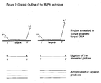
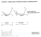
- the method of the preamble is known from e.g. WO 96/15271 (herein incorporated by reference), providing a method for copying and detecting sequence information of a target nucleic acid present in a sample, into a well characterised DNA template.
- the method comprises hybridising up to 5 different probe sets of single stranded first and second DNA probes to a target nucleic acid wherein the first and second probe, after hybridisation to the target sequence and subsequently ligation of the probes are used as a template for amplification.
- the method is suited for the copying of sequence information of RNA or DNA into a DNA template.
- Said first and/or said second probe further comprises a tag which is essentially non-complementary to said target nucleic acid.
- the tags are used for the priming of nucleic acid synthesis in the amplification reaction. Such tag can also be used for detection of the resulting amplicon.
- said amplification is initiated by binding of a nucleic acid primer specific for said tag.
- a bias due to difference in primer sequences is avoided by including into the copying action a nucleic acid tag to which amplification primers are directed.
- the sample is provided with one or more DNA probes wherein said probes comprise a first nucleic acid tag and a second nucleic acid tag, optionally denaturing nucleic acid in said sample, incubating said sample to allow hybridisation of complementary nucleic acid in said sample, functionally separating hybridised probes from non-hybridised probes, providing said hybridised probes with at least a first primer, complementary to said first tag, and a second oligomer primer, complementary to said second tag, amplifying at least part of said DNA probes after hybridisation and analysing the amplificate for the presence of amplified products.
- Said first and said second probe can only be amplified exponentially by e.g. PCR when the probes are connected. Since connection can essentially only take place when the probes are substantially adjacent to each other, exponential amplification, and thereby detection of the amplicon is only possible if said first and said second probe where hybridised to the target nucleic acid. Non hybridised probes are not exponentially amplified. Removal of non-hybridised and non-ligated probes is therefore not essential, and the reactions can be carried out in the same reaction vessel.
- oligonucleotides Dependent on the temperature, buffer-conditions, ligase-enzyme and oligonucleotides used, the difference in ligation efficiency of oligonucleotides that are perfectly matched to the target nucleic acid and mismatched oligonucleotides can be very large providing increased possibilities to discriminate closely related target sequences.
- One of the tags used for amplification which is present at the 3' end of one of the two probe oligonucleotides is however complementary to one of the PCR primers and will therefore provide a template for primer elongation during the PCR reaction.
- These unligated probe molecules only contain one of the two tags used in the PCR reaction and can therefore not be amplified exponentially but only linearly. During each PCR cycle each picomole of probe will consume one picomole of one of the PCR primers.
- the probe amounts used in the art, 200-500 femtomoles ( WO97/45559 ) of each probe, 750-1500 femtomoles ( WO96/15271 ) or 160 fmoles ( WO 98/04746 ) will consume 5 - 45 picomoles of one of the PCR primers during the 25-30 PCR cycles that are needed when nanogram amounts of human nucleic acids are being analysed.
- the invention is characterized in that the amount of at least the first probe of at least one probe set in the mixture is less than 40 femtomoles, and the molar ratio between the said first primer and the first probe being at least 200.
- the use of such substantial low probe amounts and a relatively high molar ratio between the first primer and the first probe also solves the problem of false positive signals due to extension of the probes having the target specific sequence at their 3' end when hybridized to the target sequence during the PCR reaction, followed by elongation of the complement of the second target specific probe on these extension products as described in detail in WO97/45559A and US Pat. No. 6,027,889 .
- hybridisation reactions are slower.
- hybridisation reactions typically are performed for 16 hrs. This can be reduced by inclusion of certain chemicals and/or proteins in the reactions as is well known in the art.
- Previous art methods using, or being derived from LCR reactions use typical hybridisation treatments of 1-5 minutes ( WO 97/45559 ).
- a plurality of probe sets can be used in the invention for detecting one or more specific nucleic acid sequences, without the above-mentioned drawback that the probes are significantly consumed by amplification of unligated probes.
- the first probes from the probe sets specific for hybridising to the corresponding nucleic acid sequences and containing a tag complementary to one of the amplification primers, are present in the mixture in the above-mentioned amount.
- the amount of at least the first probe of each probe set in the mixture is less than 40 femtomoles, the molar ratio between the first primer and the first probe being at least 200.
- the probe sets differ from one another in that at least one of the probes of different probe sets have different target specific regions, therewith implicating that each probe set is specific for a unique target nucleic acid sequence.
- probe sets may only differ in one of the probes, the other probe(s) being identical.
- Such primer sets can e.g. be used for the determination of a specific point mutation or polymorphism in the sample nucleic acids.
- the molar ratio between the first primer and the first probe is preferably at least 500, more preferably at least 1000, and most preferably at least 2000. The higher the said ratio, the more different primer sets for the detection of a corresponding number of different amplicons can be used. However, as indicated above, unspecific amplification reactions as a result of high primer concentrations is to be avoided. Thereto, the primer concentration preferably is below 50 pMoles, more preferably below 20 pMoles in a reaction volume of 10-100 ⁇ l.
- the molar amount of at least the first probe of at least one probe set, preferably of a plurality of probe sets, more preferably of each probe set in the mixture is less than 10 femtomoles, preferably 4-5 femtomoles.
- the molar amount of at least the first probe of at least one probe set is less than 10 femtomoles, preferably 4-5 femtomoles.
- the probes of the same probe set are present in the mixture in substantially equal amounts, although the said amounts can differ from one another, e.g. dependent on the hybridisation characteristics of the target specific regions with the target nucleic acid sequence.
- the amount of second probe may optionally be a factor 1-5 higher than that of the corresponding first probe, without negatively affecting the reaction.
- first probe of different probe sets may have different tag sequences, implicating that a plurality of different first primers are to be used in the amplification step it is highly preferred that the first tag sequences of the first nucleic acid probes of the different probe sets are identical, so that only one first primer has to be used in the amplification reaction. A bias in the amplification due to a difference in the sequence of different primers used for the amplification can thus be completely avoided, resulting in a substantially uniform amplification for all probe assemblies. According to the invention it is however also possible that a number of first nucleic acid probes comprise the same tag sequence, whereas first probes belonging to another probe set may comprise another first tag sequence.
- the amplification step comprises binding of a second nucleic acid primer, specific to the second tag sequence, to the elongation product of the first primer.
- a second primer By the use of a second primer, the amplification reaction is not linear, but exponential.
- Said first and said second probe preferably each comprise a different tag.
- said amplification of connected probes is performed with the use of the Polymerase Chain Reaction (PCR).
- the molar ratio between the second primer and the second probe is preferably at least 200, more preferably at least 500, even more preferably at least 1000 and most preferably at least 2000.
- the second tag sequences of the second nucleic acid probes of the different probe sets are identical, so that for amplification of the primer assemblies a limited amount of different primers may be used. In this way, amplification of all possible primer assemblies can be accomplished using a limited number of primer pairs, preferably only one primer pair. As in such a case, all the probes comprise the same first tag and the same second tag, thereby excluding any bias in the amplification of the probes due to sequence differences in the primers.
- the molar ratio between the second primer and the total amount of second probes present in the reaction mixture is preferably at least 5, more preferably at least 15 and most preferably at least 25.
- probes that comprise different first tags and/or different second tags.
- the primers are matches for similar priming efficiencies.
- some bias can be tolerated for non quantitative applications or when the bias is known, it can be taken into account in a quantitative application.
- the reaction mixture preferably comprises at least 10 probe sets, preferably at least 20 and most preferably 30-40 different sets of probes. It is to be understood that it is preferred to use lower probe amounts when the number of different probe sets increases. Using e.g. 10 different probe sets, the amount of each first probe is preferably less than 20-40 femtomoles, whereas when 30-40 different probe sets are used, the amount of each different first probe is preferably in the range of 1-8 femtomoles in the reaction mixture.
- the presence of a second, or further additional, distinct target nucleic acid can be detected with the method according to the present invention.
- said sample is provided with at least two probe sets, i.e. the target specific regions of at least one of the first, second, or, when present, the third probes of each set differ from one another.
- at least two different amplicons can be detected.
- a first or said second nucleic acid probe of a probe set is capable of hybridising to target nucleic acid essentially adjacent to a probe of the second probe set.
- Successful connecting of probes can then result in an amplicon resulting from the connection of said first and said second probe of the first set and an amplicon resulting from the connection of the first and second of the second set.
- one of the probes of the first and second set may be identified.
- This embodiment of the invention has applications in the detection of for instance SNPs which are different in only one nucleotide.
- a second probe set can simultaneously be used comprising the same first probe as in the first probe set, and a second probe differing from the second probe of the first probe set in the nucleotide at the site of the SNP.
- both second probes are present at the same concentration and are both able to hybridise to the target nucleic acid sequence under the incubation conditions used, half of the target nucleic acids will hybridise to probes of the first probe set and the other half will hybridise to probes of the second probe set.
- the second probe of either the first or second probe set will have a mismatch at the site of the SNP which strongly reduces the enzymatic or chemical ligation efficiency and thereby reduces the formation of the corresponding amplicon in the amplification reaction.
- both SNP alleles are present both amplicons will be formed. These can be distinguished by length if the second probes of probe sets 1 and 2 differ not only at the site of the SNP but also by the length of the sequence between the PCR tag and the end of the probes for instance by the introduction of a small stuffer sequence between the hybridising sequence and the PCR tag in one of both probes. Probes were made for the detection of polymorfisms in the human TNF gene. Although approximately 40 % of the probe pairs worked excellent and gave band of almost identical peak areas on DNA samples from heterozygotes, it was noted that the amplification reaction often resulting in a preferred amplification of one, most often the smallest, amplicon.
- the two amplicons in this particular embodiment have an almost identical sequence, not only homoduplexes but also heteroduplexes will be formed during the final part of the amplification reaction.
- the incorporation of small non-identical stuffer sequences between the hybridising sequence and the PCR tag in both the second and third probe diminished this bias in amplification efficiency.
- these non-identical stuffer regions do have the same nucleotide immediately adjacent to the primer tag sequence.
- a competition takes place between primer-binding/elongation and duplex formation of the amplicons. If a heteroduplex is formed between strands at which a PCR primer is already annealed, the PCR primer will not be as easily be displaced when a short mismatch region is present immediately adjacent to the PCR primer binding site.
- the probes preferably do not leave a gap upon hybridisation with the target sequence.
- the first and second segments of the target nucleic acids are adjacent.
- a third segment is located on the target nucleic acid.
- a third probe may be provided in a probe set complementary to the third segment of said target nucleic acid, whereby hybridisation of the third probe to said third segment allows the connecting of the first, second and third probes.
- a gap upon hybridisation of the first and second probes to the target nucleic acid is filled through the hybridisation of the third probe.
- the resulting amplicon will comprise the sequence of the third probe.
- a gap between first and second probes on said target nucleic acid is filled through extending a 3' end of a hybridised probe or an additional nucleic acid filling part of an interadjacent part, prior to said connecting.
- Applications for this particular embodiment include the determination of the breakpoint sites in chromosomal translocations.
- the probes, not hybridised in the incubation step are not removed in the course of the method according to the invention and remain in the reaction mixture together with the hybridised probes.
- reaction conditions are used that do not require unligated probe removal or buffer exchange
- portion an amount of probes is meant above trace-level that may remain present when the reaction is subjected to a treatment for complete separation of hybridised probes from unhybridised probes.
- said portion is at least 5% from the unhybridised probes, more preferably 10% or more.
- Hybridised probes can be separated from non-hybridised probes in a number of different ways.
- One way is to fix sample nucleic acid to a solid surface and wash away non-hybridised probes. Washing conditions can be chosen such that essentially only hybridised probes remain associated with the solid surface.
- the hybridised probes can be collected and used as a template for amplification.
- probe separation was accomplished by addition of a tagged third target specific oligonucleotide.
- the method according to the invention provides the possibility for an essential one-tube assay using more than 5 probes simultaneously and less than 10.000 copies of each target nucleic acid for each assay.
- the method is very attractive for the method to be carried out as a "one tube” assay; i.e. the contacting step, the connecting step and preferably also the amplification step are carried out in the same reaction vessel, the reaction mixture not being removed from the said vessel during the said steps.
- the contacting, incubation and connecting step are usually carried out in a relatively small volume of 3-20 ⁇ l, although larger volumes, as well as increase of volume of the reaction mixture in subsequent reaction steps are tolerated.
- the amplification step is usually performed in a larger volume of 20-150 ⁇ l; for this, the optionally smaller volume of the reaction mixture in the connection step is usually completed to the desired volume for the amplification by adding the additional ingredients for the amplification reaction.
- the amount of: sample nucleic acid is 10 - 1000 ng
- the first probe of each probe set is 0,5 - 40 fmol
- the second probe of each probe set is 0,5 - 40 fmol
- each first primer is 5 - 20 pmol
- each second primer is 0 - 20 pmol.
- the amount of the second probe is 0,5-40 fmol
- the amount of the said second primer is preferably 5-20 pmol.
- thermostable nucleic acid ligase active at temperatures of 50 °C or higher, but capable of being rapidly inactivated above approximately 95 °C.
- the present invention therefore in one aspect provides a method wherein ligation of probes annealed to a target nucleic acid is performed by a thermostable nucleic acid ligation enzyme, i.e. with an activity optimum higher than at least 50°C, under suitable conditions, wherein at least 95% of the ligation activity of the said ligation enzyme is inactivated by incubating said sample for 10 minutes at a temperature of approximately 95 °C.
- the length of the complementarity region with the target nucleic acid in the probe is preferably long enough to allow annealing at elevated temperatures. Typically the length of the complementarity region is at least 20 nucleotides.
- the probes also contain a tag which can be of any size, however, typically a tag comprises a nucleic acid with a length of at least 15 nucleotides. A probe comprising a tag therefore typically comprises a length of 35 or more nucleotides.
- Amplicons of connected first and second probes typically have a length of at least 70 nucleotides. This minimum length is also preferred to discriminate amplicons from primer dimers and other side products that are often formed in PCR reactions in which only very small amounts of starting template are used.
- One way is to design the multiplex amplification such that the size of each amplicon that can occur, is different. Size fractionation on for instance a gel and determination of the size of the detected amplicon then allows discrimination of the various amplicons.
- amplicons can be discriminated between on the basis of the respective sequences present in the amplicon. For instance through hybridising amplicons to specific probes. However, the latter method has the disadvantage that additional steps need to be included to detect and/or discriminate the amplicons. In the examples illustrating the present invention therefore the various amplicons were discriminated on the basis of size.
- amplicons which differ only slightly in size are difficult.
- a size difference between different amplicons of at least 4 nucleotides is preferred.
- longer probes, to allow more differences in size of the resulting amplicons are not very easily synthesised synthetically.
- at least one of the probes of a number of amplicons is more than 50-60 nucleotides in size.
- Oligonucleotides longer than 60 nucleotides typically suffer from less yield, lower purity and the reliability of the sequence of the probe becomes a problem.
- Chemically synthesised oligonucleotides are made stepwise in a 3' - 5' direction. Coupling yield for each nucleotide is usually only 98,5 %, resulting in the presence of a large number of different side products. Besides there is a risk on damaging the already synthesised part of the oligonucleotide during each new cycle of chemical polymerisation.
- a high reliability of the sequence of a probe is particularly important when already one false nucleotide can give false results.
- this problem is overcome by utilising at least one probe comprising nucleic acid that is generated through enzymatic template directed polymerisation, at least prior to the hybridisation step.
- the above-discussed probe amounts and relative primer-to-probe ratios are preferred.
- Enzymatic template directed polymerisation can be achieved for instance in a cell. It is preferably achieved through the action of a DNA polymerase, RNA polymerase and/or a reverse transcriptase.
- Such enzymatic template directed polymerisation is capable of generating large stretches of nucleic acid with a high fidelity, thereby enabling the generation of a reliable probe, that is substantially larger than currently reliably possible with the synthetic methods.
- a probe comprising nucleic acid that is generated through enzymatic template directed polymerisation is in the present invention further referred to as an enzymatic probe.
- Size differences can be generated by increasing the length of the hybridising region of a probe or by introduction of a stuffer region that is not complementary to the target nucleic acid.
- a stuffer region that is not complementary to the target nucleic acid.
- Another advantage of non-hybridising stuffer sequences is that stuffer sequences with known amplification characteristics can be selected. Certain DNA sequences have a lower amplification efficiency in amplification reactions for instance due to polymerase pause sites such as hairpins.
- Stuffer sequences provide the possibility to use long amplification products while knowing that a major part of the probe has good amplification characteristics.
- SNP / mutation screening the use of a short hybridising region in combination with a non-hybridising stuffer sequence provides the possibility to simultaneously use probes for SNP's or mutations that are close to each other without competition between probes during the hybridisation reaction while still using the advantages of long amplification products. This is also a great advantage in mRNA quantification as only a small (50-80) nucleotide cDNA fragment is needed for binding of probes, reducing the chance of reverse transcriptase pause sites or RNA breakdown influencing the results obtained.
- stuffer can of course also be used to introduce a tag, for instance for later discrimination of probe amplification products on the basis of stuffer sequence.
- a series of cloning vectors each containing different stuffer sequences is provided.
- one of the probe oligonucleotides is generated by digestion of DNA, in particular plasmid, phage or viral DNA with a restriction endonuclease (also referred to as "restriction enzyme").
- a restriction endonuclease also referred to as "restriction enzyme”
- one of the probe oligonucleotides is obtained by restriction enzyme digestion of single stranded phage DNA that is made partially double-stranded by annealing of short oligonucleotides.
- the use of single stranded phage or phagemid DNA increases the effective probe concentration during hybridisation and reduces the amount of probe DNA present as well as the possibility of non-specific amplification products formed e.g.
- restriction enzyme is capable of cutting at least one strand of the DNA outside the enzyme recognization site sequence on said DNA, resulting in DNA fragments not containing any residues of the restriction enzyme recognition sequence at their ends.
- Digestion means cleavage of both or only one strand of a double stranded DNA, such as e.g. cleavage by the restriction enzyme BsmI.
- the DNA used is single stranded DNA made partially double stranded by annealing one or more oligonucleotides.
- At least one probe comprises two separate probe parts being connected together in the step of connecting the essentially adjacent probes.
- Probe parts are herein defined as two nucleic acid sequence stretches that, once linked together, make up the probe. Said stretches may be of different length.
- at least one of said probe parts comprises enzymatic template directed polymerised nucleic acid prior to said connecting.
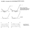
- This embodiment can in one aspect be used to add a stuffer to the probes, resulting in a larger amplicon, whereas not all of said at least one probe needs to be generated through enzymatic template directed polymerisation prior to said connecting. This embodiment is elucidated in fig. 12 below.
- RNA is the analysis of RNA.
- Non limiting examples are the relative quantification of mRNAs and SNP analysis of RNA viruses including the class of retroviruses.
- Direct detection of RNA sequences is not preferred as there is no thermostable ligase known acting on DNA-RNA duplexes.
- RNA is extremely prone to degradation during the long incubations required for complete hybridisation of probes.
- the target nucleic acid comprises RNA than one way to copy sequence information of the target nucleic acid into a DNA template is by using a reverse transcriptase.
- This retrovirus derived enzyme is capable of generating a DNA strand using RNA as a template.
- reverse transcriptase activity is notoriously difficult to standardise when long sequences are copied thereby reducing the reliability of an amplification strategy.
- the present invention provides alternative means and methods to generate amplicons substantially longer than 80 nucleotides while needing only 80 nucleotides or less copy sequence of the RNA target.
- a further application of the current invention is the detection of pathogens in a sample.
- pathogens There are many different pathogens that can contaminate food samples or be present in clinical samples. Determination of even minor quantities of a pathogen can be accomplished using nucleic acid amplification methods such as PCR, RT-PCR and 3SR.
- nucleic acid amplification methods such as PCR, RT-PCR and 3SR.
- PCR nucleic acid amplification methods
- RT-PCR RT-PCR
- 3SR 3SR
- primer sets need to be used and their performance optimised. Although possible, this is a lengthy process.
- very often not all primer sets can be added in one reaction mix thus necessitating different reactions for full coverage of the potential pathogens.
- RNA's and ribosomal RNA's may be used to design oligonucleotides that will be aligned on either (cDNA of) these abundant RNA species, or the DNA coding for them.
- the resulting ligation finger-print may provide enough information to identify the specific strain or species from which the nucleic acid was derived. Due to the high copy number of tRNA's and ribosomal RNA molecules, sensitivity of detection techniques can be extremely high.
- the invention further provides a nucleic acid probe for use in a method of the invention, the probe comprising enzymatic template directed polymerised nucleic acid.
- nucleic acids comprising two or more probes, at least one of these comprising enzymatic template directed polymerised nucleic acid.
- nucleic acid probe set for use in the current invention wherein the probes are capable of hybridising to adjacent sites on a DNA sequence which is complementary to a naturally occurring mRNA but having essentially separated target sequences on chromosomal DNA.
- probe pair is specific for the detection of a cDNA sequence, as will be explained in more detail below.
- kits for performing a method of the invention comprising a liquid medium containing at least one probe in a concentration of 20nM or less.
- the probes are provided in the required low amount to perform reliable multiplex detection reactions according to the present invention.
- a kit for performing the method according to the invention comprising a nucleic acid probe comprising enzymatic template directed polymerised nucleic acid, or a probe mixture comprising at least one of such probes.
- thermostable ligation enzyme of the invention optionally further comprising a nucleic acid probe and or a mixture of probes according to the invention.
- target nucleic acids present in the sample are amplified, but (ligated) oligonucleotide probes provided to the sample.
- Target nucleic acid sequences originally found in the sample being analysed are not amplified because such target sequences do not contain amplification primer-specific tags.
- the invention uses the polymerase chain reaction for amplification of the probes used.
- Other amplification methods for nucleic acids such as the 3SR and NASBA techniques are also known.
- MLPA Multiplex Ligatable Probe Amplification
- DNA polymorphism refers to the condition in which two or more different nucleotide sequences can exist at a particular site in the DNA.
- a complementary nucleic acid is capable of hybridising to another nucleic acid under normal hybridisation conditions. It may comprise mismatches at a small minority of the sites.
- oligonucleotide indicates any short segment of nucleic acid having a length between 10 up to at least 800 nucleotides. Oligonucleotides can be generated in any matter, including chemical synthesis, restriction endonuclease digestion of plasmids or phage DNA, DNA replication, reverse transcription, or a combination thereof. One or more of the nucleotides can be modified e.g. by addition of a methyl group, a biotin or digoxigenin moiety, a fluorescent tag or by using radioactive nucleotides.
- the term "primer” refers to an oligonucleotide, whether occurring naturally as in a purified restriction digest or produced synthetically, which is capable of acting as a point of initiation of nucleic acid sequence synthesis when placed under conditions in which synthesis of a primer extension product which is complementary to a nucleic acid strand is induced, i.e. in the presence of different nucleotide triphosphates and a polymerase in an appropriate buffer ("buffer” includes pH, ionic strength, cofactors etc.) and at a suitable temperature.
- buffer includes pH, ionic strength, cofactors etc.
- One or more of the nucleotides of the primer can be modified for instance by addition of a methyl group, a biotin or digoxigenin moiety, a fluorescent tag or by using radioactive nucleotides.
- a primer sequence need not reflect the exact sequence of the template.
- a non-complementary nucleotide fragment may be attached to the 5'end of the primer, with the remainder of the primer sequence being substantially complementary to the strand.
- target sequence and “target nucleic acid” refer to a specific nucleic acid sequence to be detected and / or quantified in the sample to be analysed.
- amplification refers to the increase in the number of copies of a particular nucleic acid. Copies of a particular nucleic acid made in vitro in an amplification reaction are called “amplicons” or “amplification products”.
- probe refers to a known sequence of a nucleic acid that is capable of selectively binding to a target nucleic acid. More specifically, “probe” refers to an oligonucleotide designed to be sufficiently complementary to a sequence of one strand of a nucleic acid that is to be probed such that the probe and nucleic acid strand will hybridise under selected stringency conditions. Additionally a “ligated probe” refers to the end product of a ligation reaction between a pair of probes.
- the term substantially "adjacent” is used in reference to nucleic acid molecules that are in close proximity to one another. The term also refers to a sufficient proximity between two nucleic acid molecules to allow the 5'end of one nucleic acid that is brought into juxtaposition with the 3'end of a second nucleic acid so that they may be ligated by a ligase enzyme. Nucleic acid segments are defined to be substantially adjacent when the 3' end and the 5' end of two probes, one hybridising to one segment and the other probe to the other segment, are sufficiently near each other to allow connection of the said ends of both probes to one another. Thus, two probes are substantially adjacent, when the ends thereof are sufficiently near each other to allow connection of the said ends of both probes to one another.
- the terms "detected” and “detection” are used interchangeably and refer to the discernment of the presence or absence of a target nucleic acid or amplified nucleic acid thereof or amplified probes specific for that target nucleic acid.
- hot-start refers to methods used to prevent polymerase activity in amplification reactions until a certain temperature is reached.
- restriction endonucleases and “restriction enzymes” refer to bacterial enzymes each of which cut double-stranded DNA at or near a specific nucleotide sequence.
- PCR refers to the polymerase chain reaction ( Mulis et al U.S.Pat.Nos. 4,683,195 , 4,683,202 and 4,800,159 ).
- the PCR amplification process results in the exponential increase of discrete DNA fragments whose length is defined by the 5' ends of the oligonucleotide primers.
- wild-type refers to a gene or gene product which has the characteristics of that gene or gene product when isolated from a naturally occurring source.
- a wild-type gene is that which is most frequently observed in a population and is thus arbitrarily designed the "normal” or “wild-type” form of the gene.
- mutant refers to a gene or gene-product having at one or more sites a different nucleic acid sequence when compared to the wild-type gene or gene product.
- sample refers to a substance that is being assayed for the presence of one or more nucleic acids of interest.
- hybridisation and “annealing” are used in reference to the pairing of complementary nucleic acids.
- At least one of the two oligonucleotides will have a length of more than 60 nucleotides in most (but not necessarily all) of the probes. Fragments substantially longer than 60 nucleotides are difficult to synthesise chemically in high yield and high quality. We discovered that fragments derived by restriction endonuclease digestion of plasmids, phages or phagemids are a preferred source of one of the two oligonucleotides used in ligatable probe amplification.
- fragments typically contain less than one mistake in every 10.000 bp as template directed enzymatic nucleotide polymerisation occurs with high fidelity and is backed in vivo by several repair mechanisms.
- fragments of a sufficient long length and having a sequence tag can be produced by in vitro enzymatic template directed nucleotide polymerisation as described in example 8.
- the other probe oligonucleotide to be ligated can be smaller and is most easily produced chemically.
- the SNP is preferably located on the small chemically synthesised fragment as only one phage or plasmid clone has to be produced for each SNP to be tested.
- oligonucleotides are made in a 3'- 5' direction. As coupling yield for each nucleotide is usually only 98,5 %, a considerable number of fragments in unpurified oligonucleotides are shorter than the required oligonucleotide.
- the oligonucleotide end involved in the ligation reaction should however be constant. For the experiment described in example 1 we therefore chose to use chemically synthesised oligo's of which the 3'-end is joined by ligation to the 5'-end of the long (enzymatic produced) fragment (Type A probe). The 5'-end of DNA fragments produced by restriction enzyme digestion is phosphorylated.
- the smaller chemically synthesised oligonucleotide does not have to be phosphorylated as only the 3'-end is used for the ligation reaction.
- the SNP site should be close to the end, preferably at the end or at the penultimate site of the chemically synthesised oligonucleotide in order to obtain the largest difference in ligation efficiency between matched and mismatched oligonucleotides.
- the long enzymatic produced oligonucleotide is made by an amplification reaction such as PCR with the use of two primers, one of which contains a sequence tag at its 5'end.
- the long oligonucleotide is produced by restriction enzyme digestion of a plasmid or phage clone.
- the 5'-end of the long fragment (type A probe) to be ligated should be complementary to the target nucleic acid.
- restriction endonucleases among which the commercially available Bsm 1 isolated from Bacillus stearothermophilus NUB36 cleave the DNA outside their DNA recognition site and provide a means to produce oligonucleotides that have a 5' end with perfect complementarity to the target nucleic acid.
- Other restriction endonucleases such as Sph I and Aat II produce oligonucleotides that have left only one nucleotide of the restriction enzyme recognition site at the 5'end of the fragment produced and can be used for the production of some type A probes.
- the vector for the production of the long ligation fragment can be double stranded, or can be obtained in both single stranded and double stranded form such as M13 phages and phagemids.
- a double stranded form of the vector is required for efficient cloning of the fragments that are complementary to the target nucleic acid sequence.
- the absence of a complementary strand of the probe has advantages during the hybridisation procedure as the concentration of the hybridisation probe does not drop during the incubation due to reannealing of the complementary strands. Also the absence of a DNA strand complementary to the probe diminishes the possibility of the formation of primer-dimers and other side products during the amplification reaction.
- FIG. 4 An outline of a phage M13 derived clone used for MLPA reactions as in Examples 1-3 and 12-14 is shown in Fig. 4 .
- this Bsm I site and a Sph 1 site can be used to insert an oligonucleotide having sequence complementarity to the target nucleic acid.
- this target sequence specific oligonucleotide has a length of at least 20 nucleotides and a melting temperature when annealed to its complementary sequence of at least 60 °C.
- a stuffer fragment is inserted such as a fragment of phage Lambda or phage T7 DNA.
- this stuffer sequence is selected for not containing any polymerase pause sites and having an equal amplification efficiency in amplification reactions as compared to the stuffer sequences of other probes.
- this stuffer sequence of one or more probes may contain a specific sequence tag used to identify ligation products for instance using real-time quantitative PCR with the use of molecular beacons as marketed by Stratagene Corp., fluorogenic probes such as Taqman probes that are based on the 5'nuclease activity of some heat stable polymerases and are marketed by the PE Biosystems Corporation or fluorescent probes using fluorescence resonance energy transfer (FRET) as used in the lightcyclers of the Roche company.
- FRET fluorescence resonance energy transfer
- the stuffer fragment of one or more probes may have a specific melting temperature that may be used to identify amplification products for instance with the use of the light cycler apparatus of the Roche company.
- sequence tag X At the 3'end of sequence tag X an EcoR5 site is located that is used to remove the type A probe from the bulk of the M13mp18 DNA.
- Each probe used preferably has a different stuffer sequence between the target specific sequence and the sequence tag in order to prevent amplification artefacts due to heteroduplex formation during later parts of the amplification reaction.
- the length between the sequence tag X which is used during the amplification reaction and the Bsm I site combined with the length between sequence tag Y and the 3'end of the other probe determine the length of the amplification product which may be used to identify the amplified probe.
- the short probes contain a sequence tag Y at their 5'end and a target specific sequence at the 3'end.
- this target sequence specific oligonucleotide has a length between 18 and 45 nucleotides and a melting temperature when annealed to its complementary sequence of at least 55 °C, preferably at least 60 °C.
- specific abundant amplification products may be limited by providing for a specific target nucleic acid a (competitor) oligonucleotide capable of annealing to the same target nucleic acid sequence as the probe used but lacking the sequence tag used for the amplification reaction. This may be useful when studying target nucleic acids sequences that differ greatly in copy number for instance when using a probe specific for the cDNA copy of an abundant mRNA sequence as well as probes specific for the cDNA copies of rare mRNA sequences.
- the target nucleic acid is rendered single stranded and exposed to the various added oligonucleotides in order to enable duplex formation.
- certain regions of chromosomal DNA may have a very high G/C content, it may be difficult to denature these stretches of DNA in the solutions of high ionic strength that are preferred for the annealing of the probes. Addition of salt after the denaturation step is therefore preferred.
- Annealing of the oligonucleotides to the template is faster in buffers of high ionic strength.
- the salt concentration of the buffer has to be reduced however to less than approximately 150 mM after the annealing reaction for optimal ligase activity.
- Inclusion of certain chemicals such as polyethyleenglycol polymers or proteins such as BSA may increase both the ligation activity as well as the oligonucleotide hybridisation speed and do not interfere below certain limits with the ligation and amplification reactions.
- An increase in hybridisation speed will also be obtained by concentration of all nucleic presents in a small region of the reactionvolume by means of applying an electric potential across the reaction volume. As shown in the examples it is possible to obtain a so called "one-tube reaction" by careful selection of the hybridisation, ligation and amplification reaction conditions.
- the duration of the probe annealing is very important. Some probes will hybridise faster than others. This is due to a difference in length of the hybridising sequence ; the presence or absence of regions with a high % G/C (GC-clamps) ; secondary structure of the probes and / or the target sequence etc.
- the purpose of the experiment is the relative quantification of nucleic acid sequences, care has to be taken that either hybridisation of each probe is complete, or that hybridisation of none of the probes is complete.
- the purpose of the experiment is the relative quantification of genomic DNA sequences, it will be advantageous to prolong the duration of the annealing reaction in order to make sure that all target sequences have probes annealed to them.
- Annealing of the probes to the target nucleic acid is concentration dependent and is preferably performed in a small volume in the order of 10 ul. and at temperatures of 50-65 °C in order to prevent annealing of probes to aspecific sites. In order to prevent evaporation the use of a thermocycler with heated lid is preferred.
- the two target specific sequences of the two probes are complementary to adjacent but not overlapping sites of the target nucleic acid.
- the probe with the target specific sequence at its 3' end can be elongated by a polymerase in the presence of a suitable buffer and dNTP's in order to make ligation of the two probes possible.
- the gap between the probes can be filled by complementary oligonucleotides that can be ligated to the probes. In this embodiment more than one ligation site is present and more than site will influence the amount of amplificationproduct obtained. This will be useful for detection of mutations or SNP's that are close to each other.
- a covalent phosphate link between the two fragments can be formed enzymatically by a ligase.
- DNA ligases are enzymes capable of forming a covalent phosphate link between two oligonucleotides bound at adjacent sites on a complementary strand. These enzymes use either NAD or ATP as a cofactor to seal nicks in ds DNA. Alternatively chemical autoligation of modified DNA-ends can be used to ligate two oligonucleotides bound at adjacent sites on a complementary strand ( Xu, Y. & Kool, E.T. (1999), Nucleic Acid Res. 27, 875-881 ).
- ligase is used that remains active at 50 - 65 °C for prolonged times, but which can be easily inactivated at the higher temperatures used during a PCR reaction.
- the only ligases commercially available at the moment are enzymes that function only at temperatures below 50 °C such as the DNA ligase encoded by E.coli and by phage T4, and thermostable enzymes that have a half-life of more than 30 minutes at temperatures of 95 °C such as the DNA ligase encoded by Thermus aquaticus.
- Ligase 65 is active at 60-65 °C. In contrast to Tth- and Taq DNA ligase however, the activity of ligase-65 is destroyed more than 90 % by incubation in the optimum reaction buffer for 10 minutes at 95 °C.
- thermostable ligase such as the ligase from Thermus aquaticus may be used and the annealing and ligation reactions can be repeated several times by alternate cycles of heat denaturation and probe annealing.
- This particular embodiment is more time consuming unless higher concentrations of probes are used to increase the speed of the annealing reaction. Higher concentrations of probes increases however the chance on primer-dimer formation during the amplification reaction.
- the amount of probes used in the preferred embodiment of the MLPA reaction (approximately 1 - 10 femtoMol in a 10 ul ligase reaction) is more than one magnitude lower than the amounts routinely used in the ligase chain reaction.
- One of the two PCR primers is complementary to the end of one of the probe oligonucleotides. This means that this probe oligonucleotide even when not ligated to the other probe oligonucleotide can be amplified by PCR but only by linear amplification.
- 160 femtomoles of this PCR primer is consumed resulting in the consumation of 4800 femtomoles or 48 % of the available 10 picoMoles PCR primer that is used in a typical PCR reaction.
- probe amounts in the previous art up to 1500 femtomoles / reaction which is 375 times the amounts used in the current invention also results in strongly increased chances on the formation of primer-dimers, other side-products and false positive signals.
- the majority of probes are present in amounts of less than 40 femtomoles / reaction.
- most of the probes containing a sequence tag complementary to one of the two PCR primers are present in amounts less than 15 femtomoles / reaction.
- the ratio between the amounts present of each probe having a sequence tag complementary to one of the two PCR primers, and the amount used of that PCR primer during the amplification reaction is less than 1:500 for the majority of these probes. This ratio is important for both enzymatic as well as synthetically produced probes when multiplex reactions with more than 5 probe pairs are performed.
- the ligation products consisting of a type A probe covalently joined to a type B probe can be amplified with the use of two oligonucleotide primers, dNTP's and a polymerase, one primer being complementary to one of the sequence tags and the other primer corresponding in sequence to the second sequence tag.
- the preferred method for amplification is PCR. As shown by Vos et al (Nucleic acid Research 23, 4407-4414; 1995 ), conditions can be found in which DNA fragments between 70 and 700 nucleotides containing the same sequence tags are amplified with almost equal efficiency as they are present in the same amplification reaction and use the same primers.
- the preferred conditions include a sufficiently long elongation time and the presence of a higher concentration of Taq polymerase than in ordinary PCR reactions.
- Other amplification methods than PCR such as NASBA or 3SR ( Guatelli et al., Proc. Natl. Acad.Sci. USA 87:1874-1878, 1990 ) can also be used in combination with the current invention.
- the sequence tags used for the PCR reactions can easily be replaced by RNA polymerase binding sites.
- the activity of the polymerase used in the polymerase chain reaction can temporarily be inhibited e.g. by chemical modification of the enzyme or by addition of antibodies to the enzyme.
- the polymerase activity will be apparent only after heating the sample permitting the development of a test in which the ligase and polymerase can be added simultaneously and wherein the ligase is active at moderate temperatures and is inactivated at high temperatures whereas the polymerase is activated only after the heating step.
- a so-called hotstart for the PCR reaction is advantageous since in case one of the two PCR primers (complementary to the primer sequence of the long enzymatic produced oligonucleotide) anneals to one of the short probes containing the other PCR primer sequence, a primer dimer is formed upon elongation of the primer.
- One of the disadvantages of the use of (denatured) double stranded long probes is the increased chance of primer dimer formation as also the second amplification primer can form primer dimers upon annealing to and elongation on the unused strand of the long probe.
- the probe oligonucleotides can easily produce primer dimers and other side products during the amplification reaction, particularly if present at high concentration.
- primer dimers can be further inhibited by using a two-step nested primer amplification reaction.
- the sequence tag on the long probes used in the examples is 36 nucleotides long which is sufficient for the design of two different primers having limited sequence similarity for use in a nested primer amplification reaction.
- All ligated probes can be amplified simultaneous in a first PCR reaction using PCR tags common to all probes. Subsequently this first PCR reaction can be divided in several aliquots and specific subsets of probes can be further amplified in a second amplification reaction using PCR tags common to probes of a specific subset only.
- amplification conditions for PCR can be equal to the conditions used for AFLP reactions ( Vos et al, Nucleic acid Research 23, 4407-4414 ; 1995 ). AFLP and MLPA reactions usually stop as a cause of all primers being consumed. Additional amplification cycles have therefore no or only limited influence on the results obtained and results obtained do not depend strongly on the amount of target nucleic acid in the sample.
- Polyethyleenglycol e.g. has only a limited influence on the PCR reaction at concentrations up to 1 %, implicating that concentrations up to at least 5 % may be present during the annealing reaction as performed in examples 1-3 and 12-14.
- the buffer composition during the ligation reaction is very similar to a standard PCR buffer, it proved also possible to use the complete volume of the ligation reaction and start the amplification reaction by the addition of primers, dNTP's, a small amount of a non-ionic detergent such as 0.01 % triton X-100 and a heat stable polymerase such as Taq polymerase.
- primers dNTP's
- a non-ionic detergent such as 0.01 % triton X-100
- a heat stable polymerase such as Taq polymerase.
- betaine are known to improve some multiplex PCR amplification reactions and do not severely inhibit the ligation reaction.
- PCR conditions that prevent a bias in the amplification of some amplicons.
- concentrations of the amplicons during the later stages of the amplification reaction do not reach very high concentrations. This can be accomplished by using only low amounts of one of the PCR primers.
- a bias in the amplification of some amplicons will be due to faster renaturation kinetics of some amplicons after each denaturation cycle and displacement of PCR primers by the complementary strand of the amplicon.
- Important in this respect is also the nature of the first nucleotide following the PCR primer, G or C being the preferred first nucleotides. This displacement will be reduced when using PCR primers containing one or more LNA residues at their 3'end.
- LNA Locked Nucleic Acids residues have an improved thermostability of duplexes towards complementary DNA strands ( Wahlestedt,C. Et al. Proc.Natl.Acad.Sci.USA 97, 5633-38 ).
- PCR conditions that promote the amplification of rare templates as compared to the amplification of more abundant templates in order to obtain bands of almost equal intensity for the different nucleic acid target sequences tested.
- These PCR conditions may include: (1) The use of higher salt concentrations which promote the annealing of complementary strands and reduce the polymerase activity ; (2) High concentrations of primers ; (3) Reduced annealing / extension temperatures during the last PCR-cycles ; (4) Additives to the PCR buffer such as betaine and DMSO.
- detection of the amplification products is accomplished after separation of the fragments by gel-electrophoresis.
- mass spectrometry is used to detect and identify the amplification products.
- the melting temperature of the amplification products which can be influenced by the choice of the stuffer fragment is used to identify the amplification-products.
- the presence of a sequence tag on the amplification products is used to detect the amplification products and to analyse the results of the experiment.
- a sequence tag can easily be incorporated in the stuffer region of the probes and can be used to discriminate e.g. probes specific for wild-type sequences and probes specific for mutant sequences. Separation of the fragments by gel electrophoresis is not necessary as the use of fluorogenic probes and the use of the 5'nuclease activity of some polymerases that can be used in the amplification reaction permits real time quantitative detection of the formation of at least two different sequence tags for instance one tag specific for a control wild-type specific probe and the other tag being specific for one or more different mutant sequences.
- the current invention employs a mixture of probes in one reaction, each probe being specific for one nucleic acid sequence.
- Each type B probe contains an oligonucleotide that has a region complementary to the target DNA sequence e.g. the sequence essentially flanking the SNP, as well as a common sequence-tag that can be used for the amplification reaction.
- For each SNP two or more type B oligonucleotides are used that differ in the nucleotide at the position of the SNP and in the number of nucleotides between the sequence tag used for the amplification reaction and the SNP site.
- a probe In addition another single stranded DNA fragment (type A probe) is provided that has at one end a nucleotide sequence complementary to the nucleotide sequence flanking the SNP, as well as a sequence tag common to all type A probes which is used in the amplification reaction.
- the length of the type A and type B probes can be chosen such that each possible ligation product results in an amplification product that has a unique size.
- the mixture is treated with a DNA ligase.
- a DNA ligase In case more than one type B probe is used, one or more of the type B probes will have a mismatch at the position of the SNP, which is at the end, or close to the end of the type B probe. These probes will be ligated at a lower efficiency to the type A probe than the type B probe(s) that has no mismatch at the SNP position.
- a DNA fragment is produced that can be amplified e.g. by PCR using a primer specific for the DNA sequence common to all type A probes and a primer specific for the sequence common to all type B probes.
- NAD requiring ligases are very sensitive to the presence of mismatches between the complementary strands that are closer than approximately 9 bp from the site of ligation. The greatest difference in ligation efficiency between perfectly complementary strands and complexes having a mismatch is however obtained when the mismatch is exactly at the site of ligation.
- Type B-probes have a preferred length of 30-60 nucleotides and differ for instance 2 - 4 nucleotides in length dependent on the nucleotide at the SNP position.
- Type A probes preferably have a length of 45 - 600 nucleotides.
- For each SNP a different set of two or more type B probes and one type A probe is added.
- a banding pattern is obtained in which the length and the relative intensity of the bands obtained depend on the length of the type A and type B probes and the efficiency of ligation of the different type B probes to the type A probes.
- a stuffer region was present in only one of the two type B primers used in example 1 and in Figure 1 .
- the only function of this 3-4 nucleotide stuffer region was to obtain a size difference between the two closely related amplicons.
- the incorporation of a (different) stuffer region in both type B probes was therefore not the first choice.
- the two amplicons in this particular embodiment have an almost identical sequence, not only homoduplexes but also heteroduplexes will be formed during the final part of the amplification reaction. During later stages of the PCR reaction a competition takes place between primer-binding/elongation and duplex formation of the amplicons.
- a heteroduplex will be formed between strands at which a PCR primer is already annealed as it takes some time for the polymerase enzyme to find the annealed primer and start its elongation. During this time the PCR primer might be displaced by the long complementary amplicon. This will not happen as easily when a short mismatch region is present immediately adjacent to the PCR primer binding site.
- Both the majority of the hybridising region as well as the PCR tag region are complementary in the heteroduplex.
- a small non identical stuffer region will accomplish that the target hybridising region and the PCR tag region behave as independent regions in the heteroduplex and will accomplish that the PCR primer will not as easily be displaced.
- This part of the invention will be of use in both assays using a enzymatic type A probe as well as assays in which a synthetic type A probe is used.
- oligonucleotide sets for different SNP's can be tested simultaneously provided that each possible amplification-product has a unique length.
- sequencing type electrophoresis systems and multicolour fluorescently labelled PCR primers more than 100 SNP's may be analysed in one lane.
- RNA or cDNA can also be used for the alignment of oligonucleotides designed for detection of the SNP specific nucleotide.
- denatured DNA For multiplex SNP detection however it is more convenient to use denatured DNA as a target as the number of targets for each SNP is than almost identical. The amount of a specific mRNA may be higher but is more variable. If the purpose of the experiment is however the identification of a strain or species, ribosomal RNA or the cDNA thereof or the multiple DNA copies coding for it may be a useful target.
- nucleic acid sequence to be detected is relatively rare, it is often to be preferred to detect only this sequence and not for instance the wild type allele as would be done in SNP analysis.
- a probe is provided that has the sequence unique for that mutation at or close to one of the ends of the probes that are ligated. No probe specific for wild-type sequences needs to be provided. Only in case a mutation recognised by one of the probes is present in the target nucleic acid, amplicons of a specific size or having a specific sequence will be generated. As the number of bands obtained is small, the amplification products can be analysed by rapid and cheap methods such as agarose gel electrophoresis.
- Samples containing a mutation can be analysed further using methods with higher separating power such as acrylamide type sequencing gels or by sequence analysis of the amplification product. In many cases it may be preferred to detect only one or a few wild-type sequences as a control for the presence of sufficient target nucleic acids and the correct performing of the MLPA assay and a large number of mutant target nucleic acid sequences.
- the signal obtained from the wild-type specific probes is reduced by the addition of competing oligonucleotides binding to the same wild-type nucleic acid sequences. As a result the relative amount of signal obtained from the mutant specific probes is increased.
- the wild-type sequences are present in both chromosomal copies while the mutant sequences are present on one chromosome only.
- An outline of this MLPA variant is shown in Figure 5 . This particular embodiment is described in further detail in Example 6.
- probes containing non hybridising stuffer regions are used.
- the use of a small hybridising region permits the screening of mutations that are close to each other. In case different probe pairs have part of their target sequence in common, they will compete with one another for binding to this common target sequence.
- the combined hybridising region of a probe pair has a length of 40-120 nucleotides. In a further preferred embodiment this length is 45-80 nucleotides.
- MLPA assays as described in Figure 5 and example 6 can be used to detect mutated nucleic acid sequences in a high background of normal DNA provided that the sequence of the mutation is known.
- MLPA assays as described in Figure 5 and example 6 have an advantage over traditional nucleic acid amplification based detection methods as traditional PCR, 3SR and Nasba in that internal controls are provided for each sample confirming that a negative test result was not due to any error made during preparation of the sample or during sample analysis.
- wild-type and mutant specific probes may be distinguished by the presence or absence of a specific sequence tag in the stuffer region of the probe oligonucleotides.
- This sequence tag can be used for the binding of complementary labelled oligonucleotides that can be used in real time amplification methods.
- Useful oligonucleotides are for example the so-called "Molecular Beacons” marketed by Stratagene Corporation and Taqman probes marketed by PE Biosystems Corp. both containing a reporter fluorescent dye as well as a quencher dye and dual fluorescently labelled hybridisation probes capable of fluorescence energy transfer as marketed by Roche company for use in the lightcycler.
- Detection of amplification products containing a specific sequence tag is accomplished by detection of increased fluorescence due to binding of the molecular beacon to the sequence tag or by degradation of target bound taqman probes by the 5'nuclease activity of some polymerases such as Taq polymerase.
- the advantage of the use of these real time fluorescence detection methods is that labour intensive gel-electrophoresis for the separation of wild-type (control) and mutant specific amplification fragments is avoided and that tubes do not have to be opened after the amplification reaction, diminishing the chance on contamination of other samples that have not yet been amplified.
- a disadvantage is that only a very limited number of different fluorescence signals can be discriminated as compared to the simultaneous discrimination of more than 50 different amplification products by gelelectrophoresis or mass spectrometry.
- probes specific for certain sequences and detect amplification products by the appearance of long double stranded DNA for instance by measuring the increased fluorescence of some DNA intercalating dyes such as SYBR Green.
- the long amplification products formed in the process of the current invention can be easily distinguished from primer dimers for instance by the measurement of the melting temperature after or during the amplification procedure.
- MLPA assays as outlined in Figure 5 can easily be combined with the MLPA variant described in example 3 and outlined in Figure 12 .
- a multiplex test for the detection of specific nucleic acid sequences can be rapidly developed.
- such test is used for the detection of relatively rare mutations or the presence of relatively rare nucleic acids such as those from specific pathogens.
- DNA rearrangements as well as amplification or deletion of large segments of chromosomal regions due to genetic instability are frequently linked to neoplasia.
- Deletions are usually detected by loss of heterozygosity (LOH) of micro-satellite sequences which is a method difficult to perform on a large number of genes.
- LHO heterozygosity
- Gene amplification or loss of gene copies can be detected by cytogenetic analysis for instance by fluorescent in situ hybridisation (“FISH”) or by comparative genomic hybridisation methods which require specialised expertise, are time consuming and require large probes. These methods cannot be used to study micro-deletions or -amplifications.
- the current invention can be used to detect amplification of chromosomal regions or loss of heterozygosity at 10-100 sites simultaneously.
- the relative number of copies of particular genes can be determined without knowledge of micro-satellites or SNP's in these genes. This provides the possibility to determine clonality of tumors in an easy way and provides the possibility to detect trisomy of human chromosomes in foetal samples.
- Other non-limiting applications are the discrimination of benign and malignant tumors using extremely small amounts of DNA available from microbiopts as malignant tumors generally have more and other chromosomal aberrations. Important in this respect is the reproducibility of the testresults.
- RNA can be a ligation template when T4 DNA-ligase is used as the ligation enzyme, preferably in the presence of Mn ions in the buffer (( Hsuih et al (1996) J. Clin.Microbiology 34, 501-507 ). RNA is however easily degraded by RNAses and is a poor template for ligation reactions in which thermophilic NAD+ requiring bacterial ligases are used.
- RNA can be efficiently reverse transcribed in the region required with the use of a specific primer located very close to the hybridisation sequences of the probes ( Fig. 8 ).
- the cDNA obtained is an efficient substrate for the alignment of the two hemiprobes used.
- the RNA can be reverse transcribed with the use of oligo-dT ( Fig.
- a reverse transcription primer sequence can be part of one of the two probes ( Fig. 11 ). After reverse transcription, the RNA can be removed from the cDNA by RnaseH treatment or by heating.
- a probe is used with a target (cDNA) specific sequence at its 5'end, a reverse transcription primer sequence at its 3'-end and the oligomer tag used for the amplification reaction in-between, a (large) hairpin will be formed easily when the target specific sequence anneals to its complementary cDNA sequence ( Fig. 11 ). In this case only one hybridisation event needs to take place before the ligation reaction apart from the rapid hairpin formation.
- a pair of probes to be ligated and designed for the detection of mRNA's can both bind within one exon in which case detection of the corresponding DNA sequence can be used to check the performance of the probes. It is however difficult to remove all contaminating DNA from a RNA preparation. Fragments smaller than 50 nucleotides are sufficient to give a positive signal in MLDA reactions but are difficult to degrade to much smaller fragments by DNAse treatment.
- one of the two probes can be designed to detect a cDNA sequence which is predominantly located in another exon. In this case the pair of probes should be specific for the cDNA sequence and the signal obtained will not be sensitive to DNA contamination of the RNA sample. These probe pairs can be tested on a synthetic DNA copy of the RNA region to be detected.
- the primers used for the reverse transcription reaction whether gene-specific or oligo-dT can also be used for the purification of the mRNA's or the cDNA produced from cell homogenates.
- reverse transcriptase primers with a gene specific sequence at the 3'-end and a hapten such as biotin, digoxigenin or a specific DNA sequence at the 5'-end mRNA's can be purified from cell homogenates with the use of immobilised hapten binding agents such as Streptavidin or a complementary DNA-sequence.
- One preferred embodiment of the current invention makes use of reverse transcription primers containing a common sequence tag such as a GT repeat as used e.g. in the oligonucleotides SEQ ID NO. 81-84.
- sequence tags can be used before or after the reverse transcription-reaction to enrich the nucleic acid sequences of interest for instance by providing a complementary CA repeat containing a hapten such as biotin and immobilised streptavidine for the binding of the biotin moieties ( Figure 10 ).
- An advantage of this indirect purification system compared to biotinylated reverse transcription primers is the possibility to remove the enriched nucleic acids from the immobilised streptavidin-biotin complex by heating.
- the CA oligonucleotides can be immobilised directly.
- relative quantification of target sequences can be accomplished by using a high enough probe concentration and long enough annealing times in order to make sure that 100 % of the target nucleic acids have probes annealed to the target sequences. This is a preferred embodiment of the MLPA method.
- relative quantification can be accomplished by ensuring that at all target sequences annealing of one probe of each probe pair is incomplete.
- the relative amount of specific amplification products can be reduced compared to the amount of other amplification products by providing a competitor oligonucleotide capable of annealing to a target sequence, preventing the annealing of one or both of the probes specific for that target sequence.
- a competitor oligonucleotide capable of annealing to a target sequence By using mixtures of target specific probes and competing oligonucleotides binding to the same target sequence, the relative amount of amplification products from abundant mRNA's can be reduced.
- the relative amounts of the cDNA targets to be detected accurately reflects the relative amounts of the RNA sequences present in the sample.
- a reverse transcriptase lacking strand displacement activity or reaction conditions in which strand displacement activity is reduced are used.
- Example 2 The application of the MLPA invention for the relative quantification of mRNA's is described in Example 2. An outline of the method is presented in Figures 6-11 .
- oligonucleotides are required for the simultaneous analysis of larger numbers of target nucleic acids however, long (60-600 nucleotides) oligonucleotides are required. With the current technology for the chemical synthesis of these molecules, oligonucleotides longer than approximately 80 nucleotides will preferably be enzymatic produced. Although technically no problem for one of ordinary skill in the art, this is time consuming as a new clone has to be produced for every SNP to be tested.
- Oligonucleotide 77 contains a sequence complementary to the target nucleic acid as well as a sequence tag to be used in the amplification reaction.
- a mixture of two or more oligonucleotides 77 can be used which differ in length as well as in the nucleotide present at the SNP site.
- Oligonucleotide 89 contains a sequence complementary to the target nucleic acid adjacent to the target specific sequence present in oligonucleotide 77, as well as a sequence complementary to oligonucleotide 90. Oligonucleotide 89 should be phosphorylated at its 5'end.
- Oligonucleotide 90 contains a sequence complementary to oligonucleotide 89 as well as a sequence complementary to oligonucleotide M227. The function of oligonucleotide 90 is only to align oligonucleotides 89 and M227 in order to make chemical or enzymatic ligation of these oligonucleotides possible.
- Oligonucleotide M227 contains a sequence complementary to oligonucleotide 90 as well as a sequence tag to be used in the amplification reaction. In case many target nucleic acids are analysed simultaneously, oligonucleotide M227 has to be relatively long for some target nucleic acids and may be preferably enzymatic produced. As this oligonucleotide has no target specific sequences, a standard set of oligonucleotides 90 and M227 can be used for many different target sequences.
- probes For a true multiplex assay, different probes can be used which result after successful template directed ligation in amplification products having different characteristics such as length, mass, sequence, presence of a sequence tag or melting behaviour.
- a chemically synthesised oligonucleotide (type B probe) is ligated at its 3'-end to the 5'-end of a long enzymatic produced type A probe.
- the probes can be made partially doublestranded next to the part that hybridises to the target nucleic acid by addition of a complementary oligonucleotide ( Fig. 14 ).
- This "viagra"-oligonucleotide reduces the internal secondary structure of the probe and results in some cases in a faster hybridisation of the probe to its target sequence.
- full length probes may be used, consisting of a single oligonucleotide containing the two different sequence tags and giving rise to an amplification product of a specific size.
- Amplification reactions such as PCR are capable of detecting less than 100 molecules containing the two sequence tags. For many purposes not the absolute signal strength but the relative strengths of the signals obtained with different probes are interesting. Amplification reactions are often allowed to proceed for more than the minimum number of cycles needed in order to obtain signals of comparable intensities for different samples that may contain different amounts of target sequences and are stopped when one of the necessary ingredients, usually the amount of primers, becomes limiting.
- a small amount of a "full length probe” e.g. 100 molecules, may therefore be provided to each sample. Detection of the amplification product of this full length probe is a warning that insufficient target specific ligation products were present at the start of the amplification reaction and that the results obtained should be regarded with caution.
- Complete probes may also be used as spiked internal controls added after or even before purification of the nucleic acids from a sample in order to check the sample preparation and to estimate the absolute amount of the ligation products specific for the target sequences that are present before the amplification reaction.
- probes containing not only the two oligomer tags needed for the amplification reaction at a certain distance from each other but also in-between or next to these oligomer tags a sequence capable of hybridising to a target sequence, can be used for multiplex detection of a large number of different target sequences.
- each probe can be distinguished by the unique length or mass of its amplification product.
- probes are preferably derived from plasmid or phage DNA by digestion with restriction-endonucleases.
- probes can be made by PCR using suitable primers as shown in Fig. 15 .
- hybridised probes After hybridisation to the target sequences in a sample to be analysed, hybridised probes have to be separated from non-hybridised probes which can be accomplished e.g. by immobilising the sample nucleic acids (dot-blots), biotinylation of sample nucleic acids and binding of these sample nucleic acids + hybridised full length probes to magnetic particles coated with streptavidin, and various other means.
- kits containing two or more probes that can be amplified with the same amplification primers wherein each probe contains a sequence complementary to one of the target nucleic acids.
- kits may also contain, in packaged combination, one or more of the following: a hybridisation/ligation buffer; a ligase enzyme; amplification primers specific for the sequence tags of the probes; and amplification reagents.
- phage M13mp18 which is available from New England Biolabs.
- the M13mp18 sequence is available from Genbank, accession number X02513.
- Double strand DNA of M13mp18 was digested with EcoR1 and Hind 3.
- the oligonucleotides of SEQ ID NO. 8 and SEQ ID NO. 9 which form together a duplex having ends that can ligate to the EcoRI and Hind III sites of the digested M13mp18 was inserted. After ligation and transformation, plaques containing the inserted oligonucleotide were selected and double stranded DNA was prepared of transformant MRCH001.
- Double stranded DNA of this virus was digested with Nco I and Acc I.
- the oligonucleotides of SEQ ID NO. 10 and SEQ ID NO. 11 which form together a duplex having ends that can ligate to Nco I and Acc I sites of the digested MRCH001 was inserted.
- plaques containing the inserted oligonucleotide were selected and double stranded DNA was prepared of transformant MRCH002.
- M13mp18 contains a Bsm 1 recognition site at position 1745-1750 which we removed from phage MRCH002 by changing the T-nucleotide at position 1748 into a C-nucleotide.
- a primer (SEQ ID NO. 12) was annealed to single stranded M13mp18 DNA. This primer was elongated by the Klenow fragment of DNA Polymerase I. After closing the resulting double stranded DNA with T4-DNA ligase, the DNA was heated 5 minutes to 95 °C in the presence of 10 pMol of an oligonucleotide (SEQ ID NO. 13). This oligonucleotide was again elongated by Klenow fragment and the resulting d.s. DNA preparation was transformed in E.coli strain JM109 (Promega). Transformants were cultured together in one bottle for 5 hrs.
- Double stranded virus DNA was purified from the mixture of transformants and was digested with Bsm I. The digested DNA was again transformed in E.coli strain JM109, and virus plaques were tested for the presence of a Bsm 1 site. One transformant (MRCH106) not containing a Bsm I site was selected.
- Double stranded DNA of this virus was digested with Nco I and EcoR I.
- the oligonucleotides of SEQ ID NO. 14 and SEQ ID NO. 15 which form together a duplex having ends that can ligate to Nco I and Hind 3 sites of the digested MRCH106 was inserted. After ligation and transformation, plaques containing the inserted oligonucleotide were selected and double stranded DNA was prepared of transformant MRCH107.
- Double stranded DNA of MRCH107 was digested with PinA1 and Acc1 and the oligonucleotide SEQ ID NO. 16 + SEQ ID NO. 17 which together form a duplex having ends that can ligate to PinA1 and Acc 1 digested MRCH107 DNA was inserted. After ligation and transformation, plaques containing the inserted oligonucleotide were selected and double stranded DNA was prepared of transformant MRCH214.
- M13mp18 derivative that lacks the Bsm I site at position 1745-1750 and has the sequence shown in SEQ ID NO. 18 inserted in the EcoR 1 and Hind 3 sites of M13mp18.
- PCR fragments derived from phage T7 DNA with the use of the following primer pairs were inserted in MRCH214 : SEQ ID NO. 19 + 20 ; 21 + 22 ; 23 + 24 or 25 + 26. These 4 PCR fragments were digested with Sph 1 and Xba 1 and ligated to Sph 1 and Xba 1 digested d.s.DNA of phage MRCH214. Primers SEQ ID NO. 19, 21, 23 and 25 have an Sph 1 site close to their 5'end. Primers SEQ ID NO. 20, 22, 24 and 26 have an Xba 1 site close to their 5'end. Phage T7 is available from the American Type Culture Collection. The T7 DNA sequence is available from Genbank as Acc. nr.
- MRCH228 has a 34 bp T7 fragment inserted ; MRCH266 has a 79 bp T7 fragment inserted ; MRCH273 has a 151 bp T7 fragment inserted ; MRCH285 has a 310 bp T7 fragment inserted and MRCH113 contains a 349 bp T7 fragment.
- each of the five clones containing a T7 stuffer fragment an oligonucleotide was inserted into Bsm 1 and Sph 1 digested double stranded DNA.
- Each oligonucleotide is identical to the sequence at the 3'-side of a known mutation in the human CFTR gene (Genbank seq. nr. M55108 - M55130).
- the partially complementary oligonucleotides SEQ ID NO. 30 and 31 were inserted in the Bsm 1 and Sph 1 sites of MRCH228; SEQ ID NO. 32 + 33 in MRCH266 ; SEQ ID NO. 34 + 35 in MRCH273 ; SEQ ID NO. 36 + 37 in MRCH285 and SEQ ID NO. 38 + 39 in MRCH113, resulting respectively in phage clones MRCH231, 236, 243, 258 and 252.
- Single stranded phage DNA from these five clones was produced as described by Reddy, P. and McKenney K. (Biotechniques 20: 854-860 ; 1996 ).
- This single stranded DNA was annealed to two oligonucleotides: Seq ID NO. 29 + 40 for the MRCH231 DNA ; SEQ ID NO. 29 + 41 for MRCH236 DNA ; SEQ ID NO. 29 + 42 for MRCH243 DNA ; SEQ ID NO. 29 + 43 for MRCH258 DNA and SEQ ID NO. 29 + 44 for MRCH252 DNA.
- Digestion was performed by incubation of 400 pMol of an M13 derivative single stranded DNA with 2.2 nMol of each of the two oligonucleotides and 8000 units EcoR5 in 10 mM Tris-HCl pH 7,6 ; 100 mM KCl; 10 mM MgCl2 and 1 mM Dithiothreitol at 37 °C. After incubation for 30 minutes, 4000 units Bsm 1 was added and the temperature raised to 50 °C. After incubation for another 30 minutes the digested DNA was phenol extracted, ethanol precipitated and dissolved in TE.
- oligonucleotides were synthesised that have a common part used to amplify ligated oligonucleotides (Sequence tag X), and a part complementary to the CFTR sequence at the position of the mutation.
- oligonucleotides differ in length (4 bp) and in the nucleotide present at the site of the mutation.
- the site of the mutation is at the penultimate position or at the 3'end of the oligonucleotide.
- Oligonucleotides SEQ ID NO. 45 and 46 can anneal to a site adjacent to the insert of clone M231 on CFTR wild-type DNA or to DNA containing mutation E60X of the CFTR gene respectively.
- Oligonucleotides SEQ ID NO. 47 and 48 can anneal to a site adjacent to the insert of clone M236 on CFTR wild-type DNA or to DNA containing mutation 621+1G>T of the CFTR gene respectively.
- Oligonucleotides SEQ ID NO. 49 and 50 can anneal to a site adjacent to the insert of clone M243 on CFTR wild-type DNA or to DNA containing mutation deltaF508 of the CFTR gene respectively.
- Oligonucleotides SEQ ID NO. 51 and 52 can anneal to a site adjacent to the insert of clone M258 on CFTR wild-type DNA or to DNA containing mutation 3659delC of the CFTR gene respectively.
- Oligonucleotides SEQ ID NO. 53 and 54 can anneal to a site adjacent to the insert of clone M252 on CFTR wild-type DNA or to DNA containing mutation 2184delA of the CFTR gene respectively.
- DNA of five different humans was mixed in a 200 ul vial with 4 FemtoMol of each of the five digested phage DNA's and 10 femtoMoles of each of the ten oligonucleotides designated SEQ.ID. 45 - 54 in a final volume of 8.5 ul.
- DNA was denatured by heating for 5 minutes at 95 °C in a thermocycler with heated lid.
- 1.5 ul salt mix 1500 mM KCl; 300 mM Tris-HCl pH 8.5 ; 1 mM EDTA. Annealing of the probes to the target DNA was for 6 hrs. at 60 °C in a thermocycler with heated lid.
- PCR was performed in 200 ul tubes in a Biometra Uno 2 thermal cycler using the following conditions:
- the probes used were designed to give rise upon successful ligation to amplification products of the following lengths when using the above mentioned amplification primers:
- Samples of human chromosomal DNA to be analysed were obtained from the Dept. of Antropogenetica, Free University of Amsterdam, and were known to contain the following mutations in the CFTR gene: Lane 1 of Figure 16 : No mutations ; Lane 2: deltaF508 mutation on both chromosomes. Lane 3: deltaF508 mutation on one chromosome only. Lane 4: 3659delC mutation on one chromosome. Lane 5: R117H mutation on one chromosome.
- the scale on Figure 16 is not in bp. but in minutes after starting the gel-electrophoresis.
- 5 bands were obtained on wild-type DNA corresponding in size with the expected fragment sizes.
- the third band is running slightly slower through the gel corresponding to a size four bp. longer than the third band of lane 1 and corresponding in size to the fragment expected from the probe specific for the deltaF508 mutation.
- This same band as well as the wild-type band appear in lane 3, proving that both the wild-type probe as well as the probe specific for the deltaF508 mutation have given rise to an amplification product and thus proving that successful ligation of the probes has occurred.
- Lane 4 an extra band has appeared corresponding in size to the probe specific for the 3659delC mutation. As expected no extra bands were observed in Lane 5 as no R117H specific probe was used.
- the number of probes used in one assay and to be distinguished by the specific length of the amplification products can be 50 or more.
- probes were made that were complementary to two abundant human mRNA's coding for beta-actin (Genbank acc. nr. M10277) and the S24 ribosomal protein (Genbank acc. nr. U12202).
- the probes were used in a MLPA assay as described in example 1 using 0.5 ug total human RNA derived from adrenal gland tissue (Clontech) as a ligation template.
- MRCH214 SEQ ID NO. 57 + 58 ; 59 + 60 ; 61 + 62 or 63 + 64.
- M13 derivatives were obtained: MRCH270 has a 115 bp T7 fragment inserted ; MRCH275 has a 169 bp T7 fragment inserted ; MRCH292 has a 208 bp T7 fragment inserted and MRCH202 contains a 304 bp T7 fragment.
- the partially complementary oligonucleotides SEQ ID NO. 65 + 66 were inserted in vector MRCH202 digested with Bsm I and Sph 1.
- the resulting clone was designated MRCH213.
- the partially complementary oligonucleotides SEQ ID NO. 67 and 68 were inserted in vector MRCH270 digested with Bsm I and Sph 1.
- the resulting clone was designated MRCH215.
- the partially complementary oligonucleotides SEQ ID NO. 69 + 70 were inserted in vector MRCH292 digested with Bsm I and Sph 1.
- the resulting clone was designated MRCH216.
- oligonucleotides SEQ ID NO. 71 and 72 were inserted in vector MRCH275 digested with Bsm I and Sph 1.
- the resulting clone was designated MRCH217.
- Single stranded DNA was prepared from each clone and was digested with Bsm I and EcoRV in the presence of oligonucleotides SEQ ID NO. 29 and 73 (S24) or SEQ ID NO. 29 and 74 (PSA) or SEQ ID NO. 29 and 75 (Thymosin) or SEQ ID NO. 29 and 76 (MDA-6) as described in example 1.
- probes were used in a MLPA assay as described in example 1 using oligonucleotide SEQ ID NO. 77 (S24) or SEQ ID NO. 78 (PSA) or SEQ ID NO. 79 (Thymosin) or SEQ ID NO. 80 (MDA-6) as the second probe.
- a cDNA copy of the RNA to be analysed was made by incubation of 1 ug total RNA from liver, prostate, salivary gland or pancreas tissue (Clontech human total RNA panel V) with one specific primer for each mRNA to be detected.
- RNA-cDNA hybrid Incubation was for 30 minutes at 37 °C in a thermocycler with heated lid followed by denaturation of the cDNA-RNA hybrids by heating 5 minutes at 98 °C.
- a RnaseH treatment can be used to remove the RNA part of the RNA-cDNA hybrid.
- RnaseH treatment has the advantage that no heat denaturation of the RNA-cDNA hybrid is necessary which is to be preferred in case the RNA preparation to be analysed is contaminated with DNA. Without heat denaturation this DNA will not be accessible for probe annealing and does not need to be removed.
- Results are shown in Fig. 18 .
- the probes were designed to produce PCR products of 404 bp (S24), 310 bp (Thymosin), 265 bp (MDA6) and 211 bp (PSA) upon successful template directed ligation of the probes.
- S24 serum
- Thymosin 310 bp
- MDA6 265 bp
- PSA 211 bp
- the amount of PSA mRNA can be compared to the results of Ishikawa et al (Jap. J. of Clin. Oncology, 28, 723-728 ; 1998 ). Using quantitative dot blot hybridisation, they found a very high expression in prostate RNA, but also expression of PSA mRNA (although at a 28 fold level) in Salivary gland RNA, Pancreatic RNA (48 x lower level) and in many other tissues. this corresponds well with the results obtained by us using the MLPA method.
- the PSA signal obtained with prostate total RNA was 115 % of the S24 signal strength. In salivary gland RNA 20 % ; In pancreas RNA 8 % and in liver RNA only 2 %.
- the signal obtained after PCR amplification with MLPA is non linear with the amount before amplification especially when using a limited number of probes.
- annealing of complementary probes competes with primer annealing for abundant fragments. This is prevented by using larger number of probes as the amplification reaction stops by depletion of primers before extremely high amounts of fragments are produced.
- M13 clone MRCH227 contains a 268 bp. stuffer fragment derived from phage T7 inserted in M13 derivative MRCH214 described in example 1. Double stranded DNA of MRCH214 as well as a DNA fragment obtained by PCR from a T7 DNA template using primer SEQ ID NO. 86 and 87 were both digested with Xba 1 and Sph 1 and ligated. An M13 clone containing the 268 bp T7 insert was designated MRCH227.
- MRCH227 single stranded DNA was digested with EcoR5 and SpaH1 which is a true isoschizomer of Sph 1. Digestion was performed by incubation of 400 pMol MRCH227 single stranded DNA with 2.2 nMol of each of the oligonucleotides SEQ ID NO. 29 and SEQ ID NO. 88 and 8000 units each of EcoR5 and SpaH1 in 10 mM Tris-HCl pH 7,6 ; 100 mM KCl; 10 mM MgCl2 and 1 mM Dithiothreitol at 37 °C. Following digestion for 1 hr, the DNA was phenol extracted, ethanol precipitated and dissolved in TE.
- a cDNA copy of the S24 mRNA was produced as described in example 2, starting with 50 ng total RNA of human adrenal gland tissue (Clontech) and 100 fMol reverse transcription primer SEQ ID NO. 81.
- the ligation and PCR reaction were performed as described in example 2 except that the probe used consisted of a mixture of 5 fMol each of three chemically produced oligonucleotides SEQ ID NO. 77, 89 and 90 and 10 ng digested MRCH227 DNA.
- Oligonucleotide SEQ ID NO. 89 was purified by PAGE and was phosphorylated with the use of T4-polynucleotide kinase. As can be seen in Fig.
- oligonucleotide SEQ ID NO. 90 binds both the MRCH227 DNA as well as oligonucleotide SEQ ID NO. 89 and functions as a ligation-template. Oligonucleotides SEQ ID NO. 77 and 89 can both be bound to, and aligned by DNA or cDNA containing sequences of the S24 gene. In the presence of S24 cDNA and oligonucleotide SEQ ID NO. 90, the oligonucleotides SEQ ID NO. 77 and 89 as well as the Sph1-EcoR5 fragment of clone MRCH227 can be ligated to one molecule which can be amplified using primers Seq. ID. 55 and 85 as described in example 1. The resulting amplification product has a length of 394 bp. and was indeed observed when only 50 ng of human total RNA was used as a template for the cDNA reaction ( Fig. 19 lane 2).
- the S24 mRNA was detected in a 5 ng sample of human total adrenal gland RNA using the probe described in example 2 consisting of Bsm 1 and EcoR5 digested MRCH213 single stranded DNA and oligonucleotide SEQ ID NO. 77.
- the amplification fragment obtained has a length of 404 bp and is indeed observed in Lane 1.
- the relative strength of the signals obtained for each probe after amplification will reflect the relative copy numbers of these oncogenes in the samples used.
- the absence of an amplification product of a particular probe in the DNA sample derived from cancer cells indicates the loss of both copies of the target sequence.
- a reduced amount of the amplification product of a particular probe relative to other probes and relative to results obtained with normal cells indicates loss of one copy of the particular target sequence (Loss of heterozygosity).
- a larger amount of amplification product of a particular probe relative to other probes and relative to results obtained with normal cells indicates amplification of the particular target sequence.
- the relative amounts of transcription of these alleles can be determined by using probes specific for these SNP sites and by comparing the relative amounts of the amplification products of each SNP specific probe with the use of either cDNA or denatured chromosomal DNA as a ligation template.
- transcription frequency of the maternal and paternal derived gene copies differ as a result of genomic imprinting.
- probes can be used specific for these mutations / SNP's without the use of probes specific for the wild-type sequence or the common SNP variant.
- the appearance of an amplification product for these mutation specific probes can e.g. be tested on simple agarose gels or by real time PCR methods.
- a series of probes was constructed for different mutations in the human CFTR gene.
- all probes of enzymatic origin contained non variable sequences adjacent to the site of the mutation, whereas the chemically produced probes contained the site of the mutation at or very close to the 3'end of the oligonucleotide.
- a enzymatic produced probe 1 was provided (10 ng single stranded M13 clone DNA, digested with Bsm I and EcoR5 and each probe containing the same oligonucleotide tag used for the amplification reaction).
- a chemically produced oligonucleotide probe 2 (4 fMol each) specific for the rare mutation sequence.
- Each combination of probes 1 and 2 specific for a certain CFTR mutation could give rise upon template directed ligation and subsequent amplification to an amplification product between 300 and 350 bp except for the probes specific for the more common deltaF508 mutation which could give rise to an amplification product of 260 bp.
- MRCH287 has a 331 bp T7 fragment inserted and MRCH294 contains a 232 bp stuffer T7 fragment.
- each of the three different clones containing a T7 stuffer fragment an oligonucleotide was inserted into Bsm 1 and Sph 1 digested double stranded DNA.
- Each oligonucleotide is identical to the sequence at the 3'-side of a known mutation in the human CFTR gene (Genbank seq. nr. M55108 - M55130).
- the partially complementary oligonucleotides SEQ ID NO. 95 and 96 were inserted in the Bsm 1 and Sph 1 digested MRCH287; SEQ ID NO. 97 + 98 in MRCH292 described in example 2 ; SEQ ID NO. 99 + 100 in MRCH294, resulting respectively in phage clones MRCH261, 308 and 314.
- Single stranded phage DNA from these three clones was produced as described by Reddy, P. and McKenney K. (Biotechniques 20: 854-860 ; 1996 ). This single stranded DNA was annealed to two oligonucleotides: Seq ID NO. 29 + 101 for the MRCH261 DNA; SEQ ID NO. 29 + 102 for MRCH308 DNA and SEQ ID NO. 29 + 103 for MRCH314 DNA. Digestion was performed as described in example 1.
- oligonucleotides For each mutation to be tested one oligonucleotides was synthesised containing a common part used to amplify ligated oligonucleotides (Sequence tag X), and a part complementary to the CFTR sequence at the position of the mutation.
- the site of the mutation is at the penultimate position or at the 3'end of the oligonucleotide.
- Oligonucleotide SEQ ID NO. 104 can anneal to a site adjacent to the insert of clone M308 on human CFTR gene DNA containing mutation 1717-1G>A.
- Oligonucleotide SEQ ID NO. 105 can anneal to a site adjacent to the insert of clone M314 on human CFTR gene DNA containing mutation R1162X.
- Other clones and oligonucleotides used are described in example 1.
- DNA of five different humans was mixed in a 200 ul vial with the following probes: 4 femtoMol EcoR5 and Bsm I digested MRCH236 DNA described in example 1 + 4 femtoMol oligonucleotide SEQ ID NO. 47 + 6 femtoMol oligonucleotide SEQ ID NO. 48 which together give rise to an amplification product of 193 bp on human DNA having a wild-type sequence in intron 4 of the CFTR gene.
- Target DNA denaturation, probe annealing, template directed probe ligation and amplification were as described in EXAMPLE 1 except that the annealing reaction was for 16 hrs at 60 °C. Detection of amplification products was performed on ethidium bromide stained 1.8 % agarose gels or on acrylamide gels with fluorescent detection as described in example 1.
- Fig. 17 Results obtained using 50-200 ng samples of human chromosomal DNA are shown in Fig. 17 .
- a control sample of human DNA (Lane 1) only the two probes specific for wild-type sequences give rise to an amplification product (193 & 436 bp.).
- Samples 2 and 3 known to be derived from a deltaF508 homozygote and a deltaF508 heterozygote respectively give in addition to the 193 and 436 bp bands rise to an amplification product of 269 bp.
- Sample 4 known to be derived from an individual with a 1717-1G>A mutation on one of the chromosomes gives rise to a band of 326 bp in addition to the 193 and 436 bp bands.
- Sample 5 was derived from an individual having a R1162X allele and gives an amplification product of 341 bp in addition to the 193 and 436 control bands.
- the relative amount of amplification product specific for the control sequences has been reduced compared to the amount of amplification product specific for the CFTR mutations by providing an oligonucleotide capable of annealing to the control sequence and preventing the annealing of the control wild-type specific short probe.
- probes were designed for a ribosomal RNA sequence that is unique to this organism or a specific variant of this organism. From a sample total nucleic acids are isolated. from this nucleic acid sample, cDNA is prepared using a primer specific for the ribosomal RNA to be detected, reverse transcriptase, dNTP's and a suitable buffer. The cDNA is made single stranded e.g. by RnaseH treatment, alkali treatment or heat denaturation, and used as a ligation template for the probes.
- nucleic acid sequences / organisms can be identified in a single assay.
- a specific amount of a unique RNA sequence or a small number of bacteria containing a unique RNA sequence can be added to the sample when starting the nucleic acid purification.
- a microbial cell contains approx. 25.000 ribosomes.
- 10.000 - 20.000 copies of a control RNA sequence generated e.g. in vitro by methods known in the art can be added to the 10 mg plant tissue immediately preceding the isolation of the total nucleic acids.
- reverse transcriptase primers specific for the agrobacterium ribosomal RNA and the control RNA sequences are provided and cDNA is made as described in example 3.
- a MLPA assay is performed as described in examples 1-3, using e.g.
- RNA samples from human prostate and salivary gland were purchased from Clontech Company.
- DNA fragments complementary to the human ribosomal protein S24 and the human prostate specific antigen were made with the use of the polymerase chain reaction using 5 ng human genomic DNA (Promega) as a template and oligonucleotides SEQ ID NO. 1 & 2 (prostate specific antigen) or SEQ ID NO 3 & 4 (ribosomal protein S24) as PCR primers. These primers contain a part complementary to the DNA fragment to be amplified, as well as a part to be used in the detection reaction.
- PCR conditions were: 2 minutes denaturation at 95 °C ; 30 cycles of 30 seconds denaturation at 95 °C, 30 seconds annealing at 60 °C and 60 seconds elongation at 72 °C.
- the concentration of the PCR fragments were estimated by ethidium bromide stained agarose gel-electrophoresis with standards. The PCR fragments were used without purification.
- the hybridisation selection reaction was carried out in a 500 ul eppendorf tube containing the following: 12,9 ul deionised formamide, 0.5 ul of the RNA sample (0.5 ug RNA), 0.2 ul each of the two PCR fragments ( ⁇ 5 ng DNA), 0.5 ul biotin-dT43 (SEQ ID NO 7, 50 pMol/ul, dissolved in TE), 1 ul of Rsa 1 digested Lambda DNA (0.5 ug/ul). This mixture was incubated for 5 minutes at 65 ⁇ C in order to denature the probe DNA fragments. To this mixture was added 2,6 ul water and 8,1 ul 20 x SSC.
- Hybridisation was performed by incubation for 2 hrs at 42 ⁇ C, after which 80 ul of paramagnetic streptavidin particles were added.
- the paramagnetic streptavidin particles (SA-PMP) preparation consisted of 40 ul SA-PMP's (Promega) + 40 ul denatured herringsperm DNA (0.5 mg/ml) mixed 20 minutes before use. After incubation at room temperature for 15 minutes in order to allow the binding of the oligo-dT-biotin to the streptavidin-particles, the streptavidin-particles were collected with the use of a magnet (Promega) and washed 4 times at room temperature with 1 ml.
- 0.5 x SSC + 0.1 % SDS twice with 0.5 x SSC and twice with 20 x SSC.
- the particles were transferred to a clean tube and washed twice in 1 x PCR buffer (10 mM Tris-HCl pH 8.5; 50 mM KCl and 1.5 mM MgCl2).
- the particles were finally suspended in 50 ul 1 x PCR buffer containing 10 ug/ml RNaseA (Roche biochemicals). After incubation for 10 minutes at room temperature, the particles were removed by centrifugation and the supernatant collected.
- PCR buffer 15 pMol of the two PCR primers (SEQ ID NO 5 and 6), 1 unit taq polymerase and dNTP's to a final concentration of 100 uM each.
- One of the PCR primers (SEQ ID NO 5) is fluorescent labelled as it contains a FITC group covalently bound to its 5'- end.
- Lanes 1-3 are control reactions. Lane 4 shows the results obtained with RNA from salivary glands. Lane 5 shows the results obtained with RNA from prostate tissue.
- RNA was added to the hybridisation reaction for the sample shown in Lane 1.
- Lane 2 was a reaction with prostate RNA, but no biotin-dT43 was added.
- lane 3 both DNA fragments used as probe (1pgram) were added to the PCR reaction as a positive control.
- the two peaks at 218 and 286 minutes correspond to fragment-lengths of 196 and 267 nucleotides as compared to DNA markers in a different lane. This is very close to the expected size of the fragment specific for the prostate specific antigen (195 nucleotides) and the expected size of the ribosomal protein S24 fragment (265 nucleotides).
- the size of the S24 peak in lane 4 is 12,1 times larger as the size of the psa specific fragment. In lane 5, the size of the S24 peak is only 2,1 times larger than the psa peak.
- the S24 mRNA is expected to be present in most if not all human tissues.
- the prostate specific antigen has been detected in several human tissues, but is expected in a relatively larger amount in human prostate tissue.
- the genomic DNA of some genes may be more or less modified by cytosine methylation in different tissues or under different growth conditions. Methylation can be detected by digestion with pairs of restriction endonucleases wherein one isoschizomer is sensitive and the other is insensitive to methylation.
- An example are the enzymes Hpa II and Msp I, both recognising the DNA sequence CCGG and both commercially available from several sources including New England Biolabs. Digestion by Hpa II is blocked when one of the two cytosine residues in the recognition site is methylated, whereas methylation of the internal cytosine residue does not affect Msp I digestion.
- MLPA probe hybridising to a DNA sequence containing a Hpa II / Msp I site close to the site of ligation, formation of amplifyable ligated probes is prevented when the target DNA is unmethylated and the target DNA is digested with either Hpa II or Msp I before denaturation and hybridisation to the probes, whereas amplifyable ligated probes are obtained when using Hpa II digested target DNA in case part or all of the target DNA is methylated at the internal C of the CCGG sequences.
- sample DNA is purified as a chromosomal complex and is not digested with a restriction-endonuclease but with increasing amounts of DNAse I
- the amount of signal obtained with the various probes will reflect the DNAse I hypersensitivity of the particular genes which differs between active and inactive genes and in some cases differ between maternal and paternal inherited copies of a gene.
- haplotype Usually only particular combinations of polymorphisms in a certain chromosomal region are present in a population. Such a combination is called a haplotype. It has for instance been shown that in the Dutch population only five out of eight possible combinations of four polymorphisms in the human TNF gene are present ( Crusius JBA et al, Eur.Cytok.Network, 1994;2:168 .).
- DNA is tested for polymorphisms using an assay as described in example 1. In case more than one locus is heterozygote, primers are designed ending at the outermost polymorphism site, one primer being specific for allele A and the other for allele B.
- primers are used to amplify either the chromosomal copy of allele A or the chromosomal copy of allele B, for instance by linear amplification using repeated cycles of denaturation, primer annealing and primer-elongation. Following amplification the DNA is again tested for the polymorphisms present as described in example 1.
- EXAMPLE 11 DETERMINATION OF THE SITE OF A CHROMOSOMAL BREAKPOINT.
- Chromosomal instability is encountered in most cancer cells. Rearrangements in which part of one chromosome is linked to a part of another chromosome is usually detected by histological methods. Some rearrangements are very common in particular types of cancer. Often specific chromosomal regions are involved but exact breakpoints in each region differ between different patients. As an example the chromosomal rearrangement called the Philadelphia chromosome is encountered in many cases of leukemia and involves the linking of part of the BCR gene with part of the ABL gene which are located on different chromosomes. The exact breakpoint can be anywhere in the first intron of these genes and may differ as much as 70.000 bp between different cases.
- Philadelphia chromosomes can be detected both histologically as well as with the use of RT-PCR on RNA from the cancer cells. Knowing the exact chromosomal breakpoint site is very useful. This information can be used to design primers that can be used to detect DNA fragments specific for cancer cells and not present in wild-type cells of the patient by for instance (nested) PCR. This can make detection of a single cancer cells in more than a thousand other cells possible and can be used to follow the effect of a therapy.
- a sample containing chromosomal DNA in single stranded form is provided with a large number of chemically synthesised type A probes. These probes each contain at the 3'end a different sequence complementary to a part of one of the chromosomal region involved at distances of approximately 1000 bp and each probe contains the same sequence tag, 5'of the hybridising sequence.
- a large number of chemically synthesised probes B are provided to the sample each containing at the 5'end a different sequence complementary to the second chromosomal region involved at distances of approximately 1000 bp and each containing a second sequence tag 3'of the hybridising sequence.
- the 3'ends of the type A probes are elongated by a DNA polymerase such as sequenase (exo- T7 DNA polymerase), the Klenow fragment of E. Coli DNA polymerase I or the Klenow fragment of Taq polymerase.
- the DNA polymerase used has preferably no or only a limited amount of strand displacement activity.
- Probes of which the elongated 3'end have become adjacent to the 5'end of a type B probe can be connected by ligation and can be amplified with the use of a primer complementary to the sequence tag of the type B probe and a primer essentially identical to the sequence tag of the type A probe.
- the resulting amplicons are separated on size and analysed in order to determine which probes have become connected and / or analysed by sequence determination in order to find the exact site of the chromosomal breakpoint.
- the resulting amplicons will be between 40 and 2000 bp.
- only one amplicon is expected.
- the size of this amplicon is most often larger than 500 bp, it is possible to chose the sequence tags of the type A and the type B probes to be each others complement thereby permitting the use of only a single primer during the amplification reaction.
- PCR reactions in which only one primer is used are efficient for amplification of longer fragments and have the advantage that amplification of short fragments such as primer dimers is reduced due to the formation of hairpin-structures in the amplicons.
- concentration of probes used in MLPA reactions is typically 10.000 fold lower than in ordinary multiplex PCR reducing the chance on artefacts and formation of primer dimers. Only during the final PCR reaction high concentrations of primers are used, but only of one primer pair specific for the sequence tags common to all probes.
- the probes used were 4 femtomol of a oligonucleotide containing a tag at its 5'end and a CFTR sequence at its 3'end (SEQ ID Nr. 113 ; complement of nucleotides 389-418 of Genbank sequence M55116) and 10 nanogram of a digested single stranded M13 clone M234 containing 169 bp stuffer DNA between the sequence tag and the 42 nucleotides CFTR specific sequence (Complement of nucleotides 252-293 of Genbank sequence M55116).
- Clone M234 was prepared as described in example 1 by inserting a 169 bp PCR fragment of phage T7 DNA obtained with primers SEQ ID 108 + 109 in clone M214. In the clone obtained (M275), a double stranded synthetic DNA fragment (SEQ ID 110 + 111) was inserted. Single stranded DNA from the clone obtained (M245) was digested with EcoR5 and Bsm I in the presence of oligonucleotides SEQ ID 112 and SEQ ID 29 as described in example 1.
- 0.5 ug human chromosomal DNA (Promega Corp.) was mixed with 4 femtomol probe SEQ ID 113 and 10 ng probe M245 and was diluted with water to 8.5 ul.
- the DNA was denatured by heating 5 minutes at 98 °C in a UNO 2 thermocycler with heated lid.
- To the DNA was added 1.5 ul of a salt solution (1500 mM KCl; 300 mM Tris-HCl pH 8.5; 1 mM EDTA.). Annealing of the probes to the target DNA was for 16 hrs. at 60 °C in a thermocycler with heated lid.
- 10 ul of the mixture was used as a template for a PCR reaction in a 50 ul volume containing 2 units Taq polymerase ; 15 mM Tris-HCl pH 8.5; 50 mM KCl; 1.5 mM MgCl2 and 0.01 Triton X-100.
- PCR was performed in 200 ul tubes in a Biometra Uno 2 thermal cycler using the following conditions:
- DNA samples derived from a female, a male or from the cell line SKBR3 were tested with three different mixes of MLPA probes each containing 4 femtomoles of each of 37-38 probe pairs. Probes were made as described in examples 1 and 2. A list of the genes towards which these probes were directed, their chromosomal locations and the length of their amplificationproducts is presented in Fig. 24 . Each probe pair was designed to detect a unique chromosomal DNA sequence of the particular gene.
- Tests were performed on 100 ng samples of the DNA essentially as described in example 1.
- DNA in 5 ul TE was denatured by heating 5 minutes to 98 oC.
- To the samples was added 1.5 ul TE containing 4 femtomoles of each probe + 1.5 ul 1500 mM KCl; 350 mM Tris-HCl pH 8,5 ; 1 mM EDTA. After mixing, the reactions were heated for 1 minute at 95 oC followed by a 16 hrs. incubation at 60 oC.
- amplification products Separation of amplification products was performed on a LICOR IR2 DNA Analyzer (IRD-800 label ; denaturing 25 cm gel containing 6,5 % acrylamide) or a Beckman CEQ2000 capillary electrophoresis apparatus (D4-label) according to the instructions of the manufacturer.
- a LICOR IR2 DNA Analyzer (IRD-800 label ; denaturing 25 cm gel containing 6,5 % acrylamide) or a Beckman CEQ2000 capillary electrophoresis apparatus (D4-label) according to the instructions of the manufacturer.
- Fig. 25 Female and male DNA samples differ in the presence or absence of the Y-chromosome and in the number of copies of the X chromosome present.
- the cell line SKBR3 is known to contain an amplified ERBB2 locus. We noticed that the MYC locus is also amplified and that both copies of the CDH1 gene on chromosome 16q22.1 are missing.
- Fig. 24 List of genes represented in probe mix 1c, 2c and 3c. Used are the HUGO names for the genes towards which the probes were directed as used by the Unigene resource of the National Centre for Biotechnology Information (www.ncbi.nlm.nih.gov/unigene). Also shown are the chromosomal locations of these genes and the size of the amplificationproducts obtained from the probe pairs used, in basepairs.
- Fig. 25 Gel image obtained after electrophoresis on a Licor IR2 DNA analyser. Only the region of the gel between 125 and 470 bp. is shown. Arrows indicate the sites of amplification in the case of ERBB2 (Chr. 17q21.1) and MYC (Chr. 8q24.12), deletion in the case of CDH1 (Chr. 16q22.1), the presence of the SRY locus of the Y-chromosome in the male DNA sample and the presence of one or two copies of the X chromosome in the male and female DNA sample respectively. Apart from the CDH1 loss and the MYC and ERBB2 amplification, the DNA of the SKBR3 cell line has several loci with a change in copy number.
- Fig. 26 Peak profiles obtained with the Beckman CEQ 2000XL DNA analysis system. By comparing the relative peak area of a probe pair amplification product obtained on a test sample with the corresponding value obtained on control DNA, the relative gene copy number in the sample can be estimated.
- Fig. 26A Profile obtained with the use of probe mix 1C. Arrows indicate the probe amplification products of the first ERBB2 and the first MYC specific probe pairs that appear to be amplified in cell line SKBR3 as well as the amplification product of the CDH1 specific probe pair.
- Fig. 26B Profile obtained with the use of probe mix 2C. Arrows indicate the probe amplification products of the second ERBB2 and the second MYC specific probe pairs that appear to be amplified in cell line SKBR3 as well as the presence or absence of the amplification product of a probe pair specific for the SRY region on the Y chromosome.
- Fig. 26C Profile obtained using probe mix 3C. Arrows indicate the probe amplification products of the third ERBB2 that appears to be amplified in cell line SKBR3 as well as the amplification product of a probe pair specific for the AR gene located on chromosome X.
- the relative peak area values obtained for the female DNA sample were compared with the corresponding relative peak value of the male DNA sample (top) and of the SKBR3 DNA sample (bottom)
- the three ERBB2 probe pairs and the two MYC probe pairs are specific for DNA sequences in different regions of these genes.
- DNA from 12 individuals was tested with three different MLPA mixes containing 37-38 probe pairs each.
- DNA samples were provided by Dr. G. Pals of the Free University of Amsterdam.
- DNA samples were obtained from 12 healthy individuals, 2 of whom were known to have a deletion of one copy of exon 13 of the BRCA1 gene, and 6 were known to contain a deletion of exon 22 of this gene ( Petrij-Bosch A, et al., Nat Genet.1977 Nov;17(3):341-5 ).
- Reaction conditions were as described in example 1 except that only 4 femtomoles of each probe was used and that one of the PCR primers was labelled with a D4 fluorescent group (Research Genetics). Approximately 100 ng DNA was used for each test. Detection of amplicons was done by capillary electrophoresis using a Beckman CEQ2000 apparatus. All 112 different probe pairs yielded a detectable amplification product. Fluorescence peak areas were measured and relative peak areas were calculated by dividing the peak area through the total area of all peaks in that sample. For each probe pair the average relative peak size was calculated. It appeared that for only 7 probe pairs the standard deviation of the average relative peak area was higher than 10 % (respectively 11; 12; 12; 12; 13; 15 and 175).
- the relative peak area of a probe pair specific for the exon 13 sequence of BRCA1 was 50 % (S.D. ⁇ 1%) in the two samples known to have a deletion of this exon in one gene copy as compared with the average relative peak area of the other 10 samples. Standard variation of these other 10 samples was below 4 %.
- the relative peak area of a probe pair specific for exon 22 sequence of the BRCA1 gene was 50 % (S.D. ⁇ 7 %) in the 6 samples known to have a deletion of one gene copy of exon 22 as compared to the average relative peak area of the other 6 samples. Standard deviation for this probe in the other 6 samples was below 10%.
- RNA samples derived from different human tissues were analysed with a mix of 34 MLPA probe pairs. Probes were made as described in example 1. Probe pairs were designed to produce amplification products ranging from 130 to 409 bp upon successful ligation. Each probe pair was designed to detect a unique cDNA sequence of an mRNA derived from the particular gene and should not detect chromosomal DNA as the recognition site of one of the probes of each probe pair was interrupted by an intron sequence. For each probe pair a gene-specific reverse transcription primer was used located within 25 nucleotides downstream in the gene as compared to the cDNA sequence recognised by the M13 derived probe.
- RNA 100 ng RNA; 500 femtoMoles of each cDNA primer; 1,25 nMol of each dNTP; 200 nMol Tris-HCl pH 8,5; 120 nMol KCl; 20 nMol MgCl2 and 25 nMol Dithiothreitol in a total volume of 4.5 ul was incubated for 1 minutes at 80 °C and 5 minutes at 45 oC. To this was added 1.5 ul MMLV-Reverse Trariscriptase (Promega, diluted to 40 units/ul). Incubation was for 15 minutes at 37 °C in a thermocycler with heated lid followed by denaturation of the cDNA-RNA hybrids by heating 2 minutes at 98 °C.
- 10 ul of the 40 ul mixture was used as a template for a PCR reaction containing 10 pMol of each PCR primer (Seq ID NO. 55 and 85), 50 uM dNTP's and 2.5 units Taq polymerase as described in example 1 except that the PCR primer with SEQ ID 55 was labelled with an IRD-800 moiety.
- Results are shown in Fig. 28 .
- a list of the genes (HUGO names) towards which these probes were directed as well as the length of their amplification products is listed.
- Lane 1 shows results obtained obtained on total RNA from human blood.
- Lane 2 shows results obtained on total RNA from human blood incubated for 4 hrs. in vitro in the presence of 1 nanogram / ml of lipopolysacharids (LPS) in order to stimulate the immune response.
- LPS lipopolysacharids
- Lane 3 shows results obtained on total RNA from human blood incubated for 4 hrs. in vitro in the presence of 10 nanogram / ml lipopolysacharids (LPS) in order to stimulate the immune response.
- LPS lipopolysacharids
- RNA samples used in lanes 1-3 were a gift from Dr. P. Reitsma of the University of Amsterdam.
- Lane 4-9 shows results obtained on 500 ng total RNA each from human salivary gland (Lane 4); human prostate tissue (Lane 5); human pancreatic tissue (Lane 6); human liver tissue (Lane 7); human adrenal gland tissue (Lane 8) and human thyroid gland tissue (Lane 9). These RNA samples were obtained from Clontech Comp.
Landscapes
- Chemical & Material Sciences (AREA)
- Organic Chemistry (AREA)
- Life Sciences & Earth Sciences (AREA)
- Chemical Kinetics & Catalysis (AREA)
- Zoology (AREA)
- Wood Science & Technology (AREA)
- Proteomics, Peptides & Aminoacids (AREA)
- Health & Medical Sciences (AREA)
- Engineering & Computer Science (AREA)
- Biophysics (AREA)
- Immunology (AREA)
- Microbiology (AREA)
- Molecular Biology (AREA)
- Biotechnology (AREA)
- Analytical Chemistry (AREA)
- Physics & Mathematics (AREA)
- Biochemistry (AREA)
- Bioinformatics & Cheminformatics (AREA)
- General Engineering & Computer Science (AREA)
- General Health & Medical Sciences (AREA)
- Genetics & Genomics (AREA)
- Measuring Or Testing Involving Enzymes Or Micro-Organisms (AREA)
Claims (31)
- Procédé de détection dans un échantillon, comprenant une pluralité d'acides nucléiques échantillons de différentes séquences, de la présence d'au moins une séquence d'acide nucléique cible spécifique à brin simple comprenant un premier et un deuxième segment, et facultativement un troisième segment situé entre les premiers et deuxième segments, les segments situés sensiblement adjacents les uns aux autres, comprenant, dans un mélange de réaction, les étapes qui consiste à :- mettre en contact les acides nucléiques échantillons avec une pluralité d'ensembles de sondes différents, chaque ensemble de sondes comprenant-- une première sonde d'acide nucléique ayant--- une première région cible spécifique complémentaire du premier segment de ladite séquence d'acide nucléique cible et--- une première région non complémentaire, en 3' de la première région, étant sensiblement non complémentaire de ladite séquence d'acide nucléique cible, comprenant une première séquence marqueur,-- une deuxième sonde d'acide nucléique ayant--- une deuxième région cible spécifique complémentaire du deuxième segment de ladite séquence d'acide nucléique cible et--- une deuxième région non complémentaire, en 5' de la deuxième région, étant sensiblement non complémentaire de ladite séquence d'acide nucléique cible, comprenant une deuxième séquence marqueur,
et facultativement,-- une troisième sonde d'acide nucléique ayant une troisième région cible spécifique, complémentaire du troisième segment,- mettre à incuber la pluralité d'acides nucléiques échantillons avec les sondes permettant l'hybridation d'acides nucléiques complémentaires,- raccorder par activité de ligature les première, deuxième et facultativement troisième sondes les unes aux autres, hybridées aux premier, deuxième et, s'il est présent, troisième segments de la même séquence d'acide nucléique cible, respectivement, les sondes hybridées étant sensiblement adjacentes les unes aux autres, formant un ensemble de sondes raccordées,- amplifier par PCR les ensembles de sondes raccordés, l'amplification étant initiée par liaison d'une première amorce d'acide nucléique spécifique de la première séquence marqueur suivie par son allongement,- détecter un amplicon, dans lequella quantité d'au moins la première sonde d'au moins un ensemble de sondes dans le mélange est inférieure ou égale à 40 femtomoles, et le rapport molaire entre la première amorce et la première sonde étant d'au moins 200, et dans lequel les sondes non hybridées restent dans le mélange réactionnel pendant l'étape d'incubation, l'étape de raccordement et l'étape d'amplification, et dans lequel l'étape de mise en contact, l'étape de raccordement et l'étape d'amplification sont réalisées dans la même cuve de réaction, le mélange réactionnel n'étant pas retiré de ladite cuve pendant lesdites étapes. - Procédé selon la revendication 1, dans lequel la quantité d'au moins la première sonde de chaque ensemble de sondes dans le mélange est inférieure ou égale à 40 femtomoles, et le rapport molaire entre la première amorce et la première sonde étant d'au moins 200.
- Procédé selon la revendication 1 ou 2, dans lequel le rapport molaire entre la première amorce et la première sonde d'au moins un ensemble de sondes, de préférence de chaque ensemble de sondes, est d'au moins 400, de préférence d'au moins 800, idéalement d'au moins 1600.
- Procédé selon l'une quelconque des revendications précédentes, dans lequel la quantité molaire d'au moins la première sonde d'au moins un ensemble de sondes, de préférence de chaque ensemble de sondes est inférieure ou égale à 40 femtomoles, 10 femtomoles, de préférence 4 à 5 femtomoles.
- Procédé selon l'une quelconque des revendications précédentes, dans lequel les premières séquences marqueurs des premières sondes d'acide nucléique des différents ensembles de sondes sont identiques.
- Procédé selon l'une quelconque des revendications précédentes, dans lequel l'étape d'amplification comprend la liaison d'une deuxième amorce d'acide nucléique, spécifique de la deuxième séquence marqueur, au produit d'allongement de la deuxième amorce.
- Procédé selon l'une quelconque des revendications précédentes, dans lequel la quantité molaire de la deuxième sonde d'au moins un ensemble de sondes, de préférence de chaque ensemble de sondes, est inférieure ou égale à 40 femtomoles, de préférence inférieure à 10 femtomoles, idéalement de 4 à 5 femtomoles.
- Procédé selon l'une quelconque des revendications précédentes, dans lequel le rapport molaire entre la deuxième amorce et la deuxième sonde est d'au moins 200, de préférence d'au moins 500, de manière davantage préférée d'au moins 1 000, idéalement d'au moins 2 000.
- Procédé selon l'une quelconque des revendications précédentes, dans lequel les deuxièmes séquences marqueurs des sondes d'acide nucléique des différents ensembles de sondes sont identiques.
- Procédé selon la revendication 9, dans lequel le rapport molaire entre la deuxième amorce et la quantité totale de sondes présentes dans le mélange réactionnel est d'au moins 5, de préférence d'au moins 15 et de manière davantage préférée d'au moins 25.
- Procédé selon l'une quelconque des revendications précédentes, dans lequel le mélange réactionnel comprend au moins 10 ensembles, de préférence au moins 20, idéalement 30 à 40 différents ensembles de sondes.
- Procédé selon l'une quelconque des revendications précédentes, dans lequel, dans un mélange réactionnel de 3 à 150 µL, la quantité de :acide nucléique échantillon est de 10 à 1 000 ng,la première sonde de chaque ensemble de sondes est de 0,5 à 40 fmol,la deuxième sonde de chaque ensemble de sondes est de 0,5 à 40 fmol,chaque première amorce est de 5 à 20 pmol,chaque deuxième amorce est de 0 à 20 pmol.
- Procédé selon l'une quelconque des revendications précédentes, dans lequel l'activité de ligature est réalisée avec une ligase d'acide nucléique thermostable, au moins 95 % de l'activité étant inactivés en 10 minutes à une température supérieure à approximativement 95°C.
- Procédé selon l'une quelconque des revendications précédentes, dans lequel au moins une sonde d'acide nucléique comprend un acide nucléique polymérisé enzymatiquement dirigé par matrice.
- Procédé selon la revendication 14, dans lequel au moins une sonde est générée par digestion d'ADN avec une endonucléase de restriction.
- Procédé selon la revendication 14 ou 15, dans lequel l'endonucléase de restriction est capable de couper au moins un brin de l'ADN hors de la séquence du site de reconnaissance enzymatique sur ledit ADN.
- Procédé selon l'une quelconque des revendications 14 à 16, dans lequel l'ADN utilisé est un ADN brin simple partiellement rendu à brin double par hybridation d'au moins un oligonucléotide.
- Procédé selon l'une quelconque des revendications précédentes, dans lequel au moins une sonde comprend deux parties de sonde distinctes raccordées l'une à l'autre dans l'étape de raccordement de sondes sensiblement adjacentes.
- Procédé selon la revendication 18, dans lequel au moins l'une desdites parties de sonde comprend un acide nucléique polymérisé enzymatiquement dirigé par matrice avant ledit raccordement.
- Procédé selon l'une quelconque des revendications précédentes, comprenant en outre l'extension d'une extrémité 3' d'une sonde hybridée avant l'étape de raccordement.
- Procédé selon l'une quelconque des revendications précédentes, comprenant en outre le fait de doter ledit échantillon d'un acide nucléique concurrent comprenant une séquence d'acide nucléique capable d'être en concurrence avec au moins une sonde pour l'hybridation à un acide nucléique cible.
- Procédé selon l'une quelconque des revendications précédentes, dans lequel ledit échantillon est en outre doté d'une quantité connue d'une séquence cible pour une ou plusieurs paires de sondes, avant l'étape de raccordement.
- Procédé selon l'une quelconque des revendications précédentes, dans lequel ledit échantillon est en outre doté d'une quantité connue d'une ou plusieurs sondes raccordées, avant ladite amplification.
- Procédé selon l'une quelconque des revendications précédentes, comprenant en outre la quantification de l'abondance relative ou absolue d'un acide nucléique cible dans ledit échantillon ou ledit sous-échantillon.
- Procédé selon l'une quelconque des revendications précédentes, pour la détection d'un polymorphisme nucléotidique, de préférence d'un polymorphisme mononucléotidique.
- Procédé selon l'une quelconque des revendications précédentes, pour la détection de multiples acides nucléiques cibles brin simple.
- Procédé selon la revendication 26, dans lequel lesdits acides nucléiques cibles à brin simple sont détectés par la détection de multiples amplicons.
- Procédé selon la revendication 27, dans lequel au moins deux desdits multiples amplicons peuvent être différenciés sur la base d'une différence de taille desdits deux amplicons ou plus.
- Procédé selon l'une quelconque des revendications précédentes, de détermination de l'abondance absolue ou relative de multiples acides nucléiques cibles à brin simple dans l'échantillon.
- Procédé selon l'une quelconque des revendications précédentes, pour la détection d'une région de point de rupture dans l'acide nucléique réarrangé.
- Utilisation d'un ensemble de sondes d'acide nucléique dans le procédé selon l'une quelconque des revendications précédentes, dans lequel les sondes sont capables de s'hybrider à des sites adjacents sur une séquence d'ADN qui est complémentaire d'un ARNm naturel mais ayant une séquence cible sensiblement séparée sur l'ADN chromosomique.
Priority Applications (1)
| Application Number | Priority Date | Filing Date | Title |
|---|---|---|---|
| EP01919289.7A EP1255871B1 (fr) | 2000-02-15 | 2001-02-15 | Amplification de sonde multiplex pouvant etre ligaturee |
Applications Claiming Priority (4)
| Application Number | Priority Date | Filing Date | Title |
|---|---|---|---|
| EP00200506 | 2000-02-15 | ||
| EP00200506A EP1130113A1 (fr) | 2000-02-15 | 2000-02-15 | Méthode d'amplification dépendant de ligatures multiples |
| PCT/EP2001/001739 WO2001061033A2 (fr) | 2000-02-15 | 2001-02-15 | Amplification de sonde multiplex pouvant etre ligaturee |
| EP01919289.7A EP1255871B1 (fr) | 2000-02-15 | 2001-02-15 | Amplification de sonde multiplex pouvant etre ligaturee |
Publications (2)
| Publication Number | Publication Date |
|---|---|
| EP1255871A2 EP1255871A2 (fr) | 2002-11-13 |
| EP1255871B1 true EP1255871B1 (fr) | 2014-07-23 |
Family
ID=8171015
Family Applications (2)
| Application Number | Title | Priority Date | Filing Date |
|---|---|---|---|
| EP00200506A Withdrawn EP1130113A1 (fr) | 2000-02-15 | 2000-02-15 | Méthode d'amplification dépendant de ligatures multiples |
| EP01919289.7A Expired - Lifetime EP1255871B1 (fr) | 2000-02-15 | 2001-02-15 | Amplification de sonde multiplex pouvant etre ligaturee |
Family Applications Before (1)
| Application Number | Title | Priority Date | Filing Date |
|---|---|---|---|
| EP00200506A Withdrawn EP1130113A1 (fr) | 2000-02-15 | 2000-02-15 | Méthode d'amplification dépendant de ligatures multiples |
Country Status (5)
| Country | Link |
|---|---|
| US (1) | US6955901B2 (fr) |
| EP (2) | EP1130113A1 (fr) |
| AU (1) | AU2001246439A1 (fr) |
| CA (1) | CA2400240C (fr) |
| WO (1) | WO2001061033A2 (fr) |
Families Citing this family (143)
| Publication number | Priority date | Publication date | Assignee | Title |
|---|---|---|---|---|
| CA2255599C (fr) | 1996-04-25 | 2006-09-05 | Bioarray Solutions, Llc | Assemblage electrocinetique de particules proches des surfaces regule par la lumiere |
| AU2001265121A1 (en) * | 2000-05-30 | 2001-12-11 | Applera Corporation | Methods for detecting target nucleic acids using coupled ligation and amplification |
| US7087414B2 (en) | 2000-06-06 | 2006-08-08 | Applera Corporation | Methods and devices for multiplexing amplification reactions |
| DE60117556T2 (de) | 2000-06-21 | 2006-11-02 | Bioarray Solutions Ltd. | Multianalytische molekularanalyse durch verwendung anwendungsspezifischer zufallspartikelarrays |
| US9709559B2 (en) | 2000-06-21 | 2017-07-18 | Bioarray Solutions, Ltd. | Multianalyte molecular analysis using application-specific random particle arrays |
| US7262063B2 (en) | 2001-06-21 | 2007-08-28 | Bio Array Solutions, Ltd. | Directed assembly of functional heterostructures |
| CA2497740C (fr) | 2001-10-15 | 2011-06-21 | Bioarray Solutions, Ltd. | Analyse multiplexee de loci polymorphes par une methode de detection basee sur le prolongement de la sonde |
| EP1319718A1 (fr) * | 2001-12-14 | 2003-06-18 | Keygene N.V. | Analyse et détection de séquences cibles multiples à haut rendement |
| WO2003060163A2 (fr) * | 2001-12-28 | 2003-07-24 | Keygene N.V. | Discrimination et detection de sequences nucleotidiques cibles utilisant la spectrometrie de masse |
| AU2003220254B2 (en) * | 2002-03-13 | 2008-09-18 | Syngenta Participations, Ag. | Nucleic acid detection method |
| EP1552001A4 (fr) * | 2002-09-19 | 2006-01-04 | Applera Corp | Procedes et compositions de detection de cibles |
| EP1540003A4 (fr) * | 2002-09-19 | 2006-06-21 | Applera Corp | Procedes et composition pour detecter des cibles |
| AU2002342093A1 (en) * | 2002-10-23 | 2004-05-25 | Applera Corporation | Methods and composition for detecting targets |
| AU2003298655A1 (en) | 2002-11-15 | 2004-06-15 | Bioarray Solutions, Ltd. | Analysis, secure access to, and transmission of array images |
| US7597936B2 (en) * | 2002-11-26 | 2009-10-06 | University Of Utah Research Foundation | Method of producing a pigmented composite microporous material |
| EP2031070B1 (fr) | 2002-12-04 | 2013-07-17 | Life Technologies Corporation | Amplification multiplexe de polynucléotides |
| WO2005047543A2 (fr) * | 2003-06-10 | 2005-05-26 | Applera Corporation | Dosage de ligation |
| EP1633888B1 (fr) | 2003-06-17 | 2009-03-25 | Keygene N.V. | Moyens et procede de detection de sequences nucleotidiques cibles, utilisant des melanges de ligation avec des paires de sondes oligonucleotidiques |
| EP1641936B1 (fr) | 2003-06-17 | 2010-08-04 | Human Genetic Signatures PTY Ltd. | Procedes d'amplification de genome |
| EP1500709A1 (fr) * | 2003-07-21 | 2005-01-26 | Academisch Ziekenhuis bij de Universiteit van Amsterdam | Methodes et moyens de detection du risk d'infarction lié à l'artériosclérose |
| WO2005026389A2 (fr) * | 2003-08-29 | 2005-03-24 | MAX-PLANCK-Gesellschaft zur Förderung der Wissenschaften e.V. | Procede a base de ligature permettant l'analyse de polymorphismes simples nucleotides sur l'adn genomique |
| EP1602733A1 (fr) * | 2004-06-02 | 2005-12-07 | Keygene N.V. | Détection de sequences cibles de nucleotidiques utilisant un essai asymetrique de ligation d'oligonucleotides |
| US8808991B2 (en) | 2003-09-02 | 2014-08-19 | Keygene N.V. | Ola-based methods for the detection of target nucleic avid sequences |
| US7846693B2 (en) | 2003-09-04 | 2010-12-07 | Human Genetic Signatures Pty. Ltd. | Nucleic acid detection assay |
| US7927796B2 (en) | 2003-09-18 | 2011-04-19 | Bioarray Solutions, Ltd. | Number coding for identification of subtypes of coded types of solid phase carriers |
| CA2539824C (fr) | 2003-09-22 | 2015-02-03 | Xinwen Wang | Polyelectrolyte immobilise en surface a plusieurs groupes fonctionnels pouvant se lier de maniere covalente a des biomolecules |
| US7862849B2 (en) * | 2003-10-17 | 2011-01-04 | Massachusetts Institute Of Technology | Nanocontact printing |
| CA2544041C (fr) | 2003-10-28 | 2015-12-08 | Bioarray Solutions Ltd. | Optimisation de l'analyse de l'expression genique a l'aide de sondes de capture immobilisees |
| CA2544202C (fr) | 2003-10-29 | 2012-07-24 | Bioarray Solutions Ltd. | Analyse multiplexee d'acide nucleique par fragmentation d'adn bicatenaire |
| US8168777B2 (en) | 2004-04-29 | 2012-05-01 | Human Genetic Signatures Pty. Ltd. | Bisulphite reagent treatment of nucleic acid |
| US7785843B2 (en) * | 2004-06-23 | 2010-08-31 | Sequenom, Inc. | Target-specific compomers and methods of use |
| US20060019289A1 (en) * | 2004-06-30 | 2006-01-26 | Applera Corporation | Compositions and methods for gene expression analysis |
| WO2006012468A2 (fr) * | 2004-07-23 | 2006-02-02 | (Osi) Eyetech, Inc. | Detection d'oligonuleotides par hybridation double |
| US7848889B2 (en) | 2004-08-02 | 2010-12-07 | Bioarray Solutions, Ltd. | Automated analysis of multiplexed probe-target interaction patterns: pattern matching and allele identification |
| EP1794173B1 (fr) | 2004-09-10 | 2010-08-04 | Human Genetic Signatures PTY Ltd | Bloqueur d'amplification comprenant des acides nucleiques intercalants (ina) contenant des pseudonucleotides intercalants (ipn) |
| US7833942B2 (en) | 2004-12-03 | 2010-11-16 | Human Genetic Signatures Pty. Ltd. | Methods for simplifying microbial nucleic acids by chemical modification of cytosines |
| US7393665B2 (en) | 2005-02-10 | 2008-07-01 | Population Genetics Technologies Ltd | Methods and compositions for tagging and identifying polynucleotides |
| WO2006099604A2 (fr) * | 2005-03-16 | 2006-09-21 | Compass Genetics, Llc | Procedes et compositions pour releves de mesures sur de multiples plate-formes analytiques |
| WO2006112780A1 (fr) * | 2005-04-20 | 2006-10-26 | Sedna Biotechnologies Ab | Procede d'amplification |
| CA2609218C (fr) | 2005-05-26 | 2016-10-11 | Human Genetic Signatures Pty Ltd | Amplification par deplacement de brin isotherme utilisant des amorces contenant une base non reguliere |
| US8486629B2 (en) | 2005-06-01 | 2013-07-16 | Bioarray Solutions, Ltd. | Creation of functionalized microparticle libraries by oligonucleotide ligation or elongation |
| US8343738B2 (en) | 2005-09-14 | 2013-01-01 | Human Genetic Signatures Pty. Ltd. | Assay for screening for potential cervical cancer |
| US20070092883A1 (en) * | 2005-10-26 | 2007-04-26 | De Luwe Hoek Octrooien B.V. | Methylation specific multiplex ligation-dependent probe amplification (MS-MLPA) |
| US8137936B2 (en) * | 2005-11-29 | 2012-03-20 | Macevicz Stephen C | Selected amplification of polynucleotides |
| WO2007101075A2 (fr) * | 2006-02-22 | 2007-09-07 | Applera Corporation | Procédé de double-ligature pour détecter le polymorphisme haplotype et grande échelle |
| EP1991698B1 (fr) * | 2006-03-01 | 2013-12-18 | Keygene N.V. | Detection de snp a haut debit basee sur des sequences a l'aide de tests de ligature |
| CA2651815A1 (fr) | 2006-05-10 | 2007-11-22 | Dxterity Diagnostics | Detection de cibles d'acides nucleiques au moyen de sondes oligonucleotidiques chimiquement reactives |
| US7897747B2 (en) * | 2006-05-25 | 2011-03-01 | The Board Of Trustees Of The Leland Stanford Junior University | Method to produce single stranded DNA of defined length and sequence and DNA probes produced thereby |
| US7833716B2 (en) | 2006-06-06 | 2010-11-16 | Gen-Probe Incorporated | Tagged oligonucleotides and their use in nucleic acid amplification methods |
| CA2672246A1 (fr) * | 2006-06-30 | 2008-03-13 | Rosetta Genomics Ltd | Procede pour detecter des acides nucleiques |
| WO2008027262A1 (fr) * | 2006-08-28 | 2008-03-06 | Massachusetts Institute Of Technology | NANO-ESTAMPAGE SUPRAMOLÉCULAIRE LIQUIDE (LiSuNS) |
| US8053191B2 (en) | 2006-08-31 | 2011-11-08 | Westend Asset Clearinghouse Company, Llc | Iterative nucleic acid assembly using activation of vector-encoded traits |
| US20080090238A1 (en) * | 2006-10-12 | 2008-04-17 | Dan-Hui Dorothy Yang | Increased sensitivity of proximity ligation assays |
| US8778846B2 (en) * | 2006-12-04 | 2014-07-15 | General Electric Company | Composition, device and associated method |
| WO2008118998A2 (fr) * | 2007-03-27 | 2008-10-02 | Primera Biosystems Inc. | Procédé de détection multiplex et de quantification d'acides nucléiques |
| EP2014774A1 (fr) * | 2007-07-11 | 2009-01-14 | Pathofinder B.V. | Analyse pour la détection simultanée de plusieurs séquences d'acides nucléiques dans un échantillon |
| US9512470B2 (en) | 2007-07-11 | 2016-12-06 | Pathofinder Holding B.V. | Method for the simultaneous detection of multiple nucleic acid sequences in a sample |
| US8685675B2 (en) | 2007-11-27 | 2014-04-01 | Human Genetic Signatures Pty. Ltd. | Enzymes for amplification and copying bisulphite modified nucleic acids |
| US20090176226A1 (en) * | 2008-01-02 | 2009-07-09 | Children's Medical Center Corporation | Method for diagnosing autism spectrum disorder |
| US8003326B2 (en) * | 2008-01-02 | 2011-08-23 | Children's Medical Center Corporation | Method for diagnosing autism spectrum disorder |
| US8852864B2 (en) * | 2008-01-17 | 2014-10-07 | Sequenom Inc. | Methods and compositions for the analysis of nucleic acids |
| JP2012507306A (ja) * | 2008-11-05 | 2012-03-29 | シュティッヒティング・サンコン・ブロードフォールジーニング | 核酸配列を調査するための手段および方法 |
| ES2716807T3 (es) | 2008-11-07 | 2019-06-17 | Univ Utah Res Found | Sesgo de amplificación de alelos |
| EP2414541B8 (fr) * | 2009-03-31 | 2014-09-10 | Centre Hospitalier Universitaire Vaudois | Macropuce pour l'identification de méthylations dépendante d'une ligature (mlm) |
| CA2757300C (fr) * | 2009-04-01 | 2018-01-09 | Dxterity Diagnostics Incorporated | Amplification de sonde dependante d'une ligature chimique |
| US20110003301A1 (en) * | 2009-05-08 | 2011-01-06 | Life Technologies Corporation | Methods for detecting genetic variations in dna samples |
| WO2011011426A2 (fr) | 2009-07-20 | 2011-01-27 | Bar Harbor Biotechnology, Inc. | Procédés d'évaluation d'un risque de maladie |
| WO2011066330A2 (fr) * | 2009-11-24 | 2011-06-03 | Life Technologies Corporation | Amplification sélective de séquences polynucléotidiques |
| US8835358B2 (en) | 2009-12-15 | 2014-09-16 | Cellular Research, Inc. | Digital counting of individual molecules by stochastic attachment of diverse labels |
| CA2780827A1 (fr) * | 2009-12-31 | 2011-07-07 | Ventana Medical Systems, Inc. | Procedes de production de sondes d'acide nucleique a specificite unique |
| GB201001088D0 (en) | 2010-01-23 | 2010-03-10 | Trillion Genomics Ltd | Detection |
| CN101864482A (zh) * | 2010-02-10 | 2010-10-20 | 深圳出入境检验检疫局动植物检验检疫技术中心 | Mlcr探针、二步反应模式和悬浮芯片检测用捕获探针 |
| US20130040375A1 (en) | 2011-08-08 | 2013-02-14 | Tandem Diagnotics, Inc. | Assay systems for genetic analysis |
| US20120034603A1 (en) | 2010-08-06 | 2012-02-09 | Tandem Diagnostics, Inc. | Ligation-based detection of genetic variants |
| US10167508B2 (en) | 2010-08-06 | 2019-01-01 | Ariosa Diagnostics, Inc. | Detection of genetic abnormalities |
| AU2015201175B2 (en) * | 2010-08-06 | 2017-09-07 | F. Hoffmann-La Roche Ag | Assay systems for determination of source contribution in a sample |
| US10533223B2 (en) | 2010-08-06 | 2020-01-14 | Ariosa Diagnostics, Inc. | Detection of target nucleic acids using hybridization |
| US20140342940A1 (en) | 2011-01-25 | 2014-11-20 | Ariosa Diagnostics, Inc. | Detection of Target Nucleic Acids using Hybridization |
| US8700338B2 (en) | 2011-01-25 | 2014-04-15 | Ariosa Diagnosis, Inc. | Risk calculation for evaluation of fetal aneuploidy |
| US11203786B2 (en) | 2010-08-06 | 2021-12-21 | Ariosa Diagnostics, Inc. | Detection of target nucleic acids using hybridization |
| US11031095B2 (en) | 2010-08-06 | 2021-06-08 | Ariosa Diagnostics, Inc. | Assay systems for determination of fetal copy number variation |
| US20130261003A1 (en) | 2010-08-06 | 2013-10-03 | Ariosa Diagnostics, In. | Ligation-based detection of genetic variants |
| AU2015202048B2 (en) * | 2010-08-06 | 2017-09-07 | F. Hoffmann-La Roche Ag | Ligation-based detection of genetic variants |
| WO2012064975A1 (fr) | 2010-11-12 | 2012-05-18 | Gen9, Inc. | Puces à protéines et leurs procédés d'utilisation et de fabrication |
| CN103502448B (zh) | 2010-11-12 | 2017-03-29 | Gen9股份有限公司 | 核酸合成的方法和设备 |
| SG190377A1 (en) | 2010-11-24 | 2013-06-28 | Kaneka Corp | Amplified nucleic acid detection method and detection device |
| WO2012090073A2 (fr) | 2010-12-30 | 2012-07-05 | The Netherlands Cancer Institute | Procédés et compositions pour prédire la sensibilité à la chimiothérapie |
| US9994897B2 (en) | 2013-03-08 | 2018-06-12 | Ariosa Diagnostics, Inc. | Non-invasive fetal sex determination |
| US8756020B2 (en) | 2011-01-25 | 2014-06-17 | Ariosa Diagnostics, Inc. | Enhanced risk probabilities using biomolecule estimations |
| US11270781B2 (en) | 2011-01-25 | 2022-03-08 | Ariosa Diagnostics, Inc. | Statistical analysis for non-invasive sex chromosome aneuploidy determination |
| US10131947B2 (en) | 2011-01-25 | 2018-11-20 | Ariosa Diagnostics, Inc. | Noninvasive detection of fetal aneuploidy in egg donor pregnancies |
| KR101322880B1 (ko) * | 2011-03-04 | 2013-10-29 | 한국과학기술원 | Sdl-pcr을 이용한 유전자 분석방법 |
| US20140296248A1 (en) | 2011-04-04 | 2014-10-02 | Stichting het Nederlands Kanker Instiuut-Antoni van Leeuwenhoek ziekenhuis | Methods and compositions for predicting resistance to anticancer treatment |
| EP2694678A2 (fr) | 2011-04-04 | 2014-02-12 | Netherland Cancer Institute | Procédés et compositions pour prédire une résistance à un traitement anti-cancéreux |
| US9745616B2 (en) | 2011-05-17 | 2017-08-29 | Dxterity Diagnostics Incorporated | Methods and compositions for detecting target nucleic acids |
| DK2729580T3 (en) | 2011-07-08 | 2015-12-14 | Keygene Nv | SEQUENCE BASED genotyping BASED ON OLIGONUKLEOTIDLIGERINGSASSAYS |
| LT2944693T (lt) | 2011-08-26 | 2019-08-26 | Gen9, Inc. | Kompozicijos ir būdai, skirti nukleorūgščių didelio tikslumo sąrankai |
| US8712697B2 (en) | 2011-09-07 | 2014-04-29 | Ariosa Diagnostics, Inc. | Determination of copy number variations using binomial probability calculations |
| EP2753710B1 (fr) | 2011-09-07 | 2017-01-11 | Human Genetic Signatures Pty Ltd | Essai de détection moléculaire |
| LT2756098T (lt) * | 2011-09-16 | 2018-09-10 | Lexogen Gmbh | Nukleorūgščių molekulių bibliotekos sudarymo būdas |
| KR20140145575A (ko) | 2011-12-14 | 2014-12-23 | 드 슈타트 데르 네덜란덴, 베르테겐부어디그트 두어 드 미니스터 반 베이웨이에스, 미니스테리 반 폭스겐트존하이트, 벨지인 엔 스포츠 | 폴리오바이러스 스트레인의 식별 |
| EP2798086B1 (fr) | 2011-12-28 | 2018-09-05 | Ricardo Mancebo | Réactifs et procédés pour réaction d'auto-assemblage en chaîne |
| WO2013106807A1 (fr) | 2012-01-13 | 2013-07-18 | Curry John D | Caractérisation échelonnable d'acides nucléiques par séquençage parallèle |
| WO2013133708A1 (fr) | 2012-03-07 | 2013-09-12 | Stichting Vu-Vumc | Compositions et méthodes pour le diagnostic et le traitement du syndrome de déficience intellectuelle, de l'autisme et de troubles apparentés à l'autisme |
| US9150853B2 (en) | 2012-03-21 | 2015-10-06 | Gen9, Inc. | Methods for screening proteins using DNA encoded chemical libraries as templates for enzyme catalysis |
| CA2871505C (fr) | 2012-04-24 | 2021-10-12 | Gen9, Inc. | Procedes de tri d'acides nucleiques et de clonage in vitro multiplex preparatoire |
| JP6219816B2 (ja) | 2012-04-27 | 2017-10-25 | 株式会社カネカ | 核酸の増幅方法、および、増幅核酸の検出方法 |
| US10289800B2 (en) | 2012-05-21 | 2019-05-14 | Ariosa Diagnostics, Inc. | Processes for calculating phased fetal genomic sequences |
| IL236303B (en) | 2012-06-25 | 2022-07-01 | Gen9 Inc | Methods for high-throughput nucleic acid assembly and sequencing |
| EP2875156A4 (fr) | 2012-07-19 | 2016-02-24 | Ariosa Diagnostics Inc | Détection à base de ligature séquentielle multiplexe de variants génétiques |
| JP6234463B2 (ja) * | 2012-09-10 | 2017-11-22 | ジーンスカイ ダイアグノスティクス(スーツォウ) インコーポレーテッドGenesky Diagnostics (Suzhou) Inc. | 核酸の多重分析の方法 |
| WO2014156513A1 (fr) * | 2013-03-29 | 2014-10-02 | 日本碍子株式会社 | Procédé de détection d'une mutation |
| CN103266180A (zh) * | 2013-06-07 | 2013-08-28 | 徐堤 | 通用型多重连接依赖探针扩增试剂盒 |
| EP3024943A1 (fr) * | 2013-07-23 | 2016-06-01 | Universitat Autònoma de Barcelona | Amplification multiplex de sonde dépendant de ligature inverse (imlpa), et procédé in vitro de génotypage d'inversions multiples |
| FR3010530B1 (fr) | 2013-09-11 | 2015-10-09 | Univ Rouen | Methode de diagnostic des hemopathies malignes et kit associe |
| WO2015076356A1 (fr) | 2013-11-22 | 2015-05-28 | 株式会社カネカ | Méthode de détection d'arn à chaîne courte |
| WO2015112619A1 (fr) * | 2014-01-22 | 2015-07-30 | Adam Platt | Procedes et systemes pour la detection de mutations genetiques |
| WO2015135935A1 (fr) * | 2014-03-11 | 2015-09-17 | INSERM (Institut National de la Santé et de la Recherche Médicale) | Procédés et trousses pour classer des lymphomes diffus à grandes cellules b (dlbcl) en gcb-dlbcl ou en abc-dlbcl |
| CN106879252B (zh) | 2014-06-10 | 2020-07-17 | 德克斯特里蒂诊断公司 | 用于收集和稳定生物样品的装置及方法 |
| US11091810B2 (en) * | 2015-01-27 | 2021-08-17 | BioSpyder Technologies, Inc. | Focal gene expression profiling of stained FFPE tissues with spatial correlation to morphology |
| KR20170064540A (ko) * | 2014-09-26 | 2017-06-09 | 투 포어 가이즈, 인코포레이티드 | 합성 프로브의 나노포어 감지에 의한 표적 서열 검출 |
| US20170349925A1 (en) * | 2014-10-21 | 2017-12-07 | Gingko Bioworks, Inc. | Methods for Nucleic Acid Assembly |
| EP3286329B1 (fr) * | 2015-04-23 | 2019-10-30 | PathoFinder B.V. | Procédé de détection simultanée de multiples séquences d'acide nucléique dans un échantillon |
| CN108026569B (zh) * | 2015-07-17 | 2021-08-24 | 卢米耐克斯公司 | 用于催化性测定的方法和组合物 |
| US11118216B2 (en) | 2015-09-08 | 2021-09-14 | Affymetrix, Inc. | Nucleic acid analysis by joining barcoded polynucleotide probes |
| WO2017046192A1 (fr) * | 2015-09-18 | 2017-03-23 | Biotype Diagnostic Gmbh | Test de vérification de produits d'amplification primaires d'acides nucléiques au cours d'un processus réactionnel continu et analyse directe au moyen d'un procédé électrophorétique |
| WO2018081178A1 (fr) | 2016-10-24 | 2018-05-03 | Two Pore Guys, Inc. | Abondance fractionnaire de séquences polynucléotidiques dans un échantillon |
| US11486873B2 (en) | 2016-03-31 | 2022-11-01 | Ontera Inc. | Multipore determination of fractional abundance of polynucleotide sequences in a sample |
| GB201612011D0 (en) * | 2016-07-11 | 2016-08-24 | Glaxosmithkline Ip Dev Ltd | Novel processes for the production of oligonucleotides |
| WO2018081830A1 (fr) * | 2016-10-31 | 2018-05-03 | Oregon Health & Science University | Combinaisons d'agents servant à traiter les hémopathies malignes |
| JP7358388B2 (ja) * | 2018-05-03 | 2023-10-10 | ベクトン・ディキンソン・アンド・カンパニー | 反対側の転写物末端における分子バーコーディング |
| AU2019287163B2 (en) | 2018-06-12 | 2025-08-21 | Keygene N.V. | Nucleic acid amplification method |
| FR3088077B1 (fr) | 2018-11-05 | 2023-04-21 | Centre Henri Becquerel | Methode de diagnostic d'un cancer et kit associe |
| AU2019375136B2 (en) | 2018-11-05 | 2025-10-16 | Centre Henri Becquerel | Method for diagnosing a cancer and associated kit |
| CN109355437A (zh) * | 2018-12-11 | 2019-02-19 | 上海捷诺生物科技有限公司 | 一种呼吸道病原体多重检测试剂盒 |
| CA3127572A1 (fr) | 2019-02-21 | 2020-08-27 | Keygene N.V. | Genotypage de polyploides |
| BR112021018728A2 (pt) | 2019-03-21 | 2022-04-05 | Sherlock Biosciences Inc | Sistema |
| CA3131874A1 (fr) | 2019-03-28 | 2020-10-01 | Philippe RUMINY | Classification de lymphomes non hodgkiniens a cellules b |
| CN111621551B (zh) * | 2019-07-31 | 2021-10-26 | 深圳闪量科技有限公司 | 多重连接探针微阵列检测 |
| WO2021146219A1 (fr) | 2020-01-13 | 2021-07-22 | Becton, Dickinson And Company | Capture de cellules à l'aide d'oligonucléotides contenant de la du |
| US20260043073A1 (en) | 2022-07-01 | 2026-02-12 | Sherlock Biosciences, Inc. | Ambient temperature nucleic acid amplification and detection |
| JPWO2024062603A1 (fr) * | 2022-09-22 | 2024-03-28 | ||
| KR20250027308A (ko) | 2023-08-16 | 2025-02-26 | (주) 제노텍 | 유전자의 복제수 및 유전자 변이형의 동시 결정 방법 및 킷트 |
Family Cites Families (10)
| Publication number | Priority date | Publication date | Assignee | Title |
|---|---|---|---|---|
| US5942391A (en) * | 1994-06-22 | 1999-08-24 | Mount Sinai School Of Medicine | Nucleic acid amplification method: ramification-extension amplification method (RAM) |
| WO1996015271A1 (fr) * | 1994-11-16 | 1996-05-23 | Abbott Laboratories | Amplification multiplex dependant des ligatures |
| US5888731A (en) * | 1995-08-30 | 1999-03-30 | Visible Genetics Inc. | Method for identification of mutations using ligation of multiple oligonucleotide probes |
| DK0862656T3 (da) * | 1995-11-21 | 2001-04-09 | Univ Yale | Unimolekylær segmentamplifikation og -detektering |
| US6852487B1 (en) * | 1996-02-09 | 2005-02-08 | Cornell Research Foundation, Inc. | Detection of nucleic acid sequence differences using the ligase detection reaction with addressable arrays |
| EP2369007B1 (fr) * | 1996-05-29 | 2015-07-29 | Cornell Research Foundation, Inc. | Détection de différences entre des séquences d'acides nucléiques faisant appel à la réaction de détection par ligation en chaîne couplée à la réaction de polymérisation en chaîne |
| US6312892B1 (en) * | 1996-07-19 | 2001-11-06 | Cornell Research Foundation, Inc. | High fidelity detection of nucleic acid differences by ligase detection reaction |
| AUPO524897A0 (en) * | 1997-02-21 | 1997-03-20 | Johnson & Johnson Research Pty. Limited | Method of amplifying specific nucleic acid target sequences |
| US6534293B1 (en) * | 1999-01-06 | 2003-03-18 | Cornell Research Foundation, Inc. | Accelerating identification of single nucleotide polymorphisms and alignment of clones in genomic sequencing |
| WO2001057268A2 (fr) * | 2000-02-07 | 2001-08-09 | Illumina, Inc. | Detection d'acides nucleiques et procedes utilisant l'amorçage universel |
-
2000
- 2000-02-15 EP EP00200506A patent/EP1130113A1/fr not_active Withdrawn
-
2001
- 2001-02-15 EP EP01919289.7A patent/EP1255871B1/fr not_active Expired - Lifetime
- 2001-02-15 WO PCT/EP2001/001739 patent/WO2001061033A2/fr not_active Ceased
- 2001-02-15 AU AU2001246439A patent/AU2001246439A1/en not_active Abandoned
- 2001-02-15 CA CA2400240A patent/CA2400240C/fr not_active Expired - Lifetime
-
2002
- 2002-08-15 US US10/218,567 patent/US6955901B2/en not_active Expired - Lifetime
Also Published As
| Publication number | Publication date |
|---|---|
| EP1255871A2 (fr) | 2002-11-13 |
| CA2400240A1 (fr) | 2001-08-23 |
| EP1130113A1 (fr) | 2001-09-05 |
| WO2001061033A2 (fr) | 2001-08-23 |
| CA2400240C (fr) | 2012-12-18 |
| WO2001061033A3 (fr) | 2002-03-28 |
| US6955901B2 (en) | 2005-10-18 |
| US20030108913A1 (en) | 2003-06-12 |
| AU2001246439A1 (en) | 2001-08-27 |
Similar Documents
| Publication | Publication Date | Title |
|---|---|---|
| EP1255871B1 (fr) | Amplification de sonde multiplex pouvant etre ligaturee | |
| US20230392191A1 (en) | Selective degradation of wild-type dna and enrichment of mutant alleles using nuclease | |
| US7175982B1 (en) | Method for the characterization of nucleic acid molecules involving generation of extendible upstream DNA fragments resulting from the cleavage of nucleic acid at an abasic site | |
| US10338062B2 (en) | Method for relative quantification of nucleic acid sequence, expression, or copy changes, using combined nuclease, ligation, and polymerase reactions | |
| US20190218611A1 (en) | Methods for variant detection | |
| US20070092883A1 (en) | Methylation specific multiplex ligation-dependent probe amplification (MS-MLPA) | |
| EP3256584B1 (fr) | Procédé et compositions pour réduire les produits d'amplification non spécifiques | |
| AU2007231546B2 (en) | Amplification of DNA fragments | |
| WO2013192292A1 (fr) | Analyse de séquence d'acide nucléique spécifique d'un locus multiplexe massivement parallèle | |
| EP1165838B1 (fr) | Reaction en chaine de la polymerase couplee, digestion de l'endonuclease de restriction, et procede de reaction de detection par ligase | |
| EP3610030B1 (fr) | Procédés de détection de variants | |
| HK1254317B (en) | Selective degradation of wild-type dna and enrichment of mutant alleles using nuclease | |
| HK1240612B (en) | Method and compositions for reducing non-specific amplification products |
Legal Events
| Date | Code | Title | Description |
|---|---|---|---|
| PUAI | Public reference made under article 153(3) epc to a published international application that has entered the european phase |
Free format text: ORIGINAL CODE: 0009012 |
|
| 17P | Request for examination filed |
Effective date: 20020904 |
|
| AK | Designated contracting states |
Kind code of ref document: A2 Designated state(s): AT BE CH CY DE DK ES FI FR GB GR IE IT LI LU MC NL PT SE TR |
|
| AX | Request for extension of the european patent |
Free format text: AL;LT;LV;MK;RO;SI |
|
| RAP1 | Party data changed (applicant data changed or rights of an application transferred) |
Owner name: DE LUWE HOEK OCTROOIEN B.V. |
|
| RIN1 | Information on inventor provided before grant (corrected) |
Inventor name: SCHOUTEN, JOHANNES PETRUS |
|
| GRAP | Despatch of communication of intention to grant a patent |
Free format text: ORIGINAL CODE: EPIDOSNIGR1 |
|
| INTG | Intention to grant announced |
Effective date: 20140214 |
|
| GRAS | Grant fee paid |
Free format text: ORIGINAL CODE: EPIDOSNIGR3 |
|
| GRAA | (expected) grant |
Free format text: ORIGINAL CODE: 0009210 |
|
| AK | Designated contracting states |
Kind code of ref document: B1 Designated state(s): AT BE CH CY DE DK ES FI FR GB GR IE IT LI LU MC NL PT SE TR |
|
| REG | Reference to a national code |
Ref country code: GB Ref legal event code: FG4D |
|
| REG | Reference to a national code |
Ref country code: CH Ref legal event code: EP |
|
| REG | Reference to a national code |
Ref country code: IE Ref legal event code: FG4D |
|
| REG | Reference to a national code |
Ref country code: AT Ref legal event code: REF Ref document number: 678928 Country of ref document: AT Kind code of ref document: T Effective date: 20140815 |
|
| REG | Reference to a national code |
Ref country code: DE Ref legal event code: R096 Ref document number: 60148922 Country of ref document: DE Effective date: 20140904 |
|
| REG | Reference to a national code |
Ref country code: NL Ref legal event code: T3 |
|
| REG | Reference to a national code |
Ref country code: CH Ref legal event code: NV Representative=s name: DR. LUSUARDI AG, CH |
|
| REG | Reference to a national code |
Ref country code: AT Ref legal event code: MK05 Ref document number: 678928 Country of ref document: AT Kind code of ref document: T Effective date: 20140723 |
|
| PG25 | Lapsed in a contracting state [announced via postgrant information from national office to epo] |
Ref country code: SE Free format text: LAPSE BECAUSE OF FAILURE TO SUBMIT A TRANSLATION OF THE DESCRIPTION OR TO PAY THE FEE WITHIN THE PRESCRIBED TIME-LIMIT Effective date: 20140723 Ref country code: FI Free format text: LAPSE BECAUSE OF FAILURE TO SUBMIT A TRANSLATION OF THE DESCRIPTION OR TO PAY THE FEE WITHIN THE PRESCRIBED TIME-LIMIT Effective date: 20140723 Ref country code: PT Free format text: LAPSE BECAUSE OF FAILURE TO SUBMIT A TRANSLATION OF THE DESCRIPTION OR TO PAY THE FEE WITHIN THE PRESCRIBED TIME-LIMIT Effective date: 20141124 Ref country code: ES Free format text: LAPSE BECAUSE OF FAILURE TO SUBMIT A TRANSLATION OF THE DESCRIPTION OR TO PAY THE FEE WITHIN THE PRESCRIBED TIME-LIMIT Effective date: 20140723 Ref country code: GR Free format text: LAPSE BECAUSE OF FAILURE TO SUBMIT A TRANSLATION OF THE DESCRIPTION OR TO PAY THE FEE WITHIN THE PRESCRIBED TIME-LIMIT Effective date: 20141024 |
|
| PG25 | Lapsed in a contracting state [announced via postgrant information from national office to epo] |
Ref country code: AT Free format text: LAPSE BECAUSE OF FAILURE TO SUBMIT A TRANSLATION OF THE DESCRIPTION OR TO PAY THE FEE WITHIN THE PRESCRIBED TIME-LIMIT Effective date: 20140723 Ref country code: CY Free format text: LAPSE BECAUSE OF FAILURE TO SUBMIT A TRANSLATION OF THE DESCRIPTION OR TO PAY THE FEE WITHIN THE PRESCRIBED TIME-LIMIT Effective date: 20140723 |
|
| REG | Reference to a national code |
Ref country code: DE Ref legal event code: R097 Ref document number: 60148922 Country of ref document: DE |
|
| PG25 | Lapsed in a contracting state [announced via postgrant information from national office to epo] |
Ref country code: IT Free format text: LAPSE BECAUSE OF FAILURE TO SUBMIT A TRANSLATION OF THE DESCRIPTION OR TO PAY THE FEE WITHIN THE PRESCRIBED TIME-LIMIT Effective date: 20140723 Ref country code: DK Free format text: LAPSE BECAUSE OF FAILURE TO SUBMIT A TRANSLATION OF THE DESCRIPTION OR TO PAY THE FEE WITHIN THE PRESCRIBED TIME-LIMIT Effective date: 20140723 |
|
| PLBE | No opposition filed within time limit |
Free format text: ORIGINAL CODE: 0009261 |
|
| STAA | Information on the status of an ep patent application or granted ep patent |
Free format text: STATUS: NO OPPOSITION FILED WITHIN TIME LIMIT |
|
| PG25 | Lapsed in a contracting state [announced via postgrant information from national office to epo] |
Ref country code: BE Free format text: LAPSE BECAUSE OF NON-PAYMENT OF DUE FEES Effective date: 20150228 |
|
| 26N | No opposition filed |
Effective date: 20150424 |
|
| PG25 | Lapsed in a contracting state [announced via postgrant information from national office to epo] |
Ref country code: LU Free format text: LAPSE BECAUSE OF FAILURE TO SUBMIT A TRANSLATION OF THE DESCRIPTION OR TO PAY THE FEE WITHIN THE PRESCRIBED TIME-LIMIT Effective date: 20150215 |
|
| PG25 | Lapsed in a contracting state [announced via postgrant information from national office to epo] |
Ref country code: MC Free format text: LAPSE BECAUSE OF FAILURE TO SUBMIT A TRANSLATION OF THE DESCRIPTION OR TO PAY THE FEE WITHIN THE PRESCRIBED TIME-LIMIT Effective date: 20140723 |
|
| REG | Reference to a national code |
Ref country code: IE Ref legal event code: MM4A |
|
| PG25 | Lapsed in a contracting state [announced via postgrant information from national office to epo] |
Ref country code: IE Free format text: LAPSE BECAUSE OF NON-PAYMENT OF DUE FEES Effective date: 20150215 |
|
| REG | Reference to a national code |
Ref country code: FR Ref legal event code: PLFP Year of fee payment: 16 |
|
| PG25 | Lapsed in a contracting state [announced via postgrant information from national office to epo] |
Ref country code: BE Free format text: LAPSE BECAUSE OF FAILURE TO SUBMIT A TRANSLATION OF THE DESCRIPTION OR TO PAY THE FEE WITHIN THE PRESCRIBED TIME-LIMIT Effective date: 20140723 |
|
| REG | Reference to a national code |
Ref country code: FR Ref legal event code: PLFP Year of fee payment: 17 |
|
| PG25 | Lapsed in a contracting state [announced via postgrant information from national office to epo] |
Ref country code: TR Free format text: LAPSE BECAUSE OF FAILURE TO SUBMIT A TRANSLATION OF THE DESCRIPTION OR TO PAY THE FEE WITHIN THE PRESCRIBED TIME-LIMIT Effective date: 20140723 |
|
| REG | Reference to a national code |
Ref country code: FR Ref legal event code: PLFP Year of fee payment: 18 |
|
| PGFP | Annual fee paid to national office [announced via postgrant information from national office to epo] |
Ref country code: NL Payment date: 20190218 Year of fee payment: 19 |
|
| PGFP | Annual fee paid to national office [announced via postgrant information from national office to epo] |
Ref country code: DE Payment date: 20190219 Year of fee payment: 19 |
|
| PGFP | Annual fee paid to national office [announced via postgrant information from national office to epo] |
Ref country code: GB Payment date: 20200219 Year of fee payment: 20 |
|
| PGFP | Annual fee paid to national office [announced via postgrant information from national office to epo] |
Ref country code: FR Payment date: 20200219 Year of fee payment: 20 |
|
| REG | Reference to a national code |
Ref country code: DE Ref legal event code: R119 Ref document number: 60148922 Country of ref document: DE |
|
| REG | Reference to a national code |
Ref country code: CH Ref legal event code: PL |
|
| REG | Reference to a national code |
Ref country code: NL Ref legal event code: MM Effective date: 20200301 |
|
| PG25 | Lapsed in a contracting state [announced via postgrant information from national office to epo] |
Ref country code: CH Free format text: LAPSE BECAUSE OF NON-PAYMENT OF DUE FEES Effective date: 20200229 Ref country code: LI Free format text: LAPSE BECAUSE OF NON-PAYMENT OF DUE FEES Effective date: 20200229 |
|
| PG25 | Lapsed in a contracting state [announced via postgrant information from national office to epo] |
Ref country code: NL Free format text: LAPSE BECAUSE OF NON-PAYMENT OF DUE FEES Effective date: 20200301 |
|
| PG25 | Lapsed in a contracting state [announced via postgrant information from national office to epo] |
Ref country code: DE Free format text: LAPSE BECAUSE OF NON-PAYMENT OF DUE FEES Effective date: 20200901 |
|
| REG | Reference to a national code |
Ref country code: GB Ref legal event code: PE20 Expiry date: 20210214 |
|
| PG25 | Lapsed in a contracting state [announced via postgrant information from national office to epo] |
Ref country code: GB Free format text: LAPSE BECAUSE OF EXPIRATION OF PROTECTION Effective date: 20210214 |