EP1258529A2 - FLIP-Gène et FLIP-Protéine - Google Patents
FLIP-Gène et FLIP-Protéine Download PDFInfo
- Publication number
- EP1258529A2 EP1258529A2 EP02013267A EP02013267A EP1258529A2 EP 1258529 A2 EP1258529 A2 EP 1258529A2 EP 02013267 A EP02013267 A EP 02013267A EP 02013267 A EP02013267 A EP 02013267A EP 1258529 A2 EP1258529 A2 EP 1258529A2
- Authority
- EP
- European Patent Office
- Prior art keywords
- flip
- dna sequence
- cells
- gene product
- protein
- Prior art date
- Legal status (The legal status is an assumption and is not a legal conclusion. Google has not performed a legal analysis and makes no representation as to the accuracy of the status listed.)
- Withdrawn
Links
- 108090000623 proteins and genes Proteins 0.000 claims abstract description 251
- 108091028043 Nucleic acid sequence Proteins 0.000 claims abstract description 171
- 102000036292 Death effector domains Human genes 0.000 claims abstract description 81
- 108091010866 Death effector domains Proteins 0.000 claims abstract description 81
- 239000013604 expression vector Substances 0.000 claims abstract description 77
- 210000004027 cell Anatomy 0.000 claims description 243
- 102000004169 proteins and genes Human genes 0.000 claims description 132
- 230000006907 apoptotic process Effects 0.000 claims description 107
- 230000014509 gene expression Effects 0.000 claims description 60
- 102000004091 Caspase-8 Human genes 0.000 claims description 59
- 108090000538 Caspase-8 Proteins 0.000 claims description 59
- 125000003275 alpha amino acid group Chemical group 0.000 claims description 49
- 238000000034 method Methods 0.000 claims description 46
- 102000005962 receptors Human genes 0.000 claims description 31
- 108020003175 receptors Proteins 0.000 claims description 31
- 210000001744 T-lymphocyte Anatomy 0.000 claims description 23
- 230000001086 cytosolic effect Effects 0.000 claims description 22
- 230000030833 cell death Effects 0.000 claims description 20
- 102000011727 Caspases Human genes 0.000 claims description 19
- 108010076667 Caspases Proteins 0.000 claims description 19
- 230000019491 signal transduction Effects 0.000 claims description 19
- 230000002401 inhibitory effect Effects 0.000 claims description 16
- 108091005804 Peptidases Proteins 0.000 claims description 14
- 239000004365 Protease Substances 0.000 claims description 14
- 210000003719 b-lymphocyte Anatomy 0.000 claims description 13
- 230000001413 cellular effect Effects 0.000 claims description 12
- 230000008569 process Effects 0.000 claims description 12
- 108091034117 Oligonucleotide Proteins 0.000 claims description 11
- 230000002068 genetic effect Effects 0.000 claims description 10
- 210000004962 mammalian cell Anatomy 0.000 claims description 10
- 230000005764 inhibitory process Effects 0.000 claims description 9
- 238000013518 transcription Methods 0.000 claims description 9
- 230000035897 transcription Effects 0.000 claims description 9
- 210000000987 immune system Anatomy 0.000 claims description 8
- 238000000338 in vitro Methods 0.000 claims description 8
- 108700028369 Alleles Proteins 0.000 claims description 7
- 238000002955 isolation Methods 0.000 claims description 7
- 239000002773 nucleotide Substances 0.000 claims description 7
- 125000003729 nucleotide group Chemical group 0.000 claims description 7
- 101150067131 DED1 gene Proteins 0.000 claims description 6
- 208000031886 HIV Infections Diseases 0.000 claims description 6
- 101100331110 Neurospora crassa (strain ATCC 24698 / 74-OR23-1A / CBS 708.71 / DSM 1257 / FGSC 987) drh-9 gene Proteins 0.000 claims description 6
- 108090000765 processed proteins & peptides Chemical group 0.000 claims description 5
- 230000000692 anti-sense effect Effects 0.000 claims description 4
- 230000003834 intracellular effect Effects 0.000 claims description 4
- 239000012528 membrane Substances 0.000 claims description 4
- 208000023275 Autoimmune disease Diseases 0.000 claims description 3
- 229910019142 PO4 Inorganic materials 0.000 claims description 3
- 108060008683 Tumor Necrosis Factor Receptor Proteins 0.000 claims description 3
- 230000000903 blocking effect Effects 0.000 claims description 3
- 201000010099 disease Diseases 0.000 claims description 3
- 208000037265 diseases, disorders, signs and symptoms Diseases 0.000 claims description 3
- 210000005260 human cell Anatomy 0.000 claims description 3
- 238000013519 translation Methods 0.000 claims description 3
- 102000003298 tumor necrosis factor receptor Human genes 0.000 claims description 3
- 125000003277 amino group Chemical group 0.000 claims description 2
- 125000003147 glycosyl group Chemical group 0.000 claims description 2
- 125000002887 hydroxy group Chemical group [H]O* 0.000 claims description 2
- NBIIXXVUZAFLBC-UHFFFAOYSA-K phosphate Chemical group [O-]P([O-])([O-])=O NBIIXXVUZAFLBC-UHFFFAOYSA-K 0.000 claims description 2
- 239000010452 phosphate Chemical group 0.000 claims description 2
- 125000006239 protecting group Chemical group 0.000 claims description 2
- 102100037486 Reverse transcriptase/ribonuclease H Human genes 0.000 claims 1
- 239000003153 chemical reaction reagent Substances 0.000 claims 1
- 150000002632 lipids Chemical group 0.000 claims 1
- 239000013598 vector Substances 0.000 abstract description 29
- 230000002424 anti-apoptotic effect Effects 0.000 abstract description 5
- 101000914211 Homo sapiens CASP8 and FADD-like apoptosis regulator Proteins 0.000 description 241
- 102100025752 CASP8 and FADD-like apoptosis regulator Human genes 0.000 description 232
- 235000018102 proteins Nutrition 0.000 description 119
- 239000000047 product Substances 0.000 description 75
- 101000911074 Homo sapiens FAS-associated death domain protein Proteins 0.000 description 64
- 102100026693 FAS-associated death domain protein Human genes 0.000 description 59
- 230000003612 virological effect Effects 0.000 description 55
- 235000001014 amino acid Nutrition 0.000 description 36
- 241000700605 Viruses Species 0.000 description 34
- 229940024606 amino acid Drugs 0.000 description 34
- 230000001640 apoptogenic effect Effects 0.000 description 34
- 238000001262 western blot Methods 0.000 description 34
- 150000001413 amino acids Chemical class 0.000 description 32
- 108010049207 Death Domain Receptors Proteins 0.000 description 30
- 102000009058 Death Domain Receptors Human genes 0.000 description 30
- 230000027455 binding Effects 0.000 description 28
- 108020004414 DNA Proteins 0.000 description 26
- 230000000875 corresponding effect Effects 0.000 description 26
- 102000046283 TNF-Related Apoptosis-Inducing Ligand Human genes 0.000 description 25
- 108700012411 TNFSF10 Proteins 0.000 description 25
- 238000002474 experimental method Methods 0.000 description 24
- 108020001775 protein parts Proteins 0.000 description 24
- 101100369992 Homo sapiens TNFSF10 gene Proteins 0.000 description 23
- 241000725578 Equid gammaherpesvirus 2 Species 0.000 description 22
- RXWNCPJZOCPEPQ-NVWDDTSBSA-N puromycin Chemical compound C1=CC(OC)=CC=C1C[C@H](N)C(=O)N[C@H]1[C@@H](O)[C@H](N2C3=NC=NC(=C3N=C2)N(C)C)O[C@@H]1CO RXWNCPJZOCPEPQ-NVWDDTSBSA-N 0.000 description 22
- 241000701062 Saimiriine gammaherpesvirus 2 Species 0.000 description 19
- 239000003446 ligand Substances 0.000 description 15
- 230000002797 proteolythic effect Effects 0.000 description 15
- 238000001890 transfection Methods 0.000 description 15
- 238000001114 immunoprecipitation Methods 0.000 description 14
- 102000035195 Peptidases Human genes 0.000 description 13
- 101000953580 Pseudomonas phage Pf1 8.6 kDa protein Proteins 0.000 description 13
- 101000953577 Pseudomonas phage Pf3 7.9 kDa protein Proteins 0.000 description 13
- 230000006870 function Effects 0.000 description 13
- 108010039471 Fas Ligand Protein Proteins 0.000 description 12
- 241001502974 Human gammaherpesvirus 8 Species 0.000 description 12
- 241001529936 Murinae Species 0.000 description 12
- 102100031988 Tumor necrosis factor ligand superfamily member 6 Human genes 0.000 description 12
- 239000000284 extract Substances 0.000 description 12
- 101710107811 Viral CASP8 and FADD-like apoptosis regulator Proteins 0.000 description 11
- 239000001963 growth medium Substances 0.000 description 11
- 230000035755 proliferation Effects 0.000 description 11
- 229950010131 puromycin Drugs 0.000 description 11
- 239000012634 fragment Substances 0.000 description 10
- 230000001965 increasing effect Effects 0.000 description 10
- 230000006698 induction Effects 0.000 description 10
- 238000003780 insertion Methods 0.000 description 10
- 230000037431 insertion Effects 0.000 description 10
- 101000611023 Homo sapiens Tumor necrosis factor receptor superfamily member 6 Proteins 0.000 description 9
- 102100022203 Tumor necrosis factor receptor superfamily member 25 Human genes 0.000 description 9
- 102100040403 Tumor necrosis factor receptor superfamily member 6 Human genes 0.000 description 9
- 210000004899 c-terminal region Anatomy 0.000 description 9
- 238000003776 cleavage reaction Methods 0.000 description 9
- 239000002299 complementary DNA Substances 0.000 description 9
- 230000036039 immunity Effects 0.000 description 9
- 208000015181 infectious disease Diseases 0.000 description 9
- 108020004999 messenger RNA Proteins 0.000 description 9
- 230000007017 scission Effects 0.000 description 9
- 238000006467 substitution reaction Methods 0.000 description 9
- 241000701066 Bovine gammaherpesvirus 4 Species 0.000 description 8
- 101710088341 Dermatopontin Proteins 0.000 description 8
- 108700026244 Open Reading Frames Proteins 0.000 description 8
- 238000004132 cross linking Methods 0.000 description 8
- 125000000151 cysteine group Chemical group N[C@@H](CS)C(=O)* 0.000 description 8
- 230000034994 death Effects 0.000 description 8
- 238000001514 detection method Methods 0.000 description 8
- 230000000694 effects Effects 0.000 description 8
- 239000003623 enhancer Substances 0.000 description 8
- 230000007246 mechanism Effects 0.000 description 8
- 230000008054 signal transmission Effects 0.000 description 8
- 108091032973 (ribonucleotides)n+m Proteins 0.000 description 7
- 101100044298 Drosophila melanogaster fand gene Proteins 0.000 description 7
- 101150064015 FAS gene Proteins 0.000 description 7
- 101000850887 Human herpesvirus 8 type P (isolate GK18) Viral FLICE protein Proteins 0.000 description 7
- 101100335198 Pneumocystis carinii fol1 gene Proteins 0.000 description 7
- 240000004808 Saccharomyces cerevisiae Species 0.000 description 7
- 235000014680 Saccharomyces cerevisiae Nutrition 0.000 description 7
- 238000004458 analytical method Methods 0.000 description 7
- 230000007423 decrease Effects 0.000 description 7
- 238000011534 incubation Methods 0.000 description 7
- 210000003292 kidney cell Anatomy 0.000 description 7
- 230000002018 overexpression Effects 0.000 description 7
- 241000588724 Escherichia coli Species 0.000 description 6
- 206010028980 Neoplasm Diseases 0.000 description 6
- 102000007056 Recombinant Fusion Proteins Human genes 0.000 description 6
- 108010008281 Recombinant Fusion Proteins Proteins 0.000 description 6
- FAPWRFPIFSIZLT-UHFFFAOYSA-M Sodium chloride Chemical compound [Na+].[Cl-] FAPWRFPIFSIZLT-UHFFFAOYSA-M 0.000 description 6
- JLCPHMBAVCMARE-UHFFFAOYSA-N [3-[[3-[[3-[[3-[[3-[[3-[[3-[[3-[[3-[[3-[[3-[[5-(2-amino-6-oxo-1H-purin-9-yl)-3-[[3-[[3-[[3-[[3-[[3-[[5-(2-amino-6-oxo-1H-purin-9-yl)-3-[[5-(2-amino-6-oxo-1H-purin-9-yl)-3-hydroxyoxolan-2-yl]methoxy-hydroxyphosphoryl]oxyoxolan-2-yl]methoxy-hydroxyphosphoryl]oxy-5-(5-methyl-2,4-dioxopyrimidin-1-yl)oxolan-2-yl]methoxy-hydroxyphosphoryl]oxy-5-(6-aminopurin-9-yl)oxolan-2-yl]methoxy-hydroxyphosphoryl]oxy-5-(6-aminopurin-9-yl)oxolan-2-yl]methoxy-hydroxyphosphoryl]oxy-5-(6-aminopurin-9-yl)oxolan-2-yl]methoxy-hydroxyphosphoryl]oxy-5-(6-aminopurin-9-yl)oxolan-2-yl]methoxy-hydroxyphosphoryl]oxyoxolan-2-yl]methoxy-hydroxyphosphoryl]oxy-5-(5-methyl-2,4-dioxopyrimidin-1-yl)oxolan-2-yl]methoxy-hydroxyphosphoryl]oxy-5-(4-amino-2-oxopyrimidin-1-yl)oxolan-2-yl]methoxy-hydroxyphosphoryl]oxy-5-(5-methyl-2,4-dioxopyrimidin-1-yl)oxolan-2-yl]methoxy-hydroxyphosphoryl]oxy-5-(5-methyl-2,4-dioxopyrimidin-1-yl)oxolan-2-yl]methoxy-hydroxyphosphoryl]oxy-5-(6-aminopurin-9-yl)oxolan-2-yl]methoxy-hydroxyphosphoryl]oxy-5-(6-aminopurin-9-yl)oxolan-2-yl]methoxy-hydroxyphosphoryl]oxy-5-(4-amino-2-oxopyrimidin-1-yl)oxolan-2-yl]methoxy-hydroxyphosphoryl]oxy-5-(4-amino-2-oxopyrimidin-1-yl)oxolan-2-yl]methoxy-hydroxyphosphoryl]oxy-5-(4-amino-2-oxopyrimidin-1-yl)oxolan-2-yl]methoxy-hydroxyphosphoryl]oxy-5-(6-aminopurin-9-yl)oxolan-2-yl]methoxy-hydroxyphosphoryl]oxy-5-(4-amino-2-oxopyrimidin-1-yl)oxolan-2-yl]methyl [5-(6-aminopurin-9-yl)-2-(hydroxymethyl)oxolan-3-yl] hydrogen phosphate Polymers Cc1cn(C2CC(OP(O)(=O)OCC3OC(CC3OP(O)(=O)OCC3OC(CC3O)n3cnc4c3nc(N)[nH]c4=O)n3cnc4c3nc(N)[nH]c4=O)C(COP(O)(=O)OC3CC(OC3COP(O)(=O)OC3CC(OC3COP(O)(=O)OC3CC(OC3COP(O)(=O)OC3CC(OC3COP(O)(=O)OC3CC(OC3COP(O)(=O)OC3CC(OC3COP(O)(=O)OC3CC(OC3COP(O)(=O)OC3CC(OC3COP(O)(=O)OC3CC(OC3COP(O)(=O)OC3CC(OC3COP(O)(=O)OC3CC(OC3COP(O)(=O)OC3CC(OC3COP(O)(=O)OC3CC(OC3COP(O)(=O)OC3CC(OC3COP(O)(=O)OC3CC(OC3COP(O)(=O)OC3CC(OC3COP(O)(=O)OC3CC(OC3CO)n3cnc4c(N)ncnc34)n3ccc(N)nc3=O)n3cnc4c(N)ncnc34)n3ccc(N)nc3=O)n3ccc(N)nc3=O)n3ccc(N)nc3=O)n3cnc4c(N)ncnc34)n3cnc4c(N)ncnc34)n3cc(C)c(=O)[nH]c3=O)n3cc(C)c(=O)[nH]c3=O)n3ccc(N)nc3=O)n3cc(C)c(=O)[nH]c3=O)n3cnc4c3nc(N)[nH]c4=O)n3cnc4c(N)ncnc34)n3cnc4c(N)ncnc34)n3cnc4c(N)ncnc34)n3cnc4c(N)ncnc34)O2)c(=O)[nH]c1=O JLCPHMBAVCMARE-UHFFFAOYSA-N 0.000 description 6
- 230000004913 activation Effects 0.000 description 6
- 210000003527 eukaryotic cell Anatomy 0.000 description 6
- 239000012133 immunoprecipitate Substances 0.000 description 6
- 230000006882 induction of apoptosis Effects 0.000 description 6
- 230000035772 mutation Effects 0.000 description 6
- 239000002953 phosphate buffered saline Substances 0.000 description 6
- 239000000523 sample Substances 0.000 description 6
- 238000012216 screening Methods 0.000 description 6
- 230000035945 sensitivity Effects 0.000 description 6
- 230000001960 triggered effect Effects 0.000 description 6
- 229920000936 Agarose Polymers 0.000 description 5
- 241000894006 Bacteria Species 0.000 description 5
- 102000010170 Death domains Human genes 0.000 description 5
- 108050001718 Death domains Proteins 0.000 description 5
- 241000699666 Mus <mouse, genus> Species 0.000 description 5
- 238000000636 Northern blotting Methods 0.000 description 5
- 230000015572 biosynthetic process Effects 0.000 description 5
- 230000003833 cell viability Effects 0.000 description 5
- 238000010367 cloning Methods 0.000 description 5
- 108010052621 fas Receptor Proteins 0.000 description 5
- 102000018823 fas Receptor Human genes 0.000 description 5
- 238000001727 in vivo Methods 0.000 description 5
- 230000001939 inductive effect Effects 0.000 description 5
- 239000002609 medium Substances 0.000 description 5
- 238000011002 quantification Methods 0.000 description 5
- 238000012360 testing method Methods 0.000 description 5
- 210000004881 tumor cell Anatomy 0.000 description 5
- 108010077333 CAP1-6D Proteins 0.000 description 4
- 101710180451 CD-NTase-associated protein 6 Proteins 0.000 description 4
- 101000836075 Homo sapiens Serpin B9 Proteins 0.000 description 4
- 101000661807 Homo sapiens Suppressor of tumorigenicity 14 protein Proteins 0.000 description 4
- 241000829100 Macaca mulatta polyomavirus 1 Species 0.000 description 4
- -1 Mch5 Proteins 0.000 description 4
- 102100029500 Prostasin Human genes 0.000 description 4
- 102100025517 Serpin B9 Human genes 0.000 description 4
- 102000035181 adaptor proteins Human genes 0.000 description 4
- 108091005764 adaptor proteins Proteins 0.000 description 4
- 230000001270 agonistic effect Effects 0.000 description 4
- 230000001580 bacterial effect Effects 0.000 description 4
- 239000000872 buffer Substances 0.000 description 4
- 239000013592 cell lysate Substances 0.000 description 4
- 238000001516 cell proliferation assay Methods 0.000 description 4
- 238000006243 chemical reaction Methods 0.000 description 4
- 238000012761 co-transfection Methods 0.000 description 4
- 230000009089 cytolysis Effects 0.000 description 4
- 238000012217 deletion Methods 0.000 description 4
- 238000001415 gene therapy Methods 0.000 description 4
- 230000003993 interaction Effects 0.000 description 4
- 238000011835 investigation Methods 0.000 description 4
- 230000003287 optical effect Effects 0.000 description 4
- 210000005105 peripheral blood lymphocyte Anatomy 0.000 description 4
- XJMOSONTPMZWPB-UHFFFAOYSA-M propidium iodide Chemical compound [I-].[I-].C12=CC(N)=CC=C2C2=CC=C(N)C=C2[N+](CCC[N+](C)(CC)CC)=C1C1=CC=CC=C1 XJMOSONTPMZWPB-UHFFFAOYSA-M 0.000 description 4
- 108010031970 prostasin Proteins 0.000 description 4
- 230000001681 protective effect Effects 0.000 description 4
- 238000003153 stable transfection Methods 0.000 description 4
- 230000000638 stimulation Effects 0.000 description 4
- 239000006228 supernatant Substances 0.000 description 4
- 241001529453 unidentified herpesvirus Species 0.000 description 4
- 241000282709 Aotus trivirgatus Species 0.000 description 3
- 102100021569 Apoptosis regulator Bcl-2 Human genes 0.000 description 3
- 101100292361 Colletotrichum gloeosporioides CAP5 gene Proteins 0.000 description 3
- 241000699800 Cricetinae Species 0.000 description 3
- 102000005927 Cysteine Proteases Human genes 0.000 description 3
- 108010005843 Cysteine Proteases Proteins 0.000 description 3
- 241000701022 Cytomegalovirus Species 0.000 description 3
- 238000002965 ELISA Methods 0.000 description 3
- LFQSCWFLJHTTHZ-UHFFFAOYSA-N Ethanol Chemical compound CCO LFQSCWFLJHTTHZ-UHFFFAOYSA-N 0.000 description 3
- 241000206602 Eukaryota Species 0.000 description 3
- 208000037357 HIV infectious disease Diseases 0.000 description 3
- 101000971171 Homo sapiens Apoptosis regulator Bcl-2 Proteins 0.000 description 3
- 101000850748 Homo sapiens Tumor necrosis factor receptor type 1-associated DEATH domain protein Proteins 0.000 description 3
- 102000003839 Human Proteins Human genes 0.000 description 3
- 108090000144 Human Proteins Proteins 0.000 description 3
- 241000124008 Mammalia Species 0.000 description 3
- 229940124158 Protease/peptidase inhibitor Drugs 0.000 description 3
- 102100033081 Tumor necrosis factor receptor type 1-associated DEATH domain protein Human genes 0.000 description 3
- 108010067390 Viral Proteins Proteins 0.000 description 3
- 208000036142 Viral infection Diseases 0.000 description 3
- 239000000427 antigen Substances 0.000 description 3
- 108091007433 antigens Proteins 0.000 description 3
- 102000036639 antigens Human genes 0.000 description 3
- 230000005735 apoptotic response Effects 0.000 description 3
- 230000033228 biological regulation Effects 0.000 description 3
- 238000004113 cell culture Methods 0.000 description 3
- 230000004663 cell proliferation Effects 0.000 description 3
- 239000003795 chemical substances by application Substances 0.000 description 3
- KRKNYBCHXYNGOX-UHFFFAOYSA-N citric acid Chemical compound OC(=O)CC(O)(C(O)=O)CC(O)=O KRKNYBCHXYNGOX-UHFFFAOYSA-N 0.000 description 3
- 238000004140 cleaning Methods 0.000 description 3
- 238000000749 co-immunoprecipitation Methods 0.000 description 3
- 238000010276 construction Methods 0.000 description 3
- 230000037430 deletion Effects 0.000 description 3
- 239000000539 dimer Substances 0.000 description 3
- 238000001502 gel electrophoresis Methods 0.000 description 3
- 208000033519 human immunodeficiency virus infectious disease Diseases 0.000 description 3
- 230000007124 immune defense Effects 0.000 description 3
- 210000003734 kidney Anatomy 0.000 description 3
- 210000004698 lymphocyte Anatomy 0.000 description 3
- 239000006166 lysate Substances 0.000 description 3
- 239000012139 lysis buffer Substances 0.000 description 3
- 239000011159 matrix material Substances 0.000 description 3
- 230000001575 pathological effect Effects 0.000 description 3
- 239000000137 peptide hydrolase inhibitor Substances 0.000 description 3
- 239000002244 precipitate Substances 0.000 description 3
- 238000001742 protein purification Methods 0.000 description 3
- 230000017854 proteolysis Effects 0.000 description 3
- 230000001105 regulatory effect Effects 0.000 description 3
- 230000010076 replication Effects 0.000 description 3
- 238000011160 research Methods 0.000 description 3
- 239000011780 sodium chloride Substances 0.000 description 3
- 239000000126 substance Substances 0.000 description 3
- 230000009466 transformation Effects 0.000 description 3
- 241000701161 unidentified adenovirus Species 0.000 description 3
- 230000009385 viral infection Effects 0.000 description 3
- QKNYBSVHEMOAJP-UHFFFAOYSA-N 2-amino-2-(hydroxymethyl)propane-1,3-diol;hydron;chloride Chemical compound Cl.OCC(N)(CO)CO QKNYBSVHEMOAJP-UHFFFAOYSA-N 0.000 description 2
- 241000304886 Bacilli Species 0.000 description 2
- LZJRNLRASBVRRX-ZDUSSCGKSA-N Boldine Chemical compound CN1CCC2=CC(O)=C(OC)C3=C2[C@@H]1CC1=C3C=C(OC)C(O)=C1 LZJRNLRASBVRRX-ZDUSSCGKSA-N 0.000 description 2
- 101710100501 CASP8 and FADD-like apoptosis regulator Proteins 0.000 description 2
- 108091026890 Coding region Proteins 0.000 description 2
- 102000053602 DNA Human genes 0.000 description 2
- HVLSXIKZNLPZJJ-TXZCQADKSA-N HA peptide Chemical compound C([C@@H](C(=O)N[C@@H](CC(O)=O)C(=O)N[C@@H](C(C)C)C(=O)N1[C@@H](CCC1)C(=O)N[C@@H](CC(O)=O)C(=O)N[C@@H](CC=1C=CC(O)=CC=1)C(=O)N[C@@H](C)C(O)=O)NC(=O)[C@H]1N(CCC1)C(=O)[C@@H](N)CC=1C=CC(O)=CC=1)C1=CC=C(O)C=C1 HVLSXIKZNLPZJJ-TXZCQADKSA-N 0.000 description 2
- QNAYBMKLOCPYGJ-REOHCLBHSA-N L-alanine Chemical compound C[C@H](N)C(O)=O QNAYBMKLOCPYGJ-REOHCLBHSA-N 0.000 description 2
- 108010060408 Member 25 Tumor Necrosis Factor Receptors Proteins 0.000 description 2
- 102000008166 Member 25 Tumor Necrosis Factor Receptors Human genes 0.000 description 2
- 241000699670 Mus sp. Species 0.000 description 2
- 101800000135 N-terminal protein Proteins 0.000 description 2
- 101800001452 P1 proteinase Proteins 0.000 description 2
- 101150055505 RFS1 gene Proteins 0.000 description 2
- 108700008625 Reporter Genes Proteins 0.000 description 2
- 101150009046 Tnfrsf1a gene Proteins 0.000 description 2
- 108020005202 Viral DNA Proteins 0.000 description 2
- 239000000556 agonist Substances 0.000 description 2
- 235000004279 alanine Nutrition 0.000 description 2
- 230000005540 biological transmission Effects 0.000 description 2
- 239000012876 carrier material Substances 0.000 description 2
- 230000010261 cell growth Effects 0.000 description 2
- 238000011072 cell harvest Methods 0.000 description 2
- 210000000349 chromosome Anatomy 0.000 description 2
- 230000004186 co-expression Effects 0.000 description 2
- 230000000295 complement effect Effects 0.000 description 2
- 235000018417 cysteine Nutrition 0.000 description 2
- XUJNEKJLAYXESH-UHFFFAOYSA-N cysteine Natural products SCC(N)C(O)=O XUJNEKJLAYXESH-UHFFFAOYSA-N 0.000 description 2
- 230000003247 decreasing effect Effects 0.000 description 2
- 230000007123 defense Effects 0.000 description 2
- 230000006735 deficit Effects 0.000 description 2
- LOKCTEFSRHRXRJ-UHFFFAOYSA-I dipotassium trisodium dihydrogen phosphate hydrogen phosphate dichloride Chemical compound P(=O)(O)(O)[O-].[K+].P(=O)(O)([O-])[O-].[Na+].[Na+].[Cl-].[K+].[Cl-].[Na+] LOKCTEFSRHRXRJ-UHFFFAOYSA-I 0.000 description 2
- 239000003814 drug Substances 0.000 description 2
- 238000005516 engineering process Methods 0.000 description 2
- 238000001943 fluorescence-activated cell sorting Methods 0.000 description 2
- 210000002865 immune cell Anatomy 0.000 description 2
- 230000010354 integration Effects 0.000 description 2
- 230000004048 modification Effects 0.000 description 2
- 238000012986 modification Methods 0.000 description 2
- 210000003205 muscle Anatomy 0.000 description 2
- 239000013642 negative control Substances 0.000 description 2
- 210000002569 neuron Anatomy 0.000 description 2
- 108020004707 nucleic acids Proteins 0.000 description 2
- 102000039446 nucleic acids Human genes 0.000 description 2
- 150000007523 nucleic acids Chemical class 0.000 description 2
- 210000003463 organelle Anatomy 0.000 description 2
- 230000008520 organization Effects 0.000 description 2
- 210000001672 ovary Anatomy 0.000 description 2
- 230000036961 partial effect Effects 0.000 description 2
- 235000021317 phosphate Nutrition 0.000 description 2
- 230000004962 physiological condition Effects 0.000 description 2
- 239000013612 plasmid Substances 0.000 description 2
- 238000001556 precipitation Methods 0.000 description 2
- 210000001236 prokaryotic cell Anatomy 0.000 description 2
- 230000006337 proteolytic cleavage Effects 0.000 description 2
- 230000009467 reduction Effects 0.000 description 2
- 230000002829 reductive effect Effects 0.000 description 2
- 230000001850 reproductive effect Effects 0.000 description 2
- 108091008146 restriction endonucleases Proteins 0.000 description 2
- 238000002415 sodium dodecyl sulfate polyacrylamide gel electrophoresis Methods 0.000 description 2
- 241000894007 species Species 0.000 description 2
- 230000010473 stable expression Effects 0.000 description 2
- 230000009897 systematic effect Effects 0.000 description 2
- 210000001541 thymus gland Anatomy 0.000 description 2
- 210000001519 tissue Anatomy 0.000 description 2
- 230000000381 tumorigenic effect Effects 0.000 description 2
- 125000001493 tyrosinyl group Chemical group [H]OC1=C([H])C([H])=C(C([H])=C1[H])C([H])([H])C([H])(N([H])[H])C(*)=O 0.000 description 2
- 241001430294 unidentified retrovirus Species 0.000 description 2
- 210000005253 yeast cell Anatomy 0.000 description 2
- IDOQDZANRZQBTP-UHFFFAOYSA-N 2-[2-(2,4,4-trimethylpentan-2-yl)phenoxy]ethanol Chemical compound CC(C)(C)CC(C)(C)C1=CC=CC=C1OCCO IDOQDZANRZQBTP-UHFFFAOYSA-N 0.000 description 1
- JKMHFZQWWAIEOD-UHFFFAOYSA-N 2-[4-(2-hydroxyethyl)piperazin-1-yl]ethanesulfonic acid Chemical compound OCC[NH+]1CCN(CCS([O-])(=O)=O)CC1 JKMHFZQWWAIEOD-UHFFFAOYSA-N 0.000 description 1
- BFSVOASYOCHEOV-UHFFFAOYSA-N 2-diethylaminoethanol Chemical compound CCN(CC)CCO BFSVOASYOCHEOV-UHFFFAOYSA-N 0.000 description 1
- OSJPPGNTCRNQQC-UWTATZPHSA-N 3-phospho-D-glyceric acid Chemical compound OC(=O)[C@H](O)COP(O)(O)=O OSJPPGNTCRNQQC-UWTATZPHSA-N 0.000 description 1
- 208000030507 AIDS Diseases 0.000 description 1
- HRPVXLWXLXDGHG-UHFFFAOYSA-N Acrylamide Chemical compound NC(=O)C=C HRPVXLWXLXDGHG-UHFFFAOYSA-N 0.000 description 1
- 102100022900 Actin, cytoplasmic 1 Human genes 0.000 description 1
- 108010085238 Actins Proteins 0.000 description 1
- 108010088751 Albumins Proteins 0.000 description 1
- 102000009027 Albumins Human genes 0.000 description 1
- 102000010565 Apoptosis Regulatory Proteins Human genes 0.000 description 1
- 108010063104 Apoptosis Regulatory Proteins Proteins 0.000 description 1
- 229940085659 Apoptosis stimulant Drugs 0.000 description 1
- 108010039627 Aprotinin Proteins 0.000 description 1
- 239000004475 Arginine Substances 0.000 description 1
- 108091007065 BIRCs Proteins 0.000 description 1
- 244000063299 Bacillus subtilis Species 0.000 description 1
- 235000014469 Bacillus subtilis Nutrition 0.000 description 1
- 241000537222 Betabaculovirus Species 0.000 description 1
- 241000588832 Bordetella pertussis Species 0.000 description 1
- 208000011691 Burkitt lymphomas Diseases 0.000 description 1
- OYPRJOBELJOOCE-UHFFFAOYSA-N Calcium Chemical compound [Ca] OYPRJOBELJOOCE-UHFFFAOYSA-N 0.000 description 1
- 208000005623 Carcinogenesis Diseases 0.000 description 1
- 108090000426 Caspase-1 Proteins 0.000 description 1
- 102100026549 Caspase-10 Human genes 0.000 description 1
- 108090000572 Caspase-10 Proteins 0.000 description 1
- 241000282693 Cercopithecidae Species 0.000 description 1
- 108091062157 Cis-regulatory element Proteins 0.000 description 1
- 108091033380 Coding strand Proteins 0.000 description 1
- 108020004705 Codon Proteins 0.000 description 1
- 208000035473 Communicable disease Diseases 0.000 description 1
- 108020003215 DNA Probes Proteins 0.000 description 1
- 239000003298 DNA probe Substances 0.000 description 1
- 229920002307 Dextran Polymers 0.000 description 1
- 108090000204 Dipeptidase 1 Proteins 0.000 description 1
- KCXVZYZYPLLWCC-UHFFFAOYSA-N EDTA Chemical compound OC(=O)CN(CC(O)=O)CCN(CC(O)=O)CC(O)=O KCXVZYZYPLLWCC-UHFFFAOYSA-N 0.000 description 1
- 102000004190 Enzymes Human genes 0.000 description 1
- 108090000790 Enzymes Proteins 0.000 description 1
- 108700024394 Exon Proteins 0.000 description 1
- 108091060211 Expressed sequence tag Proteins 0.000 description 1
- XZWYTXMRWQJBGX-VXBMVYAYSA-N FLAG peptide Chemical compound NCCCC[C@@H](C(O)=O)NC(=O)[C@H](CC(O)=O)NC(=O)[C@H](CC(O)=O)NC(=O)[C@H](CC(O)=O)NC(=O)[C@H](CC(O)=O)NC(=O)[C@H](CCCCN)NC(=O)[C@@H](NC(=O)[C@@H](N)CC(O)=O)CC1=CC=C(O)C=C1 XZWYTXMRWQJBGX-VXBMVYAYSA-N 0.000 description 1
- 241000233866 Fungi Species 0.000 description 1
- 108700028146 Genetic Enhancer Elements Proteins 0.000 description 1
- WQZGKKKJIJFFOK-GASJEMHNSA-N Glucose Natural products OC[C@H]1OC(O)[C@H](O)[C@@H](O)[C@@H]1O WQZGKKKJIJFFOK-GASJEMHNSA-N 0.000 description 1
- 241000700721 Hepatitis B virus Species 0.000 description 1
- 102000005548 Hexokinase Human genes 0.000 description 1
- 108700040460 Hexokinases Proteins 0.000 description 1
- 108010033040 Histones Proteins 0.000 description 1
- 241000282412 Homo Species 0.000 description 1
- 101000897856 Homo sapiens Adenylyl cyclase-associated protein 2 Proteins 0.000 description 1
- 101000983528 Homo sapiens Caspase-8 Proteins 0.000 description 1
- 101000929047 Homo sapiens Dermatopontin Proteins 0.000 description 1
- 101000836079 Homo sapiens Serpin B8 Proteins 0.000 description 1
- 101000798702 Homo sapiens Transmembrane protease serine 4 Proteins 0.000 description 1
- 101000830565 Homo sapiens Tumor necrosis factor ligand superfamily member 10 Proteins 0.000 description 1
- 101000679903 Homo sapiens Tumor necrosis factor receptor superfamily member 25 Proteins 0.000 description 1
- 241000701024 Human betaherpesvirus 5 Species 0.000 description 1
- 102000055031 Inhibitor of Apoptosis Proteins Human genes 0.000 description 1
- 108091092195 Intron Proteins 0.000 description 1
- 208000007766 Kaposi sarcoma Diseases 0.000 description 1
- AGPKZVBTJJNPAG-WHFBIAKZSA-N L-isoleucine Chemical compound CC[C@H](C)[C@H](N)C(O)=O AGPKZVBTJJNPAG-WHFBIAKZSA-N 0.000 description 1
- ROHFNLRQFUQHCH-YFKPBYRVSA-N L-leucine Chemical compound CC(C)C[C@H](N)C(O)=O ROHFNLRQFUQHCH-YFKPBYRVSA-N 0.000 description 1
- QIVBCDIJIAJPQS-VIFPVBQESA-N L-tryptophane Chemical compound C1=CC=C2C(C[C@H](N)C(O)=O)=CNC2=C1 QIVBCDIJIAJPQS-VIFPVBQESA-N 0.000 description 1
- KZSNJWFQEVHDMF-BYPYZUCNSA-N L-valine Chemical compound CC(C)[C@H](N)C(O)=O KZSNJWFQEVHDMF-BYPYZUCNSA-N 0.000 description 1
- GUBGYTABKSRVRQ-QKKXKWKRSA-N Lactose Natural products OC[C@H]1O[C@@H](O[C@H]2[C@H](O)[C@@H](O)C(O)O[C@@H]2CO)[C@H](O)[C@@H](O)[C@H]1O GUBGYTABKSRVRQ-QKKXKWKRSA-N 0.000 description 1
- 101710128836 Large T antigen Proteins 0.000 description 1
- ROHFNLRQFUQHCH-UHFFFAOYSA-N Leucine Natural products CC(C)CC(N)C(O)=O ROHFNLRQFUQHCH-UHFFFAOYSA-N 0.000 description 1
- GDBQQVLCIARPGH-UHFFFAOYSA-N Leupeptin Natural products CC(C)CC(NC(C)=O)C(=O)NC(CC(C)C)C(=O)NC(C=O)CCCN=C(N)N GDBQQVLCIARPGH-UHFFFAOYSA-N 0.000 description 1
- 206010025323 Lymphomas Diseases 0.000 description 1
- KDXKERNSBIXSRK-UHFFFAOYSA-N Lysine Natural products NCCCCC(N)C(O)=O KDXKERNSBIXSRK-UHFFFAOYSA-N 0.000 description 1
- 239000004472 Lysine Substances 0.000 description 1
- 241000700560 Molluscum contagiosum virus Species 0.000 description 1
- 241000186362 Mycobacterium leprae Species 0.000 description 1
- 241000187479 Mycobacterium tuberculosis Species 0.000 description 1
- 125000001429 N-terminal alpha-amino-acid group Chemical group 0.000 description 1
- 229930193140 Neomycin Natural products 0.000 description 1
- 101150092861 ORF71 gene Proteins 0.000 description 1
- 238000012408 PCR amplification Methods 0.000 description 1
- 102000016387 Pancreatic elastase Human genes 0.000 description 1
- 108010067372 Pancreatic elastase Proteins 0.000 description 1
- KHGNFPUMBJSZSM-UHFFFAOYSA-N Perforine Natural products COC1=C2CCC(O)C(CCC(C)(C)O)(OC)C2=NC2=C1C=CO2 KHGNFPUMBJSZSM-UHFFFAOYSA-N 0.000 description 1
- 208000037581 Persistent Infection Diseases 0.000 description 1
- 102000012288 Phosphopyruvate Hydratase Human genes 0.000 description 1
- 108010022181 Phosphopyruvate Hydratase Proteins 0.000 description 1
- 108091000080 Phosphotransferase Proteins 0.000 description 1
- 241001505332 Polyomavirus sp. Species 0.000 description 1
- 241000288906 Primates Species 0.000 description 1
- 101710132599 Protein E8 Proteins 0.000 description 1
- 108010076504 Protein Sorting Signals Proteins 0.000 description 1
- 108010090931 Proto-Oncogene Proteins c-bcl-2 Proteins 0.000 description 1
- 102000013535 Proto-Oncogene Proteins c-bcl-2 Human genes 0.000 description 1
- 108010011939 Pyruvate Decarboxylase Proteins 0.000 description 1
- 241000700159 Rattus Species 0.000 description 1
- 108020004511 Recombinant DNA Proteins 0.000 description 1
- 241000293869 Salmonella enterica subsp. enterica serovar Typhimurium Species 0.000 description 1
- 229920002684 Sepharose Polymers 0.000 description 1
- 108020004682 Single-Stranded DNA Proteins 0.000 description 1
- 238000002105 Southern blotting Methods 0.000 description 1
- 241000223996 Toxoplasma Species 0.000 description 1
- 102100032471 Transmembrane protease serine 4 Human genes 0.000 description 1
- 239000007983 Tris buffer Substances 0.000 description 1
- 229920004890 Triton X-100 Polymers 0.000 description 1
- 239000013504 Triton X-100 Substances 0.000 description 1
- 229920004929 Triton X-114 Polymers 0.000 description 1
- QIVBCDIJIAJPQS-UHFFFAOYSA-N Tryptophan Natural products C1=CC=C2C(CC(N)C(O)=O)=CNC2=C1 QIVBCDIJIAJPQS-UHFFFAOYSA-N 0.000 description 1
- 108060008682 Tumor Necrosis Factor Proteins 0.000 description 1
- 102100040247 Tumor necrosis factor Human genes 0.000 description 1
- 101710100170 Unknown protein Proteins 0.000 description 1
- KZSNJWFQEVHDMF-UHFFFAOYSA-N Valine Natural products CC(C)C(N)C(O)=O KZSNJWFQEVHDMF-UHFFFAOYSA-N 0.000 description 1
- 108700005077 Viral Genes Proteins 0.000 description 1
- 125000002777 acetyl group Chemical group [H]C([H])([H])C(*)=O 0.000 description 1
- 230000003213 activating effect Effects 0.000 description 1
- 239000000654 additive Substances 0.000 description 1
- 125000000539 amino acid group Chemical group 0.000 description 1
- 229960000723 ampicillin Drugs 0.000 description 1
- AVKUERGKIZMTKX-NJBDSQKTSA-N ampicillin Chemical compound C1([C@@H](N)C(=O)N[C@H]2[C@H]3SC([C@@H](N3C2=O)C(O)=O)(C)C)=CC=CC=C1 AVKUERGKIZMTKX-NJBDSQKTSA-N 0.000 description 1
- 239000002269 analeptic agent Substances 0.000 description 1
- 230000019552 anatomical structure morphogenesis Effects 0.000 description 1
- 150000001450 anions Chemical class 0.000 description 1
- 239000003242 anti bacterial agent Substances 0.000 description 1
- 230000000840 anti-viral effect Effects 0.000 description 1
- 229940088710 antibiotic agent Drugs 0.000 description 1
- 230000000890 antigenic effect Effects 0.000 description 1
- 238000003782 apoptosis assay Methods 0.000 description 1
- 230000009925 apoptotic mechanism Effects 0.000 description 1
- 238000013459 approach Methods 0.000 description 1
- 229960004405 aprotinin Drugs 0.000 description 1
- ODKSFYDXXFIFQN-UHFFFAOYSA-N arginine Natural products OC(=O)C(N)CCCNC(N)=N ODKSFYDXXFIFQN-UHFFFAOYSA-N 0.000 description 1
- 238000003556 assay Methods 0.000 description 1
- 238000005844 autocatalytic reaction Methods 0.000 description 1
- 238000000376 autoradiography Methods 0.000 description 1
- 150000001540 azides Chemical class 0.000 description 1
- 108700041737 bcl-2 Genes Proteins 0.000 description 1
- 102000006635 beta-lactamase Human genes 0.000 description 1
- 230000004071 biological effect Effects 0.000 description 1
- 239000007853 buffer solution Substances 0.000 description 1
- 239000011575 calcium Substances 0.000 description 1
- 229910052791 calcium Inorganic materials 0.000 description 1
- 238000004364 calculation method Methods 0.000 description 1
- 230000036952 cancer formation Effects 0.000 description 1
- 125000002057 carboxymethyl group Chemical group [H]OC(=O)C([H])([H])[*] 0.000 description 1
- 231100000504 carcinogenesis Toxicity 0.000 description 1
- 210000004413 cardiac myocyte Anatomy 0.000 description 1
- 239000000969 carrier Substances 0.000 description 1
- 150000001768 cations Chemical class 0.000 description 1
- 230000000453 cell autonomous effect Effects 0.000 description 1
- 230000032823 cell division Effects 0.000 description 1
- 230000006727 cell loss Effects 0.000 description 1
- 230000011748 cell maturation Effects 0.000 description 1
- 210000003855 cell nucleus Anatomy 0.000 description 1
- 108091092356 cellular DNA Proteins 0.000 description 1
- 230000019522 cellular metabolic process Effects 0.000 description 1
- 208000019065 cervical carcinoma Diseases 0.000 description 1
- 230000008859 change Effects 0.000 description 1
- 238000004587 chromatography analysis Methods 0.000 description 1
- 150000001875 compounds Chemical class 0.000 description 1
- 239000012141 concentrate Substances 0.000 description 1
- 230000001143 conditioned effect Effects 0.000 description 1
- 230000002596 correlated effect Effects 0.000 description 1
- 238000012258 culturing Methods 0.000 description 1
- 230000000120 cytopathologic effect Effects 0.000 description 1
- 210000000805 cytoplasm Anatomy 0.000 description 1
- 231100000433 cytotoxic Toxicity 0.000 description 1
- 230000001472 cytotoxic effect Effects 0.000 description 1
- 238000013479 data entry Methods 0.000 description 1
- 238000000354 decomposition reaction Methods 0.000 description 1
- 230000007850 degeneration Effects 0.000 description 1
- 238000013461 design Methods 0.000 description 1
- 238000011161 development Methods 0.000 description 1
- 230000018109 developmental process Effects 0.000 description 1
- 238000000502 dialysis Methods 0.000 description 1
- 231100000673 dose–response relationship Toxicity 0.000 description 1
- 230000002222 downregulating effect Effects 0.000 description 1
- 229940079593 drug Drugs 0.000 description 1
- 239000012636 effector Substances 0.000 description 1
- 230000005684 electric field Effects 0.000 description 1
- 238000004520 electroporation Methods 0.000 description 1
- 230000008030 elimination Effects 0.000 description 1
- 238000003379 elimination reaction Methods 0.000 description 1
- 238000010828 elution Methods 0.000 description 1
- 230000013020 embryo development Effects 0.000 description 1
- 229940082150 encore Drugs 0.000 description 1
- 239000002158 endotoxin Substances 0.000 description 1
- 230000007613 environmental effect Effects 0.000 description 1
- 230000007515 enzymatic degradation Effects 0.000 description 1
- DEFVIWRASFVYLL-UHFFFAOYSA-N ethylene glycol bis(2-aminoethyl)tetraacetic acid Chemical compound OC(=O)CN(CC(O)=O)CCOCCOCCN(CC(O)=O)CC(O)=O DEFVIWRASFVYLL-UHFFFAOYSA-N 0.000 description 1
- 230000004992 fission Effects 0.000 description 1
- 238000013467 fragmentation Methods 0.000 description 1
- 238000006062 fragmentation reaction Methods 0.000 description 1
- 230000004927 fusion Effects 0.000 description 1
- 238000002523 gelfiltration Methods 0.000 description 1
- 238000010353 genetic engineering Methods 0.000 description 1
- 102000018146 globin Human genes 0.000 description 1
- 108060003196 globin Proteins 0.000 description 1
- 230000002414 glycolytic effect Effects 0.000 description 1
- 230000013595 glycosylation Effects 0.000 description 1
- 238000006206 glycosylation reaction Methods 0.000 description 1
- 230000012010 growth Effects 0.000 description 1
- 239000003102 growth factor Substances 0.000 description 1
- 210000002064 heart cell Anatomy 0.000 description 1
- 206010073071 hepatocellular carcinoma Diseases 0.000 description 1
- 239000000833 heterodimer Substances 0.000 description 1
- 238000004128 high performance liquid chromatography Methods 0.000 description 1
- 239000000710 homodimer Substances 0.000 description 1
- 102000048448 human CASP8 Human genes 0.000 description 1
- 102000044116 human CFLAR Human genes 0.000 description 1
- 102000044949 human TNFSF10 Human genes 0.000 description 1
- 210000005104 human peripheral blood lymphocyte Anatomy 0.000 description 1
- 238000003384 imaging method Methods 0.000 description 1
- 230000001900 immune effect Effects 0.000 description 1
- 230000036737 immune function Effects 0.000 description 1
- 230000001771 impaired effect Effects 0.000 description 1
- 230000008676 import Effects 0.000 description 1
- 238000010348 incorporation Methods 0.000 description 1
- 239000012678 infectious agent Substances 0.000 description 1
- 230000002458 infectious effect Effects 0.000 description 1
- 108091006086 inhibitor proteins Proteins 0.000 description 1
- ZPNFWUPYTFPOJU-LPYSRVMUSA-N iniprol Chemical compound C([C@H]1C(=O)NCC(=O)NCC(=O)N[C@H]2CSSC[C@H]3C(=O)N[C@@H](CCCCN)C(=O)N[C@@H](C)C(=O)N[C@@H](CCCNC(N)=N)C(=O)N[C@H](C(N[C@H](C(=O)N[C@@H](CCCNC(N)=N)C(=O)N[C@@H](CC=4C=CC(O)=CC=4)C(=O)N[C@@H](CC=4C=CC=CC=4)C(=O)N[C@@H](CC=4C=CC(O)=CC=4)C(=O)N[C@@H](CC(N)=O)C(=O)N[C@@H](C)C(=O)N[C@@H](CCCCN)C(=O)N[C@@H](C)C(=O)NCC(=O)N[C@@H](CC(C)C)C(=O)N[C@@H](CSSC[C@H](NC(=O)[C@H](CC(O)=O)NC(=O)[C@H](CCC(O)=O)NC(=O)[C@H](C)NC(=O)[C@H](CO)NC(=O)[C@H](CCCCN)NC(=O)[C@H](CC=4C=CC=CC=4)NC(=O)[C@H](CC(N)=O)NC(=O)[C@H](CC(N)=O)NC(=O)[C@H](CCCNC(N)=N)NC(=O)[C@H](CCCCN)NC(=O)[C@H](C)NC(=O)[C@H](CCCNC(N)=N)NC2=O)C(=O)N[C@@H](CCSC)C(=O)N[C@@H](CCCNC(N)=N)C(=O)N[C@@H]([C@@H](C)O)C(=O)N[C@@H](CSSC[C@H](NC(=O)[C@H](CC=2C=CC=CC=2)NC(=O)[C@H](CC(O)=O)NC(=O)[C@H]2N(CCC2)C(=O)[C@@H](N)CCCNC(N)=N)C(=O)N[C@@H](CC(C)C)C(=O)N[C@@H](CCC(O)=O)C(=O)N2[C@@H](CCC2)C(=O)N2[C@@H](CCC2)C(=O)N[C@@H](CC=2C=CC(O)=CC=2)C(=O)N[C@@H]([C@@H](C)O)C(=O)NCC(=O)N2[C@@H](CCC2)C(=O)N3)C(=O)NCC(=O)NCC(=O)N[C@@H](C)C(O)=O)C(=O)N[C@@H](CCC(N)=O)C(=O)N[C@H](C(=O)N[C@@H](CC=2C=CC=CC=2)C(=O)N[C@H](C(=O)N1)C(C)C)[C@@H](C)O)[C@@H](C)CC)=O)[C@@H](C)CC)C1=CC=C(O)C=C1 ZPNFWUPYTFPOJU-LPYSRVMUSA-N 0.000 description 1
- 238000009413 insulation Methods 0.000 description 1
- 108010027775 interleukin-1beta-converting enzyme inhibitor Proteins 0.000 description 1
- 238000001155 isoelectric focusing Methods 0.000 description 1
- 229960000310 isoleucine Drugs 0.000 description 1
- AGPKZVBTJJNPAG-UHFFFAOYSA-N isoleucine Natural products CCC(C)C(N)C(O)=O AGPKZVBTJJNPAG-UHFFFAOYSA-N 0.000 description 1
- BPHPUYQFMNQIOC-NXRLNHOXSA-N isopropyl beta-D-thiogalactopyranoside Chemical compound CC(C)S[C@@H]1O[C@H](CO)[C@H](O)[C@H](O)[C@H]1O BPHPUYQFMNQIOC-NXRLNHOXSA-N 0.000 description 1
- 239000008101 lactose Substances 0.000 description 1
- 208000032839 leukemia Diseases 0.000 description 1
- 108010052968 leupeptin Proteins 0.000 description 1
- GDBQQVLCIARPGH-ULQDDVLXSA-N leupeptin Chemical compound CC(C)C[C@H](NC(C)=O)C(=O)N[C@@H](CC(C)C)C(=O)N[C@H](C=O)CCCN=C(N)N GDBQQVLCIARPGH-ULQDDVLXSA-N 0.000 description 1
- 229920006008 lipopolysaccharide Polymers 0.000 description 1
- 239000002502 liposome Substances 0.000 description 1
- 210000004185 liver Anatomy 0.000 description 1
- 210000004072 lung Anatomy 0.000 description 1
- 206010025135 lupus erythematosus Diseases 0.000 description 1
- 210000002751 lymph Anatomy 0.000 description 1
- 125000003588 lysine group Chemical class [H]N([H])C([H])([H])C([H])([H])C([H])([H])C([H])([H])C([H])(N([H])[H])C(*)=O 0.000 description 1
- 239000003550 marker Substances 0.000 description 1
- 241001515942 marmosets Species 0.000 description 1
- 230000001404 mediated effect Effects 0.000 description 1
- 201000001441 melanoma Diseases 0.000 description 1
- 238000001466 metabolic labeling Methods 0.000 description 1
- 230000029052 metamorphosis Effects 0.000 description 1
- 230000005012 migration Effects 0.000 description 1
- 238000013508 migration Methods 0.000 description 1
- 235000013336 milk Nutrition 0.000 description 1
- 239000008267 milk Substances 0.000 description 1
- 210000004080 milk Anatomy 0.000 description 1
- 201000006417 multiple sclerosis Diseases 0.000 description 1
- 210000004165 myocardium Anatomy 0.000 description 1
- 229960004927 neomycin Drugs 0.000 description 1
- 238000006386 neutralization reaction Methods 0.000 description 1
- 210000000440 neutrophil Anatomy 0.000 description 1
- 244000045947 parasite Species 0.000 description 1
- 230000003071 parasitic effect Effects 0.000 description 1
- 239000002245 particle Substances 0.000 description 1
- 229930192851 perforin Natural products 0.000 description 1
- 102000013415 peroxidase activity proteins Human genes 0.000 description 1
- 108040007629 peroxidase activity proteins Proteins 0.000 description 1
- 230000002688 persistence Effects 0.000 description 1
- 230000000144 pharmacologic effect Effects 0.000 description 1
- 150000003013 phosphoric acid derivatives Chemical class 0.000 description 1
- 102000020233 phosphotransferase Human genes 0.000 description 1
- 230000001766 physiological effect Effects 0.000 description 1
- 230000035790 physiological processes and functions Effects 0.000 description 1
- 229920000136 polysorbate Polymers 0.000 description 1
- 230000001323 posttranslational effect Effects 0.000 description 1
- 238000004321 preservation Methods 0.000 description 1
- 102000004196 processed proteins & peptides Human genes 0.000 description 1
- 238000012545 processing Methods 0.000 description 1
- 230000005522 programmed cell death Effects 0.000 description 1
- 230000002062 proliferating effect Effects 0.000 description 1
- 230000000644 propagated effect Effects 0.000 description 1
- 108020001580 protein domains Proteins 0.000 description 1
- 238000000746 purification Methods 0.000 description 1
- 230000002285 radioactive effect Effects 0.000 description 1
- 230000031539 regulation of cell division Effects 0.000 description 1
- 230000018866 regulation of programmed cell death Effects 0.000 description 1
- 206010039073 rheumatoid arthritis Diseases 0.000 description 1
- 230000005070 ripening Effects 0.000 description 1
- 239000012723 sample buffer Substances 0.000 description 1
- 229920006395 saturated elastomer Polymers 0.000 description 1
- 238000010845 search algorithm Methods 0.000 description 1
- 230000028327 secretion Effects 0.000 description 1
- 230000011664 signaling Effects 0.000 description 1
- 238000002741 site-directed mutagenesis Methods 0.000 description 1
- 210000000813 small intestine Anatomy 0.000 description 1
- 239000001509 sodium citrate Substances 0.000 description 1
- NLJMYIDDQXHKNR-UHFFFAOYSA-K sodium citrate Chemical compound O.O.[Na+].[Na+].[Na+].[O-]C(=O)CC(O)(CC([O-])=O)C([O-])=O NLJMYIDDQXHKNR-UHFFFAOYSA-K 0.000 description 1
- 238000000527 sonication Methods 0.000 description 1
- 230000009870 specific binding Effects 0.000 description 1
- 210000000952 spleen Anatomy 0.000 description 1
- 238000010186 staining Methods 0.000 description 1
- 239000000021 stimulant Substances 0.000 description 1
- 230000004936 stimulating effect Effects 0.000 description 1
- 230000004083 survival effect Effects 0.000 description 1
- 230000002194 synthesizing effect Effects 0.000 description 1
- 229940124597 therapeutic agent Drugs 0.000 description 1
- 230000030968 tissue homeostasis Effects 0.000 description 1
- 239000003053 toxin Substances 0.000 description 1
- 231100000765 toxin Toxicity 0.000 description 1
- 108700012359 toxins Proteins 0.000 description 1
- 238000012549 training Methods 0.000 description 1
- 238000010361 transduction Methods 0.000 description 1
- 230000026683 transduction Effects 0.000 description 1
- 238000012546 transfer Methods 0.000 description 1
- 238000000844 transformation Methods 0.000 description 1
- 230000001131 transforming effect Effects 0.000 description 1
- 230000001052 transient effect Effects 0.000 description 1
- 230000032258 transport Effects 0.000 description 1
- LENZDBCJOHFCAS-UHFFFAOYSA-N tris Chemical compound OCC(N)(CO)CO LENZDBCJOHFCAS-UHFFFAOYSA-N 0.000 description 1
- 102000003390 tumor necrosis factor Human genes 0.000 description 1
- 231100000588 tumorigenic Toxicity 0.000 description 1
- 238000000539 two dimensional gel electrophoresis Methods 0.000 description 1
- 241000701447 unidentified baculovirus Species 0.000 description 1
- 238000011144 upstream manufacturing Methods 0.000 description 1
- 239000004474 valine Substances 0.000 description 1
- 230000035899 viability Effects 0.000 description 1
- 230000017613 viral reproduction Effects 0.000 description 1
- 230000007810 virus-infected cell apoptotic process Effects 0.000 description 1
- 238000005406 washing Methods 0.000 description 1
Images































Classifications
-
- C—CHEMISTRY; METALLURGY
- C07—ORGANIC CHEMISTRY
- C07K—PEPTIDES
- C07K14/00—Peptides having more than 20 amino acids; Gastrins; Somatostatins; Melanotropins; Derivatives thereof
- C07K14/435—Peptides having more than 20 amino acids; Gastrins; Somatostatins; Melanotropins; Derivatives thereof from animals; from humans
- C07K14/46—Peptides having more than 20 amino acids; Gastrins; Somatostatins; Melanotropins; Derivatives thereof from animals; from humans from vertebrates
- C07K14/47—Peptides having more than 20 amino acids; Gastrins; Somatostatins; Melanotropins; Derivatives thereof from animals; from humans from vertebrates from mammals
- C07K14/4701—Peptides having more than 20 amino acids; Gastrins; Somatostatins; Melanotropins; Derivatives thereof from animals; from humans from vertebrates from mammals not used
- C07K14/4702—Regulators; Modulating activity
- C07K14/4703—Inhibitors; Suppressors
-
- A—HUMAN NECESSITIES
- A61—MEDICAL OR VETERINARY SCIENCE; HYGIENE
- A61P—SPECIFIC THERAPEUTIC ACTIVITY OF CHEMICAL COMPOUNDS OR MEDICINAL PREPARATIONS
- A61P43/00—Drugs for specific purposes, not provided for in groups A61P1/00-A61P41/00
Definitions
- DNA sequences with at least one death effector domain Expression vectors with DMA sequences with at least one Death effector domain transformed with these expression vectors Host cells, methods of expression or isolation gene products with at least one death effector domain and purified gene products or portions thereof Gene products with at least one death effector domain.
- the invention relates to DNA sequences for proteins or Encode protein segments that encode at least one death effector domain have and inhibit cell apoptosis, in addition expression vectors with the above DNA sequences with at least one death effector domain, Host cells transformed with these expression vectors are methods of expression or isolation of Gene products with at least one death effector domain and purified gene products or sections of these gene products with at least one death effector domain, the invention with the help of the various subjects of the invention in intervenes in the regulation of programmed cell death.
- the programmed cell death is under the name apoptosis known and occurs in embryogenesis, in metamorphosis, in tissue renewal, in the elimination of pathological Cells and when the immunological thymocytes die and T cells. Learn during T cell maturation in the thymus the T cells, foreign to the body's own antigens to distinguish. T cells that recognize the body's own antigens are killed in the course of the ripening process. The demise of these cells represents an apoptotic Process.
- apoptosis is also used as a defense strategy to eliminate potentially harmful ones Agents used.
- apoptosis occurs such parasites that intracellularly their reproductive cycle deny (e.g. viruses, certain intracellularly parasitizing Bacteria, e.g. Mycobacterium tuberculosis or Mycobacterium leprae or Bordetella pertussis or also Lysteria species, and also protozoa, such as trypanosome or toxoplasmas).
- the apoptotic processes in virus-infected Cells are controlled by T lymphocytes. There are various ways of doing this. Something like that the T lymphocyte can be released by proapoptotic Proteins (e.g.
- the Fas receptor plays a central role in apoptosis.
- An extracellular signal occurs via the Fas receptor to the cell and leads - after passing through a signal cascade - for cell apoptosis.
- the Fas protein CD95
- CD95 on activated T cells B cells and expressed on neutrophils. It is a 45 kD protein (Itoh et al., Cell 66: 232, 1991; Watanabe-Fukunaga et al., Nature 356: 314, 1992).
- Fas mRNA A strong expression of Fas mRNA can also be found in the thymus, in the liver, in Hearts, in the lungs and / or in the ovaries of mice (Watanbe-Fukunaga et al., Journal of Immunology, 148: 1274, 1992) can be observed.
- a cross-linking more specific monoclonal antibody against the Fas receptor leads to Induction of cell death (apoptosis) in numerous cell lines (Yonehara et al., Journal of Experimental Medicine, 169: 1747, 1989; Traut et al., Science, 245: 301, 1989).
- DD death domain
- FADD cytoplasmic sequence motif that also called death domain
- TRADD cytoplasmic sequence motif that also called death domain
- ICE-like Protease FLICE also called caspase-8, Mch5, or MACH
- DEDs death effector domains
- FADD Adapter molecule
- N-terminus of the Protease FLICE The complex of receptor, Adapter molecule and protease is also DISC ("Death Inducing Signaling Complex ”) (Kischkel et al., EMBO J. 14, 5579-5588, 1995).
- the FLICE protein bound in the DISC complex finally regulates the other proteolytic Activities of other proteins from the group of ICE proteases.
- apoptosis is a defense strategy of the immune system to kill virus-infected cells.
- viruses have developed strategies around apoptosis, which inhibit their reproductive cycle would escape.
- Certain viruses therefore carry genes whose Gene products the apoptotic signal transduction mechanism block (Shen, Y. & Schenk, Curr. Biology 5, 105-111, 1995). Examples of this are the gene products Cowpox virus CrmA or baculovirus protein p35 (Shen, Y. Schenk, Curr. Biology 5, 105-111, 1995). These gene products block as inhibitor proteins as expected in particular the effector proteases of the apoptotic signal transduction mechanism.
- the object of the present invention is to convert those genes or to identify their gene products, which also have a blocking effect on cell apoptosis and thus regulate apoptotic events.
- genes i.e. DNA sequence segments, as well as those encoded by these genes
- Gene products i.e. Proteins with inhibitory properties on the signal cascade.
- viral, human and murine genes various Gene transcripts and the correspondingly expressed ones Proteins that interrupt proteolytic signal transduction and thus block apoptosis.
- the subject of the present invention is therefore one DNA sequence that is for a gene product or a gene product segment which inhibits cell apoptosis and has at least one death effector domain.
- Gene product or gene product section are in particular encoded protein or the encoded protein segment understand.
- a primary transcript e.g. mRNA.
- all derivatives of the native DNA sequence be native or artificial, claimed according to claim 1.
- Artificial changes in the native DNA sequence can be introduced by all known methods.
- the Mutations can occur at a particular locus in the DNA sequence by synthesizing corresponding oligonucleotides, that contain a specific mutation sequence become. In this case, they are flanked by restriction sites, which is the connection of the synthesized oligonucleotide allow with the native sequence. After ligation you get a changed sequence that a certain one Amino acid or a specific amino acid insertion, -substitution or -deletion.
- oligonucleotides can be identified by a "site specific mutagenesis "can be used to change genes. This means that certain codons are changed, with which a substitution, deletion or insertion is desired kind is possible.
- Exemplary methods of introduction of changes in a DNA sequence are about Walder et al. (Gene 42: 133, 1986), Bauer et al. (Gene 37:73, 1985), Craik (Bio Techniques, January 1985, 12-19), Smith et al. (Genetic Engineering: Principles and Methods, Plenum Press, 1981).
- the variants called derivatives can be any Substitutions, insertions or deletions of a native DNA sequence as long as the inhibitory according to the invention Function on the signal cascade is given.
- they can have conservative substitutions, i.e. one amino acid against another amino acid similar physicochemical properties is exchanged.
- conservative substitutions are substitution one aliphatic amino acid against another aliphatic amino acid, such as isoleucine, valine, leucine or alanine each other or the substitution polar amino acids against other polar amino acids, e.g. the substitution of lysine for arginine.
- DNA sequences are also claimed, in which the DNA sequence according to the invention other DNA sequences is recombined. This can be done by a fusion at the C- or at the N-terminus of the invention DNA sequence happen. So you could imagine that certain "reporter genes" with a DNA sequence according to the invention are connected via the C- or the N-terminus. A statement could be made about the reporter genes Gain quantity of expression within certain limits. On the other hand, at the N and / or at the C terminus, in particular at the N-terminus of the gene product of the invention DNA sequence "leader sequences" possible. Allow this then targeted control of the import or export of the gene product in certain cell organelles or also in the extracellular Room.
- shortened forms of the physiological gene product Under sections of the gene product, with a corresponding correlate DNA sequence according to the invention shortened forms of the physiological gene product. This shortened forms can be found at the N or C terminus of the native Sequence can be shortened by methods known per se. Shortened forms with an amino acid sequence section are also conceivable missing in the internal sequence area. Several internal amino acid sequence pieces can also be used in this way be removed from the native amino acid sequence.
- the related ones DNA sequences, the degeneration of genetic codes always to a variety of inventive DNA sequences for an amino acid sequence leads correspond to the inventive concept in all of the above cases.
- a DNA sequence be it a derivative the native DNA sequence or an allele of the native DNA sequence or any section of the native DNA sequence as claimed in claim 1, is that the DNA sequence according to the invention for a gene product or encodes the portion of a gene product that (which) inhibits cell apoptosis.
- DNA sequences are for the first time disclosed using physiological conditions inhibit cell apoptosis in at least one death effector domain.
- death effector domains have only been as Binding domains of proteins with the task of being extracellular Transmit the signal to trigger apoptosis, known.
- the death effector domain is used Binding of caspase proteins to adapter molecules for triggering of apoptotic events.
- the death effector domain works activating the transmission of the signal, which eventually leads to apoptosis of the cell. That is also the previous functional definition of the death effector domain.
- DNA sequences code for proteins or Sections of protein that are not passed on to the apoptosis trigger signal, but rather the signal cascade to trigger the apoptotic events To block. Therefore, all DNA sequences belong to the inhibitory already by an external signal to apoptosis prepared cells act on the subject of this invention, provided they are at least for a death effector domain encode.
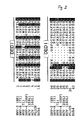
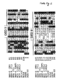
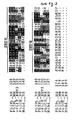
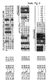
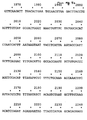
- such a DNA sequence is claimed which has a significance level of p ⁇ 10 -2 when the sequence of the death effector domain is compared with a search profile according to FIG. 1a or a search profile according to FIG. 1b ,
- a search profile is created when related sequences with functional homology are to be identified in the context of a database search.
- a search profile is described that on a generalized search profile based on six amino acid sequences, encoded by the invention DNA sequences of viral origin, based (see below).
- DNA sequences of viral origin constitute genes represents the following as FLIP genes, with viral origin but are referred to as vFLIP genes. Encode these genes for the vFLIP proteins.
- the profiles are on the Amino acid sequences of the known death effector domains aligned. Newly identified proteins or DNA sequences then include at least one profile death effector domain.
- the database search was carried out using the algorithm of Bucher et al . (Bucher, P., Karplus, K., Moeri, N. & Hofmann, KA, Computer Chem. 20, 3-24 (1996)).
- the present amino acid sequences and the DNA sequences on which they are based were identified in the "GenBank" database. However, all commercial or non-commercial databases with amino acid sequence entries can be considered as databases for the homology search.
- a search in DNA databases with a DNA search profile for death effector domains is also conceivable.
- the invention is also not limited by the current status of the entries, ie by the number of deposited protein sequences at the time of priority of the application. All future database entries with the associated DNA sequences according to the invention and the corresponding protein sequences, provided they satisfy the designed profiles of FIGS. 1A or 1B, are included in this preferred embodiment of the invention.
- a significance level of p ⁇ 10 -2 must exist if the sequence of the death effector domain is compared with a search profile according to FIG. 1A or a search profile according to FIG. 1B.
- the p values indicate statistical significance values which result from the conversion of normalized profile scores (Nscores).
- the programs pfscan and pfsearch (both are components of the program package pftools) provide so-called "normalized scores" (Nscores) in the output file as a measure of the significance of a profile / sequence comparison.
- pftools is a freely available program package for "Generalized Profile" applications.
- the randomized database which is based, for example, on the SWISSPROT database, is obtained by a so-called "section shuffling" with a window width of 20 residues (Pearson and Lipman, Proceedings of National Academy of Science, USA, 85, 2444-2448, 1988). More detailed explanations of the algorithm can also be found in the publications by Bucher et al . (Computer Chemistry 20, 3-24, 1996) and Hoffmann et al . (TIBS 20, 347-349, 1995).
- the probability of error defined here as the probability for this is that a hit of the required quality by chance is conditioned and no evolutionary relationship to the cause Has. More detailed information on the search method used with the help of a search profile in a database on the World Wide websites (WWW pages): http://ulrec3.unil.ch/profile/profile.html and http://ulrec3.unil.ch/profile/scoredoc.html.
- the presence of two death effector domains allows a strong bond to what is to be bound Protein, e.g. the adapter molecule FADD.
- the two Death effector domains of the DNA sequence according to the invention show clear similarities, but are preferred not identical. Due to amino acid sequence deviations both death effector domains will specify each Domain that is functionally important. Consequently can the binding behavior of the by the invention DNA sequences encoded proteins on proteins of the apoptosis signal transduction pathway be optimally regulated.
- About the Binding constant can occur at a given cellular concentration the intensity of the protein coded according to the invention of the apoptotic signal can be influenced.
- this invention also includes such artificial recombinants DNA sequences claimed for proteins encode with two identical death effector domains.
- Recombinant DNA sequences according to the invention e.g. DED1 from HHV-8 and DED2 from MCV (159L) or duplication from DED1 from HHV-8 or similar combinations
- DED1 from HHV-8 and DED2 from MCV (159L) or duplication from DED1 from HHV-8 or similar combinations certain differentiated inhibitory effects of apoptotic Response can be achieved.
- the present invention bind the DNA sequence according to the invention encoded proteins (or gene products) to a protein of the Apoptosesignaltransduklasweges. That way you can the gene products encoded by the DNA sequence according to the invention intervene in the apoptosis signal transduction path and regulate it with it.
- the target protein for binding come especially proteins carrying death effector domains in question.
- these target proteins as adapter proteins, such as FADD, or also as Proteases, especially as caspase-like proteases, Act.
- FADD protein carrying death effector domains in question.
- Proteases especially as caspase-like proteases, Act.
- the receptors of the tumor necrosis factor type are particularly suitable.
- the receptors TRAMP, CD95 and TNFR-1, as well as the receptor for the death-inducing ligand TRAIL (Wiley et al ., Immunity 3, 673-682 (1995)) are to be mentioned here.
- DNA sequences are those that code for gene products that target soluble intracellular Proteins of the apoptosis signal transduction pathway tie.
- FADD protein that code for gene products that target soluble intracellular Proteins of the apoptosis signal transduction pathway tie.
- the gene product of the DNA sequence according to the invention with the protein called FADD.
- This interaction happens about the respective death effector domains, is one Interaction between the adapter molecules, e.g. FADD or TRADD, with the cytoplasmic part of the receptor still possible.
- the signal transmission from is blocked Adapter molecule on the proteolytically acting proteins of the Signaling cascade. This means that the interaction between the death domains of the cytoplasmic part of the Apoptosis receptor and the adapter molecule still possible are, but the subsequent signal transmission step, e.g. from FADD to FLICE.
- DNA sequence whose gene product is attached to an adapter molecule attaches that a complex of receptor, in the form of its cytoplasmic part, from the adapter molecule and the Genetic product of the DNA sequence of the invention forms and thus the inhibition of the apopotic signal cascade is given is.
- This DISC complex is made up of several elements to which CD95 protein is attached. They wear the designation CAP1 to CAP6, where CAP1 is a FADD protein CAP2 is a hyperphosphorylated FADD, CAP3 is a previously unknown protein with an N-terminus of the FLICE protein, CAP4 a pro-FLICE protein and CAP5 or CAP6 already split off FLICE prodomains are used as indicators for activation of FLICE protein within the DISC complex can serve. In the case of an agonistic Stimulation of the CD95 receptor is stored in these components with the CD95 protein.
- CAP1 is expected to target the CD95 protein because the presence of CAP4 and CAP3 (each FLICE or FLICE-like components) strong is reduced.
- CAP5 and CAP6 undetectable in the corresponding experiments.
- the gene product of the DNA sequences according to the invention Cell apoptosis completely by stopping signal transmission prevents the proteolytic proteins in the cascade.
- the invention DNA sequences in viral, prokaryotic or eukaryotic DNA genetic information molecules.
- the DNA sequences of the invention can therefore in all Life forms occur.
- DNA sequences occur which are used to express Genetic products lead, at least one death effector domain have and inhibit cell apoptosis, at Viruses on.
- DNA sequences according to the invention occur, for example, in the Molluscum cantagiosum Virus (MCV).
- MCV Molluscum cantagiosum Virus
- MCV Molluscum cantagiosum Virus
- With this Virus occurs a DNA sequence according to the invention in ORF 159L or also in the ORF 160L.
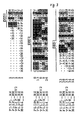
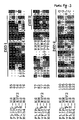
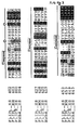
- DNA sequences according to the invention are in the GenBank database under the access code U60315 (for the ORF 159L or for the ORF 160L of the MCV). About that in addition, N-terminal amino acid sequences are shown in FIG. 2 of the two ORFs. Lie in this area two death effector domains. Both amino acid sequences point a C-terminal extension, which is shown in FIG Arrows is indicated. The extensions have a length of 66 amino acids (ORF 159L) or of 102 amino acids (ORF 160L).
- the DNA sequences according to the invention in herpes viruses, in particular in ⁇ -herpes viruses.
- the herpes viruses are viruses with double-stranded DNA, their morphogenesis runs in the core of the host cell.
- the Gene product an amino acid sequence according to the "GenBank” access code U20824 (E8), where E8 indicates the ORF, contains claimed.
- This amino acid sequence comes from the virus EHV-2. In this sequence it is noticeable that in four places the domain DED I no match with the respective otherwise under the other sequences listed in Figure 2 consists. At these positions only the sequence of EHV-2 deviations.
- the Gene product comprises an amino acid sequence according to FIG. 16, claimed. It represents an amino acid sequence of the virus HHV-8. This sequence is in ORF 71.
- One is based on it DNA sequence according to the invention is in the GenBank listed under the access code (U90534 (71)).
- a DNA sequence is also claimed in particular, whose gene product is an amino acid sequence according to FIG 17 includes.
- this viral sequence (BHV-4) there is under Z46385 a GenBank data entry of an inventive DNA sequence.
- these genes or gene products carry the name vFLIP genes or vFLIP proteins (abbreviation for FLICE viral inhibition proteins).
- the level of significance p for the search profile according to FIG. 1a or 1b is less than 10 -2 . They all have two death effector domains (DED I and DED II). These are via the search profile shown in Fig. 1a using the algorithm from Bucher at al. (see above). They thus show the characteristic sequence motifs of a death effector domain.
- At least one death effector domain is required for the effectiveness of the viral FLIP proteins, preferably two domains of this type, as under physiological conditions, are required.
- the two death effector domains are linked by a sequence of at least 16 amino acids.
- the connection sequence is preferably 16 to 19 amino acids in length.
- the DNA sequences according to the invention can code for gene products which contain at least one or both of these death effector domains, amino acid sequence segments.
- the DNA sequence according to the invention in the genetic information of Mammalian cells.
- Mammalian cells In this context everyone comes conceivable mammalian cells in question. In particular concerns the invention human DNA sequences of the invention Art.
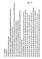
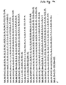
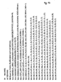
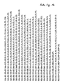
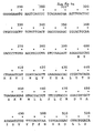
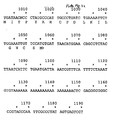
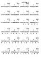
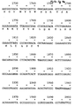
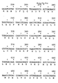
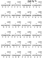
- FIGS. 4a and 4b show the amino acid sequences and the associated DNA sequences of human proteins which are homologous to the viral vFLIP proteins.
- the sequences were determined by screening cDNA libraries from activated human peripheral blood lymphocytes (PBL). Several clones were identified that code for a protein that is closely related to the viral FLIP protein. It has two death effector domains (DEDs), followed by only a short C-terminal sequence. This human protein is called FLIP S in the following.
- a human FLIP S DNA sequence according to the invention is shown in FIG. 4a. It comprises 1190 nucleotides.
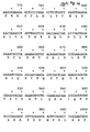
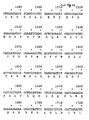
- FIG. 4b A longer version of a human FLIP DNA sequence is shown in Figure 4b.
- a DNA sequence according to the invention for coding a protein hereinafter called FLIP L
- FLIP L likewise isolated from activated peripheral blood lymphocytes
- FLIP L and FLIP S are alternative SPLICE variants of the DNA sequence according to the invention.
- RNA species exist as transcripts of the DNA sequence according to the invention, that is to say a DNA sequence with at least one, preferably two, death effector domains with apoptosis inhibitory activity.
- the shortest RNA transcript (1.1 to 1.3 kB) is particularly expressed in muscle tissue and in peripheral blood lymphocytes. This shortest transcript does not contain a caspase-homologous domain, as occurs in the longer version.
- two longer transcripts were detected (2.2 kB and 3.8 kB), both of which have a caspase-homologous domain.
- a smaller amount of longer FLIP L transcripts are synthesized in the cell. They are also essentially restricted to muscle and lymph tissue, above all to the spleen and small intestine, or to peripheral blood lymphocytes. However, small amounts of transcripts can also be detected in other tissues.
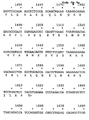
- FIG. 4c also shows a mouse DNA sequence according to the invention, a long version with a caspase homologue Domain.
- the corresponding amino acid sequence is also the same disclosed in Figure 4c.
- the DNA sequence of this cellular FLIP protein has 2452 nucleotides.
- the mouse gene was in a cDNA library of mouse heart cells identified with the help of human cDNA samples.
- such DNA sequences from cellular DNA of eukaryotes are FLIP L sequences which, in addition to at least one death effector domain, also have a caspase-homologous domain.
- FLIP S sequences only have at least one death effector domain.
- the present invention claims a DNA sequence whose gene product has two death effector domains and has a caspase homologous domain, wherein the caspase homologous domain is non-functional.
- Under caspase homologous domain becomes a domain of proteases from Understood caspase type (i.e. the type of cysteine proteases), this homologous domain gives the caspases their proteolytic Activity.
- a caspase-homologous domain that is characterized by the fact that it is not Has more cysteine proteolytic activity. Such a Loss of function can be due to a sequence-specific mutation based.
- the particularly preferred DNA sequences for gene products of the long cellular FLIP proteins is the cysteine residue in the active for the proteolysis activity Center which e.g. the FLICE protein its proteolytic Activity gives, mutates.
- cysteine residue is substituted by a tyrosine residue.
- characteristic mutations in comparison to the functional protease domains of, for example, FLICE occur at other sites in the caspase-homologous domain in the long human FLIP protein. Mutations at position -1, related to the critical cysteine residue, and at positions +1 and +2, also related to the critical cysteine residue, should be mentioned here.
- the critical cysteine residue, indicated by an asterisk in FIG. 3, is also mutated in the sequence of the mouse FLIP L protein. In this case too, the caspase-homologous domain cannot perform a proteolytic function due to the cysteine residue.
- caspase-homologous domain In the sense of the present invention, the functionality of a caspase-homologous domain is understood only to mean the loss of the proteolytic activity caused by the critical cysteine residue. Nevertheless, the caspase-homologous domain can have a different functionality. Such caspase-homologous domains are also non-functional in the sense of this invention.
- the two death effector domains of human and mouse FLIP proteins also interact with adapter proteins, e.g. FADD. Both the long protein version (55 kD) and the short form (34 kD) strongly associate with the FADD protein in the case of coexpression.
- the caspase homologue shows Sequence section of the long FLIP protein none Binding affinity to FADD. The binding is therefore preferred of a FLIP protein identified in mammalian cells with a DNA sequence according to the invention to another Protein regardless of the presence of caspase homologues Domain, via at least one death effector domain. Preferably this bond is across two death effector domains allows.
- the caspases are first activated as dimers in the course of signal transmission and then processed by autocatalysis, with a stable complex of the form (p10 / p20) 2 finally forming.
- the long FLIP proteins from eukaryotic cells, in particular mammalian or human cells, encoded by the DNA sequences according to the invention show no dimer or oligomer formation. Rather, the FLIP proteins interact with the FLICE proteins in such a way that heterodimers or heterooligomers are formed between the active FLICE and the FLIP L protein without cysteine proteolysis activity.
- a further aspect of the present invention is the use of FLIP proteins with an functional, caspase-homologous domain and at least one death effector domain for binding to proteases, in particular to cysteine proteases, which have a function as an element of the apoptotic signal cascade due to their death effector domains. This binding can then block apoptotic signal transmission.
- the FLICE protein is activated by the signal triggered at the death receptor for apoptosis, possibly by homodimer or homooligomer formation. In the course of the activation, either an autocatalytic cleavage can now take place at the p10 / p20 interface in the sequence section of the caspase domain or the FLIP protein can first be bound.
- the FLIP protein is proteolysed by the proteolytic activity of a FLICE protein in the FLICE / FLIP heterodimer or heterooligomer after the FLIP protein has bound to the infunctional caspase-homologous domain.
- FLICE protein takes on a form that largely corresponds to the small cellular FLIP protein (FLIP S ). Therefore, proteolytic cleavage occurs approximately at the junction between the C-terminal death effector domain and the caspase domain of the FLICE protein.
- DNA sequences encoded FLIP protein (human or from mice, long or short FLIP protein), shown to affect numerous, through, or via the death receptors triggered apoptosis signals have an inhibiting effect.
- An essential aspect for all DNA sequences according to the invention is their use for immortalization of cells.
- This immortalization can be done in vitro or in done in vivo; it can contain all conceivable cell types that one may be subject to apoptotic reaction.
- In Vivo methods include all gene therapy techniques known to those skilled in the art Methods.
- a DNA sequence according to the invention operable with a promoter connected.
- a promoter preferably upstream promoter with a DNA sequence according to the invention linked.
- This promoter can be a prokaryotic, depending on the experimental goal or be a eukaryotic promoter.
- Promoters used in prokaryotic host cells are the ⁇ -lactamase or the lactose promoter, furthermore the tryptophan promoter system or also hybrid promoters such as the tac promoter.
- the appropriate promoters chosen by a specialist Your nucleotide sequences are published and by information in the literature is the expert able to connect the promoters to the invention Attach DNA sequences (Siebenlist et al., Cell, 20: 269, 1980).
- Promoters in bacterial systems are generally also contain a Shine-Dalgarno (SD) sequence.
- Suitable promoter sequences in yeast host cells are e.g. the 3-phosphoglycerate kinase promoter or promoters of others glycolytic enzymes (examples include: Enolase, glyceraldehyde-3-phosphate-D-hydrokinase, hexokinase, Pyruvate decarboxylase, phosphofructokenase and others).
- the transcription of a DNA sequence according to the invention in Cells of higher eukaryotes, especially mammalian cells, is controlled by promoters that come from different native systems. It offers itself, for example to use promoters from the genomes of viruses. Examples include polyoma viruses, SV40, Adenoviruses, retroviruses, hepatitis B viruses, cytomegaloviruses and similar. Mammalian cells e.g. the ⁇ -actin promoter in question. Is particularly preferred in the present Invention of the SR ⁇ promoter.
- Cytomegaloviruses in particular also come from the early promoter of the human cytomegalovirus in question, which is a HindIII-E Restriction interface (Greenaway et al., Gene, 18: 355-360 (1982)).
- promoters also come of the respective mammalian or human host cells in Question.
- others are Control elements for transcription and / or translation added to the DNA sequence according to the invention.
- Enhancers are cis-acting elements of DNA that are common Include 10 to 300 base pairs and to the promoter Increase the transcription rate. You can in 3 'or 5' position of the DNA sequence according to the invention be arranged, but also in the coding sequence itself or in an intron that only occurs through SPLICE processes is cut out, be present.
- Enhancers of adenoviruses come into question but also enhancers of adenoviruses.
- Enhancers also known from mammalian genes (e.g. globin, Elastase or albumin).
- the enhancers are typical at the 5 'or 3' position of the coding region a DNA sequence according to the invention in the expression vector integrated.
- the enhancer is 5 'to Promoter.
- Additional control elements can be used for regulation serve the termination of the transcription, so that the expression the mRNA is affected.
- expression vectors a DNA sequence according to the invention, typically with a promoter and optionally with others of the above-described control elements of the transcription and / or translation included. They serve that the nucleotide sequence according to the invention in specific Host cells can be propagated and expressed.
- general expression vectors are used, which are can replicate autonomously regardless of the host chromosome. You have your own "origin of replication". Such Sequences are present in bacteria, in yeast or in viruses. In contrast, the origins in expression vectors are from Not required for mammals.
- the expression vectors with the invention DNA sequence as so-called "shuttle" vectors designed, i.e., they can be in a host system replicate first and then move to another host system be transfected for expression.
- a vector can first be cloned into E.coli and then expressed in a yeast or mammalian cell become. In such a case, it is no longer imperative that replication is independent of the Replication of the host cell chromosome is possible.
- a DNA sequence according to the invention is preferred then into another vector that is the stable Expression e.g. allowed in Jurkat and Raji cells.
- a selection gene that acts as a marker exercises. It is a gene that is responsible for encodes a protein that survives or grows allowed such host cells that transformed with the vector were.
- Typical selection genes code for proteins which are resistant to antibiotics or others Allow toxins.
- Puromycin is one example or ampicillin or neomycin. Within the scope of this invention are puromycin resistances for the selection of the transformed Host cells are particularly preferred.
- Another aspect of the present invention is a Process for the isolation of gene products with at least a death effector domain, the host cells with a Expression vector according to the invention transformed and then under suitable conditions that promote expression be cultivated so that the gene product is finally made the culture can be cleaned up.
- the protein of the invention DNA sequence can, depending on the expression system, from a culture medium or from cell extracts be isolated.
- the expert can easily recognize that the respective insulation methods and Process for the purification of an inventive DNA encoded recombinant protein strongly of the type the host cell or the fact whether the protein is in the medium is secreted depends.
- you can Expression systems are used for the secretion of the recombinant protein.
- the culture medium must be in in this case through commercially available protein concentration filters, e.g. Amicon or Millipore Pelicon, concentrated become.
- a Cleaning step e.g. a gel filtration step.
- an anion exchanger can also be used that has a matrix with DEAE.
- a protein encoded by a DNA according to the invention serve HPLC sections. It can be one or more Act steps. In particular, the "reversed phase" method used. These steps are for preservation an essentially homogeneous recombinant protein of DNA sequence according to the invention.
- yeast cells In addition to bacterial cell cultures for isolating the gene product transformed yeast cells can also be used become. In this case the translated protein can be secreted be so that protein purification is simplified becomes. Secreted recombinant protein from a yeast host cell can be obtained through methods like those at Urdal et al. (J. Chromato. 296: 171 (1994)).
- Another aspect of the present invention is a Process for the expression of gene products with at least a death effector domain, host cells with a Expression vector, which is a DNA sequence according to the invention contains, be transformed.
- This method of expression of gene products based on a DNA sequence according to the invention based does not serve the corresponding gene product to concentrate and purify, but rather to the cell metabolism by introducing the DNA sequences according to the invention via the expression of the to influence the associated gene product.
- expression vectors transformed host cells for the purpose of immortalization to think.
- Concentrations of sequences according to the invention based proteins are expressed. In this way is permanently triggered by or via the death receptors Prevents apoptosis.
- the corresponding cell lines are compared to a variety of apoptotic Stimulants resistant. These cells can also if necessary transferred back to the mammalian or human organism become.
- in vitro with the Expression vectors of manipulated cells according to the invention and the subsequent transmission into the organism gene therapy use of the DNA sequences according to the invention possible.
- Expression vectors with the sequences according to the invention transfected into such cells the disease-related through apoptotic influences in the organism be eliminated. Through such a process a constant restitution of these cells at risk of apoptosis possible.
- Vectors are used for this purpose (e.g. Liposomes or adenoviruses or retroviruses or the like), the DNA sequences of the invention specifically in each Insert cells of the organism that are due to pathological Influences an increased disposition to cell death demonstrate.
- the DNA sequences according to the invention, their alleles, derivatives or fragments can also be used as samples on a laboratory scale become.
- the DNA sequence the viral FLIP genes to detect the corresponding Viruses are used in in vitro test systems. This would come in particular Applications for Northern or Southern blots in Question.
- the samples have a suitable DNA sequence length depending on the experimental requirement. For example could be that of a death effector domain of the invention Type underlying DNA sequence used as a detection sample become.
- DNA sequences according to the invention are their use as a single-stranded DNA sequence. This is possible at both the RNA and DNA levels.
- the use of both DNA or RNA of the coding strand ("sense") as well as the complementary strand "antisense”) is conceivable. It depends on the choice of the target protein whether anti-sense or sense DNA is used.
- the corresponding oligonucleotides typically include between 20 and 40 nucleotides of the DNA sequence according to the invention.
- the binding of anti-sense or sense DNA to the training of duplex molecules that either translate (at the RNA level) or transcription (at the DNA level) To block. In this way, the inventive DNA sequences the expression of FLIP proteins in the Cell can be prevented.
- Another object of the present invention are Host cells with an expression vector according to the invention were transformed.
- suitable host cells for Cloning or expression of the DNA sequences according to the invention come prokaryotic yeasts or higher eukaryotic ones Cells in question. In prokaryotes are gram-negative or Gram-positive organisms expressly included. To E. coli or bacilli are mentioned here.
- preferred host cells for cloning the DNA sequences according to the invention the strains E.coli 294, E.coli B and E.coli X1776 as well as E.coli W3110.
- the bacilli are bacillus subtilis, Salmonella typhimurium or the like.
- prokaryotic cells typically a signal sequence to transport the protein into the culture medium.
- prokaryotic cells there are also eukaryotic ones Microbes as host cells with the expression vector have been transfected into question. So about flame-like fungi or yeasts as suitable host cells for vectors encoding the DNA sequences of the invention be used. Saccharomycis is particularly noteworthy cirevesiae or the ordinary baker's yeast (Stinchcomb et al., Nature, 282: 39, (1997)).
- expression vectors which have the DNA sequences according to the invention, preferably cells of the mammalian immune system all of the human immune system, transfected. Most notably preferred host cells are T and D lymphocytes.
- DNA sequences according to the invention which can immortalize the host cell, results, for example, in the case of an HIV infection.
- HIV infection cells die through apoptotic mechanisms.
- Uninfected CD4 + cells are also subject to cell death.
- immortalization of uninfected immune cells would therefore be desirable. In this way, basic functionality of the immune system could be preserved.
- Gene products of the DNA sequences according to the invention are understood both Primary transcripts, ie RNA, preferably mRNA, as well Proteins. According to the invention, these proteins have at least a death effector domain and inhibit cell apoptosis. These proteins include all encoded according to the invention Proteins, i.e. also those of DNA derivatives, DNA fragments or DNA alleles expressed proteins. About that the proteins can also be chemically modified. So For example, there may be a protecting group at the N-terminus. It can add glycosyl groups to hydroxyl or amino groups Lipids can be covalent with the invention Protein linked, as well as phosphates or acetyl groups and similar.
- any chemical substances, compounds or groups can be by any synthetic route be bound to the protein of the invention.
- additional amino acids e.g. in the form of individual amino acids or in the form of peptides or in the form of protein domains and the like, can be fused to the N and / or C terminus his.
- so-called signal or "Leader" sequences at the N-terminus of the invention Amino acid sequence are present that cotranslational the peptide or post-translational into a particular cell organelle or lead into the excellent space (or the culture medium).
- the Amino acid sequences can also be present at the N- or at the C-terminus, which as the antigen bind the invention Allow amino acid sequence on antibody.
- the flag peptide its sequence in the one-letter code of the amino acids is: DYKDDDDK.
- This Sequence has strong antigenic properties and therefore allows a quick check and easy cleaning of the recombinant Protein.
- Monoclonal antibodies containing the flag peptide bind are from Eastman Kodak Co., Scientific Imaging Systems Division, New Haven, Connecticut available.
- the DNA sequences according to the invention can also in numerous exons separated by introns are on the strand of the genetic information molecule be filed. This also includes all conceivable SPLICE variants (at the mRNA level) as gene products for the invention Object. Also those of these different SPLICE variants encoded proteins are subject to this invention.
- protein aggregates e.g. Dimers of the invention Proteins or higher aggregates, in the range of the present Invention.
- the protein as a gene product according to the invention has a level of significance of p ⁇ 10 -2 when compared with a search profile according to FIG. 1a or FIG. 1b.
- a search profile is created that can identify any death effector domains in a database.
- search profiles There are two search profiles to choose from. Both the database with the search profile and the search profile with the database can be compared.
- all gene products or sections of these gene products in one embodiment form part of the subject matter of the invention if a primary transcript (RNA) is encoded by one of claims 1 to 24.
- the invention FLIP proteins an amino acid sequence according to one of the Figures 4a, 4b or 4c (for the cellular proteins) or an amino acid sequence according to FIG. 16 or according to FIG. 17 or according to the "GenBank" access codes U60315 (MCV 159L), U60315 (MCV 160L), U20824 (E8) or X64346 (ORF 71).
- the protein according to the invention can be used in any combination with other proteins or with pharmacological Additives or other carrier materials or therapeutic agents be prepared galenically.
- the protein can be used Investigation of cell physiological processes can be used. Binding studies with other proteins are possible. In the context of such binding studies, the biological Activity of the proteins of the invention tested become.
- the invention Proteins as part of protein purification processes to think.
- a protein according to the invention can bound to a carrier material, e.g. on a pillar, this use for binding and isolation any physiologically interacting with the protein of the invention Serves cell proteins.
- Figure 1 there are two search profiles for identification described by death effector domains.
- the amino acids are according to their one-letter code, alphabetically ordered (e.g. alanine: A, in the first position). Such a systematic ranking of the amino acids takes place for each of the sequence positions.
- SY 'P' for the first sequence position of the Profile according to Figure 1a) to occur in a homologous sequence.
- M a systematic alphabetical Ranking of the amino acids with a profile weighting the probability for each amino acid at the specified sequence position.
- Figure 1a is the search profile using the sequences for FADD Mch4 and FLICE. It comprises two pages.
- FIG. 1b shows the search profile, which consists of the protein FADD, Mch4, FLICE and the viral FLIP proteins was shown. It was used to identify the murine and human FLIP sequences. This search profile too comprises two pages.
- the amino acid sequences are from his time known proteins with death effector domains (FADD, FLICE and Mch4) compared to those found via a profile search
- EHV-2 equine herpes virus 2
- HHV-8 human herpes virus 8
- HVS herpes virus Saimiri
- BHV-4 bovine herpes virus 4
- MCV Molluscum Contagiosum Virus
- the amino acid sequences of the viral FLIPs are deposited in the GenBank database the access numbers U20824 (ORF E8 in EHV-2), X64346 (ORF 71 in HVS) and U60315 (ORF 159L and ORF 160L in MCV).
- the Amino acid sequences of the viral FLIPs from HHV-8 (ORF 71) and from BHV-4 are in Figure 16 (ORF 71 from HHV-8) and shown in Figure 17 (v-FLIP of BHV-4).
- Amino acid sequence of the DNA sequence encoding viral FLIPs is deposited in the GenBank database with the access number U20824 (ORF E8 in EHV-2), Z46385 (BHV-4), X64346 (ORF 71 in HVS), U90534 (ORF 71 in HHV-8) and U60315 (ORF 159L and ORF 160L in MCV).
- FIG. 3 shows the homology of the amino acid sequences of the human (HS) and the murine (MM) form of the long (FLIP L ) and the short (FLIP S ) version of FLIP with the amino acid sequences of FLICE and Mch4.
- the N-terminal 202 amino acids of the shorter splicing variant of the human FLIP (FLIP S ) are identical to the sequence of the longer form of the human FLIP (FLIP L ), the sequence of the shorter protein (FLIP S ) ends after a C-terminal extension of a further 19 amino acids (only this extension is shown in the figure for the human FLIP S ), which are not contained in the longer form (FLIP L ).
- the murine (MM) homolog FLIP L and the human (HS) homolog FLIP L and FLIP S each contain two death effector domains (DED1 and DED2, respectively ).
- the murine and human FLIP L form also contain a C-terminal domain that is homologous to the protease domain of the caspases.
- FLIP L (2 N-terminal death effector domains combined with a caspase) thus corresponds to the structural organization of FLICE and Mch4, however, compared to the caspases, the conserved cysteine residue of the active protease domain is missing: in human and in murine FLIP L it is the corresponding amino acid position is replaced by a tyrosine residue (the corresponding position is indicated by an asterisk in the figure).
- FIG. 4a shows the DNA and the amino acid sequence of the human FLIP S , which was screened from a cDNA bank of activated T cells by screening with a 32 P-labeled DNA probe (EcoRI / RsaI fragment of the 5'-terminus of the DNA Inserts of EST clone No. 309776) was isolated.
- FIG. 4b shows the DNA and the amino acid sequence of the human c-FLIP L , which was isolated from a cDNA bank of activated T cells by screening with the probe described in FIG. 4a.
- Figure 4c shows the cDNA and the amino acid sequence of the murine c-FLIP L , which was isolated from a murine cardiac muscle cDNA library by screening with that in Figure 4a.
- Figure 5 is by cotransfection experiments in eukaryotic 293T cells using the example of the viral FLIPs from EHV-2 (ORF E8, Fig. 5a) and MCV (ORF 159L, Fig. 5b) demonstrated that the viral FLIPs attached to the adapter protein FADD bind, and that this bond is not the attachment of the Adapter protein FADD on the cytoplasmic, the domain of death containing protein part of the CD95 death receptor prevented.
- About the binding of the viral FLIPs E8 (from EHV-2) and 159L (from MCV) to FADD thus becomes an attachment the viral FLIPs via the adapter FADD with the cytoplasmic Protein part of the death receptor CD95 enables.
- FIG. 6 shows the attachment of the viral FLIP protein E8 from EHV-2 with the agonistically stimulated CD95 receptor complex in human Raji B cell clones which were stably transfected with an expression vector for the E8 protein.
- Figure 6a shows by Western blot analysis the expression of the flag-labeled E8 protein in cell extracts from two Raji B cell clones labeled RE8 / 11 and RE8 / 19, which were transfected with an expression vector for E8. No E8 protein, however, is detected in the Raji control clones designated RCo / 1 and RCo / 3, which were transfected with the corresponding expression vector without E8 insertion.
- FIG. 6 shows the attachment of the viral FLIP protein E8 from EHV-2 with the agonistically stimulated CD95 receptor complex in human Raji B cell clones which were stably transfected with an expression vector for the E8 protein.
- Figure 6a shows by Western blot analysis the expression of the flag-labeled E8 protein in cell
- FIG. 6b shows the result of the analysis of immunoprecipitations of the CD95 death receptor agonistically stimulated with the antibody APO-1 (Kischkel et al., EMBO Journal 14, 5579-5588 (1995)) from the 35 S metabolically labeled Raji cell clones RCo / 1 ( a non-E8 control clone) and RE8 / 11 (an E8 expressing clone).
- a white arrowhead marks the migration position of the radioactively labeled E8 protein, which is contained in the CD95 attachment complex of the clone RE8 / 11, but not in the corresponding complex of the control clone RCo / 1.
- FIG. 7a shows the association of 35 S-labeled proteins with the agonistically stimulated CD95 death receptor (Kischkel et al., EMBO Journal 14, 5579-5588 (1995)) in the non-transfected Raji control cells (clone RCo / 3, left part of the Figure) and in those cells that were transfected with an expression vector for the viral FLIP E8 of EHV-2 (clone RE8 / 19, right part of the figure), by a two-dimensional gel electrophoresis analysis of CD95 (anti-APO-1) - Immunoprecipitations compared.
- the figure shows that the viral protein E8 associates with the agonistically stimulated CD95 death receptor without impairing the attachment of the adapter molecule FADD (CAP 1) to the receptor (the CAP1-labeled FADD protein is in the agonistically stimulated CD95-associated signal transduction complex contained both the non-E8 expressing control clone RCo / 3 and in the E8 expressing Raji clone RE8 / 19 (compare the left and right part of the figure).
- CAP 1-labeled FADD protein is in the agonistically stimulated CD95-associated signal transduction complex contained both the non-E8 expressing control clone RCo / 3 and in the E8 expressing Raji clone RE8 / 19 (compare the left and right part of the figure).
- the figure shows that in such cells which express the viral FLIP, the attachment of FLICE (CAP4) and FLICE-like molecules (CAP3) to the receptor is prevented, and thus the conversion of FLICE into its two which takes place in the receptor attachment complex Cleavage products CAP5 and CAP6 is blocked (absence of the radioactively labeled proteins labeled with 4 to 6 in the E8-expressing clone RE8 / 19 only in the right but not in the left part of the figure).
- CAP4 FLICE
- CAP3 FLICE-like molecules
- Figure 7b shows that the treatment of cells with the agonist stimulating anti-CD95 antibody APO-1 induced FLICE cleavage activity of the agonistically stimulated Receptor of the E8 expressing Raji B cell clone RE8 / 19 is significantly less than the corresponding activity of the control clone RCo / 3 (which does not express E8).
- the Presence of FLICE cleavage activity in the CD95 attachment complex is quantifiable through the formation of the FLICE fission products p43, p26, p17, p12 and p9 from radioactive labeled FLICE by CD95 (anti-APO-1) immunoprecipitates treated by APO-1 (+), but practically not by untreated (-) cells.
- Figure 8 it is shown that eukaryotic cells by the expression of a viral FLIP (ORF E8 from EHV-2 or ORF159L from MCV) significantly more resistant to apoptosis induction through the CD95 death receptor are used as control cells.
- Figure 8a shows the percentage of apoptotic Raji B cells (ordinate) depending on the concentration of the agonistic anti-CD95 (APO-1) antibody (Abscissa) (present in the culture medium for 24 hours at 37 ° C) for two Raji B cell clones expressing E8 (RE8 / 11 and RE8 / 19) and two non-E8 expressing Raji control clones (RCo / 1 and RCo / 3) compared.
- APO-1 agonistic anti-CD95
- the two expressing E8 Clones prove to be significantly more resistant to CD95-mediated apoptosis as the control clones.
- a APO-1 concentration in the culture medium of 100ng / ml was sufficient namely under the chosen experimental conditions to in more than 60% of the non-E8 control clones To induce apoptosis while taking the same Conditions only about 20% of the cells of each of the two E8 expressing Raji clones RE8 / 11 and RE8 / 19 apoptotically were.
- FIG. 8b shows in particular that the amount of flag-expressed in the clones viral FLIPs (demonstrated by the in the literature described Western blot of cell extracts with anti-flag Antibody, upper part of Figure 8b) with a reduction the apoptosis sensitivity to the CD95 ligand (lower Part of Figure 8b) correlates because the percentage of apoptotic cells induced by the CD95 ligand (indicated on the abscissa), the lower the stronger which was detected in the western blot in the upper part of FIG. 8b E8 expression of the clones.
- the percentage of apoptotic cells for non-E8 expressing control clones (JCo / 2, JCo / 3, JCo / 4 and JE8 / 5) at least three times higher than the percentage of apoptotic cells for the Jurkat clone with the strongest E8 expression (clone JE8 / 13).
- FIG. 8c shows the protective effect of v-FLIPs (shown is an experiment with the gene product of ORF E8 from EHV-2) to those caused by overexpression of the CD95 death receptor in 293T cells (a human embryonic kidney cell line) induced apoptosis.
- 293T Cells with expression vectors for CD95 along with the sets of expression vectors indicated under part 8c transfected for the above v-FLIP and 30h harvested after transfection.
- Apoptosis induction in the Cells became apoptotic by photometric quantification Histone-DNA complexes determined (the ordinate The apoptotic index given corresponds to that with the Cell death detection ELISA system determined by Boehringer optical density (OD) of the samples at 405 nm).
- OD Boehringer optical density
- As negative controls cells were used for the induction of apoptosis transfected with expression vector without insertion (mock) were or transfected with the expression vector for CD95 Cells that are in after transfection until cell harvest Medium were incubated with 25 ⁇ M z-VAD-fmk.
- the figure shows that the gene product of E8 pre-EHV-2 293T cells protect the induction of apoptosis by CD95 overexpression, the protective effect increases with the concentration of the used v-FLIP expression vector (this corresponds to a decrease in the apoptotic index with increasing v-FLIP expression vector concentration in Figure 8c).
- the protective effect in the highest doses used (co-expression of 1 ⁇ g each of the expression vector for E8 with that given in Figure 8c Amount of CD95 expression vector) is the protective effect the viral FLIPs by the protease inhibitor z-VAD-fmk achieved comparable protection.
- Figure 9 shows the protective effect of v-FLIPs (shown are experiments with the gene products of ORFs E8 from EHV-2, ORF 159L from MCV and ORF 71 from HVS) to those caused by overexpression of the death receptor TRAMP in 293T cells (one human embryonic renal cell line) induced apoptosis.
- 293T cells with expression vectors were used for TRAMP (Bodmer et al., Immunity 6, 79-88 (1997)) together with the specified amounts of Expression vectors transfected for the above v-FLIPs and harvested 30h after transfection.
- v-FLIP expression vectors (the set of expression vectors used is given below the figures in ⁇ g) apoptosis protection of TRAMP-overexpressing Cells reached by at least 70% (compared to 100% protection by the protease inhibitor z-VAD-fmk).
- Figure 10 it is shown that human Jurkat T cells by expression of the viral FLIP of ORF E8 from EHV-2 resistance to apoptosis induction by the receptor purchase TRAIL for the death ligand. Correlated the amount of viral expressed in the clones FLIPs (detected in the Western blot of cell extracts, upper part of Figure 8b) with the extent of cell viability in the presence of the apoptosis-inducing death receptor ligand TRAIL (Figure 10).
- Clones against TRAIL-induced apoptosis were identified by Incubate with the concentrations shown in the abscissa of recombinant soluble flag-labeled TRAIL and cross-linking anti-flag antibody for 20 h at 37 ° C and subsequent determination of the ability to proliferate the cells (ordinate) with a cell profiling assay (Celltiter 96 AQ, Promega).
- Celltiter 96 AQ Celltiter 96 AQ, Promega.
- the not expressing E8 Jurkat clones (JCo / 2, JCo / 3, JCo / 4 and JE8 / 5) were identified in this test with increasing TRAIL concentration one significant decrease in proliferation on (sigmoid curve, Decrease in optical density with decreasing proliferation).
- Figure 11 demonstrates the correlation of expression of a viral FLIPs (ORF 71 of the HVS) in the course of the viral Infection of the host cell OMK (owl monkey kidney) with the Protection of the host cell against CD95 ligand-induced apoptosis.
- Figure 11a shows a Northern blot analysis for detection of transcripts of ORF71 from HVS in samples from not virus-infected control cells (OMK), in samples from OMK Cells that were treated with HVS strain C488 for 1 to 4 days (C488, OMK d1 to d4) or with HVS strain A11 for 4 days (OMKd4-A11) were infected, or from samples of a semipermissive T cell line that produces small amounts of virus particles (P-1079).
- OMK virus-infected control cells
- FIG. 12 shows by co-transfection experiments in eukaryotic 293T cells that this binds the short and the long form of the human FLIP (FLIP S and FLIP L ), but not the protein part with caspase homology (FLIP P ) to the adapter protein FADD.
- FLIP S and FLIP L the binding of FLIP S or FLIP L to the adapter protein FADD does not prevent the attachment of FADD to the cytoplasmic protein part of the CD95 death receptor containing the death domain.
- binding of the human FLIP S and FLIP L to the FADD adapter protein enables the attachment of these FLIP forms, via the adapter FADD, with the cytoplasmic protein part of the death receptor CD95.
- FIG. 13 shows by cotransfection experiments in eukaryotic 293T cells that the human proteins FLIP S and FLIP L bind to the cysteine protease FLICE.
- the figure further shows that the N-terminal protein part of FLIP L containing the death effector domains and the C-terminal protein part of FLIP L possessing the caspase homology contribute to the binding of FLIP L to FLICE.
- the expression of the HA-labeled FLICE protein in cell extracts of the transfected samples was detected in the bottom part of the figure by a Western blot with anti-HA antibody.
- the middle part of the figure shows by an anti-flag Western blot that comparable amounts of 3 different flag-labeled FLIP proteins or protein parts were precipitated from the transfected cells by anti-flag immunoprecipitation.
- the uppermost part of the figure shows by an anti-HA Western blot analysis of the anti-flag precipitates that the HA-labeled FLICE on FLIP L and FLIP S as well as on the caspase-homologous C-terminal protein part of FLIP L ( FLIP P ) binds.
- FIG. 14 shows that eukaryotic cells are significantly more resistant to apoptosis induction by the CD95 death receptor than control cells due to the expression of the human FLIP S or FLIP L. It is also shown that the longer form of the human FLIP (FLIP L ) provides more efficient protection against CD95-induced apoptosis than the shorter form FLIP S.
- FIG. 14a shows that human Jurkat T cells acquire resistance to apoptosis induction by the Fas (CD95) ligand (FasL) through the expression of the human FLIP S or the human FLIP L. The amount of human VSV-labeled FLIPS or FLIP L expressed in the clones (detected in the anti-VSV western blot of cell extracts, upper part of FIG.
- the non-transfected parental Jurkat T cells Jurkat, Co
- the non-FLIP S expressing Jurkat clone JFS1 and the weakly FLIP S expressing Jurkat clone JFS5 showed an overlapping sigmoid curve in this test (decrease in proliferation with increasing sFasL concentration ).
- a right shift of the proliferation curve was observed for the clone JFS7 expressing more FLIP S.
- an approximately 5-fold higher concentration of FasL was necessary than for the parental clone.
- An even stronger resistance to FasL was observed for the two FLIP L -transfected Jurkat clones JFL1 and JFL2.
- the protein expression of the VSV-labeled FLIP L was only weakly (JFL2) or not at all (JFL1) detectable in the Western blot (upper part of FIG. 14a), the two clones were approximately 10-fold (JFL1) or more than 25- fold (JFL2) more resistant to treatment with sFasL than the parental Jurkat clone.
- FIG. 14b shows that human Raji B cells also acquire resistance to the induction of apoptosis by the sFasL through the expression of the human FLIP S.
- the Raji clones RFS3, RFS7, RFS8 and RFS11 expressing the human FLIP S , a non-FLIP S expressing Raji clone (RFS1) and the non-transfected parent Raji clone (Raji wild-type, wt) and the sensitivity of the clones were used against sFasL-induced apoptosis as described in FIG. 14a.
- FIG. 14a shows that human Raji B cells also acquire resistance to the induction of apoptosis by the sFasL through the expression of the human FLIP S.
- FIG. 14b shows that Raji clones which express VSV-labeled FLIP S (see anti-VSV Western blot in the upper part of FIG. 14b) have partial resistance to sFasL.
- these clones were at least 5 times more resistant to sFasL-induced proliferation inhibition than the non-FLIPS-expressing control clones Raji (wt) and RFS1 under the chosen experimental conditions.
- FIG. 14c shows, using the experimental approach described in FIG. 14a, that human Raji B cells can acquire practically complete resistance to treatment with sFasL by expressing human FLIP L.
- the Raji clones with strong (RFL12) and medium (RFL2, RFL42 and RFL47) expression levels of the VSV-labeled FLIP L are practically completely resistant to treatment with sFasL at the concentrations of sFasL indicated in the abscissa (this is shown in a almost horizontal curve, ie unchanged cell proliferation behavior of the respective clones even at high (over 1 ⁇ g / ml) sFasL concentrations).
- FIG. 15 shows that human Jurkat T cells acquire resistance to the induction of apoptosis by the receptor for the death ligand TRAIL through the expression of the human FLIP S or FLIP L.
- the amount of c-FLIPs expressed in the clones correlates with the extent of cell viability in the presence of the apoptosis-inducing death receptor ligand TRAIL (FIG. 15).
- the sensitivity of the Jurkat clones to TRAIL-induced apoptosis was determined by incubation with the concentrations of recombinant soluble flag-marked TRAIL and cross-linking anti-flag antibody for 20 h at 37 ° C and subsequent determination of the ability of the cells to proliferate (ordinate) a cell proliferation assay (Celltiter 96 AQ, Promega).
- the parent, non-transfected Jurkat clone (wt) and the transfected, non-FLIP S expressing Jurkat clone JFS1 showed a significant decrease in proliferation with increasing TRAIL concentration (sigmoid curve, decrease in optical density with decreasing proliferation).
- Clone JFS5 which showed an intermediate level of VSV-labeled FLIP S in the Western blot (FIG. 14a above), showed partial resistance to TRAIL, while clone JFS7, which expressed the VSV-labeled FLIPS more strongly, was practically completely resistant to TRAIL (The horizontal curve shows that there was no impairment of the proliferation by the death receptor ligand TRAIL).
- the experimental curves further show that the two Jurkat clones JFL1 and JFL2 transfected with an expression vector for human FLIP L despite the least (clone JFL1) or weak (clone JFL2) expression of the VSV-labeled FLIP L (see Western blot Figure 14a, upper part of the figure) ) were almost (JFL1) or completely (JFL2) resistant to treatment with the death receptor ligand TRAIL.
- FIG. 16 shows the amino acid sequence of the viral FLIP of the HHV-8 (ORF 71) in the one-letter code of the amino acids.
- FIG. 17 shows the amino acid sequence of the viral FLIP of the BHV-4 in the one-letter code of the amino acids.
- One for this amino acid sequence encoding DNA sequence can be found in the GenBank database with the accession number Z46385.
- a human embryonic kidney cell line (293T cells), a human leukemia T cell line (Jurkat cells) or a human Burkitt-Lymphoma B cell line (Raji cells) were used and grown as cell lines as in Bodmer et al . (Immunity 6, 79-88 (1997)).
- the clone is called 117926 and is in GenBank registered under accession number T90422.
- As The following sequences were oligonucleotides for the PCR method used: the oligonucleotide JT403 5'-TCAGCTGCAGACCTCTGAGGAAAC-3 'and the oligonucleotide JT469 5'-ACTAGTTAGCCAACTAAAAAG-3 '.
- Cloning the section was carried out in the vector pQE-16 (from Qiagen), and in a PstI / SpeI interface - according to the Restriction enzyme abbreviations.
- the cloned sequence also contains a flag sequence and a subsequent one Linking element with the amino acid sequence GPGQVQLQ. Then the PstI and SpeI interfaces between the original BamHI / XbaI digits of the vector. Protein expression 0.5 mM IPTG was induced in the bacteria. After 6 hours of incubation at 30 ° C, the cells were harvested and lysed by sonication.
- the lysates were first extracted with 0.5% precondensed Triton X-114, to eliminate the bacterial lipopolysaccharides, and finally the Flag / TRAIL protein was through a chromatography column with M2 anti-flag agarose purified; this was followed by elution with 50 mM citric acid and finally neutralization with a 1 M Tris base and dialysis against PBS (phosphate buffered saline, phosphate buffered saline).
- PBS phosphate buffered saline, phosphate buffered saline
- a DNA fragment obtained by PCR amplification of the DNA sequence of human FLIP contained in EST clone 309776 was used as the 32 P-labeled probe. This fragment corresponds to the DNA sequence section numbered 394 to 903 in FIG. 4a.
- the screening of the ⁇ -ZAP cDNA bank of activated human lymphocytes from Stratagene, available on request from Hermann Eibel, University of Freiburg, Germany) to isolate the human forms of FLIP and the screening of a corresponding bank of murine cardiac muscle cells (Stratagene) Isolation of the murine FLIP L was carried out according to the manufacturer's recommendation.
- the complete "Open Reading Frame" E8 of the virus EHV-2 (ORF E8) was amplified from the viral DNA by PCR methods, a 5 'primer with an EcoRI sequence extension and a 3' primer with a sequence -Extension for the restriction enzymes BamHI and EcoRI were used here.
- the insertion (into the EcoR1 interface of the vector) was carried out in the same reading frame as for the N-terminal flag epitope.
- the vector is derived from the pCR-3 vector (from Invitrogen).
- the same procedure was followed with the Open Reading Frames ORF 159L from Virus MCV (from the Institute for Medical Virology at Heidelberg University, Germany) and the Open Reading Frame ORF 71 from Virus HVS.
- the FLIP-coding ORFs of these viruses were also amplified by PCR methods and then inserted in the correct reading frame with the N-terminal flag epitope in the EcoR1 interface of the vector derived from pCR-3. Furthermore, the complete ORF of the human FLIP S and the human FLIP L , as well as a HindIII / XhoI fragment of the 3 'terminus of the DNA sequence of the human FLIP L were amplified by PCR methods and in the correct reading frame in vectors derived from pCR-3 cloned, which give the respective gene products an N-terminal flag or VSV epitope.
- the constructs from Flag-E8, VSV-FLIP S and VSV-FLIP L were inserted into the multiple cloning site ( MCS) of the vector pSR ⁇ puro (a gift from R. Sekaly, IRCM, Montreal, Canada). This vector carries puromycin resistance.
- An expression vector for the cytoplasmic domain of the murine Fas receptor provided with the myc sequence was produced by inserting a PCR fragment corresponding to amino acids 166-306, in the correct reading frame with the N-terminal myc epitope, and in the pCR-3-like Vector inserted.
- the expression vector for the human TRAMP or FADD in the vector pCR-3 can be found in the publication by Bodmer et al. (Immunity 6, 79-88 (1997)).
- Human Fas (with the nucleotide sequence -24 to +1009) was by PCR methods from an EST clone (GenBank accession number X63717) and then amplified as a HindIII / XbaI fragment subcloned into the pCR-3 vector.
- the cells were washed twice with HeBS buffer, at a pH of 7.05 (0.8 mM NaH 2 PO 4 .2H 2 O, 20 mM Hepes, 137 mM NaCl, 5 mM KCl, and 5.5 mM D-glucose). Then 8x10 6 cells were resuspended in 800 ⁇ l HeBS, mixed with 20 ⁇ g of the SR ⁇ puro plasmid (both with and without Flag-E8 insertion) and finally exposed to a voltage of 250 V and a current of 960 ⁇ F. After 48 hours of transfection, the cells were distributed at a concentration of between 2000 and 20,000 cells per well on flat-bottom plates with 96 wells each and stable transfectants were selected by adding 5 ⁇ g / ml puromycin (from Sigma).
- the cells were distributed at a concentration of 1 to 2x10 6 cells / 10 cm plate or 3 to 6x10 5 cells / 5 cm plate and the following day using the calcium described in the literature Transfected phosphate precipitation method. The precipitate was left on the cells for 8 hours and the cells were finally harvested 26 to 30 hours after the transfection.
- the 293-T cells from a 10 cm plate were lysed in 200 ⁇ l lysis buffer (1% NP40, 20 mM Tris-HCl, pH 7.4, 150 mM NaCl, additionally 1 mM EGTA, 1 mM pefabloc-sc (from Serva) and 10 ⁇ g / ml leupeptin and aprotinin (each from Sigma) were added
- Postnuclear lysates were pre-cleaned for at least 1 hour on Sepharose 6B (from Pharmacia) before precipitation, followed by either 2 hours or overnight with 3 ⁇ l of anti-flag agarose, the precipitates were washed a total of four times with lysis buffer, the lysis buffer containing 1% NP40 in the first two washes and 0.1% NP40 in the second two washes were then heated in sample buffer and then analyzed by SDS-PAGE and Western blotting The blots were saturated with 5% milk in PBS with 0.5% Tween
- the detection of the proteins was enhanced by chemiluminescence (Amersham International).
- Jurkat, Raji or 293T cell clones were checked for their expression of transfected proteins. This was done by anti-flag, anti-VSV, anti-FADD, anti-myc or anti-HA western blot analysis of post-nuclear cell lysates of equivalent protein content.
- the metabolic labeling of Raji cells with 35 S, the anti-CD95 immunoprecipitation and their 2D gel electrophoresis were carried out as already described (Kischkel, FC et al ., EMBO J. 14, 5579-5588 (1995)).
- the analysis of the apoptosis induced by FasL was carried out as follows. Puromycin-resistant Jurkat clones (approx. 3 ⁇ 10 5/500 ⁇ l) were or for 3 hours at a temperature of 37 ° C. with 50 ⁇ l of a supernatant of neuro-2a cells transfected with a FasL expression vector a control supernatant from cells transfected with a mock vector (Rensing-Ehl, A. et al ., Eur. J. Immunol. 25, 2253-2258 (1995)). The Jurkat cells were washed with FACS buffer (2% FCS and 0.02% azide), and fixed in 70% ice-cold ethanol.
- RNAse After washing again with FACS buffer, treatment with RNAse followed for 5 minutes at a temperature of 37 ° C. (50 ⁇ g / ml RNAse A in 100 mM Tris HCl, pH 7.4, 100 mM NaCl, 5 mM EDTA). Staining was carried out with 250 ⁇ g / ml propidium iodide in PBS / 1% NP40 for analysis of the DNA content. The apoptotic cell fraction was analyzed and quantified in a Becton-Dickinsen FACScan device using the Lysis II software.
- the susceptibility of Raji clones to anti-APO1-induced apoptosis was analyzed by incubating cells (5x10 5 / ml) with varying concentrations of monoclonal anti-APO1 antibody (in medium for 16 hours at 37 ° C).
- the quantification of DNA fragmentation as a measure of the extent of apoptosis was carried out in this case essentially as described by Nicoletti, I. et al ., J. Immunol. Methods 139, 271-279 (1991).
- the cells were washed once with PBS and carefully resuspended in 0.1% sodium citrate and 0.1% Triton X-100 with 50 ⁇ g / ml propidium iodide. After an incubation at 4 "in the dark for at least 24 hours, the percentage of apoptotic cell nuclei was determined by FACScan (R) (Becton-Dickinsen, Heidelberg, Germany).
- sCD95L Recombinant sCD95L (sFasL) (described in Bodmer, JL et al ., Immunity 6, 79-88 (1997)) was found at a concentration of 0.3 ⁇ g / ml (Alexis, San Diego) together with cross-linking, monoclonal anti-Flag - Antibodies at a concentration of 1 ⁇ g / ml are added at various times after the infection. The samples were examined 20 hours later for the presence of histone-DNA complexes as described above.
- anti-flags were used Antibodies (attached to the N-terminal end of the FLIP protein) and anti-FADD antibodies for the detection of the stable Transfection in the 293T cells.
- the corresponding representations can be found in the lower part of Figure 5a.
- 293T cells were also constructed using a myc-CD95 construct (this is an apoptosis receptor) transfected. All in all, five were different Transfection clones made. A representation of the transfection pattern of the different clones can be found in the uppermost part of Figure 5a. For example, there are in the fourth last column, such 293T cell transfectants are plotted, which transfected with both Flag-E8, FADD and myc-CD95 have been.
- FIG. 5b An analog experiment is shown in FIG. 5b, wherein here a construct from the flag and the FLIP gene of the virus MCV (Open Reading Frame 159L) transfected the 293T cells were.
- the experimental procedure is analogous to the procedure in Figure 5a and described in detail in embodiment 7.
- the method by Bodmer et al. (Immunity 6, 79-88 (1997)).
- the aim of the second experimental embodiment was the investigation of the incorporation of the FLIP protein (in this case of the E8-FLIP protein) in the so-called DISC Complex that in the case of activation of the apoptotic Signal cascade with the cytoplasmic part of the CD95 receptor is associated.
- Raji clones were created using the E8 FLIP gene transfected stably. Expression vectors were also used for this used, being a promoter SR ⁇ promoter was used.
- the stably transfected Raji clones (RE8 / 11 and RE8 / 19) were used with control clones compared only with the vector, without insertion of the FLIP gene have been transfected. These are called RCo / 1 and RCo / 3.
- FIG. 6a is a band of each FLIP protein in the transfected clones RE8 / 11 and RE8 / 19 can be recognized, but clones RCo / 1 and RCo / 3, the control clones, no expression of the E8 protein.
- a co-immunoprecipitation was also performed here, with anti-CD95 antibody.
- the Koimmunoprecipitate were then denatured by 2D gel electrophoresis Conditions (in one axis SDS-PAGE and in the other direction by isoelectric focusing). The blot was treated with an anti-flag antibody, whereby this also simultaneously against the viral FLIP protein was targeted.
- FIG. 6b The result of this experiment is shown in FIG. 6b. This shows that only in the case of flag E8 stable Raji clones transfected a positive signal for a approximately 23 kD protein observed at a pI of approximately 5.0 could be. In the control clone without E8 expression (upper picture in Figure 6b) is not a corresponding one Detect signal. The E8 flag construct leaves a pI value of 5.0 and a molecular weight of approximately 23 kD expect.
- the aim was to show that the E8-FLIP protein influences the constitutive structure of the DISC complex.
- the DISC formation was analyzed in detail. This was shown comparatively for the control clone RCo / 3 and the clone RE8 / 19 stably transfected with flag E8-FLIP (FIG. 7a).
- Anti-Apo-1 antibodies labeled with 35 S were used for this purpose. This antibody acts agonistically on the death receptor Apo-1 (CD95). It comes from PH Krammer (DKFZ, Heidelberg, Germany).
- the immunoprecipitate obtained by the above-mentioned antibody was analyzed by 2D gel electrophoresis, and the gel was evaluated by autoradiography. The method used is also described in Kischkel et al ., (EMBO Journal 14 , 5579-5588 (1995)).
- the proteolytic activity against the FLICE protein in the DISC complex was examined in Raji cells transfected with E8 and in control Raji cells.
- the cells were either treated with anti-Apo-1 antibody for five minutes or remained untreated.
- An immunoprecipitation was then carried out with the anti-Apo-1 antibody, and the proteolytic activity of the immunoprecipitate from cells treated with or untreated with anti-Apo-1 antibody was then checked against FLICE.
- the proteolysis of 35 S-labeled FLICE protein translated in vitro was caused by incubating it with the immunoprecipitate.
- the specific cleavage products of the FLICE protein (p43, p26, p17, p12 and p9) were then examined autoradiographically on an SDS gel.
- the investigations in the Raji control clones show and the Raji clones transfected with E8 Differences.
- the number of apoptotic was first Cells depending on the concentration of the anti-Apo-1 antibody measured. The quantification of the Apoptotic cells were made as already explained above. Again, the apoptotic cells were the control clones RCo / 1 and RCo / 3 compared to those transfected with E8-FLIP Raji B cell clones examined. The induction the apoptotic signal cascade was done by agonistic anti-Apo-1 antibody ( Figure 8a).
- 293T cell single transfectants were used with an expression vector coding for CD95 as well as double transfectants with one for E8 or an expression vector coding for CD95. If CD95 (2 ⁇ g) is overexpressed, a massive cell death observed. This is the relative amount of DNA histone complexes released into the cytoplasm are measured.
- By overexpressing the CD95 receptor are subject to a typical CD95 transfection experiment approximately 50 to 90% of apoptosis cells.
- Protease inhibitor z-VAD-fmk added (25 ⁇ mol).
- the HVS-71-FLIP protein was chosen for this.
- the served as host cells for the viral infection OMK cells (Owl Monkey Kidney).
- OMK cells Owl Monkey Kidney
- the transcription was analyzed in permisive OMK cells analyzed with an HVS infection of the strain C488. For this was an mRNA determination of the viral FLIP gene in the cells infected with the HVS virus one, two, three or four Days after infection. As a control non-virus infected OMK cells examined.
- FIG. 11a shows that the permisive OMK cells, with a cytopathological HVS strain (C488 or A11) were infected on the fourth day after infection have a strong presence of a 5 kb mRNA fragment.
- This The mRNA piece is used to translate the viral FLIP protein.
- the FLIP transcript thus appears one day before the massive one cellular lysis. This can be seen from Figure 11b.
- the Invitrogen pCR-3 expression vector has this property. Therefore, DNA fragments coding for the human proteins or protein parts FLIP L , FLIP S and FLIP P were cloned for expression in 293T cells in a modified version of the vector pCR-3, which these proteins or protein parts with an N-terminal Flag epitope provides, while the DNA region coding for the cytoplasmic protein part of CD95 was inserted into an analog expression vector with an N-terminal myc epitope. As shown in FIG.
- the flag-marked FLIP L , FLIP S or FLIP P present in cell lysates from correspondingly transfected 293T cells were immunoprecipitated by means of anti-flag agarose and these immunoprecipitates were then carried out in an anti-HA Western blot on the association of HA-FLICE with FLIP L , FLIP S or FLIP P analyzed.
- the human proteins FLIP S and FLIP L were found to bind to the caspase FLICE. Both the N-terminal protein part with the two death effector domains contained in FLIP S and the C-terminal protein part FLIP P , which contains the caspase-homologous inactive protease domain, contribute to the binding of FLIP L to FLICE.
- a human Jurkat T cell line and a human Raji B cell line were used with an expression vector for human FLIP S or with an N-terminal VSV epitope FLIP L transfected.
- an expression vector with SR ⁇ promoter was used, which gives the transfected cells puromycin resistance (the vector was a gift from R. Sekaly, ICRM, Montreal, Canada).
- the cells were transfected as described above by electroporation of 8 ⁇ 10 6 cells at 250V and 960 ⁇ F in HeBS buffer solution with 20 ⁇ g of the plasmid to be transfected and, after growth for 48 hours in culture medium without puromycin, with selection in culture medium containing 5 ⁇ g / ml puromycin 2000-20000 cells per well sown in flat-bottomed cell culture plates with 96 wells. Puromycin-resistant clones grew within 2-3 weeks and were then tested for the expression of VSV-labeled FLIP S or FLIP L in a Western blot.
- Clones with different expression levels of the VSV-FLIP S or VSV-FLIP L were then tested for their resistance to apoptosis induced by sFasL (see FIG. 14).
- the clones were incubated with the concentrations of flag-labeled sFasL and 1 ⁇ g / ml cross-linking anti-flag antibody in the culture medium for 20 h at 37 ° C., and then the cell viability of the cells treated in this way with a cell proliferation assay (Celltiter 96 AQ , Promega).
- a human Jurkat T cell line was described as in the previous embodiment with an expression vector for human FLIP S or N-terminal VSV epitope FLIP L transfected and clones used with puromycin selection, which were then tested for the expression of VSV-labeled FLIP S or FLIP L in a Western blot (see upper part of FIG. 14a). Clones with different expression levels of the VSV-FLIP S or VSV-FLIP L were then tested for their resistance to TRAIL-induced apoptosis (see FIG. 15).
- the clones were incubated with the concentrations of flag-labeled TRAIL and 1 ⁇ g / ml cross-linking anti-flag antibody in the culture medium for 20 h at 37 ° C., and then the cell viability of the cells treated in this way with a cell proliferation assay (cell titer 96 AQ, Promega).
- the Jurkat human T cell line acquired resistance to the apoptosis induced by the death receptor ligand TRAIL by expression of the human FLIP S or FLIP L.
- Expression of the longer form of human FLIP (FLIP L ) offers more efficient protection against TRAIL-induced apoptosis than the shorter form of human FLIP (FLIP S ), which does not have the caspase-homologous inactive protease domain.
- Tissue homeostasis is maintained through a balanced balance between cell growth and apoptosis. While apoptotic signal transduction is responsible for the cell death of superfluous or infected cells, cell growth compensates for any cell loss. This balance is disturbed in numerous infectious diseases or tumor diseases. Tumor diseases are characterized by a location-specific or locally diffuse, accelerated cell proliferation of cells. The regulation of cell division is switched off in tumor cells. Effective apoptosis also no longer takes place in the case of tumor cells. For example, there is clear experimental evidence that tumor cells, such as melanomas or hepatomas, do not respond to the binding of CD95L with cell death.
- the effect of viral FLIP proteins is based on one Inhibition of the apoptotic signal transduction mechanism, as already described above. Viruses probably have that originally cellular genetic material integrated into their genome, the virus-specific immune defense of the immune system to escape. Due to the presence of the viral FLIP protein as an inhibitor of apoptosis, the immune system is unable to to kill the virus-infected immune cell and thus to stop the virus from multiplying. The integration of the FLIP protein in the viral genome thus promotes the virus spread or a persistent infection of the Host.
- the viral FLIP protein is of central importance also in understanding viral tumor origin. Numerous data confirm the connection between virus persistence and tumor origin. That's what it produces Virus MCV slow growing epidermal neoplasms that can escape immune defense for a long time.
- the HVS virus causes tumors in certain primate strains, and evidence from epidemiological studies indicate that the virus HHV-8 as an infectious co-factor for Kaposi's sarcoma occurs particularly in AIDS patients, as well as for certain Forms of primary lymphomas with regard to the transformation process responsible for.
- ⁇ -herpes viruses are responsible for one Encode FLIP protein, also a bcl-2 homolog.
- the anti-apoptotic bcl-2 proteins block the Cell death caused by external factors such as withdrawal growth factors, ⁇ -radiation or cytotoxic substances is triggered.
- the viral DNA sequences according to the invention are more anti-apoptotic Effect on lymphocyte cell lines through stimulation of the CD95 receptor less pronounced. Therefore indicate the above Viruses have two types of genes, the complementary Possess properties, e.g. a bcl-2 homolog and a vFLIP protein. According to the invention, therefore, by the combination of these two types of genes, e.g.
- a antiapoptotic resistant to numerous environmental influences Property of the cell can be achieved.
- According to the invention is also conceivable using a recombinant protein effective domains of bcl-2 homologues and effective domains of vFLIP proteins.
- Immortalization of the cells affected by the autoimmune disease should be considered in particular.
- these cells could be transfected with the DNA sequences according to the invention, for example also in vitro , and these could then be retransplanted.
- FLIP proteins with their potential for inhibiting the apoptotic signal transduction mechanism could be integrated, preferably ex vivo , in cells, in particular in T cells, and thus - after retransplantation of the cells - prevented from dying in vivo after extracellular stimulation. These cells could then maintain their viability indefinitely in vivo and perform their immunological functions.
- the cells of the immune defense could be saved from the massive cell death.
Landscapes
- Health & Medical Sciences (AREA)
- Chemical & Material Sciences (AREA)
- Life Sciences & Earth Sciences (AREA)
- Organic Chemistry (AREA)
- Medicinal Chemistry (AREA)
- General Health & Medical Sciences (AREA)
- Biochemistry (AREA)
- Biophysics (AREA)
- Gastroenterology & Hepatology (AREA)
- Genetics & Genomics (AREA)
- Zoology (AREA)
- Molecular Biology (AREA)
- Proteomics, Peptides & Aminoacids (AREA)
- Toxicology (AREA)
- Engineering & Computer Science (AREA)
- Public Health (AREA)
- Chemical Kinetics & Catalysis (AREA)
- General Chemical & Material Sciences (AREA)
- Nuclear Medicine, Radiotherapy & Molecular Imaging (AREA)
- Pharmacology & Pharmacy (AREA)
- Animal Behavior & Ethology (AREA)
- Bioinformatics & Cheminformatics (AREA)
- Veterinary Medicine (AREA)
- Peptides Or Proteins (AREA)
- Medicines That Contain Protein Lipid Enzymes And Other Medicines (AREA)
- Micro-Organisms Or Cultivation Processes Thereof (AREA)
- Preparation Of Compounds By Using Micro-Organisms (AREA)
- Saccharide Compounds (AREA)
Applications Claiming Priority (3)
| Application Number | Priority Date | Filing Date | Title |
|---|---|---|---|
| DE19713393 | 1997-04-01 | ||
| DE19713393A DE19713393C2 (de) | 1997-04-01 | 1997-04-01 | Flip-Gen und Flip-Protein |
| EP98919192A EP0973901B1 (fr) | 1997-04-01 | 1998-03-31 | Flip-gene et flip-proteine |
Related Parent Applications (1)
| Application Number | Title | Priority Date | Filing Date |
|---|---|---|---|
| EP98919192A Division EP0973901B1 (fr) | 1997-04-01 | 1998-03-31 | Flip-gene et flip-proteine |
Publications (2)
| Publication Number | Publication Date |
|---|---|
| EP1258529A2 true EP1258529A2 (fr) | 2002-11-20 |
| EP1258529A3 EP1258529A3 (fr) | 2003-01-29 |
Family
ID=7825114
Family Applications (2)
| Application Number | Title | Priority Date | Filing Date |
|---|---|---|---|
| EP02013267A Withdrawn EP1258529A3 (fr) | 1997-04-01 | 1998-03-31 | FLIP gene et FLIP-proteine |
| EP98919192A Expired - Lifetime EP0973901B1 (fr) | 1997-04-01 | 1998-03-31 | Flip-gene et flip-proteine |
Family Applications After (1)
| Application Number | Title | Priority Date | Filing Date |
|---|---|---|---|
| EP98919192A Expired - Lifetime EP0973901B1 (fr) | 1997-04-01 | 1998-03-31 | Flip-gene et flip-proteine |
Country Status (8)
| Country | Link |
|---|---|
| US (2) | US20020095030A1 (fr) |
| EP (2) | EP1258529A3 (fr) |
| JP (1) | JP2002501371A (fr) |
| AT (1) | ATE231552T1 (fr) |
| AU (1) | AU750797C (fr) |
| CA (1) | CA2285669A1 (fr) |
| DE (2) | DE19713393C2 (fr) |
| WO (1) | WO1998044104A2 (fr) |
Families Citing this family (18)
| Publication number | Priority date | Publication date | Assignee | Title |
|---|---|---|---|---|
| IL114615A0 (en) | 1995-07-16 | 1995-11-27 | Yeda Res & Dev | Modulators of the function of fas receptors and other proteins |
| US6399327B1 (en) | 1995-07-16 | 2002-06-04 | Yeda Research And Development Co. Ltd. | Modulators of the function of FAS receptors and other proteins |
| WO1998031801A1 (fr) | 1997-01-21 | 1998-07-23 | Human Genome Sciences, Inc. | I-flice, nouvel inhibiteur de recepteur du facteur de necrose tumorale de type 1 et d'apoptose induite par cd-95 |
| IL120759A0 (en) | 1997-03-03 | 1997-09-30 | Yeda Res & Dev | Modulators of the function of fas receptors and other proteins |
| US20040230039A1 (en) | 1997-03-01 | 2004-11-18 | Yeda Research And Development Co., Ltd. | Cash (caspase homologue) with death effector domain, modulators of the function of FAS receptors |
| WO1999058692A2 (fr) * | 1998-05-13 | 1999-11-18 | Incyte Pharmaceuticals, Inc. | Proteines associees a l'apoptose humaine |
| EP1097226A1 (fr) * | 1998-07-08 | 2001-05-09 | Merck Frosst Canada & Co. | L'usurpine, homologue mammifere de la ded-caspase, empechant le recrutement de la caspase-8 et l'activation de celle-ci par le complexe du recepteur de cd-95 (fas, apo-1) |
| WO2003035083A1 (fr) * | 2001-10-26 | 2003-05-01 | Ribopharma Ag | Medicament destine a traiter une fibrose par interference d'arn |
| US20050208151A1 (en) * | 2003-10-30 | 2005-09-22 | Entelos, Inc. | Treatment of rheumatoid arthritis with FLIP antagonists |
| WO2010011952A2 (fr) * | 2008-07-25 | 2010-01-28 | University Of Southern California | Peptides très puissants pour lutter contre le cancer et les maladies neurodégénératives |
| US9023841B2 (en) | 2011-08-03 | 2015-05-05 | University Of Southern California | Compositions and methods for the treatment of asthma and associated disorders |
| EP3139942B1 (fr) | 2014-05-05 | 2019-12-18 | Bioventures, Llc | Compositions et procédés d'inhibition de protéines antiapoptotiques bcl-2 comme agents anti-âge |
| WO2016014625A1 (fr) | 2014-07-22 | 2016-01-28 | Board Of Trustees Of The University Of Arkansas | Compositions et procédés de déplétion sélective de cellules sénescentes |
| KR20230148844A (ko) * | 2016-03-29 | 2023-10-25 | 유니버시티 오브 써던 캘리포니아 | 암을 표적하는 키메라 항원 수용체 |
| CN109152933B (zh) | 2016-04-21 | 2022-12-02 | 生物风险投资有限责任公司 | 诱导抗细胞凋亡bcl-2家族蛋白的降解的化合物及其用途 |
| WO2018133937A1 (fr) * | 2017-01-19 | 2018-07-26 | Biontech Ag | Cellules modifiées pour induire une tolérance |
| ES3041854T3 (en) | 2018-01-22 | 2025-11-17 | Bioventures Llc | Bcl-2 proteins degraders for cancer treatment |
| US12084423B2 (en) | 2018-05-18 | 2024-09-10 | Bioventures, Llc | Piperlongumine analogues and uses thereof |
Family Cites Families (5)
| Publication number | Priority date | Publication date | Assignee | Title |
|---|---|---|---|---|
| JPH07509128A (ja) * | 1992-07-13 | 1995-10-12 | バイオネブラスカ・インコーポレーテッド | 組換型ポリペプチドの修飾方法 |
| WO1998031801A1 (fr) * | 1997-01-21 | 1998-07-23 | Human Genome Sciences, Inc. | I-flice, nouvel inhibiteur de recepteur du facteur de necrose tumorale de type 1 et d'apoptose induite par cd-95 |
| US6242569B1 (en) * | 1997-02-05 | 2001-06-05 | Tularik, Inc. | Regulators of apoptosis |
| US5837837A (en) * | 1997-02-27 | 1998-11-17 | Millennium Pharmaceuticals, Inc. | Nucleic acids molecules encoding Caspase-8h and Caspase-8i |
| IL120759A0 (en) * | 1997-03-03 | 1997-09-30 | Yeda Res & Dev | Modulators of the function of fas receptors and other proteins |
-
1997
- 1997-04-01 DE DE19713393A patent/DE19713393C2/de not_active Expired - Fee Related
-
1998
- 1998-03-31 AT AT98919192T patent/ATE231552T1/de not_active IP Right Cessation
- 1998-03-31 WO PCT/EP1998/001857 patent/WO1998044104A2/fr not_active Ceased
- 1998-03-31 EP EP02013267A patent/EP1258529A3/fr not_active Withdrawn
- 1998-03-31 CA CA002285669A patent/CA2285669A1/fr not_active Abandoned
- 1998-03-31 DE DE59807012T patent/DE59807012D1/de not_active Expired - Fee Related
- 1998-03-31 AU AU72129/98A patent/AU750797C/en not_active Ceased
- 1998-03-31 JP JP54115498A patent/JP2002501371A/ja active Pending
- 1998-03-31 EP EP98919192A patent/EP0973901B1/fr not_active Expired - Lifetime
-
1999
- 1999-09-30 US US09/410,194 patent/US20020095030A1/en not_active Abandoned
-
2004
- 2004-04-26 US US10/832,218 patent/US20050084876A1/en not_active Abandoned
Also Published As
| Publication number | Publication date |
|---|---|
| AU750797C (en) | 2004-06-17 |
| CA2285669A1 (fr) | 1998-10-08 |
| JP2002501371A (ja) | 2002-01-15 |
| WO1998044104A3 (fr) | 1999-10-28 |
| EP0973901A2 (fr) | 2000-01-26 |
| DE59807012D1 (de) | 2003-02-27 |
| WO1998044104A2 (fr) | 1998-10-08 |
| EP0973901B1 (fr) | 2003-01-22 |
| EP1258529A3 (fr) | 2003-01-29 |
| DE19713393A1 (de) | 1998-10-08 |
| AU7212998A (en) | 1998-10-22 |
| AU750797B2 (en) | 2002-07-25 |
| US20050084876A1 (en) | 2005-04-21 |
| US20020095030A1 (en) | 2002-07-18 |
| DE19713393C2 (de) | 2002-12-05 |
| ATE231552T1 (de) | 2003-02-15 |
Similar Documents
| Publication | Publication Date | Title |
|---|---|---|
| EP0973901B1 (fr) | Flip-gene et flip-proteine | |
| DE69738370T2 (de) | Modulatoren des tnf-rezeptor-assoziierten faktors, deren herstellung und verwendungen | |
| DE69533255T2 (de) | ISOLIERTES p27 PROTEIN UND NUKLEINSÄURE DAFÜR KODIEREND | |
| DE69636365T2 (de) | Peptide und zusammensetzungen, die die apoptose modulieren. | |
| DE69833014T2 (de) | Menschliche toll homologen | |
| DE69937344T2 (de) | Faktoren die das tumor necrosis faktor rezeptor abspaltende enzym beeinflussen | |
| DE69839105T2 (de) | XAF Gene und Polypeptide und deren Verwendung zur Modulation der Apoptose | |
| DE69926764T2 (de) | Methoden zur bestimmung von komponenten zur modulation des körpergewichts | |
| DE69635349T2 (de) | Nukleotidsequenzen, proteine, medikamente und diagnoseagentien zur anwendung in krebsbehandlung | |
| EP1349933A2 (fr) | Proteines et sequences d'adn sous-jacentes a ces proteines, utilisees pour traiter les inflammations | |
| EP0926236A1 (fr) | Partenaires de liaison des inhibiteurs de kinases cycline-dépendantes et leur utilisation pour le dépistage d'inhibiteurs, le diagnostic et la thérapie | |
| DE60118359T2 (de) | Humanische pellino-polypeptide | |
| EP1397384A2 (fr) | Peptide a action antimicrobienne | |
| EP0842278B1 (fr) | Proteine a activite de desoxyribonuclease | |
| DE60018605T2 (de) | Ing2, ein mit iaps assoziiertes zellzyklusprotein, sowie zusammensetzungen, verfahren und verwendungen | |
| EP1485412B1 (fr) | Represseur de la differenciation des muscles du squelette, acide nucleique codant pour ledit represseur et utilisation dudit represseur a des fins de diagnostic et de therapie | |
| EP1436327A1 (fr) | Famille de proteines ee3 et sequences d'adn correspondantes | |
| DE10100280A1 (de) | DNA-Sequenzen, codierend für ein Apoptose-Signaltransduktionsprotein | |
| EP1366071A2 (fr) | Sequences d'adn codant pour une proteine de transduction du signal d'apoptose | |
| WO2000046244A1 (fr) | SEQUENCE D'ADNc D'UNE PROTEINE D'INTERACTION FANCIP1 DE LA PROTEINE DE L'ANEMIE DE FANCONI DU GROUPE DE COMPLEMENTATION A | |
| WO2001040461A2 (fr) | Sequences d'adnc de deux interacteurs fancip4 et fancip5 de la proteine d'anemie de fanconi des groupes de complementation a et c | |
| WO2003093310A1 (fr) | Nouvelles sequences d'acides nucleiques et proteines de tumeurs et neoplasies de la glande thyroide | |
| DE10163467A1 (de) | Protein 24B2 und zugrundliegende DNA-Sequenz | |
| DE10059595A1 (de) | Proteine und den Proteinen zugrundeliegende DNA-Sequenzen mit Funktion bei Entzündungsereignissen | |
| WO2000034472A1 (fr) | Proteine intervenant dans la spermatogenese |
Legal Events
| Date | Code | Title | Description |
|---|---|---|---|
| PUAI | Public reference made under article 153(3) epc to a published international application that has entered the european phase |
Free format text: ORIGINAL CODE: 0009012 |
|
| 17P | Request for examination filed |
Effective date: 20020617 |
|
| AC | Divisional application: reference to earlier application |
Ref document number: 973901 Country of ref document: EP |
|
| AK | Designated contracting states |
Kind code of ref document: A2 Designated state(s): AT BE CH DE DK ES FI FR GB GR IE IT LI LU MC NL PT SE |
|
| PUAL | Search report despatched |
Free format text: ORIGINAL CODE: 0009013 |
|
| RIN1 | Information on inventor provided before grant (corrected) |
Inventor name: BODMER, JEAN-LUC Inventor name: SCHNEIDER, PASCAL Inventor name: SCHROETER, MICHAEL Inventor name: HOFMANN, KAY Inventor name: IRMLER, MARTIN Inventor name: BURNS, KIMBERLY Inventor name: THOME, MARGOT Inventor name: TSCHOPP, JUERG |
|
| AK | Designated contracting states |
Designated state(s): AT BE CH DE DK ES FI FR GB GR IE IT LI LU MC NL PT SE |
|
| RTI1 | Title (correction) |
Free format text: FLIP GENE AND FLIP PROTEIN |
|
| RAP1 | Party data changed (applicant data changed or rights of an application transferred) |
Owner name: APOXIS SA |
|
| AKX | Designation fees paid |
Designated state(s): AT BE CH DE DK ES FI FR GB GR IE IT LI LU MC NL PT SE |
|
| STAA | Information on the status of an ep patent application or granted ep patent |
Free format text: STATUS: THE APPLICATION IS DEEMED TO BE WITHDRAWN |
|
| 18D | Application deemed to be withdrawn |
Effective date: 20061003 |