EP1270586A2 - Zusammensetzung zum zellspezifischen Transfer von Wirkstoffen - Google Patents
Zusammensetzung zum zellspezifischen Transfer von Wirkstoffen Download PDFInfo
- Publication number
- EP1270586A2 EP1270586A2 EP02014449A EP02014449A EP1270586A2 EP 1270586 A2 EP1270586 A2 EP 1270586A2 EP 02014449 A EP02014449 A EP 02014449A EP 02014449 A EP02014449 A EP 02014449A EP 1270586 A2 EP1270586 A2 EP 1270586A2
- Authority
- EP
- European Patent Office
- Prior art keywords
- vlp
- particles according
- cationic polymer
- cell
- cells
- Prior art date
- Legal status (The legal status is an assumption and is not a legal conclusion. Google has not performed a legal analysis and makes no representation as to the accuracy of the status listed.)
- Granted
Links
Images




Classifications
-
- C—CHEMISTRY; METALLURGY
- C12—BIOCHEMISTRY; BEER; SPIRITS; WINE; VINEGAR; MICROBIOLOGY; ENZYMOLOGY; MUTATION OR GENETIC ENGINEERING
- C12N—MICROORGANISMS OR ENZYMES; COMPOSITIONS THEREOF; PROPAGATING, PRESERVING, OR MAINTAINING MICROORGANISMS; MUTATION OR GENETIC ENGINEERING; CULTURE MEDIA
- C12N15/00—Mutation or genetic engineering; DNA or RNA concerning genetic engineering, vectors, e.g. plasmids, or their isolation, preparation or purification; Use of hosts therefor
- C12N15/09—Recombinant DNA-technology
- C12N15/87—Introduction of foreign genetic material using processes not otherwise provided for, e.g. co-transformation
- C12N15/88—Introduction of foreign genetic material using processes not otherwise provided for, e.g. co-transformation using microencapsulation, e.g. using amphiphile liposome vesicle
-
- A—HUMAN NECESSITIES
- A61—MEDICAL OR VETERINARY SCIENCE; HYGIENE
- A61K—PREPARATIONS FOR MEDICAL, DENTAL OR TOILETRY PURPOSES
- A61K47/00—Medicinal preparations characterised by the non-active ingredients used, e.g. carriers or inert additives; Targeting or modifying agents chemically bound to the active ingredient
- A61K47/50—Medicinal preparations characterised by the non-active ingredients used, e.g. carriers or inert additives; Targeting or modifying agents chemically bound to the active ingredient the non-active ingredient being chemically bound to the active ingredient, e.g. polymer-drug conjugates
- A61K47/51—Medicinal preparations characterised by the non-active ingredients used, e.g. carriers or inert additives; Targeting or modifying agents chemically bound to the active ingredient the non-active ingredient being chemically bound to the active ingredient, e.g. polymer-drug conjugates the non-active ingredient being a modifying agent
- A61K47/62—Medicinal preparations characterised by the non-active ingredients used, e.g. carriers or inert additives; Targeting or modifying agents chemically bound to the active ingredient the non-active ingredient being chemically bound to the active ingredient, e.g. polymer-drug conjugates the non-active ingredient being a modifying agent the modifying agent being a protein, peptide or polyamino acid
- A61K47/64—Drug-peptide, drug-protein or drug-polyamino acid conjugates, i.e. the modifying agent being a peptide, protein or polyamino acid which is covalently bonded or complexed to a therapeutically active agent
- A61K47/645—Polycationic or polyanionic oligopeptides, polypeptides or polyamino acids, e.g. polylysine, polyarginine, polyglutamic acid or peptide TAT
-
- C—CHEMISTRY; METALLURGY
- C12—BIOCHEMISTRY; BEER; SPIRITS; WINE; VINEGAR; MICROBIOLOGY; ENZYMOLOGY; MUTATION OR GENETIC ENGINEERING
- C12N—MICROORGANISMS OR ENZYMES; COMPOSITIONS THEREOF; PROPAGATING, PRESERVING, OR MAINTAINING MICROORGANISMS; MUTATION OR GENETIC ENGINEERING; CULTURE MEDIA
- C12N15/00—Mutation or genetic engineering; DNA or RNA concerning genetic engineering, vectors, e.g. plasmids, or their isolation, preparation or purification; Use of hosts therefor
- C12N15/09—Recombinant DNA-technology
- C12N15/87—Introduction of foreign genetic material using processes not otherwise provided for, e.g. co-transformation
-
- A—HUMAN NECESSITIES
- A61—MEDICAL OR VETERINARY SCIENCE; HYGIENE
- A61K—PREPARATIONS FOR MEDICAL, DENTAL OR TOILETRY PURPOSES
- A61K48/00—Medicinal preparations containing genetic material which is inserted into cells of the living body to treat genetic diseases; Gene therapy
Definitions
- the invention relates to new cell-specific compositions Transfer of active ingredients based on viral structural proteins with a cationic polymer as an anchor for binding ligands, in particular target cell-specific ligands are associated.
- VLP virus-like particles
- VP1-VLPs can be dissociated into VP1 pentamers by withdrawing Ca 2+ ions under reducing conditions and, in contrast to VP1-VLPs of other polyomaviruses, can subsequently be reassociated to complete VP1-VLPs.
- the DNA is packaged during this VP1 reassociation process under defined in vitro conditions without changing the morphological and biological properties of the VP1-VLP.
- the DNA packaged in this way is also protected against enzymatic degradation by DNase I.
- DNase I DNase I.
- low-molecular substances can also be packaged in the VP1-VLPs.
- the packaged foreign DNA is efficiently and specifically introduced into cells of renal and neuronal origin and expressed there.
- this very narrow cell tropism of the VP1-VLP corresponds to that of the natural JC virus and is very advantageous for use as a DNA transfer system.
- Target cell specificity of transfer systems is one of the decisive criteria for in vivo use for gene therapy treatment of Diseases.
- Many of the viral and non-viral currently available Transfer systems have a very broad host cell spectrum. By a targeted change or exchange of coat proteins is hereby tries to target tropism to certain cells or tissues restrict. On the one hand, these structural changes are very complex and are often at the expense of transfer efficiency.
- VP1-VLPs As cell-specific DNA transport and Transduction system has been studied in different cell lines. Immunofluorescence studies showed that the VP1-VLP was exclusive bind to cells of renal and neuronal origin and within a short time Time internalized and transported into the cell nucleus.
- An object of the invention is thus the use of VP1 - VLP for specific transduction of cells of renal and neuronal origin.
- Another object of the invention described here is a method to specifically alter the cell tropism of VP1-VLP in this way to selectively transduce specific target cells and tissues.
- This Principle enables the VP1-VLP to be used flexibly as DNA Transfer system in the treatment of diseases specific to Cell types or tissues are restricted.
- the VP1-VLP can therefore be used as a cell-specific transport system for therapeutic nucleic acids or substances are used.
- adeno-associated or adenoviral vectors in packaging cell lines can be Contamination with viral nucleic acids and the potential risk of Development of infectious viruses due to recombination events exclude.
- the VP1-VLP DNA transfer system sets biosafe platform technology that demonstrates the benefits of viral and non-viral systems combined, but without their disadvantages exhibit.
- the invention thus relates to conjugates of virus-like particles (VLP), which consists of several molecules of the viral protein VP1 from JC virus are associated with a cationic polymer that acts as an anchor can serve to bind additional ligands.
- VLP virus-like particles
- the VLP according to the invention are particularly characterized by the fact that they are free of JCV are associated nucleic acids.
- Such VLP, especially VLP recombinant VP1 molecules are described in WO97 / 19174.
- the VP1 of JVC is the main structural protein of the capsid coat of JCV, e.g. from wild type strains or mutagenized strains from JCV.
- the VLP is made from recombinantly VP1 built. Therefore, the term VP1 also includes proteins that differ from Wild type VP1 through mutations such as substitutions, insertions or / and deletions.
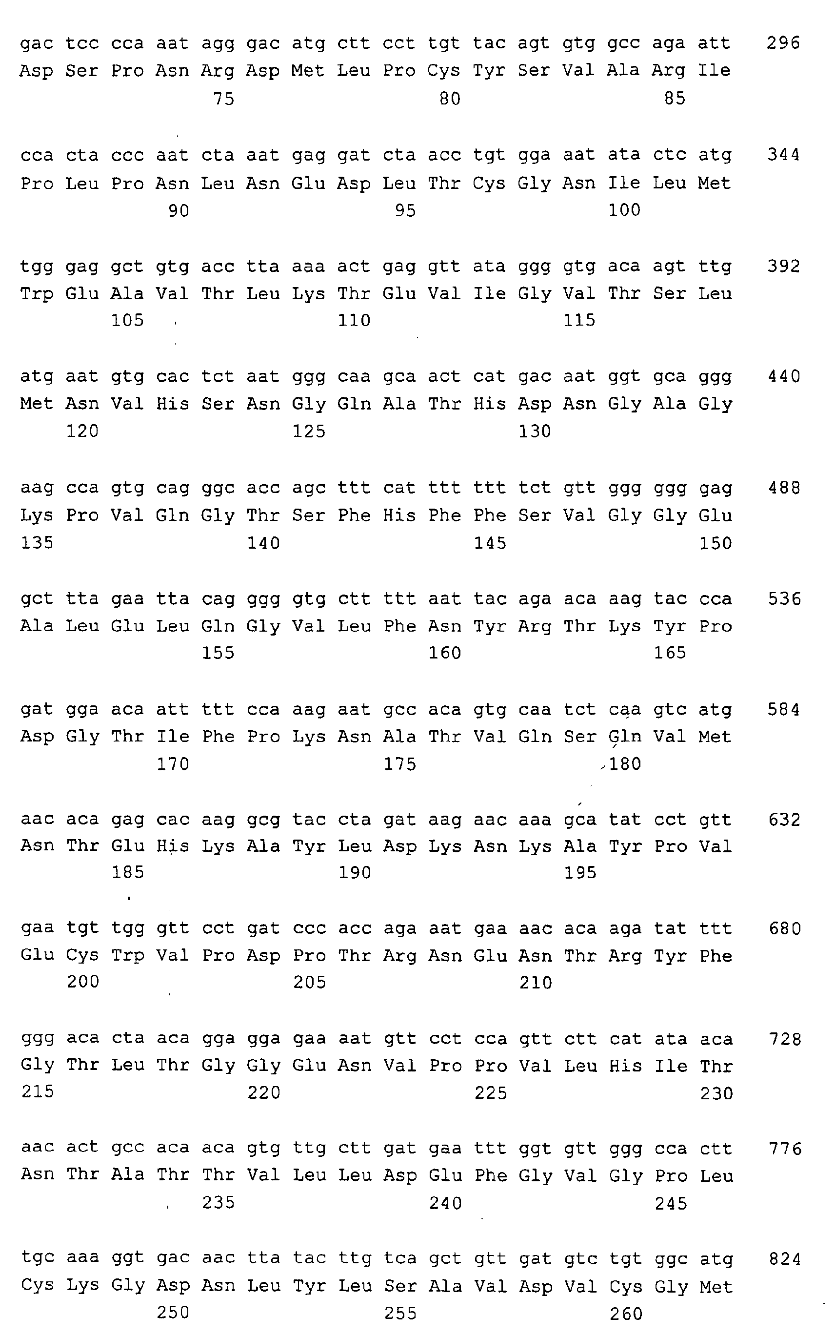
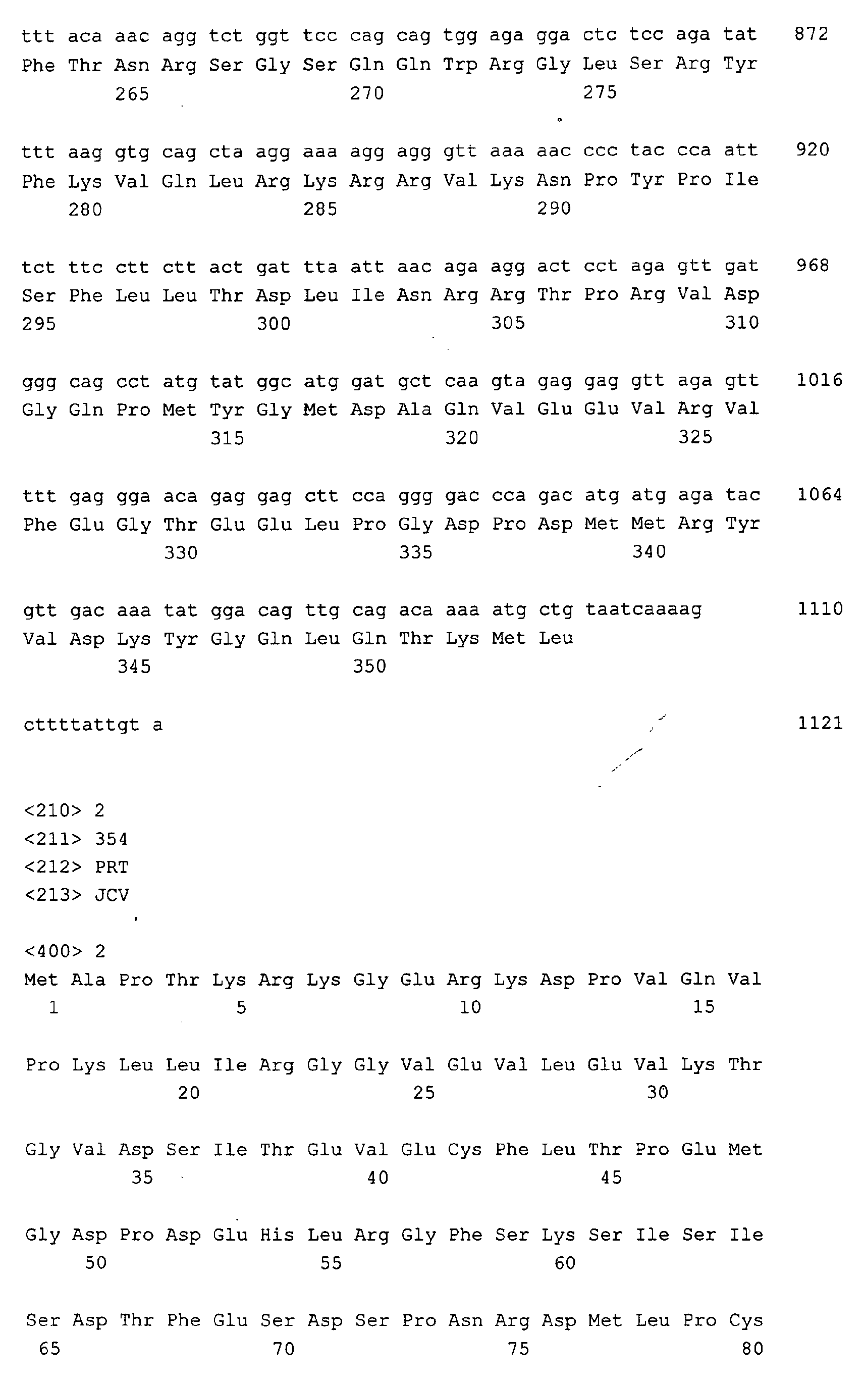
- recombinant VP1 is preferably used a nucleic acid that the in SEQ. ID NO.
- stringent Hybridization conditions are preferably defined according to Sambrook et al. (1989) Molecular Cloning A Laboratory Manual, Second Edition, Cold Spring Harbor Laboratory Press, and include a wash step of 30 min in 0.1 x SSC, 0.5% SDS at 60 ° C and preferably 68 ° C.
- the VLP according to the invention can also have one or more additional ones have heterologous proteins in the capsid structure.
- a heterologous protein is anchored in the capsid structure, preferably at least part of this protein accessible from the outside is.
- all proteins that are suitable as heterologous protein are: can be incorporated into the capsid structure and the Do not interfere with the self-assembly of the VLP.
- the cationic polymer associated with the VLP is preferably a physiologically compatible polymer.
- suitable cationic polymers are polyamines and / or polyimines, i.e. Polymers the primary, secondary or tertiary amino or / and Imino functions included in sufficient amounts to make for a positive To ensure net charge of the polymer under physiological conditions.
- the ratio of cationic groups to anionic is favorably Groups ⁇ 2: 1 particularly preferred ⁇ 5: 1.
- Most preferred the cationic polymer is essentially free of anionic groups.
- the molecular weight of the cationic polymers is preferably in the Range from 10 to 750 kD, particularly preferably in the range from 25 to 100 kD.
- cationic polymers are polymers based on essentially basic amino acids, such as polylysine, in particular poly-L-lysine, etc.
- suitable cationic polymers are polyalkyleneimines, preferably poly-C 2 -C 4 -alkyleneimines, in particular polyethyleneimine ( PEI), pAMAM (polyamidoamine) dendrimers and fractionated dendrimers as well as cationically modified polyethylene glycol.
- PEI polyethyleneimine
- pAMAM polyamidoamine dendrimers
- fractionated dendrimers as well as cationically modified polyethylene glycol.
- Polyethyleneimine is a particularly preferred cationic polymer in the sense of the present invention, since it is non-toxic and has a high density of positive charges.
- PEI is also able to bring about a pH-dependent structural change after absorption into the cells, which leads to the destabilization of endosomal and lysosomal cell compartments and thus facilitates the release of active substances, eg nucleic acids, into the cytoplasm.
- active substances eg nucleic acids
- Weight ratio of VP1-VLP to cationic polymer in the Conjugates according to the invention can be varied in a wide range. Weight ratios of 5: 1 to 1:10 have proven to be suitable proven, with weight ratios of 2: 1 and 1: 5 particularly are preferred to enable optimal binding.
- the ligand can basically be any substance provided it is direct about covalent and / or non-covalent interactions or can be bound indirectly to the cationic polymer.
- the ligand can be a target cell specific group, e.g. on Binding partner for a cell surface receptor.
- Binding partners are natural ligands or synthetic analogs thereof, high molecular weight ligands such as proteins, e.g. Transferrin, Antibodies or sugars like mannose, but also low-molecular ones synthetic ligands, e.g.
- the tripeptide motif R-G-D (Arg-Gly-Asp)
- it can also be used as a ligand a marker group, e.g. one using suitable detection methods recognizable group, such as a fluorescent label group or Biotin.
- the ligand can also be a Effector group, e.g. a cytotoxic group.
- a marker group e.g. one using suitable detection methods recognizable group, such as a fluorescent label group or Biotin.
- the ligand can also be a Effector group, e.g. a cytotoxic group.
- a cytotoxic group e.g. cytotoxic group.
- combinations of several ligands in particular Combinations of the aforementioned ligands can be used.
- the VLP can be inside the Capsid structure contain one or more active substances.
- Active substance is understood in this description of every molecule, which is not in the medium used for self-assembly is usually present.
- active substances include e.g. Macromolecules such as nucleic acids, i.e. RNA, DNA or artificial modified nucleic acids, as well as proteins and other physiologically active Substances that can be natural, synthetic or recombinant.
- physiologically active substances are e.g. lipids, Phospholipids, peptides, drugs, toxins etc.
- the invention relates to a method for Preparation of conjugates from VP1-VLP and cationic polymers, whereby several VP1 molecules assemble into a particle and a cationic polymer during or / and after assembly Association to which the particle is added.
- a cationic polymer added after assembly.
- the VLP in particular the recombinant VLP, are preferred first cleaned, then dissociated and then in the presence of the active substance annealed.
- the assembly is preferably carried out in the presence this further substance, which is then inside the capsid shell of the VLP is included.
- the VLP is preferably produced in a recombinant manner, using introduces a nucleic acid coding for a VP1 protein into a cell which transformed cell grown in a medium under conditions at which an expression of the nucleic acid takes place, and that Expression product from the cell or from the medium wins.
- the recombinant VP1 is isolated depending on the one used Host / vector system directly from the host cells or / and that Cell culture supernatant.
- the advantage of the recombinant method is there all in that simple VLP in high purity and in large Quantities can be obtained.
- As an expression system in the Practice the use of baculoviruses in conjunction with insect cells, e.g. with the insect cell line Sf 158, proven.
- VPL that is a heterologous within the capsid structure Have incorporated protein, or from VPL, inside the capsid structure contain an active substance
- the incorporation of heterologous polypeptides into the capsid envelope can, for example, by recombinant coexpression of the respective polypeptides, i.e.
- a suitable host cell e.g. a eukaryotic cell respectively.
- the incorporation of active substances into the interior of the Capsid cover can e.g. by dissociation of the capsid envelope and subsequent Reassociation in the presence of the active substance or by osmotic VLP shock in the presence of the active substance.
- the conjugates of VLP and cationic polymer according to the invention can be used for diagnostic and therapeutic purposes, e.g. for the diagnosis and treatment of diseases with a Infection associated with the JC virus, such as PML.
- the VLP can be used as a transport vehicle use, especially for the transport of active ingredients to a target cell and preferably in the target cell.
- a transport vehicle use especially for the transport of active ingredients to a target cell and preferably in the target cell.
- TNF- ⁇ antisense nucleic acids on oligodendrocytes in multiple sclerosis, since it is known that in this disease TNF- ⁇ expression in the relapse to Demyelination leads.
- VLP as a transporter system for nucleic acids in gene therapy.
- thymidine kinase gene By introducing the thymidine kinase gene from herpes viruses the cell's suicide mechanism is initiated.
- a nucleoside analogue e.g. acyclovir or gancyclovir
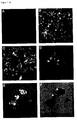
- Figure 1 VP1-specific immunofluorescence for the detection of VP1-VLP in SVG and COS-7 cells
- VP1-VLP 30 ⁇ g VP1-VLP were incubated with 5 x 10 4 cells for 24 hours.
- the VP1 was detected after fixation of the cells with a VP1-specific immune serum and subsequent incubation with a FITC-coupled anti-rabbit mAb by fluorescence microscopic methods.
- VP1-VLP transduction efficiency of VP1-VLP
- 1 ⁇ g DNA of the reporter plasmid pGL3-C was packed in 1.25 ⁇ g VP1-VLP.
- 5 x 10 4 cells each were incubated with the VLP complexes for 24 hours, then washed and cultured for a further 24 hours.
- the luciferase activity in the cell lysates was then determined luminometrically.
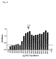
- PEI-Biotin was used to determine the binding ability of PEI to VP1-VLP used as a detectable conjugate. This was done in an ELISA Test 100 ⁇ g VP1 bound per cavity, then with different ones Concentrations of PEI-biotin were incubated and the complexes with streptavidin HRP quantified photometrically at 490 nm. The VLP / PEI ratio of 1: 3.4 was determined as the optimal binding ratio. The cut off value of the Measurement is indicated by the dashed line.
- Figure 5 VP1 specific immunofluorescence for the detection of VP1-VLP and VP1-VLP PEI / Transferrin complexes in HeLa, DU-145 and BM1604 cells
- VP1-VLP PEI / transferrin complexes 3 ⁇ g were incubated with 5x10 4 cells for 24 hours.
- the VP1 was detected after fixation of the cells with a VP1-specific immune serum and a subsequent incubation with an FITC-coupled anti-rabbit mAb by fluorescence microscopic methods.
- VP1-VLP / PEI-transferrin complexes 1 ⁇ g DNA of the reporter plasmid pGL3-C was packaged in 1.25 ⁇ g VP1-VLP. The VLP were then loaded with 1.9 ⁇ g PEI transferrin. 5x10 4 cells each were incubated with the VLP complexes for 24 hours, then washed and cultured for a further 24 hours. The luciferase activity in the cell lysates was then determined luminometrically. In addition to the VLP complexes, unloaded VLP and DNA / PEI transferrin were used as controls for the transduction of the cells.
- Example 1 Specific transduction of neuronal and renal cells Originating from JCV-VLP
- the JCV-VLP have been observed to have a specific tropism for cells of renal and neuronal origin.
- the JCV-VLP were purified from the supernatant of an insect cell line (SF158) which were infected with VP1-recombinant baculoviruses.
- SF158 insect cell line
- 50 ⁇ g of purified VP1-VLP in a total volume of 100 ⁇ l were dissociated in 10 mM Tris-HCl, pH 7.5, 10 mM EGTA, 150 mM NaCl and 5 mM DTT.
- VP1 pentamers were dialyzed against a Ca 2+ -containing buffer in the presence of the plasmids to be packaged in order to dilute the complexing agent EGTA and the reducing agent DTT and at the same time to supply Ca 2+ ions.
- the formation of the JCV-VLP could be verified by electron microscopy.
- DNasel digestion was performed to remove plasmid DNA bound to the surface of the VP1-VLP. 10 U DNasel (Pharmacia Biotech, Freiburg) were added to each reaction batch, a final concentration of 10 mM Tris-HCl, pH 7.5 and 6 mM MgCl 2 was set and incubated at 37 ° C. for one hour.
- JCV-VLP specific cells of neuronal origin such as SVG cells
- renal origin such as COS-7 cells
- 5 ⁇ 10 4 cells were incubated overnight at 37 ° C. with the DNA-containing VP1-VLPs.
- the transduction efficiency was determined using the reporter gene luciferase.
- high transduction efficiencies were measured in cells of neuronal and renal origin.
- no luciferase activity was observed in fribroblasts, T-lymphocytes, dentritic cells, chondrocytes, cells from the prostate or from breast cancer, which were also incubated with the JCV-VLP.
- biotin was first covalently identified as a well detectable group Polyethyleneimine coupled in a molar ratio of 1: 1.
- the Binding capabilities of the PEI biotin to the VP1 VLPs were determined, by binding 100 ng VP1-VLP per well of a 96 well ELISA plate was incubated with PEI biotin in various concentrations and binding with a horseradish peroxidase HPR) Streptavidin conjugate was quantified photometrically. It resulted an optimal VP1-VLP / PEI-biotin binding ratio (w / w) of 1: 3.4 ( Figure 3).
- Example 3 Cell binding test with JCV VP1-VLP PEI transferrin
- the binding, internalization and nuclear transport of the VP1-VLP / PEI-Transferrin complexes were demonstrated in cell binding tests.
- the VP1-VLP is loaded in a simple in vitro system.
- a VP1-VLP / PEI transferrin weight ratio of 1: 1.5 was set.
- Binding was carried out during a 30 minute incubation at 37 ° C.
- the cell binding tests were carried out on the cell lines HeLa (Scherer et al., J. Exp. Medicine 97 (1953), 695), DU145, BM1604 and BPH-1 (Mitchell et al., BJU International 85 (2000), 932), that express the transferrin receptor.
- the transduction efficiency of VP1-VLP PEI / Transferrin was determined by the Expression of expression of the reporter gene luciferase determined. This was done 1 ⁇ g of the plasmid pGL3-C (Promega) packed in 1.25 ⁇ g VP1-VLP. Then the VP1-VLPs with PEI transferrin, as in Example 2 described, loaded. Incubation of the VP1-VLP PEI transferrin Complexes with the HeLa, DU145, BM1604 and BPH-1 cell lines were performed as described in Example 3 for 24 hours. After that, the medium changed and the cells cultured for another 24 hours. The quantitative detection of luciferase expression was carried out luminometrically using a commercially available system (Promega).
Landscapes
- Health & Medical Sciences (AREA)
- Life Sciences & Earth Sciences (AREA)
- Genetics & Genomics (AREA)
- Engineering & Computer Science (AREA)
- Bioinformatics & Cheminformatics (AREA)
- Chemical & Material Sciences (AREA)
- Organic Chemistry (AREA)
- Biomedical Technology (AREA)
- Zoology (AREA)
- Biotechnology (AREA)
- General Engineering & Computer Science (AREA)
- Wood Science & Technology (AREA)
- General Health & Medical Sciences (AREA)
- Molecular Biology (AREA)
- Biochemistry (AREA)
- Microbiology (AREA)
- Plant Pathology (AREA)
- Biophysics (AREA)
- Physics & Mathematics (AREA)
- Proteomics, Peptides & Aminoacids (AREA)
- Animal Behavior & Ethology (AREA)
- Pharmacology & Pharmacy (AREA)
- Epidemiology (AREA)
- Medicinal Chemistry (AREA)
- Public Health (AREA)
- Veterinary Medicine (AREA)
- Micro-Organisms Or Cultivation Processes Thereof (AREA)
- Peptides Or Proteins (AREA)
- Medicines Containing Material From Animals Or Micro-Organisms (AREA)
- Medicinal Preparation (AREA)
- Medicines That Contain Protein Lipid Enzymes And Other Medicines (AREA)
- Measuring Or Testing Involving Enzymes Or Micro-Organisms (AREA)
- Seeds, Soups, And Other Foods (AREA)
Abstract
Description



Claims (27)
- Virus-ähnliches Partikel, das aus mehreren Molekülen des Virusprotein VP1 von JC-Virus aufgebaut ist, assoziiert mit einem kationischen Polymer.
- Partikel nach Anspruch 1,
dadurch gekennzeichnet, dass es aus rekombinantem VP1 aufgebaut ist. - Partikel nach Anspruch 1 oder 2,
dadurch gekennzeichnet, dass das VP1 kodiert ist von einer Nukleinsäure, welche(a) die in SEQ. ID NO. 1 gezeigte oder die dazu komplementäre Nukleotidsequenz,(b) eine der Sequenz aus (a) im Rahmen der Degeneration des genetischen Codes entsprechende Nukleotidsequenz oder/und(c) eine mit einer der Sequenzen aus (a) oder/und (b) unter stringenten Bedingungen hybridisierende Nukleotidsequenz umfasst. - Partikel nach einem der Ansprüche 1 bis 3,
dadurch gekennzeichnet, dass das kationische Polymer ein Polyamin oder Polyimin ist. - Partikel nach einem der Ansprüche 1 bis 4,
dadurch gekennzeichnet, dass das kationische Polymer ein auf Aminosäuren basierendes Polymer, insbesondere ein Polylysin ist. - Partikel nach einem der Ansprüche 1 bis 4,
dadurch gekennzeichnet, dass das kationische Polymer ein Polyalkylenimin, insbesondere ein Polyethylenimin (PEI) ist. - Partikel nach einem der Ansprüche 1 bis 6,
dadurch gekennzeichnet, dass mindestens ein Ligand an das kationische Polymer gebunden ist. - Partikel nach Anspruch 7,
dadurch gekennzeichnet, dass der Ligand eine zielzellspezifische Gruppe, insbesondere einen Bindungspartner für einen Zelloberflächenrezeptor umfasst. - Partikel nach Anspruch 7,
dadurch gekennzeichnet, dass der Ligand eine Markierungsgruppe umfasst. - Partikel nach Anspruch 7,
dadurch gekennzeichnet, dass der Ligand eine Effektorgruppe umfasst. - Partikel nach einem der Ansprüche 1 bis 10,
dadurch gekennzeichnet, dass es im Inneren der Kapsidstruktur mindestens eine Wirksubstanz enthält. - Partikel nach Anspruch 11,
dadurch gekennzeichnet, dass die Wirksubstanz ausgewählt ist aus Nukleinsäuren, Proteinen und physiologisch aktiven Stoffen. - Partikel nach Anspruch 11 oder 12,
dadurch gekennzeichnet, dass die Wirksubstanz durch einen Dissoziations/Reassoziationszyklus in die VP1-VLP verpackt worden ist. - Verfahren zur Herstellung von Partikeln nach einem der Ansprüche 1 bis 13,
dadurch gekennzeichnet, dass man(a) mehrere VP1 Moleküle zu einem Partikel assembliert und(b) vor, während oder/und nach der Assemblierung ein kationisches Polymer zur Assoziierung an das Partikel zusetzt. - Verfahren nach Anspruch 14,
dadurch gekennzeichnet, dass das kationische Polymer nach der Assemblierung zugesetzt wird. - Verfahren nach einem der Ansprüche 14 oder 15,
dadurch gekennzeichnet, dass man die Assemblierung in Gegenwart einer weiteren Substanz durchführt, wobei die Substanz im Inneren der Kapsidhülle eingeschlossen wird. - Verwendung eines Partikels nach einem der Ansprüche 1 bis 14 als diagnostisches Mittel.
- Verwendung eines Partikels nach einem der Ansprüche 1 bis 14 als therapeutisches Mittel.
- Verwendung eines Partikels nach einem der Ansprüche 1 bis 14 als Transportvehikel.
- Verwendung nach einem der Ansprüche 17 bis 19, zum Transport von Wirkstoffen zu einer Zielzelle, vorzugsweise in eine Zielzelle.
- Verwendung nach Anspruch 20,
dadurch gekennzeichnet, dass der Wirkstoff eine Nukleinsäure ist. - Verwendung nach Anspruch 20 zur spezifischen Transduktion von Zellen.
- Verwendung nach Anspruch 21 zur Gentherapie.
- Verwendung von Virus-ähnlichen Partikeln, die aus mehreren Molekülen des Virusproteins VP1 von JCV aufgebaut sind, zur spezifischen Transduktion von Zellen neuronalen oder renalen Ursprungs.
- Verwendung von Virus-ähnlichen Partikeln, die aus mehreren Molekülen des Virusproteins VP1 von JCV aufgebaut sind, zum spezifischen Transport von Wirkstoffen zu einer Zelle neuronalen oder renalen Ursprungs.
- Verwendung nach Anspruch 25,
dadurch gekennzeichnet, dass der Wirkstoff eine Nukleinsäure ist. - Verwendung nach Anspruch 26 zur Gentherapie.
Applications Claiming Priority (2)
| Application Number | Priority Date | Filing Date | Title |
|---|---|---|---|
| DE10131145 | 2001-06-28 | ||
| DE10131145A DE10131145B4 (de) | 2001-06-28 | 2001-06-28 | Zusammensetzung zum zellspezifischen Transfer von Wirkstoffen |
Publications (3)
| Publication Number | Publication Date |
|---|---|
| EP1270586A2 true EP1270586A2 (de) | 2003-01-02 |
| EP1270586A3 EP1270586A3 (de) | 2003-10-15 |
| EP1270586B1 EP1270586B1 (de) | 2006-11-29 |
Family
ID=7689740
Family Applications (1)
| Application Number | Title | Priority Date | Filing Date |
|---|---|---|---|
| EP02014449A Expired - Lifetime EP1270586B1 (de) | 2001-06-28 | 2002-06-28 | Zusammensetzung zum zellspezifischen Transfer von Wirkstoffen |
Country Status (7)
| Country | Link |
|---|---|
| US (1) | US20030044961A1 (de) |
| EP (1) | EP1270586B1 (de) |
| JP (1) | JP2003061693A (de) |
| AT (1) | ATE346859T1 (de) |
| DE (2) | DE10131145B4 (de) |
| ES (1) | ES2275781T3 (de) |
| NO (1) | NO20022898L (de) |
Cited By (6)
| Publication number | Priority date | Publication date | Assignee | Title |
|---|---|---|---|---|
| WO2004073738A3 (de) * | 2003-02-18 | 2004-10-28 | Responsif Gmbh | Zusammensetzung zur verabreichung an ein lebewesen und verfahren zur markierung von mittlen |
| EP2036980A1 (de) * | 2007-09-14 | 2009-03-18 | Gruber, Jens | Herabregulation der Genexpression mittels Nukleinsäure-beladener virusähnlicher Partikel |
| EP2554664A1 (de) * | 2011-08-02 | 2013-02-06 | Life Science Inkubator | Verfahren zur Aufreinigung von Virus-ähnlichen Partikeln (VLP) |
| EP3031820A1 (de) * | 2014-12-08 | 2016-06-15 | Life Science Inkubator | JC Polyomavirus virus-ähnliche Partikeln (VLP) mit einem Targeting-Peptid |
| US9696307B2 (en) | 2009-02-05 | 2017-07-04 | Biogen Ma Inc. | Methods for the detection of JC polyoma virus |
| WO2018141849A1 (en) | 2017-02-02 | 2018-08-09 | Deutsches Primatenzentrum Gmbh (Dpz) - Leibniz-Institut Für Primatenforschung | RETARGETING OF VIRUSES OR VLPs |
Families Citing this family (8)
| Publication number | Priority date | Publication date | Assignee | Title |
|---|---|---|---|---|
| US7468186B2 (en) * | 2005-07-22 | 2008-12-23 | City Of Hope | Polyomavirus cellular epitopes and uses therefor |
| EP2289937A1 (de) | 2009-08-24 | 2011-03-02 | Wilfried Stücker | Herstellung von monoklonalen In-vitro-Antikörpern |
| EP2548567A1 (de) * | 2011-07-22 | 2013-01-23 | Universitätsklinikum Hamburg-Eppendorf | Polyomavirus-JC-Peptide und -Proteine in Impf- und Diagnoseanwendungen |
| EP2636746A1 (de) * | 2012-03-06 | 2013-09-11 | Life Science Inkubator | Neuartiges System zur Verabreichung von Arzneimitteln auf der Grundlage von JCV-VLP |
| EP2774991B1 (de) * | 2013-03-06 | 2017-12-06 | Life Science Inkubator Betriebs GmbH & Co. KG | Arzneimittelausgabesystem zur Verwendung bei der Behandlung von neurologischen Störungen |
| EP3031821A1 (de) * | 2014-12-08 | 2016-06-15 | Life Science Inkubator | Virus-ähnliche Partikeln (VLP) aus Polyomavirus mit Fusionsprotein |
| CN105194681B (zh) * | 2015-09-17 | 2018-02-09 | 哈尔滨医科大学 | 一种基于ppv病毒样颗粒的纳米递药系统及其制备方法和应用 |
| US20180320199A1 (en) | 2015-10-28 | 2018-11-08 | Life Science Inkubator Gmbh | Use of vlp for the detection of nucleic acids |
Family Cites Families (10)
| Publication number | Priority date | Publication date | Assignee | Title |
|---|---|---|---|---|
| US5093920A (en) * | 1987-06-25 | 1992-03-03 | At&T Bell Laboratories | Programmable processing elements interconnected by a communication network including field operation unit for performing field operations |
| DE4335025A1 (de) * | 1993-10-14 | 1995-04-20 | Boehringer Ingelheim Int | Endosomolytisch wirksame Partikel |
| US6613749B1 (en) * | 1994-07-26 | 2003-09-02 | Imperial College Innovations Limited | Papovavirus pseudocapsids and use thereof for exogenous material transfer |
| DE19543553B4 (de) * | 1995-11-22 | 2009-04-09 | Deutsches Primatenzentrum Gmbh | VP-Antigene des JC-Virus |
| US6287857B1 (en) * | 1998-02-09 | 2001-09-11 | Genzyme Corporation | Nucleic acid delivery vehicles |
| JP2001222564A (ja) * | 2000-02-09 | 2001-08-17 | Hitachi Ltd | 論理エミュレーションシステム |
| JP2001249824A (ja) * | 2000-03-02 | 2001-09-14 | Hitachi Ltd | 論理エミュレーションプロセッサおよびそのモジュールユニット |
| US20030212022A1 (en) * | 2001-03-23 | 2003-11-13 | Jean-Marie Vogel | Compositions and methods for gene therapy |
| WO2002024232A2 (en) * | 2000-09-25 | 2002-03-28 | Board Of Regents, The University Of Texas System | Pei: dna vector formulations for in vitro and in vivo gene delivery |
| US7223388B2 (en) * | 2001-08-03 | 2007-05-29 | Board Of Regents, The Univeristy Of Texas System | Modified reoviral therapy |
-
2001
- 2001-06-28 DE DE10131145A patent/DE10131145B4/de not_active Expired - Fee Related
-
2002
- 2002-06-17 NO NO20022898A patent/NO20022898L/no not_active Application Discontinuation
- 2002-06-26 US US10/179,126 patent/US20030044961A1/en not_active Abandoned
- 2002-06-26 JP JP2002186686A patent/JP2003061693A/ja active Pending
- 2002-06-28 DE DE50208824T patent/DE50208824D1/de not_active Expired - Lifetime
- 2002-06-28 ES ES02014449T patent/ES2275781T3/es not_active Expired - Lifetime
- 2002-06-28 EP EP02014449A patent/EP1270586B1/de not_active Expired - Lifetime
- 2002-06-28 AT AT02014449T patent/ATE346859T1/de not_active IP Right Cessation
Cited By (12)
| Publication number | Priority date | Publication date | Assignee | Title |
|---|---|---|---|---|
| WO2004073738A3 (de) * | 2003-02-18 | 2004-10-28 | Responsif Gmbh | Zusammensetzung zur verabreichung an ein lebewesen und verfahren zur markierung von mittlen |
| EP2036980A1 (de) * | 2007-09-14 | 2009-03-18 | Gruber, Jens | Herabregulation der Genexpression mittels Nukleinsäure-beladener virusähnlicher Partikel |
| WO2009036933A3 (de) * | 2007-09-14 | 2009-08-13 | Jens Gruber | Herabregulation der genexpression mittels nukleinsäure-beladener virusähnlicher partikel |
| US8729038B2 (en) | 2007-09-14 | 2014-05-20 | Jens Gruber | Down regulation of the gene expression by means of nucleic acid-loaded virus-like particles |
| US9951329B2 (en) | 2007-09-14 | 2018-04-24 | Gabriele Jansen | Down regulation of the gene expression by means of nucleic acid-loaded virus-like particles |
| US9696307B2 (en) | 2009-02-05 | 2017-07-04 | Biogen Ma Inc. | Methods for the detection of JC polyoma virus |
| EP2554664A1 (de) * | 2011-08-02 | 2013-02-06 | Life Science Inkubator | Verfahren zur Aufreinigung von Virus-ähnlichen Partikeln (VLP) |
| WO2013017272A1 (de) * | 2011-08-02 | 2013-02-07 | Life Science Inkubator Betriebs Gmbh & Co. Kg | Verfahren zur aufreinigung von virus-ähnlichen partikeln (vlp) |
| EP3031820A1 (de) * | 2014-12-08 | 2016-06-15 | Life Science Inkubator | JC Polyomavirus virus-ähnliche Partikeln (VLP) mit einem Targeting-Peptid |
| WO2016091375A1 (en) * | 2014-12-08 | 2016-06-16 | Life Science Inkubator Gmbh | Jc polyomavirus vlp (virus-like particle) with a targeting peptide |
| WO2018141849A1 (en) | 2017-02-02 | 2018-08-09 | Deutsches Primatenzentrum Gmbh (Dpz) - Leibniz-Institut Für Primatenforschung | RETARGETING OF VIRUSES OR VLPs |
| US10975359B2 (en) | 2017-02-02 | 2021-04-13 | Deutsches Primatenzentrum Gmbh Leibniz-Institut Für Primatenforschung | Retargeting of viruses or VLPs |
Also Published As
| Publication number | Publication date |
|---|---|
| DE10131145B4 (de) | 2005-07-14 |
| EP1270586B1 (de) | 2006-11-29 |
| DE10131145A1 (de) | 2003-03-06 |
| ATE346859T1 (de) | 2006-12-15 |
| JP2003061693A (ja) | 2003-03-04 |
| US20030044961A1 (en) | 2003-03-06 |
| DE50208824D1 (de) | 2007-01-11 |
| EP1270586A3 (de) | 2003-10-15 |
| NO20022898L (no) | 2002-12-30 |
| NO20022898D0 (no) | 2002-06-17 |
| ES2275781T3 (es) | 2007-06-16 |
Similar Documents
| Publication | Publication Date | Title |
|---|---|---|
| DE69725878T2 (de) | Zusammensetzungen zur polynukleotidabgabe | |
| DE60305443T2 (de) | Pharmazeutische zusammnesetzung zur verbesserten in-vivo genübertragung | |
| DE69535540T2 (de) | Zusammensetzung enthaltend nukleinsäuren und kationische polymere, zubereitung und verwendung | |
| DE69629385T2 (de) | Peptide, die den transport über gewebe erhöhen und verfahren zu ihrer identifizierung und verwendung | |
| EP2195430B1 (de) | Herabregulation der genexpression mittels nukleinsäure-beladener virusähnlicher partikel | |
| DE69831156T2 (de) | Integrin-zielvektoren mit transfektionaktivität | |
| EP1270586B1 (de) | Zusammensetzung zum zellspezifischen Transfer von Wirkstoffen | |
| EP0571414B1 (de) | Neue, über endozytose in höhere eukaryotische zellen aufnehmbare, nukleinsäure enthaltende komplexe | |
| EP0905254B1 (de) | Verfahren zur Herstellung eines niedermolekularen Polyethylenimins | |
| EP1427837B1 (de) | Modulare transfektionssysteme auf der basis von nukleoproteinfilamenten | |
| JPH09508530A (ja) | 核酸を含む組成物、その調製および使用 | |
| CA2331384A1 (en) | Nucleic acid transfer phage | |
| EP0577648B1 (de) | Neue protein-polykation-konjugate | |
| DE10059631A1 (de) | T-Zellepitope des Papillomavirus L1-und E7-Proteins und ihre Verwendung in Diagnostik und Therapie | |
| EP1063254A1 (de) | Copolymere für den Transport von Nukleinsäuren in die Zelle | |
| DE19952957B4 (de) | Modulare Transportsysteme für molekulare Substanzen und deren Herstellung und Verwendung | |
| DE69709891T2 (de) | Chitosan enthaltende zusammensetzung | |
| DE102004032888B4 (de) | Rekombinante ScV-Partikel, ihre kodierenden Nukleinsäuren und ihre Verwendungen | |
| DE10015906B4 (de) | Gerichteter Gentransfer in Thy-1-positive Zellen | |
| WO2001032852A2 (de) | Verfahren zur gerichteten verpackung von molekularen substanzen in proteinhüllen | |
| EP1196618A2 (de) | Neue vektorkomplexe und deren verwendung für die gentherapie | |
| EP1007107A2 (de) | Verfahren zum transport molekularer substanz in vorgegebene bereiche eukaryontischer zellen | |
| DE4110410C2 (de) | Neue Protein-Polykation-Konjugate | |
| DE19610805A1 (de) | Verfahren zum Einschleusen von Makromolekülen in Eukaryontenzellen |
Legal Events
| Date | Code | Title | Description |
|---|---|---|---|
| PUAI | Public reference made under article 153(3) epc to a published international application that has entered the european phase |
Free format text: ORIGINAL CODE: 0009012 |
|
| AK | Designated contracting states |
Kind code of ref document: A2 Designated state(s): AT BE CH CY DE DK ES FI FR GB GR IE IT LI LU MC NL PT SE TR |
|
| AX | Request for extension of the european patent |
Free format text: AL;LT;LV;MK;RO;SI |
|
| PUAL | Search report despatched |
Free format text: ORIGINAL CODE: 0009013 |
|
| RIC1 | Information provided on ipc code assigned before grant |
Ipc: 7A 61K 48/00 B Ipc: 7C 07K 14/025 A Ipc: 7C 12N 15/37 B Ipc: 7C 12N 15/88 B Ipc: 7A 61K 47/48 B |
|
| AK | Designated contracting states |
Kind code of ref document: A3 Designated state(s): AT BE CH CY DE DK ES FI FR GB GR IE IT LI LU MC NL PT SE TR |
|
| AX | Request for extension of the european patent |
Extension state: AL LT LV MK RO SI |
|
| 17P | Request for examination filed |
Effective date: 20040415 |
|
| AKX | Designation fees paid |
Designated state(s): AT BE CH CY DE DK ES FI FR GB GR IE IT LI LU MC NL PT SE TR |
|
| RAP1 | Party data changed (applicant data changed or rights of an application transferred) |
Owner name: FORSCHUNGSZENTRUM FUER MEDIZINTECHNIK UND BIOTECHN |
|
| GRAP | Despatch of communication of intention to grant a patent |
Free format text: ORIGINAL CODE: EPIDOSNIGR1 |
|
| GRAS | Grant fee paid |
Free format text: ORIGINAL CODE: EPIDOSNIGR3 |
|
| GRAA | (expected) grant |
Free format text: ORIGINAL CODE: 0009210 |
|
| AK | Designated contracting states |
Kind code of ref document: B1 Designated state(s): AT BE CH CY DE DK ES FI FR GB GR IE IT LI LU MC NL PT SE TR |
|
| PG25 | Lapsed in a contracting state [announced via postgrant information from national office to epo] |
Ref country code: IT Free format text: LAPSE BECAUSE OF FAILURE TO SUBMIT A TRANSLATION OF THE DESCRIPTION OR TO PAY THE FEE WITHIN THE PRESCRIBED TIME-LIMIT;WARNING: LAPSES OF ITALIAN PATENTS WITH EFFECTIVE DATE BEFORE 2007 MAY HAVE OCCURRED AT ANY TIME BEFORE 2007. THE CORRECT EFFECTIVE DATE MAY BE DIFFERENT FROM THE ONE RECORDED. Effective date: 20061129 Ref country code: FI Free format text: LAPSE BECAUSE OF FAILURE TO SUBMIT A TRANSLATION OF THE DESCRIPTION OR TO PAY THE FEE WITHIN THE PRESCRIBED TIME-LIMIT Effective date: 20061129 Ref country code: IE Free format text: LAPSE BECAUSE OF FAILURE TO SUBMIT A TRANSLATION OF THE DESCRIPTION OR TO PAY THE FEE WITHIN THE PRESCRIBED TIME-LIMIT Effective date: 20061129 |
|
| REG | Reference to a national code |
Ref country code: GB Ref legal event code: FG4D Free format text: NOT ENGLISH |
|
| REG | Reference to a national code |
Ref country code: CH Ref legal event code: EP |
|
| REG | Reference to a national code |
Ref country code: IE Ref legal event code: FG4D Free format text: LANGUAGE OF EP DOCUMENT: GERMAN |
|
| REF | Corresponds to: |
Ref document number: 50208824 Country of ref document: DE Date of ref document: 20070111 Kind code of ref document: P |
|
| GBT | Gb: translation of ep patent filed (gb section 77(6)(a)/1977) |
Effective date: 20070102 |
|
| PG25 | Lapsed in a contracting state [announced via postgrant information from national office to epo] |
Ref country code: DK Free format text: LAPSE BECAUSE OF FAILURE TO SUBMIT A TRANSLATION OF THE DESCRIPTION OR TO PAY THE FEE WITHIN THE PRESCRIBED TIME-LIMIT Effective date: 20070228 Ref country code: SE Free format text: LAPSE BECAUSE OF FAILURE TO SUBMIT A TRANSLATION OF THE DESCRIPTION OR TO PAY THE FEE WITHIN THE PRESCRIBED TIME-LIMIT Effective date: 20070228 |
|
| REG | Reference to a national code |
Ref country code: CH Ref legal event code: NV Representative=s name: A. BRAUN, BRAUN, HERITIER, ESCHMANN AG PATENTANWAE |
|
| PG25 | Lapsed in a contracting state [announced via postgrant information from national office to epo] |
Ref country code: PT Free format text: LAPSE BECAUSE OF FAILURE TO SUBMIT A TRANSLATION OF THE DESCRIPTION OR TO PAY THE FEE WITHIN THE PRESCRIBED TIME-LIMIT Effective date: 20070430 |
|
| ET | Fr: translation filed | ||
| REG | Reference to a national code |
Ref country code: ES Ref legal event code: FG2A Ref document number: 2275781 Country of ref document: ES Kind code of ref document: T3 |
|
| REG | Reference to a national code |
Ref country code: IE Ref legal event code: FD4D |
|
| PLBE | No opposition filed within time limit |
Free format text: ORIGINAL CODE: 0009261 |
|
| STAA | Information on the status of an ep patent application or granted ep patent |
Free format text: STATUS: NO OPPOSITION FILED WITHIN TIME LIMIT |
|
| 26N | No opposition filed |
Effective date: 20070830 |
|
| BERE | Be: lapsed |
Owner name: FORSCHUNGSZENTRUM FUR MEDIZINTECHNIK UND BIOTECHN Effective date: 20070630 |
|
| PG25 | Lapsed in a contracting state [announced via postgrant information from national office to epo] |
Ref country code: MC Free format text: LAPSE BECAUSE OF NON-PAYMENT OF DUE FEES Effective date: 20070630 |
|
| PG25 | Lapsed in a contracting state [announced via postgrant information from national office to epo] |
Ref country code: BE Free format text: LAPSE BECAUSE OF NON-PAYMENT OF DUE FEES Effective date: 20070630 |
|
| PG25 | Lapsed in a contracting state [announced via postgrant information from national office to epo] |
Ref country code: GR Free format text: LAPSE BECAUSE OF FAILURE TO SUBMIT A TRANSLATION OF THE DESCRIPTION OR TO PAY THE FEE WITHIN THE PRESCRIBED TIME-LIMIT Effective date: 20070301 |
|
| REG | Reference to a national code |
Ref country code: CH Ref legal event code: PFA Owner name: FORSCHUNGSZENTRUM FUER MEDIZINTECHNIK UND BIOTECH Free format text: FORSCHUNGSZENTRUM FUER MEDIZINTECHNIK UND BIOTECHNOLOGIE E.V.#GERANIENWEG 7#99947 BAD LANGENSALZA (DE) -TRANSFER TO- FORSCHUNGSZENTRUM FUER MEDIZINTECHNIK UND BIOTECHNOLOGIE E.V.#GERANIENWEG 7#99947 BAD LANGENSALZA (DE) |
|
| PG25 | Lapsed in a contracting state [announced via postgrant information from national office to epo] |
Ref country code: LU Free format text: LAPSE BECAUSE OF NON-PAYMENT OF DUE FEES Effective date: 20070628 Ref country code: CY Free format text: LAPSE BECAUSE OF FAILURE TO SUBMIT A TRANSLATION OF THE DESCRIPTION OR TO PAY THE FEE WITHIN THE PRESCRIBED TIME-LIMIT Effective date: 20061129 |
|
| PG25 | Lapsed in a contracting state [announced via postgrant information from national office to epo] |
Ref country code: TR Free format text: LAPSE BECAUSE OF FAILURE TO SUBMIT A TRANSLATION OF THE DESCRIPTION OR TO PAY THE FEE WITHIN THE PRESCRIBED TIME-LIMIT Effective date: 20061129 |
|
| PGFP | Annual fee paid to national office [announced via postgrant information from national office to epo] |
Ref country code: ES Payment date: 20100625 Year of fee payment: 9 Ref country code: FR Payment date: 20100706 Year of fee payment: 9 |
|
| PGFP | Annual fee paid to national office [announced via postgrant information from national office to epo] |
Ref country code: AT Payment date: 20100614 Year of fee payment: 9 |
|
| PGFP | Annual fee paid to national office [announced via postgrant information from national office to epo] |
Ref country code: CH Payment date: 20100623 Year of fee payment: 9 Ref country code: NL Payment date: 20100614 Year of fee payment: 9 |
|
| PGFP | Annual fee paid to national office [announced via postgrant information from national office to epo] |
Ref country code: GB Payment date: 20100618 Year of fee payment: 9 |
|
| PGFP | Annual fee paid to national office [announced via postgrant information from national office to epo] |
Ref country code: DE Payment date: 20110729 Year of fee payment: 10 |
|
| REG | Reference to a national code |
Ref country code: NL Ref legal event code: V1 Effective date: 20120101 |
|
| REG | Reference to a national code |
Ref country code: CH Ref legal event code: PL |
|
| GBPC | Gb: european patent ceased through non-payment of renewal fee |
Effective date: 20110628 |
|
| PG25 | Lapsed in a contracting state [announced via postgrant information from national office to epo] |
Ref country code: AT Free format text: LAPSE BECAUSE OF NON-PAYMENT OF DUE FEES Effective date: 20110628 |
|
| REG | Reference to a national code |
Ref country code: AT Ref legal event code: MM01 Ref document number: 346859 Country of ref document: AT Kind code of ref document: T Effective date: 20110628 |
|
| REG | Reference to a national code |
Ref country code: FR Ref legal event code: ST Effective date: 20120229 |
|
| PG25 | Lapsed in a contracting state [announced via postgrant information from national office to epo] |
Ref country code: LI Free format text: LAPSE BECAUSE OF NON-PAYMENT OF DUE FEES Effective date: 20110630 Ref country code: FR Free format text: LAPSE BECAUSE OF NON-PAYMENT OF DUE FEES Effective date: 20110630 Ref country code: CH Free format text: LAPSE BECAUSE OF NON-PAYMENT OF DUE FEES Effective date: 20110630 |
|
| PG25 | Lapsed in a contracting state [announced via postgrant information from national office to epo] |
Ref country code: NL Free format text: LAPSE BECAUSE OF NON-PAYMENT OF DUE FEES Effective date: 20120101 |
|
| PG25 | Lapsed in a contracting state [announced via postgrant information from national office to epo] |
Ref country code: GB Free format text: LAPSE BECAUSE OF NON-PAYMENT OF DUE FEES Effective date: 20110628 |
|
| PG25 | Lapsed in a contracting state [announced via postgrant information from national office to epo] |
Ref country code: DE Free format text: LAPSE BECAUSE OF NON-PAYMENT OF DUE FEES Effective date: 20130101 |
|
| REG | Reference to a national code |
Ref country code: ES Ref legal event code: FD2A Effective date: 20130605 |
|
| PG25 | Lapsed in a contracting state [announced via postgrant information from national office to epo] |
Ref country code: ES Free format text: LAPSE BECAUSE OF NON-PAYMENT OF DUE FEES Effective date: 20110629 |
|
| REG | Reference to a national code |
Ref country code: DE Ref legal event code: R119 Ref document number: 50208824 Country of ref document: DE Effective date: 20130101 |