EP1274339B1 - Procede d'evaluation electrophysiologique objective des fonctions de la vue - Google Patents
Procede d'evaluation electrophysiologique objective des fonctions de la vue Download PDFInfo
- Publication number
- EP1274339B1 EP1274339B1 EP01969026A EP01969026A EP1274339B1 EP 1274339 B1 EP1274339 B1 EP 1274339B1 EP 01969026 A EP01969026 A EP 01969026A EP 01969026 A EP01969026 A EP 01969026A EP 1274339 B1 EP1274339 B1 EP 1274339B1
- Authority
- EP
- European Patent Office
- Prior art keywords
- vep
- recording
- multifocal
- stimulus
- virtual reality
- Prior art date
- Legal status (The legal status is an assumption and is not a legal conclusion. Google has not performed a legal analysis and makes no representation as to the accuracy of the status listed.)
- Expired - Lifetime
Links
Images



Classifications
-
- A—HUMAN NECESSITIES
- A61—MEDICAL OR VETERINARY SCIENCE; HYGIENE
- A61B—DIAGNOSIS; SURGERY; IDENTIFICATION
- A61B5/00—Measuring for diagnostic purposes; Identification of persons
- A61B5/24—Detecting, measuring or recording bioelectric or biomagnetic signals of the body or parts thereof
- A61B5/316—Modalities, i.e. specific diagnostic methods
- A61B5/398—Electrooculography [EOG], e.g. detecting nystagmus; Electroretinography [ERG]
-
- A—HUMAN NECESSITIES
- A61—MEDICAL OR VETERINARY SCIENCE; HYGIENE
- A61B—DIAGNOSIS; SURGERY; IDENTIFICATION
- A61B5/00—Measuring for diagnostic purposes; Identification of persons
- A61B5/24—Detecting, measuring or recording bioelectric or biomagnetic signals of the body or parts thereof
- A61B5/316—Modalities, i.e. specific diagnostic methods
- A61B5/369—Electroencephalography [EEG]
- A61B5/377—Electroencephalography [EEG] using evoked responses
- A61B5/378—Visual stimuli
Definitions
- Multifocal ERG recording has been performed with various electrodes (gold foil, DTL, Burian-Allen, gold lens). Good correlation was reported between the multifocal VEP and visual field loss in glaucoma (refs 2,5-7), and between the multifocal ERG and local retinal disease (ref 8), but not between the multifocal ERG and glaucoma (ref 9,10).
- the derivation of a functional map of the human visual field can be achieved from analysis of either multifocal VEP or ERG responses.
- the VEP responses tend to reflect losses at all stages of the visual pathway, whereas the ERG responses tend to correlate with local retinal disease.
- Disease states such as glaucoma or optic nerve disorders that cause blind spots in the vision (eg optic neuritis in multiple sclerosis) can the detected and mapped. Both amplitudes and latencies of the signals can be compared to normal reference values or compared between the two eyes of a subject.
- a head mounted stereo display eg virtual reality goggles
- a head mounted stereo display has good patient acceptance, and both monocular or binocular recording can be performed.
- Simultaneous binocular recording can be achieved with the application of the spread spectrum technique ( Malov, WO 0 139 659 ) and a head mounted stereo display to provide different pseudorandom stimulus patterns to the two eyes at the same time.
- the stimulus algorithm is divided into twice the number of segments and these can be distributed between the two eyes, still providing different stimulus sequences to each part of the field and with each subsequent run.
- the cross-correlations can derive VEP results from each eye independently, with minimal auto-correlation of the signals within or between eyes. This has the advantage of shortening the test time significantly. It also standardises conditions of the recording such that the two eyes are recorded under the same conditions in terms of the subject's visual attention and extraneous noise levels. This aids in the reliability of direct comparisons between eyes of an individual.
- the disclosure thus provides a method and apparatus for objectively assessing the Visual field using virtual reality goggles to present a multifocal stimulus and then recording of either retinal (ERG) or cortical (VEP) responses to that stimulus. It includes simultaneous binocular recording of the VEP, using different stimuli for the two eyes. It also includes a new scaling method to reduce inter-subject variability in the recorded multifocal VEP amplitudes by scaling the VEP response according to overall electroencephalogram activity.
- ERP retinal
- VEP cortical
- a suitable head mounted stereo display is what is commonly known as virtual reality goggles. Other head mounted displays which are able to present a suitable stimulus which can generate a retinal or cortical response would also be appropriate.
- Virtual reality is a term applied to the experience of an individual when viewing through a head-mounted display an image presented immediately before the eyes which has the appearance of being viewed at a distance from the eye. Different images can be presented to the two eyes to give a three dimensional effect.
- a method according to the invention is defined in claim 1.
- a preferred embodiment is defined in the depending claim.
- the head-mounted stereo display suitable comprises virtual reality goggles.
- the head-mounted stereo display may be used to derive a signal from the cortical visual evoked potentials. It may also be used to derive an electroretinogram signal from the eye.
- This display shows any type of multifocal stimulation directly to the eye.
- the stimulus presented to the eye may be a flash stimulus or a pattern stimulus.
- the stimulus may vary in luminance, colour or stimulus duration to elicit visual responses.
- the head-mounted stereo display suitably uses a liquid crystal display or plasma screen, for example.
- the stimulus may be presented monocularly or binocularly. The same stimulus may be presented binocularly for simultaneously recording of signals from both eyes.
- the two eyes may be simultaneously presented binocularly for simultaneous recording to signals from the two eyes.
- the results are scaled according to the overall spontaneous brain activity (i.e. electroencephalogram levels) of the subject during the recording to minimise variability.
- the disclosure utilises multifocal stimulation techniques.
- Any multifocal stimulator can be used to generate a stimulus which is then projected into virtual reality goggles using monocular or binocular displays.
- the stimulus can be diffuse (flash) or structured (pattern) and can vary in intensity, colour, size or temporal characteristics.
- Cross-correlation techniques allow for derivation of the signal from background noise.
- a topographical map of the responses can then be derived corresponding to the field of view of the subject.
- the output can displayed as a printout of results, and comparisons made with a normal data base of responses.
- the inventors have applied a scaling factor based on background electroencephalogram (EEG) levels.
- EEG background electroencephalogram
- Scaling of the VEP amplitude based on amplitude of spontaneous brain activity eliminates part of the variability between individuals caused by differences in conductivity of underlying tissues (eg bone, muscle, skin and subcutaneous fat). This also reduces the differences seen between males and females, since it is known that women have generally higher amplitude VEP signals when compared to men, presumably due to sex differences in tissue thickness and conductivity. Scaling according to EEG signals removes this difference, rendering final signals equivalent between the sexes. By reducing the range of variability between subjects it improves the sensitivity of the test for detecting abnormality.
- Figure 1 shows a schematic of the apparatus for VEP recording using virtual reality goggles (1), which present the display to the subject.
- the goggles are connected to a computer (2) with a linked video board that generates the multifocal stimulus.
- the signals are conducted to an amplifier (3), before being processed by software for presentation on the operators display (4). Results can be compared for each eye, or between the two eyes of a subject, with respect to normal reference values.
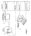
- FIG 2 shows a schematic of the apparatus for multifocal ERG recording using virtual reality goggles (1).
- the set up is the same as in Figure 1 except that the recording electrode is placed in contact with the eye or eyelid.
- a ground electrode is required (shown on the earlobe). Only one channel recording is required for the ERG.
- Figure 3 is an example of a multifocal multichannel VEP recording from the right and left eye of a normal subject.
- Figure 3A shows the responses achieved using a conventional screen (22 inch Hitachi monitor) to present the stimulus.
- a cortically scaled dartboard stimulus was generated with 60 different areas of pattern stimulation using the ObjectiVision perimeter.
- the trace array shown in the figure represents the responses generated from each part of the visual field tested out to 27 degrees of eccentricity temporally and 34 degrees nasally. For graphics purposes the central areas are relatively enlarged to show the raw VEP signal within that area.
- Figure 3B shows a multifocal multichannel VEP recordings from the same normal subject as in Figure 3A , recorded using virtual reality goggles to present the same stimulus instead of the conventional monitor.
- the same ObjectiVision system was used.
- the responses are of similar order of magnitude in the two techniques, although there is some variation in amplitude across the field. Due to the specifications of the goggles used, the display was limited to 21 degrees temporally and 27 degrees nasal
- Figure 4 provides a comparison between subjective perimetry findings and the objective VEP assessment of the visual field using virtual reality goggles.
- Figure 4A shows the grayscale and pattern deviation printout from a subjective Humphrey visual field test of the right eye of a glaucoma patient. An inferior arcuate scotoma (blind spot) is shown in the visual field.
- Figure 4B shows the multifocal multichannel VEP recording from the same eye as in Figure 4A , recorded using virtual reality goggles. Analysis of the signals demonstrates loss of VEP responses corresponding to the inferior scotoma in Figure 4A , with more extensive reductions in the superior field than seen on the Humphrey.
- the amplitude deviation plot shades areas according to probability of abnormality when compared to a reference range of normal values extrapolated from the conventional screen ObjectiVision system. This suggests that the technique is capable of detecting visual field loss in glaucoma, just as it is with the use of the conventional large screen. It may also demonstrate more significant glaucomatous damage than suspected on conventional Humphrey field testing. Five glaucoma patients have been tested with the virtual reality goggles and the scotomas were detected in all five cases.
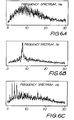
- the level of spontaneous EEG activity is calculated during the recording, it provides an indirect measure of the overall registration of brain signals for that individual for the electrode positions used. Whilst it is recognised that EEG amplitude is determined by many additional factors other than conductivity, it is proposed that scaling of an individual's VEP responses according to their EEG levels, relative to normal population EEG values, helps to reduce inter-individual VEP variability.
- the EEG amplitude is approximately 1000x the amplitude of the VEP, so it is reasonable to assume that the VEP signals themselves will have little contribution to the raw EEG levels.
- the EEG raw data is actually examined by cross-correlation techniques to extract the VEP signals.
- the overall level of the raw EEG (99% confidence interval) as recorded during each run of the VEP recording can be used to provide an individual's scaling factor.
- the VEP extracted is then scaled by the EEG scaling factor.
- the value of the technique of the invention of VEP scaling was confirmed by examining the data from 50 normals.
- the coefficient of variation for all 60 visual field test points had a mean value of 50.1 %.
- the coefficient of variation for all 60 visual field test points was reduced to 28.2%.
Landscapes
- Health & Medical Sciences (AREA)
- Life Sciences & Earth Sciences (AREA)
- Medical Informatics (AREA)
- Molecular Biology (AREA)
- Veterinary Medicine (AREA)
- Biophysics (AREA)
- Pathology (AREA)
- Engineering & Computer Science (AREA)
- Biomedical Technology (AREA)
- Heart & Thoracic Surgery (AREA)
- Public Health (AREA)
- Physics & Mathematics (AREA)
- Surgery (AREA)
- Animal Behavior & Ethology (AREA)
- General Health & Medical Sciences (AREA)
- Psychiatry (AREA)
- Psychology (AREA)
- Ophthalmology & Optometry (AREA)
- Measurement And Recording Of Electrical Phenomena And Electrical Characteristics Of The Living Body (AREA)
- Investigating Or Analysing Materials By Optical Means (AREA)
- Eye Examination Apparatus (AREA)
Claims (2)
- Procédé pour analyser au moins un enregistrement de potentiel visuel multifocal suscité à partir d'un mode quelconque de stimulation multifocale comprenant le fait d'utiliser un ordinateur pour calibrer une sortie par logiciel informatique selon un facteur de calibrage de l'électroencéphalogramme calculé à partir de l'ensemble des niveaux d'électroencéphalogrammes spontanés d'un sujet pendant l'enregistrement afin de minimiser une variabilité entre sujets, dans lequel des pics de rythme alpha ou des signaux de l'électrocardiogramme dans les données brutes sont identifiés par analyse des spectres de puissance de Fourier et éliminés avant de calculer le facteur de calibrage.
- Procédé selon la revendication 1 comprenant, de plus, une stimulation multifocale utilisant des moniteurs CRT ou LCD, des écrans plasma ou des lunettes de protection contre la réalité immédiate.
Applications Claiming Priority (5)
| Application Number | Priority Date | Filing Date | Title |
|---|---|---|---|
| AUPQ6940A AUPQ694000A0 (en) | 2000-04-17 | 2000-04-17 | Method and apparatus for objective electrophysiological assessment of visual function |
| AUPQ694000 | 2000-04-17 | ||
| AUPR1982A AUPR198200A0 (en) | 2000-12-08 | 2000-12-08 | Goggles |
| AUPR198200 | 2000-12-08 | ||
| PCT/AU2001/000423 WO2001078586A1 (fr) | 2000-04-17 | 2001-04-12 | Procede et appareil d'evaluation electrophysiologique objective des fonctions de la vue |
Publications (3)
| Publication Number | Publication Date |
|---|---|
| EP1274339A1 EP1274339A1 (fr) | 2003-01-15 |
| EP1274339A4 EP1274339A4 (fr) | 2004-05-19 |
| EP1274339B1 true EP1274339B1 (fr) | 2011-06-08 |
Family
ID=25646302
Family Applications (1)
| Application Number | Title | Priority Date | Filing Date |
|---|---|---|---|
| EP01969026A Expired - Lifetime EP1274339B1 (fr) | 2000-04-17 | 2001-04-12 | Procede d'evaluation electrophysiologique objective des fonctions de la vue |
Country Status (6)
| Country | Link |
|---|---|
| US (3) | US7740592B2 (fr) |
| EP (1) | EP1274339B1 (fr) |
| JP (1) | JP4817582B2 (fr) |
| AT (1) | ATE511784T1 (fr) |
| NZ (1) | NZ521952A (fr) |
| WO (1) | WO2001078586A1 (fr) |
Cited By (2)
| Publication number | Priority date | Publication date | Assignee | Title |
|---|---|---|---|---|
| EP3568739A4 (fr) * | 2017-03-15 | 2020-03-11 | Samsung Electronics Co., Ltd. | Systèmes et procédés permettant de déterminer des défauts dans le champ visuel d'un utilisateur |
| US10667683B2 (en) | 2018-09-21 | 2020-06-02 | MacuLogix, Inc. | Methods, apparatus, and systems for ophthalmic testing and measurement |
Families Citing this family (74)
| Publication number | Priority date | Publication date | Assignee | Title |
|---|---|---|---|---|
| GB0113533D0 (en) | 2001-06-05 | 2001-07-25 | Brigantia Software Ltd | Apparatus and method for testing visual response |
| GB2382252A (en) * | 2001-10-13 | 2003-05-21 | Alki Liasis | Portable visual stimulators |
| US7549749B2 (en) | 2002-05-06 | 2009-06-23 | Antti Valjakka | Visual stimulator |
| FI112592B (fi) * | 2002-05-06 | 2003-12-31 | Urtti Arto Olavi | Näköstimulaatiolaite |
| US6966650B2 (en) * | 2003-06-27 | 2005-11-22 | Zongqi Hu | Method and apparatus for an automated procedure to detect and monitor early-stage glaucoma |
| US8583223B2 (en) * | 2003-11-28 | 2013-11-12 | The Australian National University | Assessment of neural function |
| US7922670B2 (en) * | 2005-02-24 | 2011-04-12 | Warren Jones | System and method for quantifying and mapping visual salience |
| US7384145B2 (en) * | 2006-02-16 | 2008-06-10 | The Board Of Trustees Of The University Of Illinois | Mapping retinal function using corneal electrode array |
| US8118752B2 (en) * | 2006-02-16 | 2012-02-21 | The Board Of Trustees Of The University Of Illinois | Apparatus and methods for mapping retinal function |
| WO2007144711A2 (fr) * | 2006-06-12 | 2007-12-21 | National University Of Ireland Maynooth | Procédé et appareil destinés à susciter une réponse chez un sujet |
| WO2008091399A2 (fr) * | 2006-09-01 | 2008-07-31 | Board Of Regents Of The University Of Texas | Procédé et dispositif pour mesurer la vitesse de traitement d'une information par le cerveau |
| WO2008098310A1 (fr) | 2007-02-16 | 2008-08-21 | Objectivision Limited | Procédé de stimulus pour un potentiel évoqué visuel multifocal |
| US20090040296A1 (en) * | 2007-08-06 | 2009-02-12 | Moscato Jonathan D | Head mounted display assembly |
| US8083354B2 (en) * | 2007-10-03 | 2011-12-27 | Diopsys, Inc. | Simultaneously multi-temporal visual test and method and apparatus therefor |
| JP5832747B2 (ja) * | 2007-11-09 | 2015-12-16 | ジ・オーストラリアン・ナショナル・ユニバーシティー | 感覚野の機能を評価するための装置の作動方法および装置 |
| JP5076065B2 (ja) * | 2008-06-13 | 2012-11-21 | 国立大学法人福井大学 | 視神経活動測定支援装置 |
| CN102264277B (zh) | 2008-07-09 | 2015-04-29 | 劳伦斯·M·麦金利 | 视觉功能监控处理和装置 |
| DE102008056976B4 (de) * | 2008-11-07 | 2010-09-02 | Technische Universität Ilmenau | Verfahren und Vorrichtung zur multifokalen, farbkanalselektiven Stimulation des visuellen Systems |
| WO2010105370A1 (fr) * | 2009-03-20 | 2010-09-23 | Jocelyn Faubert | Dispositif et procédé pour mesurer une défaillance perceptuelle bénigne |
| US20120172419A1 (en) | 2009-09-15 | 2012-07-05 | Medical College Of Wisconsin Research Foundation Inc. | Reagents and methods for modulating cone photoreceptor activity |
| US20110065073A1 (en) * | 2009-09-16 | 2011-03-17 | Duffy Charles J | Method and system for quantitative assessment of word detection latency |
| US20110066068A1 (en) * | 2009-09-16 | 2011-03-17 | Duffy Charles J | Method and system for quantitative assessment of functional impairment |
| US20110065072A1 (en) * | 2009-09-16 | 2011-03-17 | Duffy Charles J | Method and system for quantitative assessment of word recognition sensitivity |
| US8562541B2 (en) * | 2009-09-16 | 2013-10-22 | Cerebral Assessment Systems, Inc. | Method and system for quantitative assessment of visual motion discrimination |
| US8777630B2 (en) | 2009-09-16 | 2014-07-15 | Cerebral Assessment Systems, Inc. | Method and system for quantitative assessment of facial emotion sensitivity |
| US20110066082A1 (en) * | 2009-09-16 | 2011-03-17 | Duffy Charles J | Method and system for quantitative assessment of visual motor response |
| US8475391B2 (en) * | 2009-09-16 | 2013-07-02 | Cerebral Assessment Systems | Method and system for quantitative assessment of spatial distractor tasks |
| US20110065070A1 (en) * | 2009-09-16 | 2011-03-17 | Duffy Charles J | Method and system for quantitative assessment of letter identification latency |
| US20110065071A1 (en) * | 2009-09-16 | 2011-03-17 | Duffy Charles J | Method and system for quantitative assessment of word identification latency |
| US20110066069A1 (en) * | 2009-09-16 | 2011-03-17 | Duffy Charles J | Method and system for quantitative assessment of visual form discrimination |
| US20110065078A1 (en) * | 2009-09-16 | 2011-03-17 | Duffy Charles J | Method and system for quantitative assessment of social interactions nulling testing |
| US20110065069A1 (en) * | 2009-09-16 | 2011-03-17 | Duffy Charles J | Method and system for quantitative assessment of verbal recognition memory |
| JP5308305B2 (ja) * | 2009-10-20 | 2013-10-09 | 株式会社トーメーコーポレーション | 網膜電位測定装置 |
| US20120029302A1 (en) * | 2010-07-29 | 2012-02-02 | Shih-Chung Hsu | Apparatus and method for dynamic detection of electrophysiological signals for stillness feedback |
| RU2441571C1 (ru) * | 2010-10-07 | 2012-02-10 | Государственное образовательное учреждение высшего профессионального образования Марийский государственный технический университет | Способ определения лабильности зрительной системы человека |
| AU2011334593B2 (en) * | 2010-11-23 | 2014-09-18 | ResMed Pty Ltd | Method and apparatus for detecting cardiac signals |
| JP6340534B2 (ja) * | 2011-06-01 | 2018-06-13 | 東海光学株式会社 | 大脳視覚野等の誘発活動による眼鏡レンズの評価方法 |
| TWI698240B (zh) | 2012-05-15 | 2020-07-11 | 澳大利亞商艾佛蘭屈澳洲私營有限公司 | 使用腺相關病毒(aav)sflt-1治療老年性黃斑部退化(amd) |
| BR112015013489A2 (pt) | 2012-12-11 | 2017-07-11 | Klin Ami | sistemas e métodos para a detecção de blink inibição como um marcador de engajamento e percebido saliência estímulo |
| US9072478B1 (en) * | 2013-06-10 | 2015-07-07 | AutismSees LLC | System and method for improving presentation skills |
| AU2014334870B2 (en) | 2013-10-17 | 2019-06-13 | Children's Healthcare Of Atlanta, Inc. | Methods for assessing infant and child development via eye tracking |
| KR101541619B1 (ko) * | 2013-12-31 | 2015-08-03 | 한양대학교 산학협력단 | 일시적 시각 유발 전위를 이용한 신호 생성 방법 및 장치 |
| EP3800191B1 (fr) | 2014-03-17 | 2025-08-20 | Adverum Biotechnologies, Inc. | Compositions et procédés pour améliorer l'expression des gènes dans les cônes rétiniens |
| CA2946301A1 (fr) * | 2014-04-17 | 2015-10-22 | The Regents Of The University Of California | Plate-forme de detection de l'activite du cerveau portable pour l'evaluation de deficits de champ visuel |
| DE102014113682A1 (de) | 2014-09-22 | 2016-03-24 | Carl Zeiss Meditec Ag | Vorrichtung zur Gesichtsfeldmessung |
| US20170150907A1 (en) * | 2015-02-04 | 2017-06-01 | Cerebral Assessment Systems, LLC | Method and system for quantitative assessment of visual motor response |
| US11021519B2 (en) | 2015-03-02 | 2021-06-01 | Adverum Biotechnologies, Inc. | Compositions and methods for intravitreal delivery of polynucleotides to retinal cones |
| NZ773822A (en) | 2015-03-16 | 2022-07-29 | Magic Leap Inc | Methods and systems for diagnosing and treating health ailments |
| WO2016182974A1 (fr) * | 2015-05-08 | 2016-11-17 | Ngoggle | Dispositif d'eeg à affichage monté sur la tête |
| US11357442B2 (en) | 2015-05-12 | 2022-06-14 | Diagnosys LLC | Combined stimulator and electrode assembly for mouse electroretinography (ERG) |
| EP3305206A4 (fr) * | 2015-06-05 | 2018-06-20 | Shikuukankoubou Co., Ltd. | Programme et système de détection précoce et de prévention de la démence légère |
| US10893823B2 (en) * | 2015-11-10 | 2021-01-19 | Diagnosys LLC | Method and apparatus for the assessment of electrophysiological signals |
| GB2545763A (en) | 2015-12-23 | 2017-06-28 | Adverum Biotechnologies Inc | Mutant viral capsid libraries and related systems and methods |
| EP3440497B1 (fr) | 2016-04-08 | 2023-08-16 | Magic Leap, Inc. | Systèmes et procédés de réalité augmentée comprenant des éléments de lentille à focale variable |
| US10888222B2 (en) | 2016-04-22 | 2021-01-12 | Carl Zeiss Meditec, Inc. | System and method for visual field testing |
| US20180110409A1 (en) * | 2016-10-20 | 2018-04-26 | Stylianos Georgios Tsapakis | Visual field test method/perimeter using virtual reality glasses/headset and a smartphone or tablet or other portable device |
| IL307602A (en) | 2017-02-23 | 2023-12-01 | Magic Leap Inc | Variable focus virtual imagers based on polarization conversion |
| CN107898459B (zh) * | 2017-09-12 | 2025-06-06 | 深圳麦格米特电气股份有限公司 | 一种便携式视觉电生理检测系统 |
| WO2019060298A1 (fr) | 2017-09-19 | 2019-03-28 | Neuroenhancement Lab, LLC | Procédé et appareil de neuro-activation |
| US11717686B2 (en) | 2017-12-04 | 2023-08-08 | Neuroenhancement Lab, LLC | Method and apparatus for neuroenhancement to facilitate learning and performance |
| US10413172B2 (en) | 2017-12-11 | 2019-09-17 | 1-800 Contacts, Inc. | Digital visual acuity eye examination for remote physician assessment |
| US11478603B2 (en) | 2017-12-31 | 2022-10-25 | Neuroenhancement Lab, LLC | Method and apparatus for neuroenhancement to enhance emotional response |
| US12280219B2 (en) | 2017-12-31 | 2025-04-22 | NeuroLight, Inc. | Method and apparatus for neuroenhancement to enhance emotional response |
| US11364361B2 (en) | 2018-04-20 | 2022-06-21 | Neuroenhancement Lab, LLC | System and method for inducing sleep by transplanting mental states |
| CN109222970A (zh) * | 2018-07-09 | 2019-01-18 | 司法鉴定科学研究院 | 视力客观评定的设备及视觉诱发电位的检测系统与方法 |
| US11497911B2 (en) | 2018-07-18 | 2022-11-15 | Diagnosys LLC | Electrically evoked response (EER) stimulator/amplifier combination |
| US11452839B2 (en) | 2018-09-14 | 2022-09-27 | Neuroenhancement Lab, LLC | System and method of improving sleep |
| US11786694B2 (en) | 2019-05-24 | 2023-10-17 | NeuroLight, Inc. | Device, method, and app for facilitating sleep |
| US12290315B2 (en) * | 2019-09-16 | 2025-05-06 | Eyeonic Pty Limited | Method and/or system for testing visual function |
| US12343157B2 (en) * | 2020-04-14 | 2025-07-01 | Neurotype Inc. | Assessing motivated attention with cue reactivity |
| WO2022011006A1 (fr) * | 2020-07-08 | 2022-01-13 | Veyezer, Llc | Système de réfraction en espace réel holographique |
| US20240000308A1 (en) * | 2020-11-20 | 2024-01-04 | Diagnosys LLC | Methods and apparatus for performing enhanced full-field stimulus threshold (fst) tests |
| IT202300017343A1 (it) | 2023-08-17 | 2025-02-17 | Opto Plus S R L | Sistema di personalizzazione e di analisi della visione per la produzione di lenti multifocali |
| CN121176840A (zh) * | 2025-09-28 | 2025-12-23 | 中山大学中山眼科中心 | 一种大数据视觉功能智能解析方法及系统 |
Family Cites Families (31)
| Publication number | Priority date | Publication date | Assignee | Title |
|---|---|---|---|---|
| US4255023A (en) * | 1979-02-12 | 1981-03-10 | House Harold D | Objective retinal response recorder |
| US4474186A (en) * | 1979-07-17 | 1984-10-02 | Georgetown University | Computerized electro-oculographic (CEOG) system with feedback control of stimuli |
| US4320768A (en) * | 1979-07-17 | 1982-03-23 | Georgetown University Medical Center | Computerized electro-oculographic (CEOG) system |
| US4493539A (en) * | 1982-06-30 | 1985-01-15 | The United States Of America As Represented By The Secretary Of The Air Force | Method and apparatus for objective determination of visual contrast sensitivity functions |
| US4697598A (en) * | 1985-04-25 | 1987-10-06 | Westinghouse Electric Corp. | Evoked potential autorefractometry system |
| US4832480A (en) * | 1986-06-24 | 1989-05-23 | Quintron, Inc. | Differential diagnosis of sensory abnormalities using a normalized, ratiometric analysis of steady state evoked potentials |
| US4861154A (en) * | 1986-08-06 | 1989-08-29 | Westinghouse Electric Corp. | Automated visual assessment system with steady state visual evoked potential stimulator and product detector |
| US4846567A (en) | 1986-08-06 | 1989-07-11 | Sutter Erich E | Retinal area response mapping using simultaneous multi-area stimulation with binary sequences and objective response analysis |
| US4736751A (en) * | 1986-12-16 | 1988-04-12 | Eeg Systems Laboratory | Brain wave source network location scanning method and system |
| EP0301322A1 (fr) * | 1987-07-27 | 1989-02-01 | Siemens Aktiengesellschaft | Appareil pour la stimulation visuelle |
| US5458117A (en) * | 1991-10-25 | 1995-10-17 | Aspect Medical Systems, Inc. | Cerebral biopotential analysis system and method |
| US5539482A (en) | 1992-02-28 | 1996-07-23 | The Australian National University | Glaucoma testing using non-linear systems identification techniques |
| DE4301483A1 (de) * | 1993-01-21 | 1994-08-11 | Otto Sembritzki | Gerät zur Erzeugung von Lichtreizen und zum Messen des Elektroretinogramms sowie des visuell evozierten kortikalen Potentials |
| JP2819401B2 (ja) * | 1994-08-23 | 1998-10-30 | 里子 佐藤 | 脳波および心電図の記録装置 |
| US5609158A (en) * | 1995-05-01 | 1997-03-11 | Arrhythmia Research Technology, Inc. | Apparatus and method for predicting cardiac arrhythmia by detection of micropotentials and analysis of all ECG segments and intervals |
| JPH09114543A (ja) * | 1995-10-02 | 1997-05-02 | Xybernaut Corp | ハンドフリーコンピュータ装置 |
| US5946075A (en) * | 1996-05-21 | 1999-08-31 | Horn; Gerald | Vision screening system |
| KR100349709B1 (ko) | 1997-12-23 | 2002-11-23 | 삼성전자 주식회사 | 리니어압축기의고정자 |
| US6086206A (en) * | 1998-01-05 | 2000-07-11 | Sutter; Erich E. | Analysis method for enhancing and extracting second order nonlinear response components of the multi-area electroretinogram |
| AUPP266198A0 (en) * | 1998-03-30 | 1998-04-23 | Australian National University, The | Simultaneous binocular assessment of multiple optic nerve and cortical regions in diseases affecting nerve conduction |
| DE69937234T2 (de) * | 1998-05-08 | 2008-07-03 | The University Of Sydney, Sydney | Elektrophysiologische messung des gesichtfeldes |
| US6364845B1 (en) * | 1998-09-17 | 2002-04-02 | University Of Rochester | Methods for diagnosing visuospatial disorientation or assessing visuospatial orientation capacity |
| US6044292A (en) * | 1998-09-21 | 2000-03-28 | Heyrend; F. Lamarr | Apparatus and method for predicting probability of explosive behavior in people |
| JP2003515375A (ja) | 1999-12-03 | 2003-05-07 | イオウリ マロヴ, | 拡散スペクトル技術による現場試験 |
| IL138926A0 (en) * | 2000-10-06 | 2001-11-25 | Notal Vision Ltd | Method and system for detecting eye disease |
| US7682021B2 (en) * | 2002-02-08 | 2010-03-23 | Novavision, Inc. | System and methods for the treatment of retinal diseases |
| WO2003070089A1 (fr) * | 2002-02-19 | 2003-08-28 | Notal Vision Ltd. | Procede et systeme d'evaluation d'une maladie de l'oeil |
| CA2503658A1 (fr) * | 2002-11-05 | 2004-05-21 | University Of Rochester Medical Center | Procede d'evaluation du sens de l'orientation |
| WO2004098447A2 (fr) * | 2003-05-05 | 2004-11-18 | Notal Vision Ltd. | Mappage de l'oeil |
| US6966650B2 (en) * | 2003-06-27 | 2005-11-22 | Zongqi Hu | Method and apparatus for an automated procedure to detect and monitor early-stage glaucoma |
| US7384145B2 (en) * | 2006-02-16 | 2008-06-10 | The Board Of Trustees Of The University Of Illinois | Mapping retinal function using corneal electrode array |
-
2001
- 2001-04-12 EP EP01969026A patent/EP1274339B1/fr not_active Expired - Lifetime
- 2001-04-12 JP JP2001575892A patent/JP4817582B2/ja not_active Expired - Lifetime
- 2001-04-12 AT AT01969026T patent/ATE511784T1/de not_active IP Right Cessation
- 2001-04-12 US US10/257,565 patent/US7740592B2/en not_active Expired - Fee Related
- 2001-04-12 NZ NZ521952A patent/NZ521952A/en not_active IP Right Cessation
- 2001-04-12 WO PCT/AU2001/000423 patent/WO2001078586A1/fr not_active Ceased
-
2008
- 2008-10-17 US US12/253,331 patent/US7972278B2/en not_active Expired - Fee Related
-
2011
- 2011-05-17 US US13/109,047 patent/US20110218456A1/en not_active Abandoned
Non-Patent Citations (1)
| Title |
|---|
| SHIMOYAMA ICHIRO ET AL: "Normalized wave energy for background activity in visual evoked potentials", JOURNAL OF BRAIN SCIENCE, JAPAN BRAIN SCIENCE SOCIETY, OKAYAMA, JP, vol. 24, no. 1, 1 January 1998 (1998-01-01), pages 13 - 18, XP008082701, ISSN: 1341-5301 * |
Cited By (3)
| Publication number | Priority date | Publication date | Assignee | Title |
|---|---|---|---|---|
| EP3568739A4 (fr) * | 2017-03-15 | 2020-03-11 | Samsung Electronics Co., Ltd. | Systèmes et procédés permettant de déterminer des défauts dans le champ visuel d'un utilisateur |
| US10667683B2 (en) | 2018-09-21 | 2020-06-02 | MacuLogix, Inc. | Methods, apparatus, and systems for ophthalmic testing and measurement |
| US11089954B2 (en) | 2018-09-21 | 2021-08-17 | MacuLogix, Inc. | Method and apparatus for guiding a test subject through an ophthalmic test |
Also Published As
| Publication number | Publication date |
|---|---|
| ATE511784T1 (de) | 2011-06-15 |
| US20030158497A1 (en) | 2003-08-21 |
| EP1274339A4 (fr) | 2004-05-19 |
| NZ521952A (en) | 2004-03-26 |
| US7740592B2 (en) | 2010-06-22 |
| JP2003532460A (ja) | 2003-11-05 |
| EP1274339A1 (fr) | 2003-01-15 |
| WO2001078586A1 (fr) | 2001-10-25 |
| US20110218456A1 (en) | 2011-09-08 |
| US20090076406A1 (en) | 2009-03-19 |
| US7972278B2 (en) | 2011-07-05 |
| JP4817582B2 (ja) | 2011-11-16 |
Similar Documents
| Publication | Publication Date | Title |
|---|---|---|
| EP1274339B1 (fr) | Procede d'evaluation electrophysiologique objective des fonctions de la vue | |
| Klistorner et al. | Electroencephalogram-based scaling of multifocal visual evoked potentials: effect on intersubject amplitude variability | |
| Graham et al. | Objective VEP perimetry in glaucoma: asymmetry analysis to identify early deficits | |
| Goldberg et al. | Multifocal objective perimetry in the detection of glaucomatous field loss | |
| Thut et al. | Effects of single-pulse transcranial magnetic stimulation (TMS) on functional brain activity: a combined event-related TMS and evoked potential study | |
| Klistorner et al. | Objective perimetry in glaucoma | |
| US20030149350A1 (en) | Glaucoma screening system and method | |
| US20200060573A1 (en) | Head mountable device | |
| NZ519897A (en) | Electroretinogram (ERG) using image segments which stimulate output signals with very little cross-correlation | |
| Horn et al. | Visual evoked potentials under luminance contrast and color contrast stimulation in glaucoma diagnosis | |
| Bearse et al. | Glaucomatous dysfunction revealed in higher order components of the electroretinogram | |
| Brázdil et al. | Effect of subthreshold target stimuli on event-related potentials | |
| Trick | PRRP abnormalities in glaucoma and ocular hypertension. | |
| Balachandran et al. | Effect of stimulus check size on multifocal visual evoked potentials | |
| AU2007200577B2 (en) | Method and apparatus for objective electrophysiological assessment of visual function | |
| Mastropasqua et al. | Right frontal eye field has perceptual and oculomotor functions during optokinetic stimulation and nystagmus | |
| Abdullah et al. | Contrast-response functions of the multifocal steady-state VEP (MSV) | |
| AU2001295167A1 (en) | Method and apparatus for objective electrophysiological assessment of visual function | |
| ES2363126T3 (es) | Método para la evaluación electrofisiologica objetiva de la función visual. | |
| Yoshii et al. | Relationship between visual field defect and multifocal electroretinogram | |
| Yoshii et al. | Analysis of second-order kernel response components of multifocal electroretinograms elicited from normal subjects | |
| Yagi et al. | Spatial and temporal variations in eye‐fixation‐related potentials | |
| AU2019100634A4 (en) | Head mountable device | |
| Vernon Odom et al. | Visually evoked potentials evoked by moving unidimensional noise stimuli: effects of contrast, spatial frequency, active electrode location, reference electrode location, and stimulus type | |
| Agrawal et al. | Normative data for peak latencies and amplitudes of P100 wave of pattern reversal visual evoked potential in central indian population |
Legal Events
| Date | Code | Title | Description |
|---|---|---|---|
| PUAI | Public reference made under article 153(3) epc to a published international application that has entered the european phase |
Free format text: ORIGINAL CODE: 0009012 |
|
| 17P | Request for examination filed |
Effective date: 20021016 |
|
| AK | Designated contracting states |
Kind code of ref document: A1 Designated state(s): AT BE CH CY DE DK ES FI FR GB GR IE IT LI LU MC NL PT SE TR |
|
| AX | Request for extension of the european patent |
Free format text: AL;LT;LV;MK;RO;SI |
|
| A4 | Supplementary search report drawn up and despatched |
Effective date: 20040407 |
|
| RIC1 | Information provided on ipc code assigned before grant |
Ipc: 7A 61B 3/024 B Ipc: 7A 61B 3/10 A |
|
| RAP1 | Party data changed (applicant data changed or rights of an application transferred) |
Owner name: THE UNIVERSITY OF SYDNEY |
|
| RIN1 | Information on inventor provided before grant (corrected) |
Inventor name: KOZLOVSKI, ALEX Inventor name: KLISTORNER, ALEXANDER Inventor name: GRAHAM, STUART Inventor name: MALOV, IOURI |
|
| GRAP | Despatch of communication of intention to grant a patent |
Free format text: ORIGINAL CODE: EPIDOSNIGR1 |
|
| RTI1 | Title (correction) |
Free format text: METHOD FOR OBJECTIVE ELECTROPHYSIOLOGICAL ASSESSMENT OF VISUAL FUNCTION |
|
| GRAS | Grant fee paid |
Free format text: ORIGINAL CODE: EPIDOSNIGR3 |
|
| GRAA | (expected) grant |
Free format text: ORIGINAL CODE: 0009210 |
|
| AK | Designated contracting states |
Kind code of ref document: B1 Designated state(s): AT BE CH CY DE DK ES FI FR GB GR IE IT LI LU MC NL PT SE TR |
|
| REG | Reference to a national code |
Ref country code: GB Ref legal event code: FG4D |
|
| REG | Reference to a national code |
Ref country code: CH Ref legal event code: EP |
|
| REG | Reference to a national code |
Ref country code: IE Ref legal event code: FG4D |
|
| REG | Reference to a national code |
Ref country code: DE Ref legal event code: R096 Ref document number: 60144746 Country of ref document: DE Effective date: 20110721 Ref country code: ES Ref legal event code: FG2A Ref document number: 2363126 Country of ref document: ES Kind code of ref document: T3 Effective date: 20110721 |
|
| REG | Reference to a national code |
Ref country code: NL Ref legal event code: VDEP Effective date: 20110608 |
|
| PG25 | Lapsed in a contracting state [announced via postgrant information from national office to epo] |
Ref country code: SE Free format text: LAPSE BECAUSE OF FAILURE TO SUBMIT A TRANSLATION OF THE DESCRIPTION OR TO PAY THE FEE WITHIN THE PRESCRIBED TIME-LIMIT Effective date: 20110608 |
|
| PG25 | Lapsed in a contracting state [announced via postgrant information from national office to epo] |
Ref country code: GR Free format text: LAPSE BECAUSE OF FAILURE TO SUBMIT A TRANSLATION OF THE DESCRIPTION OR TO PAY THE FEE WITHIN THE PRESCRIBED TIME-LIMIT Effective date: 20110909 Ref country code: AT Free format text: LAPSE BECAUSE OF FAILURE TO SUBMIT A TRANSLATION OF THE DESCRIPTION OR TO PAY THE FEE WITHIN THE PRESCRIBED TIME-LIMIT Effective date: 20110608 Ref country code: CY Free format text: LAPSE BECAUSE OF FAILURE TO SUBMIT A TRANSLATION OF THE DESCRIPTION OR TO PAY THE FEE WITHIN THE PRESCRIBED TIME-LIMIT Effective date: 20110608 Ref country code: FI Free format text: LAPSE BECAUSE OF FAILURE TO SUBMIT A TRANSLATION OF THE DESCRIPTION OR TO PAY THE FEE WITHIN THE PRESCRIBED TIME-LIMIT Effective date: 20110608 |
|
| PG25 | Lapsed in a contracting state [announced via postgrant information from national office to epo] |
Ref country code: BE Free format text: LAPSE BECAUSE OF FAILURE TO SUBMIT A TRANSLATION OF THE DESCRIPTION OR TO PAY THE FEE WITHIN THE PRESCRIBED TIME-LIMIT Effective date: 20110608 Ref country code: NL Free format text: LAPSE BECAUSE OF FAILURE TO SUBMIT A TRANSLATION OF THE DESCRIPTION OR TO PAY THE FEE WITHIN THE PRESCRIBED TIME-LIMIT Effective date: 20110608 |
|
| PG25 | Lapsed in a contracting state [announced via postgrant information from national office to epo] |
Ref country code: PT Free format text: LAPSE BECAUSE OF FAILURE TO SUBMIT A TRANSLATION OF THE DESCRIPTION OR TO PAY THE FEE WITHIN THE PRESCRIBED TIME-LIMIT Effective date: 20111010 |
|
| PLBE | No opposition filed within time limit |
Free format text: ORIGINAL CODE: 0009261 |
|
| STAA | Information on the status of an ep patent application or granted ep patent |
Free format text: STATUS: NO OPPOSITION FILED WITHIN TIME LIMIT |
|
| 26N | No opposition filed |
Effective date: 20120309 |
|
| PG25 | Lapsed in a contracting state [announced via postgrant information from national office to epo] |
Ref country code: DK Free format text: LAPSE BECAUSE OF FAILURE TO SUBMIT A TRANSLATION OF THE DESCRIPTION OR TO PAY THE FEE WITHIN THE PRESCRIBED TIME-LIMIT Effective date: 20110608 |
|
| REG | Reference to a national code |
Ref country code: DE Ref legal event code: R097 Ref document number: 60144746 Country of ref document: DE Effective date: 20120309 |
|
| PG25 | Lapsed in a contracting state [announced via postgrant information from national office to epo] |
Ref country code: MC Free format text: LAPSE BECAUSE OF NON-PAYMENT OF DUE FEES Effective date: 20120430 |
|
| REG | Reference to a national code |
Ref country code: CH Ref legal event code: PL |
|
| REG | Reference to a national code |
Ref country code: IE Ref legal event code: MM4A |
|
| PG25 | Lapsed in a contracting state [announced via postgrant information from national office to epo] |
Ref country code: LI Free format text: LAPSE BECAUSE OF NON-PAYMENT OF DUE FEES Effective date: 20120430 Ref country code: CH Free format text: LAPSE BECAUSE OF NON-PAYMENT OF DUE FEES Effective date: 20120430 Ref country code: IE Free format text: LAPSE BECAUSE OF NON-PAYMENT OF DUE FEES Effective date: 20120412 |
|
| PG25 | Lapsed in a contracting state [announced via postgrant information from national office to epo] |
Ref country code: TR Free format text: LAPSE BECAUSE OF FAILURE TO SUBMIT A TRANSLATION OF THE DESCRIPTION OR TO PAY THE FEE WITHIN THE PRESCRIBED TIME-LIMIT Effective date: 20110608 |
|
| PG25 | Lapsed in a contracting state [announced via postgrant information from national office to epo] |
Ref country code: LU Free format text: LAPSE BECAUSE OF NON-PAYMENT OF DUE FEES Effective date: 20120412 |
|
| REG | Reference to a national code |
Ref country code: FR Ref legal event code: PLFP Year of fee payment: 15 |
|
| REG | Reference to a national code |
Ref country code: FR Ref legal event code: PLFP Year of fee payment: 16 |
|
| REG | Reference to a national code |
Ref country code: FR Ref legal event code: PLFP Year of fee payment: 17 |
|
| REG | Reference to a national code |
Ref country code: FR Ref legal event code: PLFP Year of fee payment: 18 |
|
| PGFP | Annual fee paid to national office [announced via postgrant information from national office to epo] |
Ref country code: FR Payment date: 20200420 Year of fee payment: 20 Ref country code: ES Payment date: 20200629 Year of fee payment: 20 Ref country code: DE Payment date: 20200420 Year of fee payment: 20 |
|
| PGFP | Annual fee paid to national office [announced via postgrant information from national office to epo] |
Ref country code: GB Payment date: 20200427 Year of fee payment: 20 Ref country code: IT Payment date: 20200428 Year of fee payment: 20 |
|
| REG | Reference to a national code |
Ref country code: DE Ref legal event code: R071 Ref document number: 60144746 Country of ref document: DE |
|
| REG | Reference to a national code |
Ref country code: GB Ref legal event code: PE20 Expiry date: 20210411 |
|
| REG | Reference to a national code |
Ref country code: ES Ref legal event code: FD2A Effective date: 20210726 |
|
| PG25 | Lapsed in a contracting state [announced via postgrant information from national office to epo] |
Ref country code: GB Free format text: LAPSE BECAUSE OF EXPIRATION OF PROTECTION Effective date: 20210411 |
|
| PG25 | Lapsed in a contracting state [announced via postgrant information from national office to epo] |
Ref country code: ES Free format text: LAPSE BECAUSE OF EXPIRATION OF PROTECTION Effective date: 20210413 |