EP1287847A1 - Verbesserte Mikronadeln und Verfahren zu deren Herstellung sowie deren Verwendung - Google Patents
Verbesserte Mikronadeln und Verfahren zu deren Herstellung sowie deren Verwendung Download PDFInfo
- Publication number
- EP1287847A1 EP1287847A1 EP02254617A EP02254617A EP1287847A1 EP 1287847 A1 EP1287847 A1 EP 1287847A1 EP 02254617 A EP02254617 A EP 02254617A EP 02254617 A EP02254617 A EP 02254617A EP 1287847 A1 EP1287847 A1 EP 1287847A1
- Authority
- EP
- European Patent Office
- Prior art keywords
- micro
- needle
- base
- needles
- vertex
- Prior art date
- Legal status (The legal status is an assumption and is not a legal conclusion. Google has not performed a legal analysis and makes no representation as to the accuracy of the status listed.)
- Withdrawn
Links
- 238000000034 method Methods 0.000 title claims abstract description 45
- 238000004519 manufacturing process Methods 0.000 title claims description 25
- 239000012530 fluid Substances 0.000 claims abstract description 31
- 239000013060 biological fluid Substances 0.000 claims abstract description 24
- 238000005070 sampling Methods 0.000 claims abstract description 20
- 238000005259 measurement Methods 0.000 claims abstract description 19
- 239000000470 constituent Substances 0.000 claims abstract description 10
- 239000000463 material Substances 0.000 claims description 34
- 239000012491 analyte Substances 0.000 claims description 21
- 238000004891 communication Methods 0.000 claims description 5
- 239000012472 biological sample Substances 0.000 claims description 3
- 239000002991 molded plastic Substances 0.000 claims 1
- 239000003814 drug Substances 0.000 abstract description 23
- 229940079593 drug Drugs 0.000 abstract description 22
- 239000000203 mixture Substances 0.000 abstract description 9
- 238000009472 formulation Methods 0.000 abstract description 8
- 210000003491 skin Anatomy 0.000 description 19
- 230000035515 penetration Effects 0.000 description 14
- 238000001746 injection moulding Methods 0.000 description 12
- 229910052751 metal Inorganic materials 0.000 description 11
- 239000002184 metal Substances 0.000 description 11
- 239000004033 plastic Substances 0.000 description 11
- 229920003023 plastic Polymers 0.000 description 11
- 210000001519 tissue Anatomy 0.000 description 10
- 230000004888 barrier function Effects 0.000 description 9
- 230000008569 process Effects 0.000 description 9
- 239000000758 substrate Substances 0.000 description 9
- 238000012360 testing method Methods 0.000 description 9
- WQZGKKKJIJFFOK-GASJEMHNSA-N Glucose Natural products OC[C@H]1OC(O)[C@H](O)[C@@H](O)[C@@H]1O WQZGKKKJIJFFOK-GASJEMHNSA-N 0.000 description 7
- 239000003153 chemical reaction reagent Substances 0.000 description 7
- 239000008103 glucose Substances 0.000 description 7
- 239000008280 blood Substances 0.000 description 6
- 210000004369 blood Anatomy 0.000 description 6
- HVYWMOMLDIMFJA-DPAQBDIFSA-N cholesterol Chemical compound C1C=C2C[C@@H](O)CC[C@]2(C)[C@@H]2[C@@H]1[C@@H]1CC[C@H]([C@H](C)CCCC(C)C)[C@@]1(C)CC2 HVYWMOMLDIMFJA-DPAQBDIFSA-N 0.000 description 6
- 238000001514 detection method Methods 0.000 description 6
- 239000012620 biological material Substances 0.000 description 5
- 150000002739 metals Chemical class 0.000 description 5
- 229920005989 resin Polymers 0.000 description 5
- 239000011347 resin Substances 0.000 description 5
- 239000000523 sample Substances 0.000 description 5
- 239000003795 chemical substances by application Substances 0.000 description 4
- 238000003780 insertion Methods 0.000 description 4
- 230000037431 insertion Effects 0.000 description 4
- 229920000642 polymer Polymers 0.000 description 4
- 210000000434 stratum corneum Anatomy 0.000 description 4
- 230000008901 benefit Effects 0.000 description 3
- WQZGKKKJIJFFOK-VFUOTHLCSA-N beta-D-glucose Chemical compound OC[C@H]1O[C@@H](O)[C@H](O)[C@@H](O)[C@@H]1O WQZGKKKJIJFFOK-VFUOTHLCSA-N 0.000 description 3
- 230000015572 biosynthetic process Effects 0.000 description 3
- 235000012000 cholesterol Nutrition 0.000 description 3
- 201000010099 disease Diseases 0.000 description 3
- 208000037265 diseases, disorders, signs and symptoms Diseases 0.000 description 3
- 238000012377 drug delivery Methods 0.000 description 3
- 238000009760 electrical discharge machining Methods 0.000 description 3
- 210000003722 extracellular fluid Anatomy 0.000 description 3
- 239000012768 molten material Substances 0.000 description 3
- 230000037368 penetrate the skin Effects 0.000 description 3
- 229920000728 polyester Polymers 0.000 description 3
- 239000011343 solid material Substances 0.000 description 3
- 239000004593 Epoxy Substances 0.000 description 2
- 239000004696 Poly ether ether ketone Substances 0.000 description 2
- 238000003491 array Methods 0.000 description 2
- 239000011230 binding agent Substances 0.000 description 2
- 210000004204 blood vessel Anatomy 0.000 description 2
- 210000004027 cell Anatomy 0.000 description 2
- 238000006243 chemical reaction Methods 0.000 description 2
- 239000002131 composite material Substances 0.000 description 2
- 230000007423 decrease Effects 0.000 description 2
- 230000001419 dependent effect Effects 0.000 description 2
- 238000003745 diagnosis Methods 0.000 description 2
- 238000005516 engineering process Methods 0.000 description 2
- 125000003700 epoxy group Chemical group 0.000 description 2
- 239000012535 impurity Substances 0.000 description 2
- NOESYZHRGYRDHS-UHFFFAOYSA-N insulin Chemical compound N1C(=O)C(NC(=O)C(CCC(N)=O)NC(=O)C(CCC(O)=O)NC(=O)C(C(C)C)NC(=O)C(NC(=O)CN)C(C)CC)CSSCC(C(NC(CO)C(=O)NC(CC(C)C)C(=O)NC(CC=2C=CC(O)=CC=2)C(=O)NC(CCC(N)=O)C(=O)NC(CC(C)C)C(=O)NC(CCC(O)=O)C(=O)NC(CC(N)=O)C(=O)NC(CC=2C=CC(O)=CC=2)C(=O)NC(CSSCC(NC(=O)C(C(C)C)NC(=O)C(CC(C)C)NC(=O)C(CC=2C=CC(O)=CC=2)NC(=O)C(CC(C)C)NC(=O)C(C)NC(=O)C(CCC(O)=O)NC(=O)C(C(C)C)NC(=O)C(CC(C)C)NC(=O)C(CC=2NC=NC=2)NC(=O)C(CO)NC(=O)CNC2=O)C(=O)NCC(=O)NC(CCC(O)=O)C(=O)NC(CCCNC(N)=N)C(=O)NCC(=O)NC(CC=3C=CC=CC=3)C(=O)NC(CC=3C=CC=CC=3)C(=O)NC(CC=3C=CC(O)=CC=3)C(=O)NC(C(C)O)C(=O)N3C(CCC3)C(=O)NC(CCCCN)C(=O)NC(C)C(O)=O)C(=O)NC(CC(N)=O)C(O)=O)=O)NC(=O)C(C(C)CC)NC(=O)C(CO)NC(=O)C(C(C)O)NC(=O)C1CSSCC2NC(=O)C(CC(C)C)NC(=O)C(NC(=O)C(CCC(N)=O)NC(=O)C(CC(N)=O)NC(=O)C(NC(=O)C(N)CC=1C=CC=CC=1)C(C)C)CC1=CN=CN1 NOESYZHRGYRDHS-UHFFFAOYSA-N 0.000 description 2
- 238000012986 modification Methods 0.000 description 2
- 230000004048 modification Effects 0.000 description 2
- 239000002245 particle Substances 0.000 description 2
- 239000008188 pellet Substances 0.000 description 2
- 229920000058 polyacrylate Polymers 0.000 description 2
- 229920000515 polycarbonate Polymers 0.000 description 2
- 239000004417 polycarbonate Substances 0.000 description 2
- 229920000647 polyepoxide Polymers 0.000 description 2
- 229920002530 polyetherether ketone Polymers 0.000 description 2
- 229920000098 polyolefin Polymers 0.000 description 2
- 229920000915 polyvinyl chloride Polymers 0.000 description 2
- 239000004800 polyvinyl chloride Substances 0.000 description 2
- 239000011148 porous material Substances 0.000 description 2
- 239000000843 powder Substances 0.000 description 2
- 102000004169 proteins and genes Human genes 0.000 description 2
- 108090000623 proteins and genes Proteins 0.000 description 2
- 238000007789 sealing Methods 0.000 description 2
- 229910001220 stainless steel Inorganic materials 0.000 description 2
- 239000010935 stainless steel Substances 0.000 description 2
- 230000001225 therapeutic effect Effects 0.000 description 2
- 238000012546 transfer Methods 0.000 description 2
- LFQSCWFLJHTTHZ-UHFFFAOYSA-N Ethanol Chemical compound CCO LFQSCWFLJHTTHZ-UHFFFAOYSA-N 0.000 description 1
- 102000004877 Insulin Human genes 0.000 description 1
- 108090001061 Insulin Proteins 0.000 description 1
- JVTAAEKCZFNVCJ-UHFFFAOYSA-M Lactate Chemical compound CC(O)C([O-])=O JVTAAEKCZFNVCJ-UHFFFAOYSA-M 0.000 description 1
- 229920000106 Liquid crystal polymer Polymers 0.000 description 1
- 229930182556 Polyacetal Natural products 0.000 description 1
- 239000004952 Polyamide Substances 0.000 description 1
- 239000004642 Polyimide Substances 0.000 description 1
- 239000004721 Polyphenylene oxide Substances 0.000 description 1
- 239000004734 Polyphenylene sulfide Substances 0.000 description 1
- 239000004793 Polystyrene Substances 0.000 description 1
- VYPSYNLAJGMNEJ-UHFFFAOYSA-N Silicium dioxide Chemical compound O=[Si]=O VYPSYNLAJGMNEJ-UHFFFAOYSA-N 0.000 description 1
- NIXOWILDQLNWCW-UHFFFAOYSA-N acrylic acid group Chemical group C(C=C)(=O)O NIXOWILDQLNWCW-UHFFFAOYSA-N 0.000 description 1
- 229940035674 anesthetics Drugs 0.000 description 1
- 239000003242 anti bacterial agent Substances 0.000 description 1
- 229940088710 antibiotic agent Drugs 0.000 description 1
- 238000013459 approach Methods 0.000 description 1
- 238000003556 assay Methods 0.000 description 1
- 239000000560 biocompatible material Substances 0.000 description 1
- 230000005540 biological transmission Effects 0.000 description 1
- 230000000740 bleeding effect Effects 0.000 description 1
- 238000010241 blood sampling Methods 0.000 description 1
- 210000001124 body fluid Anatomy 0.000 description 1
- 239000010839 body fluid Substances 0.000 description 1
- 238000004364 calculation method Methods 0.000 description 1
- 238000004140 cleaning Methods 0.000 description 1
- 229920001577 copolymer Polymers 0.000 description 1
- 238000005520 cutting process Methods 0.000 description 1
- 239000000850 decongestant Substances 0.000 description 1
- 229940124581 decongestants Drugs 0.000 description 1
- 238000013461 design Methods 0.000 description 1
- 230000006866 deterioration Effects 0.000 description 1
- 206010012601 diabetes mellitus Diseases 0.000 description 1
- 229940039227 diagnostic agent Drugs 0.000 description 1
- 239000000032 diagnostic agent Substances 0.000 description 1
- 238000007812 electrochemical assay Methods 0.000 description 1
- 239000003792 electrolyte Substances 0.000 description 1
- 239000003193 general anesthetic agent Substances 0.000 description 1
- 239000008187 granular material Substances 0.000 description 1
- 239000011361 granulated particle Substances 0.000 description 1
- 238000000227 grinding Methods 0.000 description 1
- 239000003102 growth factor Substances 0.000 description 1
- -1 growth factors Chemical class 0.000 description 1
- 239000002117 illicit drug Substances 0.000 description 1
- 208000015181 infectious disease Diseases 0.000 description 1
- 238000002347 injection Methods 0.000 description 1
- 239000007924 injection Substances 0.000 description 1
- 229940125396 insulin Drugs 0.000 description 1
- 230000007794 irritation Effects 0.000 description 1
- 239000007788 liquid Substances 0.000 description 1
- 238000007726 management method Methods 0.000 description 1
- 238000012544 monitoring process Methods 0.000 description 1
- 210000001640 nerve ending Anatomy 0.000 description 1
- 102000039446 nucleic acids Human genes 0.000 description 1
- 108020004707 nucleic acids Proteins 0.000 description 1
- 150000007523 nucleic acids Chemical class 0.000 description 1
- 230000003647 oxidation Effects 0.000 description 1
- 238000007254 oxidation reaction Methods 0.000 description 1
- TWNQGVIAIRXVLR-UHFFFAOYSA-N oxo(oxoalumanyloxy)alumane Chemical compound O=[Al]O[Al]=O TWNQGVIAIRXVLR-UHFFFAOYSA-N 0.000 description 1
- RVTZCBVAJQQJTK-UHFFFAOYSA-N oxygen(2-);zirconium(4+) Chemical compound [O-2].[O-2].[Zr+4] RVTZCBVAJQQJTK-UHFFFAOYSA-N 0.000 description 1
- 238000004806 packaging method and process Methods 0.000 description 1
- 206010033675 panniculitis Diseases 0.000 description 1
- 230000000149 penetrating effect Effects 0.000 description 1
- 229920002647 polyamide Polymers 0.000 description 1
- 229920001721 polyimide Polymers 0.000 description 1
- 229920006324 polyoxymethylene Polymers 0.000 description 1
- 229920006380 polyphenylene oxide Polymers 0.000 description 1
- 229920000069 polyphenylene sulfide Polymers 0.000 description 1
- 229920002223 polystyrene Polymers 0.000 description 1
- 230000009467 reduction Effects 0.000 description 1
- 230000004044 response Effects 0.000 description 1
- HBMJWWWQQXIZIP-UHFFFAOYSA-N silicon carbide Chemical compound [Si+]#[C-] HBMJWWWQQXIZIP-UHFFFAOYSA-N 0.000 description 1
- 229910010271 silicon carbide Inorganic materials 0.000 description 1
- 229910052814 silicon oxide Inorganic materials 0.000 description 1
- 238000005245 sintering Methods 0.000 description 1
- 239000007787 solid Substances 0.000 description 1
- 239000002904 solvent Substances 0.000 description 1
- 150000003431 steroids Chemical class 0.000 description 1
- 238000007920 subcutaneous administration Methods 0.000 description 1
- 210000004304 subcutaneous tissue Anatomy 0.000 description 1
- 238000009121 systemic therapy Methods 0.000 description 1
- 230000008685 targeting Effects 0.000 description 1
- 229940126585 therapeutic drug Drugs 0.000 description 1
- 238000005491 wire drawing Methods 0.000 description 1
- 229910001928 zirconium oxide Inorganic materials 0.000 description 1
Images



Classifications
-
- A—HUMAN NECESSITIES
- A61—MEDICAL OR VETERINARY SCIENCE; HYGIENE
- A61B—DIAGNOSIS; SURGERY; IDENTIFICATION
- A61B5/00—Measuring for diagnostic purposes; Identification of persons
- A61B5/14—Devices for taking samples of blood ; Measuring characteristics of blood in vivo, e.g. gas concentration within the blood, pH-value of blood
- A61B5/1405—Devices for taking blood samples
-
- A—HUMAN NECESSITIES
- A61—MEDICAL OR VETERINARY SCIENCE; HYGIENE
- A61M—DEVICES FOR INTRODUCING MEDIA INTO, OR ONTO, THE BODY; DEVICES FOR TRANSDUCING BODY MEDIA OR FOR TAKING MEDIA FROM THE BODY; DEVICES FOR PRODUCING OR ENDING SLEEP OR STUPOR
- A61M37/00—Other apparatus for introducing media into the body; Percutany, i.e. introducing medicines into the body by diffusion through the skin
- A61M37/0015—Other apparatus for introducing media into the body; Percutany, i.e. introducing medicines into the body by diffusion through the skin by using microneedles
-
- A—HUMAN NECESSITIES
- A61—MEDICAL OR VETERINARY SCIENCE; HYGIENE
- A61B—DIAGNOSIS; SURGERY; IDENTIFICATION
- A61B5/00—Measuring for diagnostic purposes; Identification of persons
- A61B5/145—Measuring characteristics of blood in vivo, e.g. gas concentration or pH-value ; Measuring characteristics of body fluids or tissues, e.g. interstitial fluid or cerebral tissue
- A61B5/14507—Measuring characteristics of blood in vivo, e.g. gas concentration or pH-value ; Measuring characteristics of body fluids or tissues, e.g. interstitial fluid or cerebral tissue specially adapted for measuring characteristics of body fluids other than blood
- A61B5/1451—Measuring characteristics of blood in vivo, e.g. gas concentration or pH-value ; Measuring characteristics of body fluids or tissues, e.g. interstitial fluid or cerebral tissue specially adapted for measuring characteristics of body fluids other than blood for interstitial fluid
- A61B5/14514—Measuring characteristics of blood in vivo, e.g. gas concentration or pH-value ; Measuring characteristics of body fluids or tissues, e.g. interstitial fluid or cerebral tissue specially adapted for measuring characteristics of body fluids other than blood for interstitial fluid using means for aiding extraction of interstitial fluid, e.g. microneedles or suction
-
- A—HUMAN NECESSITIES
- A61—MEDICAL OR VETERINARY SCIENCE; HYGIENE
- A61B—DIAGNOSIS; SURGERY; IDENTIFICATION
- A61B5/00—Measuring for diagnostic purposes; Identification of persons
- A61B5/145—Measuring characteristics of blood in vivo, e.g. gas concentration or pH-value ; Measuring characteristics of body fluids or tissues, e.g. interstitial fluid or cerebral tissue
- A61B5/14532—Measuring characteristics of blood in vivo, e.g. gas concentration or pH-value ; Measuring characteristics of body fluids or tissues, e.g. interstitial fluid or cerebral tissue for measuring glucose, e.g. by tissue impedance measurement
-
- A—HUMAN NECESSITIES
- A61—MEDICAL OR VETERINARY SCIENCE; HYGIENE
- A61B—DIAGNOSIS; SURGERY; IDENTIFICATION
- A61B5/00—Measuring for diagnostic purposes; Identification of persons
- A61B5/15—Devices for taking samples of blood
- A61B5/150007—Details
- A61B5/150015—Source of blood
- A61B5/150022—Source of blood for capillary blood or interstitial fluid
-
- A—HUMAN NECESSITIES
- A61—MEDICAL OR VETERINARY SCIENCE; HYGIENE
- A61B—DIAGNOSIS; SURGERY; IDENTIFICATION
- A61B5/00—Measuring for diagnostic purposes; Identification of persons
- A61B5/15—Devices for taking samples of blood
- A61B5/150007—Details
- A61B5/150206—Construction or design features not otherwise provided for; manufacturing or production; packages; sterilisation of piercing element, piercing device or sampling device
- A61B5/150274—Manufacture or production processes or steps for blood sampling devices
- A61B5/150282—Manufacture or production processes or steps for blood sampling devices for piercing elements, e.g. blade, lancet, canula, needle
-
- A—HUMAN NECESSITIES
- A61—MEDICAL OR VETERINARY SCIENCE; HYGIENE
- A61B—DIAGNOSIS; SURGERY; IDENTIFICATION
- A61B5/00—Measuring for diagnostic purposes; Identification of persons
- A61B5/15—Devices for taking samples of blood
- A61B5/150977—Arrays of piercing elements for simultaneous piercing
- A61B5/150984—Microneedles or microblades
-
- A—HUMAN NECESSITIES
- A61—MEDICAL OR VETERINARY SCIENCE; HYGIENE
- A61B—DIAGNOSIS; SURGERY; IDENTIFICATION
- A61B10/00—Instruments for taking body samples for diagnostic purposes; Other methods or instruments for diagnosis, e.g. for vaccination diagnosis, sex determination or ovulation-period determination; Throat striking implements
- A61B10/0045—Devices for taking samples of body liquids
- A61B2010/008—Interstitial fluid
-
- A—HUMAN NECESSITIES
- A61—MEDICAL OR VETERINARY SCIENCE; HYGIENE
- A61M—DEVICES FOR INTRODUCING MEDIA INTO, OR ONTO, THE BODY; DEVICES FOR TRANSDUCING BODY MEDIA OR FOR TAKING MEDIA FROM THE BODY; DEVICES FOR PRODUCING OR ENDING SLEEP OR STUPOR
- A61M37/00—Other apparatus for introducing media into the body; Percutany, i.e. introducing medicines into the body by diffusion through the skin
- A61M37/0015—Other apparatus for introducing media into the body; Percutany, i.e. introducing medicines into the body by diffusion through the skin by using microneedles
- A61M2037/003—Other apparatus for introducing media into the body; Percutany, i.e. introducing medicines into the body by diffusion through the skin by using microneedles having a lumen
-
- A—HUMAN NECESSITIES
- A61—MEDICAL OR VETERINARY SCIENCE; HYGIENE
- A61M—DEVICES FOR INTRODUCING MEDIA INTO, OR ONTO, THE BODY; DEVICES FOR TRANSDUCING BODY MEDIA OR FOR TAKING MEDIA FROM THE BODY; DEVICES FOR PRODUCING OR ENDING SLEEP OR STUPOR
- A61M37/00—Other apparatus for introducing media into the body; Percutany, i.e. introducing medicines into the body by diffusion through the skin
- A61M37/0015—Other apparatus for introducing media into the body; Percutany, i.e. introducing medicines into the body by diffusion through the skin by using microneedles
- A61M2037/0053—Methods for producing microneedles
Definitions
- This invention is related to micro-needles, fabrication of micro-needles, and methods of using the micro-needles for obtaining biological fluid samples and for delivering drugs, agents, formulations or biological molecules across biological tissue barriers.
- Analyte detection assays find use in a variety of applications, including clinical laboratory testing, home testing, etc., where the results of such testing play a prominent role in the diagnosis and management of a variety of disease conditions.
- Common analytes of interest include glucose, cholesterol, and the like.
- a common technique for collecting a sample of blood for analyte determination is to pierce the skin at least into the subcutaneous layer to access the underlining blood vessels in order to produce localized bleeding on the body surface.
- the accessed blood is then collected into a small tube for delivery and analyzed by testing equipment, often in the form of a hand-held instrument having a reagent sample onto which the blood sample is placed.
- the fingertip is the most frequently used site for this method of blood collection due to the large number of small blood vessels located therein.
- This method has the significant disadvantage of being painful because subcutaneous tissue of the fingertip has a large concentration of nerve endings.
- This technique of blood sampling also runs the risk of infection and the transmission of disease to the patient, particularly when done on a high-frequency basis.
- Micro-needles may be combined with analyte measurement systems to provide a minimally invasive fluid retrieval and analyte sensing system. These systems may include one or more micro-needles that penetrate tissue to obtain body fluid samples.
- Micro-needles are typically made from stainless steel or other metals.
- Metal needles are subject to numerous disadvantages. Some of the major disadvantages include the manufacturing complexities of metal needles, such as wire drawing, grinding, deburing and cleaning steps involved in the manufacturing process. Further, impurities in the metals can cause oxidation and deterioration of the needles. As such, the manufacturing process for metal micro-needles may also involve steps where impurities are eliminated from the metals. Another challenge is the difficulty in handling the micro-needles, which are very small and delicate, during each manufacturing step. It may be desirable to provide a certain feature, such as a customized or atypically designed needle tip, that is very difficult if not impossible to do in the conventional fabrication of metal needles.
- the customized feature may require a means for providing or making such a feature which are not completely automated.
- Customization of metal micro-needles is difficult due to an increase in the number of steps involved in a manufacturing process, the cost of manufacturing and the probability for inconsistent products increase. As such, there is a need in the art for micro-needles that overcome the disadvantages of metal micro-needles.
- micro-needles that is less expensive and easier to fabricate and customize.
- Micro-needles and methods for making and using such micro-needles are provided for the sampling of biological fluid from tissue and/or for the delivery of drugs, etc. to within tissue.
- the subject micro-needles are useful in the context of analyte concentration measurement and is particularly suited for use in the measurement of glucose concentration in interstitial fluid.
- the subject micro-needles are also useful for the delivery of drugs for local or systemic therapy or diagnosis.
- Types of drugs and agents suitable for delivery with the devices of the present invention include, but are not limited to, nucleic acids, proteins, such as growth factors, and other agents such as antibiotics, steroids decongestants anesthetics, etc.
- the subject micro-needle has a configuration optimized for minimally invasive sampling of biological fluids and, in particular, interstitial fluid sampling, as well as for the delivery of small doses of drugs or other formulations.
- a feature of the subject micro-needle is the superior sharpness of its distal tip which may be advantageous in minimizing and eliminating the pain felt by patients, for example, when delivering materials to or withdrawing materials from the skin.
- the subject micro-needle is fabricated by means of micro-replication techniques, such as injection molding of a plastic material or the like.
- micro-replication techniques such as injection molding of a plastic material or the like.
- the plastic molded injection process allows flexibility in the design of the subject micro-needle and, in particular, allows it to be optimized or customized for a particular intended use.
- the micro-needle of the present invention may be used in conjunction with a biological fluid sampling and analyte measuring device or system or with a drug or biological material reservoir or the like.
- the present invention further includes arrays of the subject micro-needle. Also provided by the present invention are methods of making, methods for using the micro-needle, and kits including one or more of the subject micro-needles or arrays thereof.
- the subject micro-needle has a structure or a body having a customized configuration defined by a structure, such as an oblique cone (sometimes referred to as a "scalene cone") where the apex of the cone defines a distal end having a distal tip and the base of the cone defines the proximal or base end of the micro-needle structure.
- the distal tip has a sharp point for penetrating the skin surface and for accessing biological fluids therein.
- the micro-needle structure further includes a lumen which extends from the distal tip to the base end of the micro-needle and serves as a delivery conduit or channel for biological fluid accessed at the distal tip of the micro-needle.
- the diameter of the lumen is sized and configured to exert a capillary force on the biological fluid, thereby wicking it into the micro-needle structure.
- the fluid sampling or transport capabilities of the micro-needle may be accomplished by vacuum assist means.
- the base end of the micro-needle may be placed in fluid communication with another structure, such as a means for measuring certain characteristics of sampled fluid or a reservoir for holding a drug or biological material for delivery across a biological barrier, e.g. , skin.
- the micro-needle lumen acts to transfer wicked biological fluid into the measurement means, such as the reaction zone or chamber of an electrochemical cell, a porous structure containing a reagent material, or the like, commonly used in the detection and measurement of constituents within biological fluids.
- the subject micro-needle may be integrally provided with such measurement means, collectively referred to as a "sensor device," for determining the concentration of a constituent within the sampled fluid.
- the subject sensor device may function as a part of an analyte sensing system that includes a means for controlling the sensor device.
- the micro-needle lumen acts to transfer drugs, formulations or biological materials from a reservoir containing the fluid or material to be delivered.
- the subject micro-needle may be integrally provided with such a reservoir means, collectively referred to as a "delivery device.”
- the subject delivery device may function as a part of a drug delivery system that includes a means for controlling, for example, the volume and rate of the material being delivered to the subject.
- the micro-needles may be integrated in a single device having both sensor and delivery capabilities and means.
- the subject micro-needles, sensor devices, delivery devices and systems are useful in the minimally invasive sampling of fluids, such as interstitial fluid, and the minimally invasive delivery of fluids, such as a drug or other agent.
- the sampling and sensor components and functions are particularly useful in the detection and measurement of various analytes, e.g., glucose, cholesterol, electrolytes, pharmaceuticals, or illicit drugs, and the like, present in the sampled fluid.
- the delivery components and functions are particularly useful in delivery an amount of drug, such as insulin, and the like, for treatment of a disease or condition, such as diabetes.
- FIGS. 1A and 1B illustrate an exemplary prior art micro-needle 40 and Figs. 2A and 2B illustrate one embodiment of a micro-needle 80 of the present invention.
- Figs. 1A and 1B illustrate cross-sectional and perspective views, respectively, of prior art micro-needle 40 having a frustum conical shape (i.e., a cone truncated by a plane parallel to its base) defining a longitudinal axis or centerline 45.
- Micro-needle 40 has a body 35 and a tip 20 at its distal end and a base 30 at its proximal end. The plane defined by the annular distal edge of tip 20 is generally substantially parallel to base 30.
- Micro-needle 40 further includes a lumen 15 having the same longitudinal axis 45 as conically-shaped micro-needle body 35 ( i.e. , body 35 and lumen 15 are co-axial), extending from the distal end to the base 30 of micro-needle body 35.
- the walls 17 of lumen 15 are equidistant from centerline 45 along the entire length of lumen 15.
- micro-needle 40 is operatively mounted and in fluid communication with a planar substrate 10, having sufficient rigidity to support micro-needle 40.
- Micro-needle 40 generally has the following dimensions: a height ranging from about 50 to 3,000 ⁇ m, a luminal diameter ranging from about 10 to 200 ⁇ m and a base diameter ranging from about 100 to 400 ⁇ m.
- the frustum conical shape of prior art micro-needle 40 is designed to penetrate the skin with a planar, annular footprint (i.e. , the annular ring of the edge of the distal tip) which distributes the penetration force applied to micro-needle 40 over a surface area of the skin generally in the range from about 3,000 to 10,000 ⁇ m 2 , depending on the desired depth of penetration.
- the greater the penetration surface area the greater the force necessary to cause micro-needle 40 to disrupt the surface of the stratum corneum.
- the necessary penetration force increases as the number of micro-needles is increased.
- the amount of penetration force increases greatly. The greater the penetration force and the penetration surface area, the more pain likely to be felt by the patient.
- micro-needle that has an extremely pointed and sharp tip with a footprint having a miniscule surface area, requiring very little force for the tip to disrupt the surface of the stratum corneum.
- forming or creating such a tip is difficult with the micro-needle materials and fabrication processes of prior art.
- Micro-needle 80 includes a body 75 having a distal end 58 and a proximal end or base 70, and defining a structural or body axis 65 which extends from vertex 60 to the center 72 of base 70.
- Micro-needle 80 further includes an open lumen 55 that extends longitudinally from distal end 58 to base 70, defining a lumenal axis 56 of micro-needle 80 which is perpendicular to base 70.
- body 75 has a substantially oblique shape defined by vertex 60 (also defined as the apex of micro-needle 80) at distal end 58, base 70, and the surface area defined between vertex 60 and base 70, defined, in part, by phantom line 62.
- An oblique structure is one that has a vertex or apex and a substantially flat base whose structural or body axis, i.e., the line extending between the vertex and the center of its base, is not per perpendicular to the base.
- body 75 may have a shape that is "regular" and not oblique, i.e., the line extending between the vertex and the center of its base is perpendicular to the base.
- the resulting structure defines a vertex angle ⁇ and a structural or body axis 65, that extends from vertex 60 to the center 72 of base 70.
- Angle ⁇ is determined by the selected height and base diameter (or width) of micro-needle 80, discussed in more detail below, and may have any practical value for a selected application of the subject micro-needles.
- the volume of body 75 is determined by the height of vertex 60 and the diameter of base 70, discussed in further detail below.
- base 70 has an annular-shaped cross-section (e.g., circular or elliptical), and as such, forming a conical-shaped oblique structure.
- Lumenal axis 56 intersects with structural or body axis 65 defining an intersection angle ⁇ there between.
- the intersection angle ⁇ is zero, i.e. , lumenal axis 56 and structural axis 65 do not intersect but, instead, are parallel and not co-axial.
- the intersection angle ⁇ is greater than zero ( i.e. , lumenal axis 56 and structural axis 65 are not co-axial nor are they parallel with each other) and has a value less than half of the vertex angle ⁇ . In other words the vertex angle ⁇ is greater than twice the intersection angle ⁇ . This relationship is represented by the following equations: ⁇ ⁇ ⁇ /2 and ⁇ > 2 ⁇ .
- micro-needle should have cone and wall strengths sufficient to withstand biological barrier penetration pressures for a given application.
- Lumen 55 may have an annular or any suitable non-annular cross-sectional shape. As illustrated in Fig. 2B, lumen 55 has a cylindrical configuration. The wall defined by lumen 55 intersects with apex 60 to define the distal-most point of micro-needle 80. Diametrically opposite to apex 60, the lumenal wall intersects at a location 63 of micro-needle body 75 to define the most proximal point of distal end 58. The linear distance between apex 60 and proximal point 63 is indicated by bracket 67 of Fig. 2A.
- an angled or sliced distal tip configuration is provided which facilitates easy penetration of apex 60 into the skin with minimal pressure applied to micro-needle 80, which may in turn reduce the pain felt by the patient.
- the angled distal tip may further be beveled or contoured along distance 67 between apex 60 and proximal point 63 to further facilitate penetration of micro-needle 80 into the skin.
- structural axis 65 of micro-needle 80 intersects lumenal axis 56 at a center 72 of base 70.
- axial intersection i.e., the "axial intersection" (AI)
- proximal to i.e., below
- proximal to i.e., above
- base 70 proximal to, i.e., above
- This point of axial intersection determines, at least in part, the diameter of lumen 55, i.e., the distance 67 between apex 60 and proximal point 63.
- micro-needle and, more specifically, the desired fluid flow rate through lumen 55 (dependent upon the lumenal diameter) and the depth of penetration (dependent upon the lumenal length) dictates the relative location, i.e., proximal to base 70, at base 70, or distal to base 70, of the axial intersection of the subject micro-needles.
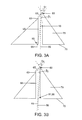
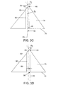
- Figs. 3A-D illustrate schematic representations of various embodiments of micro-needle lumen 55 and the portion of micro-needle body 75 defining apex 60 having different axial intersections; however, such are intended to be exemplary and are not intended to limit the possible locations of axial intersection.
- a feature of the micro-needles of the present invention is that such an axial intersection can occur at any point below apex 60.
- the axial intersection for each embodiment will be discussed relative to the micro-needle base 70.
- AI is the point of intersection between lumenal axis 56 and structural axis 65, wherein structural axis 65 is the line between apex 60 and the center point 95 of base 70.
- Fig. 3A illustrates an embodiment where AI 90 is above micro-needle base 70.
- vertex angle ⁇ is defined as ⁇ 1 and intersection angle ⁇ is defined as ⁇ 1 .
- Fig. 3B illustrates an embodiment, such as the embodiment of Figs. 2A and 2B, where AI 91 and the micro-needle base 70 are superimposed, i.e., lumenal axis 56 intersects structural axis 65 at a point along the plane which defines base 70.
- vertex angle ⁇ is defined as ⁇ 2 and intersection angle ⁇ is defined as ⁇ 2 .
- Fig. 3C illustrates an embodiment where AI 92 is below micro-needle base 70.
- vertex angle ⁇ is defined as ⁇ 3 and intersection angle ⁇ is defined as ⁇ 3 .
- angles ⁇ and ⁇ both increase, respectively, as the AI approaches apex 60.
- the distance 67 between apex 60 and proximal point 63 decreases.
- angles ⁇ and ⁇ both decrease, respectively, and distance 67 increases.
- angle ⁇ preferably ranges from about 30 to 60° but may be more or less depending on the particular application.
- the micro-needles of the present invention may have the following dimensions: a height or length generally in the range from about 100 to 10,000 ⁇ m, usually in the range from about 200 to 2,000 ⁇ m, and more usually in the range from 500 to 1,200 ⁇ m although the actual penetration depth of the micro-needle may be less; a base diameter generally in the range from about 100 to about 2,000 ⁇ m, and typically in the range from about 100 to 600 ⁇ m, and more typically in the range from about 250 to 500 ⁇ m; and a lumenal diameter capable of exerting a capillary force on fluid and, thus, is generally in the range from about 10 to 200 ⁇ m and more typically in the range from about 70 to 150 ⁇ m. Any appropriate aspect ratio between the height and base diameter of the micro-needle may be used, however, the aspect ratio is at least 1:1, and is usually between about 2:1 to 5:1.
- the subject micro-needles are configured to be mechanically stable and strong enough to penetrate the stratum corneum.
- the micro-needles are made of a biocompatible material so as not to cause irritation to the skin or an undesirable tissue response.
- the micro-needles are made of a plastic or resin material. Specific examples of such materials include, but are not limited to, acrylic, polyacrylates, polycarbonate, epoxies, polyesters polyetheretherketone, polyvinylchloride, polyolefins, liquid crystalline polyesters, or their composites.
- the subject micro-needles may be made of any suitable solid material, including stainless steel and other metals, provided such material is suitable for treatment in a micro-replication process.
- the micro-needles of the present invention may be integrally provided with a measurement means to provide a fluid sampling and constituent measurement device, also referred to as a "sensor device", for determining the concentration of one or more constituents within a sampled fluid.
- a fluid sampling and constituent measurement device also referred to as a "sensor device”
- the subject micro-needles may be integrally provided with a reservoir chamber for holding drugs or the like for the delivery of such for therapeutic and/or diagnostic applications.
- the micro-needles may be integrated with a device which includes both fluid sampling/measurement means and fluid holding/delivery means.
- Micro-needle 80 is operatively mounted or affixed to a substrate 50 or the like which may house, for example, an analyte measurement means (not shown) and/or a fluid reservoir (not shown).
- the measurement means may include an electrochemical means, a photometric means or any other suitable means for measuring the level of a target constituent(s).
- the fluid reservoir may include a chamber containing a selected volume or one or more doses of a drug or other material.
- Lumen 55 of micro-needle 80 is shown in fluid communication with pore 52 of substrate 50 having the same or substantially the same diameter dimensions as lumen 55.
- substrate housing an analyte measurement means is able to receive a sampled biological fluid through lumen 55 by means of a capillary force when micro-needle 80 is penetrated within the skin.
- pore 52 and lumen 55 may have the same or different diameter dimensions, depending on the desired delivery protocol.
- the sensor/delivery device may provide at least one subject micro-needle 80, or multiple micro-needles in the form of an array in which the micro-needles are mounted to or formed on, in close proximity with each other, to a base structure or substrate.
- the micro-needles and the associated substrate may be separately fabricated components which are integrally connected or may be formed together to establish a single, generally continuous device if desired.
- micro-needles may be employed with the sensor/delivery devices of the present invention. The optimal number will depend upon various factors including the type of fluid being sampled or delivered, the location and surface area of the biological tissue into which the micro-needles are inserted, the rate of sampling or delivery desired, the size of the device and the margin of accuracy desired. Regardless of the number micro-needles, they are sufficiently separated from each other so as to ensure that the stratum corneum can be penetrated without undue pressure on the skin. In general, micro-needles are separated from adjacent micro-needles a distance in the range from about 100 to about 1,000 ⁇ m, and typically from about 200 to 600 ⁇ m. Additionally, the micro-needle array may comprise micro-needles of the present invention having varying shapes, lengths, widths and tip configurations.
- the subject sensor/delivery devices may be part of a system for sampling biological fluid and measuring the target analyte concentration therein or for deiverying therapeutic or diagnostic materials across a biological tissue barrier.
- a system of the present invention may include a control unit for automatically controlling the function of the sensor/delivery device and for deriving specific information about the sampled fluid and its constituents and for displaying such information and/or for controlling the volume and rate of delivery of a fluid.
- the control unit may be configured to generate and send input signals to the sensor/delivery device and to receive output signals from the sensor/delivery device. These functions, among others, may be controlled by a software algorithm programmed within the control unit.
- control unit functions such as the automatic calculation of the concentration of a target analyte in a biological sample such that a user need only insert a micro-needle of the subject invention into the skin and then read the final analyte concentration result from a display of the device.
- a control device is further described in U.S. Patent No. 6,193,873 entitled “Sample Detection to Initiate Timing of an Electrochemical Assay," the disclosure of which is herein incorporated by reference.
- the micro-needles of the present invention are fabricated by micro-replication technology using any solid material.
- suitable materials include, but are not limited to, plastics, polymers or resins, and the like which are suitable for forming components by means of injection molding.
- Other solid materials, suitable for use in micro-replication technology, may also be used to form the micro-needles and devices of the present invention.
- Plastic injection molding is one type of micro-fabrication technique which is particularly suitable for fabricating the micro-needles of the present invention.
- An advantage of plastic injection molding is the ability to customize and precisely detail a micro-needle for its intended application far more quickly than by prior art micro-needle fabrication techniques.
- the oblique structures, beveled tips and sharp points or apexes of the subject micro-needles discussed above may be fabricated by plastic injection-molding techniques in an automated fashion.
- plastic resins and polymers are typical materials used in the form of pellets or small particles.
- Particularly suitable resins and polymers include, but are not limited to, polyacrylate, polycarbonate, epoxies, polyester, polyetherether-ketone, polyvinylchloride, polyolefins, liquid crystalline polymers, polyphenylene sulfide, polyphenylene oxide, polyacetal, polyimide, polyamide, polystyrene, copolymers or their composites or the like.
- These pellets or particles are fed into an injection molding machine and heated until they become molten.
- the molten material is then forced into a mold having a reverse image of the micro-needle structure to be formed.
- the molten material is then allowed to cool and become solidified within the mold.
- the resulting solid structure is then ejected from the mold by means of ejection pins which are run through the mold.
- FIG. 4A and 4B illustrate cross-sections of exemplary embodiments of molds that are suitable for use in the injection molding of the micro-needles of the present invention.
- Fig. 4A shows a mold assembly 100 having a base portion 102 and structural portion 104 which are alignable in a sealing engagement with each other at rests 106a and 106b.
- Base portion 102 provides a surface area 103 which forms either the base end of a micro-needle or, as here, the back-side of a substrate structure, such as those described above.
- Structural portion 104 provides a negative image of the desired micro-needle structure 108, such as the micro-needles structures of the present invention.
- Structural portion 104 also provides a pin formation 110 within micro-needle structure 108 which forms the lumen within the micro-needle structure as well as provides a spacing 116 forming the structure and side walls of a substrate.
- Fig. 4B illustrates another embodiment of a mold 120 having base portion 122 and structural portion 124 which are alignable in sealing engagement with each other at rests 126a and 126b.
- Structural portion 124 provides a negative or opposite image of the desired micro-needle structure 132, such as the micro-needles structures of the present invention.
- Base portion 102 provides a surface area 128 which forms either the base end of a micro-needle or, as here, the back-side and side walls of a substrate structure, such as those described above.
- base portion 122 also provides a pin formation 130 for forming a lumen within micro-needle structure 132.
- a granulated material is selected for filling the mold cavities.
- Suitable materials for powder injection molding are metals, silicon carbide, silicon oxide, aluminum oxide, zirconium oxide, and their mixtures.
- the granulated particles are then mixed with a binder material and heated to a high temperature until molten, then transferred into a mold, such as the molds of the present invention described above.
- the molten material is then allowed to cool and harden.
- the hardened structure is then removed from the mold, such as by means of the ejection pins discussed above, and placed in a solvent ore material to extract the binder material from the resulting structure.
- the structure is then further hardened by means of a sintering process.
- the cavities of the molds used in the above-described fabrication processes may be formed in the desired negative or opposite image, e.g ., an oblique cone shape, of a micro-needle by means of an electrical discharge machining (EDM).
- EDM electrical discharge machining
- an electrode is used to cut or form the desired configuration of a micro-needle structure into the base and structural portions of the mold.
- the formation pin used to form the micro-needle lumen may be formed by the EDM process or, alternatively, formed by a modular pin fastened within the cavity of either the base or structural portions of the mold.
- the present invention also provides for a method of manufacturing tiny oblique structures.
- These oblique structures have a base end and a vertex end.
- the base end has a diameter generally in the range from about 100 to 2,000 ⁇ m and more typically in the range from about 100 to 500 ⁇ m.
- the base-to-vertex height is generally in the range from about 100 to 10,000 ⁇ m, and more typically in the range from about 200 to 2,000 ⁇ m.
- This method includes providing a suitable material from which said oblique structure can be fabricated by means of one or more micro-replication techniques, such as the injection molding technique and electrical discharge machining discussed above.
- the manufacturing process further includes forming an open lumen within the oblique structure either in conjunction with the fabrication of the oblique structure or thereafter.
- the open lumen is formed to extend from the base end of the structure to the vertex end and may have dimensions the same as or similar to those discussed with respect to the micro-needle embodiment of Figs. 2A and 2B.
- the process additionally includes customizing a tip at the vertex end of the structure. Again, such customization may be performed in conjunction with the fabrication of the oblique structure and/or in conjunction with the fabrication of the open lumen within the oblique structure.
- the customized tip may be selectively beveled, for a particular application, such as those mentioned above.
- the first step is to provide a device having one or more subject micro-needles or an array of subject micro-needles, as described above.
- the subject micro-needle(s) may be made of material formed by a micro-replication technique, such as plastic injection molding. More specifically, that material may be a type of plastic, polymer or resin.
- the micro-needle(s) may be provide as an integral part of a subject sensor device, which device may be particularly configured (i.e., containing the appropriate reagent) for targeting the analyte(s) of interest.
- the sensor device may be a stand alone device, while in other embodiments of the present invention, the sensor device may be operatively engaged and interfaced with a control unit, described above, that can be manually held and controlled by the user.
- the control unit is programmed for testing the targeted analyte(s).
- the user positions sensor device over a selected area of the patient's skin and, with slight pressure, the micro-needle(s) of the sensor device is/are caused to penetrate into the skin.
- the depth to which the micro-needles are inserted will depend on the length of the respective micro-needles or by some other means associated with the sensor unit for limiting the insertion depth.
- an amount i.e., a sample
- biological fluid present at the open tips of the micro-needles is wicked into the lumen by means of a capillary force, or by a vacuum assist device or the like.
- the sampled fluid is then transported to a measurement area, e.g. , a reaction zone of an electrochemical cell, of the measurement means.
- a signal producing and receiving system of the measurement means defined by the type of measurement being made, e. g. electrochemical or photometric, measures the concentration of the analyte within the sampled fluid.
- Representative analytes include glucose, cholesterol, lactate, alcohol, and the like.
- the micro-needle(s) may be provide as an integral part of a delivery device containing a reservoir for holding a volume of drug or material to be delivered, which device may be particularly configured for controlling the rate of delivery such drug or material.
- the delivery device may be a stand alone device, while in other embodiments of the present invention, the delivery device may be operatively engaged and interfaced with a control unit, described above, that can be manually held and controlled by the user.
- the control unit may be programmed for delivering a selected amount of drug or material to be delivered.
- the user positions the delivery device over a selected area of the patient's skin and, with slight pressure, the micro-needle(s) of the delivery device is/are caused to penetrate into the skin.
- the depth to which the micro-needles are inserted will depend on the length of the respective micro-needles or by some other means associated with the delivery device for limiting the insertion depth.
- an amount of the drug or material held within the delivery devices reservoir is injected or is otherwise caused to be transported via the micro-needle(s) to within the biological tissue.
- Various drugs and materials to be delivered include therapeutic drugs diagnostic agents, genetic material biological material, and the like.
- kits for use in practicing the subject methods include at least one subject micro-needle and/or one or more subject sensor and/or delivery devices having one or more subject micro-needles.
- the sensor/delivery devices may include an array of micro-needles of the present invention having the same or different lengths.
- Certain kits may include various sensor devices each containing the same or different reagents.
- more than one reagent may be provides within a single micro-needle array, wherein one or more of the micro-needles are provided with a first reagent for testing a first target analyte and one or more other micro-needles are provided with other reagents for testing other targeted analytes.
- kits may include various delivery devices each containing the same or different drug or formulation.
- the kits may also include a reusable or disposable control unit that may be used with reusable or disposable sensor/delivery devices of the kit of the subject invention.
- the kits preferably include instructions for using the subject devices in the determination of an analyte concentration in a biological sample and/or for delivering a drug or formulation across a biological barrier. These instructions may be present on one or more of the packaging, a label insert, or containers present in the kits, and the like.
- the features of the subject micro-needles overcome many of the disadvantages of prior art micro-needles, and provide certain advantages including, but not limited to, providing micro-needles having extremely sharp tips for penetration into the skin, providing a customized micro-needle for the particular application at hand and providing easier and less costly manufacturing techniques.
- Other advantages of the subject micro-needles is the reduction in pain experienced by a patient as a result of the reduced footprint of the micro-needle tip and the minimal pressure required to cause the micro-needle to penetrate the skin.
- the subject invention represents a significant contribution to the field of fluid sampling and delivery across a biological barrier.
Landscapes
- Health & Medical Sciences (AREA)
- Life Sciences & Earth Sciences (AREA)
- Engineering & Computer Science (AREA)
- Physics & Mathematics (AREA)
- Veterinary Medicine (AREA)
- Public Health (AREA)
- General Health & Medical Sciences (AREA)
- Animal Behavior & Ethology (AREA)
- Biomedical Technology (AREA)
- Heart & Thoracic Surgery (AREA)
- Medical Informatics (AREA)
- Surgery (AREA)
- Molecular Biology (AREA)
- Pathology (AREA)
- Biophysics (AREA)
- Hematology (AREA)
- Manufacturing & Machinery (AREA)
- Optics & Photonics (AREA)
- Emergency Medicine (AREA)
- Dermatology (AREA)
- Anesthesiology (AREA)
- Measurement Of The Respiration, Hearing Ability, Form, And Blood Characteristics Of Living Organisms (AREA)
- Media Introduction/Drainage Providing Device (AREA)
- Infusion, Injection, And Reservoir Apparatuses (AREA)
Applications Claiming Priority (2)
| Application Number | Priority Date | Filing Date | Title |
|---|---|---|---|
| US09/901,535 US6749792B2 (en) | 2001-07-09 | 2001-07-09 | Micro-needles and methods of manufacture and use thereof |
| US901535 | 2001-07-09 |
Publications (1)
| Publication Number | Publication Date |
|---|---|
| EP1287847A1 true EP1287847A1 (de) | 2003-03-05 |
Family
ID=25414377
Family Applications (1)
| Application Number | Title | Priority Date | Filing Date |
|---|---|---|---|
| EP02254617A Withdrawn EP1287847A1 (de) | 2001-07-09 | 2002-07-01 | Verbesserte Mikronadeln und Verfahren zu deren Herstellung sowie deren Verwendung |
Country Status (12)
| Country | Link |
|---|---|
| US (2) | US6749792B2 (de) |
| EP (1) | EP1287847A1 (de) |
| JP (1) | JP2003088514A (de) |
| KR (1) | KR20030007039A (de) |
| CN (1) | CN1494876A (de) |
| AR (1) | AR034723A1 (de) |
| CA (1) | CA2391810A1 (de) |
| IL (1) | IL150468A0 (de) |
| MX (1) | MXPA02006647A (de) |
| PL (1) | PL354857A1 (de) |
| RU (1) | RU2002118232A (de) |
| TW (1) | TW590782B (de) |
Cited By (8)
| Publication number | Priority date | Publication date | Assignee | Title |
|---|---|---|---|---|
| WO2005082593A1 (en) * | 2004-02-17 | 2005-09-09 | Avery Dennison Corporation | Method of making microneedles |
| US7076987B2 (en) | 2004-08-05 | 2006-07-18 | Becton, Dickinson And Company | Method of producing tapered or pointed cannula |
| US7086266B2 (en) | 2004-08-05 | 2006-08-08 | Becton, Dickinson And Company | Method of producing tapered or pointed cannula |
| WO2006062974A3 (en) * | 2004-12-07 | 2006-09-28 | 3M Innovative Properties Co | Method of molding a microneedle |
| CN1321706C (zh) * | 2003-05-29 | 2007-06-20 | 财团法人工业技术研究院 | 一种数组式微针软质基材结构及其制造方法 |
| US7497980B2 (en) | 2003-11-10 | 2009-03-03 | Agency For Science, Technology And Research | Microneedles and microneedle fabrication |
| US8671544B2 (en) | 2004-03-12 | 2014-03-18 | Agency For Science, Technology And Research | Methods and moulds for use in fabricating side-ported microneedles |
| WO2024040387A1 (en) * | 2022-08-22 | 2024-02-29 | Wenzhou Institute, University Of Chinese Academy Of Sciences (Wiucas) | Microneedle arrays and method of making |
Families Citing this family (214)
| Publication number | Priority date | Publication date | Assignee | Title |
|---|---|---|---|---|
| SE9704076D0 (sv) * | 1997-11-06 | 1997-11-06 | Holdingbolaget Vid Goeteborgs | Method for permeabilisation of cell structures and use thereof |
| US6036924A (en) | 1997-12-04 | 2000-03-14 | Hewlett-Packard Company | Cassette of lancet cartridges for sampling blood |
| US6391005B1 (en) | 1998-03-30 | 2002-05-21 | Agilent Technologies, Inc. | Apparatus and method for penetration with shaft having a sensor for sensing penetration depth |
| US6884427B1 (en) * | 1999-02-08 | 2005-04-26 | Aderans Research Institute, Inc. | Filamentary means for introducing agents into tissue of a living host |
| KR20030043928A (ko) * | 2000-08-08 | 2003-06-02 | 바이오아미드 인코포레이티드 | 조직 조작된 모발용 지지체 |
| US7131987B2 (en) | 2000-10-16 | 2006-11-07 | Corium International, Inc. | Microstructures and method for treating and conditioning skin which cause less irritation during exfoliation |
| US7828827B2 (en) | 2002-05-24 | 2010-11-09 | Corium International, Inc. | Method of exfoliation of skin using closely-packed microstructures |
| US7108681B2 (en) | 2000-10-16 | 2006-09-19 | Corium International, Inc. | Microstructures for delivering a composition cutaneously to skin |
| SE0003841D0 (sv) * | 2000-10-20 | 2000-10-20 | Daniel Chiu | A method and apparatus for penetration of lipid bilayer membranes |
| US8641644B2 (en) | 2000-11-21 | 2014-02-04 | Sanofi-Aventis Deutschland Gmbh | Blood testing apparatus having a rotatable cartridge with multiple lancing elements and testing means |
| WO2002100460A2 (en) | 2001-06-12 | 2002-12-19 | Pelikan Technologies, Inc. | Electric lancet actuator |
| US7981056B2 (en) | 2002-04-19 | 2011-07-19 | Pelikan Technologies, Inc. | Methods and apparatus for lancet actuation |
| US7537571B2 (en) | 2001-06-12 | 2009-05-26 | Pelikan Technologies, Inc. | Integrated blood sampling analysis system with multi-use sampling module |
| AU2002348683A1 (en) | 2001-06-12 | 2002-12-23 | Pelikan Technologies, Inc. | Method and apparatus for lancet launching device integrated onto a blood-sampling cartridge |
| US7041068B2 (en) | 2001-06-12 | 2006-05-09 | Pelikan Technologies, Inc. | Sampling module device and method |
| US9427532B2 (en) | 2001-06-12 | 2016-08-30 | Sanofi-Aventis Deutschland Gmbh | Tissue penetration device |
| CA2448902C (en) | 2001-06-12 | 2010-09-07 | Pelikan Technologies, Inc. | Self optimizing lancing device with adaptation means to temporal variations in cutaneous properties |
| US7682318B2 (en) | 2001-06-12 | 2010-03-23 | Pelikan Technologies, Inc. | Blood sampling apparatus and method |
| US9795747B2 (en) | 2010-06-02 | 2017-10-24 | Sanofi-Aventis Deutschland Gmbh | Methods and apparatus for lancet actuation |
| WO2002100461A2 (en) | 2001-06-12 | 2002-12-19 | Pelikan Technologies, Inc. | Method and apparatus for improving success rate of blood yield from a fingerstick |
| US9226699B2 (en) | 2002-04-19 | 2016-01-05 | Sanofi-Aventis Deutschland Gmbh | Body fluid sampling module with a continuous compression tissue interface surface |
| US8337419B2 (en) | 2002-04-19 | 2012-12-25 | Sanofi-Aventis Deutschland Gmbh | Tissue penetration device |
| US7344507B2 (en) | 2002-04-19 | 2008-03-18 | Pelikan Technologies, Inc. | Method and apparatus for lancet actuation |
| US8201330B1 (en) * | 2001-09-07 | 2012-06-19 | Orbital Research Inc | Physiological recording device or electrode |
| US6785569B2 (en) | 2001-09-07 | 2004-08-31 | Orbital Research | Dry physiological recording electrode |
| US6782283B2 (en) * | 2001-09-07 | 2004-08-24 | Robert N. Schmidt | Dry penetrating recording device |
| US7286864B1 (en) | 2001-09-07 | 2007-10-23 | Orbital Research, Inc. | Dry physiological recording device |
| US7344894B2 (en) | 2001-10-16 | 2008-03-18 | Agilent Technologies, Inc. | Thermal regulation of fluidic samples within a diagnostic cartridge |
| AU2002353741A1 (en) * | 2001-11-27 | 2003-06-10 | Cellectricon Ab | A method for combined sequential agent delivery and electroporation for cell structures and use thereof |
| US20040068284A1 (en) * | 2002-01-29 | 2004-04-08 | Barrows Thomas H. | Method for stimulating hair growth and kit for carrying out said method |
| JP2005518873A (ja) * | 2002-03-04 | 2005-06-30 | ナノ パス テクノロジーズ リミテッド | 生体障壁を横切る流体輸送の為の装置及び方法 |
| US7141058B2 (en) | 2002-04-19 | 2006-11-28 | Pelikan Technologies, Inc. | Method and apparatus for a body fluid sampling device using illumination |
| US9795334B2 (en) | 2002-04-19 | 2017-10-24 | Sanofi-Aventis Deutschland Gmbh | Method and apparatus for penetrating tissue |
| US7297122B2 (en) | 2002-04-19 | 2007-11-20 | Pelikan Technologies, Inc. | Method and apparatus for penetrating tissue |
| US7485128B2 (en) | 2002-04-19 | 2009-02-03 | Pelikan Technologies, Inc. | Method and apparatus for penetrating tissue |
| US7901362B2 (en) | 2002-04-19 | 2011-03-08 | Pelikan Technologies, Inc. | Method and apparatus for penetrating tissue |
| US7232451B2 (en) | 2002-04-19 | 2007-06-19 | Pelikan Technologies, Inc. | Method and apparatus for penetrating tissue |
| US7582099B2 (en) | 2002-04-19 | 2009-09-01 | Pelikan Technologies, Inc | Method and apparatus for penetrating tissue |
| US7410468B2 (en) | 2002-04-19 | 2008-08-12 | Pelikan Technologies, Inc. | Method and apparatus for penetrating tissue |
| US8267870B2 (en) | 2002-04-19 | 2012-09-18 | Sanofi-Aventis Deutschland Gmbh | Method and apparatus for body fluid sampling with hybrid actuation |
| US7491178B2 (en) | 2002-04-19 | 2009-02-17 | Pelikan Technologies, Inc. | Method and apparatus for penetrating tissue |
| US8579831B2 (en) | 2002-04-19 | 2013-11-12 | Sanofi-Aventis Deutschland Gmbh | Method and apparatus for penetrating tissue |
| US7244265B2 (en) | 2002-04-19 | 2007-07-17 | Pelikan Technologies, Inc. | Method and apparatus for penetrating tissue |
| US9314194B2 (en) | 2002-04-19 | 2016-04-19 | Sanofi-Aventis Deutschland Gmbh | Tissue penetration device |
| US7291117B2 (en) | 2002-04-19 | 2007-11-06 | Pelikan Technologies, Inc. | Method and apparatus for penetrating tissue |
| US7226461B2 (en) | 2002-04-19 | 2007-06-05 | Pelikan Technologies, Inc. | Method and apparatus for a multi-use body fluid sampling device with sterility barrier release |
| US8360992B2 (en) | 2002-04-19 | 2013-01-29 | Sanofi-Aventis Deutschland Gmbh | Method and apparatus for penetrating tissue |
| US7563232B2 (en) | 2002-04-19 | 2009-07-21 | Pelikan Technologies, Inc. | Method and apparatus for penetrating tissue |
| US7892183B2 (en) | 2002-04-19 | 2011-02-22 | Pelikan Technologies, Inc. | Method and apparatus for body fluid sampling and analyte sensing |
| US7674232B2 (en) | 2002-04-19 | 2010-03-09 | Pelikan Technologies, Inc. | Method and apparatus for penetrating tissue |
| US7909778B2 (en) | 2002-04-19 | 2011-03-22 | Pelikan Technologies, Inc. | Method and apparatus for penetrating tissue |
| US8702624B2 (en) | 2006-09-29 | 2014-04-22 | Sanofi-Aventis Deutschland Gmbh | Analyte measurement device with a single shot actuator |
| US7524293B2 (en) | 2002-04-19 | 2009-04-28 | Pelikan Technologies, Inc. | Method and apparatus for penetrating tissue |
| US7331931B2 (en) | 2002-04-19 | 2008-02-19 | Pelikan Technologies, Inc. | Method and apparatus for penetrating tissue |
| US7374544B2 (en) | 2002-04-19 | 2008-05-20 | Pelikan Technologies, Inc. | Method and apparatus for penetrating tissue |
| US7547287B2 (en) | 2002-04-19 | 2009-06-16 | Pelikan Technologies, Inc. | Method and apparatus for penetrating tissue |
| US7892185B2 (en) | 2002-04-19 | 2011-02-22 | Pelikan Technologies, Inc. | Method and apparatus for body fluid sampling and analyte sensing |
| US7648468B2 (en) | 2002-04-19 | 2010-01-19 | Pelikon Technologies, Inc. | Method and apparatus for penetrating tissue |
| US9248267B2 (en) | 2002-04-19 | 2016-02-02 | Sanofi-Aventis Deustchland Gmbh | Tissue penetration device |
| US8221334B2 (en) | 2002-04-19 | 2012-07-17 | Sanofi-Aventis Deutschland Gmbh | Method and apparatus for penetrating tissue |
| US7229458B2 (en) | 2002-04-19 | 2007-06-12 | Pelikan Technologies, Inc. | Method and apparatus for penetrating tissue |
| AU2003231749A1 (en) | 2002-04-19 | 2003-11-03 | Pelikan Technologies, Inc. | Device and method for variable speed lancet |
| US8784335B2 (en) | 2002-04-19 | 2014-07-22 | Sanofi-Aventis Deutschland Gmbh | Body fluid sampling device with a capacitive sensor |
| US7717863B2 (en) | 2002-04-19 | 2010-05-18 | Pelikan Technologies, Inc. | Method and apparatus for penetrating tissue |
| US7976476B2 (en) | 2002-04-19 | 2011-07-12 | Pelikan Technologies, Inc. | Device and method for variable speed lancet |
| US7371247B2 (en) | 2002-04-19 | 2008-05-13 | Pelikan Technologies, Inc | Method and apparatus for penetrating tissue |
| US8574895B2 (en) | 2002-12-30 | 2013-11-05 | Sanofi-Aventis Deutschland Gmbh | Method and apparatus using optical techniques to measure analyte levels |
| DE10305831B4 (de) * | 2003-02-12 | 2007-01-04 | Siemens Ag | Diagnosegerät |
| US7578954B2 (en) | 2003-02-24 | 2009-08-25 | Corium International, Inc. | Method for manufacturing microstructures having multiple microelements with through-holes |
| US7415299B2 (en) * | 2003-04-18 | 2008-08-19 | The Regents Of The University Of California | Monitoring method and/or apparatus |
| EP2238892A3 (de) | 2003-05-30 | 2011-02-09 | Pelikan Technologies Inc. | Vorrichtung zur Entnahme von Körperflüssigkeit |
| ES2490740T3 (es) | 2003-06-06 | 2014-09-04 | Sanofi-Aventis Deutschland Gmbh | Aparato para toma de muestras de fluido sanguíneo y detección de analitos |
| WO2006001797A1 (en) | 2004-06-14 | 2006-01-05 | Pelikan Technologies, Inc. | Low pain penetrating |
| US7604592B2 (en) | 2003-06-13 | 2009-10-20 | Pelikan Technologies, Inc. | Method and apparatus for a point of care device |
| EP1671096A4 (de) | 2003-09-29 | 2009-09-16 | Pelikan Technologies Inc | Verfahren und apparatur für eine verbesserte probeneinfangvorrichtung |
| GB2406794B (en) | 2003-10-06 | 2008-03-05 | Inverness Medical Ltd | A lancing device using a piezoelectric actuator |
| EP1680014A4 (de) | 2003-10-14 | 2009-01-21 | Pelikan Technologies Inc | Verfahren und gerät für eine variable anwenderschnittstelle |
| WO2005060621A2 (en) * | 2003-11-21 | 2005-07-07 | The Regents Of The University Of California | Method and/or apparatus for puncturing a surface for extraction, in situ analysis, and/or substance delivery using microneedles |
| US7822454B1 (en) | 2005-01-03 | 2010-10-26 | Pelikan Technologies, Inc. | Fluid sampling device with improved analyte detecting member configuration |
| WO2005065414A2 (en) | 2003-12-31 | 2005-07-21 | Pelikan Technologies, Inc. | Method and apparatus for improving fluidic flow and sample capture |
| CA2560840C (en) | 2004-03-24 | 2014-05-06 | Corium International, Inc. | Transdermal delivery device |
| US7597885B2 (en) * | 2004-03-26 | 2009-10-06 | Aderans Research Institute, Inc. | Tissue engineered biomimetic hair follicle graft |
| US8828203B2 (en) | 2004-05-20 | 2014-09-09 | Sanofi-Aventis Deutschland Gmbh | Printable hydrogels for biosensors |
| EP1765194A4 (de) | 2004-06-03 | 2010-09-29 | Pelikan Technologies Inc | Verfahren und gerät für eine flüssigkeitsentnahmenvorrichtung |
| US9775553B2 (en) | 2004-06-03 | 2017-10-03 | Sanofi-Aventis Deutschland Gmbh | Method and apparatus for a fluid sampling device |
| AR050212A1 (es) * | 2004-08-13 | 2006-10-04 | Aderans Res Inst Inc | Organogenesis a partir de celulas disociadas |
| SE0402100D0 (sv) * | 2004-08-30 | 2004-08-30 | Bonsens Ab | Molded micro-needles |
| US7132054B1 (en) * | 2004-09-08 | 2006-11-07 | Sandia Corporation | Method to fabricate hollow microneedle arrays |
| US7785459B2 (en) | 2004-10-22 | 2010-08-31 | Hewlett-Packard Development Company, L.P. | Microneedles and methods of fabricating |
| US7097776B2 (en) * | 2004-10-22 | 2006-08-29 | Hewlett-Packard Development Company, L.P. | Method of fabricating microneedles |
| US8652831B2 (en) | 2004-12-30 | 2014-02-18 | Sanofi-Aventis Deutschland Gmbh | Method and apparatus for analyte measurement test time |
| WO2006075716A1 (ja) * | 2005-01-14 | 2006-07-20 | Fujikura Ltd. | 医薬物運搬用器具とその製造方法 |
| WO2006105146A2 (en) * | 2005-03-29 | 2006-10-05 | Arkal Medical, Inc. | Devices, systems, methods and tools for continuous glucose monitoring |
| JP4826711B2 (ja) * | 2005-03-31 | 2011-11-30 | ニプロ株式会社 | 注射針 |
| AU2006304516A1 (en) * | 2005-10-17 | 2007-04-26 | Aderans Research Institute, Inc. | Method of delivering hair follicle progenitor cells to the skin |
| AR057628A1 (es) * | 2005-11-22 | 2007-12-05 | Aderans Res Inst Inc | Injertos capilares derivados de cabello extirpado |
| TW200800240A (en) * | 2005-11-22 | 2008-01-01 | Aderans Res Inst Inc | Hair follicle graft from tissue engineered skin |
| EP1959828B1 (de) * | 2005-12-14 | 2013-02-27 | SciBase AB | Diagnose eines Krankheitszustands der Haut mit Impedanz |
| US9636035B2 (en) * | 2005-12-14 | 2017-05-02 | Scibase Ab | Medical apparatus for determination of biological conditions using impedance measurements |
| EP2520324A2 (de) * | 2006-02-09 | 2012-11-07 | Aderans Research Institute, Inc. | Vorrichtung und Verfahren zur Abgabe von Flüssigkeit und Material an eine Person |
| US20070202186A1 (en) | 2006-02-22 | 2007-08-30 | Iscience Interventional Corporation | Apparatus and formulations for suprachoroidal drug delivery |
| US20090131778A1 (en) * | 2006-03-28 | 2009-05-21 | Jina Arvind N | Devices, systems, methods and tools for continuous glucose monitoring |
| US20080154107A1 (en) * | 2006-12-20 | 2008-06-26 | Jina Arvind N | Device, systems, methods and tools for continuous glucose monitoring |
| US20100049021A1 (en) * | 2006-03-28 | 2010-02-25 | Jina Arvind N | Devices, systems, methods and tools for continuous analyte monitoring |
| US8197435B2 (en) | 2006-05-02 | 2012-06-12 | Emory University | Methods and devices for drug delivery to ocular tissue using microneedle |
| TWI300055B (en) * | 2006-05-26 | 2008-08-21 | Nat Univ Tsing Hua | Method of manufacturing hollow micro-needle structures |
| US20070276330A1 (en) * | 2006-05-28 | 2007-11-29 | Beck Patricia A | Microneedles and methods of fabricating thereof |
| US8012566B2 (en) * | 2006-07-12 | 2011-09-06 | Hewlett-Packard Development Company, L.P. | Microneedles formed by electroplating and selectively releasing temperature sensitive layers |
| US8250729B2 (en) * | 2006-07-12 | 2012-08-28 | University Of Utah Research Foundation | 3D fabrication of needle tip geometry and knife blade |
| US8865288B2 (en) * | 2006-07-17 | 2014-10-21 | University Of Utah Research Foundation | Micro-needle arrays having non-planar tips and methods of manufacture thereof |
| US20080138581A1 (en) * | 2006-07-17 | 2008-06-12 | Rajmohan Bhandari | Masking high-aspect aspect ratio structures |
| JP2010502267A (ja) * | 2006-08-28 | 2010-01-28 | エージェンシー・フォー・サイエンス・テクノロジー・アンド・リサーチ | マイクロニードルおよびマイクロニードルの製造方法 |
| US20080058726A1 (en) * | 2006-08-30 | 2008-03-06 | Arvind Jina | Methods and Apparatus Incorporating a Surface Penetration Device |
| US20080097352A1 (en) * | 2006-09-12 | 2008-04-24 | Beck Patricia A | Methods of fabricating microneedles with bio-sensory functionality |
| WO2008036043A1 (en) * | 2006-09-18 | 2008-03-27 | Agency For Science, Technology And Research | Needle structures and methods for fabricating needle structures |
| KR100869277B1 (ko) * | 2006-12-22 | 2008-11-18 | 호남석유화학 주식회사 | 마이크로니들 어레이와 그의 성형 몰드 |
| US8523926B2 (en) * | 2007-01-19 | 2013-09-03 | Joseph Neev | Devices and methods for generation of subsurface microdisruptions for biomedical applications |
| EP2121111B1 (de) | 2007-01-22 | 2018-03-14 | Corium International, Inc. | Applikator für mikronadelanordnung |
| CN100431487C (zh) * | 2007-01-25 | 2008-11-12 | 中国科学院上海微系统与信息技术研究所 | 一种三维植入式微电极阵列的制作方法 |
| USD606190S1 (en) | 2007-02-08 | 2009-12-15 | Aderans Research Institute, Inc. | Device for delivering cellular material and physiologic fluids to a subject |
| US20080234562A1 (en) * | 2007-03-19 | 2008-09-25 | Jina Arvind N | Continuous analyte monitor with multi-point self-calibration |
| EP2146689B1 (de) | 2007-04-16 | 2020-08-12 | Corium, Inc. | Einen wirkstoff enthaltende lösungsmittel-gegossene mikronadelanordnungen |
| US8911749B2 (en) | 2007-04-16 | 2014-12-16 | Corium International, Inc. | Vaccine delivery via microneedle arrays |
| US7985537B2 (en) * | 2007-06-12 | 2011-07-26 | Aderans Research Institute, Inc. | Methods for determining the hair follicle inductive properties of a composition |
| US20080312518A1 (en) * | 2007-06-14 | 2008-12-18 | Arkal Medical, Inc | On-demand analyte monitor and method of use |
| US20100196446A1 (en) | 2007-07-10 | 2010-08-05 | Morteza Gharib | Drug delivery and substance transfer facilitated by nano-enhanced device having aligned carbon nanotubes protruding from device surface |
| US20090053673A1 (en) * | 2007-08-23 | 2009-02-26 | Zimmer, Inc. | Method for localized treatment of periodontal tissue |
| JP5559054B2 (ja) * | 2007-09-28 | 2014-07-23 | ザ クイーンズ ユニヴァーシティ オブ ベルファスト | 送達装置および方法 |
| US20090099427A1 (en) * | 2007-10-12 | 2009-04-16 | Arkal Medical, Inc. | Microneedle array with diverse needle configurations |
| WO2009064380A2 (en) | 2007-11-09 | 2009-05-22 | California Institute Of Technology | Fabrication of anchored carbon nanotube array devices for integrated light collection and energy conversion |
| US8398397B2 (en) * | 2008-03-12 | 2013-03-19 | Ultradent Products, Inc. | Dental intraligamentary injection needles and related methods of manufacture |
| EP2265324B1 (de) | 2008-04-11 | 2015-01-28 | Sanofi-Aventis Deutschland GmbH | Integriertes System zur Messung von Analyten |
| US20090301994A1 (en) * | 2008-05-12 | 2009-12-10 | Rajmohan Bhandari | Methods for Wafer Scale Processing of Needle Array Devices |
| BRPI0913578A2 (pt) | 2008-05-14 | 2017-06-06 | Dermtech Int | diagnose de melanoma e lentigo solar por análise de ácido nucléico |
| WO2009149197A2 (en) | 2008-06-03 | 2009-12-10 | University Of Utah Research Foundation | High aspect ratio microelectrode arrays enabled to have customizable lengths and methods of making the same |
| EP2316339A4 (de) * | 2008-08-01 | 2012-10-24 | Lightnix Inc | Sensor mit feiner nadel mit darin gebildetem kanal |
| US9375169B2 (en) | 2009-01-30 | 2016-06-28 | Sanofi-Aventis Deutschland Gmbh | Cam drive for managing disposable penetrating member actions with a single motor and motor and control system |
| WO2010101621A1 (en) | 2009-03-02 | 2010-09-10 | Seventh Sense Biosystems, Inc. | Devices and methods for the analysis of an extractable medium |
| US9033898B2 (en) * | 2010-06-23 | 2015-05-19 | Seventh Sense Biosystems, Inc. | Sampling devices and methods involving relatively little pain |
| US9295417B2 (en) | 2011-04-29 | 2016-03-29 | Seventh Sense Biosystems, Inc. | Systems and methods for collecting fluid from a subject |
| JP2012523270A (ja) | 2009-04-10 | 2012-10-04 | スリーエム イノベイティブ プロパティズ カンパニー | 中空マイクロニードルアレイを製造する方法並びにそれに由来する製品及びその使用法 |
| EP2260764A1 (de) * | 2009-06-10 | 2010-12-15 | F. Hoffmann-La Roche AG | Mikronadel und Verfahren zu deren Herstellung |
| KR101213223B1 (ko) * | 2009-07-07 | 2013-01-09 | 한국전자통신연구원 | 할로우를 가지는 미세 바늘 및 미세 바늘 구조체의 제조 방법 |
| JP5568324B2 (ja) * | 2010-01-15 | 2014-08-06 | 凸版印刷株式会社 | マイクロニードル製造方法 |
| WO2011094573A1 (en) | 2010-01-28 | 2011-08-04 | Seventh Sense Biosystems, Inc. | Monitoring or feedback systems and methods |
| US9115424B2 (en) | 2010-04-07 | 2015-08-25 | California Institute Of Technology | Simple method for producing superhydrophobic carbon nanotube array |
| US8965476B2 (en) | 2010-04-16 | 2015-02-24 | Sanofi-Aventis Deutschland Gmbh | Tissue penetration device |
| JP6327852B2 (ja) | 2010-05-04 | 2018-05-23 | コリウム インターナショナル, インコーポレイテッド | 微小突起アレイを使用した副甲状腺ホルモンの経皮送達のための方法及びデバイス |
| JP2013538069A (ja) | 2010-07-16 | 2013-10-10 | セブンス センス バイオシステムズ,インコーポレーテッド | 流体移動デバイスのための低圧環境 |
| US20130158482A1 (en) | 2010-07-26 | 2013-06-20 | Seventh Sense Biosystems, Inc. | Rapid delivery and/or receiving of fluids |
| US20120039809A1 (en) | 2010-08-13 | 2012-02-16 | Seventh Sense Biosystems, Inc. | Systems and techniques for monitoring subjects |
| EP2627292B1 (de) | 2010-10-15 | 2018-10-10 | Clearside Biomedical, Inc. | Vorrichtung für okularen zugang |
| EP2637562B1 (de) | 2010-11-09 | 2016-01-27 | Seventh Sense Biosystems, Inc. | Systeme und schnittstellen zur blutabnahme |
| US8609458B2 (en) | 2010-12-10 | 2013-12-17 | California Institute Of Technology | Method for producing graphene oxide with tunable gap |
| JP2014519344A (ja) * | 2011-03-18 | 2014-08-14 | ユニヴェルシテ リブル ドゥ ブリュッセル | 人間または動物の体の膜に穴をあけるための装置 |
| US20120245445A1 (en) * | 2011-03-21 | 2012-09-27 | Michael Darryl Black | Glucose Monitoring System |
| WO2012135238A1 (en) | 2011-03-29 | 2012-10-04 | California Institute Of Technology | Method to increase the capacitance of electrochemical carbon nanotube capacitors by conformal deposition of nanoparticles |
| JP2014516644A (ja) | 2011-04-29 | 2014-07-17 | セブンス センス バイオシステムズ,インコーポレーテッド | 血斑または他の体液の収集および/または操作のためのデバイスおよび方法 |
| US20130158468A1 (en) | 2011-12-19 | 2013-06-20 | Seventh Sense Biosystems, Inc. | Delivering and/or receiving material with respect to a subject surface |
| EP2701600B1 (de) | 2011-04-29 | 2016-06-08 | Seventh Sense Biosystems, Inc. | Ausgabe und/oder aufnahme von flüssigkeiten |
| JP2013090837A (ja) * | 2011-10-26 | 2013-05-16 | Toppan Printing Co Ltd | 針状体の製造方法および針状体 |
| US8764681B2 (en) | 2011-12-14 | 2014-07-01 | California Institute Of Technology | Sharp tip carbon nanotube microneedle devices and their fabrication |
| JP5856475B2 (ja) * | 2011-12-28 | 2016-02-09 | Asti株式会社 | マイクロニードルの製造方法 |
| CN102526870B (zh) * | 2012-01-09 | 2013-08-28 | 上海交通大学 | 基于表面微细加工工艺的异平面空心微针及其制备方法 |
| EP2806940B1 (de) * | 2012-01-24 | 2019-05-22 | International Scientific PTY Ltd | Abgabegerät |
| JP2013166363A (ja) * | 2012-02-17 | 2013-08-29 | Toppan Printing Co Ltd | 微細針チップ射出成形用金型及びその微細針チップを射出成形する方法 |
| USD690004S1 (en) | 2012-03-16 | 2013-09-17 | Aderans Research Institute, Inc. | Holder for a device for delivering cellular material and physiologic fluids |
| US9962535B2 (en) | 2012-05-11 | 2018-05-08 | 10X Technology Llc | Hollow silica glass microneedle arrays and method and apparatus for manufacturing same |
| US9349543B2 (en) | 2012-07-30 | 2016-05-24 | California Institute Of Technology | Nano tri-carbon composite systems and manufacture |
| EP2887982B1 (de) * | 2012-08-27 | 2022-11-23 | Clearside Biomedical, Inc. | Vorrichtung zur arzneimittelverabreichung mit mikronadeln |
| KR102467714B1 (ko) | 2012-11-08 | 2022-11-16 | 클리어사이드 바이오메디컬, 인코포레이드 | 인간 대상체에서 안구 질병을 치료하기 위한 방법 및 장치 |
| AU2013364053B2 (en) | 2012-12-21 | 2018-08-30 | Corium Pharma Solutions, Inc. | Microarray for delivery of therapeutic agent and methods of use |
| CA2903748C (en) | 2013-03-12 | 2021-11-02 | Corium International, Inc. | Microprojection applicators |
| US9192313B1 (en) | 2013-03-14 | 2015-11-24 | Orbital Research Inc. | Dry physiological recording device and method of manufacturing |
| US10820860B2 (en) | 2013-03-14 | 2020-11-03 | One Drop Biosensor Technologies, Llc | On-body microsensor for biomonitoring |
| CN105228522B (zh) | 2013-03-14 | 2018-06-26 | 萨诺智能公司 | 用于生物监测的人体上的微传感器 |
| US9962534B2 (en) | 2013-03-15 | 2018-05-08 | Corium International, Inc. | Microarray for delivery of therapeutic agent, methods of use, and methods of making |
| BR112015022625B1 (pt) | 2013-03-15 | 2023-01-31 | Corium, Inc | Aparelho de microestrutura para entrega de agente terapêutico |
| AU2014237279B2 (en) | 2013-03-15 | 2018-11-22 | Corium Pharma Solutions, Inc. | Microarray with polymer-free microstructures, methods of making, and methods of use |
| WO2014150285A2 (en) | 2013-03-15 | 2014-09-25 | Corium International, Inc. | Multiple impact microprojection applicators and methods of use |
| AU2014259694B2 (en) | 2013-05-03 | 2018-11-08 | Clearside Biomedical, Inc. | Apparatus and methods for ocular injection |
| US10188550B2 (en) | 2013-06-03 | 2019-01-29 | Clearside Biomedical, Inc. | Apparatus and methods for drug delivery using multiple reservoirs |
| JP6533520B2 (ja) * | 2013-06-13 | 2019-06-19 | マイクロダーミクス インコーポレイテッドMicrodermics Inc. | 金属マイクロニードルの作製方法 |
| EP3021930B1 (de) * | 2013-07-16 | 2020-10-07 | 3M Innovative Properties Company | Hohle mikronadel mit abgeschrägter spitze |
| JP6430501B2 (ja) * | 2013-07-16 | 2018-11-28 | スリーエム イノベイティブ プロパティズ カンパニー | 斜角開口部を有する中空マイクロニードル |
| US10595754B2 (en) | 2014-03-13 | 2020-03-24 | Sano Intelligence, Inc. | System for monitoring body chemistry |
| CN106102578A (zh) | 2014-03-13 | 2016-11-09 | 萨诺智能公司 | 用于监控身体化学性质的系统 |
| WO2015196085A2 (en) | 2014-06-20 | 2015-12-23 | Clearside Biomedical, Inc. | Variable diameter cannula and methods for controlling insertion depth for medicament delivery |
| WO2016036866A1 (en) | 2014-09-04 | 2016-03-10 | Corium International, Inc. | Microstructure array, methods of making, and methods of use |
| JP6906885B2 (ja) | 2014-11-14 | 2021-07-21 | ロレアル | しわを減少させるためのマイクロニードルシート |
| JP6909366B2 (ja) * | 2015-01-21 | 2021-07-28 | キンデーバ ドラッグ デリバリー リミティド パートナーシップ | マイクロニードルアレイ及び使用方法 |
| EP3260160A4 (de) * | 2015-02-16 | 2018-09-05 | Toppan Printing Co., Ltd. | Mikronadel |
| JP6317690B2 (ja) * | 2015-03-03 | 2018-04-25 | 富士フイルム株式会社 | 経皮吸収シート、及びその製造方法 |
| JP6482323B2 (ja) | 2015-03-03 | 2019-03-13 | 富士フイルム株式会社 | 経皮吸収シート |
| EP3272387A4 (de) * | 2015-03-18 | 2018-12-12 | Toppan Printing Co., Ltd. | Arzneimittelverabreichungsvorrichtung und verfahren zur herstellung der arzneimittelverabreichungsvorrichtung |
| US10857093B2 (en) | 2015-06-29 | 2020-12-08 | Corium, Inc. | Microarray for delivery of therapeutic agent, methods of use, and methods of making |
| WO2017139375A1 (en) | 2016-02-10 | 2017-08-17 | Clearside Biomedical, Inc. | Ocular injection kit, packaging, and methods of use |
| WO2017192565A1 (en) | 2016-05-02 | 2017-11-09 | Clearside Biomedical, Inc. | Systems and methods for ocular drug delivery |
| US10973681B2 (en) | 2016-08-12 | 2021-04-13 | Clearside Biomedical, Inc. | Devices and methods for adjusting the insertion depth of a needle for medicament delivery |
| US12090294B2 (en) | 2017-05-02 | 2024-09-17 | Georgia Tech Research Corporation | Targeted drug delivery methods using a microneedle |
| CN109091728A (zh) * | 2017-08-01 | 2018-12-28 | 中国科学院深圳先进技术研究院 | 一种微量液体注射器及对薄壁组织进行注射的方法 |
| US11976332B2 (en) | 2018-02-14 | 2024-05-07 | Dermtech, Inc. | Gene classifiers and uses thereof in non-melanoma skin cancers |
| IT201800006526A1 (it) * | 2018-06-20 | 2019-12-20 | Microago cavo per la somministrazione transdermica di molecole attive e/o per il campionamento di fluidi biologici e metodo di fabbricazione di tale microago cavo | |
| US11213667B2 (en) * | 2018-07-11 | 2022-01-04 | Santa Clara University | 3D printed microneedles for microencapsulated mammalian cell extrusion |
| JP6730720B1 (ja) * | 2019-02-12 | 2020-07-29 | 近畿精工株式会社 | マイクロニードルの成形方法及び成形金型 |
| AU2020247911A1 (en) | 2019-03-26 | 2021-11-11 | Dermtech, Llc | Novel gene classifiers and uses thereof in skin cancers |
| BR112022022039A2 (pt) | 2020-04-28 | 2022-12-13 | Ticona Llc | Conjunto de microagulhas |
| KR102531521B1 (ko) | 2021-01-05 | 2023-05-15 | 인싸이토(주) | 마이크로 니들을 이용한 경사 주사침 세트 및 경사 주사침 |
| US20230415383A1 (en) * | 2021-02-11 | 2023-12-28 | The Boeing Company | Method and apparatus for fabricating brittle microneedle |
| US20240167102A1 (en) * | 2021-03-02 | 2024-05-23 | Dermtech, Inc. | Predicting therapeutic response |
| USD1076079S1 (en) | 2021-04-21 | 2025-05-20 | One Health Biosensing Inc. | Applicator assembly |
| USD1086030S1 (en) | 2021-04-21 | 2025-07-29 | One Health Biosensing Inc. | Charging station |
| USD988882S1 (en) | 2021-04-21 | 2023-06-13 | Informed Data Systems Inc. | Sensor assembly |
| US20250283847A1 (en) * | 2022-04-29 | 2025-09-11 | Board Of Regents, The University Of Texas System | Microneedle-based plant sensors and methods of making and using thereof |
Citations (6)
| Publication number | Priority date | Publication date | Assignee | Title |
|---|---|---|---|---|
| US3964482A (en) * | 1971-05-17 | 1976-06-22 | Alza Corporation | Drug delivery device |
| US5733266A (en) * | 1996-07-26 | 1998-03-31 | Gravlee, Jr.; Joseph F. | Hypodermic needle |
| WO2000074763A2 (en) * | 1999-06-04 | 2000-12-14 | Georgia Tech Research Corporation | Devices and methods for enhanced microneedle penetration of biological barriers |
| US6256533B1 (en) * | 1999-06-09 | 2001-07-03 | The Procter & Gamble Company | Apparatus and method for using an intracutaneous microneedle array |
| WO2001049362A1 (en) * | 2000-01-06 | 2001-07-12 | The Regents Of The University Of California | Method of forming vertical, hollow needles within a semiconductor substrate |
| WO2002100474A2 (en) * | 2001-06-13 | 2002-12-19 | Hospira, Inc. | Microneedles for minimally invasive drug delivery and method of manufacturing the same |
Family Cites Families (20)
| Publication number | Priority date | Publication date | Assignee | Title |
|---|---|---|---|---|
| US4838877A (en) * | 1985-08-06 | 1989-06-13 | Massau Bruce A | Polymeric hypodermic device |
| US5217671A (en) * | 1988-09-22 | 1993-06-08 | Terumo Kabushiki Kaisha | Method of making a tubular body |
| US5078700A (en) * | 1990-03-19 | 1992-01-07 | Becton, Dickinson And Company | Liquid crystalline catheter |
| US5250066A (en) * | 1990-03-19 | 1993-10-05 | Becton Dickinson And Company | Plastic pointed articles and method for their preparation |
| CN1054802C (zh) * | 1993-03-09 | 2000-07-26 | 史蒂文斯·史密斯和巴特利特公司 | 制造针的方法 |
| US5403291A (en) * | 1993-08-02 | 1995-04-04 | Quinton Instrument Company | Catheter with elongated side holes |
| US5928207A (en) * | 1997-06-30 | 1999-07-27 | The Regents Of The University Of California | Microneedle with isotropically etched tip, and method of fabricating such a device |
| US6310886B1 (en) * | 1997-08-28 | 2001-10-30 | Tivo, Inc. | Method and apparatus implementing a multimedia digital network |
| US6077462A (en) * | 1998-02-20 | 2000-06-20 | 3M Innovative Properties Company | Method and apparatus for seamless microreplication using an expandable mold |
| US6117386A (en) * | 1998-04-30 | 2000-09-12 | Medronic Inc. | Centering perfusion delivery catheter and method of manufacture |
| US6233389B1 (en) * | 1998-07-30 | 2001-05-15 | Tivo, Inc. | Multimedia time warping system |
| US6312612B1 (en) * | 1999-06-09 | 2001-11-06 | The Procter & Gamble Company | Apparatus and method for manufacturing an intracutaneous microneedle array |
| US6379324B1 (en) | 1999-06-09 | 2002-04-30 | The Procter & Gamble Company | Intracutaneous microneedle array apparatus |
| US6558361B1 (en) * | 2000-03-09 | 2003-05-06 | Nanopass Ltd. | Systems and methods for the transport of fluids through a biological barrier and production techniques for such systems |
| US6612111B1 (en) * | 2000-03-27 | 2003-09-02 | Lifescan, Inc. | Method and device for sampling and analyzing interstitial fluid and whole blood samples |
| US6607513B1 (en) * | 2000-06-08 | 2003-08-19 | Becton, Dickinson And Company | Device for withdrawing or administering a substance and method of manufacturing a device |
| US7310543B2 (en) * | 2001-03-26 | 2007-12-18 | Kumetrix, Inc. | Silicon microprobe with integrated biosensor |
| US6501976B1 (en) * | 2001-06-12 | 2002-12-31 | Lifescan, Inc. | Percutaneous biological fluid sampling and analyte measurement devices and methods |
| US6721586B2 (en) * | 2001-06-12 | 2004-04-13 | Lifescan, Inc. | Percutaneous biological fluid sampling and analyte measurement devices and methods |
| US6875613B2 (en) * | 2001-06-12 | 2005-04-05 | Lifescan, Inc. | Biological fluid constituent sampling and measurement devices and methods |
-
2001
- 2001-07-09 US US09/901,535 patent/US6749792B2/en not_active Expired - Fee Related
-
2002
- 2002-06-27 CA CA002391810A patent/CA2391810A1/en not_active Abandoned
- 2002-06-27 IL IL15046802A patent/IL150468A0/xx unknown
- 2002-07-01 EP EP02254617A patent/EP1287847A1/de not_active Withdrawn
- 2002-07-03 PL PL02354857A patent/PL354857A1/xx not_active Application Discontinuation
- 2002-07-03 MX MXPA02006647A patent/MXPA02006647A/es unknown
- 2002-07-04 AR ARP020102507A patent/AR034723A1/es unknown
- 2002-07-08 RU RU2002118232/14A patent/RU2002118232A/ru not_active Application Discontinuation
- 2002-07-08 TW TW091115036A patent/TW590782B/zh not_active IP Right Cessation
- 2002-07-08 KR KR1020020039231A patent/KR20030007039A/ko not_active Withdrawn
- 2002-07-08 CN CNA02141825XA patent/CN1494876A/zh active Pending
- 2002-07-08 JP JP2002199085A patent/JP2003088514A/ja active Pending
-
2003
- 2003-04-16 US US10/417,541 patent/US20030208138A1/en not_active Abandoned
Patent Citations (6)
| Publication number | Priority date | Publication date | Assignee | Title |
|---|---|---|---|---|
| US3964482A (en) * | 1971-05-17 | 1976-06-22 | Alza Corporation | Drug delivery device |
| US5733266A (en) * | 1996-07-26 | 1998-03-31 | Gravlee, Jr.; Joseph F. | Hypodermic needle |
| WO2000074763A2 (en) * | 1999-06-04 | 2000-12-14 | Georgia Tech Research Corporation | Devices and methods for enhanced microneedle penetration of biological barriers |
| US6256533B1 (en) * | 1999-06-09 | 2001-07-03 | The Procter & Gamble Company | Apparatus and method for using an intracutaneous microneedle array |
| WO2001049362A1 (en) * | 2000-01-06 | 2001-07-12 | The Regents Of The University Of California | Method of forming vertical, hollow needles within a semiconductor substrate |
| WO2002100474A2 (en) * | 2001-06-13 | 2002-12-19 | Hospira, Inc. | Microneedles for minimally invasive drug delivery and method of manufacturing the same |
Cited By (12)
| Publication number | Priority date | Publication date | Assignee | Title |
|---|---|---|---|---|
| CN1321706C (zh) * | 2003-05-29 | 2007-06-20 | 财团法人工业技术研究院 | 一种数组式微针软质基材结构及其制造方法 |
| US7497980B2 (en) | 2003-11-10 | 2009-03-03 | Agency For Science, Technology And Research | Microneedles and microneedle fabrication |
| WO2005082593A1 (en) * | 2004-02-17 | 2005-09-09 | Avery Dennison Corporation | Method of making microneedles |
| US8551391B2 (en) | 2004-02-17 | 2013-10-08 | Avery Dennison Corporation | Method of making microneedles |
| US8671544B2 (en) | 2004-03-12 | 2014-03-18 | Agency For Science, Technology And Research | Methods and moulds for use in fabricating side-ported microneedles |
| US7076987B2 (en) | 2004-08-05 | 2006-07-18 | Becton, Dickinson And Company | Method of producing tapered or pointed cannula |
| US7086266B2 (en) | 2004-08-05 | 2006-08-08 | Becton, Dickinson And Company | Method of producing tapered or pointed cannula |
| WO2006062974A3 (en) * | 2004-12-07 | 2006-09-28 | 3M Innovative Properties Co | Method of molding a microneedle |
| US8088321B2 (en) | 2004-12-07 | 2012-01-03 | 3M Innovative Properties Company | Method of molding a microneedle |
| US8246893B2 (en) | 2004-12-07 | 2012-08-21 | 3M Innovative Properties Company | Method of molding a microneedle |
| US8821779B2 (en) | 2004-12-07 | 2014-09-02 | 3M Innovative Properties Company | Method of molding a microneedle |
| WO2024040387A1 (en) * | 2022-08-22 | 2024-02-29 | Wenzhou Institute, University Of Chinese Academy Of Sciences (Wiucas) | Microneedle arrays and method of making |
Also Published As
| Publication number | Publication date |
|---|---|
| CN1494876A (zh) | 2004-05-12 |
| US20030208138A1 (en) | 2003-11-06 |
| JP2003088514A (ja) | 2003-03-25 |
| IL150468A0 (en) | 2002-12-01 |
| MXPA02006647A (es) | 2004-06-29 |
| RU2002118232A (ru) | 2004-01-27 |
| CA2391810A1 (en) | 2003-01-09 |
| US20030009113A1 (en) | 2003-01-09 |
| US6749792B2 (en) | 2004-06-15 |
| PL354857A1 (en) | 2003-01-13 |
| KR20030007039A (ko) | 2003-01-23 |
| TW590782B (en) | 2004-06-11 |
| AR034723A1 (es) | 2004-03-17 |
Similar Documents
| Publication | Publication Date | Title |
|---|---|---|
| US6749792B2 (en) | Micro-needles and methods of manufacture and use thereof | |
| KR100869655B1 (ko) | 생물학적 유체 성분 샘플링 및 측정 장치 | |
| JP4668535B2 (ja) | 動物の皮膚を通して物質を供給または引き抜くデバイス | |
| CN102791197B (zh) | 取样装置接口 | |
| US11202895B2 (en) | Rapid delivery and/or receiving of fluids | |
| CN107115115B (zh) | 输送和/或抽出流体的装置 | |
| US6837988B2 (en) | Biological fluid sampling and analyte measurement devices and methods | |
| JP2003535625A (ja) | 物質を抜き取り又は投与するための皮膚透過性強化装置及びその装置を製造するための方法 | |
| KR20020094896A (ko) | 생물학적 유체 성분의 경피적 샘플링 및 측정 장치, 및측정방법 | |
| CA2661033A1 (en) | Methods and apparatus incorporating a surface penetration device | |
| HK1051328A (en) | Improved micro-needles and methods of manufactures and use thereof | |
| JP3855048B2 (ja) | 微量試料の採取具 | |
| HK1154192B (en) | Biological fluid constituent sampling and measurement devices and methods | |
| HK1050128B (en) | Biological fluid constituent sampling and measurement devices and methods |
Legal Events
| Date | Code | Title | Description |
|---|---|---|---|
| PUAI | Public reference made under article 153(3) epc to a published international application that has entered the european phase |
Free format text: ORIGINAL CODE: 0009012 |
|
| AK | Designated contracting states |
Kind code of ref document: A1 Designated state(s): AT BE BG CH CY CZ DE DK EE ES FI FR GB GR IE IT LI LU MC NL PT SE SK TR Designated state(s): AT BE BG CH CY CZ DE DK EE ES FI FR GB GR IE IT LI LU MC NL PT SE SK TR |
|
| AX | Request for extension of the european patent |
Extension state: AL LT LV MK RO SI |
|
| 17P | Request for examination filed |
Effective date: 20030811 |
|
| 17Q | First examination report despatched |
Effective date: 20030929 |
|
| AKX | Designation fees paid |
Designated state(s): AT BE BG CH CY CZ DE DK EE ES FI FR GB GR IE IT LI LU MC NL PT SE SK TR |
|
| STAA | Information on the status of an ep patent application or granted ep patent |
Free format text: STATUS: THE APPLICATION IS DEEMED TO BE WITHDRAWN |
|
| 18D | Application deemed to be withdrawn |
Effective date: 20050621 |
|
| REG | Reference to a national code |
Ref country code: HK Ref legal event code: WD Ref document number: 1051328 Country of ref document: HK |