EP1299563B1 - Procede de detection qualitative et/ou quantitative d'interactions moleculaires sur des microplaques - Google Patents
Procede de detection qualitative et/ou quantitative d'interactions moleculaires sur des microplaques Download PDFInfo
- Publication number
- EP1299563B1 EP1299563B1 EP01954002A EP01954002A EP1299563B1 EP 1299563 B1 EP1299563 B1 EP 1299563B1 EP 01954002 A EP01954002 A EP 01954002A EP 01954002 A EP01954002 A EP 01954002A EP 1299563 B1 EP1299563 B1 EP 1299563B1
- Authority
- EP
- European Patent Office
- Prior art keywords
- probe
- precipitate
- array
- targets
- detection
- Prior art date
- Legal status (The legal status is an assumption and is not a legal conclusion. Google has not performed a legal analysis and makes no representation as to the accuracy of the status listed.)
- Expired - Lifetime
Links
- 239000000523 sample Substances 0.000 title claims abstract description 265
- 238000000034 method Methods 0.000 title claims abstract description 81
- 238000001514 detection method Methods 0.000 title claims abstract description 80
- 238000003491 array Methods 0.000 title claims abstract description 55
- 230000004001 molecular interaction Effects 0.000 title claims description 7
- 238000006243 chemical reaction Methods 0.000 claims abstract description 82
- 239000002244 precipitate Substances 0.000 claims abstract description 55
- 238000001556 precipitation Methods 0.000 claims abstract description 49
- 230000003993 interaction Effects 0.000 claims abstract description 44
- 230000015572 biosynthetic process Effects 0.000 claims abstract description 37
- 239000000047 product Substances 0.000 claims abstract description 18
- 238000009396 hybridization Methods 0.000 claims description 107
- 229910052709 silver Inorganic materials 0.000 claims description 52
- 239000004332 silver Substances 0.000 claims description 52
- 238000005755 formation reaction Methods 0.000 claims description 38
- 108091034117 Oligonucleotide Proteins 0.000 claims description 36
- 239000000758 substrate Substances 0.000 claims description 32
- YBJHBAHKTGYVGT-ZKWXMUAHSA-N (+)-Biotin Chemical compound N1C(=O)N[C@@H]2[C@H](CCCCC(=O)O)SC[C@@H]21 YBJHBAHKTGYVGT-ZKWXMUAHSA-N 0.000 claims description 26
- PCHJSUWPFVWCPO-UHFFFAOYSA-N gold Chemical compound [Au] PCHJSUWPFVWCPO-UHFFFAOYSA-N 0.000 claims description 20
- 238000005259 measurement Methods 0.000 claims description 20
- 230000000295 complement effect Effects 0.000 claims description 19
- 239000002245 particle Substances 0.000 claims description 15
- 229960002685 biotin Drugs 0.000 claims description 13
- 235000020958 biotin Nutrition 0.000 claims description 13
- 239000011616 biotin Substances 0.000 claims description 13
- 238000002360 preparation method Methods 0.000 claims description 11
- 102000004190 Enzymes Human genes 0.000 claims description 10
- 108090000790 Enzymes Proteins 0.000 claims description 10
- WSFSSNUMVMOOMR-UHFFFAOYSA-N Formaldehyde Chemical compound O=C WSFSSNUMVMOOMR-UHFFFAOYSA-N 0.000 claims description 10
- 108010090804 Streptavidin Proteins 0.000 claims description 10
- 230000027455 binding Effects 0.000 claims description 10
- 229940088598 enzyme Drugs 0.000 claims description 10
- SQGYOTSLMSWVJD-UHFFFAOYSA-N silver(1+) nitrate Chemical compound [Ag+].[O-]N(=O)=O SQGYOTSLMSWVJD-UHFFFAOYSA-N 0.000 claims description 10
- 239000000427 antigen Substances 0.000 claims description 9
- 108091007433 antigens Proteins 0.000 claims description 9
- 102000036639 antigens Human genes 0.000 claims description 9
- 239000003054 catalyst Substances 0.000 claims description 9
- 230000000694 effects Effects 0.000 claims description 9
- 229910052751 metal Inorganic materials 0.000 claims description 8
- 239000002184 metal Substances 0.000 claims description 8
- 239000002773 nucleotide Substances 0.000 claims description 8
- 125000003729 nucleotide group Chemical group 0.000 claims description 8
- 108060003951 Immunoglobulin Proteins 0.000 claims description 7
- 239000003638 chemical reducing agent Substances 0.000 claims description 7
- 238000004453 electron probe microanalysis Methods 0.000 claims description 7
- 102000018358 immunoglobulin Human genes 0.000 claims description 7
- 238000002372 labelling Methods 0.000 claims description 7
- 239000002923 metal particle Substances 0.000 claims description 7
- 229920000447 polyanionic polymer Polymers 0.000 claims description 7
- 238000004630 atomic force microscopy Methods 0.000 claims description 6
- 238000000376 autoradiography Methods 0.000 claims description 6
- YCIMNLLNPGFGHC-UHFFFAOYSA-N catechol Chemical compound OC1=CC=CC=C1O YCIMNLLNPGFGHC-UHFFFAOYSA-N 0.000 claims description 6
- 229920001519 homopolymer Polymers 0.000 claims description 6
- 238000002465 magnetic force microscopy Methods 0.000 claims description 6
- 238000004626 scanning electron microscopy Methods 0.000 claims description 6
- 238000004574 scanning tunneling microscopy Methods 0.000 claims description 6
- 230000008878 coupling Effects 0.000 claims description 5
- 238000010168 coupling process Methods 0.000 claims description 5
- 238000005859 coupling reaction Methods 0.000 claims description 5
- 229910001961 silver nitrate Inorganic materials 0.000 claims description 5
- 238000003325 tomography Methods 0.000 claims description 5
- OXEUETBFKVCRNP-UHFFFAOYSA-N 9-ethyl-3-carbazolamine Chemical compound NC1=CC=C2N(CC)C3=CC=CC=C3C2=C1 OXEUETBFKVCRNP-UHFFFAOYSA-N 0.000 claims description 4
- 108010001336 Horseradish Peroxidase Proteins 0.000 claims description 4
- QIGBRXMKCJKVMJ-UHFFFAOYSA-N Hydroquinone Chemical compound OC1=CC=C(O)C=C1 QIGBRXMKCJKVMJ-UHFFFAOYSA-N 0.000 claims description 4
- 238000010521 absorption reaction Methods 0.000 claims description 4
- 238000000386 microscopy Methods 0.000 claims description 4
- HSTOKWSFWGCZMH-UHFFFAOYSA-N 3,3'-diaminobenzidine Chemical compound C1=C(N)C(N)=CC=C1C1=CC=C(N)C(N)=C1 HSTOKWSFWGCZMH-UHFFFAOYSA-N 0.000 claims description 3
- RXGJTUSBYWCRBK-UHFFFAOYSA-M 5-methylphenazinium methyl sulfate Chemical compound COS([O-])(=O)=O.C1=CC=C2[N+](C)=C(C=CC=C3)C3=NC2=C1 RXGJTUSBYWCRBK-UHFFFAOYSA-M 0.000 claims description 3
- 102000002260 Alkaline Phosphatase Human genes 0.000 claims description 3
- 108020004774 Alkaline Phosphatase Proteins 0.000 claims description 3
- 108010015776 Glucose oxidase Proteins 0.000 claims description 3
- 239000004366 Glucose oxidase Substances 0.000 claims description 3
- 108091028043 Nucleic acid sequence Proteins 0.000 claims description 3
- IYXMNTLBLQNMLM-UHFFFAOYSA-N benzene-1,4-diamine;hydron;dichloride Chemical compound Cl.Cl.NC1=CC=C(N)C=C1 IYXMNTLBLQNMLM-UHFFFAOYSA-N 0.000 claims description 3
- 150000001875 compounds Chemical class 0.000 claims description 3
- 238000009792 diffusion process Methods 0.000 claims description 3
- 229940116332 glucose oxidase Drugs 0.000 claims description 3
- 235000019420 glucose oxidase Nutrition 0.000 claims description 3
- 150000002343 gold Chemical class 0.000 claims description 3
- 239000003446 ligand Substances 0.000 claims description 3
- 238000012545 processing Methods 0.000 claims description 3
- INCIMLINXXICKS-UHFFFAOYSA-M pyronin Y Chemical compound [Cl-].C1=CC(=[N+](C)C)C=C2OC3=CC(N(C)C)=CC=C3C=C21 INCIMLINXXICKS-UHFFFAOYSA-M 0.000 claims description 3
- 230000009870 specific binding Effects 0.000 claims description 3
- KJCVRFUGPWSIIH-UHFFFAOYSA-N 1-naphthol Chemical compound C1=CC=C2C(O)=CC=CC2=C1 KJCVRFUGPWSIIH-UHFFFAOYSA-N 0.000 claims description 2
- KCVIRDLVBXYYKD-UHFFFAOYSA-O 1-nitrotetrazol-2-ium Chemical compound [O-][N+](=O)[NH+]1C=NN=N1 KCVIRDLVBXYYKD-UHFFFAOYSA-O 0.000 claims description 2
- UAIUNKRWKOVEES-UHFFFAOYSA-N 3,3',5,5'-tetramethylbenzidine Chemical compound CC1=C(N)C(C)=CC(C=2C=C(C)C(N)=C(C)C=2)=C1 UAIUNKRWKOVEES-UHFFFAOYSA-N 0.000 claims description 2
- LVSPDZAGCBEQAV-UHFFFAOYSA-N 4-chloronaphthalen-1-ol Chemical compound C1=CC=C2C(O)=CC=C(Cl)C2=C1 LVSPDZAGCBEQAV-UHFFFAOYSA-N 0.000 claims description 2
- 108090001008 Avidin Proteins 0.000 claims description 2
- SHIBSTMRCDJXLN-UHFFFAOYSA-N Digoxigenin Natural products C1CC(C2C(C3(C)CCC(O)CC3CC2)CC2O)(O)C2(C)C1C1=CC(=O)OC1 SHIBSTMRCDJXLN-UHFFFAOYSA-N 0.000 claims description 2
- 208000030984 MIRAGE syndrome Diseases 0.000 claims description 2
- 230000001745 anti-biotin effect Effects 0.000 claims description 2
- 238000004364 calculation method Methods 0.000 claims description 2
- QONQRTHLHBTMGP-UHFFFAOYSA-N digitoxigenin Natural products CC12CCC(C3(CCC(O)CC3CC3)C)C3C11OC1CC2C1=CC(=O)OC1 QONQRTHLHBTMGP-UHFFFAOYSA-N 0.000 claims description 2
- SHIBSTMRCDJXLN-KCZCNTNESA-N digoxigenin Chemical compound C1([C@@H]2[C@@]3([C@@](CC2)(O)[C@H]2[C@@H]([C@@]4(C)CC[C@H](O)C[C@H]4CC2)C[C@H]3O)C)=CC(=O)OC1 SHIBSTMRCDJXLN-KCZCNTNESA-N 0.000 claims description 2
- ZUVOYUDQAUHLLG-OLXYHTOASA-L disilver;(2r,3r)-2,3-dihydroxybutanedioate Chemical compound [Ag+].[Ag+].[O-]C(=O)[C@H](O)[C@@H](O)C([O-])=O ZUVOYUDQAUHLLG-OLXYHTOASA-L 0.000 claims description 2
- MHMNJMPURVTYEJ-UHFFFAOYSA-N fluorescein-5-isothiocyanate Chemical compound O1C(=O)C2=CC(N=C=S)=CC=C2C21C1=CC=C(O)C=C1OC1=CC(O)=CC=C21 MHMNJMPURVTYEJ-UHFFFAOYSA-N 0.000 claims description 2
- TVLSRXXIMLFWEO-UHFFFAOYSA-N prochloraz Chemical compound C1=CN=CN1C(=O)N(CCC)CCOC1=C(Cl)C=C(Cl)C=C1Cl TVLSRXXIMLFWEO-UHFFFAOYSA-N 0.000 claims description 2
- 238000006722 reduction reaction Methods 0.000 claims description 2
- CQLFBEKRDQMJLZ-UHFFFAOYSA-M silver acetate Chemical compound [Ag+].CC([O-])=O CQLFBEKRDQMJLZ-UHFFFAOYSA-M 0.000 claims description 2
- 229940071536 silver acetate Drugs 0.000 claims description 2
- 229940100890 silver compound Drugs 0.000 claims description 2
- 150000003379 silver compounds Chemical class 0.000 claims description 2
- LMEWRZSPCQHBOB-UHFFFAOYSA-M silver;2-hydroxypropanoate Chemical compound [Ag+].CC(O)C([O-])=O LMEWRZSPCQHBOB-UHFFFAOYSA-M 0.000 claims description 2
- 108091007491 NSP3 Papain-like protease domains Proteins 0.000 claims 1
- 239000000243 solution Substances 0.000 description 52
- BQCADISMDOOEFD-UHFFFAOYSA-N Silver Chemical compound [Ag] BQCADISMDOOEFD-UHFFFAOYSA-N 0.000 description 48
- 239000010931 gold Substances 0.000 description 41
- 229910052737 gold Inorganic materials 0.000 description 41
- JLCPHMBAVCMARE-UHFFFAOYSA-N [3-[[3-[[3-[[3-[[3-[[3-[[3-[[3-[[3-[[3-[[3-[[5-(2-amino-6-oxo-1H-purin-9-yl)-3-[[3-[[3-[[3-[[3-[[3-[[5-(2-amino-6-oxo-1H-purin-9-yl)-3-[[5-(2-amino-6-oxo-1H-purin-9-yl)-3-hydroxyoxolan-2-yl]methoxy-hydroxyphosphoryl]oxyoxolan-2-yl]methoxy-hydroxyphosphoryl]oxy-5-(5-methyl-2,4-dioxopyrimidin-1-yl)oxolan-2-yl]methoxy-hydroxyphosphoryl]oxy-5-(6-aminopurin-9-yl)oxolan-2-yl]methoxy-hydroxyphosphoryl]oxy-5-(6-aminopurin-9-yl)oxolan-2-yl]methoxy-hydroxyphosphoryl]oxy-5-(6-aminopurin-9-yl)oxolan-2-yl]methoxy-hydroxyphosphoryl]oxy-5-(6-aminopurin-9-yl)oxolan-2-yl]methoxy-hydroxyphosphoryl]oxyoxolan-2-yl]methoxy-hydroxyphosphoryl]oxy-5-(5-methyl-2,4-dioxopyrimidin-1-yl)oxolan-2-yl]methoxy-hydroxyphosphoryl]oxy-5-(4-amino-2-oxopyrimidin-1-yl)oxolan-2-yl]methoxy-hydroxyphosphoryl]oxy-5-(5-methyl-2,4-dioxopyrimidin-1-yl)oxolan-2-yl]methoxy-hydroxyphosphoryl]oxy-5-(5-methyl-2,4-dioxopyrimidin-1-yl)oxolan-2-yl]methoxy-hydroxyphosphoryl]oxy-5-(6-aminopurin-9-yl)oxolan-2-yl]methoxy-hydroxyphosphoryl]oxy-5-(6-aminopurin-9-yl)oxolan-2-yl]methoxy-hydroxyphosphoryl]oxy-5-(4-amino-2-oxopyrimidin-1-yl)oxolan-2-yl]methoxy-hydroxyphosphoryl]oxy-5-(4-amino-2-oxopyrimidin-1-yl)oxolan-2-yl]methoxy-hydroxyphosphoryl]oxy-5-(4-amino-2-oxopyrimidin-1-yl)oxolan-2-yl]methoxy-hydroxyphosphoryl]oxy-5-(6-aminopurin-9-yl)oxolan-2-yl]methoxy-hydroxyphosphoryl]oxy-5-(4-amino-2-oxopyrimidin-1-yl)oxolan-2-yl]methyl [5-(6-aminopurin-9-yl)-2-(hydroxymethyl)oxolan-3-yl] hydrogen phosphate Polymers Cc1cn(C2CC(OP(O)(=O)OCC3OC(CC3OP(O)(=O)OCC3OC(CC3O)n3cnc4c3nc(N)[nH]c4=O)n3cnc4c3nc(N)[nH]c4=O)C(COP(O)(=O)OC3CC(OC3COP(O)(=O)OC3CC(OC3COP(O)(=O)OC3CC(OC3COP(O)(=O)OC3CC(OC3COP(O)(=O)OC3CC(OC3COP(O)(=O)OC3CC(OC3COP(O)(=O)OC3CC(OC3COP(O)(=O)OC3CC(OC3COP(O)(=O)OC3CC(OC3COP(O)(=O)OC3CC(OC3COP(O)(=O)OC3CC(OC3COP(O)(=O)OC3CC(OC3COP(O)(=O)OC3CC(OC3COP(O)(=O)OC3CC(OC3COP(O)(=O)OC3CC(OC3COP(O)(=O)OC3CC(OC3COP(O)(=O)OC3CC(OC3CO)n3cnc4c(N)ncnc34)n3ccc(N)nc3=O)n3cnc4c(N)ncnc34)n3ccc(N)nc3=O)n3ccc(N)nc3=O)n3ccc(N)nc3=O)n3cnc4c(N)ncnc34)n3cnc4c(N)ncnc34)n3cc(C)c(=O)[nH]c3=O)n3cc(C)c(=O)[nH]c3=O)n3ccc(N)nc3=O)n3cc(C)c(=O)[nH]c3=O)n3cnc4c3nc(N)[nH]c4=O)n3cnc4c(N)ncnc34)n3cnc4c(N)ncnc34)n3cnc4c(N)ncnc34)n3cnc4c(N)ncnc34)O2)c(=O)[nH]c1=O JLCPHMBAVCMARE-UHFFFAOYSA-N 0.000 description 27
- 230000035772 mutation Effects 0.000 description 27
- 239000011521 glass Substances 0.000 description 25
- HEMHJVSKTPXQMS-UHFFFAOYSA-M Sodium hydroxide Chemical compound [OH-].[Na+] HEMHJVSKTPXQMS-UHFFFAOYSA-M 0.000 description 21
- DBMJMQXJHONAFJ-UHFFFAOYSA-M Sodium laurylsulphate Chemical compound [Na+].CCCCCCCCCCCCOS([O-])(=O)=O DBMJMQXJHONAFJ-UHFFFAOYSA-M 0.000 description 21
- 230000002787 reinforcement Effects 0.000 description 20
- 108020005187 Oligonucleotide Probes Proteins 0.000 description 18
- 239000002751 oligonucleotide probe Substances 0.000 description 18
- 230000021615 conjugation Effects 0.000 description 16
- LYCAIKOWRPUZTN-UHFFFAOYSA-N Ethylene glycol Chemical compound OCCO LYCAIKOWRPUZTN-UHFFFAOYSA-N 0.000 description 15
- FAPWRFPIFSIZLT-UHFFFAOYSA-M Sodium chloride Chemical compound [Na+].[Cl-] FAPWRFPIFSIZLT-UHFFFAOYSA-M 0.000 description 14
- 238000011534 incubation Methods 0.000 description 14
- 238000005406 washing Methods 0.000 description 14
- 239000000872 buffer Substances 0.000 description 13
- 230000003321 amplification Effects 0.000 description 12
- 238000000151 deposition Methods 0.000 description 12
- 238000003199 nucleic acid amplification method Methods 0.000 description 12
- 238000010276 construction Methods 0.000 description 11
- 238000012217 deletion Methods 0.000 description 11
- 230000037430 deletion Effects 0.000 description 11
- 230000008021 deposition Effects 0.000 description 11
- 230000003287 optical effect Effects 0.000 description 11
- 230000008569 process Effects 0.000 description 11
- 238000001035 drying Methods 0.000 description 10
- 238000005516 engineering process Methods 0.000 description 10
- 108091032973 (ribonucleotides)n+m Proteins 0.000 description 9
- 239000000203 mixture Substances 0.000 description 9
- 150000007523 nucleic acids Chemical class 0.000 description 9
- 238000003780 insertion Methods 0.000 description 8
- 230000037431 insertion Effects 0.000 description 8
- 108020004707 nucleic acids Proteins 0.000 description 8
- 102000039446 nucleic acids Human genes 0.000 description 8
- 238000011002 quantification Methods 0.000 description 8
- 108020004414 DNA Proteins 0.000 description 7
- KCXVZYZYPLLWCC-UHFFFAOYSA-N EDTA Chemical compound OC(=O)CN(CC(O)=O)CCN(CC(O)=O)CC(O)=O KCXVZYZYPLLWCC-UHFFFAOYSA-N 0.000 description 7
- 208000037065 Subacute sclerosing leukoencephalitis Diseases 0.000 description 7
- 206010042297 Subacute sclerosing panencephalitis Diseases 0.000 description 7
- 125000003700 epoxy group Chemical group 0.000 description 7
- 239000008363 phosphate buffer Substances 0.000 description 7
- 108090000623 proteins and genes Proteins 0.000 description 7
- 239000011780 sodium chloride Substances 0.000 description 7
- 101150029409 CFTR gene Proteins 0.000 description 6
- 201000003883 Cystic fibrosis Diseases 0.000 description 6
- 238000004458 analytical method Methods 0.000 description 6
- 238000003556 assay Methods 0.000 description 6
- 239000003623 enhancer Substances 0.000 description 6
- -1 for example Inorganic materials 0.000 description 6
- 239000003999 initiator Substances 0.000 description 6
- 238000012986 modification Methods 0.000 description 6
- 230000004048 modification Effects 0.000 description 6
- GPRLSGONYQIRFK-MNYXATJNSA-N triton Chemical compound [3H+] GPRLSGONYQIRFK-MNYXATJNSA-N 0.000 description 6
- XLYOFNOQVPJJNP-UHFFFAOYSA-N water Substances O XLYOFNOQVPJJNP-UHFFFAOYSA-N 0.000 description 6
- 241000186226 Corynebacterium glutamicum Species 0.000 description 5
- PMZURENOXWZQFD-UHFFFAOYSA-L Sodium Sulfate Chemical compound [Na+].[Na+].[O-]S([O-])(=O)=O PMZURENOXWZQFD-UHFFFAOYSA-L 0.000 description 5
- 230000000903 blocking effect Effects 0.000 description 5
- 230000008859 change Effects 0.000 description 5
- 238000002425 crystallisation Methods 0.000 description 5
- 230000008025 crystallization Effects 0.000 description 5
- 238000013461 design Methods 0.000 description 5
- 238000002474 experimental method Methods 0.000 description 5
- 230000014509 gene expression Effects 0.000 description 5
- 229910052938 sodium sulfate Inorganic materials 0.000 description 5
- 235000011152 sodium sulphate Nutrition 0.000 description 5
- 239000000126 substance Substances 0.000 description 5
- MHAJPDPJQMAIIY-UHFFFAOYSA-N Hydrogen peroxide Chemical compound OO MHAJPDPJQMAIIY-UHFFFAOYSA-N 0.000 description 4
- 108700018351 Major Histocompatibility Complex Proteins 0.000 description 4
- 238000013459 approach Methods 0.000 description 4
- 210000004027 cell Anatomy 0.000 description 4
- 238000010790 dilution Methods 0.000 description 4
- 239000012895 dilution Substances 0.000 description 4
- 239000012153 distilled water Substances 0.000 description 4
- 239000003814 drug Substances 0.000 description 4
- 239000000975 dye Substances 0.000 description 4
- 238000011156 evaluation Methods 0.000 description 4
- 230000005284 excitation Effects 0.000 description 4
- 238000013537 high throughput screening Methods 0.000 description 4
- 239000000463 material Substances 0.000 description 4
- 239000003068 molecular probe Substances 0.000 description 4
- 239000007787 solid Substances 0.000 description 4
- 230000020382 suppression by virus of host antigen processing and presentation of peptide antigen via MHC class I Effects 0.000 description 4
- 238000002604 ultrasonography Methods 0.000 description 4
- 108700028369 Alleles Proteins 0.000 description 3
- 238000000018 DNA microarray Methods 0.000 description 3
- 206010013710 Drug interaction Diseases 0.000 description 3
- 206010052779 Transplant rejections Diseases 0.000 description 3
- 239000012472 biological sample Substances 0.000 description 3
- 230000001900 immune effect Effects 0.000 description 3
- 238000012417 linear regression Methods 0.000 description 3
- 239000003550 marker Substances 0.000 description 3
- 238000002493 microarray Methods 0.000 description 3
- 238000002156 mixing Methods 0.000 description 3
- 230000010287 polarization Effects 0.000 description 3
- 230000000405 serological effect Effects 0.000 description 3
- 241000894007 species Species 0.000 description 3
- 238000012360 testing method Methods 0.000 description 3
- 210000001519 tissue Anatomy 0.000 description 3
- 238000002054 transplantation Methods 0.000 description 3
- 208000002109 Argyria Diseases 0.000 description 2
- KFZMGEQAYNKOFK-UHFFFAOYSA-N Isopropanol Chemical compound CC(C)O KFZMGEQAYNKOFK-UHFFFAOYSA-N 0.000 description 2
- 241001460678 Napo <wasp> Species 0.000 description 2
- 229910019142 PO4 Inorganic materials 0.000 description 2
- CDBYLPFSWZWCQE-UHFFFAOYSA-L Sodium Carbonate Chemical compound [Na+].[Na+].[O-]C([O-])=O CDBYLPFSWZWCQE-UHFFFAOYSA-L 0.000 description 2
- 238000004125 X-ray microanalysis Methods 0.000 description 2
- 150000001450 anions Chemical class 0.000 description 2
- 238000004061 bleaching Methods 0.000 description 2
- 230000001413 cellular effect Effects 0.000 description 2
- 239000012297 crystallization seed Substances 0.000 description 2
- 239000000412 dendrimer Substances 0.000 description 2
- 229920000736 dendritic polymer Polymers 0.000 description 2
- 230000001419 dependent effect Effects 0.000 description 2
- 238000009826 distribution Methods 0.000 description 2
- 239000000386 donor Substances 0.000 description 2
- 229940079593 drug Drugs 0.000 description 2
- 238000010894 electron beam technology Methods 0.000 description 2
- 230000002255 enzymatic effect Effects 0.000 description 2
- 238000010195 expression analysis Methods 0.000 description 2
- 238000003384 imaging method Methods 0.000 description 2
- 229910052747 lanthanoid Inorganic materials 0.000 description 2
- 150000002602 lanthanoids Chemical class 0.000 description 2
- 230000005381 magnetic domain Effects 0.000 description 2
- 239000011159 matrix material Substances 0.000 description 2
- 230000007246 mechanism Effects 0.000 description 2
- 238000012544 monitoring process Methods 0.000 description 2
- FSVCQIDHPKZJSO-UHFFFAOYSA-L nitro blue tetrazolium dichloride Chemical compound [Cl-].[Cl-].COC1=CC(C=2C=C(OC)C(=CC=2)[N+]=2N(N=C(N=2)C=2C=CC=CC=2)C=2C=CC(=CC=2)[N+]([O-])=O)=CC=C1[N+]1=NC(C=2C=CC=CC=2)=NN1C1=CC=C([N+]([O-])=O)C=C1 FSVCQIDHPKZJSO-UHFFFAOYSA-L 0.000 description 2
- 230000006911 nucleation Effects 0.000 description 2
- 238000010899 nucleation Methods 0.000 description 2
- 210000004940 nucleus Anatomy 0.000 description 2
- NBIIXXVUZAFLBC-UHFFFAOYSA-K phosphate Chemical compound [O-]P([O-])([O-])=O NBIIXXVUZAFLBC-UHFFFAOYSA-K 0.000 description 2
- 239000010452 phosphate Substances 0.000 description 2
- 229920006254 polymer film Polymers 0.000 description 2
- ZCCUUQDIBDJBTK-UHFFFAOYSA-N psoralen Chemical compound C1=C2OC(=O)C=CC2=CC2=C1OC=C2 ZCCUUQDIBDJBTK-UHFFFAOYSA-N 0.000 description 2
- 238000004445 quantitative analysis Methods 0.000 description 2
- 230000002285 radioactive effect Effects 0.000 description 2
- 108020003175 receptors Proteins 0.000 description 2
- 102000005962 receptors Human genes 0.000 description 2
- 238000012876 topography Methods 0.000 description 2
- 238000006276 transfer reaction Methods 0.000 description 2
- 108020004465 16S ribosomal RNA Proteins 0.000 description 1
- HNLXNOZHXNSSPN-UHFFFAOYSA-N 2-[2-[2-[2-[2-[2-[2-[4-(2,4,4-trimethylpentan-2-yl)phenoxy]ethoxy]ethoxy]ethoxy]ethoxy]ethoxy]ethoxy]ethanol Chemical compound CC(C)(C)CC(C)(C)C1=CC=C(OCCOCCOCCOCCOCCOCCOCCO)C=C1 HNLXNOZHXNSSPN-UHFFFAOYSA-N 0.000 description 1
- NKDFYOWSKOHCCO-YPVLXUMRSA-N 20-hydroxyecdysone Chemical compound C1[C@@H](O)[C@@H](O)C[C@]2(C)[C@@H](CC[C@@]3([C@@H]([C@@](C)(O)[C@H](O)CCC(C)(O)C)CC[C@]33O)C)C3=CC(=O)[C@@H]21 NKDFYOWSKOHCCO-YPVLXUMRSA-N 0.000 description 1
- VXGRJERITKFWPL-UHFFFAOYSA-N 4',5'-Dihydropsoralen Natural products C1=C2OC(=O)C=CC2=CC2=C1OCC2 VXGRJERITKFWPL-UHFFFAOYSA-N 0.000 description 1
- 101710095339 Apolipoprotein E Proteins 0.000 description 1
- 101100242035 Bacillus subtilis (strain 168) pdhA gene Proteins 0.000 description 1
- 206010005003 Bladder cancer Diseases 0.000 description 1
- XMUUHJNPXMZKRI-UHFFFAOYSA-L C(=O)([O-])C(O)C(O)C(=O)[O-].[Ag+].[N+](=O)(O)[O-].[Ag+] Chemical compound C(=O)([O-])C(O)C(O)C(=O)[O-].[Ag+].[N+](=O)(O)[O-].[Ag+] XMUUHJNPXMZKRI-UHFFFAOYSA-L 0.000 description 1
- 108091028026 C-DNA Proteins 0.000 description 1
- XOQFEHCWPMIKCT-UHFFFAOYSA-N C1(=CC=CC2=CC=CC=C12)O.CC=1C=C(C=C(C1N)C)C1=CC(=C(N)C(=C1)C)C Chemical compound C1(=CC=CC2=CC=CC=C12)O.CC=1C=C(C=C(C1N)C)C1=CC(=C(N)C(=C1)C)C XOQFEHCWPMIKCT-UHFFFAOYSA-N 0.000 description 1
- FVYUIUUYNVUJOP-UHFFFAOYSA-N C1=CC=C2C(O)=CCC(Cl)(C#N)C2=C1 Chemical compound C1=CC=C2C(O)=CCC(Cl)(C#N)C2=C1 FVYUIUUYNVUJOP-UHFFFAOYSA-N 0.000 description 1
- 101150001086 COB gene Proteins 0.000 description 1
- 201000009030 Carcinoma Diseases 0.000 description 1
- KRKNYBCHXYNGOX-UHFFFAOYSA-K Citrate Chemical compound [O-]C(=O)CC(O)(CC([O-])=O)C([O-])=O KRKNYBCHXYNGOX-UHFFFAOYSA-K 0.000 description 1
- 108010026925 Cytochrome P-450 CYP2C19 Proteins 0.000 description 1
- 108010000543 Cytochrome P-450 CYP2C9 Proteins 0.000 description 1
- 108010001237 Cytochrome P-450 CYP2D6 Proteins 0.000 description 1
- 108010001202 Cytochrome P-450 CYP2E1 Proteins 0.000 description 1
- 108010015742 Cytochrome P-450 Enzyme System Proteins 0.000 description 1
- 102100036194 Cytochrome P450 2A6 Human genes 0.000 description 1
- 102100029363 Cytochrome P450 2C19 Human genes 0.000 description 1
- 102100029358 Cytochrome P450 2C9 Human genes 0.000 description 1
- 102100021704 Cytochrome P450 2D6 Human genes 0.000 description 1
- 102100024889 Cytochrome P450 2E1 Human genes 0.000 description 1
- 239000003298 DNA probe Substances 0.000 description 1
- 238000002965 ELISA Methods 0.000 description 1
- 102000009109 Fc receptors Human genes 0.000 description 1
- 108010087819 Fc receptors Proteins 0.000 description 1
- 206010071602 Genetic polymorphism Diseases 0.000 description 1
- 102100036534 Glutathione S-transferase Mu 1 Human genes 0.000 description 1
- 102100038055 Glutathione S-transferase theta-1 Human genes 0.000 description 1
- 108010070675 Glutathione transferase Proteins 0.000 description 1
- 108010058597 HLA-DR Antigens Proteins 0.000 description 1
- 102000006354 HLA-DR Antigens Human genes 0.000 description 1
- 241000282412 Homo Species 0.000 description 1
- 101000875170 Homo sapiens Cytochrome P450 2A6 Proteins 0.000 description 1
- 101001071694 Homo sapiens Glutathione S-transferase Mu 1 Proteins 0.000 description 1
- 101001032462 Homo sapiens Glutathione S-transferase theta-1 Proteins 0.000 description 1
- 101100123255 Komagataeibacter xylinus aceC gene Proteins 0.000 description 1
- 102000003960 Ligases Human genes 0.000 description 1
- 108090000364 Ligases Proteins 0.000 description 1
- 206010058467 Lung neoplasm malignant Diseases 0.000 description 1
- 101150053771 MT-CYB gene Proteins 0.000 description 1
- 241001446467 Mama Species 0.000 description 1
- 108020005196 Mitochondrial DNA Proteins 0.000 description 1
- 206010052641 Mitochondrial DNA mutation Diseases 0.000 description 1
- 101710202061 N-acetyltransferase Proteins 0.000 description 1
- 206010028980 Neoplasm Diseases 0.000 description 1
- 238000012408 PCR amplification Methods 0.000 description 1
- 101100134871 Pseudomonas aeruginosa (strain ATCC 15692 / DSM 22644 / CIP 104116 / JCM 14847 / LMG 12228 / 1C / PRS 101 / PAO1) aceE gene Proteins 0.000 description 1
- 230000010799 Receptor Interactions Effects 0.000 description 1
- ABBQHOQBGMUPJH-UHFFFAOYSA-M Sodium salicylate Chemical compound [Na+].OC1=CC=CC=C1C([O-])=O ABBQHOQBGMUPJH-UHFFFAOYSA-M 0.000 description 1
- 210000001744 T-lymphocyte Anatomy 0.000 description 1
- 241000251539 Vertebrata <Metazoa> Species 0.000 description 1
- 101150094017 aceA gene Proteins 0.000 description 1
- 230000000890 antigenic effect Effects 0.000 description 1
- SCJNCDSAIRBRIA-DOFZRALJSA-N arachidonyl-2'-chloroethylamide Chemical compound CCCCC\C=C/C\C=C/C\C=C/C\C=C/CCCC(=O)NCCCl SCJNCDSAIRBRIA-DOFZRALJSA-N 0.000 description 1
- 238000000429 assembly Methods 0.000 description 1
- 230000000712 assembly Effects 0.000 description 1
- 230000005784 autoimmunity Effects 0.000 description 1
- 101150070136 axeA gene Proteins 0.000 description 1
- 230000001580 bacterial effect Effects 0.000 description 1
- 239000011324 bead Substances 0.000 description 1
- 230000008901 benefit Effects 0.000 description 1
- 230000033228 biological regulation Effects 0.000 description 1
- 230000005540 biological transmission Effects 0.000 description 1
- 230000029918 bioluminescence Effects 0.000 description 1
- 238000005415 bioluminescence Methods 0.000 description 1
- 239000000090 biomarker Substances 0.000 description 1
- 201000001531 bladder carcinoma Diseases 0.000 description 1
- 210000001124 body fluid Anatomy 0.000 description 1
- 239000000969 carrier Substances 0.000 description 1
- 230000003197 catalytic effect Effects 0.000 description 1
- 238000012512 characterization method Methods 0.000 description 1
- 239000013522 chelant Substances 0.000 description 1
- 238000007385 chemical modification Methods 0.000 description 1
- 238000007705 chemical test Methods 0.000 description 1
- 238000002591 computed tomography Methods 0.000 description 1
- 238000011109 contamination Methods 0.000 description 1
- 238000004132 cross linking Methods 0.000 description 1
- 101150006264 ctb-1 gene Proteins 0.000 description 1
- 230000002559 cytogenic effect Effects 0.000 description 1
- 230000003111 delayed effect Effects 0.000 description 1
- 238000011161 development Methods 0.000 description 1
- 230000018109 developmental process Effects 0.000 description 1
- 201000010099 disease Diseases 0.000 description 1
- 208000037265 diseases, disorders, signs and symptoms Diseases 0.000 description 1
- 239000012636 effector Substances 0.000 description 1
- 230000002708 enhancing effect Effects 0.000 description 1
- 230000002349 favourable effect Effects 0.000 description 1
- 230000005669 field effect Effects 0.000 description 1
- 238000002875 fluorescence polarization Methods 0.000 description 1
- 239000007850 fluorescent dye Substances 0.000 description 1
- 239000008098 formaldehyde solution Substances 0.000 description 1
- 230000002068 genetic effect Effects 0.000 description 1
- 238000003205 genotyping method Methods 0.000 description 1
- 230000036541 health Effects 0.000 description 1
- 238000010438 heat treatment Methods 0.000 description 1
- 210000003917 human chromosome Anatomy 0.000 description 1
- 229910052739 hydrogen Inorganic materials 0.000 description 1
- 239000001257 hydrogen Substances 0.000 description 1
- 239000000852 hydrogen donor Substances 0.000 description 1
- 238000005286 illumination Methods 0.000 description 1
- 238000003365 immunocytochemistry Methods 0.000 description 1
- 229940072221 immunoglobulins Drugs 0.000 description 1
- 238000010348 incorporation Methods 0.000 description 1
- 229910010272 inorganic material Inorganic materials 0.000 description 1
- 239000011147 inorganic material Substances 0.000 description 1
- 230000010354 integration Effects 0.000 description 1
- 238000009830 intercalation Methods 0.000 description 1
- 230000002687 intercalation Effects 0.000 description 1
- 229910000765 intermetallic Inorganic materials 0.000 description 1
- 238000011835 investigation Methods 0.000 description 1
- 238000002955 isolation Methods 0.000 description 1
- 238000009533 lab test Methods 0.000 description 1
- 210000000265 leukocyte Anatomy 0.000 description 1
- 230000031700 light absorption Effects 0.000 description 1
- 238000012886 linear function Methods 0.000 description 1
- 239000007788 liquid Substances 0.000 description 1
- 210000004072 lung Anatomy 0.000 description 1
- 201000005296 lung carcinoma Diseases 0.000 description 1
- 239000006249 magnetic particle Substances 0.000 description 1
- 238000004949 mass spectrometry Methods 0.000 description 1
- 238000012067 mathematical method Methods 0.000 description 1
- 230000001404 mediated effect Effects 0.000 description 1
- 108020004999 messenger RNA Proteins 0.000 description 1
- 239000007769 metal material Substances 0.000 description 1
- 239000011859 microparticle Substances 0.000 description 1
- 230000009149 molecular binding Effects 0.000 description 1
- 101150088166 mt:Cyt-b gene Proteins 0.000 description 1
- 229910000510 noble metal Inorganic materials 0.000 description 1
- 238000005457 optimization Methods 0.000 description 1
- 239000012071 phase Substances 0.000 description 1
- 230000035790 physiological processes and functions Effects 0.000 description 1
- 229920000642 polymer Polymers 0.000 description 1
- 239000011148 porous material Substances 0.000 description 1
- 230000001376 precipitating effect Effects 0.000 description 1
- 102000004169 proteins and genes Human genes 0.000 description 1
- 238000004451 qualitative analysis Methods 0.000 description 1
- 239000002096 quantum dot Substances 0.000 description 1
- 238000010791 quenching Methods 0.000 description 1
- 230000000171 quenching effect Effects 0.000 description 1
- 230000005855 radiation Effects 0.000 description 1
- 239000012429 reaction media Substances 0.000 description 1
- 230000009257 reactivity Effects 0.000 description 1
- 230000008844 regulatory mechanism Effects 0.000 description 1
- 230000003014 reinforcing effect Effects 0.000 description 1
- 238000011160 research Methods 0.000 description 1
- 230000004044 response Effects 0.000 description 1
- 150000003839 salts Chemical class 0.000 description 1
- 238000012216 screening Methods 0.000 description 1
- 239000004054 semiconductor nanocrystal Substances 0.000 description 1
- 230000035945 sensitivity Effects 0.000 description 1
- 230000008054 signal transmission Effects 0.000 description 1
- 239000011734 sodium Substances 0.000 description 1
- 229910000029 sodium carbonate Inorganic materials 0.000 description 1
- 229960004025 sodium salicylate Drugs 0.000 description 1
- 238000010532 solid phase synthesis reaction Methods 0.000 description 1
- 230000002269 spontaneous effect Effects 0.000 description 1
- 238000005728 strengthening Methods 0.000 description 1
- 238000001847 surface plasmon resonance imaging Methods 0.000 description 1
- 238000002198 surface plasmon resonance spectroscopy Methods 0.000 description 1
- 230000009897 systematic effect Effects 0.000 description 1
- 230000009885 systemic effect Effects 0.000 description 1
- 230000002123 temporal effect Effects 0.000 description 1
- 238000012546 transfer Methods 0.000 description 1
- 230000009466 transformation Effects 0.000 description 1
- 238000013519 translation Methods 0.000 description 1
- 230000001960 triggered effect Effects 0.000 description 1
- 230000005641 tunneling Effects 0.000 description 1
- 208000010570 urinary bladder carcinoma Diseases 0.000 description 1
- 230000000007 visual effect Effects 0.000 description 1
- 239000002676 xenobiotic agent Substances 0.000 description 1
Images


Classifications
-
- C—CHEMISTRY; METALLURGY
- C12—BIOCHEMISTRY; BEER; SPIRITS; WINE; VINEGAR; MICROBIOLOGY; ENZYMOLOGY; MUTATION OR GENETIC ENGINEERING
- C12Q—MEASURING OR TESTING PROCESSES INVOLVING ENZYMES, NUCLEIC ACIDS OR MICROORGANISMS; COMPOSITIONS OR TEST PAPERS THEREFOR; PROCESSES OF PREPARING SUCH COMPOSITIONS; CONDITION-RESPONSIVE CONTROL IN MICROBIOLOGICAL OR ENZYMOLOGICAL PROCESSES
- C12Q1/00—Measuring or testing processes involving enzymes, nucleic acids or microorganisms; Compositions therefor; Processes of preparing such compositions
- C12Q1/68—Measuring or testing processes involving enzymes, nucleic acids or microorganisms; Compositions therefor; Processes of preparing such compositions involving nucleic acids
- C12Q1/6813—Hybridisation assays
- C12Q1/6834—Enzymatic or biochemical coupling of nucleic acids to a solid phase
- C12Q1/6837—Enzymatic or biochemical coupling of nucleic acids to a solid phase using probe arrays or probe chips
Definitions
- the invention relates to a device and a method for qualitative and / or quantitative detection of particular molecular targets with the aid of Probe arrays.
- Biomedical tests are often based on the determination of the interaction between a molecule that is present in a known amount and position (the molecular probe) and the molecule to be detected or to be detected Molecules (the molecular target). Modern tests are usually done with one Probe was performed in parallel on some probes (D.J. Lockhart, E.A. Winzeler; Genomics, gene expression and DNA arrays; Nature 2000, 405, 827-836).
- the Probes are usually in a predetermined manner on a suitable, for example, immobilized matrix described in WO 00/12575 (see, for example, U.S. 5,412,087, WO 98/36827) or synthetically produced (see, e.g., U.S. Pat 5,143,854, US 5,658,734, WO 90/03382).
- the detection of such an interaction is usually as follows: The probe or the probes are in a predetermined manner to a fixed to certain matrix.
- the targets are in solution with the probes in Contact and incubated under defined conditions. As a result of Incubation takes place between probe and target a specific interaction.
- the resulting bond is significantly more stable than the binding of molecules for which the probe is nonspecific. Subsequently, the system with appropriate Washed solutions so that the molecules that are not specifically bound be removed.
- CCD-based detectors known to discriminate against optical effects such as scattering and reflections the excitation of the fluorophores in the Dark field by incident light or transmitted light (see, e.g., C.E. Hooper et al. "Quantitative Photon Imaging in Life Sciences Using Intensified CCD Cameras", Journal of Bioluminescence and Chemiluminescence (1990), pp. 337-344.).
- the Assays of the assays take place either in an exposure or through Scanning using higher-resolution optics.
- the usage of multispectral exposure sources provide relatively easy access to different fluorophores through the use of different excitation filters (combinations).
- the disadvantage is that autofluorescence and systemic optical effects such as illumination homogeneity over the assay complicated Require lighting optics and filter systems.
- a detector would Light emission components (e.g., laser, LED, high pressure lamps), a system for modulating the excitation and detection light (e.g., chopper wheels, electronic shutter) and detection of the time delayed signal (e.g. CMOS camera).
- CMOS camera a system for modulating the excitation and detection light
- detection of the time delayed signal e.g. CMOS camera
- Object of the present invention is thus the above Problems of the prior art, in particular due to the complex Structure of the detection system, the high signal background u.a. by bleaching the Signal and the lack of compatibility of the assay with the test system, to overcome.
- Another object of the present invention is that a high dynamic Resolution is achieved in the detection, i. the evidence weaker Probe / target interactions are ensured in addition to strong signals.
- probes probe molecules
- Targets Target molecules
- the detection is carried out in the inventive method by a reaction that provides a product with a particular solubility product, precipitation of the product on an array element of the probe array, where an interaction between probe and Target has occurred.
- the bound targets are provided with a mark that the Reaction of a soluble substrate to a sparingly soluble precipitate on the An array element catalyzed by a probe / target interaction has taken place, or as a seed for the conversion of a soluble substrate to a poorly soluble precipitate on the array element acts on which a probe / target interaction has taken place.
- probe arrays on non-porous carriers allows in this way the simultaneous qualitative and quantitative analysis of a large number of probe / target interactions, where individual probe elements with a size of ⁇ 1000 ⁇ m, preferably of ⁇ 100 ⁇ m and particularly preferably ⁇ 50 ⁇ m can.
- the relative quantification of the concentration of bound targets on one Probe array by detection of a precipitate or a precipitate occurs at the method according to the invention on the concentration of the target coupled labels which indicate the reaction of a soluble substrate to a poorly soluble precipitate on the array element, where a probe / target interaction has occurred, catalyze or as a seed for such reactions act.
- concentration of the target coupled labels which indicate the reaction of a soluble substrate to a poorly soluble precipitate on the array element, where a probe / target interaction has occurred, catalyze or as a seed for such reactions act.
- the ratio of bound Target to gold particles 1 1.
- the invention may be multiple or even a fraction thereof.
- concentration of the markers coupled to the targets is included the concentration of the deposited precipitate (c (N)) on the array element in following relationship:
- c (M) [F * c (N)] / t; where F is a curve function representing the time course of Precipitation reaction characterized and t is the time.
- a Molecule understood that to detect other molecules by a particular, characteristic binding behavior or a specific reactivity related becomes.
- a probe array is in the context of the present invention, a Arrangement of molecular probes on a surface understood, the Position of each probe is determined separately.
- an array element Under an array element is in the context of the present invention, a for the Deposition of a molecular probe specific area on a surface understood, the sum of all occupied array elements is the probe array.
- dissolved molecule present or a combination of Understood molecules with the aid of a catalyst or a Crystallization nucleus and a reducing agent is deposited locally.
- a virtual signal intensity is in the context of the present invention understood a value that determines the interaction between probe and target on a Array element and thus the amount of bound targets quantified and is determined by a curve function that determines the precipitation formation as Function of the time describes.
- An essential feature of the method according to the invention is the determination of a virtual signal intensity for an array element as a function of time Course of precipitation formation. Precipitation as a function of time on an array element is inventively preferably by a Curve function described on the basis of which determines a virtual signal intensity becomes. This virtual signal intensity is due to the consideration of the Over time the formation of precipitation is an unadulterated measure of the quantity on bound targets.
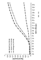
- partial areas or the entire area describe the time course of precipitation formation as a regression line become.
- the virtual signal intensity is in this embodiment in Depending on the slope of the regression line. This slope is a direct measure of the concentration of bound targets, i. the bigger the Slope of the regression line is, the more targets are bound.
- This slope is a direct measure of the concentration of bound targets, i. the bigger the Slope of the regression line is, the more targets are bound.
- the present invention corresponds to Regression line of a tangent to the curve function with which the Precipitation as a function of time can be described by the Turning point of the curve function.
- the inflection point of the curve function are through Determination of the maximum of the 1st derivative of the curve function determined.
- the Determines regression line in which the vertices of the curve function, with the The precipitation formation can be described as a function of time, by a Just to be connected.
- the vertices of the curve function are through Determination of the maxima of the 2nd derivative of the curve function determined.
- the inventive method has the further advantage that detection systems can be used, which are inexpensive and additionally inexpensive.
- detection systems can be used, which are inexpensive and additionally inexpensive.
- a camera with only 8 bits, i. 256 gray values, Gray depth can be used, which after evaluation and creation of a virtual image a real depth of field of 24 bit (16777216 gray values) or 48 bit (33554432 gray values) provides a significantly improved possibility the simultaneous detection of low and strong interactions of targets with probes on a probe array offers.
- a reference target of known concentration with the sample interacts with at least one probe of the probe array.
- This one Probes / reference-target interaction corresponding virtual signal intensity, determined as a function of the precipitation of precipitation in Dependence on time serves as a reference for the quantification of the rest Target concentrations corresponding to the respective virtual signal intensities, which are induced by the probe / target interactions, relative to the Concentration of the reference target concentration.
- the targets to be examined may be in any type of sample, preferably in one biological sample.
- the targets become prior to their detection and quantification by the method according to the invention isolated, purified, copied and / or amplified.
- a probe array used in the context of the present invention with at The immobilized probes are conventionally known Process produced.
- a probe array according to the present invention comprises a support that allows the formation of arrays of probes on its surface.
- Such a carrier can be made of materials selected from the Group consisting of glass, filters, electronic devices, polymers, metallic materials and the like, as well as combinations of these materials.
- the array comprises defined locations, so-called array elements, which particularly preferably arranged in a particular pattern, each Array element usually includes only one species of probes.
- the signal intensities at least every minute, preferably all 30 seconds, more preferably every 10 seconds.
- the signal intensities at least every minute, preferably all 30 seconds, more preferably every 10 seconds.
- the virtual signal intensity for an array element is, for example, through Multiplication of the detected signal intensity at a given time, preferably the signal intensity of the last measurement, with the slope of the for the Array element determined regression line and with the measurement time to the determined at certain time.
- the time is identical for the detection of signal intensity for all array elements.
- the method according to the invention is further characterized in that the targets are provided with markings which are the reaction of a soluble substrate to a sparingly soluble one Precipitate on the array element where a probe / target interaction has taken place, catalyze, or as a seed of crystallization for such Reactions work.
- the targets be provided directly with such markings.
- Another preferred way of coupling the targets with a label represents the synthetic or enzymatic addition of a homopolymeric region, For example, a polyA Seqeunz, the targets to form a continuous sequence, as described for example in US 6,103,474.
- labeling is preferably by sandwich hybridization labeled with a complementary to the homopolymer sequence Oligonucleotide with the variations described above.
- a signal amplification by amplification of parts of the target attached homopolymer sequence with simultaneous incorporation of labeled bases especially preferred via an RCA mechanism by using a circular single-stranded templates that complement the Homopolymer sequence.
- the targets are with a catalyst, provided in front of an enzyme, which is the transformation of a soluble Substrate catalysed in an insoluble product.
- the reaction leading to the formation of a Precipitation at the array elements leads, in this case, the conversion a soluble substrate in an insoluble product in the presence of a with the Targets coupled catalyst, preferably enzyme.
- the enzyme will preferably selected from the group consisting of horseradish peroxidase, alkaline phosphatase and glucose oxidase.
- the soluble substrate becomes preferably selected from the group consisting of 3,3'-diaminobenzidine, 4-chloro-1-naphthol, 3-amino-9-ethylcarbazole, p-phenylenediamine HCl / pyrocatechol, 3,3 ', 5,5'-tetramethylbenzidine, naphthol / pyronine, bromochloroindoyl phosphate, Nitrotetraazolium blue and phenazine methosulfate.
- a colorless soluble hydrogen donor e.g. 3,3'-diaminobenzidine
- the enzyme Horseradish peroxidase transfers hydrogen ions from the donors Hydrogen peroxide to form water.
- a metallic precipitate is particularly preferred.
- a silver compound preferably silver nitrate, silver lactate, Silver acetate or silver tartrate
- a reducing agent is preferably formaldehyde and / or hydroquinone used.
- the precipitation of the metallic compound takes place in Presence of metal clusters coupled to the targets or colloidal ones Metal particles, in particular gold clusters or colloidal gold particles. That in In this case, the metal clusters or colloidal metal particles with the Targets coupled markers dar.
- metal clusters or colloidal metal particles with the Targets coupled markers dar.
- silver nitrate in implemented elemental silver wherein silver ions from the solution of gold as Incubate seed and in a second step with the help of a Reducing agent such. Formaldehyde) can be reduced. This creates a insoluble precipitate of elemental silver.
- the precipitation of the metallic takes place Compound in the presence of polyanions coupled to the targets. If it If the target itself is not a poly anion, then there is the Possibility to use such for nucleation. That with a poly-anion For example, labeled target is exposed to a silver nitrate solution. Store it the silver cations on the poly-anion selectively. After that, be with one Reducing agent converted silver ions into elemental silver.
- the coupling of enzymes or catalysts or metal clusters or colloidal Metal particles or polyanions to the targets can be directly or via to the targets coupled anchor molecules take place. Basically there is no need that Target directly with the markers described above. It exists also the possibility of using suitable anchor molecules, e.g. Streptavidin, attached to the Target coupled, a subsequent coupling of the marker to to reach.
- suitable anchor molecules e.g. Streptavidin
- a conjugate consisting of the respective catalyst or crystallization seed and a specific binding partner for the anchor molecule also allows the Implementation of the procedures described above.
- the reaction leading to education precipitation at the array elements, then the bond of a specific binding partner to an anchor molecule coupled to the target represents.
- binding partner / anchor molecule pairs are preferably selected from the group consisting of biotin / avidin or streptavidin or anti-biotin antibodies, Digoxigenin / Antidigoxigenin Immunoglobulin, FITC / Anti-FITC Immunoglobulin and DNP / anti-DNP immunoglobulin.
- catalytically soluble substrate converted into an insoluble and precipitating product. by virtue of Close to the surface, the product becomes directly on the surface deposited and forms a solid, insensitive to various washes Rainfall.
- the markings in particular the enzymes or metal clusters or colloidal metal particles or Polyanions, to the targets before, during or after the interaction with the Probes are coupled.
- the interaction between the target and the probe is a hybridization between two nucleotide sequences.
- the hybridization of the targets with the probes arranged on a probe array takes place according to one of the known standard protocols (see, inter alia, Lottspeich and Zorbas, 1998).
- the resulting hybrids can be stabilized by covalent bonding, for example via psoralen intercalation and subsequent cross-linking, or as described in US Pat. No. 4,599,303, by noncovalent binding, for example by binding of intercalators.
- the interaction between the target and the probe is one Reaction between an antigenic structure and the corresponding antibody or a hypervariable section thereof or a reaction between a receptor and a corresponding ligand.
- the binding or recognition of targets by specific probes is common a spontaneous non-covalent reaction under optimal conditions. This includes are also non-covalent chemical bonds.
- the composition of the Medium and other chemical and physical factors affect the Speed and strength of binding. Thus, for example, at the Nucleic acid detection has a lower stringency and higher temperatures Rate and strength of bond between two not perfectly complementary strands.
- the optimization of the binding conditions is for antigen / antibody or Ligand / receptor interactions also required However, binding conditions are usually less specific.
- Evidence of the presence of a precipitate on an array element is made in one embodiment of the present invention by reflection, absorption or diffusion of a light beam, preferably a laser beam or a LED, by the precipitation. Modified due to its granular form the precipitate is the reflection of a ray of light. Furthermore, the precipitation leads to a strong light diffusion through conventional detection devices can be recorded. If the precipitation such as the Silver precipitate appears as a dark surface, can also reduce the absorption of Light detected and recorded. The resolution of the detection then depends from the number of pixels of the camera.
- the detection of the amplified by the specific reaction Areas by means of a very simple optical structure in transmitted light (contrast by shading) or reflected light (contrast by reflection).
- the Detected intensity of the shaded area is directly proportional to Occupancy density with markings such as gold particles and the Nucleation state of the particles.
- the electrical measurements can be made by means of conductivity measurements Microelectrode array arrangements or via an array of Micro capacitance sensors or via potential measurements by means of field-effect transistor arrays (FET arrays) take place.
- FET arrays field-effect transistor arrays
- For conductivity measurements by means of Microelectrodes will change the electrical resistance between two Electrodes in a deposition reaction (E. Braun, Y. Eichen, U. Sivan, G. Ben-Yoseph, Nature, 775, vol 391, 1998).
- For dielectric measurements with Microcapacity sensors will change the capacity of two to each other arranged electrodes (M. Madou, Fundamentals of Microfabrication, CRC Press, Boca Raton, 1997).
- For potential measurements using FET arrays the Change in the potential on the sensor surfaces measured (M. Madou, Fundamentals of Microfabrication, CRC Press, Boca Raton, 1997).
- the Evidence of the presence of a precipitate on an array element Scanning Electron Microscopy, Electron Probe Microanalysis (EPMA), Magneto-optical Kerr microscopy, magnetic force microscopy (MFM), atomic force microscopy (AFM), measurement of the mirage effect, scanning tunneling microscopy (STM) and / or ultrasound reflection tomography.
- EPMA Electron Probe Microanalysis
- MFM magnetic force microscopy
- AFM atomic force microscopy
- STM scanning tunneling microscopy
- STM scanning tunneling microscopy
- magneto-optical Kerr microscopy When using a substrate that is magnetic or with magnetic particles Marked, the detection of the reaction by magneto-optical Kerr microscopy or MFM.
- magneto-optic Kerr microscopy is the rotation of the polarization plane of the light by magnetic fields (Kerr and Faraday effect) (A.Hubert, R. Schafer, Magnetic Domains, Springer, 1998).
- the change in optical density through the substrate on the surface as a result of Reaction can be detected by means of the Mirrage effect.
- Mirrage effect is the local heating of a surface by absorption of a bundled Laser beam over the associated refractive index change measured.
- By the raster of the surface gives a picture of the local Surface Absorption Properties (A. Mandelis, Progress in Photothermal and Photoacoustic Science and Technology, Volume 1, Elsevier, New York 1992).
- One Another thermal spatially resolved method for the detection of Interaction reaction through the substrate is an array arrangement of Microthermophiles containing the enthalpies of crystallization of the Substrate deposits (J.M. Köhler, M. Zieren, Thermochimica acta, 25, vol 310, 1998).
- STM and AFM are also suitable for detecting reaction by substrate.
- AFM atomic force microscope
- a micro- or nano-tip feels its way Surfaces, thereby measuring the surface topography (E. Braun, Y. Eichen, U. Sivan, G. Ben-Yoseph, Nature, 775, vol 391, 1998).
- the magnetic force microscope MFM detects local magnetic via a nanotip Susceptibility differences (A.Hubert, R. Schfer, Magnetic Domains, Springer, 1998).
- the scanning tunneling microscop STM the nano tip of the Tunnel current measured to determine nano-surface topography (O. Marti, M. Amrein, STM and SFM in biology, Academic Press Inc., San Diego, 1993)
- Exotic procedures such as ultrasound reflection tomography can also be used.
- the tomographies are Procedures in which a 3-dimensional picture is collected piecemeal (F. Natterer, Mathematical Methods of Computer Tomography, Westdt. Vlg., Wiesbaden, 1997).
- the Measurement of the ultrasound reflection used to generate the tomogram V. Fleischer, F. Bergner, DGZfP NDT Conference Dresden 1997.
- a group of Pixels understood to be an illustration of the measured signal intensities for represents a probe array and directly to, for example, a screen or a printer can be sent for recording.
- a Group of pixels understood which is an illustration of according to the invention represents certain virtual signal intensities for a probe array and the also be displayed for example on a screen or a printer can.
- the detection device is preferably a camera, in particular a CCD or CMOS camera or similar cameras, usually the whole Area of the probe array records.
- the dynamic resolution extremely increase the measurement data even when using an 8-bit detection technique.
- the structure of a device necessary for this differs by the mechanical recording of a reaction chamber and a modified acquisition software.
- the software is characterized in that it handles the processing of successively recorded recordings allowed. These are the over the individual probe array elements determined gray values at any time determined. For all array elements, the virtual event intensity becomes dependent the precipitation is calculated from the time.
- the device according to the invention comprises additionally a light source, which is particularly preferably selected from the Group consisting of a laser, a light-emitting diode (LED) and a high pressure lamp.
- a light source which is particularly preferably selected from the Group consisting of a laser, a light-emitting diode (LED) and a high pressure lamp.
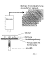
- the components of an exemplary construction of an inventive Apparatus for optically detecting precipitation consist of a low power (500mcd) light source, e.g. an LED, to the homogeneous Lighting and a detector, e.g. a CCD camera. Due to the Reinforcing effect on the catalytic deposition of the substrate, in particular When using a gold / silver system, the changes are the optical Properties of the surface are so pronounced that a simple flatbed scanner, a slide scanner or similar device for the detection of precipitation sufficient.
- a low power (500mcd) light source e.g. an LED
- a detector e.g. a CCD camera
- Typical detection times are well below 1s, while comparable sensitive CCD systems for the detection of fluorescence need about 10 s to 80 s, so that inexpensive consumer cameras can use their signal transmission the video standard.
- the miniaturization potential of such a design is very high, the whole System can be designed as an autonomous handheld device for field use. Further can the device according to the invention in a particularly preferred Embodiment be realized as a highly integrated autonomous unit. This is highly sensitive applications of microarrays, e.g. medical diagnostics, Forensics, bacterial screening, etc. also independent of medical or biological laboratories quickly performed by the layman.
- microarrays e.g. medical diagnostics, Forensics, bacterial screening, etc. also independent of medical or biological laboratories quickly performed by the layman.
- the analysis of the structure, expression and inheritance of immunologically relevant genes for transplantation and autoimmunity is of particular interest since highly polymorphic systems exist both for specific antigen recognition (histocompatibility antigens, T lymphocyte receptor) and for the effector mechanisms (antibodies, Fc receptors) subject to these complex genetic regulatory mechanisms.
- the strong transplantation antigens contribute to, for example, transplant rejection.
- These strong antigens are called major histocompatibility antigens and are genetically encoded within the major histocompatibility complex (MHC).
- MHC major histocompatibility complex
- HLA human leukocyte antigen
- the MHC which has so far only been detected in vertebrates, is referred to in humans as HLA (human leukocyte antigen).
- HLA complex is located on the short arm of human chromosome 6 (6p21.1-6p21.3) and covers an area of approximately 3500 kilobases.
- the HLA molecules can be subdivided into two classes (class I and class II), which are then subdivided into further subgroups.
- class I and class II The respective gene loci for the HLA products, which in their summation are responsible for the corresponding immunological properties of the organism, occur through the processes of heredity in very numerous allelic variations, the known number of which is steadily increasing.
- the number of possible allele types of the HLA system which can be accurately performed on an organism by serological and molecular biological analysis, allows any depth depending on the medical relevance of the cell species to be transplanted. The deeper the typing and the greater the subsequent match between donor and recipient, the less likely to be problems such as tissue incompatibility and transplant rejection. In addition to various transplants but also transfusions, disease associations and the forensic field for clear identification of persons play a role.
- the respective loci for the HLA products which are in their Summation for the corresponding immunological properties of the organism are responsible, through the processes of inheritance in very numerous Allelvariationen whose known number is steadily increasing, on.
- the number of possible Allelic typing of the HLA system attached to an organism by serological and molecular biological analysis can be made exactly depending on the medical relevance of the cell species to be transplanted, any Depth. The deeper the typing and the greater the following match between donor and recipient, the less problems there are Tissue intolerance and graft rejection expected. Since the Discovery of MHC molecules has numerous characterization methods developed and used the polymorphism of these molecules and their genes. A fundamental difference exists between biochemical, cellular and serological on the one hand and molecular biological techniques on the other Page.
- the former analyze by e.g. the use of specific antibodies excluding the expression products while the second group is replaced by e.g. the Techniques of hybridization and amplification of nucleic acids Sequence differences in the region of coding and non-coding sequences (Bidwell J., 1994 Advances in DNA-based HLA-typing methods. Immunol Today, 15 (7): 303-7).
- the invention described would be that after isolation, appropriate labeling and optionally Amplification for highlighting diagnostically relevant allele structures to one Sequence background of the genomic DNA of an individual, a massively parallel Hybridization against a probe array (DNA chip) of all known allelic variants with the goal of the lowest possible HLA typing. In doing so, by the described hybridization detection and the signal amplification in Combination with a simple detector compared to other methods, in minimal genomic typing at a minimum diagnostic time Use of known allele-specific probes in a very favorable economic framework accessible.
- an amino-modified oligonucleotide with a length of 20 nucleotides of the sequence 5'-NH 2 -CCTCTGCAGACTACTATTAC-3 ' was covalently immobilized at a defined location ("array element").
- array element 0.1 .mu.l of a 5 .mu.M solution of the oligonucleotide in 0.5 M phosphate buffer were deposited on the glass surface and finally dried at 37.degree.
- the covalent attachment of the deposited oligonucleotides with the epoxide groups on the glass surface was carried out by baking the probe arrays at 60 ° C for 30 minutes.
- the probe arrays were rinsed vigorously with distilled water and then washed for 30 min in 100 mM KCl. After further brief rinsing in 100 mM KCl and then distilled water, the probe arrays were dried for 10 minutes at 37 ° C.
- a complementary biotin-labeled 20 bp oligonucleotide sequence 5'-Bio-GTAATAGTAGTCTGCAGAGG-3 ' was used.
- the reaction was taken up in the following concentration steps in a buffer (0.25 M NaPO 4 , 4.5% SDS, 1 mM EDTA in 1xSSC) in a total volume of 50 ⁇ l each: 10 nM, 1 nM, 100 pM, 10 pM , 1 pm, 100 dc, 10 dc, 1 dc.
- Each concentration level was a prepared probe array in the Given hybridization solution.
- the hybridization mixture thus obtained became 5 min at 95 ° C and then incubated at 50 ° C for 60 min. Subsequently, the Shake each probe array for 10 min in 2xSSC + 0.2% SDS, 2xSSC and 0.2xSSC (Maniatis et al., 1989) and blown dry with compressed air.
- streptavidin-gold conjugate was (filled 52.5 g NaCl, 26.4 g NaH 2 PO 4 x H 2 O, 2.22 g NaOH with H 2 O to 1 total volume) in a 1:50 dilution in 6 ⁇ SSPE + 0.005% Triton solution was added to the probe array and incubated at 30 ° C for 15 min. The probe arrays were then shaken for 10 min in 2xSSC (17.5 g NaCl, 8.8 g Na citrate in 1 l H 2 O, adjusted to pH 7.0 with 10 N NaOH) + 0.2% SDS (sodium dodecyl sulfate), Washed 2xSSC and 0.2xSSC and blown dry with compressed air.
- gold conjugate of streptavidin also directly with gold particles modified targets were used.
- the detection limit was found to be ⁇ 10 pM.
- Example 2 Proof of Principle for the Use of the Method in Expression Profiling - Detection of Hybridization of Genomic RNA from Corynebacterium glutamicum to a Probe Array of 356 Probes
- DNA arrays are often used to measure the global physiological state of cells (expression profiling).
- RNA is isolated from the corresponding cells, labeled with a suitable method and hybridized against a probe array with complementary probes.
- the method of the invention for the detection of cellular mRNAs from Corynebacterium glutamicum was used.
- a probe array of 356 different amino-modified oligonucleotides of 25 or 30 bases in length and c-DNAs of different lengths was generated. All oligonucleotides were complementary to partial sequences of the aceA and icd genes.
- the probe arrays were constructed by arraying with the Micro-Grid I Arrayer from Biorobotics Ltd. (United Kingdom) according to the manufacturer's instructions by applying the amino-modified DNAs in a final concentration of 5 .mu.M in 0.5 M phosphate buffer on the slide and finally dried.
- the covalent attachment of the deposited oligonucleotides with the epoxide groups on the glass surface was carried out by baking the slides at 60 ° C. for 30 minutes. Subsequently, the slides were rinsed vigorously with distilled water and then washed for 30 min in 100 mM KCl. After further brief rinsing first in 100 mM KCl and then in distilled water, the probe arrays were dried at 37 ° C. for 10 min.
- RNA from Corynebacterium glutamicum was obtained by means of the Fast RNA kit (Bio 101) according to the manufacturer's instructions. 50 ⁇ g of RNA were biotinylated using Biotin Chem Link (Boehringer Mannheim, Germany) according to the manufacturer's instructions at 85 ° C. for 30 minutes. The RNA was then concentrated on Microcon-30 columns (Millipore) according to the manufacturer and then washed several times with deionized, RNase-free water. The eluate was concentrated in vacuo to 5 ul.
- the biotinylated RNA was taken up in 100 ⁇ l hybridization buffer (0.25 M NaPO 4 , 4.5% SDS, 1 mM EDTA in 1xSSC) and denatured for 3 minutes at 65 ° C.
- the DNA-coated surface of the slide was capped with a hybridization chamber (Hybrislip, Sigma, Deisenhofen, Germany).
- the slide was pre-heated to 50 ° C. on a thermo shaker with microtiter plate attachment (Eppendorf, Hamburg, Germany). Subsequently, the denatured hybridization solution was filled and sealed the hybridization chamber according to the manufacturer. The incubation was continued for 60 min at 50 ° C.
- hybridization solution was then removed, the hybridization chamber removed and the slides washed with shaking for 10 min at 30 ° C in 2xSSC + 0.2% SDS and 10 min at room temperature in 2xSSC and 0.2xSSC (Maniatis et al., 1989) and Blown dry with compressed air.
- a streptavidin-gold conjugate (EM.STP5, British BioCell International) was added to the slide in a 1:50 dilution in 6xSSPE + 0.005% Triton (Maniatis et al., 1989) and incubated at 30 ° C for 15 min. Subsequently, the probe arrays were washed with shaking for 10 min in 2xSSC + 0.2% SDS, 2xSSC and 0.2xSSC and dried with compressed air. The subsequent amplification of the gold particles now immobilized on the probe array was carried out with the LM / EM Silver Enhancing Kit (SEKL15, British BioCell International).
- oligonucleotides each with a length of 16 nucleotides were deposited at defined locations with a MicroGrid II arrayer (BioRobotics) and covalently immobilized (array elements).
- the sequences of the oligonucleotides were as follows (each with a 3'-NH 2 modification):
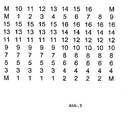
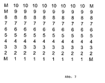
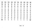
- Each of the 16 Oligonucleotide probes were in at least 5-fold repetition on the Probe array filed (array structure: see Fig. 5).
- the probes had one 0.2 mm apart, the entire probe array covered a 2 mm x 2 area mm. In total, more than 100 identical slides could be used per slide Probe arrays are generated.
- the probes were deposited from a respective 10 ⁇ M solution of the oligonucleotides in 0.1 M phosphate buffer / 5% sodium sulfate. After deposition and drying, the probes were covalently linked to the epoxide groups on the glass surface by baking for 30 minutes at 60 ° C.
- chips 3.25 mm x 3.25 mm pieces of glass
- the complementary biotin-labeled 16-bp oligonucleotides were available as targets for all 16 oligonucleotide probes.
- the target "9b" complementary to the oligonucleotide probe 9 is listed here with the following sequence:
- the hybridization reaction was carried out in 6x SSPE buffer (52.59 g NaCl, 8.28 g NaH 2 PO 4 ⁇ H 2 O, 2.22 g EDTA ⁇ 2H 2 O in 1 1 H 2 O bidest., PH 7 , 4 adjusted with NaOH) / 0.1% SDS in a total volume of 70 .mu.l with the target concentration levels 100 nM, 10 nM, 1nM, 100 pM, 10 pM, 1 pM performed.
- a chip with the probe array was added to the hybridization solution, heated at 95 ° C. for 5 minutes, then incubated at 30 ° C. for 60 minutes with shaking.
- the chip was transferred to a new reaction vessel with 500 ⁇ l hybridization buffer (without target) and washed for 10 min at 55 ° C or 60 ° C with shaking. Then the chips were then each 10 min in 2x SSC / 0.2% SDS (500 ul at 30 ° C), 2x SSC (500 ul at 20 ° C) and 0.2x SSC (500 ul at 20 ° C) washed with shaking and then dried (Eppendorf Concentrator).
- the hybridized and dried chips were in a new reaction vessel with ⁇ l of strepiavidin-gold conjugate solution in 6x SSPE / 0.1% SDS buffer and incubated there at 15 ° C for 15 min.
- streptavidin-gold conjugate were 5 nm size gold particles used (British Biocell International, EM.STP5).
- the conjugate was present in the experiment in a concentration of 500 pg streptavidin / ⁇ l.
- the chips were incubated for 10 min in 2x SSC / 0.2% SDS (500 ⁇ l at 30 ° C), 2x SSC (500 ⁇ l at 20 ° C) and 0.2x SSC (500 ⁇ l at 20 ° C) with shaking washed and then dried (Eppendorf Concentrator).
- streptavidin-gold conjugate coupling also performed directly in the hybridization solution. This was after the 60-minute hybridization, the streptavidin-gold conjugate directly into the Hybridization solution was added and then incubated at 30 ° C for a further 15 min. Thereafter, the chip was placed in a new reaction vessel with 500 ⁇ l Hybridization buffer (without target) transferred and at 55 ° C or 60 ° C for 10 min washed with shaking.
- the chips were transferred to a new reaction vessel and with shaking for 10 min at 25 ° C in about 100 ul of a silver developing solution (British Biocell International, SEKL15).
- the incubation solution was prepared by mixing one drop each of initiator and enhancer solution. Subsequently, the chip was washed for 2 min in 500 ul 0.2 x SSC and dried (Eppendorf Concentrator).
- oligonucleotides on an epoxidized 3D slide (75 mm x 25 mm) glass surface (Fa. Ealia) were treated with a MicroGrid II arrayer (BioRobotics) 10 amino-modified oligonucleotides (probes) with a length of 16 to 22 Nucleotides deposited at defined sites and covalently immobilized (Array elements).
- the 10 probes are divided into 5 pairs, with the first each the Wild type and the second represents the mutation.
- the probe pair 1 and 2 represents one Point mutation, pair 3 and 4 a deletion and the pairs 5/6, 7/8, 9/10 Insertions.
- the sequences of the oligonucleotides were as follows:
- Probe pair 3 (wild-type) and 4 (deletion) contain the most common mutation (70%). all cases) that codes for cystic fibrosis.
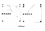
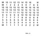
- Each of the 10th Oligonucleotide probes were in 8 to 10-fold repetition on the Probe array filed (array structure: see Figure 7).
- the probes had one 0.2 mm apart, the entire probe array covered a 2 mm x 2 area mm. In total, more than 100 identical slides could be used per slide Probe arrays are generated.
- the probes were deposited from a respective 10 ⁇ M solution of the oligonucleotides in 0.1 M phosphate buffer / 5% sodium sulfate. After deposition and drying, the probes were covalently linked to the epoxide groups on the glass surface by baking for 30 minutes at 60 ° C.
- chips 3.25 mm x 3.25 mm pieces of glass
- Target 1 covered the probe pair 1 and 2
- target 2 the pair 3 and 4
- target 3 the probe pairs 5/6, 7/8 and 9/10.
- sequences of the targets were:
- the hybridization reaction was carried out in 6 x SSPE buffer (52.59 g NaCl, 8.28 g NaH 2 PO 4 .H 2 O, 2.22 g EDTA x 2H 2 O in 11H 2 O bidist., To pH 7 4 adjusted with NaOH) / 0.1% SDS in a total volume of 70 ⁇ l with different target concentration steps.
- a chip with the probe array was added to the hybridization solution, heated for 5 min at 95 ° C, then incubated for 60 min at 30 ° C with shaking.
- streptavidin-gold conjugate After hybridization for 60 minutes, a streptavidin-gold conjugate became direct in the hybridization solution and then at 30 ° C for an additional 15 minutes incubated.
- streptavidin-gold conjugate gold particles of size 5 nm used (British Biocell International, EM.STP5). The conjugate was in the experiment in a concentration of 500 pg streptavidin / ⁇ l.
- the chip became a new one Transfer reaction vessel with 500 ⁇ l hybridization buffer (without target) and 10 Washed at 55 ° C with shaking. Then the chips were then 10 min each in 2x SSC / 0.2% SDS (500 ⁇ l at 30 ° C.), 2 ⁇ SSC (500 ⁇ l at 20 ° C.) and 0.2x SSC (500 ⁇ l at 20 ° C) with shaking and then dried (Eppendorf Concentrator).
- the chips were transferred to a new reaction vessel and with shaking for 10 min at 25 ° C in about 100 ul of a silver developing solution (British Biocell International, SEKL15).
- the incubation solution was prepared by mixing one drop each of initiator and enhancer solution. Subsequently, the chip was washed for 2 min in 500 ul 0.2x SSC and dried (Eppendorf Concentrator).
- oligonucleotides each with a length of 16 nucleotides were deposited at defined locations with a MicroGrid II arrayer (BioRobotics) and covalently immobilized (array elements).
- the sequences of the oligonucleotides were as follows:
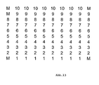
- Each of the 16 Oligonucleotide probes were in at least 5-fold repetition on the Probe array filed (array structure: see Figure 11).
- the probes had one 0.2 mm apart, the entire probe array covered a 2 mm x 2 area mm. In total, more than 100 identical slides could be used per slide Probe arrays are generated.
- the probes were deposited from a respective 10 ⁇ M solution of the oligonucleotides in 0.1 M phosphate buffer / 5% sodium sulfate. After deposition and drying, the probes were covalently linked to the epoxide groups on the glass surface by baking for 30 minutes at 60 ° C.
- chips 3.25 mm x 3.25 mm pieces of glass
- the complementary 36-bp oligonucleotides were available as targets for all 16 oligonucleotide probes, which had a polyA tail as a modification at the 3 'end.
- the target "9c" complementary to the oligonucleotide probe 9 is listed here with the following sequence:
- the hybridization reaction was carried out in 6x SSPE buffer (52.59 g NaCl, 8.28 g NaH 2 PO 4 ⁇ H 2 O, 2.22 g EDTA ⁇ 2H 2 O in 1 1 H 2 O bidest., PH 7 4 adjusted with NaOH) / 0.1% SDS in a total volume of 70 ⁇ l with different target concentration steps.
- a chip with the probe array was added to the hybridization solution, heated at 95 ° C. for 5 minutes, then incubated at 30 ° C. for 60 minutes with shaking. Thereafter, the T20 nanogold conjugate was added to the hybridization solution at various dilution levels and incubated at 30 ° C for a further 30 minutes.
- the chip was placed in a new reaction vessel with 500 ⁇ l Hybridization buffer (without target and T20 nanogold) transferred and 10 min at 55 ° C or 60 ° C with shaking. Finally, the chips were then 10 min each in 2x SSC / 0.2% SDS (500 ⁇ l at 30 ° C.), 2 ⁇ SSC (500 ⁇ l at 20 ° C.) and 0.2x SSC (500 ⁇ l at 20 ° C) with shaking and then dried (Eppendorf Concentrator).
- the chips were transferred to a new reaction vessel and with shaking for 10 min at 25 ° C in about 100 ul of a silver developing solution (British Biocell International, SEKL15).
- the incubation solution was prepared by mixing one drop each of initiator and enhancer solution. Subsequently, the chip was washed for 2 min in 500 ul 0.2x SSC and dried (Eppendorf Concentrator).
- oligonucleotides on an epoxidized 3D slide (75 mm x 25 mm) glass surface (Fa. Ealia) were treated with a MicroGrid II arrayer (BioRobotics) 10 amino-modified oligonucleotides (probes) with a length of 16 to 22 Nucleotides deposited at defined sites and covalently immobilized (Array elements).
- the 10 probes are divided into 5 pairs, with the first each the Wild type and the second represents the mutation.
- the probe pair 1 and 2 represents one Point mutation, pair 3 and 4 a deletion and the pairs 5/6, 7/8, 9/10 Insertions.
- the sequences of the oligonucleotides were as follows:
- Probe pair 3 (wild-type) and 4 (deletion) contain the most common mutation (70%). all cases) that codes for cystic fibrosis.
- Each of the 10th Oligonucleotide probes were in 8 to 10-fold repetition on the Probe array deposited (array structure: see Figure 13).
- the probes had one 0.2 mm apart, the entire probe array covered a 2 mm x 2 area mm. In total, more than 100 identical slides could be used per slide Probe arrays are generated.
- the probes were deposited from a respective 10 ⁇ M solution of the oligonucleotides in 0.1 M phosphate buffer / 5% sodium sulfate. After deposition and drying, the probes were covalently linked to the epoxide groups on the glass surface by baking for 30 minutes at 60 ° C.
- chips 3.25 mm x 3.25 mm pieces of glass
- Target 1 covered the probe pair 1 and 2
- target 2 the pair 3 and 4
- target 3 the probe pairs 5/6, 7/8 and 9/10.
- sequences of the targets were:
- the hybridization reaction was carried out in 6 ⁇ SSPE buffer (52.59 g NaCl, 8.28 g NaH 2 PO 4 ⁇ H 2 O, 2.22 g EDTA ⁇ 2H 2 O in 11 H 2 O bidistilled, to pH 7, 4 adjusted with NaOH) / 0.1% SDS in a total volume of 70 ⁇ l with all three targets in a 100 pM concentration.
- a chip with the probe array was added to the hybridization solution, heated for 5 min at 95 ° C, then incubated for 60 min at 30 ° C with shaking.
- a streptavidin-gold conjugate was added directly to the hybridization solution and then incubated at 30 ° C for an additional 15 minutes.
- streptavidin-gold conjugate gold particles of size 5 nm were used (British Biocell International, EM.STP5). The conjugate was present in the experiment in a concentration of 500 pg streptavidin / ⁇ l.
- the chip became a new one Transfer reaction vessel with 500 ⁇ l hybridization buffer (without target) and 10 Washed at 55 ° C with shaking. Then the chips were then 10 min each in 2x SSC / 0.2% SDS (500 ⁇ l at 30 ° C.), 2 ⁇ SSC (500 ⁇ l at 20 ° C.) and 0.2x SSC (500 ⁇ l at 20 ° C) with shaking and then dried (Eppendorf Concentrator).
- the chips were placed in a sealed reaction chamber (see Figure 1) and fixed with a silver developing solution (British Biocell International, SECL15).
- the incubation solution was mixed made of one drop of initiator and enhancer solution.
- the 30-minute Incubation at 21 ° C was the time course of silver amplification documented by one photo per minute (the light source was a red LED).
- Figs. 14a and 14b the chip photos are 5 and 10 minutes after the beginning, respectively the silver reinforcement was shown (hybridized with the target 2).
- Fig. 15 shows the time course of this reaction.
- oligonucleotides on an epoxidized 3D slide (75 mm x 25 mm) glass surface (Fa. Ealia) were treated with a MicroGrid II arrayer (BioRobotics) 10 amino-modified oligonucleotides (probes) with a length of 16 to 22 Nucleotides deposited at defined sites and covalently immobilized (Array elements).
- the 10 probes are divided into 5 pairs, with the first each the Wild type and the second represents the mutation.
- the probe pair 1 and 2 represents one Point mutation, pair 3 and 4 a deletion and the pairs 5/6, 7/8, 9/10 Insertions.
- the sequences of the oligonucleotides were as follows:
- Probe pair 3 (wild-type) and 4 (deletion) contain the most common mutation (70%). all cases) that codes for cystic fibrosis.
- Each of the 10th Oligonucleotide probes were in 8 to 10-fold repetition on the Probe array filed (array structure: see Fig. 16).
- the probes had one 0.2 mm apart, the entire probe array covered a 2 mm x 2 area mm. In total, more than 100 identical slides could be used per slide Probe arrays are generated.
- the probes were deposited from a respective 10 ⁇ M solution of the oligonucleotides in 0.1 M phosphate buffer / 5% sodium sulfate. After deposition and drying, the probes were covalently linked to the epoxide groups on the glass surface by baking for 30 minutes at 60 ° C.
- chips 3.25 mm x 3.25 mm pieces of glass
- Target 1 covered the probe pair 1 and 2
- target 2 the pair 3 and 4
- target 3 the probe pairs 5/6, 7/8 and 9/10.
- sequences of the targets were:
- the hybridization reaction was carried out in 6 ⁇ SSPE buffer (52.59 g NaCl, 8.28 g NaH 2 PO 4 ⁇ H 2 O, 2.22 g EDTA ⁇ 2H 2 O in 1 l H 2 O bidistilled, to pH 7, 4 adjusted with NaOH) / 0.1% SDS in a total volume of 70 ⁇ l with the target 3 in a 100 nM and 1 nM concentration.
- a chip with the probe array was added to the hybridization solution, heated for 5 min at 95 ° C, then incubated for 60 min at 55 ° C with shaking.
- the hybridized chips were then incubated for 10 min in 2 x SSC / 0.2% SDS (500 ⁇ l at 30 ° C.), 2 ⁇ SSC (500 ⁇ l at 20 ° C.) and 0.2 ⁇ SSC (500 ⁇ l at 20 ° C.) Washed shake.
- a new reaction vessel closed a 15 minute Incubate (at 30 ° C) with a streptavidin-gold conjugate.
- streptavidin-gold conjugate gold particles of size 5 nm were used (British Biocell International, EM.STP5). The conjugate was in a concentration of 125 ⁇ g streptavidin / ⁇ l.
- the chip was transferred to a new reaction vessel and each 10 min in 2 x SSC / 0.2% SDS (500 ⁇ l at 30 ° C), 2 x SSC (500 ⁇ l at 20 ° C) and 0.2 x SSC (500 ⁇ l at 20 ° C) with shaking and then dried (Eppendorf Concentrator).
- the chips were placed in a sealed reaction chamber (see Figure 1) fixed and with a silver developing solution (British Biocell International, SEKL15).
- the incubation solution was passed through Mix each one drop of initiator and enhancer solution. While the 20 minute incubation at 27 ° C, the time course of Silver reinforcement through a photo series (every 10 s a picture) documented (as Light source served a red LED). The pictures were subsequently taken with the Image evaluation software IconoClust (Clondiag) evaluated.
- Figure 1 Device for the qualitative and / or quantitative detection of Interactions between probes and targets.
- FIG. 1 Playback of the time course of the one shown in Figure 3 Hybridization results.
- Figure 4 Evidence of hybridization of genomic RNA from Corynebacterium glutamicum to a probe array of 356 probes - resulting pattern after 15 minutes incubation.
- the numbers 1-16 each stand for an oligonucleotide probe which is in 5 or 6 times Repetition is stored on the array; "M” stands for a brand mix (contains, inter alia, an immobilized biotin-labeled oligonucleotide); 1 position on + the array is not occupied.
- Numbers 1-10 each represent an oligonucleotide probe that is 8-10 times larger Repetition is stored on the array; "M” stands for a brand mixture (contains et al an immobilized biotin-labeled oligonucleotide).
- Figure 8 Probe array after hybridization, conjugation and Silver reinforcement (for array construction see Fig. 7).
- Target 1 100 pM hybridization at 30 ° C followed by direct Streptavidin-gold conjugate addition (500 pg / ⁇ l); 1st washing step at 55 ° C, Silver reinforcement: 10 min at 25 ° C.
- Figure 9 Probe array after hybridization, conjugation and Silver reinforcement (for array construction see Fig. 1).
- Target 2 100 pM hybridization at 30 ° C followed by direct Streptavidin-gold conjugate addition (500 pg / ⁇ l); 1st washing step at 55 ° C, Silver reinforcement: 10 min at 25 ° C.
- Figure 10 Probe array after hybridization, conjugation and Silver reinforcement (for array construction see Fig. 1).
- Target 3 100 pM hybridization at 30 ° C followed by direct Streptavidin-gold conjugate addition (500 pg / ⁇ l); 1st washing step at 55 ° C, Silver reinforcement: 10 min at 25 ° C.
- the numbers 1-16 each stand for an oligonucleotide probe which is in 5 or 6 times Repetition is stored on the array; "M” stands for a brand mix (contains, inter alia, an immobilized biotin-labeled oligonucleotide); 1 position up the array is not occupied.
- Figure 12 Probe array after hybridization, conjugation and Silver reinforcement of Example 5 (for array construction, see Fig. 11).
- Target 9c (1 nM) - 60 min hybridization at 30 ° C; then adding T20 nanogold (1: 100 dilution) and further incubation for 30 min at 30 ° C, 1st wash at 55 ° C; Silver reinforcement: 10 min at 25 ° C.
- Numbers 1-10 each represent an oligonucleotide probe that is 8-10 times larger Repetition is stored on the array; "M” stands for a brand mixture (contains et al an immobilized biotin-labeled oligonucleotide).
- Figure 14 Probe array after hybridization and conjugation (for Array construction cf. Fig. 1).
- FIG 15 Time course of the silver reinforcement (see figures 13 and 14).
- each measurement point is an average of 10 spot repetitions pm: Perfect match probe (Probe No. 3) mm: mismatch probe (probe No. 4)
- Target 2 100 pM hybridization at 30 ° C followed by direct Streptavidin-gold conjugate addition (500 pg / ⁇ l)
- the numbers 1-10 each stand for an oligonucleotide probe, which is filed in 8 to 10 repetitions on the array; "M” stands for a brand mixture (contains inter alia an immobilized biotin-labeled oligonucleotide in the concentration 10 ⁇ M).
- the hybridized target 3 was complementary to probes 5, 7, 9 (each perfect match) and probes 6, 8, 10 (each mismatch with one insertion).
- Each probe point is an average of 7 to 10 spot repetitions.
- pm Perfect match probe (Probe No. 5)
- mm mismatch probe (probe No. 6)
- a Target 3 (1 nM): hybridization at 55 ° C; Streptavidin-gold conjugate (125 pg / ⁇ l)
- b Target 3 (100 nM): hybridization at 55 ° C; Streptavidin-gold conjugate (125 pg / ⁇ l)
- Figure 20 Determined linear regression lines after hybridization with Target 3 (100 nM and 1 nM) for two selected probes of the probe array, valid for the Range from 4 to 13 minutes after silver enhancement begins.
Landscapes
- Chemical & Material Sciences (AREA)
- Organic Chemistry (AREA)
- Life Sciences & Earth Sciences (AREA)
- Zoology (AREA)
- Wood Science & Technology (AREA)
- Proteomics, Peptides & Aminoacids (AREA)
- Health & Medical Sciences (AREA)
- Engineering & Computer Science (AREA)
- Bioinformatics & Cheminformatics (AREA)
- General Engineering & Computer Science (AREA)
- Microbiology (AREA)
- Molecular Biology (AREA)
- Analytical Chemistry (AREA)
- Biotechnology (AREA)
- Physics & Mathematics (AREA)
- Biochemistry (AREA)
- Biophysics (AREA)
- Immunology (AREA)
- General Health & Medical Sciences (AREA)
- Genetics & Genomics (AREA)
- Measuring Or Testing Involving Enzymes Or Micro-Organisms (AREA)
- Apparatus Associated With Microorganisms And Enzymes (AREA)
- Investigating Or Analyzing Non-Biological Materials By The Use Of Chemical Means (AREA)
- Investigating Or Analyzing Materials By The Use Of Electric Means (AREA)
- Investigating Or Analysing Biological Materials (AREA)
- Analysing Materials By The Use Of Radiation (AREA)
- Investigating Or Analyzing Materials By The Use Of Ultrasonic Waves (AREA)
- Investigating Or Analysing Materials By Optical Means (AREA)
Claims (39)
- Procédé de détection qualitative et/ou quantitative de cibles dans un échantillon par interactions moléculaires entre sondes et cibles sur des arrays de sondes, comportant les étapes suivantes :a) réalisation d'un array de sondes comportant des sondes immobilisées à des endroits définis ;b) interaction entre les cibles et les sondes disposées sur l'array de sondes ;c) réalisation d'une réaction conduisant à un précipité aux éléments de l'array auxquels une interaction s'est produite ;d) détection de l'évolution dans le temps de la formation du précipité sur les éléments de l'array sous la forme d'intensités de signaux ;e) détermination d'une intensité virtuelle de signal pour un élément de l'array à l'aide d'une fonction de courbe qui décrit la formation de précipité en tant que fonction du temps.
- Procédé selon la revendication 1,
caractérisé en ce que l'intensité virtuelle de signal pour un élément de l'array est déterminée en fonction de la pente d'une droite de régression qui décrit la formation de précipité en tant que fonction du temps. - Procédé selon la revendication 2,
caractérisé en ce que la droite de régression est déterminée dans la zone de croissance exponentielle de la formation de précipité dans le temps sur l'élément de l'array. - Procédé selon la revendication 2 ou 3,
caractérisé en ce que l'intensité virtuelle de signal pour un élément de l'array est définie par multiplication de l'intensité de signal détectée à un instant déterminé, de préférence l'intensité de signal de la dernière mesure, par la pente de la droite de régression déterminée pour l'élément de l'array, ainsi que par la durée de mesure à l'instant déterminé. - Procédé selon l'une quelconque des revendications précédentes,
caractérisé en ce que dans l'échantillon se présente une cible de référence de concentration connue qui interagit avec au moins une sonde de l'array de sondes. - Procédé selon l'une quelconque des revendications précédentes,
caractérisé en ce que pour la détection de l'évolution dans le temps de la formation de précipité aux éléments de l'array, les intensités de signal sont enregistrées au moins chaque minute, de préférence toutes les 30 secondes, de manière particulièrement préférée, toutes les 10 secondes. - Procédé selon l'une quelconque des revendications précédentes,
caractérisé en ce que la réaction conduisant à la formation d'un précipité aux éléments de l'array est la transformation d'un substrat soluble en un produit insoluble en présence d'un catalyseur couplé aux cibles. - Procédé selon la revendication 7,
caractérisé en ce que le catalyseur est une enzyme. - Procédé selon la revendication 7 ou 8,
caractérisé en ce que l'enzyme est choisie dans le groupe se composant de la peroxidase de raifort, la phosphatase alcaline et la glucose oxidase. - Procédé selon l'une quelconque des revendications 7 à 9,
caractérisé en ce que le substrat soluble est choisi dans le groupe se composant de 3,3'-diaminobenzidine, 4-chlore-1-naphthole, 3-amino-9-éthyle carbazole, p-diamine phénylène-HCl/pyrocatéchole, 3,3',5,5'-tétraméthylbenzidine, naphthole / pyronine, bromchlorindoylphosphate, bleu de nitrotétraazolium et méthosulfate de phénazine. - Procédé selon l'une quelconque des revendications 1 à 6,
caractérisé en ce que la réaction conduisant à la formation d'un précipité aux éléments de l'array est la transformation d'un substrat soluble en un précipité métallique. - Procédé selon la revendication 11,
caractérisé en ce que la réaction conduisant à la formation d'un précipité aux éléments de l'array est la réduction chimique d'un composé d'argent, de préférence de nitrate d'argent, de lactate d'argent, d'acétate d'argent ou de tartrate d'argent, en argent élémentaire. - Procédé selon la revendication 12,
caractérisé en ce que le réducteur est choisi dans le groupe se composant de formaldéhyde et d'hydroquinone. - Procédé selon l'une quelconque des revendications 11 à 13,
caractérisé en ce que la transformation d'un substrat soluble en un précipité métallique s'effectue en présence de clusters métalliques ou de particules métalliques colloïdales couplés aux cibles. - Procédé selon la revendication 14,
caractérisé en ce que la transformation d'un substrat soluble en un précipité métallique s'effectue en présence de clusters d'or ou de particules d'or colloïdales. - Procédé selon l'une quelconque des revendications 11 à 13,
caractérisé en ce que la transformation d'un substrat soluble en un précipité métallique s'effectue en présence de polyanions couplés aux cibles. - Procédé selon l'une quelconque des revendications 7 à 16,
caractérisé en ce que les catalyseurs ou clusters métalliques ou particules métalliques colloïdales ou polyanions sont couplés aux cibles avant, pendant ou après l'interaction avec les sondes. - Procédé selon l'une quelconque des revendications 7 à 17,
caractérisé en ce que le couplage des enzymes ou clusters métalliques ou particules métalliques colloïdales ou polyanions aux cibles s'effectue directement ou par des molécules d'ancrage couplées aux cibles. - Procédé selon la revendication 18,
caractérisé en ce que la molécule d'ancrage est choisie dans le groupe consistant en la streptavidine ou un anticorps. - Procédé selon l'une quelconque des revendications 1 à 6,
caractérisé en ce que la réaction conduisant à la formation d'un précipité aux éléments de l'array représente la liaison d'un partenaire de liaison spécifique avec une molécule d'ancrage couplée à la cible. - Procédé selon la revendication 20,
caractérisé en ce que les paires partenaire de liaison / molécule d'ancrage sont choisies dans le groupe se composant de biotine / avidine ou streptavidine ou anticorps anti-biotine, digoxigénine / immunoglobuline anti-digoxigénine, FITC / immunoglobuline anti-FITC et DNP / immunoglobuline anti-DNP. - Procédé selon l'une quelconque des revendications précédentes,
caractérisé en ce que les cibles sont pourvues directement d'un marquage. - Procédé selon l'une quelconque des revendications 1 à 21,
caractérisé en ce qu'un marquage des cibles s'effectue par des réactions sandwich ou des hybridations sandwich des sondes interagissant avec les cibles et un composé marqué. - Procédé selon l'une quelconque des revendications 1 à 21,
caractérisé en ce qu'un marquage des cibles s'effectue par ajout aux cibles d'une séquence nucléotidique homopolymère en formant une séquence continue, puis par hybridation sandwich avec un oligonucléotide marqué complémentaire à la séquence nucléotidique homopolymère. - Procédé selon l'une quelconque des revendications précédentes,
caractérisé en ce que l'interaction entre la cible et la sonde est une hybridation entre deux séquences nucléotidiques. - Procédé selon l'une quelconque des revendications 1 à 24,
caractérisé en ce que l'interaction entre la cible et la sonde est une réaction entre une structure antigénique et l'anticorps correspondant ou une partie hypervariable de celui-ci. - Procédé selon l'une quelconque des revendications précédentes,
caractérisé en ce que l'interaction entre la cible et la sonde est une réaction entre un récepteur et un ligand correspondant. - Procédé selon l'une quelconque des revendications précédentes,
caractérisé en ce que la détection de la présence d'un précipité sur un élément de l'array s'effectue par réflexion, absorption ou diffusion d'un rayon lumineux, de préférence d'un rayon laser ou d'une diode lumineuse, par le précipité. - Procédé selon l'une quelconque des revendications 1 à 27,
caractérisé en ce que la détection de la présence d'un précipité sur un élément de l'array s'effectue électriquement. - Procédé selon la revendication 29,
caractérisé en ce que la détection électrique s'effectue par des mesures de conductibilité, des mesures de capacité ou des mesures de potentiels. - Procédé selon l'une quelconque des revendications 1 à 27,
caractérisé en ce que la détection de la présence d'un précipité sur un élément de l'array s'effectue par autoradiographie, fluorographie et/ou autoradiographie indirecte. - Procédé selon l'une quelconque des revendications 1 à 27,
caractérisé en ce que la détection de la présence d'un précipité sur un élément de l'array s'effectue par microscopie électronique à balayage, microanalyse par sonde électronique (EPMA), microscopie Kerr magnéto-optique, microscopie à force magnétique (MFM), microscopie à force atomique (AFM), mesure de l'effet mirage, microscopie à balayage à effet tunnel (STM) et/ou tomographie de réflexion d'ultrasons. - Procédé selon l'une quelconque des revendications précédentes, comprenant les étapes suivantes :détection de l'évolution dans le temps de la formation de précipité aux éléments de l'array par la prise d'images par une caméra ;transformation en forme numérique de l'information analogique contenue dans les images ;calcul d'une intensité virtuelle de signal pour chaque élément de l'array à l'aide d'une fonction de courbe décrivant la formation de précipité en tant que fonction du temps ;transformation des intensités virtuelles de signal en une image synthétique qui représente les intensités virtuelles de signal de tous les éléments de l'array.
- Dispositif pour appliquer le procédé selon l'une quelconque des revendications 1 à 33, comprenant :a) un substrat d'array avec un array de sondes,b) une chambre de réaction,c) un dispositif pour la détection d'un précipité sur un élément de l'array auquel une interaction entre cibles et sondes s'est produite, etd) un ordinateur programmé pour :collecter les intensités de signal enregistrées par le dispositif de détection ;assurer le traitement des intensités de signal enregistrées successivement de manière à ce que l'évolution dans le temps de la formation de précipité sur un élément de l'array est déterminée dans le temps et qu'une intensité virtuelle de signal est déterminée à l'aide d'une fonction de courbe qui décrit la formation de précipité en tant que fonction du temps ; etassurer, le cas échéant, la transformation des intensités virtuelles de signal en une image analogique.
- Dispositif selon la revendication 34,
caractérisé en ce que le dispositif de détection est une caméra. - Dispositif selon la revendication 35,
caractérisé en ce que la caméra est une caméra CCD ou CMOS. - Dispositif selon l'une quelconque des revendications 34 à 36,
caractérisé en ce que le dispositif comprend en outre une source lumineuse. - Dispositif selon la revendication 37,
caractérisé en ce que la source lumineuse est choisie dans le groupe se composant d'un laser, d'une diode électroluminescente (DEL) et d'une lampe haute pression. - Dispositif selon l'une quelconque des revendications 24 à 38,
caractérisé en ce que le dispositif se présente en tant qu'unité autonome fortement intégrée.
Applications Claiming Priority (3)
| Application Number | Priority Date | Filing Date | Title |
|---|---|---|---|
| DE10033334 | 2000-07-01 | ||
| DE10033334 | 2000-07-01 | ||
| PCT/EP2001/007575 WO2002002810A2 (fr) | 2000-07-01 | 2001-07-02 | Procede de detection qualitative et/ou quantitative d'interactions moleculaires sur des microplaques |
Publications (2)
| Publication Number | Publication Date |
|---|---|
| EP1299563A2 EP1299563A2 (fr) | 2003-04-09 |
| EP1299563B1 true EP1299563B1 (fr) | 2005-12-14 |
Family
ID=7648308
Family Applications (1)
| Application Number | Title | Priority Date | Filing Date |
|---|---|---|---|
| EP01954002A Expired - Lifetime EP1299563B1 (fr) | 2000-07-01 | 2001-07-02 | Procede de detection qualitative et/ou quantitative d'interactions moleculaires sur des microplaques |
Country Status (9)
| Country | Link |
|---|---|
| US (1) | US20030215891A1 (fr) |
| EP (1) | EP1299563B1 (fr) |
| JP (1) | JP2004501667A (fr) |
| AT (1) | ATE312944T1 (fr) |
| AU (1) | AU7637601A (fr) |
| CA (1) | CA2412915C (fr) |
| DE (1) | DE50108396D1 (fr) |
| ES (1) | ES2256274T3 (fr) |
| WO (1) | WO2002002810A2 (fr) |
Cited By (1)
| Publication number | Priority date | Publication date | Assignee | Title |
|---|---|---|---|---|
| CN103713134A (zh) * | 2013-12-17 | 2014-04-09 | 武汉大学 | 一种可视化检测病毒的检测试剂盒及其检测方法 |
Families Citing this family (18)
| Publication number | Priority date | Publication date | Assignee | Title |
|---|---|---|---|---|
| DE10201463B4 (de) | 2002-01-16 | 2005-07-21 | Clondiag Chip Technologies Gmbh | Reaktionsgefäß zur Durchführung von Array-Verfahren |
| DE10315074A1 (de) | 2003-04-02 | 2004-10-14 | Clondiag Chip Technologies Gmbh | Vorrichtung zur Vervielfältigung und zum Nachweis von Nukleinsäuren |
| DE10322912A1 (de) * | 2003-05-21 | 2004-12-16 | Bayer Technology Services Gmbh | Verfahren zum Nachweis von Nukleinsäuren |
| US7642064B2 (en) | 2003-06-24 | 2010-01-05 | Ventana Medical Systems, Inc. | Enzyme-catalyzed metal deposition for the enhanced detection of analytes of interest |
| DE102004003860A1 (de) | 2004-01-26 | 2005-08-18 | Clondiag Chip Technologies Gmbh | Verfahren zur Geno- und Pathotypisierung von Pseudomonas aeruginosa |
| DE102004015272A1 (de) * | 2004-03-29 | 2005-11-03 | Infineon Technologies Ag | Biosensor-Anordnung zum Erfassen von Biomolekülen und Verfahren zum Erfassen von Biomolekülen |
| DE102004022263A1 (de) | 2004-05-06 | 2005-12-15 | Clondiag Chip Technologies Gmbh | Vorrichtung und Verfahren zum Nachweis von molekularen Wechselwirkungen |
| DE102005052713A1 (de) | 2005-11-04 | 2007-05-16 | Clondiag Chip Tech Gmbh | Vorrichtung und Verfahren zum Nachweis von molekularen Wechselwirkungen |
| US7635562B2 (en) | 2004-05-25 | 2009-12-22 | Helicos Biosciences Corporation | Methods and devices for nucleic acid sequence determination |
| DE102004056735A1 (de) | 2004-11-09 | 2006-07-20 | Clondiag Chip Technologies Gmbh | Vorrichtung für die Durchführung und Analyse von Mikroarray-Experimenten |
| KR100680658B1 (ko) | 2005-03-29 | 2007-02-08 | (주)나노스토리지 | Mo방식을 이용한 바이오칩 정보 기록/재생방법 및 장치 |
| WO2008019693A2 (fr) * | 2006-08-17 | 2008-02-21 | Atonomics A/S | Amplification de biorésonateur à ondes acoustiques de surface (saw) pour la détection d'une substance à analyser cible |
| JP5155565B2 (ja) * | 2007-01-04 | 2013-03-06 | 三井金属鉱業株式会社 | CoCrPt系スパッタリングターゲットおよびその製造方法 |
| EP2015059A1 (fr) * | 2007-07-09 | 2009-01-14 | Nederlandse Organisatie voor toegepast- natuurwetenschappelijk onderzoek TNO | Capteur pour fluides avec détection electrique et optique |
| GB0904934D0 (en) | 2009-03-23 | 2009-05-06 | Geneseqe As | Method and apparatus for detecting molecules |
| CN103323448B (zh) * | 2013-01-20 | 2018-01-16 | 温州医科大学 | 一种辣根过氧化物酶黑色显色剂及其制备方法与应用 |
| CN107690906B (zh) * | 2017-11-22 | 2023-10-20 | 河南科技大学 | 一种钵苗盘漏苗检测和定位寻苗换苗装置及方法 |
| CN114755212B (zh) * | 2022-05-30 | 2024-11-05 | 泸州品创科技有限公司 | 基于苯二胺和苯二酚反应的有机小分子、白酒鉴别方法及荧光阵列传感器 |
Family Cites Families (18)
| Publication number | Priority date | Publication date | Assignee | Title |
|---|---|---|---|---|
| DE3269567D1 (en) * | 1981-04-29 | 1986-04-10 | Ciba Geigy Ag | New devices and kits for immunological analysis |
| US5200313A (en) * | 1983-08-05 | 1993-04-06 | Miles Inc. | Nucleic acid hybridization assay employing detectable anti-hybrid antibodies |
| AU593151B2 (en) * | 1986-11-12 | 1990-02-01 | Molecular Diagnostics, Inc. | Method for the detection of nucleic acid hybrids |
| US5744101A (en) * | 1989-06-07 | 1998-04-28 | Affymax Technologies N.V. | Photolabile nucleoside protecting groups |
| US5667976A (en) * | 1990-05-11 | 1997-09-16 | Becton Dickinson And Company | Solid supports for nucleic acid hybridization assays |
| US5364787A (en) * | 1992-03-23 | 1994-11-15 | Idaho Research Foundation | Genes and enzymes involved in the microbial degradation of pentachlorophenol |
| US5837832A (en) * | 1993-06-25 | 1998-11-17 | Affymetrix, Inc. | Arrays of nucleic acid probes on biological chips |
| US5599668A (en) * | 1994-09-22 | 1997-02-04 | Abbott Laboratories | Light scattering optical waveguide method for detecting specific binding events |
| EP2369007B1 (fr) * | 1996-05-29 | 2015-07-29 | Cornell Research Foundation, Inc. | Détection de différences entre des séquences d'acides nucléiques faisant appel à la réaction de détection par ligation en chaîne couplée à la réaction de polymérisation en chaîne |
| DE69737598T2 (de) * | 1996-07-29 | 2007-12-27 | Nanosphere Inc., Skokie | Nanopartikel mit daran angehefteten oligonukleotiden sowie deren verwendungen |
| US5939281A (en) * | 1996-09-16 | 1999-08-17 | Case Western Reserve University | Detecting alloreactivity |
| WO1998035012A2 (fr) * | 1997-02-12 | 1998-08-13 | Chan Eugene Y | Procedes et produits permettant d'analyser des polymeres |
| DE60000583T3 (de) * | 1999-05-19 | 2009-04-30 | Eppendorf Array Technologies | Verfahren zur identifizierung und/oder quantifizierung einer zielverbindung |
| US6077674A (en) * | 1999-10-27 | 2000-06-20 | Agilent Technologies Inc. | Method of producing oligonucleotide arrays with features of high purity |
| DE60235491D1 (de) * | 2001-11-28 | 2010-04-08 | Bio Rad Laboratories | Paralleles scoring von polymorphismen mittels amplifikation und fehlerkorrektur |
| DE60229890D1 (de) * | 2002-09-30 | 2008-12-24 | Hoffmann La Roche | Oligonukleotide zur genotypisierung des thymidylat-synthase gens |
| US7354706B2 (en) * | 2003-09-09 | 2008-04-08 | The Regents Of The University Of Colorado, A Body Corporate | Use of photopolymerization for amplification and detection of a molecular recognition event |
| WO2005047521A2 (fr) * | 2003-11-10 | 2005-05-26 | Investigen, Inc. | Procedes de preparation d'acides nucleiques en vue de leur detection |
-
2001
- 2001-07-02 CA CA002412915A patent/CA2412915C/fr not_active Expired - Fee Related
- 2001-07-02 AU AU7637601A patent/AU7637601A/xx active Pending
- 2001-07-02 EP EP01954002A patent/EP1299563B1/fr not_active Expired - Lifetime
- 2001-07-02 ES ES01954002T patent/ES2256274T3/es not_active Expired - Lifetime
- 2001-07-02 DE DE50108396T patent/DE50108396D1/de not_active Expired - Lifetime
- 2001-07-02 WO PCT/EP2001/007575 patent/WO2002002810A2/fr not_active Ceased
- 2001-07-02 AT AT01954002T patent/ATE312944T1/de active
- 2001-07-02 JP JP2002507054A patent/JP2004501667A/ja active Pending
-
2002
- 2002-12-27 US US10/331,109 patent/US20030215891A1/en not_active Abandoned
Cited By (2)
| Publication number | Priority date | Publication date | Assignee | Title |
|---|---|---|---|---|
| CN103713134A (zh) * | 2013-12-17 | 2014-04-09 | 武汉大学 | 一种可视化检测病毒的检测试剂盒及其检测方法 |
| CN103713134B (zh) * | 2013-12-17 | 2015-09-02 | 武汉大学 | 一种可视化检测病毒的检测试剂盒及其检测方法 |
Also Published As
| Publication number | Publication date |
|---|---|
| ES2256274T3 (es) | 2006-07-16 |
| WO2002002810A3 (fr) | 2002-09-19 |
| EP1299563A2 (fr) | 2003-04-09 |
| ATE312944T1 (de) | 2005-12-15 |
| US20030215891A1 (en) | 2003-11-20 |
| JP2004501667A (ja) | 2004-01-22 |
| AU7637601A (en) | 2002-01-14 |
| AU2001276376B2 (en) | 2006-08-03 |
| WO2002002810A2 (fr) | 2002-01-10 |
| CA2412915C (fr) | 2009-04-21 |
| CA2412915A1 (fr) | 2002-01-10 |
| DE50108396D1 (de) | 2006-01-19 |
Similar Documents
| Publication | Publication Date | Title |
|---|---|---|
| EP1299563B1 (fr) | Procede de detection qualitative et/ou quantitative d'interactions moleculaires sur des microplaques | |
| EP1642644B1 (fr) | Appareil de détection des molecules ciblées | |
| EP2266699B1 (fr) | Appareil pour l'amplification et la détection d'acide nucléique | |
| Raap | Advances in fluorescence in situ hybridization | |
| DE60119170T2 (de) | Nachweismethode mittels verstärkter silberfärbung | |
| DE60128720T2 (de) | Mikroarray-verfahren zur genotypisierung mehrerer proben für mehrere loci | |
| Yan et al. | Nano rolling-circle amplification for enhanced SERS hot spots in protein microarray analysis | |
| EP3018215A1 (fr) | Analyses d'expression génique effectuées par analyse élémentaire | |
| CN109477095A (zh) | 用于单分子检测的阵列及其应用 | |
| EP2776585A1 (fr) | Sonde oligonucléotidique bifonctionnelle pour la détection multianalyte en temps réel universelle | |
| DE10036457A1 (de) | Verwendung eines bildgebenden photoelektrischen Flächensensors zur Auswertung von Biochips und Bildgebungsverfahren hierfür | |
| Park et al. | Fluorescence-based assay formats and signal amplification strategies for DNA microarray analysis | |
| Gunnarsson et al. | Liposome-based chemical barcodes for single molecule DNA detection using imaging mass spectrometry | |
| EP1332365B1 (fr) | Procede et necessaire d'essai pour la detection d'analytes dans un echantillon | |
| DE10141691A1 (de) | Verdrängungsassay zur Detektion von Ligat-Ligand-Assoziationsereignissen | |
| DE102007031137A1 (de) | Verfahren und Sonden/Primärsystem zum "real time" Nachweis eines Nukleinsäuretargets | |
| WO2003014695A2 (fr) | Biocapteur et procede de detection de biopolymeres macromoleculaires au moyen d'au moins une unite pour immobiliser des biopolymeres macromoleculaires | |
| AU2006235770B2 (en) | Method for the qualitative and/or quantitative detection of molecular interactions on probe arrays | |
| JP4227092B2 (ja) | 走査型電子顕微鏡を用いる生体物質アッセイシステムおよびアッセイ法 | |
| DE102013000682B3 (de) | Verfahren zur Detektion von Molekülen | |
| TW200827703A (en) | Photographic determination of analytes | |
| WO2003040679A2 (fr) | Liaison reversible d'un fluorophore sur une surface pour detecter des phenomenes d'association ligat-ligand par extinction de fluorescence | |
| US20040161862A1 (en) | Method of visualization and quantification of biopolymer molecules immobilized on solid support | |
| EP1364211A1 (fr) | Procede de detection de biopolymeres macromoleculaires a l'aide d'au moins une unite d'immobilisation dotee d'une molecule de fixation marquee | |
| HK40016364A (en) | Gene expression assays conducted by elemental analysis |
Legal Events
| Date | Code | Title | Description |
|---|---|---|---|
| PUAI | Public reference made under article 153(3) epc to a published international application that has entered the european phase |
Free format text: ORIGINAL CODE: 0009012 |
|
| 17P | Request for examination filed |
Effective date: 20030123 |
|
| AK | Designated contracting states |
Kind code of ref document: A2 Designated state(s): AT BE CH CY DE DK ES FI FR GB GR IE IT LI LU MC NL PT SE TR |
|
| AX | Request for extension of the european patent |
Extension state: AL LT LV MK RO SI |
|
| RIN1 | Information on inventor provided before grant (corrected) |
Inventor name: KAISER, THOMAS Inventor name: WAGNER, GERD Inventor name: EHRICHT, RALF Inventor name: SCHULZ, TORSTEN Inventor name: BICKEL, RALF Inventor name: ERMANTRAUT, EUGEN Inventor name: ELLINGER, THOMAS |
|
| 17Q | First examination report despatched |
Effective date: 20040217 |
|
| GRAP | Despatch of communication of intention to grant a patent |
Free format text: ORIGINAL CODE: EPIDOSNIGR1 |
|
| RTI1 | Title (correction) |
Free format text: METHOD FOR QUALITATIVE AND/OR QUANTITATIVE DETECTION OF MOLECULAR INTERACTIONS ON PROBE ARRAYS |
|
| GRAS | Grant fee paid |
Free format text: ORIGINAL CODE: EPIDOSNIGR3 |
|
| GRAA | (expected) grant |
Free format text: ORIGINAL CODE: 0009210 |
|
| AK | Designated contracting states |
Kind code of ref document: B1 Designated state(s): AT BE CH CY DE DK ES FI FR GB GR IE IT LI LU MC NL PT SE TR |
|
| AX | Request for extension of the european patent |
Extension state: LT LV SI |
|
| PG25 | Lapsed in a contracting state [announced via postgrant information from national office to epo] |
Ref country code: FI Free format text: LAPSE BECAUSE OF FAILURE TO SUBMIT A TRANSLATION OF THE DESCRIPTION OR TO PAY THE FEE WITHIN THE PRESCRIBED TIME-LIMIT Effective date: 20051214 |
|
| REG | Reference to a national code |
Ref country code: GB Ref legal event code: FG4D Free format text: NOT ENGLISH |
|
| REG | Reference to a national code |
Ref country code: CH Ref legal event code: EP |
|
| REG | Reference to a national code |
Ref country code: IE Ref legal event code: FG4D Free format text: LANGUAGE OF EP DOCUMENT: GERMAN |
|
| REG | Reference to a national code |
Ref country code: CH Ref legal event code: NV Representative=s name: BOVARD AG PATENTANWAELTE |
|
| REF | Corresponds to: |
Ref document number: 50108396 Country of ref document: DE Date of ref document: 20060119 Kind code of ref document: P |
|
| GBT | Gb: translation of ep patent filed (gb section 77(6)(a)/1977) |
Effective date: 20060123 |
|
| PG25 | Lapsed in a contracting state [announced via postgrant information from national office to epo] |
Ref country code: SE Free format text: LAPSE BECAUSE OF FAILURE TO SUBMIT A TRANSLATION OF THE DESCRIPTION OR TO PAY THE FEE WITHIN THE PRESCRIBED TIME-LIMIT Effective date: 20060314 Ref country code: DK Free format text: LAPSE BECAUSE OF FAILURE TO SUBMIT A TRANSLATION OF THE DESCRIPTION OR TO PAY THE FEE WITHIN THE PRESCRIBED TIME-LIMIT Effective date: 20060314 Ref country code: GR Free format text: LAPSE BECAUSE OF FAILURE TO SUBMIT A TRANSLATION OF THE DESCRIPTION OR TO PAY THE FEE WITHIN THE PRESCRIBED TIME-LIMIT Effective date: 20060314 |
|
| PG25 | Lapsed in a contracting state [announced via postgrant information from national office to epo] |
Ref country code: PT Free format text: LAPSE BECAUSE OF FAILURE TO SUBMIT A TRANSLATION OF THE DESCRIPTION OR TO PAY THE FEE WITHIN THE PRESCRIBED TIME-LIMIT Effective date: 20060515 |
|
| LTIE | Lt: invalidation of european patent or patent extension |
Effective date: 20051214 |
|
| REG | Reference to a national code |
Ref country code: ES Ref legal event code: FG2A Ref document number: 2256274 Country of ref document: ES Kind code of ref document: T3 |
|
| PG25 | Lapsed in a contracting state [announced via postgrant information from national office to epo] |
Ref country code: MC Free format text: LAPSE BECAUSE OF NON-PAYMENT OF DUE FEES Effective date: 20060731 |
|
| ET | Fr: translation filed | ||
| PLBI | Opposition filed |
Free format text: ORIGINAL CODE: 0009260 |
|
| 26 | Opposition filed |
Opponent name: EPPENDORF AG Effective date: 20060913 |
|
| NLR1 | Nl: opposition has been filed with the epo |
Opponent name: EPPENDORF AG |
|
| PLAX | Notice of opposition and request to file observation + time limit sent |
Free format text: ORIGINAL CODE: EPIDOSNOBS2 |
|
| PLAB | Opposition data, opponent's data or that of the opponent's representative modified |
Free format text: ORIGINAL CODE: 0009299OPPO |
|
| PLAB | Opposition data, opponent's data or that of the opponent's representative modified |
Free format text: ORIGINAL CODE: 0009299OPPO |
|
| R26 | Opposition filed (corrected) |
Opponent name: EPPENDORF AG Effective date: 20060913 |
|
| R26 | Opposition filed (corrected) |
Opponent name: EPPENDORF AG Effective date: 20060913 |
|
| PLBB | Reply of patent proprietor to notice(s) of opposition received |
Free format text: ORIGINAL CODE: EPIDOSNOBS3 |
|
| NLR1 | Nl: opposition has been filed with the epo |
Opponent name: EPPENDORF AG |
|
| NLR1 | Nl: opposition has been filed with the epo |
Opponent name: EPPENDORF AG |
|
| PLBP | Opposition withdrawn |
Free format text: ORIGINAL CODE: 0009264 |
|
| PLCK | Communication despatched that opposition was rejected |
Free format text: ORIGINAL CODE: EPIDOSNREJ1 |
|
| PG25 | Lapsed in a contracting state [announced via postgrant information from national office to epo] |
Ref country code: LU Free format text: LAPSE BECAUSE OF NON-PAYMENT OF DUE FEES Effective date: 20060702 Ref country code: TR Free format text: LAPSE BECAUSE OF FAILURE TO SUBMIT A TRANSLATION OF THE DESCRIPTION OR TO PAY THE FEE WITHIN THE PRESCRIBED TIME-LIMIT Effective date: 20051214 |
|
| PLBN | Opposition rejected |
Free format text: ORIGINAL CODE: 0009273 |
|
| STAA | Information on the status of an ep patent application or granted ep patent |
Free format text: STATUS: OPPOSITION REJECTED |
|
| 27O | Opposition rejected |
Effective date: 20080504 |
|
| NLR2 | Nl: decision of opposition |
Effective date: 20080504 |
|
| PG25 | Lapsed in a contracting state [announced via postgrant information from national office to epo] |
Ref country code: CY Free format text: LAPSE BECAUSE OF FAILURE TO SUBMIT A TRANSLATION OF THE DESCRIPTION OR TO PAY THE FEE WITHIN THE PRESCRIBED TIME-LIMIT Effective date: 20051214 |
|
| REG | Reference to a national code |
Ref country code: CH Ref legal event code: PFA Owner name: CLONDIAG CHIP TECHNOLOGIES GMBH Free format text: CLONDIAG CHIP TECHNOLOGIES GMBH#LOEBSTEDTER STRASSE 105#07743 JENA (DE) -TRANSFER TO- CLONDIAG CHIP TECHNOLOGIES GMBH#LOEBSTEDTER STRASSE 105#07743 JENA (DE) |
|
| REG | Reference to a national code |
Ref country code: FR Ref legal event code: PLFP Year of fee payment: 16 |
|
| REG | Reference to a national code |
Ref country code: FR Ref legal event code: PLFP Year of fee payment: 17 |
|
| REG | Reference to a national code |
Ref country code: FR Ref legal event code: PLFP Year of fee payment: 18 |
|
| PGFP | Annual fee paid to national office [announced via postgrant information from national office to epo] |
Ref country code: NL Payment date: 20180613 Year of fee payment: 18 |
|
| PGFP | Annual fee paid to national office [announced via postgrant information from national office to epo] |
Ref country code: FR Payment date: 20180612 Year of fee payment: 18 Ref country code: BE Payment date: 20180614 Year of fee payment: 18 |
|
| PGFP | Annual fee paid to national office [announced via postgrant information from national office to epo] |
Ref country code: DE Payment date: 20180619 Year of fee payment: 18 Ref country code: ES Payment date: 20180801 Year of fee payment: 18 Ref country code: GB Payment date: 20180627 Year of fee payment: 18 Ref country code: IE Payment date: 20180710 Year of fee payment: 18 Ref country code: IT Payment date: 20180713 Year of fee payment: 18 |
|
| PGFP | Annual fee paid to national office [announced via postgrant information from national office to epo] |
Ref country code: CH Payment date: 20180713 Year of fee payment: 18 Ref country code: AT Payment date: 20180625 Year of fee payment: 18 |
|
| REG | Reference to a national code |
Ref country code: DE Ref legal event code: R119 Ref document number: 50108396 Country of ref document: DE |
|
| REG | Reference to a national code |
Ref country code: CH Ref legal event code: PL |
|
| REG | Reference to a national code |
Ref country code: AT Ref legal event code: MM01 Ref document number: 312944 Country of ref document: AT Kind code of ref document: T Effective date: 20190702 |
|
| GBPC | Gb: european patent ceased through non-payment of renewal fee |
Effective date: 20190702 |
|
| REG | Reference to a national code |
Ref country code: BE Ref legal event code: MM Effective date: 20190731 |
|
| PG25 | Lapsed in a contracting state [announced via postgrant information from national office to epo] |
Ref country code: AT Free format text: LAPSE BECAUSE OF NON-PAYMENT OF DUE FEES Effective date: 20190702 Ref country code: GB Free format text: LAPSE BECAUSE OF NON-PAYMENT OF DUE FEES Effective date: 20190702 Ref country code: DE Free format text: LAPSE BECAUSE OF NON-PAYMENT OF DUE FEES Effective date: 20200201 Ref country code: NL Free format text: LAPSE BECAUSE OF NON-PAYMENT OF DUE FEES Effective date: 20190801 |
|
| REG | Reference to a national code |
Ref country code: NL Ref legal event code: MM Effective date: 20190801 |
|
| PG25 | Lapsed in a contracting state [announced via postgrant information from national office to epo] |
Ref country code: LI Free format text: LAPSE BECAUSE OF NON-PAYMENT OF DUE FEES Effective date: 20190731 Ref country code: CH Free format text: LAPSE BECAUSE OF NON-PAYMENT OF DUE FEES Effective date: 20190731 Ref country code: BE Free format text: LAPSE BECAUSE OF NON-PAYMENT OF DUE FEES Effective date: 20190731 |
|
| PG25 | Lapsed in a contracting state [announced via postgrant information from national office to epo] |
Ref country code: FR Free format text: LAPSE BECAUSE OF NON-PAYMENT OF DUE FEES Effective date: 20190731 |
|
| PG25 | Lapsed in a contracting state [announced via postgrant information from national office to epo] |
Ref country code: IE Free format text: LAPSE BECAUSE OF NON-PAYMENT OF DUE FEES Effective date: 20190702 |
|
| PG25 | Lapsed in a contracting state [announced via postgrant information from national office to epo] |
Ref country code: IT Free format text: LAPSE BECAUSE OF NON-PAYMENT OF DUE FEES Effective date: 20190702 |
|
| REG | Reference to a national code |
Ref country code: ES Ref legal event code: FD2A Effective date: 20201127 |
|
| PG25 | Lapsed in a contracting state [announced via postgrant information from national office to epo] |
Ref country code: ES Free format text: LAPSE BECAUSE OF NON-PAYMENT OF DUE FEES Effective date: 20190703 |