EP1321900A2 - Appareil et procédé pour la réduction de bruit dans des systèmes de tomographie - Google Patents
Appareil et procédé pour la réduction de bruit dans des systèmes de tomographie Download PDFInfo
- Publication number
- EP1321900A2 EP1321900A2 EP02258038A EP02258038A EP1321900A2 EP 1321900 A2 EP1321900 A2 EP 1321900A2 EP 02258038 A EP02258038 A EP 02258038A EP 02258038 A EP02258038 A EP 02258038A EP 1321900 A2 EP1321900 A2 EP 1321900A2
- Authority
- EP
- European Patent Office
- Prior art keywords
- projection
- projection data
- angle
- detector
- noise
- Prior art date
- Legal status (The legal status is an assumption and is not a legal conclusion. Google has not performed a legal analysis and makes no representation as to the accuracy of the status listed.)
- Granted
Links
Images

Classifications
-
- G—PHYSICS
- G06—COMPUTING OR CALCULATING; COUNTING
- G06T—IMAGE DATA PROCESSING OR GENERATION, IN GENERAL
- G06T12/00—Tomographic reconstruction from projections
- G06T12/10—Image preprocessing, e.g. calibration, positioning of sources or scatter correction
-
- A—HUMAN NECESSITIES
- A61—MEDICAL OR VETERINARY SCIENCE; HYGIENE
- A61B—DIAGNOSIS; SURGERY; IDENTIFICATION
- A61B6/00—Apparatus or devices for radiation diagnosis; Apparatus or devices for radiation diagnosis combined with radiation therapy equipment
- A61B6/02—Arrangements for diagnosis sequentially in different planes; Stereoscopic radiation diagnosis
- A61B6/03—Computed tomography [CT]
- A61B6/032—Transmission computed tomography [CT]
-
- A—HUMAN NECESSITIES
- A61—MEDICAL OR VETERINARY SCIENCE; HYGIENE
- A61B—DIAGNOSIS; SURGERY; IDENTIFICATION
- A61B6/00—Apparatus or devices for radiation diagnosis; Apparatus or devices for radiation diagnosis combined with radiation therapy equipment
- A61B6/58—Testing, adjusting or calibrating thereof
- A61B6/582—Calibration
- A61B6/583—Calibration using calibration phantoms
-
- G—PHYSICS
- G06—COMPUTING OR CALCULATING; COUNTING
- G06T—IMAGE DATA PROCESSING OR GENERATION, IN GENERAL
- G06T5/00—Image enhancement or restoration
- G06T5/70—Denoising; Smoothing
-
- G—PHYSICS
- G06—COMPUTING OR CALCULATING; COUNTING
- G06T—IMAGE DATA PROCESSING OR GENERATION, IN GENERAL
- G06T2207/00—Indexing scheme for image analysis or image enhancement
- G06T2207/10—Image acquisition modality
- G06T2207/10072—Tomographic images
- G06T2207/10081—Computed x-ray tomography [CT]
-
- G—PHYSICS
- G06—COMPUTING OR CALCULATING; COUNTING
- G06T—IMAGE DATA PROCESSING OR GENERATION, IN GENERAL
- G06T2207/00—Indexing scheme for image analysis or image enhancement
- G06T2207/30—Subject of image; Context of image processing
- G06T2207/30004—Biomedical image processing
-
- Y—GENERAL TAGGING OF NEW TECHNOLOGICAL DEVELOPMENTS; GENERAL TAGGING OF CROSS-SECTIONAL TECHNOLOGIES SPANNING OVER SEVERAL SECTIONS OF THE IPC; TECHNICAL SUBJECTS COVERED BY FORMER USPC CROSS-REFERENCE ART COLLECTIONS [XRACs] AND DIGESTS
- Y10—TECHNICAL SUBJECTS COVERED BY FORMER USPC
- Y10S—TECHNICAL SUBJECTS COVERED BY FORMER USPC CROSS-REFERENCE ART COLLECTIONS [XRACs] AND DIGESTS
- Y10S378/00—X-ray or gamma ray systems or devices
- Y10S378/901—Computer tomography program or processor
Definitions
- This invention relates to tomographic imaging, and more particularly to methods and apparatus for reducing reconstructed image noise in a computerized tomographic (CT) imaging system.
- CT computerized tomographic
- an x-ray source projects a fan-shaped beam which is collimated to lie within an X-Y plane of a Cartesian coordinate system and generally referred to as an "imaging plane".
- the x-ray beam passes through an object being imaged, such as a patient.
- the beam after being attenuated by the object, impinges upon an array of radiation detectors.
- the intensity of the attenuated beam radiation received at the detector array is dependent upon the attenuation of an x-ray beam by the object.
- Each detector element of the array produces a separate electrical signal that is a measurement of the beam attenuation at the detector location.
- the attenuation measurements from all the detectors are acquired separately to produce a transmission profile.
- the x-ray source and the detector array are rotated with a gantry within the imaging plane and around the object to be imaged so that the angle at which the x-ray beam intersects the object constantly changes.
- a group of x-ray attenuation measurements, i.e., projection data, from the detector array at one gantry angle is referred to as a "view”.
- a "scan" of the object comprises a set of views made at different gantry angles, or view angles, during one revolution of the x-ray source and detector.
- the projection data is processed to construct an image that corresponds to a two-dimensional slice taken through the object.
- One method for reconstructing an image from a set of projection data is referred to in the art as the filtered back projection technique. This process converts the attenuation measurements from a scan into integers called “CT numbers” or “Hounsfield units”, which are used to control the brightness of a corresponding pixel on a cathode ray tube display.
- a signal loss can be caused by gaps between individual detector cells resulting in the number of detected x-ray photons decreasing by more than a factor of two thereby facilitating a reduction in image quality.
- an increased x-ray dosage may produce adverse effects in a human body, therefore, a reduced x-ray photon flux facilitates a reduction in the x-ray dosage a patient receives since an x-ray dosage received by a patient should be minimized. This is generally accomplished with a reduced x-ray tube current, which typically results in a decreased number of x-ray photons.
- a method for facilitating a reduction in reconstructed image noise in a computed tomography imaging system includes generating projection data, characterizing a noise distribution of the projection data, performing an adaptive noise reduction operation on the projection data using the noise distribution characterization, and reconstructing an image.
- a method for facilitating a reduction in reconstructed image noise in a computed tomography imaging system includes generating projection data, pre-processing the projection data using at least one of an offset correction, a primary speed correction, a reference channel correction, and an air-calibration.
- a computed tomographic (CT) imaging system for facilitating a reduction in reconstructed image noise.
- the computed tomographic (CT) imaging system includes a detector array, at least one radiation source, and a computer coupled to the detector array and radiation source and configured to generate projection data.
- the computer is also configured to characterize a noise distribution of the projection data, perform an adaptive noise reduction operation on the projection data using the noise distribution characterization, and reconstruct an image.
- a computed tomography (CT) imaging system for facilitating a reduction in reconstructed image noise.
- the computed tomography (CT) imaging system includes a detector array, at least one radiation source, and a computer coupled to the detector array and radiation source and configured to generate projection data, pre-process the projection data using at least one of an offset correction, a primary speed correction, a reference channel correction, and an air-calibration.
- a computer readable medium encoded with a program executable by a computer for facilitating a reduction in reconstructed image noise in a computed tomography imaging system is provided.
- the program is configured to instruct the computer to generate projection data, characterize a noise distribution of the projection data, perform an adaptive noise reduction operation on the projection data using the noise distribution characterization, and reconstruct an image.
- a computer readable medium encoded with a program executable by a computer for facilitating a reduction in reconstructed image noise in a computed tomography imaging system is provided.
- the program is configured to instruct the computer to generate projection data, pre-process the projection data using at least one of an offset correction, a primary speed correction, a reference channel correction, and an air-calibration.
- the phrase "reconstructing an image” is not intended to exclude embodiments of the present invention in which data representing an image is generated but a viewable image is not. However, many embodiments generate (or are configured to generate) at least one viewable image.
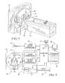
- a computed tomography (CT) imaging system 10 is shown as including a gantry 12 representative of a "third generation" twin beam CT scanner.
- Gantry 12 has an x-ray radiation source 14 that projects a beam of x-ray radiation 16 toward a detector array 18 on the opposite side of gantry 12.
- Detector array 18 is formed by detector elements 20 that together sense the projected x-rays that pass through an object 22, for example a medical patient.
- Each detector element 20 produces an electrical signal that represents the intensity of an impinging x-ray beam and hence the attenuation of the beam as it passes through patient 22.
- gantry 12 and the components mounted thereon rotate about a center of rotation 24.
- Detector array 18 is fabricated in a multi-slice configuration such that detector array 18 has a plurality of rows of detector elements or cells 20, only one of which is shown in Figure 2. During a twin beam helical scan, data is acquired from two detector rows at the same time. One or more additional rows of detector elements 20 in such configurations are arranged parallel to the illustrated row, and each row is transverse to the translation direction of patient 22 (i.e., the z-axis or patient axis).
- Control mechanism 26 includes an x-ray controller 28 that provides power and timing signals to x-ray source 14 and a gantry motor controller 30 that controls the rotational speed and position of gantry 12.
- a data acquisition system (DAS) 32 in control mechanism 26 samples analog data from detector elements or cells 20 and converts the data to digital signals for subsequent processing.
- An image reconstructor 34 receives sampled and digitized x-ray data from DAS 32 and performs highspeed image reconstruction. The reconstructed image is applied as an input to a computer 36 which stores the image in a storage device 38. Computer 36 also receives commands and scanning parameters from an operator via console 40 that has a keyboard.
- An associated display 42 such as a cathode ray tube or a liquid crystal display, allows the operator to observe the reconstructed image and other data from computer 36.
- the operator supplied commands and parameters are used by computer 36 to provide control signals and information to DAS 32, x-ray controller 28 and gantry motor controller 30.
- computer 36 operates a table motor controller 44 which controls a motorized table 46 to position patient 22 in gantry 12. Particularly, table 46 moves portions of patient 22 through gantry opening 48.
- computer 36 includes a device 50 for reading and writing onto removable media 52.
- device 50 is a floppy disk drive, a CD-R/W drive, or a DVD drive.
- media 52 is either a floppy disk, a compact disk, or a DVD.
- Device 50 and media 52 are used in one embodiment to transfer acquired projection data from imaging system 10 to another computer for further processing, or in another embodiment to input machine readable instructions that are processed by computer 36.
- Computer 36 and/or image reconstructor 34 of imaging system 10 provide the processing power necessary to perform the computational steps described herein in at least one embodiment of the present invention. Instructions for performing the computational steps are stored in an associated memory, such as storage device 38, read only or read/write memory (not shown separately in Figure 1), or media 52.
- an associated memory such as storage device 38, read only or read/write memory (not shown separately in Figure 1), or media 52.
- One obstacle in performing smoothing operations on the projection data is the difficulty in separating the real signal variation from the statistical fluctuation.
- a low-pass filter operation can be performed on the projection data without impacting the spatial resolution.
- the noise characteristics of the measured signal, i.e. projection data facilitate differentiating between variations caused by the statistical fluctuation and the real structure in object 22.
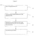
- Figure 3 is a flow diagram of a method 60 for facilitating a reduction in statistical noise in a computed tomography imaging system.
- the noise is a x-ray photon noise.
- the noise is a combination of electronic noise and photon noise.
- Method 60 includes generating 62 projection data, characterizing 64 a noise distribution of the projection data, performing 66 an adaptive noise reduction operation on the projection data using the noise distribution characterization, and reconstructing 68 an image.
- generating 62 projection data includes scanning an object 22 (shown in Figure 1) using a computed tomography (CT) imaging system 10 (shown in Figure 1).
- the projection data is pre-processed.
- pre-processing includes, but is not limited to, an offset correction, a primary speed correction, a reference channel correction, and an air-calibration.
- the projection data is pre-processed up to the step of minus logarithm operation. Pre-processing the projection data and calibrating imaging system 10 facilitates a reduction in artifacts in the image.
- Characterizing 64 a noise distribution of the projection data includes generating an air calibration vector, performing a polynomial fit of the air calibration vector to generate a bowtie shape vector, and approximating a standard deviation of the of the noise distribution using the bowtie shape vector.
- an air-calibration vector is generated with a bowtie filter. Therefore, the signal after air-calibration no longer represents the actual measured x-ray photon flux.
- a bowtie shape vector which estimates a bowtie effect is then generated.
- the bowtie shape vector is generated by performing a polynomial fit of the air-calibration vector in accordance with Equation 1.
- B( ⁇ ) poly_fit(x 2 ,Aircal( ⁇ )) where B( ⁇ ) is the bowtie shaped vector, and x is a detector channel index with an iso-channel equal to zero.
- a fifth order polynomial fit is used.
- an n th order polynomial is used, i.e.
- the bowtie vector, B( ⁇ ) can be determined directly from the x-ray photon flux measurement.
- a first set of air-scans i.e. a scan without an object inside the scanning plane, is collected with a bowtie.
- a second set of air scans is collected without a bowtie.
- the ratio of the first set of air scans and the second set of air-scans represents the bowtie vector B( ⁇ ).
- ⁇ ( ⁇ ) B ( ⁇ ) p ( ⁇ ) p ( ⁇ 0 )
- p ( ⁇ ) is the calibrated projection data before minus log
- p ( ⁇ 0 ) is a reference channel signal.
- An index for detector rows is not used since this method is identical to all rows.
- an index for a projection angle has been omitted.
- equation 2 is applied to every projection sample in the data set, i.e. each channel, each detector row, and each projection view.
- Figure 4 is a graphical representation of a calibrated projection of a quality assurance (QA) phantom.
- Figure 5 is a graphical representation of an approximated standard deviation based on equation (2) wherein the approximated standard deviation is represented by a solid line, and a corresponding measured standard deviation is represented by a dotted line.
- Performing 66 an adaptive noise reduction operation on the projection data using the noise distribution characterization includes defining a high threshold t high and a low threshold t low . If the measured variation of the projection sample over its neighborhood is less than ⁇ ( ⁇ )t low , a fully smoothing operation is applied. If the measured variation is greater than ⁇ ( ⁇ )t high , no smoothing operation is performed. When the variation is between approximately ⁇ ( ⁇ )t high and ⁇ ( ⁇ )t low ., the amount of smoothing is adjusted based on the variation level. In one embodiment, smoothing is performed on a channel-by-channel basis. The variation can be measured by calculating the standard deviation in a small region of interest. In one embodiment, a plurality of different orientations is used for the variation measurement.
- a variance can be calculated across channels, across views, and across detector rows.
- a plurality of intermediate directions such as, but not limited to, diagonal across both channels, and views can be used.
- a plurality of multi-resolution type of approaches can be used. For example, two, three, or four adjacent detector channels can be summed to form a single channel and a variation analysis and filtering can be performed on the compressed projections.
- Filtering the projection data includes filtering along the directions where the variations fall within the previously defined threshold, i.e. between t low and t high .
- a plurality of filtering methods can be used to derive a filtered projection. For example, any type of low pass filter or statistical based filter may be used.
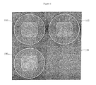
- Figure 6 is a plurality of images of a reconstructed phantom used to illustrate the effectiveness of the filtering method.
- Image 100 illustrates an example of a QA phantom scanned and reconstructed with a conventional reconstruction algorithm.
- Image 102 illustrates the same QA phantom processed in accordance with one method described herein. Image noise is significantly reduced with the adaptive processing.
- Image 104 illustrates is a difference image of image 100 and image 102. In one embodiment, no structural information is present in the difference image, indicating the algorithms ability to preserve spatial resolution.
- Image 106 illustrates a QA phantom scanned with 1.8 times the tube current and reconstructed with the conventional reconstruction algorithm. Note that the image noise level is comparable to that of image 102.
Landscapes
- Engineering & Computer Science (AREA)
- Health & Medical Sciences (AREA)
- Life Sciences & Earth Sciences (AREA)
- Physics & Mathematics (AREA)
- Medical Informatics (AREA)
- Theoretical Computer Science (AREA)
- Pathology (AREA)
- Heart & Thoracic Surgery (AREA)
- High Energy & Nuclear Physics (AREA)
- Veterinary Medicine (AREA)
- Nuclear Medicine, Radiotherapy & Molecular Imaging (AREA)
- Optics & Photonics (AREA)
- General Physics & Mathematics (AREA)
- Radiology & Medical Imaging (AREA)
- Biomedical Technology (AREA)
- Biophysics (AREA)
- Molecular Biology (AREA)
- Surgery (AREA)
- Animal Behavior & Ethology (AREA)
- General Health & Medical Sciences (AREA)
- Public Health (AREA)
- Pulmonology (AREA)
- Apparatus For Radiation Diagnosis (AREA)
- Image Processing (AREA)
Applications Claiming Priority (2)
| Application Number | Priority Date | Filing Date | Title |
|---|---|---|---|
| US990016 | 1997-12-12 | ||
| US09/990,016 US6493416B1 (en) | 2001-11-21 | 2001-11-21 | Method and apparatus for noise reduction in computed tomographic systems |
Publications (3)
| Publication Number | Publication Date |
|---|---|
| EP1321900A2 true EP1321900A2 (fr) | 2003-06-25 |
| EP1321900A3 EP1321900A3 (fr) | 2006-07-26 |
| EP1321900B1 EP1321900B1 (fr) | 2008-01-23 |
Family
ID=25535666
Family Applications (1)
| Application Number | Title | Priority Date | Filing Date |
|---|---|---|---|
| EP02258038A Expired - Lifetime EP1321900B1 (fr) | 2001-11-21 | 2002-11-21 | Appareil et procédé pour la réduction de bruit dans des systèmes de tomographie |
Country Status (4)
| Country | Link |
|---|---|
| US (1) | US6493416B1 (fr) |
| EP (1) | EP1321900B1 (fr) |
| JP (1) | JP4150900B2 (fr) |
| DE (1) | DE60224770T2 (fr) |
Families Citing this family (29)
| Publication number | Priority date | Publication date | Assignee | Title |
|---|---|---|---|---|
| US6529575B1 (en) * | 2002-04-29 | 2003-03-04 | Ge Medical Systems Global Technology Company, Llc | Adaptive projection filtering scheme for noise reduction |
| ATE508443T1 (de) * | 2003-06-17 | 2011-05-15 | Univ Brown | Verfahren und vorrichtung zur modelbasierten detektion einer struktur in projektionsdaten |
| US6931094B2 (en) * | 2003-10-10 | 2005-08-16 | Ge Medical Systems Global Technology Company, Llc | Methods and systems for smoothing |
| US7254261B2 (en) * | 2003-12-09 | 2007-08-07 | General Electric Co. | Signal-adaptive noise reduction in digital radiographic images |
| US20050201605A1 (en) * | 2004-03-11 | 2005-09-15 | Jianying Li | Methods and apparatus for CT smoothing to reduce artifacts |
| US7376255B2 (en) * | 2004-06-23 | 2008-05-20 | General Electric Company | System and method for image reconstruction |
| CN100563570C (zh) * | 2004-07-07 | 2009-12-02 | 皇家飞利浦电子股份有限公司 | 心脏锥面光束ct重建中条纹伪影的减少 |
| US8538099B2 (en) * | 2005-03-23 | 2013-09-17 | General Electric Company | Method and system for controlling image reconstruction |
| EP1731100B9 (fr) * | 2005-06-06 | 2013-01-23 | Kabushiki Kaisha Toshiba | Appareil et système medical d'affichage d'images |
| JP4786306B2 (ja) * | 2005-11-16 | 2011-10-05 | 株式会社東芝 | X線感度補正における補正データの作成方法及びx線ct装置 |
| US7660481B2 (en) * | 2005-11-17 | 2010-02-09 | Vital Images, Inc. | Image enhancement using anisotropic noise filtering |
| US7706497B2 (en) * | 2008-03-14 | 2010-04-27 | General Electric Company | Methods and apparatus for noise estimation for multi-resolution anisotropic diffusion filtering |
| US8553959B2 (en) * | 2008-03-21 | 2013-10-08 | General Electric Company | Method and apparatus for correcting multi-modality imaging data |
| CN102067177B (zh) * | 2008-06-25 | 2015-05-20 | 皇家飞利浦电子股份有限公司 | 优化剂量控制的图像生成装置 |
| US8837666B2 (en) * | 2008-09-30 | 2014-09-16 | Hitachi Medical Corporation | X-ray CT apparatus |
| EP2504811B1 (fr) | 2009-11-25 | 2014-06-18 | Koninklijke Philips N.V. | Réduction de dose/données d'image améliorées |
| JP5937093B2 (ja) * | 2010-10-27 | 2016-06-22 | コーニンクレッカ フィリップス エヌ ヴェKoninklijke Philips N.V. | 低線量ctノイズを除去するためのシステム及び方法 |
| US8538114B2 (en) * | 2011-06-06 | 2013-09-17 | Kabushiki Kaisha Toshiba | Method and system utilizing parameter-less filter for substantially reducing streak and or noise in computer tomography (CT) images |
| JP6139821B2 (ja) * | 2012-03-22 | 2017-05-31 | 東芝メディカルシステムズ株式会社 | X線ct装置 |
| JP6312401B2 (ja) * | 2012-11-30 | 2018-04-18 | キヤノン株式会社 | 画像処理装置、画像処理方法、及びプログラム |
| CN103969269B (zh) * | 2013-01-31 | 2018-09-18 | Ge医疗系统环球技术有限公司 | 用于几何校准ct扫描仪的方法和装置 |
| US9031297B2 (en) * | 2013-02-01 | 2015-05-12 | Kabushiki Kaisha Toshiba | Alternative noise map estimation methods for CT images |
| CN103136731B (zh) * | 2013-02-05 | 2015-11-25 | 南方医科大学 | 一种动态pet图像的参数成像方法 |
| US9076237B2 (en) * | 2013-03-12 | 2015-07-07 | Wisconsin Alumni Research Foundation | System and method for estimating a statistical noise map in x-ray imaging applications |
| JP6214226B2 (ja) | 2013-06-06 | 2017-10-18 | キヤノン株式会社 | 画像処理装置、断層撮影装置、画像処理方法およびプログラム |
| US10339634B2 (en) * | 2015-12-11 | 2019-07-02 | Shanghai United Imaging Healthcare Co., Ltd. | System and method for image reconstruction |
| US10255696B2 (en) | 2015-12-11 | 2019-04-09 | Shanghai United Imaging Healthcare Co., Ltd. | System and method for image reconstruction |
| US11222404B2 (en) * | 2016-03-25 | 2022-01-11 | Koninklijke Philips N.V. | Image reconstruction |
| JP6824133B2 (ja) * | 2017-09-28 | 2021-02-03 | 富士フイルム株式会社 | 画像処理装置、画像処理方法、及び画像処理プログラム |
Family Cites Families (9)
| Publication number | Priority date | Publication date | Assignee | Title |
|---|---|---|---|---|
| US4761819A (en) * | 1987-02-27 | 1988-08-02 | Picker International, Inc. | Adaptive noise reduction filter for reconstructed images |
| US5454019A (en) | 1991-03-15 | 1995-09-26 | Hitachi, Ltd. | Computed tomography system |
| GB9226376D0 (en) | 1992-12-18 | 1993-02-10 | British Tech Group | Tomography |
| DE19502576B4 (de) | 1994-02-25 | 2004-04-15 | Siemens Ag | Computertomograph mit Spiralabtastung |
| US5625660A (en) | 1995-06-30 | 1997-04-29 | Picker International, Inc. | Image reconstruction from helical partial cone-beam data |
| WO1997023844A1 (fr) * | 1995-12-21 | 1997-07-03 | Philips Electronics N.V. | Attenuation directionnelle et adaptative de bruit |
| US5818896A (en) | 1996-11-18 | 1998-10-06 | General Electric Company | Methods and apparatus for three-dimensional and maximum intensity projection image reconstruction in a computed tomography system |
| JP3033508B2 (ja) | 1997-01-20 | 2000-04-17 | 日本電気株式会社 | 生体内活動部位推定方法 |
| DE19853143C2 (de) * | 1998-11-18 | 2000-09-07 | Vamp Verfahren Und Apparate De | Computertomograph mit reduzierter Dosisbelastung bzw. reduziertem Bildpunktrauschen |
-
2001
- 2001-11-21 US US09/990,016 patent/US6493416B1/en not_active Expired - Lifetime
-
2002
- 2002-11-21 JP JP2002337728A patent/JP4150900B2/ja not_active Expired - Fee Related
- 2002-11-21 EP EP02258038A patent/EP1321900B1/fr not_active Expired - Lifetime
- 2002-11-21 DE DE60224770T patent/DE60224770T2/de not_active Expired - Lifetime
Also Published As
| Publication number | Publication date |
|---|---|
| DE60224770T2 (de) | 2009-01-15 |
| EP1321900A3 (fr) | 2006-07-26 |
| JP4150900B2 (ja) | 2008-09-17 |
| JP2003180675A (ja) | 2003-07-02 |
| EP1321900B1 (fr) | 2008-01-23 |
| DE60224770D1 (de) | 2008-03-13 |
| US6493416B1 (en) | 2002-12-10 |
Similar Documents
| Publication | Publication Date | Title |
|---|---|---|
| US6493416B1 (en) | Method and apparatus for noise reduction in computed tomographic systems | |
| US6266388B1 (en) | Methods and apparatus for two-pass cone beam image reconstruction | |
| US6421411B1 (en) | Methods and apparatus for helical image artifact reduction | |
| EP1048008B1 (fr) | Methodes et appareil de numeration de calcifications | |
| US5416815A (en) | Adaptive filter for reducing streaking artifacts in x-ray tomographic images | |
| US5727041A (en) | Methods and apparatus for reducing partial volume image artifacts | |
| JP4152649B2 (ja) | Ctスカウト画像処理のための方法及び装置 | |
| US6035012A (en) | Artifact correction for highly attenuating objects | |
| US7415145B2 (en) | Methods and apparatus for artifact reduction | |
| EP1358848A2 (fr) | Méthode de filtrage adaptif des données de projection pour la réduction de bruit | |
| US5606585A (en) | Methods and apparatus for multislice helical image reconstruction in a computer tomography system | |
| EP1149558A2 (fr) | Système d'imagerie multicouche pour une zone déterminée | |
| JP2004174249A (ja) | ボリューム灌流を計算するための方法及び装置 | |
| US6408042B1 (en) | Methods and apparatus for cone beam artifact suppression in scanning imaging systems | |
| JP2007307417A (ja) | 像データの処理方法及び像データの処理装置 | |
| US6587537B1 (en) | Methods and apparatus for multi-slice image reconstruction | |
| US7239730B2 (en) | Method and apparatus for volume scoring calcification concentrations of a CT scan | |
| US6438195B1 (en) | Methods and apparatus for compensating for view aliasing artifacts | |
| US5812628A (en) | Methods and apparatus for detecting partial volume image artifacts | |
| US5974110A (en) | Helical reconstruction algorithm | |
| JPH10225453A (ja) | 計算機式断層写真法システムにおいて基準チャンネルの閉塞を検出する方法及びシステム | |
| EP0989521A2 (fr) | Réconstruction d'image fluoroscopique | |
| US6980681B1 (en) | Methods and apparatus for helical reconstruction for multislice CT scan | |
| JP3484288B2 (ja) | X線断層撮影装置 | |
| US6647084B1 (en) | Method and apparatus for filtering projection data of a helical scan |
Legal Events
| Date | Code | Title | Description |
|---|---|---|---|
| PUAI | Public reference made under article 153(3) epc to a published international application that has entered the european phase |
Free format text: ORIGINAL CODE: 0009012 |
|
| AK | Designated contracting states |
Designated state(s): AT BE BG CH CY CZ DE DK EE ES FI FR GB GR IE IT LI LU MC NL PT SE SK TR |
|
| AX | Request for extension of the european patent |
Extension state: AL LT LV MK RO SI |
|
| PUAL | Search report despatched |
Free format text: ORIGINAL CODE: 0009013 |
|
| AK | Designated contracting states |
Kind code of ref document: A3 Designated state(s): AT BE BG CH CY CZ DE DK EE ES FI FR GB GR IE IT LI LU MC NL PT SE SK TR |
|
| AX | Request for extension of the european patent |
Extension state: AL LT LV MK RO SI |
|
| RIC1 | Information provided on ipc code assigned before grant |
Ipc: G06T 11/00 20060101AFI20030429BHEP Ipc: G06T 5/00 20060101ALI20060616BHEP |
|
| 17P | Request for examination filed |
Effective date: 20070126 |
|
| 17Q | First examination report despatched |
Effective date: 20070226 |
|
| AKX | Designation fees paid |
Designated state(s): DE NL |
|
| GRAP | Despatch of communication of intention to grant a patent |
Free format text: ORIGINAL CODE: EPIDOSNIGR1 |
|
| GRAS | Grant fee paid |
Free format text: ORIGINAL CODE: EPIDOSNIGR3 |
|
| GRAA | (expected) grant |
Free format text: ORIGINAL CODE: 0009210 |
|
| AK | Designated contracting states |
Kind code of ref document: B1 Designated state(s): DE NL |
|
| REF | Corresponds to: |
Ref document number: 60224770 Country of ref document: DE Date of ref document: 20080313 Kind code of ref document: P |
|
| PLBE | No opposition filed within time limit |
Free format text: ORIGINAL CODE: 0009261 |
|
| STAA | Information on the status of an ep patent application or granted ep patent |
Free format text: STATUS: NO OPPOSITION FILED WITHIN TIME LIMIT |
|
| 26N | No opposition filed |
Effective date: 20081024 |
|
| PGFP | Annual fee paid to national office [announced via postgrant information from national office to epo] |
Ref country code: NL Payment date: 20111129 Year of fee payment: 10 |
|
| REG | Reference to a national code |
Ref country code: NL Ref legal event code: V1 Effective date: 20130601 |
|
| PG25 | Lapsed in a contracting state [announced via postgrant information from national office to epo] |
Ref country code: NL Free format text: LAPSE BECAUSE OF NON-PAYMENT OF DUE FEES Effective date: 20130601 |
|
| PGFP | Annual fee paid to national office [announced via postgrant information from national office to epo] |
Ref country code: DE Payment date: 20191021 Year of fee payment: 18 |
|
| REG | Reference to a national code |
Ref country code: DE Ref legal event code: R119 Ref document number: 60224770 Country of ref document: DE |
|
| PG25 | Lapsed in a contracting state [announced via postgrant information from national office to epo] |
Ref country code: DE Free format text: LAPSE BECAUSE OF NON-PAYMENT OF DUE FEES Effective date: 20210601 |