EP1324802B1 - Katheter für den myogenen zelltransfer - Google Patents
Katheter für den myogenen zelltransfer Download PDFInfo
- Publication number
- EP1324802B1 EP1324802B1 EP01979245A EP01979245A EP1324802B1 EP 1324802 B1 EP1324802 B1 EP 1324802B1 EP 01979245 A EP01979245 A EP 01979245A EP 01979245 A EP01979245 A EP 01979245A EP 1324802 B1 EP1324802 B1 EP 1324802B1
- Authority
- EP
- European Patent Office
- Prior art keywords
- catheter
- needle
- cells
- tip
- electrodes
- Prior art date
- Legal status (The legal status is an assumption and is not a legal conclusion. Google has not performed a legal analysis and makes no representation as to the accuracy of the status listed.)
- Expired - Lifetime
Links
- 230000001114 myogenic effect Effects 0.000 title claims abstract description 49
- 210000002216 heart Anatomy 0.000 claims abstract description 61
- 210000003205 muscle Anatomy 0.000 claims abstract description 37
- 210000004165 myocardium Anatomy 0.000 claims abstract description 37
- 238000002347 injection Methods 0.000 claims abstract description 33
- 239000007924 injection Substances 0.000 claims abstract description 33
- 238000000034 method Methods 0.000 claims abstract description 21
- 238000001514 detection method Methods 0.000 claims abstract description 12
- 230000002159 abnormal effect Effects 0.000 claims description 8
- 208000010428 Muscle Weakness Diseases 0.000 claims description 4
- 206010028289 Muscle atrophy Diseases 0.000 claims description 4
- 206010028372 Muscular weakness Diseases 0.000 claims description 4
- 239000012190 activator Substances 0.000 claims description 4
- 238000003780 insertion Methods 0.000 claims description 4
- 230000037431 insertion Effects 0.000 claims description 4
- 230000004044 response Effects 0.000 claims description 4
- WFKWXMTUELFFGS-UHFFFAOYSA-N tungsten Chemical compound [W] WFKWXMTUELFFGS-UHFFFAOYSA-N 0.000 claims description 3
- 238000012544 monitoring process Methods 0.000 claims 6
- 230000003412 degenerative effect Effects 0.000 abstract description 20
- 238000002659 cell therapy Methods 0.000 abstract description 12
- 230000008439 repair process Effects 0.000 abstract description 11
- 208000019622 heart disease Diseases 0.000 abstract description 6
- 230000007246 mechanism Effects 0.000 abstract description 5
- 238000001356 surgical procedure Methods 0.000 abstract description 5
- 210000002784 stomach Anatomy 0.000 abstract description 3
- 206010007559 Cardiac failure congestive Diseases 0.000 abstract description 2
- 210000000936 intestine Anatomy 0.000 abstract description 2
- 210000003932 urinary bladder Anatomy 0.000 abstract description 2
- 230000001413 cellular effect Effects 0.000 abstract 2
- 208000010125 myocardial infarction Diseases 0.000 abstract 1
- 210000004027 cell Anatomy 0.000 description 113
- 210000001519 tissue Anatomy 0.000 description 39
- 230000033001 locomotion Effects 0.000 description 16
- 239000006285 cell suspension Substances 0.000 description 14
- 239000000463 material Substances 0.000 description 11
- 238000005259 measurement Methods 0.000 description 10
- 229920000642 polymer Polymers 0.000 description 8
- 230000000694 effects Effects 0.000 description 7
- 210000005003 heart tissue Anatomy 0.000 description 7
- 239000003814 drug Substances 0.000 description 6
- 230000002107 myocardial effect Effects 0.000 description 6
- 210000000056 organ Anatomy 0.000 description 6
- 239000000725 suspension Substances 0.000 description 6
- 230000000747 cardiac effect Effects 0.000 description 5
- 229940079593 drug Drugs 0.000 description 5
- 210000001367 artery Anatomy 0.000 description 4
- 238000010009 beating Methods 0.000 description 4
- 238000010586 diagram Methods 0.000 description 4
- 239000007943 implant Substances 0.000 description 4
- 230000004913 activation Effects 0.000 description 3
- 230000003416 augmentation Effects 0.000 description 3
- 230000008602 contraction Effects 0.000 description 3
- 238000012377 drug delivery Methods 0.000 description 3
- 238000002513 implantation Methods 0.000 description 3
- 210000005240 left ventricle Anatomy 0.000 description 3
- 239000000523 sample Substances 0.000 description 3
- 239000007787 solid Substances 0.000 description 3
- 208000021642 Muscular disease Diseases 0.000 description 2
- 230000003190 augmentative effect Effects 0.000 description 2
- 230000008901 benefit Effects 0.000 description 2
- 239000011230 binding agent Substances 0.000 description 2
- 210000001124 body fluid Anatomy 0.000 description 2
- 239000010839 body fluid Substances 0.000 description 2
- 230000009087 cell motility Effects 0.000 description 2
- 230000008859 change Effects 0.000 description 2
- 210000000038 chest Anatomy 0.000 description 2
- 230000000052 comparative effect Effects 0.000 description 2
- 239000004020 conductor Substances 0.000 description 2
- 210000004351 coronary vessel Anatomy 0.000 description 2
- 230000007423 decrease Effects 0.000 description 2
- 210000001105 femoral artery Anatomy 0.000 description 2
- 210000001035 gastrointestinal tract Anatomy 0.000 description 2
- 238000013507 mapping Methods 0.000 description 2
- 210000000663 muscle cell Anatomy 0.000 description 2
- 210000003098 myoblast Anatomy 0.000 description 2
- 239000008188 pellet Substances 0.000 description 2
- 230000002572 peristaltic effect Effects 0.000 description 2
- BASFCYQUMIYNBI-UHFFFAOYSA-N platinum Chemical compound [Pt] BASFCYQUMIYNBI-UHFFFAOYSA-N 0.000 description 2
- 238000012545 processing Methods 0.000 description 2
- 108090000623 proteins and genes Proteins 0.000 description 2
- 229910052709 silver Inorganic materials 0.000 description 2
- 239000004332 silver Substances 0.000 description 2
- 230000005236 sound signal Effects 0.000 description 2
- 239000000126 substance Substances 0.000 description 2
- 210000000115 thoracic cavity Anatomy 0.000 description 2
- 230000001960 triggered effect Effects 0.000 description 2
- 206010002091 Anaesthesia Diseases 0.000 description 1
- 206010002383 Angina Pectoris Diseases 0.000 description 1
- OKTJSMMVPCPJKN-UHFFFAOYSA-N Carbon Chemical compound [C] OKTJSMMVPCPJKN-UHFFFAOYSA-N 0.000 description 1
- 208000032843 Hemorrhage Diseases 0.000 description 1
- HTTJABKRGRZYRN-UHFFFAOYSA-N Heparin Chemical compound OC1C(NC(=O)C)C(O)OC(COS(O)(=O)=O)C1OC1C(OS(O)(=O)=O)C(O)C(OC2C(C(OS(O)(=O)=O)C(OC3C(C(O)C(O)C(O3)C(O)=O)OS(O)(=O)=O)C(CO)O2)NS(O)(=O)=O)C(C(O)=O)O1 HTTJABKRGRZYRN-UHFFFAOYSA-N 0.000 description 1
- 102000007625 Hirudins Human genes 0.000 description 1
- 108010007267 Hirudins Proteins 0.000 description 1
- BQCADISMDOOEFD-UHFFFAOYSA-N Silver Chemical compound [Ag] BQCADISMDOOEFD-UHFFFAOYSA-N 0.000 description 1
- 229910021607 Silver chloride Inorganic materials 0.000 description 1
- 230000001594 aberrant effect Effects 0.000 description 1
- 230000009471 action Effects 0.000 description 1
- 230000004103 aerobic respiration Effects 0.000 description 1
- 230000037005 anaesthesia Effects 0.000 description 1
- 239000003242 anti bacterial agent Substances 0.000 description 1
- 229940088710 antibiotic agent Drugs 0.000 description 1
- 210000000709 aorta Anatomy 0.000 description 1
- 238000013459 approach Methods 0.000 description 1
- 239000007900 aqueous suspension Substances 0.000 description 1
- 210000004204 blood vessel Anatomy 0.000 description 1
- 239000000872 buffer Substances 0.000 description 1
- 229910052799 carbon Inorganic materials 0.000 description 1
- 210000004413 cardiac myocyte Anatomy 0.000 description 1
- 229940030602 cardiac therapy drug Drugs 0.000 description 1
- 230000006037 cell lysis Effects 0.000 description 1
- 238000006243 chemical reaction Methods 0.000 description 1
- 239000003795 chemical substances by application Substances 0.000 description 1
- 239000011248 coating agent Substances 0.000 description 1
- 238000000576 coating method Methods 0.000 description 1
- 230000003750 conditioning effect Effects 0.000 description 1
- 230000003292 diminished effect Effects 0.000 description 1
- 201000010099 disease Diseases 0.000 description 1
- 208000037265 diseases, disorders, signs and symptoms Diseases 0.000 description 1
- 238000004090 dissolution Methods 0.000 description 1
- 230000002526 effect on cardiovascular system Effects 0.000 description 1
- 239000012530 fluid Substances 0.000 description 1
- 230000002068 genetic effect Effects 0.000 description 1
- 230000035876 healing Effects 0.000 description 1
- 229960002897 heparin Drugs 0.000 description 1
- 229920000669 heparin Polymers 0.000 description 1
- WQPDUTSPKFMPDP-OUMQNGNKSA-N hirudin Chemical compound C([C@@H](C(=O)N[C@@H](CCC(O)=O)C(=O)N[C@@H](CCC(O)=O)C(=O)N[C@@H]([C@@H](C)CC)C(=O)N1[C@@H](CCC1)C(=O)N[C@@H](CCC(O)=O)C(=O)N[C@@H](CCC(O)=O)C(=O)N[C@@H](CC=1C=CC(OS(O)(=O)=O)=CC=1)C(=O)N[C@@H](CC(C)C)C(=O)N[C@@H](CCC(N)=O)C(O)=O)NC(=O)[C@H](CC(O)=O)NC(=O)CNC(=O)[C@H](CC(O)=O)NC(=O)[C@H](CC(N)=O)NC(=O)[C@H](CC=1NC=NC=1)NC(=O)[C@H](CO)NC(=O)[C@H](CCC(N)=O)NC(=O)[C@H]1N(CCC1)C(=O)[C@H](CCCCN)NC(=O)[C@H]1N(CCC1)C(=O)[C@@H](NC(=O)CNC(=O)[C@H](CCC(O)=O)NC(=O)CNC(=O)[C@@H](NC(=O)[C@@H](NC(=O)[C@H]1NC(=O)[C@H](CCC(N)=O)NC(=O)[C@H](CC(N)=O)NC(=O)[C@H](CCCCN)NC(=O)[C@H](CCC(O)=O)NC(=O)CNC(=O)[C@H](CC(O)=O)NC(=O)[C@H](CO)NC(=O)CNC(=O)[C@H](CC(C)C)NC(=O)[C@H]([C@@H](C)CC)NC(=O)[C@@H]2CSSC[C@@H](C(=O)N[C@@H](CCC(O)=O)C(=O)NCC(=O)N[C@@H](CO)C(=O)N[C@@H](CC(N)=O)C(=O)N[C@H](C(=O)N[C@H](C(NCC(=O)N[C@@H](CCC(N)=O)C(=O)NCC(=O)N[C@@H](CC(N)=O)C(=O)N[C@@H](CCCCN)C(=O)N2)=O)CSSC1)C(C)C)NC(=O)[C@H](CC(C)C)NC(=O)[C@H]1NC(=O)[C@H](CC(C)C)NC(=O)[C@H](CC(N)=O)NC(=O)[C@H](CCC(N)=O)NC(=O)CNC(=O)[C@H](CO)NC(=O)[C@H](CCC(O)=O)NC(=O)[C@H]([C@@H](C)O)NC(=O)[C@@H](NC(=O)[C@H](CC(O)=O)NC(=O)[C@@H](NC(=O)[C@H](CC=2C=CC(O)=CC=2)NC(=O)[C@@H](NC(=O)[C@@H](N)C(C)C)C(C)C)[C@@H](C)O)CSSC1)C(C)C)[C@@H](C)O)[C@@H](C)O)C1=CC=CC=C1 WQPDUTSPKFMPDP-OUMQNGNKSA-N 0.000 description 1
- 229940006607 hirudin Drugs 0.000 description 1
- 238000003384 imaging method Methods 0.000 description 1
- 238000002847 impedance measurement Methods 0.000 description 1
- 238000001727 in vivo Methods 0.000 description 1
- 238000010255 intramuscular injection Methods 0.000 description 1
- 239000007927 intramuscular injection Substances 0.000 description 1
- 239000007928 intraperitoneal injection Substances 0.000 description 1
- 238000010253 intravenous injection Methods 0.000 description 1
- 210000004185 liver Anatomy 0.000 description 1
- 210000002540 macrophage Anatomy 0.000 description 1
- 229910052751 metal Inorganic materials 0.000 description 1
- 239000002184 metal Substances 0.000 description 1
- 238000012986 modification Methods 0.000 description 1
- 230000004048 modification Effects 0.000 description 1
- 230000004220 muscle function Effects 0.000 description 1
- 201000006938 muscular dystrophy Diseases 0.000 description 1
- 230000035515 penetration Effects 0.000 description 1
- 239000004033 plastic Substances 0.000 description 1
- 229910052697 platinum Inorganic materials 0.000 description 1
- 238000002278 reconstructive surgery Methods 0.000 description 1
- 230000033764 rhythmic process Effects 0.000 description 1
- 230000002000 scavenging effect Effects 0.000 description 1
- HKZLPVFGJNLROG-UHFFFAOYSA-M silver monochloride Chemical compound [Cl-].[Ag+] HKZLPVFGJNLROG-UHFFFAOYSA-M 0.000 description 1
- 210000002460 smooth muscle Anatomy 0.000 description 1
- 239000000243 solution Substances 0.000 description 1
- 230000006641 stabilisation Effects 0.000 description 1
- 238000011105 stabilization Methods 0.000 description 1
- 230000000638 stimulation Effects 0.000 description 1
- 210000003699 striated muscle Anatomy 0.000 description 1
- 230000008685 targeting Effects 0.000 description 1
- 238000002560 therapeutic procedure Methods 0.000 description 1
- 239000002562 thickening agent Substances 0.000 description 1
- 230000017423 tissue regeneration Effects 0.000 description 1
- 238000002054 transplantation Methods 0.000 description 1
Images

Classifications
-
- A—HUMAN NECESSITIES
- A61—MEDICAL OR VETERINARY SCIENCE; HYGIENE
- A61B—DIAGNOSIS; SURGERY; IDENTIFICATION
- A61B5/00—Measuring for diagnostic purposes; Identification of persons
- A61B5/05—Detecting, measuring or recording for diagnosis by means of electric currents or magnetic fields; Measuring using microwaves or radio waves
- A61B5/053—Measuring electrical impedance or conductance of a portion of the body
-
- A—HUMAN NECESSITIES
- A61—MEDICAL OR VETERINARY SCIENCE; HYGIENE
- A61B—DIAGNOSIS; SURGERY; IDENTIFICATION
- A61B5/00—Measuring for diagnostic purposes; Identification of persons
- A61B5/05—Detecting, measuring or recording for diagnosis by means of electric currents or magnetic fields; Measuring using microwaves or radio waves
- A61B5/053—Measuring electrical impedance or conductance of a portion of the body
- A61B5/0538—Measuring electrical impedance or conductance of a portion of the body invasively, e.g. using a catheter
-
- A—HUMAN NECESSITIES
- A61—MEDICAL OR VETERINARY SCIENCE; HYGIENE
- A61B—DIAGNOSIS; SURGERY; IDENTIFICATION
- A61B5/00—Measuring for diagnostic purposes; Identification of persons
- A61B5/48—Other medical applications
- A61B5/4836—Diagnosis combined with treatment in closed-loop systems or methods
- A61B5/4839—Diagnosis combined with treatment in closed-loop systems or methods combined with drug delivery
-
- A—HUMAN NECESSITIES
- A61—MEDICAL OR VETERINARY SCIENCE; HYGIENE
- A61M—DEVICES FOR INTRODUCING MEDIA INTO, OR ONTO, THE BODY; DEVICES FOR TRANSDUCING BODY MEDIA OR FOR TAKING MEDIA FROM THE BODY; DEVICES FOR PRODUCING OR ENDING SLEEP OR STUPOR
- A61M25/00—Catheters; Hollow probes
- A61M25/0067—Catheters; Hollow probes characterised by the distal end, e.g. tips
- A61M25/0082—Catheter tip comprising a tool
- A61M25/0084—Catheter tip comprising a tool being one or more injection needles
-
- A—HUMAN NECESSITIES
- A61—MEDICAL OR VETERINARY SCIENCE; HYGIENE
- A61B—DIAGNOSIS; SURGERY; IDENTIFICATION
- A61B17/00—Surgical instruments, devices or methods
- A61B17/00234—Surgical instruments, devices or methods for minimally invasive surgery
- A61B2017/00238—Type of minimally invasive operation
- A61B2017/00243—Type of minimally invasive operation cardiac
- A61B2017/00247—Making holes in the wall of the heart, e.g. laser Myocardial revascularization
-
- A—HUMAN NECESSITIES
- A61—MEDICAL OR VETERINARY SCIENCE; HYGIENE
- A61B—DIAGNOSIS; SURGERY; IDENTIFICATION
- A61B18/00—Surgical instruments, devices or methods for transferring non-mechanical forms of energy to or from the body
- A61B2018/00315—Surgical instruments, devices or methods for transferring non-mechanical forms of energy to or from the body for treatment of particular body parts
- A61B2018/00345—Vascular system
- A61B2018/00351—Heart
- A61B2018/00392—Transmyocardial revascularisation
-
- A—HUMAN NECESSITIES
- A61—MEDICAL OR VETERINARY SCIENCE; HYGIENE
- A61B—DIAGNOSIS; SURGERY; IDENTIFICATION
- A61B90/00—Instruments, implements or accessories specially adapted for surgery or diagnosis and not covered by any of the groups A61B1/00 - A61B50/00, e.g. for luxation treatment or for protecting wound edges
- A61B90/06—Measuring instruments not otherwise provided for
- A61B2090/064—Measuring instruments not otherwise provided for for measuring force, pressure or mechanical tension
- A61B2090/065—Measuring instruments not otherwise provided for for measuring force, pressure or mechanical tension for measuring contact or contact pressure
-
- A—HUMAN NECESSITIES
- A61—MEDICAL OR VETERINARY SCIENCE; HYGIENE
- A61B—DIAGNOSIS; SURGERY; IDENTIFICATION
- A61B5/00—Measuring for diagnostic purposes; Identification of persons
- A61B5/45—For evaluating or diagnosing the musculoskeletal system or teeth
- A61B5/4519—Muscles
-
- A—HUMAN NECESSITIES
- A61—MEDICAL OR VETERINARY SCIENCE; HYGIENE
- A61M—DEVICES FOR INTRODUCING MEDIA INTO, OR ONTO, THE BODY; DEVICES FOR TRANSDUCING BODY MEDIA OR FOR TAKING MEDIA FROM THE BODY; DEVICES FOR PRODUCING OR ENDING SLEEP OR STUPOR
- A61M25/00—Catheters; Hollow probes
- A61M25/0067—Catheters; Hollow probes characterised by the distal end, e.g. tips
- A61M25/0082—Catheter tip comprising a tool
- A61M25/0084—Catheter tip comprising a tool being one or more injection needles
- A61M2025/0089—Single injection needle protruding axially, i.e. along the longitudinal axis of the catheter, from the distal tip
-
- A—HUMAN NECESSITIES
- A61—MEDICAL OR VETERINARY SCIENCE; HYGIENE
- A61M—DEVICES FOR INTRODUCING MEDIA INTO, OR ONTO, THE BODY; DEVICES FOR TRANSDUCING BODY MEDIA OR FOR TAKING MEDIA FROM THE BODY; DEVICES FOR PRODUCING OR ENDING SLEEP OR STUPOR
- A61M2210/00—Anatomical parts of the body
- A61M2210/12—Blood circulatory system
- A61M2210/125—Heart
Definitions
- the invention relates to materials for repairing and augmenting tissues in a host. More specifically the invention relates to cardiovascular catheters for automatically injecting large quantities of cells such as myogenic cells for repairing and augmenting muscle and other tissues.
- myogenic cell therapy such as myogenic cell therapy promises to revolutionize the treatment of serious muscular diseases such as muscular dystrophy and heart disease as pioneered by Law et al. (See Gene Ther. Mol. Biol. pp. 345-363 (March 1998), Cell Transplantation 6: 95-100 (1997) and U.S. Pat. No. 5,130,141).
- myogenic cells are cultured and then introduced into a target muscle tissue such as striated muscle or heart muscle.
- the introduced cells fuse with pre-existing muscle cells, transferring the normal genome in their nuclei to effect genetic repair.
- the introduced cells also fuse among themselves to form normal cells to replenish cells that have degenerated.
- Myogenic cell therapy is very promising and promises to revolutionize the treatment of heart disease, with enormous consequences to the society.
- a major impediment to the widespread use of myogenic cell therapy is the need to implant very high numbers of cells in order to overcome the initial cell rejection, presumably due to the action of scavenging macrophages, as reviewed by Law et al, in Gene Ther. Mol. Biol. ( Id. p. 351).
- the cell number problem is a major impediment to progress in this field. That is, there is no good tool and procedure for implanting large numbers of cells to region(s) of body structures such as muscle that are degenerated or diseased and which can benefit from the cell treatment. Accordingly, new techniques and tools for implanting large numbers of cells are key to success and can unlock the potential of cell therapy to treat diseases such as heart disease and other muscular disorders.
- U.S. Nos. 5,865,787 and 5,634,899 describe delivery of a drug using electric current/voltage control. Such delivery by electric current is not compatible with living cells.
- U.S. No. 6,056,969 describes implantation of drug in pellet form by piercing the heart muscle wall (see Fig. 3 and Fig. 4). However, such pellets are incompatible with cells that are delivered in a suspension and that need to remain mobile, particularly for the large volumes needed for cell therapy.
- a second problem often encountered is that there is no sufficiently reliable way to implant material into a moving target such as the heart wall from the inside of the heart while the heart is beating.
- Present methods are hampered by, for example, the need to pace or interrupt the heart beat as injection is not carried out easily on a moving target.
- a particular need of cell therapy in the context of heart repair is to detect which portion(s) of the heart need to be treated and receive cells.
- a diseased part will exhibit electrical silence, diminished electrical activity, and/or abnormal electrocardiogram.
- Catheters have been developed that can map local cardiac signals with electrodes on their ring and tip surfaces, as reviewed in U.S. Pat. No. 5,450,846. Some of those catheters have been used for making an intracardiac electrogram, as described in U.S. Pat. No. 5,156,151.
- the art has not progressed sufficiently to conveniently identify specific small spots throughout the entire myocardium that are degenerated or weak in a manner that the spot can be repaired via cell therapy, particularly during the same procedure.
- an obstacle to cell therapy of myocardial tissue is that a suitable diagnostic catheter for that purpose is not available.
- preferred embodiments of the invention provide new tools that can 1) access the interior of a body or body structure such as the heart via a percutaneous and endovascular route, 2) electrically detect even minute degenerative areas of a body surface such as the endomyocardium, 3) insert a needle of predetermined diameter (gauge) and length, preferably 2 to 8 mm long, more preferably 3 to 5 mm long, preferably 29 gauge to 10 gauge, more preferably 28 gauge to 18 gauge, even more preferably 27 gauge to 22 gauge, preferably diagonally into the structure, at and/or around the degenerative area, 4) injecting a predetermined number of cells (such as myoblasts) through the needle, preferably 10 to 800 million cells, more preferably 25 to 200 million cells and even more preferably 50 to 100 million cells, 5) through an electrically coupled feed-back circuit and mechanical system, 6) within a predetermined period of time, preferably less than one second and 7) without the need of extensive opening of the body through, for example, an open chest or open heart
- the invention provides an apparatus for a method of delivering myogenic cells into a degenerated or weak muscle in a host, comprising the step of placing a catheter within the host, the catheter comprising a sensor on its tip to detect degenerated or weak muscle tissue on contact; and expelling the myogenic cells through a needle in the tip of the catheter into and around the degenerative or weak muscle areas upon sensing contact with degenerated or weak muscle tissue.
- the invention is a catheter for injection of myogenic cells into a target portion of a heart
- a flexible needle within the catheter having a bore of twenty-two to twenty seven gauge; a tip on the catheter that is tapered to allow close contact with the inside wall of the heart and that contains at least one electrode to monitor an electrical signal of the heart muscle portion that it contacts; a tip on the needle that protrudes from the catheter tip no more than 5 millimeters to prevent perforation of the heart; a needle fluidically connected to a source of myogenic cells; and an electrical feedback circuit that transduces any aberrant electrical signal from the catheter tip into automatically injecting the myogenic cells into a target heart material within one second.
- Another embodiment of the apparatus of the invention is useful for a method for treating angina pectoris in a patient with a catheter having a reservoir, and a tip, the tip comprising two or more electrodes and at least one needle for delivering myogenic cells, comprising the steps of providing myogenic cells for use with the catheter; placing the myogenic cells in the reservoir and placing the catheter inside the patient and/or into the heart of the patient; detecting one or more degenerative portions on the inside surface of the heart tissue; and automatically injecting myogenic cells from the needle into and around the portion of the degenerative or weak heart tissue.
- the invention is a catheter for detecting and treating local areas of muscle degeneration and weakness in a patient, comprising a long body that can be inserted into the patient; a tip at the end of the long body; at least three electrodes on the tip; and a needle for injecting cells into the muscle, the needle having a point; wherein the three electrodes form a flat surface on the end of the tip, the long axis of the needle is positioned between the three electrodes and is perpendicular to the flat surface.
- the invention is an apparatus for treating a body tissue by implantation of cells into selected parts of an organ of a patient in vivo comprising a catheter, a retracted needle and at least two electrodes in its tip; a reservoir of cells to be implanted; an actuator for moving the needle from the catheter and into a selected part of the organ; an actuator for forcibly expelling cells through the needle and into the selected part of the organ; and a controller that activates the actuators wherein the controller monitors electrical signals obtained by the electrodes, compares those signals with stored or calculated information and activates needle movement and cell movement upon detection of proximity of the selected part, whether normal or abnormal, of the organ to the catheter.
- An automated catheter according to the invention has an electrode-feedback system that detects damaged tissue on, for example, heart muscle and automatically and rapidly injects large quantities of myogenic cells into the detected tissue in real time. That is, the catheter system detects a suitable site at virtually the same time as it implants cells to improve that site, thus overcoming the severe problem that the heart is moving during the procedure.
- the catheter tip has a needle that is triggered to protrude by a defined distance that is long enough to safely inject cells into the myocardial tissue but not too long as to puncture the outer wall of the heart.
- the needle is completely retracted into a sheath during insertion of the catheter into a patient.
- the system minimizes operator error by virtue of an electronic-mechanical coupled system that electronically detects one or more weak or degenerative spots on the myocardium and automatically injects a controlled amount of myogenic cells into and/or around that same site, all without human intervention .
- the catheter for heart repair is that the injection be carried out rapidly while the heart is beating. During anesthesia, the heart may contract approximately once per second.
- the catheter solves the timing problem by detecting contact with a weak or degenerated spot and then quickly injecting cells automatically. Preferably this time is less than one second, more preferably less than 0.7 seconds, most preferably in less than 0.5 seconds.
- the catheter accesses the interior of the heart via a percutaneous and endovascular route without necessitating opening of the chest cavity or an open heart surgery; (2) electrically detects even minute degenerative areas of the endomyocardium; 3) inserts a needle of predetermined diameter (gauge) and length, preferably 2 to 8 mm long, more preferably 3 to 5 mm long, preferably 29 gauge to 10 gauge, more preferably 28 gauge to 18 gauge, even more preferably 27 gauge to 22 gauge, preferably diagonally into the myocardium, at and/or around the degenerative area, 4) injecting a predetermined number of cells (myoblasts) through the needle, preferably 10 to 800 million cells, more preferably 25 to 200 million cells and even more preferably 50 to 100 million cells, 5) through an electrically coupled feed-back circuit and mechanical system, 6) within a predetermined period of time, preferably less than one second and 7) without the need for an open chest or an open heart surgery.
- a variety of catheters and catheter control systems for their use to treat muscle degeneration and weakness in a host are contemplated according to the invention.
- cells are loaded into a reservoir and are injected into the muscle under timing control by an electronic feedback circuit.
- the feedback circuit is one link in a control system that comprises the catheter itself with electrodes in the catheter tip and a controller that contains electrical circuits for detecting, modifying and analyzing signals obtained from the electrodes.
- the feedback circuit further comprises an actuating mechanism for expelling a controlled volume or amount of myogenic cell suspension and optionally, to thrust a recessed myogenic cell delivery needle out of the catheter tip into the selected muscle tissue surface.
- catheters are commonly used in percutaneous procedures to, for example, dilate stenosed blood vessels or arteries, to deliver medicines, to deliver electric shocks and the like. Aspects of such catheters may be combined with the present invention.
- wire catheters illustrated generally in U.S. Pat. No. 4,323,071; fixed wire catheters of the type illustrated in U.S. Pat. No. 4,582,181; rapid exchange catheters as illustrated in U.S. Pat. No. 4,762,129 and other catheters for drug delivery and mapping as illustrated in U.S. Pat. Nos. 5,865,787; 6,016,437; 5,941,845; 5,941,845; 5,634,899, 6,011,889, 5,450,846; 6,059,759; and 5,002,067.
- a catheter in practice, comprises proximal and distal ends (with respect to placement of the medical practitioner user), a suspension of myogenic cells, and one or more needles that are fluidically connected to a source of medicinal material such as a suspension of myogenic cells.
- the catheter is soft so as not to cause hemorrhage of the body part such as the heart.
- the catheter body may comprise pliable plastic.
- a needle is pushed out of the catheter tip between 3-12 mm and preferably 5-10 mm and more preferably, for regular non-degenerative adult hearts, 6-8 mm from the surface of the catheter. The needle preferably exits from the very distal end surface of the tip.
- the needle may exit from a corner of the tip end, or even from the side of the tip.
- the tip presses against the wall of the heart and the needle enters the heart muscle by the defined distance.
- the needle exits preferably within a short time interval and the cells are expelled through the needle into the myocardium, all within less than the time of a single heart beat.
- the needle normally remains within the tip of the distal end of the catheter and moves out of the catheter during injection of cells.
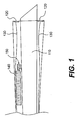
- Figure 1 shows an embodiment where needle 110 is within catheter 100 or protruding a small distance. Electrode surfaces 120 send electric signals via wires 130 to a controller (not shown). The controller activates solenoid 140 to automatically push cells out of needle 110.
- the catheter tip preferably is tapered to allow insertion into or penetration of a body cavity such as a femoral artery or coronary artery.
- the catheter body should be flexible enough (as described in many of the cited patents) to allow the tip to be manipulated to the desired site such as the interior of the heart.
- the catheter preferably has two or more electrodes on a flat surface of the tip. The centers of the electrodes are axially displaced 50 to 2000 ⁇ m from each other, preferably 100 to 1000 ⁇ m from each other and most preferably between 150 to 500 ⁇ m from each other. Preferably the electrodes are within the plane of a flat surface that terminates at the catheter tip, as shown in Figure 3. In some embodiments one or more electrodes are present on the side of the catheter tip as shown, for example in U.S. Pat. No. 5,450,846.
- the electrodes are electrically connected to conductive wires in the catheter, printed conductive (and insulated) lines on the catheter surface or other conductive material that extend from the tip to the proximal end of the catheter where they are connected electrically to a control system.
- conductive wires in the catheter printed conductive (and insulated) lines on the catheter surface or other conductive material that extend from the tip to the proximal end of the catheter where they are connected electrically to a control system.
- direct conductivity is preferred, it will be recognized that other means of transferring information is possible, including for example, use of a miniature radio transmitter from inside the catheter to a receiver outside the host (patient).
- the exterior surface of the catheter may comprise a coating of heparin, hirudin, antibiotics or the like or other substance chosen for biocompatibility with the host.
- the catheter further contains an electro-mechanical device for pushing a needle within the catheter tip into muscle tissue after sensing of mechanical contact between the catheter tip and the muscle tissue and activation via the controller.
- the needle in the tip of the catheter extends throughout the catheter and is connected to a reservoir of myogenic cells outside of the catheter.
- cells are useful for the invention, as described for example in U.S. Pat. Nos. 5,130,141 and 6,015,671.
- the cells are provided in aqueous suspension with optional polymer for mechanical stabilization and/or to improve subsequent attachment to the site of delivery.
- suspension media and suitable optional polymers are provided in U.S. Pat. No. 5,567,612.
- Specialized cell media may be used in conjunction with the catheter.
- a polymer is included to minimize leakage and/or movement of cells after injection into the myocardium.
- Polymers preferred for this use include for example, those described in U.S. Pat. No. 6,051,648.
- Polymers and binding agents that adhere to muscle cells and myocardium tissue are particularly preferred, and may be used in combination with other polymers or substances that bind and hold myogenic cells in place for a period of time before dissolution or release of the binding agents.
- concentration and average sizes of polymers used for these purposes will be readily determined through routine experimentation and chosen to minimize viscosity of the injected cell suspension.
- freshly oxygenated red cells are included in the media to maintain aerobic respiration of myogenic cells that are loaded and remain within the catheter for a period of time prior to their injection.
- Myogenic cells according to the invention pass through the needle in the catheter tip and are injected into a suitable myocardial (or other tissue) inner surface by operation of the catheter and its control system.
- the cells are loaded into a reservoir of the lumen catheter shortly before use of the catheter.
- the reservoir is fluidically connected to the injection needle.
- the cells are within a reservoir attached to the catheter and pass through the catheter length to the needle shortly before or during use of the catheter.
- the space within the needle optionally may be purged of air by pulling body fluid from inside the patient into the needle and from there down to the proximal end of the needle. Then the body fluid is expelled by forcing in the other direction back out of the needle opening.
- a catheter according to embodiments of the invention contains preferably one needle but may have more than one for delivering cells to the target muscle.
- the needle preferably is between 19 to 30 gauge, more preferably between 21-28 gauge and most preferably between 22 to 27 gauge.
- the needle has an internal bore with no constriction, thus allowing smooth cell movement.
- the needle preferably is within the tip of the catheter either on the leading edge or on the side of the tip within 10 mm from the leading edge.
- the tip preferably protrudes between 3 and 12 mm and more preferably between 5 and 8 mm from the surface of the catheter.
- the needle is retracted within the catheter tip during insertion of the catheter into a patient and then is pushed out after entering the target organ such as the interior of a heart.
- the needle does not protrude beyond the tip under normal (non-injecting) conditions and is activated by a controller, which pushes the needle outside of the tip through a distance that preferably is between 3 and 12 mm and more preferably between 5 and 8 mm.
- the tip is flat and, as manipulated by the user, can sit flush on the surface of the myocardium.
- the needle comprises metal and acts as a common electrode to form one or more detecting circuits with one or more other electrodes at the catheter tip.
- the needle-electrode monitors electrical activity to detect the condition of a muscle, particularly heart muscle that the tip contacts or is adjacent to.
- two needles are present and arranged on opposite sides of the tip. Both needles can be used as electrodes. In the latter case, a controller may select one needle or the other to deliver cells.
- the bore or the inside of the needle is polished and made smooth by rubbing with a tungsten wire.
- tungsten wire is preferably threaded inside and used to polish the inside surface(s) of the needle until they become smooth. This embodiment limits cell lysis during injection of cells through the needle.
- the needle in one embodiment exists within the catheter lumen and is pushed out along the axis of the catheter when activated as shown in Figure 1.
- the needle is perpendicular to a plate or flat surface of the catheter tip and gets pushed out of that surface at an oblique angle or perpendicular with respect to the catheter axis, when activated.
- actuation of needle movement optionally may be performed by a solenoid or piezo electric device.
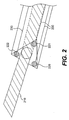
- the needle permanently is extended from the catheter surface as shown in Figure 2.
- the catheter is maneuvered within the heart and allows the needle to penetrate the myocardium.
- the electrical activity within the muscle is monitored by circuits outside the catheter to determine whether that portion of the muscle is degenerative or weak (decrease in or absence of normal electrical activity). If the electrical activity (or lack thereof) indicates a denerative or weak spot then myogenic cells are automatically expelled out of the needle into the muscle.
- Electrodes have been used in prior art catheters to detect special locations of the heart, such as, for example, the AV junction as described in U.S. Pat. No. 6,059,726 and for mapping fine detail in the heart, including pairs of sensors as described, for example by U.S. Pat. Nos. 5,865,787 and 5,450,846. Sensor materials such as carbon, silver, silver chloride plated silver and platinum are well known in the art and are suitable. Such electrodes are connected to wires or other conductors within and/or on the outer surface of the catheter. The wires connect to circuitry and create signals that can be analyzed by a computer.
- Two or more electrodes according to the invention may be used together to identify a localized area of abnormal myocardium as a site for injecting myogenic cells.
- the electrodes can be provided in various geometric forms, such as a flat surface perpendicular to the axis of the catheter tip, a flat orthogonal surface parallel to the axis of the catheter tip, a hollow cylinder, a sphere with a hole formed through the sphere, or a metallic bump on a flat insulating surface but preferably are two dots at corners of a flat probe tip and are used to determine when the probe tip is flush against the myocardium surface.
- One consideration in choosing the shape of the electrode is to confine the signal measurement area to a small region to allow detection of a localized damaged or weak muscle area.
- At least 2 electrodes are arranged on the catheter tip and an injection needle within the catheter tip is pushed out (perpendicular with) the contacted myocardium wall by a defined distance in response to signal(s) from the electrodes that indicate flush contact with a damaged myocardial surface.
- one electrode has a large surface area, or may be the catheter exterior surface (if electrically conductive) and is used as a ground to help provide more precise information to control circuitry about the electrical signal status of the immediate localized area.
- the catheter is manipulated by the medical practitioner inside a patient and, while doing so, the electrodes participate in one or more electrical circuits to provide electrical information feedback to a "control system" about the environment of the catheter tip.
- the electrodes may be used to provide, for example, electrical conductivity (resistance), high impedance voltage measurements or low impedance current measurements to the control system.
- the electrodes are electronically connected, preferably by wires, to a sensor/controller that monitors the electrical environment of the catheter tip.
- the sensor/controller controls injection of the myogenic cells through a needle in the catheter tip into the myocardium and preferably is integrated with a computer that records the history of injections and triggers injection in real time according to comparison of electrode signals with stored or computed comparative information.
- the injection is controlled by hardware and the parameters to control triggering of the actuator(s) are set by characteristics of the hardware and/or by electronic controls that are set by the user.
- two electrode pairs are connected in a circuit at any given time and either the conductivity or voltage between the electrodes is monitored.
- electrode pairs are coupled in a pair-wise fashion to differential amplifiers for generating one or more difference signals between the electrodes.
- the difference signals are subject to additional signal processing in conventional circuitry not explicitly depicted, such as signal conditioning and logical processing to develop a signal which ultimately is stored in a recording medium such as computer memory and also used to control injection of the myogenic cell suspension and (preferably) movement of a catheter needle as well. Further detail of the circuitry in the sensor/controller readily will be ascertained by a skilled artisan such as an electronics engineer.
- the sensor/controller of the control system preferably comprises a computer, which optionally collects data in real time both from the catheter electrodes and from an imaging tool using, for example, sonography to monitor the position of the catheter.
- the computer may store information related to the location of each myocardium locus assayed by electrode measurements, the electrical properties, which reveal the state of that locus, and the amount of cell suspension material injected into the locus.
- the circuit that alters the analog signals into digital signals for entry into the computer determines when the catheter tip surface has contacted a suitable target myocardium site.
- the computer or other controlling circuit quickly (preferably within 25 milliseconds, more preferably within 1 millisecond actuates efflux of myogenic cell suspension through the needle and into the target myocardium) and, according to the preferred mode where a recessed needle is used, actuates (starts) movement of a recessed needle within the catheter tip, causing it to lunge out of the catheter tip and imbed into the target myocardium.
- Electromechanical actuators such as a solenoid or piezo-electric device are preferred for these movements.
- the actuator for pushing the cell suspension through the needle may be outside the catheter.
- the pump for expelling cells may be within the catheter.
- the pump may for example, be a large surface piezo electric device (more than 1 cm 2 surface exposed to the cell suspension) having a surface that twists in response to an electrical signal, or may be a solenoid plunger that is connected to a closed flexible reservoir that causes increased pressure in the reservoir, which is fluidically connected to the catheter needle.
- the automatic control system monitors the heart beat and triggers an injection sequence at a suitable time between contractions to maximize the amount of time for injecting the cells.
- a computer is used that monitors (a) the heart beat cycle and (b) detection of contact with a weak or degenerated portion of the muscle. When the timing is right (eg. beginning of a rest period of the cycle) and the tip portion containing the needle tip contacts a weak or degenerated spot, as determined by absent or abnormal electrical signal(s) then the computer triggers the needle to plunge into the heart wall and triggers the efflux of cells, all in short time that preferably is less than one second.
- the signal for actuating efflux of cells may occur at the same time as the signal for triggering the needle to plunge into the heart.
- triggering of cell efflux may occur slightly after triggering needle movement.
- needle movement is controlled by a first piezo electric device within the catheter and the cells are pushed out of the needle by a second pump that is connected to a catheter, and both movements are started simultaneously.
- the needle plunge and cell efflux with the proper timing of the heart beat and to carry out the entire sequence of events, from proximity detection of the needle tip to a weak or degenerated region of the heart, to the needle plunge and cell efflux during the time when the particular chamber of the heart being treated is not actively contracting.
- the contraction of the heart itself is simultaneously controlled to lengthen the time for injecting the cells.
- a skilled artisan can determine suitable timing for efflux of myogenic cells through the needle, and plunging a recessed needle out of the catheter tip based on known or calculated transients for the respective movements. That is, depending on the physics of the system such as the reservoir size and response to the actuator force, the needle diameter and distance, the properties (particularly viscosity) of the fluid, etc., a timing sequence can be readily determined by a skilled artisan.
- a polymer or other thickening agent is added to the cell suspension to limit leakage of cell suspension out of the needle when not in use.
- a longer pause occurs between actuation of the pump and actual efflux of material from the needle.
- the time between optional electromechanical movement of the needle into the muscle is timed to coincide with the time for efflux of cells from the needle.
- myogenic cell suspension is forced out the needle tip and deposited into the myocardium.
- at least 5 million cells are expelled in one injection within at least 0.1ml of volume. More preferably at least 15 million cells are expelled within at least 0.25 ml volume. Most preferably between 25 and 100 million cells are expelled in a volume of between 0.3 and 0.7 ml.
- a total of between 500 million cells and 5 billion cells are administered among at least 10 such injections and preferably between 25 and 60 injections.
- an adult heart in need of repair may receive 40 0.5 ml injections for a total of 20 ml of suspension having a cell density of 100 million cells per ml. For this cell density, a 22 gauge needle is most preferred. Larger and smaller gauges are also suitable depending on the viscosity of the cell suspension that is used.
- Degenerative or weak sites of muscle tissue can be detected by its absent or weak electrical signals.
- an electrical signal emanating from that normal tissue can be detected by a reference electrical signal, typically determined by periodicity, waveform, amplitude, and/or duration of an electrical signal measured between two electrodes on the catheter surface.
- a signal from a damaged or weak target location will be absent, of lower strength (measured as a lower voltage or lower current depending on the impedance of the measurement) or of a different pattern of periodicity, waveform, amplitude and/or duration.
- This comparative information particularly is useful in two ways.
- the information can be used to inform the operator (and the computer) when the catheter has contacted damaged or weak tissue.
- Such information may be provided as an audio signal to the operator, the amplitude of which increases as the catheter moves away from healthy tissue, indicating movement to or arrival at diseased tissue.
- the information can be used by the controller to automatically inject myogenic cells upon contact with the diseased tissue.
- the controller preferably will have a set of stored or determined threshold values that, once exceeded indicates that the tissue next to the catheter tip is weak enough to warrant an injection of cells.
- a combination of measurements are made. For example, resistance can be measured between electrodes to detect placement of the catheter tip on a surface. Upon flush contact, the space between the electrodes is filled by tissue and solution phase conductivity is lost. When in contact with a tissue, the conductivity is greater.
- This conductivity (resistance) measurement preferably is carried out using an alternating current electrical drive source (preferably above 120 Hz, more preferably above 1,000, even more preferably above 50,000 Hz) in order to distinguish that signal from the noisy electrical environment produced by the muscle tissue.
- a filter tuned to the frequency of the drive source preferably is used to remove the background noise components of the signal.
- a background control resistance is determined by conductivity between electrodes when the tip is not in contact with (in this case cardiac) tissue.
- the measured resistance increases (conductivity decreases), by a threshold amount, denoting that the space between the electrodes is occluded by the less conducting solid.
- the actual change in conductivity in one embodiment is provided as an audio signal to the operator to indicate when the tip is in contact with a solid (muscle) surface.
- this impedance measurement is carried out simultaneously with detection of electrical signals by the electrodes from heart tissue.
- Contact with heart tissue is detected by the change in impedance and a determination of whether the contacted tissue has been degenerative or became weak, is made by detecting whether the tissue produces normal healthy electrical pulses or abnormal electrical pulses (or no pulses) that indicate damaged or abnormal tissue.
- both detection of electrical signals produced from the myocardium surface and the detection of impedance to determine independently, whether the tip is touching a solid surface are carried out simultaneously with the same electrodes using one or more hardware and/or computer software filters to separate the signals from each other.
- the electrodes at the catheter tip detect the position of the needle and provide positional feedback information to the control system.
- closed circuits between pairs of adjacent electrodes are established by electronic switches in the controller/sensor circuitry (the switches connect specific electrode combinations), the signals produced are buffered by operational amplifiers, and the analog signals are used, either directly to indicate a damaged target tissue, or indirectly by operation of a software program after their conversion into digital signals and input into a computer.
- the electrical conductivity between the needle and other electrode(s) increases.
- This signal is used by the sensor/control circuitry and/or the computer to feedback control the cell efflux (and optional needle movement) mechanisms.
- a signal is received from the needle-electrode circuit indicating that the needle has moved by a certain amount. The signal informs the computer of the time and duration of needle movement.
- a circuit is established between the patient's body and one or more electrodes (optionally including the needle as an electrode) and used to relax the heart muscle or otherwise momentarily alter the normal rhythm of the heart to facilitate detection and/or delivery of cells and/or other material into the target tissue. That is, a circuit is established between an electrode(s) and "ground,” wherein the ground is created by contacting a large surface electrode to the skin of the body. Skilled medical practitioners in this field will readily appreciate voltages and patterns needed for cardiac pacing, as exemplified in U.S. Pat. No. 5,634,899.
- a stimulation signal may be generated from the catheter itself to the heart to alter the heart's movement and improve chances for moving the catheter to a better position.
- the catheter electrode can provide two dimensional information that informs the user which direction to move the catheter, which is particularly useful for the embodiment having a retractable needle.
- Figure 2 shows needle 210 that extends 7mm from a surface of catheter 200.
- two or more embodiment conductivity measurements and/or two or more measurements of electrical signals produced by muscle tissue are made using electrodes 220, 221, and 222.
- the electrodes are connected to wires 230. Measurements can be made from the three electrode system shown in Figure 2 to provide two dimensional information about which direction to move the tip to encounter damaged myocardium.
- a user may manipulate the catheter tip shown in Figure 2 to sit flush on the surface of a myocardium.
- the control system compares conductivity (or voltage produced by myocardium itself) in a circuit formed between electrode 220 and electrode 221, in a circuit formed between electrode 220 and electrode 222, and in a circuit formed between electrode 222 and electrode 221.
- conductivity or voltage produced by myocardium itself
- the control system monitors and compares signals, both temporally and (preferably) spacially to obtain this information.
- a data base is obtained in a model system such as a pig heart system to generate representative voltages, impedances and conductivities for a given multiple electrode catheter system.
- the system described herein can be used for injecting cells into a muscle lining any body space such as found in stomach, intestine, bladder, arteries, heart. Most preferred uses are for within the heart although both epicardial tissue can be treated as well as endocardial tissue.
- the medical practitioner inserts the catheter into the femoral artery leading to the left ventricle or via the coronary artery through the aorta and into the left ventricle as depicted in Figure 3.
- the operator manipulates the catheter tip to contact various regions of the heart that are suspected of containing degenerative or weak tissue, such as the lateral wall of the left ventricle.
- the control system interfaced with the catheter monitors the electrical environment of the catheter tip and detects when the tip contacts a degenerative or weak area of the myocardial tissue. The heart is beating during this time. The control system automatically ejects cell suspension material through the needle tip and into the tissue.
- the control system optionally may activate a needle to come out of the catheter tip and imbed into the endo-myocardial tissue prior to ejection of cell suspension in the embodiment that uses a retractable needle.
- the needle then retracts and a series of such automated injections are made at various locations.
- FIG. 4 An example of a system for cardiac therapy is shown in Figure 4.
- the distal end of catheter 400 is seen at the top of this figure, and has a tip to the right in which needle 410 protrudes 7 mm.
- Electrode surfaces 420 are arranged around needle 410 and electrically connect by wires 430 to hardware 440.
- Hardware 440 buffers the electrical signals and converts them into a digital form that is input into computer controller 450.
- Needle 410 is fluidically connected to reservoir 460, which comprises pressure activator 470.
- computer 450 receives signals from hardware 440, compares those signals with information stored inside the computer and then turns on activator 470 upon detection of a degenerative or weak area of myocardium at the tip of catheter 400.
- the injection of cells into degenerative or weak portions of myocardial tissue are carried out within a total time of less than 2 seconds, more preferably less than 1 second and most preferably less than 0.5 seconds.
- the catheter is removed and the injected cells (if myogenic cells) gradually differentiate into heart muscle cells over time. Improved heart muscle function is detected by any of a number of techniques known to the skilled artisan.
- inventions discussed herein concern the repair and/or augmentation of heart tissue.
- the invention also is useful for repair and augmentation of other body parts such as regular smooth muscle arteries, the stomach, the diaphram, the bladder, the liver and other tissues.
- reconstructive surgery of bladder may, in some aspects benefit from administration of myogenic cells in a polymeric suspension to plug a gap or hole in a pre-existing muscle.
- reconstruction is performed with a catheter according to the invention in a two step procedure. In the first step, myogenic cells are injected into areas that respond poorly to peristaltic signals or that poorly generate peristaltic signals.
- a second type of material is then injected to cover the myogenic cells of the first injection to make a patch that protects the cells from the gastrointestinal tract lining during healing.
- the second type of material may be administered by the same catheter or by a second device.
- the cells of choice are myogenic cells, which are known to fuse with other cells both in muscle tissue and fat tissue.
Landscapes
- Health & Medical Sciences (AREA)
- Life Sciences & Earth Sciences (AREA)
- Engineering & Computer Science (AREA)
- General Health & Medical Sciences (AREA)
- Animal Behavior & Ethology (AREA)
- Biophysics (AREA)
- Veterinary Medicine (AREA)
- Public Health (AREA)
- Biomedical Technology (AREA)
- Heart & Thoracic Surgery (AREA)
- Medical Informatics (AREA)
- Molecular Biology (AREA)
- Surgery (AREA)
- Physics & Mathematics (AREA)
- Pathology (AREA)
- Nuclear Medicine, Radiotherapy & Molecular Imaging (AREA)
- Radiology & Medical Imaging (AREA)
- Hematology (AREA)
- Anesthesiology (AREA)
- Pulmonology (AREA)
- Chemical & Material Sciences (AREA)
- Bioinformatics & Cheminformatics (AREA)
- Medicinal Chemistry (AREA)
- Pharmacology & Pharmacy (AREA)
- Media Introduction/Drainage Providing Device (AREA)
- Electrotherapy Devices (AREA)
- Materials For Medical Uses (AREA)
- Measurement And Recording Of Electrical Phenomena And Electrical Characteristics Of The Living Body (AREA)
- Infusion, Injection, And Reservoir Apparatuses (AREA)
- Micro-Organisms Or Cultivation Processes Thereof (AREA)
Claims (18)
- Ein Apparat zum Nachweis und zur Behandlung von lokalen Flächen mit einer Degenerierung und Schwäche von Muskeln in einem Patienten, umfassend ein Reservoir (460) von myogenen Zellen, ein Katheter (100; 400) und eine fluide Verbindung zwischen dem Reservoir von myogenen Zellen und dem Katheter (100; 400), wobei der Katheter (100; 400) umfasst:a) einen langen Körper, der in einen Patienten eingeführt werden kann;b) eine Düsenspitze am Ende des langen Körpers;c) einen Sensor auf der Düsenspitze; undd) eine Nadel (110; 210; 410), die mit der fluiden Verbindung verbunden ist, die zur Injektion von myogenen Zellen in den Muskel geeignet ist und eine Spitze hat;worin der Sensor derart positioniert ist, um eine Oberfläche am Ende der Düsenspitze zu detektieren und die lange Achse der Nadel (110; 210; 410) am Ende der Düsenspitze positioniert ist.
- Ein Apparat, wie er in Anspruch 1 beschrieben ist, worin der Sensor mindestens zwei Elektroden umfasst.
- Ein Apparat, wie er in Anspruch 1 beschrieben ist, worin die Spitze der Nadel (110; 210; 410) während der Einführung des langen Körpers des Katheters (100; 400) in den Patienten innerhalb des Katheters (100; 400) bleibt und nach der Detektion einer elektrisch ruhigen oder untätigen Muskeloberfläche aus der Düsenspitze ausgestoßen wird.
- Ein Apparat, wie er in Anspruch 1 beschrieben ist, weiterhin umfassend einen Auslöser (470) für das Reservoir zum Herausdrücken der Zellen aus dem Reservoir (460) und durch die fluide Verbindung zwischen dem Reservoir (460) und der Nadel (410).
- Ein Apparat, wie er in Anspruch 1 beschrieben ist, worin die Nadel (110; 210; 410) in die Eintiefung der Düsenspitze zurückgezogen ist und aus der Eintiefung durch einen Auslöser für die Nadel als Reaktion auf von den Elektroden (120; 220; 221; 222) erhaltene Signale herausgedrückt wird.
- Ein Apparat, wie er in Anspruch 1 beschrieben ist, worin die Nadel (110; 210; 410) für die Injektion von Zellen durch eine Methode hergestellt wird, bei der ein Draht aus Wolfram mehrere Male durch die Nadel (110; 210; 410) hindurch gezogen wird.
- Ein Apparat, wie er in Anspruch 1 beschrieben ist, worin die Nadel (110; 210; 410) flexibel ist und einen äußeren Durchmesser von achtzehn bis achtundzwanzig Gauge (G) (1,20 mm bis 0,37 mm) hat und die Düsenspitze des Katheters spitz zuläuft, um einen engen Kontakt der Nadel (110; 210; 410) mit der Innenwand des Herzen zu ermöglichen.
- Ein Apparat, wie er in Anspruch 1 beschrieben ist, worin die mindestens zwei Elektroden (120; 220; 221; 222) in der Lage sind, einen mechanischen Kontakt zwischen der Düsenspitze des Katheters und dem Muskelgewebe zu ermöglichen wie z.B. durch Überwachung der inneren Oberfläche eines elektrisch abnormalen Herzmuskels.
- Ein Apparat, wie er in Anspruch 1 beschrieben ist, der weiterhin einen Regelkreis umfasst, der mehrfache Injektionen von Zellen von der Quelle der myogenen Zellen regelt.
- Ein Apparat, wie er in Anspruch 1 beschrieben ist, worin die Nadel nicht mehr als 5 Millimeter aus der Düsenspitze des Katheters herausragt.
- Ein Apparat, wie er in Anspruch 1 beschrieben ist, der zusätzlich eine Regelung umfasst, die Teile eines Herzens überwacht und die Lieferung von Zellen zu einem Teil durch den Katheter (100; 400) regelt, wobei die Regelung umfasst:a) zwei oder mehr Regelkreise zur Überwachung, welch die von den Elektroden in der Düsenspitze des Katheters erzeugten Signale beobachten.b) einen Vergleicher (450), der Hardware - Schaltkreise, einen Computer oder beides umfasst, der die von den Regelkreisen zur Überwachung erhaltenen Signale analysiert; undc) einen Aktivator (470) für die Injektion von Zellen durch die Nadel (110; 210; 410) und in den Teil des Herzen,worin der Vergleicher (450) in der Lage ist, den Aktivator (470) innerhalb von 50 Millisekunden nach Erhalt von Signalen von den Regelkreisen zur Überwachung anzusteuern.
- Ein Apparat, wie er in Anspruch 11 beschrieben ist, worin die Regelkreise zur Überwachung mindestens 3 Regelkreise von 3 Elektroden umfassen und worin die drei Elektroden auf der Düsenspitze des Katheters eine ebene Oberfläche bilden.
- Ein Apparat, wie er in Anspruch 11 beschrieben ist, worin das Reservoir (460) zusätzlich frisch mit Sauerstoff angereicherte rote Zellen umfasst.
- Ein Apparat, wie er in Anspruch 1 beschrieben ist, worin die Nähe der Düsenspitze des Katheters zu der Oberfläche einer lokalen Fläche von Degenerierung und Schwäche von Muskeln durch die Beobachtung des elektrischen Widerstands bestimmt wird.
- Der Apparat von Anspruch 1, worin der Sensor mindestens drei Elektroden (120; 220; 221; 222) auf der Düsenspitze umfasst und die mindestens drei Elektroden (120; 220; 221; 222) eine flache Oberfläche bilden, so dass die lange Achse der Nadel (110; 210; 410) zwischen den mindestens drei Elektroden (120; 220; 221; 222) angeordnet ist.
- Ein Apparat, wie er in Anspruch 1 beschrieben ist, der weiterhin einen Signalerzeuger umfasst, in dem die von dem Sensor (120; 220; 221; 222) erhaltenen elektrischen Signale von dem Signalerzeuger erzeugt werden und der elektrische Widerstand beobachtet wird, um den Kontakt des Katheters (100; 400) mit einer Oberfläche zu bestimmen.
- Ein Apparat, wie er in Anspruch 15 beschrieben ist, der weiterhin einen Regelkreis umfasst, der mehrfache Injektionen der Zellen regelt.
- Ein Apparat, wie er in Anspruch 15 beschrieben ist, worin die Nadel (110; 210; 410) nicht mehr als 5 Millimeter aus der Düsenspitze des Katheters herausragt.
Applications Claiming Priority (3)
| Application Number | Priority Date | Filing Date | Title |
|---|---|---|---|
| US23188000P | 2000-09-12 | 2000-09-12 | |
| US231880P | 2000-09-12 | ||
| PCT/US2001/028712 WO2002028470A1 (en) | 2000-09-12 | 2001-09-11 | Myogenic cell transfer catheter and method |
Publications (3)
| Publication Number | Publication Date |
|---|---|
| EP1324802A1 EP1324802A1 (de) | 2003-07-09 |
| EP1324802A4 EP1324802A4 (de) | 2005-08-31 |
| EP1324802B1 true EP1324802B1 (de) | 2006-11-15 |
Family
ID=22870981
Family Applications (1)
| Application Number | Title | Priority Date | Filing Date |
|---|---|---|---|
| EP01979245A Expired - Lifetime EP1324802B1 (de) | 2000-09-12 | 2001-09-11 | Katheter für den myogenen zelltransfer |
Country Status (8)
| Country | Link |
|---|---|
| US (1) | US20050124975A1 (de) |
| EP (1) | EP1324802B1 (de) |
| CN (1) | CN1474707A (de) |
| AT (1) | ATE345158T1 (de) |
| AU (1) | AU1123002A (de) |
| CA (1) | CA2422065A1 (de) |
| DE (1) | DE60124574T2 (de) |
| WO (1) | WO2002028470A1 (de) |
Families Citing this family (30)
| Publication number | Priority date | Publication date | Assignee | Title |
|---|---|---|---|---|
| US7169127B2 (en) * | 2002-02-21 | 2007-01-30 | Boston Scientific Scimed, Inc. | Pressure apron direct injection catheter |
| US7108685B2 (en) * | 2002-04-15 | 2006-09-19 | Boston Scientific Scimed, Inc. | Patch stabilization of rods for treatment of cardiac muscle |
| WO2004073484A2 (en) | 2003-02-24 | 2004-09-02 | Yossi Gross | Fully-implantable cardiac recovery system |
| US20050021091A1 (en) * | 2003-04-23 | 2005-01-27 | Laske Timothy G. | System for the delivery of a biologic therapy with device monitoring and back-up |
| US7713232B2 (en) | 2005-11-04 | 2010-05-11 | Medrad, Inc. | System for washing and processing of cells for delivery thereof to tissue |
| US8182444B2 (en) * | 2005-11-04 | 2012-05-22 | Medrad, Inc. | Delivery of agents such as cells to tissue |
| US8038595B2 (en) | 2006-01-25 | 2011-10-18 | Beth Israel Deaconess Medical Center | Devices and methods for tissue transplant and regeneration |
| US20080086111A1 (en) * | 2006-10-09 | 2008-04-10 | Medrad, Inc. | Fluid delivery systems and volume metering in cell delivery |
| US20080147007A1 (en) * | 2006-12-19 | 2008-06-19 | Toby Freyman | Delivery device with pressure control |
| US7951144B2 (en) * | 2007-01-19 | 2011-05-31 | Mahajan Roop L | Thermal and electrical conductivity probes and methods of making the same |
| US8292873B2 (en) * | 2007-08-09 | 2012-10-23 | Boston Scientific Scimed, Inc. | Catheter devices for myocardial injections or other uses |
| US8086293B2 (en) * | 2009-04-14 | 2011-12-27 | Medtronic Ablation Frontiers Llc | Catheter |
| US9278220B2 (en) * | 2013-07-23 | 2016-03-08 | Medtronic, Inc. | Identification of healthy versus unhealthy substrate for pacing from a multipolar lead |
| US20150050300A1 (en) | 2013-08-16 | 2015-02-19 | Peter K. Law | Disease prevention and alleviation by human myoblast transplantation |
| GB2567122C (en) | 2016-09-21 | 2023-06-14 | Law Peter | Autonomously controllable pull wire injection catheter, robotic system comprising said catheter and method for operating the same |
| US11559298B2 (en) | 2018-07-16 | 2023-01-24 | Cilag Gmbh International | Surgical visualization of multiple targets |
| US12257013B2 (en) | 2019-03-15 | 2025-03-25 | Cilag Gmbh International | Robotic surgical systems with mechanisms for scaling camera magnification according to proximity of surgical tool to tissue |
| US11832996B2 (en) | 2019-12-30 | 2023-12-05 | Cilag Gmbh International | Analyzing surgical trends by a surgical system |
| US12002571B2 (en) | 2019-12-30 | 2024-06-04 | Cilag Gmbh International | Dynamic surgical visualization systems |
| US11744667B2 (en) | 2019-12-30 | 2023-09-05 | Cilag Gmbh International | Adaptive visualization by a surgical system |
| US11776144B2 (en) | 2019-12-30 | 2023-10-03 | Cilag Gmbh International | System and method for determining, adjusting, and managing resection margin about a subject tissue |
| US12453592B2 (en) | 2019-12-30 | 2025-10-28 | Cilag Gmbh International | Adaptive surgical system control according to surgical smoke cloud characteristics |
| US11648060B2 (en) | 2019-12-30 | 2023-05-16 | Cilag Gmbh International | Surgical system for overlaying surgical instrument data onto a virtual three dimensional construct of an organ |
| US11759283B2 (en) | 2019-12-30 | 2023-09-19 | Cilag Gmbh International | Surgical systems for generating three dimensional constructs of anatomical organs and coupling identified anatomical structures thereto |
| US11219501B2 (en) | 2019-12-30 | 2022-01-11 | Cilag Gmbh International | Visualization systems using structured light |
| US12053223B2 (en) | 2019-12-30 | 2024-08-06 | Cilag Gmbh International | Adaptive surgical system control according to surgical smoke particulate characteristics |
| US11284963B2 (en) | 2019-12-30 | 2022-03-29 | Cilag Gmbh International | Method of using imaging devices in surgery |
| US12207881B2 (en) | 2019-12-30 | 2025-01-28 | Cilag Gmbh International | Surgical systems correlating visualization data and powered surgical instrument data |
| US11896442B2 (en) | 2019-12-30 | 2024-02-13 | Cilag Gmbh International | Surgical systems for proposing and corroborating organ portion removals |
| WO2022060696A1 (en) * | 2020-09-15 | 2022-03-24 | University Of South Carolina | Minimally invasive and semi-automated myocardial injection device |
Family Cites Families (27)
| Publication number | Priority date | Publication date | Assignee | Title |
|---|---|---|---|---|
| US4323071A (en) * | 1978-04-24 | 1982-04-06 | Advanced Catheter Systems, Inc. | Vascular guiding catheter assembly and vascular dilating catheter assembly and a combination thereof and methods of making the same |
| US4582181A (en) * | 1983-08-12 | 1986-04-15 | Advanced Cardiovascular Systems, Inc. | Steerable dilatation catheter |
| DE3442736C2 (de) * | 1984-11-23 | 1987-03-05 | Tassilo Dr.med. 7800 Freiburg Bonzel | Dilatationskatheter |
| US5130141A (en) * | 1988-05-24 | 1992-07-14 | Law Peter K | Compositions for and methods of treating muscle degeneration and weakness |
| US5002067A (en) * | 1989-08-23 | 1991-03-26 | Medtronic, Inc. | Medical electrical lead employing improved penetrating electrode |
| US5156151A (en) * | 1991-02-15 | 1992-10-20 | Cardiac Pathways Corporation | Endocardial mapping and ablation system and catheter probe |
| US5634899A (en) * | 1993-08-20 | 1997-06-03 | Cortrak Medical, Inc. | Simultaneous cardiac pacing and local drug delivery method |
| US5385146A (en) * | 1993-01-08 | 1995-01-31 | Goldreyer; Bruce N. | Orthogonal sensing for use in clinical electrophysiology |
| UA40634C2 (uk) * | 1993-11-15 | 2001-08-15 | Зенека Лімітед | Мікрокапсула, яка містить суспензію твердої біологічно активної сполуки, і спосіб її одержання |
| US6015671A (en) * | 1995-06-07 | 2000-01-18 | Indiana University Foundation | Myocardial grafts and cellular compositions |
| US5602301A (en) * | 1993-11-16 | 1997-02-11 | Indiana University Foundation | Non-human mammal having a graft and methods of delivering protein to myocardial tissue |
| US5902290A (en) * | 1994-03-14 | 1999-05-11 | Advanced Cardiovascular Systems, Inc. | Catheter providing intraluminal access |
| JP3006425B2 (ja) * | 1994-09-09 | 2000-02-07 | 日本電気株式会社 | 半導体装置及びその製造方法 |
| EP0743820A4 (de) * | 1994-12-13 | 1997-06-11 | Peter K Law | Myoblast-therapie für säugetierkrankheiten |
| PT876165E (pt) * | 1995-12-18 | 2006-10-31 | Angiotech Biomaterials Corp | Composicoes de polimeros reticulados e processos para a sua utilizacao |
| US6011889A (en) * | 1996-04-29 | 2000-01-04 | Eclipse Surgical Technologies, Inc. | Piercing point optical fiber device for laser surgery procedures |
| DE29609350U1 (de) * | 1996-05-24 | 1996-08-29 | Dr.-Ing. P. Osypka mbH Gesellschaft für Medizintechnik, 79639 Grenzach-Wyhlen | Vorrichtung zum Perforieren der Herzwand |
| US5916194A (en) * | 1996-05-24 | 1999-06-29 | Sarcos, Inc. | Catheter/guide wire steering apparatus and method |
| US6016437A (en) * | 1996-10-21 | 2000-01-18 | Irvine Biomedical, Inc. | Catheter probe system with inflatable soft shafts |
| US6059726A (en) * | 1996-11-08 | 2000-05-09 | The Regents Of The University Of California | Method for locating the atrio-ventricular (AV) junction of the heart and injecting active substances therein |
| WO1998022021A1 (en) * | 1996-11-20 | 1998-05-28 | Myotech | Multi-electrode and needle injection device for diagnosis and treatment of muscle injury and pain |
| US6200303B1 (en) * | 1997-04-30 | 2001-03-13 | Beth Israel Deaconess Medical Center, Inc. | Method and kit for transvenously accessing the pericardial space via the right atrium |
| US5941845A (en) * | 1997-08-05 | 1999-08-24 | Irvine Biomedical, Inc. | Catheter having multiple-needle electrode and methods thereof |
| EP0932427A1 (de) * | 1997-07-22 | 1999-08-04 | Emed Corporation | Nadel für iontophoretische verabreichung eines wirkstoffes |
| US6059759A (en) * | 1997-10-14 | 2000-05-09 | Merit Medical Systems, Inc. | Infusion catheter systems with tactile sensing feedback |
| JP3473386B2 (ja) * | 1998-03-26 | 2003-12-02 | 株式会社デンソー | 車両用交流発電機のステータコイル |
| US6224566B1 (en) * | 1999-05-04 | 2001-05-01 | Cardiodyne, Inc. | Method and devices for creating a trap for confining therapeutic drugs and/or genes in the myocardium |
-
2001
- 2001-09-11 CA CA002422065A patent/CA2422065A1/en not_active Abandoned
- 2001-09-11 EP EP01979245A patent/EP1324802B1/de not_active Expired - Lifetime
- 2001-09-11 AT AT01979245T patent/ATE345158T1/de not_active IP Right Cessation
- 2001-09-11 WO PCT/US2001/028712 patent/WO2002028470A1/en not_active Ceased
- 2001-09-11 US US10/486,111 patent/US20050124975A1/en not_active Abandoned
- 2001-09-11 DE DE60124574T patent/DE60124574T2/de not_active Expired - Lifetime
- 2001-09-11 AU AU1123002A patent/AU1123002A/xx not_active Withdrawn
- 2001-09-11 CN CNA018187102A patent/CN1474707A/zh active Pending
Also Published As
| Publication number | Publication date |
|---|---|
| ATE345158T1 (de) | 2006-12-15 |
| EP1324802A1 (de) | 2003-07-09 |
| WO2002028470A9 (en) | 2003-11-13 |
| US20050124975A1 (en) | 2005-06-09 |
| DE60124574D1 (de) | 2006-12-28 |
| CA2422065A1 (en) | 2002-04-11 |
| CN1474707A (zh) | 2004-02-11 |
| AU1123002A (en) | 2002-04-15 |
| WO2002028470A1 (en) | 2002-04-11 |
| DE60124574T2 (de) | 2007-06-21 |
| EP1324802A4 (de) | 2005-08-31 |
Similar Documents
| Publication | Publication Date | Title |
|---|---|---|
| US20050124975A1 (en) | Myogenic cell transfer catheter and method | |
| CN104159636B (zh) | 心肌药剂递送装置和方法 | |
| EP1563866B1 (de) | Gerät zum Freisetzen eines Medikaments im Herzen | |
| US7749215B1 (en) | Intracardiac cell delivery and cell transplantation | |
| US20110014181A1 (en) | Microneedle Delivery Device and Methods of Using Same | |
| US20030233126A1 (en) | Injection devices and methods for testing implants | |
| EP1415660A1 (de) | Zielansteuernde Donorzellen an eine Gewebezone zur Abgabe von Translokationsstimulatoren | |
| US20090312617A1 (en) | Needle injection catheter | |
| EP1415661A1 (de) | Zielansteuernde embryonale Stammzellen an eine Gewebezone zur Abgabe von Translokationsstimulatoren | |
| EP1415662A1 (de) | Zielansteuernde autologe Zellen an eine Gewebezone zur Abgabe von Translokationsstimulatoren | |
| JP2009536560A (ja) | 診断用センサを備える心臓ハーネスおよびその使用方法 | |
| WO1999039624A1 (en) | Intracardiac drug delivery | |
| WO2005014084A1 (en) | Diastolic coronary perfusion detection for timed delivery of therapeutic and/or diagnostic agents | |
| EP3773885B1 (de) | Freisetzungsvorrichtung für implantierbare oder einsetzbare medizinprodukte | |
| WO2008076653A1 (en) | Catheters having linear electrode arrays and their method of use | |
| AU2007202290B2 (en) | Myogenic cell transfer catheter and method | |
| AU2002211230B2 (en) | Myogenic cell transfer catheter and method | |
| AU2012200651B2 (en) | Myogenic cell transfer catheter and method | |
| AU2002211230A1 (en) | Myogenic cell transfer catheter and method | |
| CA2284998C (en) | Intracardiac drug delivery | |
| HK1033284A (en) | Apparatus for intracardiac cell delivery and cell transplantation | |
| IL132073A (en) | Intracardiac drug delivery apparatus and method | |
| HK1079718B (en) | Intracardiac drug delivery |
Legal Events
| Date | Code | Title | Description |
|---|---|---|---|
| PUAI | Public reference made under article 153(3) epc to a published international application that has entered the european phase |
Free format text: ORIGINAL CODE: 0009012 |
|
| 17P | Request for examination filed |
Effective date: 20030410 |
|
| AK | Designated contracting states |
Designated state(s): AT BE CH CY DE DK ES FI FR GB GR IE IT LI LU MC NL PT SE TR |
|
| AX | Request for extension of the european patent |
Extension state: AL LT LV MK RO SI |
|
| A4 | Supplementary search report drawn up and despatched |
Effective date: 20050718 |
|
| RTI1 | Title (correction) |
Free format text: MYOGENIC CELL TRANSFER CATHETER |
|
| GRAP | Despatch of communication of intention to grant a patent |
Free format text: ORIGINAL CODE: EPIDOSNIGR1 |
|
| GRAS | Grant fee paid |
Free format text: ORIGINAL CODE: EPIDOSNIGR3 |
|
| GRAA | (expected) grant |
Free format text: ORIGINAL CODE: 0009210 |
|
| AK | Designated contracting states |
Kind code of ref document: B1 Designated state(s): AT BE CH CY DE DK ES FI FR GB GR IE IT LI LU MC NL PT SE TR |
|
| PG25 | Lapsed in a contracting state [announced via postgrant information from national office to epo] |
Ref country code: IT Free format text: LAPSE BECAUSE OF FAILURE TO SUBMIT A TRANSLATION OF THE DESCRIPTION OR TO PAY THE FEE WITHIN THE PRESCRIBED TIME-LIMIT;WARNING: LAPSES OF ITALIAN PATENTS WITH EFFECTIVE DATE BEFORE 2007 MAY HAVE OCCURRED AT ANY TIME BEFORE 2007. THE CORRECT EFFECTIVE DATE MAY BE DIFFERENT FROM THE ONE RECORDED. Effective date: 20061115 Ref country code: FI Free format text: LAPSE BECAUSE OF FAILURE TO SUBMIT A TRANSLATION OF THE DESCRIPTION OR TO PAY THE FEE WITHIN THE PRESCRIBED TIME-LIMIT Effective date: 20061115 Ref country code: BE Free format text: LAPSE BECAUSE OF FAILURE TO SUBMIT A TRANSLATION OF THE DESCRIPTION OR TO PAY THE FEE WITHIN THE PRESCRIBED TIME-LIMIT Effective date: 20061115 Ref country code: NL Free format text: LAPSE BECAUSE OF FAILURE TO SUBMIT A TRANSLATION OF THE DESCRIPTION OR TO PAY THE FEE WITHIN THE PRESCRIBED TIME-LIMIT Effective date: 20061115 Ref country code: LI Free format text: LAPSE BECAUSE OF FAILURE TO SUBMIT A TRANSLATION OF THE DESCRIPTION OR TO PAY THE FEE WITHIN THE PRESCRIBED TIME-LIMIT Effective date: 20061115 Ref country code: AT Free format text: LAPSE BECAUSE OF FAILURE TO SUBMIT A TRANSLATION OF THE DESCRIPTION OR TO PAY THE FEE WITHIN THE PRESCRIBED TIME-LIMIT Effective date: 20061115 |
|
| REG | Reference to a national code |
Ref country code: GB Ref legal event code: FG4D |
|
| RAP2 | Party data changed (patent owner data changed or rights of a patent transferred) |
Owner name: LAW, PETER K. |
|
| RIN2 | Information on inventor provided after grant (corrected) |
Inventor name: LAW, PETER K. |
|
| REG | Reference to a national code |
Ref country code: CH Ref legal event code: EP |
|
| REF | Corresponds to: |
Ref document number: 60124574 Country of ref document: DE Date of ref document: 20061228 Kind code of ref document: P |
|
| REG | Reference to a national code |
Ref country code: IE Ref legal event code: FG4D |
|
| NLT2 | Nl: modifications (of names), taken from the european patent patent bulletin |
Owner name: LAW, PETER K. Effective date: 20061213 |
|
| PG25 | Lapsed in a contracting state [announced via postgrant information from national office to epo] |
Ref country code: DK Free format text: LAPSE BECAUSE OF FAILURE TO SUBMIT A TRANSLATION OF THE DESCRIPTION OR TO PAY THE FEE WITHIN THE PRESCRIBED TIME-LIMIT Effective date: 20070215 Ref country code: SE Free format text: LAPSE BECAUSE OF FAILURE TO SUBMIT A TRANSLATION OF THE DESCRIPTION OR TO PAY THE FEE WITHIN THE PRESCRIBED TIME-LIMIT Effective date: 20070215 |
|
| PG25 | Lapsed in a contracting state [announced via postgrant information from national office to epo] |
Ref country code: ES Free format text: LAPSE BECAUSE OF FAILURE TO SUBMIT A TRANSLATION OF THE DESCRIPTION OR TO PAY THE FEE WITHIN THE PRESCRIBED TIME-LIMIT Effective date: 20070226 |
|
| PG25 | Lapsed in a contracting state [announced via postgrant information from national office to epo] |
Ref country code: PT Free format text: LAPSE BECAUSE OF FAILURE TO SUBMIT A TRANSLATION OF THE DESCRIPTION OR TO PAY THE FEE WITHIN THE PRESCRIBED TIME-LIMIT Effective date: 20070416 |
|
| NLV1 | Nl: lapsed or annulled due to failure to fulfill the requirements of art. 29p and 29m of the patents act | ||
| REG | Reference to a national code |
Ref country code: CH Ref legal event code: AEN Free format text: DAS PATENT IST AUF GRUND DES WEITERBEHANDLUNGSANTRAGS VOM 16.04.2007 REAKTIVIERT WORDEN. |
|
| ET | Fr: translation filed | ||
| REG | Reference to a national code |
Ref country code: CH Ref legal event code: PL |
|
| REG | Reference to a national code |
Ref country code: CH Ref legal event code: NV Representative=s name: HANS ULRICH SEIFERT SEIFERT & PARTNER |
|
| PLBE | No opposition filed within time limit |
Free format text: ORIGINAL CODE: 0009261 |
|
| STAA | Information on the status of an ep patent application or granted ep patent |
Free format text: STATUS: NO OPPOSITION FILED WITHIN TIME LIMIT |
|
| 26N | No opposition filed |
Effective date: 20070817 |
|
| PG25 | Lapsed in a contracting state [announced via postgrant information from national office to epo] |
Ref country code: GR Free format text: LAPSE BECAUSE OF FAILURE TO SUBMIT A TRANSLATION OF THE DESCRIPTION OR TO PAY THE FEE WITHIN THE PRESCRIBED TIME-LIMIT Effective date: 20070216 Ref country code: MC Free format text: LAPSE BECAUSE OF NON-PAYMENT OF DUE FEES Effective date: 20070930 |
|
| PG25 | Lapsed in a contracting state [announced via postgrant information from national office to epo] |
Ref country code: IE Free format text: LAPSE BECAUSE OF NON-PAYMENT OF DUE FEES Effective date: 20070911 |
|
| PG25 | Lapsed in a contracting state [announced via postgrant information from national office to epo] |
Ref country code: CY Free format text: LAPSE BECAUSE OF FAILURE TO SUBMIT A TRANSLATION OF THE DESCRIPTION OR TO PAY THE FEE WITHIN THE PRESCRIBED TIME-LIMIT Effective date: 20061115 Ref country code: LU Free format text: LAPSE BECAUSE OF NON-PAYMENT OF DUE FEES Effective date: 20070911 |
|
| PG25 | Lapsed in a contracting state [announced via postgrant information from national office to epo] |
Ref country code: TR Free format text: LAPSE BECAUSE OF FAILURE TO SUBMIT A TRANSLATION OF THE DESCRIPTION OR TO PAY THE FEE WITHIN THE PRESCRIBED TIME-LIMIT Effective date: 20061115 |
|
| REG | Reference to a national code |
Ref country code: FR Ref legal event code: ST Effective date: 20100531 |
|
| PG25 | Lapsed in a contracting state [announced via postgrant information from national office to epo] |
Ref country code: FR Free format text: LAPSE BECAUSE OF NON-PAYMENT OF DUE FEES Effective date: 20090930 |
|
| REG | Reference to a national code |
Ref country code: FR Ref legal event code: RN |
|
| REG | Reference to a national code |
Ref country code: FR Ref legal event code: D3 |
|
| PGRI | Patent reinstated in contracting state [announced from national office to epo] |
Ref country code: FR Effective date: 20101115 |
|
| REG | Reference to a national code |
Ref country code: GB Ref legal event code: 746 Effective date: 20120724 |
|
| REG | Reference to a national code |
Ref country code: FR Ref legal event code: PLFP Year of fee payment: 16 |
|
| REG | Reference to a national code |
Ref country code: FR Ref legal event code: PLFP Year of fee payment: 17 |
|
| REG | Reference to a national code |
Ref country code: FR Ref legal event code: PLFP Year of fee payment: 18 |
|
| PGFP | Annual fee paid to national office [announced via postgrant information from national office to epo] |
Ref country code: FR Payment date: 20190723 Year of fee payment: 19 |
|
| PGFP | Annual fee paid to national office [announced via postgrant information from national office to epo] |
Ref country code: GB Payment date: 20190719 Year of fee payment: 19 |
|
| PGFP | Annual fee paid to national office [announced via postgrant information from national office to epo] |
Ref country code: CH Payment date: 20190805 Year of fee payment: 19 Ref country code: DE Payment date: 20190930 Year of fee payment: 19 |
|
| REG | Reference to a national code |
Ref country code: DE Ref legal event code: R119 Ref document number: 60124574 Country of ref document: DE |
|
| REG | Reference to a national code |
Ref country code: CH Ref legal event code: PL |
|
| GBPC | Gb: european patent ceased through non-payment of renewal fee |
Effective date: 20200911 |
|
| PG25 | Lapsed in a contracting state [announced via postgrant information from national office to epo] |
Ref country code: DE Free format text: LAPSE BECAUSE OF NON-PAYMENT OF DUE FEES Effective date: 20210401 Ref country code: FR Free format text: LAPSE BECAUSE OF NON-PAYMENT OF DUE FEES Effective date: 20200930 |
|
| PG25 | Lapsed in a contracting state [announced via postgrant information from national office to epo] |
Ref country code: CH Free format text: LAPSE BECAUSE OF FAILURE TO SUBMIT A TRANSLATION OF THE DESCRIPTION OR TO PAY THE FEE WITHIN THE PRESCRIBED TIME-LIMIT Effective date: 20200930 Ref country code: LI Free format text: LAPSE BECAUSE OF FAILURE TO SUBMIT A TRANSLATION OF THE DESCRIPTION OR TO PAY THE FEE WITHIN THE PRESCRIBED TIME-LIMIT Effective date: 20200930 Ref country code: GB Free format text: LAPSE BECAUSE OF NON-PAYMENT OF DUE FEES Effective date: 20200911 |