EP1338238A2 - Ophthalmo-Operationsmikroskop - Google Patents
Ophthalmo-Operationsmikroskop Download PDFInfo
- Publication number
- EP1338238A2 EP1338238A2 EP03001157A EP03001157A EP1338238A2 EP 1338238 A2 EP1338238 A2 EP 1338238A2 EP 03001157 A EP03001157 A EP 03001157A EP 03001157 A EP03001157 A EP 03001157A EP 1338238 A2 EP1338238 A2 EP 1338238A2
- Authority
- EP
- European Patent Office
- Prior art keywords
- refractometer
- surgical microscope
- patient
- microscope according
- data
- Prior art date
- Legal status (The legal status is an assumption and is not a legal conclusion. Google has not performed a legal analysis and makes no representation as to the accuracy of the status listed.)
- Withdrawn
Links
Images

Classifications
-
- A—HUMAN NECESSITIES
- A61—MEDICAL OR VETERINARY SCIENCE; HYGIENE
- A61B—DIAGNOSIS; SURGERY; IDENTIFICATION
- A61B3/00—Apparatus for testing the eyes; Instruments for examining the eyes
- A61B3/10—Objective types, i.e. instruments for examining the eyes independent of the patients' perceptions or reactions
- A61B3/13—Ophthalmic microscopes
-
- A—HUMAN NECESSITIES
- A61—MEDICAL OR VETERINARY SCIENCE; HYGIENE
- A61B—DIAGNOSIS; SURGERY; IDENTIFICATION
- A61B90/00—Instruments, implements or accessories specially adapted for surgery or diagnosis and not covered by any of the groups A61B1/00 - A61B50/00, e.g. for luxation treatment or for protecting wound edges
- A61B90/36—Image-producing devices or illumination devices not otherwise provided for
-
- A—HUMAN NECESSITIES
- A61—MEDICAL OR VETERINARY SCIENCE; HYGIENE
- A61B—DIAGNOSIS; SURGERY; IDENTIFICATION
- A61B90/00—Instruments, implements or accessories specially adapted for surgery or diagnosis and not covered by any of the groups A61B1/00 - A61B50/00, e.g. for luxation treatment or for protecting wound edges
- A61B90/20—Surgical microscopes characterised by non-optical aspects
Definitions
- the invention relates to an ophthalmic surgical microscope with a Device for 'in situ' determination of the optical data of a Patient's eye during surgery.
- the object of the invention is now to provide a device which it enables the optical data of a patient's eye during the To determine surgery without the operation itself to have to interrupt.
- a refractometer is placed in a standard surgical microscope (Auto Refractive Generator and Analyzer) integrated, which the Determination of the optical data of the patient's eye 'in situ' - i.e. during the operation - allowed. This is preferably done on following way:
- the light generated by a refractometer is transmitted through optics and for example a beam splitter in the main beam path of a Mirrored microscope and projected onto the patient's eye. That from the refractometer light is reflected in the retina of the patient's eye again using, for example, a beam splitter from the Beam path of the microscope mirrored and on an optic an analysis unit of the refractometer mapped.
- the refractometer or its evaluation unit continuously determines the quality correction by the lens implanted in the eye and communicates this a display, a device for reflecting data and / or to the surgeon via signal tone / signal light.
- the inputs and The refractometer beam paths are reflected, for example directly above the zoom or between the zoom and the main lens. It can be done with the basic principle of the invention build different variants.
- FIG. 1 shows the light emanating from the patient's eye 1, which a main lens 3 and with a zoom 4 on the tube 5 with Eyepieces.
- the right main beam path of the microscope 2 is, for example, between the zoom 4 and the tube 5 Beam splitter 11 arranged, which is the light for a video documentation device 10 reflects.
- the main beam path is, for example, between the zoom 4 and the Tube 5 is a beam splitter 7 for reflecting the one Image reflection device 6 generated data in the Viewing area of the surgeon arranged.
- one of Illumination beam path 9 emanating from illumination 8 can be seen which the light via a beam splitter 18 and a Deflecting mirror 17 projected onto the patient's eye 1.
- the light generated by a refractometer 20 is according to the invention via optics 19 for the refractometer beam path 12 at position A - directly above the zoom 4 located - by means of the beam splitter 13, for example in the right Main beam path of the microscope 2 is shown.
- This light will via the zoom 4 and the main lens 3 on the cornea of the Patient's eye 1 shown.
- the refractometer light reflected from there is in turn on the main lens 3 and the zoom 4 on the Beam splitter 13 passed, which reflects the refractometer light and via the optics 19 for the refractometer beam path 12 Analysis leads to the measuring unit of the refractometer 20.
- the refractometer light is at position B, which is direct lies above the main objective 3, via a deflecting mirror 15 for the Refractometer beam path 14 projected onto the patient's eye 1 and registered.
- the refractometer light is at position C, the is located centrally above the main objective 3 by means of the deflecting mirror 17 for the refractometer beam path 16 and the illumination 9 with the Illumination optics 26 deflected to the beam splitter 18, which the Illumination beam path 9 and the refractometer beam path 16 merges.
- the refractometer beam path is via optics 19 for the refractometer beam path onto the refractometer 20 directed.
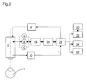
- Fig.2 shows the basic circuit diagram of all components:
- the from Refractometer 20 measured data are via an evaluation and Control unit 21 processed and stored.
- the data output takes place, for example, via a monitor 22
- Patient's eye 1 shown optionally with that from the refractometer measured optical data of the patient's eye and any further patient data.
- These can in turn be made using an image printer 23 or a pure data printer 24 printed or in the Patient file 25 are transmitted.
- the one for the operation necessary patient data are retrieved from the patient file if necessary 25 transferred to the evaluation and control unit 21.
- the evaluation and control unit 21 prepares the data of the video documentation device 10, preferably a digital camera, and provides them via an image reflection device 6 Reflection of data to the surgeon (29) in the microscope image Available.
Landscapes
- Health & Medical Sciences (AREA)
- Life Sciences & Earth Sciences (AREA)
- Surgery (AREA)
- Molecular Biology (AREA)
- General Health & Medical Sciences (AREA)
- Veterinary Medicine (AREA)
- Engineering & Computer Science (AREA)
- Biomedical Technology (AREA)
- Heart & Thoracic Surgery (AREA)
- Medical Informatics (AREA)
- Public Health (AREA)
- Animal Behavior & Ethology (AREA)
- Pathology (AREA)
- Nuclear Medicine, Radiotherapy & Molecular Imaging (AREA)
- Oral & Maxillofacial Surgery (AREA)
- Physics & Mathematics (AREA)
- Biophysics (AREA)
- Ophthalmology & Optometry (AREA)
- Microscoopes, Condenser (AREA)
- Eye Examination Apparatus (AREA)
Abstract
Description
- Durch die 'in situ'-Ermittlung der optischen Daten des Patientenauges während der Operation lassen sich durch den Chirurgen frühzeitig Gegenmaßnahmen bei Abweichungen von den Soll-Werten einleiten.
- Durch die frühzeitige Messung und Korrektur von potentiellen Abweichungen lässt sich die übrigbleibende Fehlsichtigkeit nach dem Heilungsprozess minimieren.
- Insgesamt kann damit sowohl die Qualität des Operations-Ergebnisses wie auch die Gefahr einer größeren Abweichung während der Operation auf ein Minimum reduziert werden.
- Gemäß einer Weiterentwicklung der Erfindung kann der Chirurg bei Abweichungen über einem festzulegenden Schwellenwert während der Operation mittels eines Warnmechanismus - beispielsweise eines Signaltons und/oder einer Blinkleuchte und/oder eines Einspiegelungmoduls - auf eine akute Gefahr aufmerksam gemacht werden.
- Fig.1:
- eine erfindungsgemäße Integration eines Refraktometers in ein Operationsmikroskop;
- Fig.2:
- das prinzipielle Schaltbild aller Komponenten.
- 1
- Patientenauge
- 2
- Operationsmikroskop
- 3
- Hauptobjektiv
- 4
- Zoom
- 5
- Tubus mit Okularen
- 6
- Bildeinspiegelungs-Vorrichtung
- 7
- Strahlenteiler für (6)
- 8
- Beleuchtung
- 9
- Beleuchtungsstrahlengang
- 10
- Video-Dokumentations-Vorrichtung
- 11
- Strahlenteiler für (10)
- 12
- Refraktometer-Strahlengang an der Position A (Ein-/Ausspiegelung)
- 13
- Strahlenteiler für (12)
- 14
- Refraktometer-Strahlengang an der Position B (Ein-/Ausspiegelung)
- 15
- Umlenkspiegel für (14)
- 16
- Refraktometer-Strahlengang an der Position C (Ein-/Ausspiegelung)
- 17
- Umlenkspiegel für (16) und (8)
- 18
- Strahlenteiler für (16)
- 19
- Optik für Refraktometer-Strahlengang (Positionen A, B und/oder C)
- 20
- Refraktometer
- 21
- Auswertungs- und Steuereinheit
- 22
- Monitor
- 23
- Bildprinter
- 24
- Datenprinter
- 25
- Patientendatei
- 26
- Beleuchtungsoptik
- 27
- Shutter (Blende(n))
- 28
- (Teil-)Strahlengänge für (29)
- 29
- Beobachter (Chirurg)
Claims (12)
- Ophthalmo-Operationsmikroskop (2) zur Beobachtung eines Patientenauges (1) während der Operation, dadurch gekennzeichnet, dass das Mikroskop (2) ein integriertes Refraktometer (20) (Auto-Refractive-Generator und -Analysator) umfasst, welches die optischen Daten des Patientenauges 'in situ' während der Operation ermittelt und dem Chirurgen bei Bedarf mitteilt.
- Operationsmikroskop nach Anspruch 1, dadurch gekennzeichnet, dass die Einspiegelung der vom Refraktometer (20) generierten Strahlung an mindestens einer der drei Positionenerfolgt.(A): zwischen Zoom (4) und Tubus (5) und/oder(B): zwischen Hauptoptikiv (3) und Zoom (4) und/oder(C): unmittelbar oberhalb des Hauptoptikivs (3) gelegen,
- Operationsmikroskop nach einem der vorhergehenden Ansprüche, dadurch gekennzeichnet, dass die Ausspiegelung der am Patientenauge (1) reflektierten Refraktometer-Strahlung an mindestens einer der drei Positionenerfolgt.(A): zwischen Zoom (4) und Tubus (5) und/oder(B): zwischen Hauptoptik (3) und Zoom (4) und/oder(C): unmittelbar oberhalb des Hauptoptikivs (3) gelegen,
- Operationsmikroskop nach einem der vorhergehenden Ansprüche, dadurch gekennzeichnet, dass für die Ein- und Ausspiegelung der Refraktometer-Strahlengänge (12, 14, 16) Strahlenteiler (13, 18) bzw. Prismen oder Spiegel (15, 17) vorgesehen sind.
- Operationsmikroskop nach einem der vorhergehenden Ansprüche, dadurch gekennzeichnet, dass die für die Ausspiegelung der Refraktometer-Strahlengänge bestimmten Strahlenteiler (13, 18) ausschließlich bestimmte Lichtwellenlängenbereiche transmittieren bzw. reflektieren, beispielsweise IR-Licht.
- Operationsmikroskop nach einem der vorhergehenden Ansprüche, dadurch gekennzeichnet, dass die Adaptierung der Optik des Refraktometers (20) an die Optik des Operationsmikroskops (2) mittels einer dazwischen liegenden Optik (19) für die Refraktometer-Strahlengänge (12, 14, 16) erfolgt, welche beispielsweise aus Linsen, Prismen und/oder Spiegeln besteht.
- Operationsmikroskop nach einem der vorhergehenden Ansprüche, dadurch gekennzeichnet, dass es mit mindestens einer Video-Dokumentations-Vorrichtung (10) ausrüstbar ist, welche das Patientenauge (1) aufnimmt und gegebenenfalls über einen Monitor (22) darstellbar macht.
- Operationsmikroskop nach einem der vorhergehenden Ansprüche, dadurch gekennzeichnet, dass es mit mindestens einer Bildeinspiegelungs-Vorrichtung (6) ausrüstbar ist, die im Betriebszustand und bei Bedarf die für die Operation notwendigen Daten - wie Refraktometer-, Patienten- und/oder Video-Daten - ins Blickfeld des Chirurgen (29) einspiegelt.
- Operationsmikroskop nach einem der vorhergehenden Ansprüche, dadurch gekennzeichnet, dass das Refraktometer (20) bidirektional mit dem Autofokus des Operationsmikroskops (2) koppelbar ist.
- Operationsmikroskop nach einem der vorhergehenden Ansprüche, dadurch gekennzeichnet, dass die mittels des Refraktometers (20) bestimmten Patientendaten über eine Auswertungseinheit (21) in eine Patientendatei (25) übermittelbar und/oder auf einen Datenprinter (24) ausgebbar sind.
- Operationsmikroskop nach einem der vorhergehenden Ansprüche, dadurch gekennzeichnet, dass die für die Operation relevanten Patientendaten im Betriebszustand automatisch und - vorzugsweise -elektronisch aus der Patientendatei (25) in die Auswertungs- und Steuereinheit (21) übernehmbar sind.
- Operationsmikroskop nach einem der vorhergehenden Ansprüche, dadurch gekennzeichnet, dass zur besseren Sichtbarmachung der eingespiegelten Daten Shutter (27) wahlweise in die Strahlengänge (28) für den Beobachter (29) einbringbar sind.
Applications Claiming Priority (2)
| Application Number | Priority Date | Filing Date | Title |
|---|---|---|---|
| DE10202509 | 2002-01-23 | ||
| DE10202509A DE10202509A1 (de) | 2002-01-23 | 2002-01-23 | Ophthalmo-Operationsmikroskop |
Publications (2)
| Publication Number | Publication Date |
|---|---|
| EP1338238A2 true EP1338238A2 (de) | 2003-08-27 |
| EP1338238A3 EP1338238A3 (de) | 2004-07-28 |
Family
ID=7712873
Family Applications (1)
| Application Number | Title | Priority Date | Filing Date |
|---|---|---|---|
| EP03001157A Withdrawn EP1338238A3 (de) | 2002-01-23 | 2003-01-21 | Ophthalmo-Operationsmikroskop |
Country Status (4)
| Country | Link |
|---|---|
| US (1) | US20030139736A1 (de) |
| EP (1) | EP1338238A3 (de) |
| JP (1) | JP2003265515A (de) |
| DE (1) | DE10202509A1 (de) |
Cited By (4)
| Publication number | Priority date | Publication date | Assignee | Title |
|---|---|---|---|---|
| WO2007028564A1 (de) * | 2005-09-07 | 2007-03-15 | Carl Zeiss Surgical Gmbh | Ophthalmo-operationsmikroskop mit messeinrichtung |
| DE102010008146A1 (de) | 2010-02-12 | 2011-08-18 | Carl Zeiss Surgical GmbH, 73447 | Messsystem und Verfahren zum Ermitteln der Refraktion eines Auges, des Krümmungsradius der Hornhaut oder des Innendrucks eines Auges |
| DE102009053208A1 (de) | 2008-11-06 | 2011-08-18 | Leica Instruments (Singapore) Pte. Ltd. | Vorrichtung zur Überwachung der Implantation einer IOL (Orientierungsüberwachung) |
| DE102010015691A1 (de) | 2010-04-21 | 2011-10-27 | Carl Zeiss Microlmaging Gmbh | Mikroskopiereinrichtung zur Vergleichs- und Mitbeobachtung |
Families Citing this family (21)
| Publication number | Priority date | Publication date | Assignee | Title |
|---|---|---|---|---|
| US7556378B1 (en) | 2003-04-10 | 2009-07-07 | Tsontcho Ianchulev | Intraoperative estimation of intraocular lens power |
| EP2444021B8 (de) | 2004-04-20 | 2018-04-18 | Alcon Research, Ltd. | Integriertes chirurgisches Mikroskop und Wellenfrontsensor |
| EP1972984B1 (de) * | 2005-09-22 | 2019-01-09 | Olympus Corporation | Mikroskop |
| US8820929B2 (en) * | 2006-01-20 | 2014-09-02 | Clarity Medical Systems, Inc. | Real-time measurement/display/record/playback of wavefront data for use in vision correction procedures |
| US7594729B2 (en) | 2007-10-31 | 2009-09-29 | Wf Systems, Llc | Wavefront sensor |
| WO2010054268A2 (en) | 2008-11-06 | 2010-05-14 | Wavetec Vision Systems, Inc. | Optical angular measurement system for ophthalmic applications and method for positioning of a toric intraocular lens with increased accuracy |
| CH699887A1 (de) * | 2008-11-08 | 2010-05-14 | Leica Instr Singapore Pte Ltd | Vorrichtung und Verfahren zur Positionierung einer künstlichen intraokularen Linse (IOL) im Zuge der Implantation dieser IOL. |
| US8876290B2 (en) | 2009-07-06 | 2014-11-04 | Wavetec Vision Systems, Inc. | Objective quality metric for ocular wavefront measurements |
| ES2524618T3 (es) | 2009-07-14 | 2014-12-10 | Wavetec Vision Systems, Inc. | Determinación de la posición efectiva de la lente de una lente intraocular utilizando potencia refractiva afática |
| WO2011008609A1 (en) | 2009-07-14 | 2011-01-20 | Wavetec Vision Systems, Inc. | Ophthalmic surgery measurement system |
| DE102010016623A1 (de) | 2010-04-23 | 2011-10-27 | Leica Microsystems (Schweiz) Ag | Beleuchtungseinrichtung für ein ophthalmologisches Operationsmikroskop |
| US8864309B2 (en) | 2011-12-05 | 2014-10-21 | Bioptigen, Inc. | Optical imaging systems having input beam shape control and path length control |
| US8777412B2 (en) | 2012-04-05 | 2014-07-15 | Bioptigen, Inc. | Surgical microscopes using optical coherence tomography and related methods |
| US9072462B2 (en) | 2012-09-27 | 2015-07-07 | Wavetec Vision Systems, Inc. | Geometric optical power measurement device |
| WO2014197553A2 (en) | 2013-06-04 | 2014-12-11 | Bioptigen, Inc. | Hybrid telescope for optical beam delivery and related systems and methods |
| US10456030B2 (en) | 2013-07-29 | 2019-10-29 | Bioptigen, Inc. | Procedural optical coherence tomography (OCT) for surgery and related methods |
| EP3039474A1 (de) | 2013-08-28 | 2016-07-06 | Bioptigen, Inc. | Informationsanzeigen für in eine optische kohärenztomografie integrierte operationsmikroskope |
| DE102017124545B3 (de) | 2017-10-20 | 2019-01-24 | Carl Zeiss Meditec Ag | Mikroskop |
| DE102017124548B3 (de) | 2017-10-20 | 2018-07-26 | Carl Zeiss Meditec Ag | Mikroskop mit einer OCT-Einrichtung und einer Wellenfrontmesseinrichtung |
| CA3168826A1 (en) | 2020-01-22 | 2021-07-29 | Photonic Medical Inc. | Open view, multi-modal, calibrated digital loupe with depth sensing |
| DE202020000635U1 (de) | 2020-02-17 | 2020-06-19 | Carl Zeiss Microscopy Gmbh | Binokularer Digitaltubus für ein Mikroskop |
Family Cites Families (13)
| Publication number | Priority date | Publication date | Assignee | Title |
|---|---|---|---|---|
| DE2643344A1 (de) * | 1976-09-25 | 1978-03-30 | Zeiss Carl Fa | Vorrichtung zur ermittlung von hornhautastigmatismus am menschlichen auge |
| US4544243A (en) * | 1984-05-24 | 1985-10-01 | Cooper Lasersonics, Inc. | Heads up display for microscope using remotely controlled instrument |
| JPS62268523A (ja) * | 1986-05-17 | 1987-11-21 | キヤノン株式会社 | 眼科装置 |
| US5249004A (en) * | 1989-12-28 | 1993-09-28 | Kabushiki Kaisha Topcon | Microscope for an operation |
| US5684561A (en) * | 1992-05-26 | 1997-11-04 | Daphne Eye Technologies | Device and method for evaluation of refraction of the eye |
| DE4310561A1 (de) * | 1993-03-26 | 1994-09-29 | Klaus Prof Dipl Phys Dietrich | Vorrichtung und Verfahren zur Bestimmung von Sehschärfe, Refraktion und zur Beobachtung der Augenoberfläche |
| DE59609547D1 (de) * | 1995-05-17 | 2002-09-12 | Leica Mikroskopie Systeme Ag H | Mikroskop |
| JPH09135811A (ja) * | 1995-11-15 | 1997-05-27 | Nikon Corp | 眼科装置 |
| US6193710B1 (en) * | 1998-07-16 | 2001-02-27 | Visx, Incorporated | Method for scanning non-overlapping patterns of laser energy with diffractive optics |
| UA46833C2 (uk) * | 1998-10-07 | 2002-06-17 | Інститут Біомедичної Техніки Та Технологій Академії Технологічних Наук України | Вимірювач абераційної рефракції ока |
| CN1165792C (zh) * | 1999-10-13 | 2004-09-08 | 莱卡微系统(瑞士)股份公司 | 具有信息馈入装置的体视手术显微镜 |
| US6419671B1 (en) * | 1999-12-23 | 2002-07-16 | Visx, Incorporated | Optical feedback system for vision correction |
| US6550917B1 (en) * | 2000-02-11 | 2003-04-22 | Wavefront Sciences, Inc. | Dynamic range extension techniques for a wavefront sensor including use in ophthalmic measurement |
-
2002
- 2002-01-23 DE DE10202509A patent/DE10202509A1/de not_active Withdrawn
-
2003
- 2003-01-13 US US10/341,111 patent/US20030139736A1/en not_active Abandoned
- 2003-01-21 EP EP03001157A patent/EP1338238A3/de not_active Withdrawn
- 2003-01-23 JP JP2003014499A patent/JP2003265515A/ja not_active Withdrawn
Cited By (9)
| Publication number | Priority date | Publication date | Assignee | Title |
|---|---|---|---|---|
| WO2007028564A1 (de) * | 2005-09-07 | 2007-03-15 | Carl Zeiss Surgical Gmbh | Ophthalmo-operationsmikroskop mit messeinrichtung |
| DE102009053208A1 (de) | 2008-11-06 | 2011-08-18 | Leica Instruments (Singapore) Pte. Ltd. | Vorrichtung zur Überwachung der Implantation einer IOL (Orientierungsüberwachung) |
| DE102009053208B4 (de) | 2008-11-06 | 2022-05-19 | Leica Instruments (Singapore) Pte. Ltd. | Vorrichtung zur Überwachung der Implantation einer IOL (Orientierungsüberwachung) |
| DE102010008146A1 (de) | 2010-02-12 | 2011-08-18 | Carl Zeiss Surgical GmbH, 73447 | Messsystem und Verfahren zum Ermitteln der Refraktion eines Auges, des Krümmungsradius der Hornhaut oder des Innendrucks eines Auges |
| US10080493B2 (en) | 2010-02-12 | 2018-09-25 | Carl Zeiss Meditec Ag | Measurement system and method for establishing the refraction of an eye, the radius of curvature of the cornea or the internal pressure of an eye |
| US11039743B2 (en) | 2010-02-12 | 2021-06-22 | Carl Zeiss Meditec Ag | Measurement system and method for establishing the refraction of an eye, the radius of curvature of the cornea or the internal pressure of an eye |
| US11039744B2 (en) | 2010-02-12 | 2021-06-22 | Carl Zeiss Meditec Ag | Measurement system and method for establishing the refraction of an eye, the radius of curvature of the cornea or the internal pressure of an eye |
| DE102010008146B4 (de) | 2010-02-12 | 2022-03-31 | Carl Zeiss Meditec Ag | Messsystem und Verfahren zum Ermitteln des Innendrucks eines Auges sowie Verfahren und System zum Einstellen des Augeninnendrucks |
| DE102010015691A1 (de) | 2010-04-21 | 2011-10-27 | Carl Zeiss Microlmaging Gmbh | Mikroskopiereinrichtung zur Vergleichs- und Mitbeobachtung |
Also Published As
| Publication number | Publication date |
|---|---|
| JP2003265515A (ja) | 2003-09-24 |
| US20030139736A1 (en) | 2003-07-24 |
| EP1338238A3 (de) | 2004-07-28 |
| DE10202509A1 (de) | 2003-07-31 |
Similar Documents
| Publication | Publication Date | Title |
|---|---|---|
| EP1338238A2 (de) | Ophthalmo-Operationsmikroskop | |
| EP1327412B1 (de) | Anordnung zur Beleuchtung der Linse eines menschlichen Auges | |
| DE19731301C2 (de) | Vorrichtung zum Steuern eines Mikroskopes mittels Blickrichtungsanalyse | |
| JP5248926B2 (ja) | 眼屈折力測定装置 | |
| DE102010008146A1 (de) | Messsystem und Verfahren zum Ermitteln der Refraktion eines Auges, des Krümmungsradius der Hornhaut oder des Innendrucks eines Auges | |
| DE60115126T2 (de) | Funduskamera mit sichtbarer und unsichtbarer Beleuchtung | |
| DE10302401A1 (de) | Operationsmikroskop | |
| DE102011088038B4 (de) | Operationsmikroskopsystem für die Ophthalmologie und zugehörige Detektionseinheit | |
| DE10242983B4 (de) | Ophthalmo-Operationsmikroskop mit Objektbeleuchtung | |
| DE102005032501A1 (de) | Vorrichtung zur Untersuchung vorderer und hinterer Augenabschnitte | |
| DE102017105580B4 (de) | Operationsmikroskop | |
| DE19502337C2 (de) | Vorrichtung und Verfahren zur Prüfung von Sehfunktionen | |
| EP1396747A1 (de) | Beleuchtung für Operationsmikroskope zum Schutz von menschlichem Gewebe | |
| DE2544561A1 (de) | Ophthalmologisches geraet | |
| DE10314944A1 (de) | Beleuchtungs- und Bestrahlungseinheit für ophthalmologische Geräte | |
| WO2009143976A1 (de) | Optisches system für ophthalmologische geräte, insbesondere funduskameras | |
| WO2005039404A1 (de) | Beleuchtungseinheit für funduskameras und/oder ophthalmoskope: | |
| DE10009532A1 (de) | Vorrichtung und Verfahren zur Qualitätssicherung bei Augenoperationen | |
| DE102021107297A1 (de) | Optisches System für die Augenchirurgie und Verfahren zum Vermeiden einer zu hohen Lichtintensität an einem digitalen Bildsensor eines Operationsmikroskops | |
| DE102006061932A1 (de) | Anordnung für ophthalmologische Geräte zur Verbesserung von Fundusbildern | |
| DE202010008225U1 (de) | Optisches Instrument mit Dioptrienkorrektur | |
| DE3338400A1 (de) | Sehschaerfepruefer | |
| EP1437085A1 (de) | Augenrefraktometer und Verfahren zum Betrieb eines Augenrefraktometers | |
| DE102024112777B4 (de) | Optisches Beobachtungsgerät und Verfahren zum Versehen eines optischen Beobachtungsgerätes mit einem Laserschutzfilter | |
| FI131953B1 (en) | Optical eye apparatus and method for focusing retinal image |
Legal Events
| Date | Code | Title | Description |
|---|---|---|---|
| PUAI | Public reference made under article 153(3) epc to a published international application that has entered the european phase |
Free format text: ORIGINAL CODE: 0009012 |
|
| AK | Designated contracting states |
Designated state(s): AT BE BG CH CY CZ DE DK EE ES FI FR GB GR HU IE IT LI LU MC NL PT SE SI SK TR |
|
| AX | Request for extension of the european patent |
Extension state: AL LT LV MK RO |
|
| RIC1 | Information provided on ipc code assigned before grant |
Ipc: 7A 61B 3/103 B Ipc: 7A 61B 19/00 B Ipc: 7A 61B 3/13 A |
|
| PUAL | Search report despatched |
Free format text: ORIGINAL CODE: 0009013 |
|
| RIC1 | Information provided on ipc code assigned before grant |
Ipc: 7G 02B 21/18 B Ipc: 7A 61B 3/103 B Ipc: 7A 61B 19/00 B Ipc: 7A 61B 3/13 A |
|
| AK | Designated contracting states |
Kind code of ref document: A3 Designated state(s): AT BE BG CH CY CZ DE DK EE ES FI FR GB GR HU IE IT LI LU MC NL PT SE SI SK TR |
|
| AX | Request for extension of the european patent |
Extension state: AL LT LV MK RO |
|
| 17P | Request for examination filed |
Effective date: 20050128 |
|
| AKX | Designation fees paid |
Designated state(s): CH DE FR GB LI |
|
| STAA | Information on the status of an ep patent application or granted ep patent |
Free format text: STATUS: THE APPLICATION IS DEEMED TO BE WITHDRAWN |
|
| 18D | Application deemed to be withdrawn |
Effective date: 20060707 |