EP1356860A2 - Qualitätskontrollverfahren von DNA-Mikroarray - Google Patents
Qualitätskontrollverfahren von DNA-Mikroarray Download PDFInfo
- Publication number
- EP1356860A2 EP1356860A2 EP02023726A EP02023726A EP1356860A2 EP 1356860 A2 EP1356860 A2 EP 1356860A2 EP 02023726 A EP02023726 A EP 02023726A EP 02023726 A EP02023726 A EP 02023726A EP 1356860 A2 EP1356860 A2 EP 1356860A2
- Authority
- EP
- European Patent Office
- Prior art keywords
- dna
- quality control
- fluorescent
- chip
- spots
- Prior art date
- Legal status (The legal status is an assumption and is not a legal conclusion. Google has not performed a legal analysis and makes no representation as to the accuracy of the status listed.)
- Withdrawn
Links
Images

Classifications
-
- C—CHEMISTRY; METALLURGY
- C12—BIOCHEMISTRY; BEER; SPIRITS; WINE; VINEGAR; MICROBIOLOGY; ENZYMOLOGY; MUTATION OR GENETIC ENGINEERING
- C12Q—MEASURING OR TESTING PROCESSES INVOLVING ENZYMES, NUCLEIC ACIDS OR MICROORGANISMS; COMPOSITIONS OR TEST PAPERS THEREFOR; PROCESSES OF PREPARING SUCH COMPOSITIONS; CONDITION-RESPONSIVE CONTROL IN MICROBIOLOGICAL OR ENZYMOLOGICAL PROCESSES
- C12Q1/00—Measuring or testing processes involving enzymes, nucleic acids or microorganisms; Compositions therefor; Processes of preparing such compositions
- C12Q1/68—Measuring or testing processes involving enzymes, nucleic acids or microorganisms; Compositions therefor; Processes of preparing such compositions involving nucleic acids
- C12Q1/6813—Hybridisation assays
- C12Q1/6834—Enzymatic or biochemical coupling of nucleic acids to a solid phase
- C12Q1/6837—Enzymatic or biochemical coupling of nucleic acids to a solid phase using probe arrays or probe chips
-
- C—CHEMISTRY; METALLURGY
- C12—BIOCHEMISTRY; BEER; SPIRITS; WINE; VINEGAR; MICROBIOLOGY; ENZYMOLOGY; MUTATION OR GENETIC ENGINEERING
- C12Q—MEASURING OR TESTING PROCESSES INVOLVING ENZYMES, NUCLEIC ACIDS OR MICROORGANISMS; COMPOSITIONS OR TEST PAPERS THEREFOR; PROCESSES OF PREPARING SUCH COMPOSITIONS; CONDITION-RESPONSIVE CONTROL IN MICROBIOLOGICAL OR ENZYMOLOGICAL PROCESSES
- C12Q1/00—Measuring or testing processes involving enzymes, nucleic acids or microorganisms; Compositions therefor; Processes of preparing such compositions
- C12Q1/68—Measuring or testing processes involving enzymes, nucleic acids or microorganisms; Compositions therefor; Processes of preparing such compositions involving nucleic acids
Definitions
- the present invention relates to a DNA microarray, and more particularly, to a quality control method of a DNA chip.
- a DNA microarray refers to a DNA chip manufactured by immobilizing oligonucleotide probes, each probe having a known sequence of a few to hundreds of nucleotides, at hundreds to hundreds of thousands of appropriate positions on a solid surface made of, for example, silicon, surface-modified glass, polypropylene, or activated polyacrylamide.
- a target DNA to be assayed is applied to the DNA chip, a fragment of the target DNA complementarily hybridizes to the oligonucleotide probes immobilized on the DNA chip.
- the hybridization is optically or radiochemically detected and analyzed to identify the nucleotide sequence of the target DNA, which is called sequencing by hybridization (SBH).
- the DNA chip manufactured as a microarray reduces the size of a DNA assay system and enables genetic assay with a trace of a sample.
- multiple sequences of a target DNA can be simultaneously assayed, thereby rapidly providing the genetic information of the target DNA at low costs.
- the DNA microarray chip can assay a large amount of genetic information within a short period of time and the relevancy of the genes. Accordingly, the DNA chip is expected to have wide applications, for example, to genetic disorder and cancer diagnosis, mutant and pathogen detection, gene expression assay, drug discovery, etc.
- the DNA chip can be used as a microorganism or pollutant detector to find out antidotal genes and further to produce antidotes on a large scale based on genetic recombination technologies.
- the DNA chip can lead to great improvements in most biological industries, including the production of medicinal crops or low-fat meat.
- DNA microarrays are classified into an oligo-chip and a cDNA chip according to the type of probes immobilized thereon. According to the manufacturing method, DNA microarrays are classified into a lithography chip, a pin-type spotting chip, and an ink-jet type spotting chip.
- a DNA microarray is manufactured through diversified and complex chemical processes according to the type of immobilized probes. Since a trace of a DNA probe is immobilized in a very small area on a glass surface, quantitative spot-to-spot and chip-to-chip variations of probes are considerably great, and thus the results from the hybridization are inconsistent.
- Brown et al. ( "Quantitative Monitoring of Gene Expression Patterns with a Complimentary DNA Microarray” M. Schena, D. Shalon, R.W. Davis and P.O. Brown. Science 270:467-470 (1995).) developed a method for inspecting DNA probe spot uniformity and glass surface damage by laser scanning a light scattering due to salts present in the DNA probe spots immobilized on the glass surface. This method can be conveniently applied only immediately after DNA probe spotting but is limited after completion of the DNA chip manufacturing because the salts are removed from the DNA probe spots through immobilization and washing processes following the spotting.
- the fluorescent staining method can be applied to inspect the DNA microarray after the completion of the chip manufacturing, but needs a number of washing processes for staining and dye-removing.
- the fluorescent material is likely to remain on the glass surface after the washing and thus affects the subsequent hybridization process.
- the present invention provides a convenient, non-destructive method for controlling the quality and size of DNA spots on a DNA microarray, which does not need staining and dye-removing processes.
- the quality control method of the DNA chip according to the present invention comprises: preparing a DNA spotting solution containing a fluorescent dye; applying the DNA spotting solution to a substrate as spots to manufacture the DNA chip; and detecting fluorescent signals from the DNA spots.
- a DNA spotting solution to be applied to the DNA chip substrate is prepared with a fluorescent dye, and the fluorescent dye is covalently bound to the substrate surface while DNA probes are immobilized on the DNA chip substrate. After completion of the DNA chip manufacturing, fluorescence from the DNA spots is scanned to non-destructively determine DNA spot uniformity.
- Uniformity of fluorescent signals from the DNA probe spots is determined by the diameter and shape of the DNA probe spots and the intensity of the fluorescence.
- the fluorescent dye used in the present invention has a dye group (F) for generating a fluorescent signal and a functional group (R) that covalently binds to a solid substrate, which is expressed as follows:
- Suitable dye groups for generating the fluorescent signal have no spectral interference with a dye for target DNA labelling, used to detect hybridization after the manufacture of the DNA chip, and include fluorescein isothiocyanate (FIFC), fluorescein, Cy3, Cy5, Texas Red, N,N,N',N'-tetramethyl-6-carboxyrhodamine (TAMRA), etc., which have no spectral interference.
- FIFC fluorescein isothiocyanate
- TAMRA Texas Red
- the functional group that binds to the solid substrate should be capable of covalently binding to a functional group on the solid substrate surface, such as amine, aldehyde, or poly-lysine, according to the type of the DNA microarray.
- Suitable functional groups of the fluorescent dye include amine, isothiocyanate (-NCS), activated ester, and aldehyde.
- the quality control method of the DNA spots on the DNA chip manufactured as described above can be modified according to the type of the fluorescent dye used.
- the dye group of the fluorescent dye is FIFC or fluorescein
- the dye group is excited at 488 nm wave length by an argon ion laser, and a fluorescent signal is analysed using a 520-nm wave length filter.
- the quality control method of the DNA microarray according to the present invention can be applied to another type of microarray, such as an RNA chip or a protein chip.
- Example 1 Spot quality control in oligonucleotide probe-immobilized DNA chip
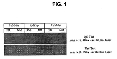
- oligonucleotide probes Two types of oligonucleotide probes (perfect match (PM)-5'-TGTAGACACGCACCTCCGTG-3'; or mismatch (MM) - 5'-TGTAGACACCCACCTCCGTG-3') and 4'-(aminomethyl)fluorescein (0 ⁇ M, 1 ⁇ M, and 2 ⁇ M) as a fluorescent dye were added to a gel matrix solution with stirring and left at 37°C for 14 hours to form matrix-DNA conjugates. The resultant solution was used as a spotting solution. The spotting solution was spotted on a glass surface, which had been treated to expose amine groups, and left in a wet chamber at 37°C for 4 hours.
- a region of the glass surface to which the spotting solution was not applied was treated to negatively charge amine groups of the region and thus to prevent the target DNA from adhering to the non-spotting region, which is a process necessary for background noise control.
- 5 g of succinic anhydride was dissolved in 315 mL of 1-methyl-2-pyrrolidone (NMP), and 35 ml of sodium borate was added to the solution with stirring.
- NMP 1-methyl-2-pyrrolidone
- the glass surface was treated with the solution, washed with distilled water and left in a dryer.
- the 4-(aminomethyl)fluorescein used as the fluorescent material has the following formula:
- a fluorescent signal from the fluorescent-labelled target material hybridized to the probes on the DNA microarray was detected using a scanner (ScanArray Scanner, GSI Lunonics) on a 10- ⁇ m-pixel resolution.
- the intensity of Spots were produced using GenePix software.
- the DNA spots were excited by scanning with an argon ion laser at 488 nm, and a fluorescent emission signal was obtained using a 520 nm wave length filter.
- the target DNA, which is hybridezed to the DNA spots was excited at 550 nm, and a fluorescent emission signal was obtained using 570 nm wave length filter.
- a recombinant gene capable of producing glyceraldehyde-3-phosphate dehydrogenase was isolated using a DNA extraction kit (GAIGEN). The isolated recombinant GAPDH gene was selectively amplified through PCR in a reaction solution having a final volume of 100 ⁇ L.
- telomere sequence 50 g of the pure recombinant GAPDH DNA, 2.5 U of thermally stable DNA polymerase, 200 ⁇ M of each dNTP (dATP, dTTP, dGTP, and dCTP), 50 mM Tris-HCI, 40 mM KCI, 1.5 mM MgCl 2 , and 100 pmols of each of a sense primer (5'-CTGGTAAGTTTAGTCTTTTTGTC-3') and an anti-sense primer (5'-CAGTGCCAAGCTTGCATGCCT-3') were used. Thirty five cycles of PCR, one cycle including denaturation at 94°C for 30 seconds, annealing at 55°C for 60 seconds, and extension at 72°C for 90 seconds, were carried out. Pre-denaturation at 94°C and last-extension at 72°C were performed for 5 minutes each.
- the amplicon was purified using the DNA extraction kit (GAIGEN) and used as probes to be immobilized on
- the amplified GAPDH gene was mixed to a final concentration of 0.25 mg/mL with 50% DMSO in two separate tubes. To one of the tubes, FITC dye was added to a final concentration of 8 ⁇ M. The amplified gene, DNSO, and FITC dye in each of the tubes were thoroughly mixed by Voltex mixer. The reagent mixtures were transferred into 384-well plates for spotting. The spotting solution was spotted on a glass substrate, which had been treated to have amine groups, in a 20X4 array, and heated at 80°C for 4 hours.
- a region of the glass surface to which the spotting solution was not applied was treated to negatively charge amine groups of the region and thus to prevent the target DNA from adhering to the non-spotting region, which is a process necessary for controlling background noise.
- 5 g succinic anhydride was dissolved in 315 mL of 1-methyl-2-pyrrolidone_ (NMP), and 35 ml of sodium borate was added to the solution with stirring.
- the glass surface was treated with the solution, washed with distilled water and left in a dryer.
- the FITC used as the fluorescent dye has the following formula:
- the GAPHD producing recombinant gene isolated using the DNA extraction in the above experiment was used to prepare a fluorescent-labelled target DNA.
- the isolated recombinant GAPDH was selectively amplified through PCR in a reaction solution having a final volume of 50 ⁇ L.
- the amplicon was purified using the DNA extraction kit (GAIGEN), and its concentration was measured by spectrophotometry. For hybridization efficiency, the concentration of the purified GAPDH DNA was constantly adjusted to 50 ng/ ⁇ L.
- the target DNA of the GAPDH gene was fluorescently labelled with Cy3 during the PCR with the addition of Cy3-dUTP and included 600 basepairs.
- the cDNA chip manufactured above was preliminarily hybridized at 50°C for 45 minutes in a solution of 4X SSC (standard saline-citrate), 0.1% SDS (sodium dodecyl sulfate), and 10 mg/mL BSA (bovine serum albumin) and washed with isopropanol and pure water. Water was removed from the cDNA chip by centrifugation at 500 rpm for 3 minutes.
- 4X SSC standard saline-citrate
- SDS sodium dodecyl sulfate
- BSA bovine serum albumin
- 1 ⁇ g of the fluorescent-labelled target DNA prepared in the above experiment was denatured at 95°C for 5 minutes, left in ice for 3 minutes, mixed with a solution of 3X SSC, 0.1% SDS, 0.5 mg/mL yeast tRNA, and 30 ⁇ g/mL herring sperm DNA, and dropped on the cDNA chip.
- the cDNA chip was covered with a cover glass to allow wide spreading of the solution and hybridized overnight at 50°C.
- the chip substrate was washed once for 5 minutes with a solution of 0.1X SSC (1.5 mM sodium citrate, 15 mM NaCI, pH 7.0) and 0.1% SDS, and twice for 5 minutes with 0.1X SSC at 25°C. Next, water was removed from the chip substrate by centrifugation in the same conditions as the above.
- Hybridization sensitivity was measured in the same manner as in Example 1.
- the DNA probe spots were excited by scanning with an argon ion laser at 488 nm, and a fluorescent emission signal was obtained using a 520 nm wave length filter.
- the target DNA which is hybridized to the DNA probe spots, was excited at 550nm, and a fluorescent emission signal was obtained using a 570 nm wave length filter.
- the spot diameter was 170 ⁇ 15 ⁇ m, and the spot interval was adjusted to 375 ⁇ m.
- the fluorescent intensity was compared in two cases, i.e., when FITC dye was used and when no fluorescent dye was used, a shown in Table 2 below.
- a signal from a fluorescent dye covalently immobilized on the solid surface of the DNA chip together with DNA probe is detected and used for the quality control of the DNA microarray.
- a signal from a fluorescent dye covalently immobilized on the solid surface of the DNA chip together with DNA probe is detected and used for the quality control of the DNA microarray.
- the fluorescent dye covalently bound to the solid substrate of the DNA chip together with DNA probes hardly affects the fluorescent intensity of the DNA spots after hybridization to a target DNA, thereby eliminating the likelihood of the fluorescent dye affecting the hybridization in conventional methods.
- the DNA microarray manufactured by the above-describe method can be quality-controlled without a staining process and provides a reliable, consistent result of hybridization.
- the fluorescent dye is covalently bound to reactive groups on the glass chip surface together with DNA probes, the intensity of a fluorescent signal is unaffected by the sequence and length of the DNA probes, thereby enabling an accurate quality comparison of the separate DNA probe spots.
Landscapes
- Chemical & Material Sciences (AREA)
- Organic Chemistry (AREA)
- Life Sciences & Earth Sciences (AREA)
- Zoology (AREA)
- Wood Science & Technology (AREA)
- Proteomics, Peptides & Aminoacids (AREA)
- Health & Medical Sciences (AREA)
- Engineering & Computer Science (AREA)
- Microbiology (AREA)
- Biochemistry (AREA)
- Physics & Mathematics (AREA)
- Molecular Biology (AREA)
- Biotechnology (AREA)
- Biophysics (AREA)
- Analytical Chemistry (AREA)
- Immunology (AREA)
- Bioinformatics & Cheminformatics (AREA)
- General Engineering & Computer Science (AREA)
- General Health & Medical Sciences (AREA)
- Genetics & Genomics (AREA)
- Measuring Or Testing Involving Enzymes Or Micro-Organisms (AREA)
- Investigating Or Analysing Materials By The Use Of Chemical Reactions (AREA)
- Investigating, Analyzing Materials By Fluorescence Or Luminescence (AREA)
Applications Claiming Priority (2)
| Application Number | Priority Date | Filing Date | Title |
|---|---|---|---|
| KR10-2002-0012158A KR100450817B1 (ko) | 2002-03-07 | 2002-03-07 | Dna 마이크로어레이 스팟의 품질관리방법 |
| KR2002012158 | 2002-03-07 |
Publications (2)
| Publication Number | Publication Date |
|---|---|
| EP1356860A2 true EP1356860A2 (de) | 2003-10-29 |
| EP1356860A3 EP1356860A3 (de) | 2004-05-06 |
Family
ID=27786001
Family Applications (1)
| Application Number | Title | Priority Date | Filing Date |
|---|---|---|---|
| EP02023726A Withdrawn EP1356860A3 (de) | 2002-03-07 | 2002-10-23 | Qualitätskontrollverfahren von DNA-Mikroarray |
Country Status (5)
| Country | Link |
|---|---|
| US (1) | US20030170672A1 (de) |
| EP (1) | EP1356860A3 (de) |
| JP (1) | JP3872762B2 (de) |
| KR (1) | KR100450817B1 (de) |
| CN (1) | CN1222624C (de) |
Families Citing this family (21)
| Publication number | Priority date | Publication date | Assignee | Title |
|---|---|---|---|---|
| US7368082B1 (en) * | 2002-12-12 | 2008-05-06 | Tung-Lian Huang | Formulation of spotting solution to achieve uniform spot size and morphology and for nondestructive quality control of assay articles |
| WO2004081570A1 (fr) * | 2003-03-13 | 2004-09-23 | Chengdu Kuachang Medical Industrial Limited | Matrice de puce, puce comprenant une matrice et leur preparation et application |
| DE10361137A1 (de) * | 2003-12-23 | 2005-07-28 | Alopex Gmbh | Mikroarray, Kit und Verfahren zum Validieren und/oder Kalibrieren eines Systems von Hybridisierungsexperimenten |
| WO2005075682A1 (en) * | 2004-02-04 | 2005-08-18 | Hee-Kyung Park | Microarray comprising qc probes and method for fabricating the same |
| JP4789518B2 (ja) * | 2005-06-30 | 2011-10-12 | キヤノン株式会社 | 製造条件付きプローブ固定担体を用いた標的物質の検出方法ならびにそのための装置、キット及びシステム |
| JP4538746B2 (ja) * | 2005-09-21 | 2010-09-08 | 横河電機株式会社 | バイオチップおよび分析装置 |
| JP4955244B2 (ja) * | 2005-09-27 | 2012-06-20 | 横河電機株式会社 | バイオチップ読み取り装置およびバイオチップ読み取り方法 |
| JP4009311B2 (ja) * | 2005-10-28 | 2007-11-14 | 三菱レイヨン株式会社 | 核酸アレイにおけるプローブ搭載位置の確認方法 |
| DE102006027517A1 (de) * | 2006-06-09 | 2007-12-13 | Euroimmun Medizinische Labordiagnostika Ag | Verfahren zur Erzeugung perfekter Macro- und Microarrays durch Kombinieren vorselektierter beschichteter Festphasen-Fragmente |
| KR100832742B1 (ko) * | 2007-02-28 | 2008-05-27 | 한국화학연구원 | 형광 링커를 포함하는 pna 프로브, 및 이를 이용한pna 마이크로어레이의 제조방법 및 품질관리방법 |
| KR20100086827A (ko) | 2009-01-23 | 2010-08-02 | 삼성전자주식회사 | 프로브 핵산의 서열을 분석하는 방법, 그를 위한 마이크로어레이 및 키트 및 프로브 핵산의 합성 효율을 결정하는 방법 |
| CN103575894B (zh) * | 2013-11-07 | 2015-08-05 | 南京祥中生物科技有限公司 | 一种可视化生物芯片的检测方法 |
| CN103543274B (zh) * | 2013-11-07 | 2015-09-09 | 南京祥中生物科技有限公司 | 一种可视化的生物芯片 |
| CN103983793A (zh) * | 2014-05-29 | 2014-08-13 | 上海理工大学 | 一种含丽春红的蛋白质芯片点样缓冲液及其制备方法 |
| CN104561292A (zh) * | 2014-12-30 | 2015-04-29 | 东南大学 | 一种基于温度差异性探针基因芯片检测方法 |
| CN104561290A (zh) * | 2014-12-30 | 2015-04-29 | 东南大学 | 一种基于混合探针基因芯片检测方法 |
| JPWO2020145124A1 (ja) * | 2019-01-09 | 2021-10-07 | 株式会社日立ハイテク | 核酸分析用基板、核酸分析用フローセル、及び画像解析方法 |
| US12577514B2 (en) | 2019-10-29 | 2026-03-17 | Toray Industries, Inc. | Method of producing biochips |
| CN111458494B (zh) * | 2020-03-31 | 2024-07-05 | 京东方科技集团股份有限公司 | 免疫芯片的质量检测方法 |
| CN115963285A (zh) * | 2022-12-30 | 2023-04-14 | 深圳市曙芯生物科技有限公司 | 一种用于微阵列芯片快速验证的溶液及验证方法 |
| WO2024138666A1 (zh) * | 2022-12-30 | 2024-07-04 | 深圳华大生命科学研究院 | 时空组定位芯片的质量检测方法、装置、设备和存储介质 |
Family Cites Families (12)
| Publication number | Priority date | Publication date | Assignee | Title |
|---|---|---|---|---|
| US5677197A (en) * | 1994-11-14 | 1997-10-14 | Hewlett-Packard Company | Biochemical assay plate and method for making the same |
| US5843655A (en) * | 1995-09-18 | 1998-12-01 | Affymetrix, Inc. | Methods for testing oligonucleotide arrays |
| US5812272A (en) * | 1997-01-30 | 1998-09-22 | Hewlett-Packard Company | Apparatus and method with tiled light source array for integrated assay sensing |
| US6101946A (en) * | 1997-11-21 | 2000-08-15 | Telechem International Inc. | Microarray printing device including printing pins with flat tips and exterior channel and method of manufacture |
| WO2000014197A1 (en) * | 1998-09-09 | 2000-03-16 | Hitachi Software Engineering Co., Ltd. | Biochip and method of using biochip |
| DE19842164B4 (de) * | 1998-09-15 | 2004-06-03 | Deutsches Krebsforschungszentrum Stiftung des öffentlichen Rechts | Verfahren zur Qualitätskontrolle beim Aufbau von Oligomerrastern |
| US6245518B1 (en) * | 1998-12-11 | 2001-06-12 | Hyseq, Inc. | Polynucleotide arrays and methods of making and using the same |
| JP2001021558A (ja) * | 1999-04-30 | 2001-01-26 | Agilent Technol Inc | ポリヌクレオチドアレイの製作法 |
| US6558623B1 (en) * | 2000-07-06 | 2003-05-06 | Robodesign International, Inc. | Microarray dispensing with real-time verification and inspection |
| JP3913978B2 (ja) * | 2000-12-13 | 2007-05-09 | 富士フイルム株式会社 | 画像解析方法および装置 |
| EP1234887B1 (de) * | 2001-02-27 | 2005-09-28 | Deutsches Krebsforschungszentrum Stiftung Des Öffentlichen Rechts | Verfahren zur Bindung von Nukleinsäuren an einen Träger |
| KR100794698B1 (ko) * | 2001-06-28 | 2008-01-14 | (주)바이오니아 | 생화학 물질 미세 배열 칩의 품질 검사 방법 |
-
2002
- 2002-03-07 KR KR10-2002-0012158A patent/KR100450817B1/ko not_active Expired - Fee Related
- 2002-09-30 US US10/262,401 patent/US20030170672A1/en not_active Abandoned
- 2002-10-23 EP EP02023726A patent/EP1356860A3/de not_active Withdrawn
- 2002-10-25 CN CNB021471991A patent/CN1222624C/zh not_active Expired - Fee Related
-
2003
- 2003-02-25 JP JP2003047159A patent/JP3872762B2/ja not_active Expired - Fee Related
Also Published As
| Publication number | Publication date |
|---|---|
| JP2003279576A (ja) | 2003-10-02 |
| CN1222624C (zh) | 2005-10-12 |
| KR100450817B1 (ko) | 2004-10-01 |
| KR20030072883A (ko) | 2003-09-19 |
| EP1356860A3 (de) | 2004-05-06 |
| CN1443854A (zh) | 2003-09-24 |
| US20030170672A1 (en) | 2003-09-11 |
| JP3872762B2 (ja) | 2007-01-24 |
Similar Documents
| Publication | Publication Date | Title |
|---|---|---|
| US20030170672A1 (en) | Quality control method of DNA microarray | |
| CN102292635B (zh) | 微阵列底物上的生物材料的选择性处理 | |
| US6232066B1 (en) | High throughput assay system | |
| US6458533B1 (en) | High throughput assay system for monitoring ESTs | |
| AU775659B2 (en) | High throughput assay system | |
| JP2003504011A5 (de) | ||
| EP1520045A1 (de) | Neue biochips mit höher dichte und verfahren zur analyse von analyten | |
| JP5258755B2 (ja) | 捕捉プローブを含み、pcr槽を開けることなしにハイブリダイゼーションによるpcr産物の検出を可能にするリアルタイムpcr用反応チェンバ | |
| WO2000037684A1 (en) | High throughput assay system using mass spectrometry | |
| JP2003130875A (ja) | 生化学物質微細配列チップの品質検査方法 | |
| CN1926244B (zh) | 具有参比范围的分析芯片、试剂盒和分析方法 | |
| JP4672234B2 (ja) | 細胞または組織試料におけるポリヌクレオチド量の変動の定量のための方法および試験キット | |
| US20050074781A1 (en) | Nucleic acid braided J-probes | |
| US20030039967A1 (en) | High throughput assay system using mass spectrometry | |
| US20260125746A1 (en) | Methods and compositions for substrate surface chemistry | |
| KR20050106759A (ko) | 단일가닥핵산의 이중기능을 이용한 특정물질의 동정 및분석방법 | |
| KR100590901B1 (ko) | Qc 프로브를 포함하는 마이크로어레이 및 그 제조방법 | |
| US20040185464A1 (en) | High throughput assay system | |
| US20050239078A1 (en) | Sequence tag microarray and method for detection of multiple proteins through DNA methods | |
| AU2006201464B2 (en) | High Throughput Assay System | |
| AU2007231625B2 (en) | High Throughput Assay System | |
| CN115404157A (zh) | 交叉污染控制 | |
| WO2005106029A1 (ja) | 核酸の解析方法 |
Legal Events
| Date | Code | Title | Description |
|---|---|---|---|
| PUAI | Public reference made under article 153(3) epc to a published international application that has entered the european phase |
Free format text: ORIGINAL CODE: 0009012 |
|
| 17P | Request for examination filed |
Effective date: 20021023 |
|
| AK | Designated contracting states |
Kind code of ref document: A2 Designated state(s): AT BE BG CH CY CZ DE DK EE ES FI FR GB GR IE IT LI LU MC NL PT SE SK TR |
|
| AX | Request for extension of the european patent |
Extension state: AL LT LV MK RO SI |
|
| PUAL | Search report despatched |
Free format text: ORIGINAL CODE: 0009013 |
|
| AK | Designated contracting states |
Kind code of ref document: A3 Designated state(s): AT BE BG CH CY CZ DE DK EE ES FI FR GB GR IE IT LI LU MC NL PT SE SK TR |
|
| AX | Request for extension of the european patent |
Extension state: AL LT LV MK RO SI |
|
| STAA | Information on the status of an ep patent application or granted ep patent |
Free format text: STATUS: THE APPLICATION HAS BEEN WITHDRAWN |
|
| 18W | Application withdrawn |
Effective date: 20041008 |