EP1374785A1 - Lithotripter mit einer Doppler-Ultraschalleinheit zur Trefferüberwachung - Google Patents
Lithotripter mit einer Doppler-Ultraschalleinheit zur Trefferüberwachung Download PDFInfo
- Publication number
- EP1374785A1 EP1374785A1 EP03014395A EP03014395A EP1374785A1 EP 1374785 A1 EP1374785 A1 EP 1374785A1 EP 03014395 A EP03014395 A EP 03014395A EP 03014395 A EP03014395 A EP 03014395A EP 1374785 A1 EP1374785 A1 EP 1374785A1
- Authority
- EP
- European Patent Office
- Prior art keywords
- doppler signal
- lithotripter
- duration
- signal
- doppler
- Prior art date
- Legal status (The legal status is an assumption and is not a legal conclusion. Google has not performed a legal analysis and makes no representation as to the accuracy of the status listed.)
- Granted
Links
Images





Classifications
-
- A—HUMAN NECESSITIES
- A61—MEDICAL OR VETERINARY SCIENCE; HYGIENE
- A61B—DIAGNOSIS; SURGERY; IDENTIFICATION
- A61B17/00—Surgical instruments, devices or methods
- A61B17/22—Implements for squeezing-off ulcers or the like on inner organs of the body; Implements for scraping-out cavities of body organs, e.g. bones; for invasive removal or destruction of calculus using mechanical vibrations; for removing obstructions in blood vessels, not otherwise provided for
- A61B17/22004—Implements for squeezing-off ulcers or the like on inner organs of the body; Implements for scraping-out cavities of body organs, e.g. bones; for invasive removal or destruction of calculus using mechanical vibrations; for removing obstructions in blood vessels, not otherwise provided for using mechanical vibrations, e.g. ultrasonic shock waves
-
- A—HUMAN NECESSITIES
- A61—MEDICAL OR VETERINARY SCIENCE; HYGIENE
- A61B—DIAGNOSIS; SURGERY; IDENTIFICATION
- A61B8/00—Diagnosis using ultrasonic, sonic or infrasonic waves
- A61B8/48—Diagnostic techniques
- A61B8/488—Diagnostic techniques involving Doppler signals
-
- A—HUMAN NECESSITIES
- A61—MEDICAL OR VETERINARY SCIENCE; HYGIENE
- A61B—DIAGNOSIS; SURGERY; IDENTIFICATION
- A61B17/00—Surgical instruments, devices or methods
- A61B17/22—Implements for squeezing-off ulcers or the like on inner organs of the body; Implements for scraping-out cavities of body organs, e.g. bones; for invasive removal or destruction of calculus using mechanical vibrations; for removing obstructions in blood vessels, not otherwise provided for
- A61B17/225—Implements for squeezing-off ulcers or the like on inner organs of the body; Implements for scraping-out cavities of body organs, e.g. bones; for invasive removal or destruction of calculus using mechanical vibrations; for removing obstructions in blood vessels, not otherwise provided for for extracorporeal shock wave lithotripsy [ESWL], e.g. by using ultrasonic waves
- A61B17/2255—Means for positioning patient, shock wave apparatus or locating means, e.g. mechanical aspects, patient beds, support arms or aiming means
-
- A—HUMAN NECESSITIES
- A61—MEDICAL OR VETERINARY SCIENCE; HYGIENE
- A61B—DIAGNOSIS; SURGERY; IDENTIFICATION
- A61B17/00—Surgical instruments, devices or methods
- A61B17/22—Implements for squeezing-off ulcers or the like on inner organs of the body; Implements for scraping-out cavities of body organs, e.g. bones; for invasive removal or destruction of calculus using mechanical vibrations; for removing obstructions in blood vessels, not otherwise provided for
- A61B17/225—Implements for squeezing-off ulcers or the like on inner organs of the body; Implements for scraping-out cavities of body organs, e.g. bones; for invasive removal or destruction of calculus using mechanical vibrations; for removing obstructions in blood vessels, not otherwise provided for for extracorporeal shock wave lithotripsy [ESWL], e.g. by using ultrasonic waves
- A61B17/2256—Implements for squeezing-off ulcers or the like on inner organs of the body; Implements for scraping-out cavities of body organs, e.g. bones; for invasive removal or destruction of calculus using mechanical vibrations; for removing obstructions in blood vessels, not otherwise provided for for extracorporeal shock wave lithotripsy [ESWL], e.g. by using ultrasonic waves with means for locating or checking the concrement, e.g. X-ray apparatus, imaging means
Definitions
- the present invention relates to a lithotripter for fragmentation of a Target object, in particular a concrement, preferably in one human body comprising a shock wave generator for generation focused shock waves, an ultrasound transmitter / receiver unit with a Ultrasound transducer for sending ultrasound waves into the body and for receiving reflected in a target area of the shock wave generator Ultrasonic waves, and one to the ultrasonic transmitter / receiver unit connected Doppler signal unit for generating and evaluating a Doppler signal from the emitted and received ultrasonic waves.
- Lithotripters are used today as medical devices for smashing Concrements, such as kidney stones, in the body of a patient with the help focused shockwaves widespread.
- Such lithotripters are from the Applicant offered commercially, for example under the name “Dornier Lithotripter S “or” Dornier Compact Delta ". With all these devices this has to be done Concretions can be located before the start of treatment so that the patient can help a sliding couch can be positioned so that, for example his kidney stone is in the focus of the shock waves, which with the help of the Shock wave generator of the lithotripter are generated.
- This initial “Adjustment” i.e.
- the patient is usually positioned with a imaging locating device, such as an ultrasound imaging scanner or an X-ray location device.
- the imaging locating device mentioned is also used for this purpose used the success of lithotripsy during the course of treatment monitor, i.e. the fragmentation of the target. Because a treatment typically takes about 30 minutes, due to the high radiation exposure are not x-rayed continuously, but at intervals of 3 to 5 at most Minutes. In the meantime, the target object is shifted, for example, by a Movement of the patient, so the body is until the next control recording impacted by shock waves without the target being further fragmented because it is no longer in shock wave focus.
- the ultrasonic transducer Ultrasound waves are emitted into the body from the body to the ultrasound transducer back-reflected ultrasound waves detected, one Doppler signal unit from the transmitted and received ultrasonic waves generates and evaluates a Doppler signal, essentially the amount of a Frequency shift of the reflected signal compared to the emitted Waves is calculated and the accuracy is inferred from this.
- the Doppler signal is precisely in the Proximity of its temporal zero point, i.e. immediately after sending out the Shock wave containing artifacts.
- the lithotripters according to EP 0 367 116 B1 and EP 0 548 048 B1 means for time synchronization between the Shock wave generator and the Doppler signal unit on what these devices are expensive and makes it expensive.
- the Doppler signal unit is designed to increase the duration of the Doppler signal determine and specify an associated time duration signal.
- the duration of the Doppler signal is largely independent of the above mentioned artifacts and a characteristic measure for a hit or Fire shot.
- this parameter can be displayed via the Treatment also determine the disintegration effect.
- the configuration of the Doppler signal unit according to the invention is based on new ones in vitro and clinical experiments, each with the entire temporal Course of the Doppler signal, i.e. the one occurring in the reflected signal Frequency shift compared to the transmitted signal as a function of time was investigated.
- the main result was found that the duration of the Doppler signal was a represents a characteristic measure of a "hit". In other words, it can assigned time signal, which of the invention Doppler signal unit is output, can be taken whether and in which Measures a shock wave has hit the target.
- the basic principle here is that the duration of the Doppler signal when the shock wave has a stronger effect on the Target object increases. However, the target object is outside the Shock wave focus, the duration of the Doppler signal is significantly shorter than in one hit. If the target object, for example a concrement, decays in the course of Treatment in an increasing number of smaller fragments, so the Duration of the Doppler signal. Due to the increase in signal length over the course Treatment can disintegrate the shock wave therapy be read.
- time duration signal Hit information can be done in a number of ways:
- the lithotripter In a preferred development of the lithotripter according to the invention provided that it also connected to the Doppler signal unit Display device for displaying Doppler signals and / or a Has alarm device to which the time duration signal is supplied.
- the Current Doppler signals are, as already mentioned above, in one Coordinate system shown, the abscissa the time and their coordinate the by Fourier transformation from the reflected ultrasonic wave amplitudes calculated frequency shift indicates.
- the measured duration of the Doppler signals in the form of a continuous display to show the development of the Track Doppler signal length. With successful progressive smashing of the target object, the Doppler signal length should increase continuously.
- the display device can use the screen, on which also the images supplied by the imaging locating device being represented.
- the alarm device proposed according to the invention can work acoustically and / or optically and send out a warning tone, for example or switch on a warning light as soon as it comes out of the supplied Time duration signal shows that the duration of the Doppler signal is less than one predetermined value has dropped, indicating a "miss”. additionally or alternatively, the alarm can be triggered when the Time signal has a negative trend.
- the shock wave generator with the Doppler signal unit is connected and is designed to generate To stop or continue shock waves as a function of the time duration signal. This allows the shock wave generator to be switched off automatically reach when the target object is apparently no longer in shock wave focus. Consequently is regardless of the attention or response time of the Medical personnel operating lithotripters place an unnecessary burden on the Body of the patient avoided by shock wave shots.
- the ultrasound transducer of the ultrasound transmitter / receiver unit is expediently mounted on an adjustable holder.
- the ultrasound transducer can then be used independently of other parts of the lithotripter Optimization of the Doppler signal adjusted and determined in the optimal position become. In particular, this can ensure that the ultrasound transducer is directed to the shock wave focus.
- Isocentric scanner guidance can also be a so-called in-line transducer be used, i.e. an ultrasonic transducer that is in the shock wave source is integrated.
- a line is defined along which, for example, in a PW method (pulse wave) a transmitted ultrasound pulse spreads in the tissue and Echoes are generated.
- PW method pulse wave
- the isocentric design ensures that that the shock wave focus, i.e. the target area, located on this line.
- the distance of the Determine transducers to focus Due to the known duration of Ultrasonic pulses in the tissue can in turn be taken from this distance Define a time window that cuts out that part of the echo that is in the Target area was generated.
- a Doppler signal also generated by a CW transducer (continuous wave) in which a transmitting element continuously emits ultrasonic waves and a receiving element continuously receives the echo.
- the essential information of the Doppler signal can be determined in the Doppler signal unit in various ways: for example, the Doppler signal unit can be designed to determine a time interval as the duration of the Doppler signal, within which the amount of the Doppler signal dropped to a predetermined fraction of an initial amount is, especially on the e-th part of the starting amount.
- the Doppler signal unit is advantageous if the amount of a Doppler signal each largely an exponentially falling envelope curve corresponds, since then the duration of the Doppler signal by automated Adaptation of test exponential functions (fits) determined quickly and reliably can be.
- the Doppler signal unit is designed to be a time duration of the Doppler signal Time interval between a signal start time and a signal end time to determine, whereby the signal start time is determined, if the amount of the Doppler signal exceeds a first threshold, and the signal end time is determined when the amount of the Doppler signal falls below a second threshold, in a simple development
- the first and second thresholds are identical can.
- these threshold values in the Doppler signal unit of the Lithotripters according to the invention can be preset and both technical data of the shock wave generator (especially its performance) as also take typical patient data into account.
- the Doppler signal unit setting means for setting the first and / or the second threshold can be set individually.
- the thresholds can also be determined automatically from the signal itself. From the namely, signals acquired before the signal start time can be entered Determine noise floor. The thresholds can then be considered a predetermined multiple of the noise amplitude and be defined by the Doppler signal unit according to the invention can be set automatically.
- the Doppler signal unit can also be designed to determine the duration of the Doppler signal to be averaged over several shock waves.
- the following are geometrical for this Consider the circumstances of a typical lithotripsy treatment:
- the Shock wave focus is typically approximately 4 mm wide.
- a typical one Concrement for example a kidney stone, indicates the beginning of treatment Dimensions between 5 and 20 mm on, and its back and forth movement due to the patient's pure breathing with an amplitude of approx. 30 mm respectively. Under these conditions, there would be the emission of shock waves typically without considering the respiratory condition of the Patient occurs, some shock waves inevitably miss the concrement.
- the Doppler signal durations measured immediately following such shock waves are - as already explained above - particularly short, so that depending on Using the assigned time signal, for example Alarm device is actuated or the shock wave generator is switched off, although there is no misalignment. The next would rather be Shock waves hit the concrement again as they hit another The patient's breathing condition.
- averaging the duration of Doppler signal over several shock waves can initially in an initial setting Doppler signal unit is averaged over five shock waves, for example, however, it is recommended to further increase treatment efficiency
- Number of Doppler signals that are averaged at the start of treatment of the Patients depend on their typical breathing behavior, on the size of the patient fragmenting concretions etc. individually. Are basically Instead of averaging, other types of signal filtering are also possible, e.g. a Median filtering.
- the ultrasound transmitter / receiver unit as part of an imaging Ultrasound scanner is designed (duplex scanner).
- the ultrasound transducer can be designed as an inexpensive pin probe be what it can be used flexibly when used with the holder mentioned above to make sure, for example, that he is always on the shock wave focus is directed.
- the Lithotripter according to the invention further comprises an X-ray locating device.
- the imaging takes place at the beginning of the positioning of the patient as well as with the detailed check with help every three to five minutes this X-ray imaging device while the continuous Hit control using the ultrasound transducer designed as a pen probe he follows. It is understood that the display of the Doppler signals also in this case on the display device also used for the display of the X-ray images can be done.
- the present invention further relates to a general method for Monitoring the fragmentation of a target, especially one Concretions, in a preferably human body, comprising the Steps: sending ultrasound waves into the body, receiving am Target object reflected ultrasound waves, and generating and evaluating one Doppler signal from the emitted and received ultrasound waves, which is characterized in that the evaluating step is a determination the duration of the Doppler signal, and further a step of outputting a assigned time signal is provided.
- this procedure can be further developed in lithotripsy that it further comprises the steps of: placing the body in one Lithotripter with a shock wave generator for generating focused shock waves, Displaying the target and focus of the shock wave generator on one Display device of an imaging locating device, adjusting the body such that the target object is in the focus of the shock wave generator, send out a shock wave towards the target object and determining the duration of the Doppler signal, storing the time period as a reference time period, and the later step: normalizing the duration of a Doppler signal measured later to the reference period. In this way, a standardization of the measured Doppler signal time duration to one for this patient and this one Typical reference time period reached.
- Fig. 1 shows a schematic view of essential mechanical components of the lithotripter 10 according to the invention not shown adjustable bed mounted that a coupling pad 14 a Shock wave generator 16 at the desired location on the patient's body 12 can be pressed to shock waves towards one in the body of the To send patients 12 to fragmenting concretions 18.
- this concretion 18 is a kidney stone in the Kidney 20 of patient 12.
- Patient 12 is moved using the adjustable couch thus "adjusted", i.e. positioned that that indicated by a cross in Fig. 1 Focus of shock waves from a shock wave source 22 of the Shock wave generator 16 generated and using the coupling pad 14 in the Body of the patient 12 are sent in, lies in the kidney stone 18.
- This Adjustment is usually carried out using an imaging locating device, for example an x-ray device or an ultrasound scanner.
- the lithotripter 10 further comprises an ultrasound transducer 24, which is mounted on an adjustable holder 26.
- this holder 26 is in the form of an articulated arm and enables precise positioning of the ultrasound transducer 24 on a desired location of the patient's body 12 such that the ultrasound transducer 24 - as indicated in FIG. 1 by dotted lines - on the Shock wave focus is directed.
- This arrangement is called one isocentric scanner guide.
- the ultrasound transducer 24 transmits continuously (continuous wave, CW) or in Pulses (pulsed wave, PW) from ultrasound waves in the direction of the shock wave focus and also receives ultrasonic waves that are in the patient's body 12, in particular from the field of shock wave focus.
- the received ultrasonic signals are, as indicated in Fig. 2, by the ultrasonic transducer 24 fed to a control unit 28, which is not only part of a transmitting / receiving unit controls the actual operation of the ultrasound transducer 24, for example, the piezoelectric elements it contains, and those of Ultrasonic transducer 24 detects measured reflected ultrasonic signals, but also forwards them to downstream electronics units.
- the control unit 28 could turn on the received ultrasound signals forward an image processing module, not shown in FIG. 2, with its Help displayed on a display device 30 ultrasound images of the kidney stone can be.
- the control unit 28 guides the lithotripter according to the invention 10 to the received ultrasound signals of a Doppler signal unit 32 based on the known frequency of the ultrasound transducer 24 emitted ultrasound waves and the Doppler shifted frequencies of the Ultrasonic waves received by the ultrasonic transducer 24 after each A shock wave is first emitted by the shock wave generator 16 Doppler signal generated.
- a Doppler signal comprises the time-resolved one Representation of the measured frequency shifts.
- An example of one generated by the Doppler signal unit 32 and on one to the Doppler signal unit 32 connected display device 30 shown Doppler signal is plotted in Fig. 3 below.
- FIG. 3 corresponds to a part a possible display of the screen of the display device 30 of the Lithotripters 10 according to the invention Usual ultrasound image of a body area of the patient 12, which Concrement 18 contains approximately in the middle.
- Fig. 3 below that is after a shock wave measured Doppler signal in the form of the measured Frequency shifts (note the unit kHz on the ordinate of the shown coordinate system) as a function of time.
- a shock wave measured Doppler signal in the form of the measured Frequency shifts (note the unit kHz on the ordinate of the shown coordinate system) as a function of time.
- the Doppler signal in the Doppler signal unit 32 cached and the amount formed from all stored values in order not having to work with positive and negative frequency shifts. Then a curve can be attached to the corresponding curve of the absolute values, which oscillates strongly Envelope by the vertices of the continuously shrinking Half waves are created.
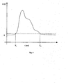
- a schematic example of the so determined Envelope is shown in Fig. 4.
- this envelope can be known way, for example a falling e-function of the form exp (-t / T) adjust (begin), where t is the time from the emission of the shock wave or from the Reception of the first back-reflected sound wave and where T is the is the desired period of the Doppler signal.
- a signal start time T 1 and a signal end time T 2 can be determined in the amount-based Doppler signal curve already mentioned above, where T 1 corresponds to the time at which the described
- T 2 corresponds to the time at which the envelope falls below a second threshold value for the first time.
- the Doppler signal unit 32 In addition to the already mentioned output of the Doppler signal to the Display device 30 for the purpose of pictorial representation corresponding to FIG. 3 below 4 is the Doppler signal unit 32, as shown in FIG. 2, also with a Alarm device 34 connected to which they have an associated time duration signal outputs. As also below. will be explained, the Alarm device 34 trigger an acoustic and / or visual alarm if the time duration signal results in a too short duration of the Doppler signal, which suggests a miss, i.e. that the concretion 18th is outside the shock wave focus.
- the Doppler signal unit 32 is also with the Shock wave generator 16 connected to this in the event of misses to be able to stop automatically if necessary. In this way, one unnecessary stress on the patient's body 12 due to shock wave misses avoided particularly safely and effectively without any intervention of medical personnel.
- Essential steps of using the lithotripter 10 according to the invention feasible method according to the invention are described below with reference to 5 to 8 are explained:
- the method according to the invention comprises after the start of the lithotripter according to the invention essentially three Sections, namely a first section S10 with steps for presetting the Lithotripters before the actual therapy, a subsequent section S20, in the relevant during therapy in various procedural steps Duration of a Doppler signal is determined, and a further section S30, in the several process steps, the corresponding measures as a function the relevant signal duration determined in section S20.
- step S11 the above-mentioned and in the state of the Technique known adjustment of the patient with the help of the adjustable couch Lithotripters 10.
- the patient 12 is positioned in such a way that his Concrement 18 is located in the shock wave focus.
- step S12 the ultrasonic transducer 24 and the first emission of a shock wave Using the shock wave generator 16.
- step S13 the Doppler signal unit 2 is generated using the reflected ultrasonic waves as well as the known frequency of the transducer 24 transmitted ultrasound waves a Doppler signal, which is then in Step S14 is shown on the display device 30 corresponding to FIG. 3.
- the envelope can correspond to the Doppler signal 4 are shown on the display device 30.
- This Doppler signal is used by the medical personnel in step S15 assessed whether it indicates a hit or a hit Fire shot.
- This first emission of a shock wave results in step S12 usually a hit, since after step S11 the concrement 18 in any case lies in the shock wave focus, and since you send out a "test shock wave” can ask the patient 12 to hold their breath for a short time so that there is no risk of a miss due to the breathing movement of the calculus.
- this assessment of Doppler signals as to whether a hit or a If there is a miss, it is important, among other things, that the respective signal noise, i.e. to determine the respective "background" for each Doppler signal.
- step S15 If it is determined in step S15 that there is no hit, the process is returned to step S11 back. If, on the other hand, a hit is found, this becomes in step S16 Doppler signal already measured as a reference Doppler signal in the memory 32a stored. With the help of this reference Doppler signal are then in step S17 defines the threshold values discussed above, in particular by Consideration of the signal noise in the background of the reference Doppler signal. Furthermore, in step S17, the time period of the reference Doppler signal is used as the reference time period stored in the memory 32a.
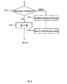
- step S18 An initialization then takes place in step S18, i.e. a reset of a Counting parameter z, the meaning of which is clear from FIG. 7 below will be.
- FIG. 7 shows the essential steps within the second section S20 according to Fig. 5, which are used to determine the relevant signal duration.
- step S21 the counting parameter z is incremented, i.e. increased by 1.
- step S22 a shock wave from the shock wave generator 16 is transmitted and in step S23 a Doppler signal from the Doppler signal unit 32 generated, which is then generated in step S24 on the display device 30 is displayed, as is its envelope according to FIG. 4.
- step S25 with the aid of the step S17 according to FIG. 6 defined threshold values the duration of this current Doppler signal certainly.
- this time period is also divided by the 6 into a normalized reference time period determined in step S17 Converted period, which subsequently in step S27 in the Doppler signal unit 32 is stored, for example in the memory 32a or in a separate memory for recording measured values.
- step S29a the last calculated mean of a continuous display added, which are those measured throughout the course of treatment Time duration of the Doppler signals continuously represents the development of the Track Doppler signal length.
- This can expediently be continuous Display can be shown on the display device 30, but it can also act as a separate screen etc.
- An example of such Continuous display is shown in Fig. 9 and will be explained further below.
- FIG. 8 shows the essential steps of the third section S30 according to FIGS. 5 and the measures in the context of the inventive method at a Lithotripsy treatment with the lithotripter 10 according to the invention as a function of Average calculated in step S29 can be taken:
- step S31 what is executed in the Doppler signal unit 32
- the program checks whether the mean value calculated in step S29 is greater than one predetermined limit. As explained above, large duration values indicate Hits, whereas small duration values are typical for misses. Accordingly, a positive result of the check in step S31 means that obviously the concrement 18 is still in the shock wave focus and thus Hits. Accordingly, this is done in the Doppler signal unit 32 executed program in this case via a step S32, in which the Counting parameter z is again reset to 0, back to step S21, i.e. it a measurement of five standardized Doppler signal time periods begins again subsequent averaging according to FIG. 7.
- step S31 if the check in step S31 leads to a negative result, i.e. to, that the averaged duration of the last five Doppler signals is too short, which is due to indicates possible misses, the program branches to a step S33, in which said alarm device 34 is triggered, and then to a step S34 in which the shock wave generator 16 is stopped by one unnecessary strain on the patient's body 12 due to misses avoid.
- 9 is a schematic illustration of an exemplary progress control display on display 30 during shock wave therapy. you 9 recognizes a coordinate system, the abscissa of which is the number emitted Shock waves and their ordinate following the respective shock wave determines the determined absolute or standardized double signal duration. You can see from Start the shock wave therapy until shortly before its end a slow increase in Doppler signal times. Individual "drops” in this curve indicate "misses” due to movement of the patient 12 or the concretions 18 within the patient 12, which are immediately replaced by a Repositioning of patient 12 was corrected. 9 represents one horizontal dashed line the amount of the threshold whose Fall below the alarm device 34 triggers. One can see that the local "Slump" in the middle of the curve to fall below this Threshold and thus led to an alarm.
- step S17 determination of the reference time period (step S17) and the later normalization of measured signal durations with the aid of this reference duration (Step S26) can also be omitted, so that the progress check absolute values of the measured Doppler signal duration during the shock wave therapy the display device 30 are displayed and evaluated.
- lithotripter 10 which is only schematically indicated with reference to FIG. 1 it is understood that this can have numerous other components that are known from the prior art, for example a X-ray locating device or an imaging ultrasound scanner.
Landscapes
- Health & Medical Sciences (AREA)
- Life Sciences & Earth Sciences (AREA)
- Surgery (AREA)
- Engineering & Computer Science (AREA)
- Medical Informatics (AREA)
- General Health & Medical Sciences (AREA)
- Veterinary Medicine (AREA)
- Public Health (AREA)
- Biomedical Technology (AREA)
- Heart & Thoracic Surgery (AREA)
- Nuclear Medicine, Radiotherapy & Molecular Imaging (AREA)
- Molecular Biology (AREA)
- Animal Behavior & Ethology (AREA)
- Mechanical Engineering (AREA)
- Orthopedic Medicine & Surgery (AREA)
- Vascular Medicine (AREA)
- Physics & Mathematics (AREA)
- Biophysics (AREA)
- Pathology (AREA)
- Radiology & Medical Imaging (AREA)
- Ultra Sonic Daignosis Equipment (AREA)
- Measurement Of Velocity Or Position Using Acoustic Or Ultrasonic Waves (AREA)
- Surgical Instruments (AREA)
Abstract
Description
- Fig. 1
- eine vereinfachte Darstellung wesentlicher Bauteile des erfindungsgemäßen Lithotripters;
- Fig. 2
- eine schematische Ansicht der Dopplersignaleinheit und ihrer Anschlüsse beim erfindungsgemäßen Lithotripter;
- Fig. 3
- eine Darstellung von Ultraschall- und Dopplersignalen auf der Anzeigevorrichtung des erfindungsgemäßen Lithotripters;
- Fig.4
- ein schematisches Beispiel für eine der weiteren Signalauswertung zugrundzulegende Einhüllende an das Dopplersignal;
- Fig. 5.
- ein schematisches Flußdiagramm zur Verdeutlichung der Hauptabschnitte des erfindungsgemäßen Verfahrens;
- Fig. 6
- ein Flußdiagramm zur Verdeutlichung wesentlicher Verfahrensschritte innerhalb des ersten Hauptabschnitts gemäß Fig. 5;
- Fig. 7
- ein Flußdiagramm zur Verdeutlichung wesentlicher Verfahrensschritte innerhalb des zweiten Hauptabschnitts gemäß Fig. 5;
- Fig. 8
- ein Flußdiagramm zur Verdeutlichung wesentlicher Verfahrensschritte des dritten Hauptabschnitts gemäß Fig. 5; und
- Fig. 9
- ein schematisches Beispiel für eine Signaldauer-Verlaufskontrolle während einer Stoßwellentherapie.
- 10
- Lithotripter
- 12
- Patient
- 14
- Ankoppelkissten
- 16
- Stoßwellengenerator
- 18
- Konkrement (Nierenstein)
- 20
- Niere
- 22
- Stoßwellenquelle
- 24
- Ultraschall-Transducer
- 26
- Einstellbarer Halter
- 28
- Steuereinheit
- 30
- Anzeigevorrichtung
- 32
- Dopplersignaleinheit
- 32a
- Speicher
- 34
- Alarmvorrichtung
Claims (17)
- Lithotripter (10) zur Fragmentierung eines Zielobjekts, insbesondere eines Konkrements (18), in einem vorzugsweise menschlichen Körper, umfassend:dadurch gekennzeichnet, daß die Dopplersignaleinheit (32) dazu ausgelegt ist, die Zeitdauer des Dopplersignals zu bestimmen und ein zugeordnetes Zeitdauersignal auszugeben.einen Stoßwellengenerator (16) zur Erzeugung fokussierter Stoßwellen,eine Ultraschall-Sende/Empfangseinheit mit einem Ultraschall-Transducer (24) zum Aussenden von Ultraschallwellen in den Körper und zum Empfangen von in einem Zielgebiet des Stoßwellengenerators (16) reflektierten Ultraschallwellen, undeine an die Ultraschall-Sende/Empfangseinheit angeschlossene Dopplersignaleinheit (32) zum Generieren und Auswerten eines Dopplersignals aus den ausgesandten und empfangenen Ultraschallwellen,
- Lithotripter (10) nach Anspruch 1, dadurch gekennzeichnet, daß er ferner eine mit der Dopplersignaleinheit (32) verbundene Anzeigevorrichtung (30) zur Anzeige von Dopplersignalen und/oder eine Alarmvorrichtung (34) aufweist, der das Zeitdauersignal zugeführt wird.
- Lithotripter (10) nach Anspruch 1 oder 2, dadurch gekennzeichnet, daß der Stoßwellengenerator (16) mit der Dopplersignaleinheit (32) verbunden und dazu ausgelegt ist, die Erzeugung von Stoßwellen als Funktion des Zeitdauersignals zu stoppen bzw. fortzusetzen.
- Lithotripter (10) nach einem der vorhergehenden Ansprüche, dadurch gekennzeichnet, daß der Ultraschall-Transducer (24) der Ultraschallsende/Empfangseinheit an einem einstellbaren Halter (26) montiert ist.
- Lithotripter (10) nach einem der vorhergehenden Ansprüche, dadurch gekennzeichnet, daß die Dopplersignaleinheit (32) dazu ausgelegt ist, als Zeitdauer des Dopplersignals ein Zeitintervall zu bestimmen, innerhalb dessen der Betrag des Dopplersignals auf einen vorbestimmten Bruchteil eines Anfangsbetrags gefallen ist, insbesondere auf den e-ten Teil des Anfangsbetrags.
- Lithotripter (10) nach einem der Ansprüche 1 bis 4, dadurch gekennzeichnet, daß die Dopplersignaleinheit (32) dazu ausgelegt ist, als Zeitdauer des Dopplersignals ein Zeitintervall zwischen einem Signal-Anfangszeitpunkt und einem Signal-Endzeitpunkt zu bestimmen, wobei der Signal-Anfangszeitpunkt festgestellt wird, wenn der Betrag des Dopplersignals einen ersten Schwellenwert überschreitet, und der Signal-Endzeitpunkt festgestellt wird, wenn der Betrag des Dopplersignals einen zweiten Schwellenwert unterschreitet.
- Lithotripter (10) nach Anspruch 6, dadurch gekennzeichnet, daß der erste und der zweite Schwellenwert identisch sind.
- Lithotripter (10) nach Anspruch 6 oder 7, dadurch gekennzeichnet, daß die Dopplersignaleinheit (32) Einstellmittel zum Einstellen des ersten und/oder des zweiten Schwellenwerts aufweist.
- Lithotripter (10) nach einem der vorhergehenden Ansprüche, dadurch gekennzeichnet, daß die Dopplersignaleinheit (32) femer dazu ausgelegt ist, die Zeitdauer des Dopplersignals über mehrere Stoßwellen zu mitteln.
- Lithotripter (10) nach einem der vorhergehenden Ansprüche, dadurch gekennzeichnet, daß die Dopplersignaleinheit (32) femer dazu ausgelegt ist, die Zeitdauer eines Dopplersignals auf die Referenz-Zeitdauer eines Referenz-Dopplersignals zu normieren.
- Lithotripter (10) nach einem der vorhergehenden Ansprüche, dadurch gekennzeichnet, daß die Ultraschall-Sende/Empfangseinheit als Teil eines bildgebenden Ultraschallscanners ausgebildet ist.
- Lithotripter (10) nach einem der vorhergehenden Ansprüche, dadurch gekennzeichnet, daß der Ultraschall-Transducer (24) als Stiftsonde ausgebildet ist.
- Lithotripter (10) nach Anspruch 12, dadurch gekennzeichnet, daß er femer eine Röntgenortungsvorrichtung aufweist.
- Lithotriper (10) nach einem der vorhergehenden Ansprüche, dadurch gekennzeichnet, daß er Mittel zur fortlaufenden Darstellung der Zeitdauer der Dopplersignale über den Behandlungsverlauf umfaßt.
- Verfahren zur Überwachung der Fragmentierung eines Zielobjekts, insbesondere eines Konkrements (18), in einem vorzugsweise menschlichen Körper, umfassend die Schritte:dadurch gekennzeichnet, daß der Schritt des Auswertens eine Bestimmung der Zeitdauer des Dopplersignals umfaßt und ferner ein Schritt der Ausgabe eines zugeordneten Zeitdauersignals vorgesehen ist.Aussenden von Ultraschallwellen in den Körper,Empfangen von am Zielobjekt reflektierten Ultraschallwellen, undGenerieren und Auswerten eines Dopplersignals aus den ausgesandten und den empfangenen Ultraschallwellen,
- Verfahren nach Anspruch 15, dadurch gekennzeichnet, daß es femer die Schritte umfaßt:sowie den späteren Schritt:Anordnen des Körpers in einem Lithotripter (10) mit einem Stoßwellengenerator (16) zur Erzeugung fokussierter Stoßwellen,Anzeigen des Zielobjekts (18) und des Fokus des Stoßwellengenerators (16) auf einer Anzeigevorrichtung (30) einer bildgebenden Ortungsvorrichtung,Justieren des Körpers derart, daß das Zielobjekt (18) im Fokus des Stoßwellengenerators (16) liegt,Aussenden einer Stoßwelle in Richtung des Zielobjekts (18) und Bestimmen der Zeitdauer des Dopplersignals,Speichem der Zeitdauer als Referenz-Zeitdauer,Normieren der Zeitdauer eines später gemessenen Dopplersignals auf die Referenz-Zeitdauer.
- Verfahren nach Anspruch 15 oder 16, gekennzeichnet durch den weiteren Schritt des fortlaufenden Darstellens der Zeitdauer der Dopplersignale über den Behandlungsverlauf.
Applications Claiming Priority (2)
| Application Number | Priority Date | Filing Date | Title |
|---|---|---|---|
| DE10228550A DE10228550B3 (de) | 2002-06-26 | 2002-06-26 | Lithotripter zur Fragmentierung eines Zielobjekts in einem Körper und Verfahren zur Überwachung der Fragmentierung eines Zielobjekts in einem Körper |
| DE10228550 | 2002-06-26 |
Publications (2)
| Publication Number | Publication Date |
|---|---|
| EP1374785A1 true EP1374785A1 (de) | 2004-01-02 |
| EP1374785B1 EP1374785B1 (de) | 2008-05-21 |
Family
ID=29716666
Family Applications (1)
| Application Number | Title | Priority Date | Filing Date |
|---|---|---|---|
| EP03014395A Expired - Lifetime EP1374785B1 (de) | 2002-06-26 | 2003-06-26 | Lithotripter mit einer Doppler-Ultraschalleinheit zur Trefferüberwachung |
Country Status (3)
| Country | Link |
|---|---|
| EP (1) | EP1374785B1 (de) |
| AT (1) | ATE395870T1 (de) |
| DE (2) | DE10228550B3 (de) |
Cited By (21)
| Publication number | Priority date | Publication date | Assignee | Title |
|---|---|---|---|---|
| CN100353918C (zh) * | 2004-01-28 | 2007-12-12 | 多恩尼尔医疗技术有限责任公司 | 碎石机 |
| EP2903688A4 (de) * | 2012-10-05 | 2016-06-15 | Univ Michigan | Blaseninduziertes farbdoppler-feedback während einer histotripsie |
| US9526923B2 (en) | 2009-08-17 | 2016-12-27 | Histosonics, Inc. | Disposable acoustic coupling medium container |
| US9636133B2 (en) | 2012-04-30 | 2017-05-02 | The Regents Of The University Of Michigan | Method of manufacturing an ultrasound system |
| US9642634B2 (en) | 2005-09-22 | 2017-05-09 | The Regents Of The University Of Michigan | Pulsed cavitational ultrasound therapy |
| US9901753B2 (en) | 2009-08-26 | 2018-02-27 | The Regents Of The University Of Michigan | Ultrasound lithotripsy and histotripsy for using controlled bubble cloud cavitation in fractionating urinary stones |
| US9943708B2 (en) | 2009-08-26 | 2018-04-17 | Histosonics, Inc. | Automated control of micromanipulator arm for histotripsy prostate therapy while imaging via ultrasound transducers in real time |
| US10071266B2 (en) | 2011-08-10 | 2018-09-11 | The Regents Of The University Of Michigan | Lesion generation through bone using histotripsy therapy without aberration correction |
| US10219815B2 (en) | 2005-09-22 | 2019-03-05 | The Regents Of The University Of Michigan | Histotripsy for thrombolysis |
| US10293187B2 (en) | 2013-07-03 | 2019-05-21 | Histosonics, Inc. | Histotripsy excitation sequences optimized for bubble cloud formation using shock scattering |
| US10780298B2 (en) | 2013-08-22 | 2020-09-22 | The Regents Of The University Of Michigan | Histotripsy using very short monopolar ultrasound pulses |
| US11135454B2 (en) | 2015-06-24 | 2021-10-05 | The Regents Of The University Of Michigan | Histotripsy therapy systems and methods for the treatment of brain tissue |
| US11432900B2 (en) | 2013-07-03 | 2022-09-06 | Histosonics, Inc. | Articulating arm limiter for cavitational ultrasound therapy system |
| US11648424B2 (en) | 2018-11-28 | 2023-05-16 | Histosonics Inc. | Histotripsy systems and methods |
| US11813485B2 (en) | 2020-01-28 | 2023-11-14 | The Regents Of The University Of Michigan | Systems and methods for histotripsy immunosensitization |
| US12318636B2 (en) | 2022-10-28 | 2025-06-03 | Histosonics, Inc. | Histotripsy systems and methods |
| US12343568B2 (en) | 2020-08-27 | 2025-07-01 | The Regents Of The University Of Michigan | Ultrasound transducer with transmit-receive capability for histotripsy |
| US12446905B2 (en) | 2023-04-20 | 2025-10-21 | Histosonics, Inc. | Histotripsy systems and associated methods including user interfaces and workflows for treatment planning and therapy |
| US12527976B2 (en) | 2020-06-18 | 2026-01-20 | Histosonics, Inc. | Histotripsy acoustic and patient coupling systems and methods |
| US12582848B2 (en) | 2021-06-07 | 2026-03-24 | The Regents Of The University Of Michigan | Minimally invasive histotripsy systems and methods |
| US12599787B2 (en) | 2021-06-07 | 2026-04-14 | The Regents Of The University Of Michigan | All-in-one ultrasound systems and methods including histotripsy |
Families Citing this family (2)
| Publication number | Priority date | Publication date | Assignee | Title |
|---|---|---|---|---|
| DE102005020115A1 (de) * | 2005-04-29 | 2006-08-24 | Siemens Ag | Verfahren und Einrichtung zum Ermitteln des Zerstörungsgrades eines Konkrementes bei der Lithotripsie |
| DE102006021049A1 (de) * | 2006-05-05 | 2007-11-08 | Siemens Ag | Stoßwellenkopf für eine Stoßwellenbehandlungsvorrichtung und Verfahren zur Fragmentierung und zur Kontrolle der Fragmentierung eines in einem Untersuchungsobjekt angeordneten Fragmentierungsobjekts |
Citations (3)
| Publication number | Priority date | Publication date | Assignee | Title |
|---|---|---|---|---|
| DE4012760A1 (de) * | 1990-04-21 | 1992-05-07 | G M T I Ges Fuer Medizintechni | Ultraschall-doppler-verfahren zur bestimmung der steingroesse aus der sinkgeschwindigkeit waehrend der gallen-lithotripsie sowie vorrichtung zur durchfuehrung des verfahrens |
| EP0367116B1 (de) | 1988-10-26 | 1994-06-08 | Kabushiki Kaisha Toshiba | Gerät zur Stosswellen-Behandlung |
| DE4446192A1 (de) | 1994-12-23 | 1996-07-04 | Wolf Gmbh Richard | Verfahren zur Trefferkontrolle |
-
2002
- 2002-06-26 DE DE10228550A patent/DE10228550B3/de not_active Expired - Fee Related
-
2003
- 2003-06-26 DE DE50309870T patent/DE50309870D1/de not_active Expired - Lifetime
- 2003-06-26 AT AT03014395T patent/ATE395870T1/de not_active IP Right Cessation
- 2003-06-26 EP EP03014395A patent/EP1374785B1/de not_active Expired - Lifetime
Patent Citations (4)
| Publication number | Priority date | Publication date | Assignee | Title |
|---|---|---|---|---|
| EP0367116B1 (de) | 1988-10-26 | 1994-06-08 | Kabushiki Kaisha Toshiba | Gerät zur Stosswellen-Behandlung |
| EP0548048B1 (de) | 1988-10-26 | 1996-02-14 | Kabushiki Kaisha Toshiba | Gerät zur Stosswellen-Behandlung |
| DE4012760A1 (de) * | 1990-04-21 | 1992-05-07 | G M T I Ges Fuer Medizintechni | Ultraschall-doppler-verfahren zur bestimmung der steingroesse aus der sinkgeschwindigkeit waehrend der gallen-lithotripsie sowie vorrichtung zur durchfuehrung des verfahrens |
| DE4446192A1 (de) | 1994-12-23 | 1996-07-04 | Wolf Gmbh Richard | Verfahren zur Trefferkontrolle |
Non-Patent Citations (1)
| Title |
|---|
| HÖRBARTH K, HOFBAUER J, MARBERGER M: "Color flow Doppler sonography for extracorporal shock wave lithotripsy", JOURNAL OF UROLOGY, vol. 150, no. 6, 1993, pages 1768 - 1770, XP009019789 * |
Cited By (37)
| Publication number | Priority date | Publication date | Assignee | Title |
|---|---|---|---|---|
| CN100353918C (zh) * | 2004-01-28 | 2007-12-12 | 多恩尼尔医疗技术有限责任公司 | 碎石机 |
| US11364042B2 (en) | 2005-09-22 | 2022-06-21 | The Regents Of The University Of Michigan | Histotripsy for thrombolysis |
| US12303152B2 (en) | 2005-09-22 | 2025-05-20 | The Regents Of The University Of Michigan | Histotripsy for thrombolysis |
| US12150661B2 (en) | 2005-09-22 | 2024-11-26 | The Regents Of The University Of Michigan | Histotripsy for thrombolysis |
| US9642634B2 (en) | 2005-09-22 | 2017-05-09 | The Regents Of The University Of Michigan | Pulsed cavitational ultrasound therapy |
| US10219815B2 (en) | 2005-09-22 | 2019-03-05 | The Regents Of The University Of Michigan | Histotripsy for thrombolysis |
| US11701134B2 (en) | 2005-09-22 | 2023-07-18 | The Regents Of The University Of Michigan | Histotripsy for thrombolysis |
| US9526923B2 (en) | 2009-08-17 | 2016-12-27 | Histosonics, Inc. | Disposable acoustic coupling medium container |
| US9901753B2 (en) | 2009-08-26 | 2018-02-27 | The Regents Of The University Of Michigan | Ultrasound lithotripsy and histotripsy for using controlled bubble cloud cavitation in fractionating urinary stones |
| US9943708B2 (en) | 2009-08-26 | 2018-04-17 | Histosonics, Inc. | Automated control of micromanipulator arm for histotripsy prostate therapy while imaging via ultrasound transducers in real time |
| US10071266B2 (en) | 2011-08-10 | 2018-09-11 | The Regents Of The University Of Michigan | Lesion generation through bone using histotripsy therapy without aberration correction |
| US9636133B2 (en) | 2012-04-30 | 2017-05-02 | The Regents Of The University Of Michigan | Method of manufacturing an ultrasound system |
| EP2903688A4 (de) * | 2012-10-05 | 2016-06-15 | Univ Michigan | Blaseninduziertes farbdoppler-feedback während einer histotripsie |
| US11058399B2 (en) | 2012-10-05 | 2021-07-13 | The Regents Of The University Of Michigan | Bubble-induced color doppler feedback during histotripsy |
| US10293187B2 (en) | 2013-07-03 | 2019-05-21 | Histosonics, Inc. | Histotripsy excitation sequences optimized for bubble cloud formation using shock scattering |
| US11432900B2 (en) | 2013-07-03 | 2022-09-06 | Histosonics, Inc. | Articulating arm limiter for cavitational ultrasound therapy system |
| US11819712B2 (en) | 2013-08-22 | 2023-11-21 | The Regents Of The University Of Michigan | Histotripsy using very short ultrasound pulses |
| US10780298B2 (en) | 2013-08-22 | 2020-09-22 | The Regents Of The University Of Michigan | Histotripsy using very short monopolar ultrasound pulses |
| US12350525B2 (en) | 2013-08-22 | 2025-07-08 | The Regents Of The University Of Michigan | Histotripsy using very short ultrasound pulses |
| US12220602B2 (en) | 2015-06-24 | 2025-02-11 | The Regents Of The University Of Michigan | Histotripsy therapy systems and methods for the treatment of brain tissue |
| US11135454B2 (en) | 2015-06-24 | 2021-10-05 | The Regents Of The University Of Michigan | Histotripsy therapy systems and methods for the treatment of brain tissue |
| US11813484B2 (en) | 2018-11-28 | 2023-11-14 | Histosonics, Inc. | Histotripsy systems and methods |
| US12491384B2 (en) | 2018-11-28 | 2025-12-09 | Histosonics, Inc. | Histotripsy systems and methods |
| US11648424B2 (en) | 2018-11-28 | 2023-05-16 | Histosonics Inc. | Histotripsy systems and methods |
| US11980778B2 (en) | 2018-11-28 | 2024-05-14 | Histosonics, Inc. | Histotripsy systems and methods |
| US12599784B2 (en) | 2018-11-28 | 2026-04-14 | Histosonics, Inc. | Histotripsy systems and methods |
| US12589261B2 (en) | 2018-11-28 | 2026-03-31 | Histosonics, Inc. | Histotripsy systems and methods |
| US12491382B2 (en) | 2018-11-28 | 2025-12-09 | Histosonics, Inc. | Histotripsy systems and methods |
| US12420118B2 (en) | 2018-11-28 | 2025-09-23 | Histosonics, Inc. | Histotripsy systems and methods |
| US11813485B2 (en) | 2020-01-28 | 2023-11-14 | The Regents Of The University Of Michigan | Systems and methods for histotripsy immunosensitization |
| US12527976B2 (en) | 2020-06-18 | 2026-01-20 | Histosonics, Inc. | Histotripsy acoustic and patient coupling systems and methods |
| US12343568B2 (en) | 2020-08-27 | 2025-07-01 | The Regents Of The University Of Michigan | Ultrasound transducer with transmit-receive capability for histotripsy |
| US12582848B2 (en) | 2021-06-07 | 2026-03-24 | The Regents Of The University Of Michigan | Minimally invasive histotripsy systems and methods |
| US12599787B2 (en) | 2021-06-07 | 2026-04-14 | The Regents Of The University Of Michigan | All-in-one ultrasound systems and methods including histotripsy |
| US12390665B1 (en) | 2022-10-28 | 2025-08-19 | Histosonics, Inc. | Histotripsy systems and methods |
| US12318636B2 (en) | 2022-10-28 | 2025-06-03 | Histosonics, Inc. | Histotripsy systems and methods |
| US12446905B2 (en) | 2023-04-20 | 2025-10-21 | Histosonics, Inc. | Histotripsy systems and associated methods including user interfaces and workflows for treatment planning and therapy |
Also Published As
| Publication number | Publication date |
|---|---|
| DE50309870D1 (de) | 2008-07-03 |
| EP1374785B1 (de) | 2008-05-21 |
| ATE395870T1 (de) | 2008-06-15 |
| DE10228550B3 (de) | 2004-02-12 |
Similar Documents
| Publication | Publication Date | Title |
|---|---|---|
| DE10228550B3 (de) | Lithotripter zur Fragmentierung eines Zielobjekts in einem Körper und Verfahren zur Überwachung der Fragmentierung eines Zielobjekts in einem Körper | |
| EP1384444B1 (de) | Lithotripter | |
| DE4125950C1 (de) | ||
| DE69208141T2 (de) | Vorrichtung zum Zerstören von Konkrementen | |
| EP1115338B1 (de) | Rotierendes chirurgisches werkzeug | |
| DE10158519B4 (de) | Stoß- und Druckwellen-Therapiegerät | |
| DE102005031116B4 (de) | Stoßwellensystem | |
| DE3338745A1 (de) | Ultraschalleinrichtung fuer augenmessungen | |
| DE3900893C2 (de) | ||
| DE102005037043B4 (de) | Stoßwellentherapiegerät mit Bildgewinnung | |
| DE29908259U1 (de) | Rotierendes chirurgisches Werkzeug | |
| DE69104551T2 (de) | Vorrichtung für Lithotripsie. | |
| DE19548000C1 (de) | Vorrichtung zur Ortung von Konkrementen im Körper eines Patienten | |
| DE3913023C2 (de) | Zertrümmerungswellen-Behandlungsgerät | |
| DE68911724T2 (de) | Nierensteinzertrümmerer. | |
| DE69114811T2 (de) | Vorrichtung zum Zerstören von Konkrementen. | |
| EP1166148A1 (de) | Vorrichtung und verfahren für die 3d-echtzeitsonographie | |
| DE102005039178B4 (de) | Vorrichtung und Verfahren zur Bestimmung der Ankoppelgüte zwischen Koppelbalg und Patienten bei einem Gerät zur extrakorporalen Behandlung mit akustischen Stoßwellen | |
| DE10260594B4 (de) | Vorrichtung zum extrakorporalen Erzeugen von fokussierten Stoßwellen | |
| WO2022175235A1 (de) | Medizinisches instrument und ultraschallsystem mit einem solchen instrument | |
| DE10304904A1 (de) | Ultraschall-Stosswellen-Lithotripter | |
| DE102006031356B4 (de) | Vorrichtung zum lokalen Abtrag von menschlichem oder tierischem Knochenmaterial | |
| DE4446192A1 (de) | Verfahren zur Trefferkontrolle | |
| DE3943644C2 (de) | Stoßwellenbehandlungsgerät | |
| DE102009004226B4 (de) | Verfahren zum Erzeugen eines den Stoßwellenfokus einer Stoßwellenquelle einer Lithotripsie-Einrichtung enthaltenden Ultraschallbildes sowie nach diesem Verfahren betriebene Lithotripsie-Einrichtung |
Legal Events
| Date | Code | Title | Description |
|---|---|---|---|
| PUAI | Public reference made under article 153(3) epc to a published international application that has entered the european phase |
Free format text: ORIGINAL CODE: 0009012 |
|
| AK | Designated contracting states |
Kind code of ref document: A1 Designated state(s): AT BE BG CH CY CZ DE DK EE ES FI FR GB GR HU IE IT LI LU MC NL PT RO SE SI SK TR |
|
| AX | Request for extension of the european patent |
Extension state: AL LT LV MK |
|
| 17P | Request for examination filed |
Effective date: 20040701 |
|
| AKX | Designation fees paid |
Designated state(s): AT BE BG CH CY CZ DE DK EE ES FI FR GB GR HU IE IT LI LU MC NL PT RO SE SI SK TR |
|
| 17Q | First examination report despatched |
Effective date: 20060707 |
|
| GRAP | Despatch of communication of intention to grant a patent |
Free format text: ORIGINAL CODE: EPIDOSNIGR1 |
|
| GRAS | Grant fee paid |
Free format text: ORIGINAL CODE: EPIDOSNIGR3 |
|
| GRAA | (expected) grant |
Free format text: ORIGINAL CODE: 0009210 |
|
| AK | Designated contracting states |
Kind code of ref document: B1 Designated state(s): AT BE BG CH CY CZ DE DK EE ES FI FR GB GR HU IE IT LI LU MC NL PT RO SE SI SK TR |
|
| REG | Reference to a national code |
Ref country code: GB Ref legal event code: FG4D Free format text: NOT ENGLISH |
|
| REG | Reference to a national code |
Ref country code: CH Ref legal event code: EP Ref country code: CH Ref legal event code: NV Representative=s name: BOVARD AG PATENTANWAELTE |
|
| REF | Corresponds to: |
Ref document number: 50309870 Country of ref document: DE Date of ref document: 20080703 Kind code of ref document: P |
|
| REG | Reference to a national code |
Ref country code: IE Ref legal event code: FG4D Free format text: LANGUAGE OF EP DOCUMENT: GERMAN |
|
| PG25 | Lapsed in a contracting state [announced via postgrant information from national office to epo] |
Ref country code: SI Free format text: LAPSE BECAUSE OF FAILURE TO SUBMIT A TRANSLATION OF THE DESCRIPTION OR TO PAY THE FEE WITHIN THE PRESCRIBED TIME-LIMIT Effective date: 20080521 |
|
| PG25 | Lapsed in a contracting state [announced via postgrant information from national office to epo] |
Ref country code: ES Free format text: LAPSE BECAUSE OF FAILURE TO SUBMIT A TRANSLATION OF THE DESCRIPTION OR TO PAY THE FEE WITHIN THE PRESCRIBED TIME-LIMIT Effective date: 20080901 Ref country code: FI Free format text: LAPSE BECAUSE OF FAILURE TO SUBMIT A TRANSLATION OF THE DESCRIPTION OR TO PAY THE FEE WITHIN THE PRESCRIBED TIME-LIMIT Effective date: 20080521 |
|
| NLV1 | Nl: lapsed or annulled due to failure to fulfill the requirements of art. 29p and 29m of the patents act | ||
| PG25 | Lapsed in a contracting state [announced via postgrant information from national office to epo] |
Ref country code: NL Free format text: LAPSE BECAUSE OF FAILURE TO SUBMIT A TRANSLATION OF THE DESCRIPTION OR TO PAY THE FEE WITHIN THE PRESCRIBED TIME-LIMIT Effective date: 20080521 |
|
| BERE | Be: lapsed |
Owner name: DORNIER MEDTECH SYSTEMS G.M.B.H. Effective date: 20080630 |
|
| REG | Reference to a national code |
Ref country code: IE Ref legal event code: FD4D |
|
| PG25 | Lapsed in a contracting state [announced via postgrant information from national office to epo] |
Ref country code: IE Free format text: LAPSE BECAUSE OF FAILURE TO SUBMIT A TRANSLATION OF THE DESCRIPTION OR TO PAY THE FEE WITHIN THE PRESCRIBED TIME-LIMIT Effective date: 20080521 Ref country code: MC Free format text: LAPSE BECAUSE OF NON-PAYMENT OF DUE FEES Effective date: 20080630 Ref country code: SE Free format text: LAPSE BECAUSE OF FAILURE TO SUBMIT A TRANSLATION OF THE DESCRIPTION OR TO PAY THE FEE WITHIN THE PRESCRIBED TIME-LIMIT Effective date: 20080821 Ref country code: PT Free format text: LAPSE BECAUSE OF FAILURE TO SUBMIT A TRANSLATION OF THE DESCRIPTION OR TO PAY THE FEE WITHIN THE PRESCRIBED TIME-LIMIT Effective date: 20081021 Ref country code: DK Free format text: LAPSE BECAUSE OF FAILURE TO SUBMIT A TRANSLATION OF THE DESCRIPTION OR TO PAY THE FEE WITHIN THE PRESCRIBED TIME-LIMIT Effective date: 20080521 Ref country code: CZ Free format text: LAPSE BECAUSE OF FAILURE TO SUBMIT A TRANSLATION OF THE DESCRIPTION OR TO PAY THE FEE WITHIN THE PRESCRIBED TIME-LIMIT Effective date: 20080521 |
|
| PG25 | Lapsed in a contracting state [announced via postgrant information from national office to epo] |
Ref country code: SK Free format text: LAPSE BECAUSE OF FAILURE TO SUBMIT A TRANSLATION OF THE DESCRIPTION OR TO PAY THE FEE WITHIN THE PRESCRIBED TIME-LIMIT Effective date: 20080521 Ref country code: RO Free format text: LAPSE BECAUSE OF FAILURE TO SUBMIT A TRANSLATION OF THE DESCRIPTION OR TO PAY THE FEE WITHIN THE PRESCRIBED TIME-LIMIT Effective date: 20080521 |
|
| PLBE | No opposition filed within time limit |
Free format text: ORIGINAL CODE: 0009261 |
|
| STAA | Information on the status of an ep patent application or granted ep patent |
Free format text: STATUS: NO OPPOSITION FILED WITHIN TIME LIMIT |
|
| PG25 | Lapsed in a contracting state [announced via postgrant information from national office to epo] |
Ref country code: BE Free format text: LAPSE BECAUSE OF NON-PAYMENT OF DUE FEES Effective date: 20080630 |
|
| 26N | No opposition filed |
Effective date: 20090224 |
|
| GBPC | Gb: european patent ceased through non-payment of renewal fee |
Effective date: 20080821 |
|
| PG25 | Lapsed in a contracting state [announced via postgrant information from national office to epo] |
Ref country code: EE Free format text: LAPSE BECAUSE OF FAILURE TO SUBMIT A TRANSLATION OF THE DESCRIPTION OR TO PAY THE FEE WITHIN THE PRESCRIBED TIME-LIMIT Effective date: 20080521 Ref country code: BG Free format text: LAPSE BECAUSE OF FAILURE TO SUBMIT A TRANSLATION OF THE DESCRIPTION OR TO PAY THE FEE WITHIN THE PRESCRIBED TIME-LIMIT Effective date: 20080821 |
|
| PG25 | Lapsed in a contracting state [announced via postgrant information from national office to epo] |
Ref country code: IT Free format text: LAPSE BECAUSE OF FAILURE TO SUBMIT A TRANSLATION OF THE DESCRIPTION OR TO PAY THE FEE WITHIN THE PRESCRIBED TIME-LIMIT Effective date: 20080521 Ref country code: AT Free format text: LAPSE BECAUSE OF NON-PAYMENT OF DUE FEES Effective date: 20080626 |
|
| PG25 | Lapsed in a contracting state [announced via postgrant information from national office to epo] |
Ref country code: GB Free format text: LAPSE BECAUSE OF NON-PAYMENT OF DUE FEES Effective date: 20080821 |
|
| PG25 | Lapsed in a contracting state [announced via postgrant information from national office to epo] |
Ref country code: CY Free format text: LAPSE BECAUSE OF FAILURE TO SUBMIT A TRANSLATION OF THE DESCRIPTION OR TO PAY THE FEE WITHIN THE PRESCRIBED TIME-LIMIT Effective date: 20080521 Ref country code: HU Free format text: LAPSE BECAUSE OF FAILURE TO SUBMIT A TRANSLATION OF THE DESCRIPTION OR TO PAY THE FEE WITHIN THE PRESCRIBED TIME-LIMIT Effective date: 20081122 Ref country code: LU Free format text: LAPSE BECAUSE OF NON-PAYMENT OF DUE FEES Effective date: 20080626 |
|
| PG25 | Lapsed in a contracting state [announced via postgrant information from national office to epo] |
Ref country code: TR Free format text: LAPSE BECAUSE OF FAILURE TO SUBMIT A TRANSLATION OF THE DESCRIPTION OR TO PAY THE FEE WITHIN THE PRESCRIBED TIME-LIMIT Effective date: 20080521 |
|
| PG25 | Lapsed in a contracting state [announced via postgrant information from national office to epo] |
Ref country code: GR Free format text: LAPSE BECAUSE OF FAILURE TO SUBMIT A TRANSLATION OF THE DESCRIPTION OR TO PAY THE FEE WITHIN THE PRESCRIBED TIME-LIMIT Effective date: 20080822 |
|
| REG | Reference to a national code |
Ref country code: CH Ref legal event code: PFA Owner name: DORNIER MEDTECH SYSTEMS GMBH Free format text: DORNIER MEDTECH SYSTEMS GMBH#ARGELSRIEDER FELD 7#82234 WESSLING (DE) -TRANSFER TO- DORNIER MEDTECH SYSTEMS GMBH#ARGELSRIEDER FELD 7#82234 WESSLING (DE) |
|
| PGFP | Annual fee paid to national office [announced via postgrant information from national office to epo] |
Ref country code: DE Payment date: 20140629 Year of fee payment: 12 |
|
| PGFP | Annual fee paid to national office [announced via postgrant information from national office to epo] |
Ref country code: CH Payment date: 20140708 Year of fee payment: 12 |
|
| PGFP | Annual fee paid to national office [announced via postgrant information from national office to epo] |
Ref country code: FR Payment date: 20140623 Year of fee payment: 12 |
|
| REG | Reference to a national code |
Ref country code: DE Ref legal event code: R119 Ref document number: 50309870 Country of ref document: DE |
|
| REG | Reference to a national code |
Ref country code: CH Ref legal event code: PL |
|
| REG | Reference to a national code |
Ref country code: FR Ref legal event code: ST Effective date: 20160229 |
|
| PG25 | Lapsed in a contracting state [announced via postgrant information from national office to epo] |
Ref country code: LI Free format text: LAPSE BECAUSE OF NON-PAYMENT OF DUE FEES Effective date: 20150630 Ref country code: CH Free format text: LAPSE BECAUSE OF NON-PAYMENT OF DUE FEES Effective date: 20150630 Ref country code: DE Free format text: LAPSE BECAUSE OF NON-PAYMENT OF DUE FEES Effective date: 20160101 |
|
| PG25 | Lapsed in a contracting state [announced via postgrant information from national office to epo] |
Ref country code: FR Free format text: LAPSE BECAUSE OF NON-PAYMENT OF DUE FEES Effective date: 20150630 |