EP1406088A2 - Reagenzsatz und Verfahren zum Nachweis von Malariaparasiten, wobei eine Hämolyse in zwei Schritten mittels Tensiden durchgeführt wird - Google Patents
Reagenzsatz und Verfahren zum Nachweis von Malariaparasiten, wobei eine Hämolyse in zwei Schritten mittels Tensiden durchgeführt wird Download PDFInfo
- Publication number
- EP1406088A2 EP1406088A2 EP03022501A EP03022501A EP1406088A2 EP 1406088 A2 EP1406088 A2 EP 1406088A2 EP 03022501 A EP03022501 A EP 03022501A EP 03022501 A EP03022501 A EP 03022501A EP 1406088 A2 EP1406088 A2 EP 1406088A2
- Authority
- EP
- European Patent Office
- Prior art keywords
- surfactant
- malaria parasite
- sample
- hemolytic agent
- malaria
- Prior art date
- Legal status (The legal status is an assumption and is not a legal conclusion. Google has not performed a legal analysis and makes no representation as to the accuracy of the status listed.)
- Withdrawn
Links
Images
Classifications
-
- G—PHYSICS
- G01—MEASURING; TESTING
- G01N—INVESTIGATING OR ANALYSING MATERIALS BY DETERMINING THEIR CHEMICAL OR PHYSICAL PROPERTIES
- G01N33/00—Investigating or analysing materials by specific methods not covered by groups G01N1/00 - G01N31/00
- G01N33/48—Biological material, e.g. blood, urine; Haemocytometers
- G01N33/50—Chemical analysis of biological material, e.g. blood, urine; Testing involving biospecific ligand binding methods; Immunological testing
- G01N33/53—Immunoassay; Biospecific binding assay; Materials therefor
- G01N33/569—Immunoassay; Biospecific binding assay; Materials therefor for microorganisms, e.g. protozoa, bacteria, viruses
- G01N33/56905—Protozoa
-
- G—PHYSICS
- G01—MEASURING; TESTING
- G01N—INVESTIGATING OR ANALYSING MATERIALS BY DETERMINING THEIR CHEMICAL OR PHYSICAL PROPERTIES
- G01N2333/00—Assays involving biological materials from specific organisms or of a specific nature
- G01N2333/435—Assays involving biological materials from specific organisms or of a specific nature from animals; from humans
- G01N2333/44—Assays involving biological materials from specific organisms or of a specific nature from animals; from humans from protozoa
- G01N2333/445—Plasmodium
-
- Y—GENERAL TAGGING OF NEW TECHNOLOGICAL DEVELOPMENTS; GENERAL TAGGING OF CROSS-SECTIONAL TECHNOLOGIES SPANNING OVER SEVERAL SECTIONS OF THE IPC; TECHNICAL SUBJECTS COVERED BY FORMER USPC CROSS-REFERENCE ART COLLECTIONS [XRACs] AND DIGESTS
- Y02—TECHNOLOGIES OR APPLICATIONS FOR MITIGATION OR ADAPTATION AGAINST CLIMATE CHANGE
- Y02A—TECHNOLOGIES FOR ADAPTATION TO CLIMATE CHANGE
- Y02A50/00—TECHNOLOGIES FOR ADAPTATION TO CLIMATE CHANGE in human health protection, e.g. against extreme weather
- Y02A50/30—Against vector-borne diseases, e.g. mosquito-borne, fly-borne, tick-borne or waterborne diseases whose impact is exacerbated by climate change
Definitions
- the present invention relates to methods for detecting malaria parasites and reagent kits therefore. More particularly, the present invention relates to methods for detecting malaria parasites and reagent kits therefor which enable correct and convenient distinction of malaria parasites using a flow cytometer by allowing an erythrocyte in a sample to undergo hemolysis in two steps.
- Malaria is a parasitic infection disease which is prevalent in tropical and subtropical zones. Based on statistics from the World Health Organization (WHO), 40% or more of the worldwide population lives in malaria-prevalent regions, 3 to 5 hundred million people per year are infected, and 1.5 to 3 million cases are assumed to result in death.
- WHO World Health Organization
- Malaria is mediated by a mosquito called Anopheles.
- Anopheles mosquito bites human skin, a parasite is injected into human blood together with the saliva of the Anopheles mosquito. The parasite travels into a hepatocyte, where it proliferates, and then is released again into the blood.
- the morphology of the parasite is referred to as a merozoite, which invades an erythrocyte soon after being released into the blood and then grows while changing its morphology.
- This change in the morphology is referred to as a life cycle, each stage of which is referred to as a ring form, tropozoite or schizonte.
- a parasite which has grown up to the stage of the schizonte destroys an erythrocyte, and then is released as a merozoite again into the blood.
- the released merozoite invades an erythrocyte, and repeats the life cycle again, thereby proliferating repeatedly.
- the malaria parasite proliferates by repeating this cycle, while destroying erythrocytes in the blood continuously.
- the merozoite is differentiated partly into a morphology referred to as a gametocyte without infecting the erythrocyte.
- the gametocyte serves as a further source of infection via the bloodsucking of an Anopheles mosquito.
- the measurement method typically employed in diagnosing malaria is a method in which a blood smear preparation is stained, for example by a Giemsa staining method or a nucleic acid-staining fluorescent dye method, and then examined visually by microscopic observation using an optical microscope or a fluorescent microscope.
- this method requires a complicated and time-consuming process involving smear preparation, fixation and staining.
- it requires a well-trained operator for ensuring correct detection and identification since detection and identification of the malaria parasite are performed visually.
- it requires a long observation period and the observation of a large number of erythrocytes in order to ensure highly sensitive detection since a specimen to be tested from a malaria patient may have a low % infection. Accordingly, the results of the measurement may frequently be inaccurate.
- malaria parasite is stained with a nucleic acid-staining dye and then measured by a flow cytometer.
- This method mostly involves automatic measurement and involves extremely low error as compared to microscopic examination. It is also rapid and convenient since measurement times are on the order of tens of seconds to several minutes and a large number of specimens may be handled at once (for example, see J.M. Whaum et al., Cytometry , 1983, 4 , 117; J.W. Jacobberger, Cytometry , 1984, 5 , 589; J.D. Hare, J. Histochem. Cytochem., 1986, 34, (2) 215; and A. E. Bianco et al., Experi. Parasitol., 1986, 62 , 275).
- the position corresponding to the appearance of malaria-infected erythrocyte may sometimes overlap with reticulocyte on the scattergram, thereby resulting complicating the measurement of malaria-infected erythrocyte, which leads to measurement error as well as to a reduction in measurement sensitivity.
- erythrocyte is allowed to undergo hemolysis using hemolytic agent containing formaldehyde and ethylene glycol to release the malaria parasite which is then stained and measured by a flow cytometer (for example, see: P.H. Vienen et al., Cytometry , 1993, 14 , 276).
- a flow cytometer for example, see: P.H. Vienen et al., Cytometry , 1993, 14 , 276).
- an erythrocyte is separated exclusively by centrifugation and then allowed to undergo hemolysis chemically or ultrasonically prior to counting (see: GB-A 2270752).
- erythrocyte is allowed to undergo hemolysis using a surfactant, and a blood sample containing a fluorescent-stained malaria parasite is measured using a flow cytometer to obtain forward scattering light intensity and lateral fluorescent light intensity from which a scattergram is produced and used for distinguishing and detecting various blood cells, debris (e.g., pieces of hemolytically broken erythrocyte), and malaria parasite (see: A. Saito-Ito et al., Parasitol. Int. , 2001 , 50 (4), 249; and Japanese Unexamined Patent Publication No. 11-75892).
- a flow cytometer to obtain forward scattering light intensity and lateral fluorescent light intensity from which a scattergram is produced and used for distinguishing and detecting various blood cells, debris (e.g., pieces of hemolytically broken erythrocyte), and malaria parasite (see: A. Saito-Ito et al., Parasitol. Int. , 2001 , 50 (4), 249
- a first method for detecting a malaria parasite embodying features of the present invention includes: releasing the malaria parasite by allowing an erythrocyte in a specimen to undergo hemolysis; staining the released malaria parasite with a fluorescent dye to prepare a measurement sample; detecting optical information from the measurement sample; and distinguishing the malaria parasite based on the optical information obtained.
- the specimen is treated with a first hemolytic agent containing a first surfactant to form a treated specimen.
- the treated specimen is further treated with a second hemolytic agent containing a second surfactant having a hemolytic activity higher than that of the first surfactant.
- a second method embodying features of the present invention for detecting a malaria parasite in a sample includes: treating the sample with a first hemolytic agent containing a first surfactant to form a first treated sample; treating the first treated sample with a second hemolytic agent containing a second surfactant to form a second treated sample, wherein the second surfactant has a hemolytic activity higher than a hemolytic activity of the first surfactant; staining the second treated sample with a fluorescent dye to form a stained sample; introducing the stained sample into a flow cytometer; detecting fluorescent light information; and distinguishing the malaria parasite based on the fluorescent light information.
- An apparatus for automatically detecting a malaria parasite embodying features of the present invention includes: (a) a chamber configured to receive a sample containing a malaria parasite to prepare a measurement sample, wherein the measurement sample is obtained by (i) treating the sample containing the malaria parasite with a first hemolytic agent containing a first surfactant to form a first treated sample, (ii) treating the first treated sample with a second hemolytic agent containing a second surfactant to form a second treated sample, wherein the second surfactant has a hemolytic activity higher than a hemolytic activity of the first surfactant, and (iii) staining the second treated sample with a fluorescent dye; (b) a flow cytometer for detecting fluorescent light information from the measurement sample; and (c) a control unit for distinguishing the malaria parasite based on the fluorescent light information.
- a reagent kit for detecting a malaria parasite embodying features of the present invention includes: a first hemolytic agent containing a first surfactant; a second hemolytic agent containing a second surfactant having a hemolytic activity higher than a hemolytic activity of the first surfactant; and a fluorescent dye for staining the malaria parasite.
- an erythrocyte in a specimen is first allowed to undergo hemolysis to release malaria parasite.
- the specimen for use in accordance with the present invention may be a clinical blood sample (for example, peripheral blood) that contains malaria-infected erythrocyte, as well as an experimentally cultured fluid or the like.
- the sample is not limited with regard to the degree of infection as long as it is infected with malaria parasite.
- the % infection may be 10% or more, and may be 0.01% or less.
- the sample may also be one which has been treated, for example, with an anticoagulant.
- the anticoagulant is not limited and may, for example, be EDTA, heparin, citric acid, citrate or the like.
- the hemolysis is accomplished by treating a specimen with a first hemolytic agent containing a first surfactant followed by treating the specimen with a second hemolytic agent containing a second surfactant.
- the first surfactant contained in the first hemolytic agent preferably does not cause damage to malaria parasite which would affect the measurement.
- the first surfactant is preferably selected in view of the amount of malaria parasite contained in the specimen (i.e., the % infection, the dilution magnitude and the like).
- a surfactant having a low erythrocyte hemolytic effect is employed.
- the surfactant contained in the first hemolytic agent may be a single surfactant or may be a mixture of several surfactants, as long as the desired effect is achieved.
- Representative surfactants for use in accordance with the present invention include nonionic, anionic, and cationic surfactants.
- a nonionic surfactant for use in accordance with the present invention is a polyoxyethylene-based and represented by a formula R 1 -R 2 - (CH 2 CH 2 O) n -H, wherein R 1 represents an alkyl, alkenyl or alkynyl group having 10 to 22 carbon atoms, R 2 represents -O-, -(C 6 H 4 )-O- or -COO-, and n represents an integer from 8 to 120.
- the alkyl group having 10 to 22 carbon atoms may be straight or branched, and includes but is not limited to decyl, lauryl, myristyl, palmityl, stearyl, eicosyl, and the like.
- the alkenyl group having 10 to 22 carbon atoms may be an alkyl group as listed above having one or more double bonds at any suitable positions.
- the alkynyl group having 10 to 22 carbon atoms may an alkyl group as listed above having one or more triple bonds at any suitable positions.
- Representative groups include but are not limited to polyoxyethylene (n) alkylphenyl ether. More particularly, representative groups include but are not limited to polyoxyethylene (40) nonylphenyl ether, polyoxyethylene (100) nonylphenyl ether, polyoxyethylene (10) lauryl ether, and the like.
- the concentration of nonionic surfactant may vary depending on the type of surfactant employed. An excessive concentration of surfactant may cause damage to the malaria parasite, which leads to a reduction in the scattering light and/or fluorescent light intensity emitted by the parasite and to undesirable debris from hemolytically broken erythrocytes.
- a suitable concentration is between about 0.1 and about 20 mg/L based on the entire quantity containing the specimen upon treatment, while it is between about 0.1 and about 30 mg/L when using polyoxyethylene (100) nonylphenyl ether.
- an anionic surfactant for use in accordance with the present invention is a polyoxyethylene-based anionic surfactant represented by formula R 1 -R 2 - (CH 2 CH 2 O) n -X, wherein R 1 , R 2 and n are as defined above, and X represents SO 3 Na, COONa, OSO 3 Na or ONa.
- the concentration of anionic surfactant may vary depending on the type of surfactant employed.
- a cationic surfactant for use in accordance with the present invention is represented by formula wherein R a represents an alkyl, alkenyl or alkynyl group having 8 to 20 carbon atoms, each of R b , R c and R d , independently, represents an alkyl, alkenyl or alkynyl group having 1 to 8 carbon atoms, each of X-and Y - , independently, represents an anion, and m represents an integer from 7 to 19.
- the alkyl having 8 to 20 carbon atom may be straight or branched, and includes but is not limited to octyl, decyl, lauryl, myristyl, palmityl, stearyl, eicosyl and the like.
- the alkenyl group having 8 to 20 carbon atoms may be an alkyl group as listed above having one or more double bonds in any suitable positions.
- the alkynyl group having 8 to 20 carbon atoms may be an alkyl group as listed above having one or more triple bonds in any suitable positions.
- Representative alkyl groups having 1 to 8 carbon atoms include but are not limited to methyl, ethyl, propyl, butyl, isobutyl, pentyl, hexyl, heptyl, octyl, and the like.
- Representative alkenyl groups having 1 to 8 carbon atoms may be an alkyl as listed above having one or more double bonds at any suitable positions, while representative alkynyl groups having 1 to 8 carbon atoms may be an alkyl as listed above having one or more triple bonds at any suitable positions.
- Presently preferred R a groups include octyl, decyl, lauryl, myristyl, cetyl, and the like.
- Presently preferred R b to R d groups include methyl, ethyl, propyl, isopropyl, and the like.
- Representative anions include halide ions such as fluorine, chlorine, bromine and iodine ions, as well as CF 3 SO 3 - , BF 4 - , ClO 4 - , and the like.
- Representative cationic surfactants include octyltrimethylammonium bromide (OTAB), decyltrimethylammonium bromide (DTAB), lauryltrimethylammonium chloride (LTAC), myristyltrimethylammonium bromide (MTAB), cetylpyridinium chloride (CPC), and the like.
- OTAB octyltrimethylammonium bromide
- DTAB decyltrimethylammonium bromide
- LTAC lauryltrimethylammonium chloride
- MTAB myristyltrimethylammonium bromide
- CPC cetylpyridinium chloride
- the concentration of cationic surfactant should be selected in consideration of the fact that an increase in the total number of carbons in R a to R d contained in the surfactant results in increased hemolytic activity. An excessive surfactant concentration is not desirable since it also dissolves malaria parasite.
- Triton X-100 polyethylene glycol mono[p-(1,1,3,3-tetramethylbutyl)phenyl] ether
- CHAPS 3-[(3-chloroamidopropyl)dimethylammonio] propanesulfonic acid
- CHAPSO 3-[(3-chloroamidopropyl)dimethylammonio]-2-hydroxypropanesulfonic acid
- BIGCHAP N,N-bis(3-D-gluconamidopropyl)deoxychloramide
- sucrose monocaprate sucrose monocholate, n-octyl- ⁇ -D-glucopyranoside, n-heptyl- ⁇ -D-thioglucopyranoside, n-octyl- ⁇ -D-thioglucopyranoside, n-dodecyl- ⁇ -D-maltopyranoside, n-nonyl- ⁇ -D
- a first hemolytic agent containing a first surfactant usually contains water or an organic solvent containing a buffering agent or a chelating agent described above in addition to the surfactant.
- an organic solvent may be one having no effect on an erythrocyte or malaria parasite contained in a blood sample, and is preferably one capable of dissolving and retaining a nucleic acid-staining dye in a stable manner when such a dye is contained in the first hemolytic agent, as described below.
- Representative water-soluble, non-aqueous solvents include methanol, ethanol, ethylene glycol, dimethyl sulfoxide, and the like.
- the treatment with a first hemolytic agent should be done so that sufficient accuracy can be achieved in the subsequent quantitative measurement by flow cytometry.
- the reaction temperature is about 20 to about 50°C, with the reaction time being suitably between about 5 and about 300 seconds. While accuracy is generally higher when the hemolytic reaction is completed, the reaction is not necessarily complete provided that the resultant accuracy is within an acceptable range.
- the second surfactant contained in the second hemolytic agent is preferably one that can accomplish sufficient hemolysis of an erythrocyte, especially reticulocyte remaining after hemolytic treatment with the first hemolytic agent, without causing any damage to malaria parasite that would affect the measurement.
- the second surfactant may be selected from the representative first surfactants in view of the type of the first surfactant, the type and condition of the sample, and the like, and can be used in controlled concentrations.
- the second surfactant is preferably one having a hemolytic activity which is higher than that of the first surfactant.
- the composition of the hemolytic agent can also be adjusted as described above.
- the surfactant is preferably one of a type that is different from the first surfactant.
- the second surfactant is preferably a cationic or anionic surfactant.
- the second surfactant is preferably about 300 to about 10000 mg/L of lauryltrimethylammonium chloride.
- the treatment with the second hemolytic agent can suitably be conducted under conditions similar to those used for the treatment with a first hemolytic agent.
- a released malaria parasite is further stained with a fluorescent dye to prepare a measurement sample.
- This staining may be conducted simultaneously with or subsequently to the treatment with a first hemolytic agent, or may be conducted simultaneously with or subsequently to the treatment with a second hemolytic agent.
- the first hemolytic agent and/or the second hemolytic agent may be supplemented with a dye in addition to the surfactant.
- the hemolytic agent may be used to treat the sample thereby accomplishing the staining, or the staining may be conducted by adding a dye to the sample after treatment with the first hemolytic agent or the second hemolytic agent.
- the fluorescent dye when it is unstable in aqueous solution, it may be dissolved in a water-soluble, non-aqueous solvent as described above, and mixed with the treated sample immediately before being introduced into a flow cytometer, thereby staining malaria parasite.
- the fluorescent dye is not limited as long as it can stain a nucleic acid.
- Representative dyes include but are not limited to acridine orange, thiazole orange, ethidium bromide, propidium iodide, auramine O, Hoechst 33258, Hoechst 33342, rhodamine 123, and DiOC1(3). Any of these fluorescent dyes can be used when an argon laser (488 nm) is employed as the light source.
- a dye capable of being used therewith is preferably employed.
- An example of such other dyes is represented by formula wherein R 1 represents a hydrogen atom or a lower alkyl group; each of R 2 and R 3 , independently, represents a hydrogen atom, a lower alkyl group or a lower alkoxy group; R 4 represents a hydrogen atom, an acyl group or a lower alkyl group; R 5 represents a hydrogen atom, an optionally substituted lower alkyl group; Z represents a sulfur atom, an oxygen atom or a carbon atom substituted by two lower alkyl groups; n represents 1 or 2; and X- represents an anion.
- R 1 represents a hydrogen atom or an alkyl group
- each of R 2 and R 3 independently, represents a hydrogen atom, a lower alkyl group or a lower alkoxy group
- R 4 represents a hydrogen atom, an acyl group or an alkyl group
- Z represents a sulfur atom, oxygen atom or carbon atom substituted by two lower alkyl groups
- n represents 0, 1 or 2
- X- represents an anion.
- dyes available from Molecular Probes, Inc.
- that may be employed include but are not limited to acridine homodimer, actinomycin D, 7-aminoactinomycin D (7-AAD), 9-amino-6-chloro-2-methoxyacridine (ACMA), BOBO-1, BOBO-3, BO-PRO-1, BO-PRO-3, 4',6-diamidino-2-phenylindole (DAPI), dihydroethidium, 4',6-(diimidazolin-2-yl)-2-phenylindole, ethidium-acridine heterodimer, ethidium diazide, ethidium homodimer-1, ethidium homodimer-2, ethidium monoazide, hexidium iodide, hydroxystilbamidine, methanesulfonate, LDS 751, OilGreen ssDNA quantitation reagent, PicoGreen dsDNA quantit
- the amount of fluorescent dye may vary depending on the type of dye employed, a preferred amount is generally between about 0.001 and about 10000 ppm based on the total quantity containing the specimen upon treatment, more preferably about 0.005 to about 1000 ppm, and more preferably about 0.01 to about 500 ppm.
- a sample, a first hemolytic agent and/or a second hemolytic agent according to the invention may contain a buffering agent for the purpose of maintaining a constant pH.
- the buffering agent is employed so that a stable result of the malaria parasite staining will be obtained via maintenance of a constant pH. Usually it is used in a concentration ranging from several mM to about 100 mM.
- the type of buffering agent is not limited, and is preferably employed in conventional amounts.
- Representative buffering agents include but are not limited to carboxylic acids, phosphoric acid, Good's buffer such as tricine, taurine, and the like, which may be selected according to the pH suitable for staining.
- the suitable pH may vary depending on the dye employed, and is preferably between about 3 and about 11, more preferably between about 4 and about 9.
- An osmotic compensation agent may be added. It is preferable to adjust the osmotic pressure to usually about 150 to about 600 mOsm/kg based on the total quantity containing the specimen upon treatment in order to prevent hypotonic dissolution of malaria parasite.
- the osmotic compensation agent is not limited, and may for example be an alkaline metal salt of propionic acid or the like, as well as a saccharide such as glucose, mannose or the like.
- An alkaline metal halide such as NaCl or an alkaline earth metal halide may also be employed. It is also possible to use a polyvalent anion.
- Representative polyvalent anions include but are not limited to sulfate ion, phosphate ion, carbonate ion and polyvalent carboxylate ions, the donors of which may, for example, be citric acid, sulfuric acid, phosphoric acid, EDTA as well as alkaline metal salts thereof.
- the buffering agent may also serve as an osmotic compensation agent.
- preservatives include but are not limited to sodium 2-pyridylthio-1-oxide, ⁇ -phenethyl alcohol, and the like.
- malaria parasite is distinguished after the procedure described above.
- Stained malaria parasite can be distinguished based on optical information.
- the optical information may be obtained, for example, by using a flow cytometer.
- the flow cytometer may be any known flow cytometer.
- the light source may be selected appropriately depending on the dye employed.
- a measurement sample is first introduced into the flow cytometer, a cell flowing through a sheath flow is irradiated with an excitation light and the intensity of the light emitted by the cell measured, and malaria parasite is distinguished from other cell components utilizing the difference in light intensity.
- the light emitted by a cell may, for example, be scattering light and/or fluorescent light.
- the scattering light may, for example, be lateral scattering light and/or forward scattering light
- the fluorescent light may, for example, be lateral fluorescent light and/or forward fluorescent light.
- the distinction may be accomplished, for example, by utilizing the difference in scattering light intensity and fluorescent light intensity.
- the scattering light intensity and the fluorescent light intensity are employed to make a two dimensional distribution diagram (scattergram), which is gated so that the malaria parasites are surrounded (i.e., zoning), and then the cells in each gate are counted, thereby accomplishing the distinction. It is also possible to distinguish malaria parasite from leukocyte on the basis of the fluorescent light intensity.
- peripheral blood can be diagnosed to have either the P. falciparum malaria exhibiting only the ring form and the gametocyte or the other three malaria exhibiting all life stages.
- methods embodying features of the present invention are useful in conducting rapid measurement and early diagnosis.
- the distinction may be determined based on the frequency distribution of malaria parasite in the measurement sample having a certain range of fluorescent light intensity detected by the flow cytometer.
- the judgment of the type of the malaria parasite can be accomplished based on the results of the comparison of a standard value with a highest frequency value in the frequency distribution of malaria parasite having a certain range of fluorescent light intensity. It is also possible that the judgment of the malaria parasite is a judgment of whether it is P. falciparum malaria or not.
- the distinction may further include classifying malaria parasite having a certain range of fluorescent light intensity as a ring form.
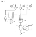
- Fig. 6 schematically shows an apparatus in accordance with the present invention for detecting malaria parasite.
- the dispenser includes an aspiration pump (1), a sampling valve (2), and an aspiration pipette for a sample (3).
- a reaction chamber can be employed as the means for preparing the sample.
- the flow cytometer comprises a flow cell (13), a detector for forward scattering light (17), a detector for fluorescence (18), and a data processor (19).
- the supplier comprises a first lysing agent container (5), a second lysing agent container (6), a staining solution container (7), and aspiration-emission pumps (9) ⁇ (11) connected with each container via tubes.
- a negative pressure pump (14) can be employed as the introduction means.
- the sample of blood in test tube (4) is introduced into the sampling valve (2) through the tube using aspiration pump (1) from the aspiration pipette (3) aspirating the sample.
- the appropriate amount is quantitatively collected.
- the quantified sample of blood is introduced into the reaction chamber (12) via the sampling valve (2) via the aspiration -emission pump (9) via tube.
- the sampling valve in accordance with the present invention is defined to overlap three round-form discs, which may be made of ceramic.
- the sampling valve comprises a mobile element sandwiched between two fixed elements, in which a flow-in route and/or a flow-out route are set for the fixed elements, and a quantitative analysis route is set for the mobile element.
- the sample is passed through the flow-in route, quantification route, and flow-out route in order.
- the reagent flow-in route and/or the flow-out route are set, and the mobile elements are rotated to pass the reagent into the flow-in route, quantification route, and flow-out route in order.
- the quantified sample is flowed out into the reaction chamber with the reagent.
- Such a sampling valve is used in the analytical apparatus for performing blood assay.
- the first lysing agent collected from the first lysing agent container (5) with the aspiration-emission pump (9) is introduced via the tube through the sampling valve (2) and is mixed with the sample of blood in the chamber (12).
- the second lysing solution collected in an appropriate amount from the second lysing container (6) with the aspiration-emission pump (10) is introduced into the chamber (12) and mixed with the blood treated with the first lysing solution.
- the staining solution collected in an appropriate amount from the staining container (7) with the aspiration-emission pump (11) is introduced into the chamber (12) to stain malaria parasite and other material.
- the sample thus obtained is introduced from the chamber (12) to the flow cell (13) via a tube by using the negative pressure pump (14), and scattering light and fluorescence are measured. After measurement, the data for scattering light and fluorescence are detected in the detector for forward scattering light (17) and the detector for fluorescence (18). Then, malaria parasites are distinguished and counted with the data processor (19).
- the present invention further provides a reagent kit for detecting malaria parasites.
- the reagent kit comprises a first hemolytic agent containing a first surfactant, a second hemolytic agent containing a second surfactant and a fluorescent dye for staining malaria parasite, which may be in a two-fluid system containing reagents to be prepared before use or in a three-fluid system.
- a fluorescent dye for staining malaria parasite may be contained in either or both of the first hemolytic agent and the second hemolytic agent, thereby providing a two-fluid system.
- the fluorescent dye may be supplied as a solid to be prepared just prior to use by dissolving it in the first hemolytic agent and/or the second hemolytic agent, thereby providing a two-fluid system.
- the fluorescent dye for staining malaria parasite may be packaged separately from the first hemolytic agent and the second hemolytic agent, thereby providing a three-fluid system. Any types and concentrations of the components contained in these systems (e.g., surfactants, etc.) may be employed in the preparation for enabling the measurement described above.
- a reagent for measuring malaria parasite was prepared as follows.
- polyoxyethylene (40) nonylphenyl ether was mixed with 500 ml of a 120 mM phosphate buffer (pH9) to prepare a first hemolytic agent.
- the osmotic pressure of the first hemolytic agent was 307 mOsm/kg.
- the resultant sample was introduced into a flow cytometer provided with an argon laser with a wavelength of 488 nm as an excitation light source, and examined for forward scattering light intensity and lateral fluorescent light intensity.
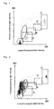
- Fig. 1 The scattergram obtained from the forward scattering light intensity and the lateral fluorescent light intensity is shown in Fig. 1.
- the rectangles A to C indicate the gating of the zones where the malaria parasites (ring form, tropozoite, and schizonte, respectively) appeared, while D indicates the gating of the zone where leukocytes appeared.
- the dotted-lined oval indicates the zone where reticulocytes appeared, while the solid-lined circle indicates the zone where the debris appeared.
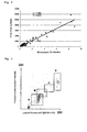
- Fig. 2 The scattergram obtained is shown in Fig. 2.
- the rectangles A to C indicate the zones where the malaria parasites (ring form, tropozoite and schizonte, respectively) appeared, while D indicates the zone where leukocytes appeared.
- the dotted-lined oval exhibited a large number of reticulocytes which had not undergone sufficient hemolysis.
- the concentration of the surfactant was increased, for example, up to 1000 mg to 10 g as LTAC and the measurement was performed with no change in other compositions.
- a higher concentration enabled the hemolysis of the reticulocytes but also dissolved the parasites, resulting in a reduction in both the scattering light intensity and the fluorescent light intensity, which caused overlapping with erythrocyte debris.
- Method B a large number of the malaria-infected specimens the % infections of which had been verified microscopically were measured by the method of Example 1 above (hereinafter referred to as Method B), and the correlation between the % infection and the number of plots appearing in the ring form zone was obtained. The correlation is shown in Fig. 3.
- Method A the specimen of a patient who had been verified to have no malaria was measured by the method described in Example 1 of Japanese Unexamined Patent Publication No. 11-75892 (hereinafter referred to as Method A) and Method B.
- Method B The numbers of the plots in the ring form zones on the scattergrams were compared with each other.
- Method A when the detection sensitivity is assumed to correspond to 0.1% infection, Method A appears to give a false indication that 52.5% of the 137 specimens which were actually not infected with malaria were malaria-infected specimens.
- Method B gave only 4.1% as the ratio of specimens mistakenly judged to be malaria-infected specimens, thus showing a substantially improved measurement accuracy.
- the malaria parasites appear together as a population within a certain zone.
- the peripheral blood specimen of a malaria-infected patient was measured by the method in Example 1 described above, and a scattergram was obtained. The scattergram is shown in Fig. 4.
- Fig. 4 the ordinate represents the forward scattering light intensity, while the abscissa represents the lateral fluorescent light intensity.
- Each detection intensity range of the forward scattering light intensity and the lateral fluorescent light intensity was divided into 255 sections and the coordinates were scaled.
- the population of the malaria parasites appears in the range of the fluorescent light intensity corresponding to the zone where the malaria parasites appear (coordinates 65 to 210), exhibiting a mount-like shape.
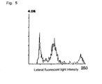
- Fig. 5 shows the histogram corresponding to the scattergram in Fig. 4, in which the ordinate represents the frequency (%) and the abscissa represents the fluorescent light intensity.
- the population of the malaria parasite appearing in the histogram was revealed to be within the range from 30 to 80 as the width of this population at the half of the peak height (hereinafter referred to as half-peak width).
- Method B enabled malaria detection in specimens having a lower % infection than is possible with Method A.
- the present invention enables rapid and specific detection of malaria parasites in samples without requiring complicated operations (e.g., centrifugation).
Landscapes
- Health & Medical Sciences (AREA)
- Life Sciences & Earth Sciences (AREA)
- Immunology (AREA)
- Engineering & Computer Science (AREA)
- Hematology (AREA)
- Chemical & Material Sciences (AREA)
- Urology & Nephrology (AREA)
- Biomedical Technology (AREA)
- Molecular Biology (AREA)
- Microbiology (AREA)
- Physics & Mathematics (AREA)
- Biotechnology (AREA)
- Virology (AREA)
- Tropical Medicine & Parasitology (AREA)
- Food Science & Technology (AREA)
- Medicinal Chemistry (AREA)
- Cell Biology (AREA)
- Analytical Chemistry (AREA)
- Biochemistry (AREA)
- General Health & Medical Sciences (AREA)
- General Physics & Mathematics (AREA)
- Pathology (AREA)
- Investigating Or Analysing Biological Materials (AREA)
Applications Claiming Priority (2)
| Application Number | Priority Date | Filing Date | Title |
|---|---|---|---|
| JP2002292612 | 2002-10-04 | ||
| JP2002292612 | 2002-10-04 |
Publications (2)
| Publication Number | Publication Date |
|---|---|
| EP1406088A2 true EP1406088A2 (de) | 2004-04-07 |
| EP1406088A3 EP1406088A3 (de) | 2004-04-14 |
Family
ID=31987221
Family Applications (1)
| Application Number | Title | Priority Date | Filing Date |
|---|---|---|---|
| EP03022501A Withdrawn EP1406088A3 (de) | 2002-10-04 | 2003-10-02 | Reagenzsatz und Verfahren zum Nachweis von Malariaparasiten, wobei eine Hämolyse in zwei Schritten mittels Tensiden durchgeführt wird |
Country Status (2)
| Country | Link |
|---|---|
| US (1) | US7236236B2 (de) |
| EP (1) | EP1406088A3 (de) |
Cited By (7)
| Publication number | Priority date | Publication date | Assignee | Title |
|---|---|---|---|---|
| EP1715345A1 (de) * | 2005-03-29 | 2006-10-25 | Sysmex Corporation | Reagens zur teilweisen Auflösung der Zellmembran einer roten Blutzelle, Reagens zur Erkennung von mit Malaria infizierten roten Blutkörperchen, und Probenanalyseverfahren zur Erkennung von mit Malaria infizierten roten Blutkörperchen. |
| CN102016573A (zh) * | 2008-05-09 | 2011-04-13 | 希森美康株式会社 | 血液分析装置,血液分析方法,溶血剂及染色剂 |
| EP2677317A1 (de) * | 2012-06-20 | 2013-12-25 | ARKRAY, Inc. | Verfahren zur Behandlung eines eine Probe enthaltenden Blutbestandteils |
| CN105372216A (zh) * | 2014-08-08 | 2016-03-02 | 希森美康株式会社 | 血液分析方法及血液分析装置 |
| EP3276330A1 (de) * | 2016-07-25 | 2018-01-31 | Sysmex Corporation | Blutanalysator und blutanalyseverfahren |
| WO2022115982A1 (zh) | 2020-12-01 | 2022-06-09 | 深圳迈瑞生物医疗电子股份有限公司 | 样本分析方法、样本分析仪及计算机可读存储介质 |
| WO2025138090A1 (zh) * | 2023-12-29 | 2025-07-03 | 深圳迈瑞动物医疗科技股份有限公司 | 血液细胞分析仪和血液细胞分析方法 |
Families Citing this family (9)
| Publication number | Priority date | Publication date | Assignee | Title |
|---|---|---|---|---|
| WO2005088301A1 (en) * | 2004-02-27 | 2005-09-22 | Beckman Coulter, Inc. | Method for detection of malaria and other parasite infections |
| JP2007024844A (ja) | 2005-07-21 | 2007-02-01 | Sysmex Corp | 血液分析方法及び血液分析装置 |
| US20130096081A1 (en) * | 2011-06-03 | 2013-04-18 | Allergan, Inc. | Dermal filler compositions |
| CN103282574B (zh) | 2011-12-08 | 2015-11-25 | Lg电子株式会社 | 烘干机 |
| US20140186859A1 (en) | 2012-05-09 | 2014-07-03 | Advanced Animal Diagnostic, Inc. | Autofocus method for imaging a biological sample and cartridge for use therein |
| SE537208C2 (sv) | 2012-12-03 | 2015-03-03 | Tommy Forsell | Blodanalysapparat för malariaanalys |
| JP6635720B2 (ja) * | 2015-08-31 | 2020-01-29 | シスメックス株式会社 | 血液分析装置及び血液分析方法 |
| JP7425597B2 (ja) * | 2019-12-25 | 2024-01-31 | シスメックス株式会社 | 血液分析方法 |
| EP4222479A1 (de) | 2020-09-29 | 2023-08-09 | Universidade do Minho | Automatische vorrichtung zur nichtinvasiven malariadiagnose durch optische reflexionstechniken, verfahren und verwendungen davon |
Family Cites Families (4)
| Publication number | Priority date | Publication date | Assignee | Title |
|---|---|---|---|---|
| GB2270752B (en) | 1992-09-16 | 1996-04-10 | Azim Fazal | Diagnostic method |
| US5830764A (en) * | 1996-11-12 | 1998-11-03 | Bayer Corporation | Methods and reagent compositions for the determination of membrane surface area and sphericity of erythrocytes and reticulocytes for the diagnosis of red blood cell disorders |
| JP4063363B2 (ja) * | 1997-09-16 | 2008-03-19 | シスメックス株式会社 | マラリア原虫の検出方法 |
| JP3886271B2 (ja) * | 1998-11-27 | 2007-02-28 | シスメックス株式会社 | 赤芽球の分類計数用試薬及び分類計数方法 |
-
2003
- 2003-10-02 EP EP03022501A patent/EP1406088A3/de not_active Withdrawn
- 2003-10-03 US US10/678,536 patent/US7236236B2/en not_active Expired - Fee Related
Cited By (24)
| Publication number | Priority date | Publication date | Assignee | Title |
|---|---|---|---|---|
| EP1715345A1 (de) * | 2005-03-29 | 2006-10-25 | Sysmex Corporation | Reagens zur teilweisen Auflösung der Zellmembran einer roten Blutzelle, Reagens zur Erkennung von mit Malaria infizierten roten Blutkörperchen, und Probenanalyseverfahren zur Erkennung von mit Malaria infizierten roten Blutkörperchen. |
| US7354775B2 (en) | 2005-03-29 | 2008-04-08 | Sysmex Corporation | Reagent for partially lysing a cell membrane of a red blood cell, a reagent for detecting malaria infected red blood cells, and a sample analyzing method for detecting malaria infected red blood cells |
| US9328375B2 (en) | 2008-05-09 | 2016-05-03 | Sysmex Corporation | Blood analyzer, blood analysis method, hemolytic agent and staining agent |
| EP2889620A1 (de) * | 2008-05-09 | 2015-07-01 | Sysmex Corporation | Blutanalysator, Blutanalyseverfahren, Hämolytikum und Färbemittel |
| CN102016573A (zh) * | 2008-05-09 | 2011-04-13 | 希森美康株式会社 | 血液分析装置,血液分析方法,溶血剂及染色剂 |
| EP2293062A4 (de) * | 2008-05-09 | 2011-05-25 | Sysmex Corp | Blutanalysator, blutanalyseverfahren, hämolytikum und färbemittel |
| CN102016573B (zh) * | 2008-05-09 | 2014-11-12 | 希森美康株式会社 | 血液分析装置,血液分析方法,溶血剂及染色剂 |
| US8920726B2 (en) | 2008-05-09 | 2014-12-30 | Sysmex Corporation | Blood analyzer, blood analysis method, hemolytic agent and staining agent |
| CN104297463A (zh) * | 2008-05-09 | 2015-01-21 | 希森美康株式会社 | 血液分析装置,血液分析方法,溶血剂及染色剂 |
| US9429501B2 (en) | 2012-06-20 | 2016-08-30 | Arkray, Inc. | Method for treating a blood component containing sample |
| US9816994B2 (en) | 2012-06-20 | 2017-11-14 | Arkray, Inc. | Method for treating a blood component containing sample |
| CN103513021A (zh) * | 2012-06-20 | 2014-01-15 | 爱科来株式会社 | 含血液成分的试样的处理方法 |
| EP2677317A1 (de) * | 2012-06-20 | 2013-12-25 | ARKRAY, Inc. | Verfahren zur Behandlung eines eine Probe enthaltenden Blutbestandteils |
| CN103513021B (zh) * | 2012-06-20 | 2016-02-17 | 爱科来株式会社 | 含血液成分的试样的处理方法 |
| CN109916802A (zh) * | 2014-08-08 | 2019-06-21 | 希森美康株式会社 | 血液分析方法及血液分析装置 |
| CN105372216A (zh) * | 2014-08-08 | 2016-03-02 | 希森美康株式会社 | 血液分析方法及血液分析装置 |
| CN109916802B (zh) * | 2014-08-08 | 2022-05-27 | 希森美康株式会社 | 血液分析方法及血液分析装置 |
| US10241048B2 (en) | 2014-08-08 | 2019-03-26 | Sysmex Corporation | Blood analyzing method and blood analyzer |
| CN107655865A (zh) * | 2016-07-25 | 2018-02-02 | 希森美康株式会社 | 血液分析装置及血液分析方法 |
| US10451638B2 (en) | 2016-07-25 | 2019-10-22 | Sysmex Corporation | Blood based Plasmodium infection analyzer |
| CN107655865B (zh) * | 2016-07-25 | 2021-10-19 | 希森美康株式会社 | 血液分析装置及血液分析方法 |
| EP3276330A1 (de) * | 2016-07-25 | 2018-01-31 | Sysmex Corporation | Blutanalysator und blutanalyseverfahren |
| WO2022115982A1 (zh) | 2020-12-01 | 2022-06-09 | 深圳迈瑞生物医疗电子股份有限公司 | 样本分析方法、样本分析仪及计算机可读存储介质 |
| WO2025138090A1 (zh) * | 2023-12-29 | 2025-07-03 | 深圳迈瑞动物医疗科技股份有限公司 | 血液细胞分析仪和血液细胞分析方法 |
Also Published As
| Publication number | Publication date |
|---|---|
| US7236236B2 (en) | 2007-06-26 |
| EP1406088A3 (de) | 2004-04-14 |
| US20040067575A1 (en) | 2004-04-08 |
Similar Documents
| Publication | Publication Date | Title |
|---|---|---|
| AU750108B2 (en) | Fully automated method and reagent composition therefor for rapid identification and characterization of reticulocytes, erythrocytes and platelets in whole blood | |
| US7236236B2 (en) | Methods for detecting malaria parasites and reagent kits therefor | |
| JP3886271B2 (ja) | 赤芽球の分類計数用試薬及び分類計数方法 | |
| US8367358B2 (en) | Reagent, kit and method for differentiating and counting leukocytes | |
| US12072330B2 (en) | Method for classifying/counting leukocytes, reagent kit for classifying leukocytes, and reagent for classifying leukocytes | |
| US7892841B2 (en) | Method and apparatus for measuring hematological sample | |
| US5858667A (en) | Method for the enumeration of micronucleated erythrocyte populations with a single laser flow cytometer | |
| US20030219850A1 (en) | Method for automatically analyzing nucleated bone marrow cells | |
| US6100038A (en) | Method for the enumeration of micronucleated erythrocyte populations with a single laser flow cytometer | |
| US6911313B2 (en) | Process for discriminating and counting erythroblasts | |
| JPH11508353A (ja) | 網状赤血球を迅速に分析するための組成物及び方法 | |
| JP4567082B2 (ja) | 赤芽球の分類計数方法 | |
| US20060223137A1 (en) | Reagent for partially lysing a cell membrane of a red blood cell, a reagent for detecting malaria infected red blood cells, and a sample analyzing method for detecting malaria infected red blood cells | |
| JP4388779B2 (ja) | マラリア原虫の検出方法、検出装置、およびその試薬キット | |
| US8597952B2 (en) | Reagent, reagent kit and analyzing method | |
| JP4063363B2 (ja) | マラリア原虫の検出方法 | |
| JP3673137B2 (ja) | 精子計数方法及び試薬 | |
| KR101993965B1 (ko) | 소변 시료 분석 방법, 소변 시료 분석용 시약 및 소변 시료 분석용 시약 키트 | |
| KR20160126066A (ko) | 소변 시료 분석 방법, 소변 시료 분석용 시약 및 소변 시료 분석용 시약 키트 | |
| JP3485436B2 (ja) | 網状赤血球測定用試薬及び測定方法 | |
| JP4794414B2 (ja) | 血小板測定用試薬、血小板測定用試薬キット並びに血小板測定方法 | |
| CN120721608A (zh) | 尿试样的分析方法及尿试样分析用试剂 |
Legal Events
| Date | Code | Title | Description |
|---|---|---|---|
| PUAI | Public reference made under article 153(3) epc to a published international application that has entered the european phase |
Free format text: ORIGINAL CODE: 0009012 |
|
| PUAL | Search report despatched |
Free format text: ORIGINAL CODE: 0009013 |
|
| AK | Designated contracting states |
Kind code of ref document: A2 Designated state(s): AT BE BG CH CY CZ DE DK EE ES FI FR GB GR HU IE IT LI LU MC NL PT RO SE SI SK TR |
|
| AX | Request for extension of the european patent |
Extension state: AL LT LV MK |
|
| AK | Designated contracting states |
Kind code of ref document: A3 Designated state(s): AT BE BG CH CY CZ DE DK EE ES FI FR GB GR HU IE IT LI LU MC NL PT RO SE SI SK TR |
|
| AX | Request for extension of the european patent |
Extension state: AL LT LV MK |
|
| 17P | Request for examination filed |
Effective date: 20040601 |
|
| AKX | Designation fees paid |
Designated state(s): AT BE BG CH CY CZ DE DK EE ES FI FR GB GR HU IE IT LI LU MC NL PT RO SE SI SK TR |
|
| STAA | Information on the status of an ep patent application or granted ep patent |
Free format text: STATUS: THE APPLICATION IS DEEMED TO BE WITHDRAWN |
|
| 18D | Application deemed to be withdrawn |
Effective date: 20081208 |