EP1406202A2 - Medizinisches Bildverwaltungssystem und Vorrichtung - Google Patents
Medizinisches Bildverwaltungssystem und Vorrichtung Download PDFInfo
- Publication number
- EP1406202A2 EP1406202A2 EP03255577A EP03255577A EP1406202A2 EP 1406202 A2 EP1406202 A2 EP 1406202A2 EP 03255577 A EP03255577 A EP 03255577A EP 03255577 A EP03255577 A EP 03255577A EP 1406202 A2 EP1406202 A2 EP 1406202A2
- Authority
- EP
- European Patent Office
- Prior art keywords
- image
- medical image
- medical
- information
- generating apparatus
- Prior art date
- Legal status (The legal status is an assumption and is not a legal conclusion. Google has not performed a legal analysis and makes no representation as to the accuracy of the status listed.)
- Withdrawn
Links
- 238000012545 processing Methods 0.000 claims abstract description 84
- 238000004891 communication Methods 0.000 claims description 5
- 238000012790 confirmation Methods 0.000 description 17
- 238000000034 method Methods 0.000 description 10
- 230000006870 function Effects 0.000 description 6
- 238000010586 diagram Methods 0.000 description 4
- 238000002591 computed tomography Methods 0.000 description 3
- 230000036544 posture Effects 0.000 description 3
- 238000012937 correction Methods 0.000 description 2
- 238000002595 magnetic resonance imaging Methods 0.000 description 2
- 238000003672 processing method Methods 0.000 description 2
- 238000002601 radiography Methods 0.000 description 2
- OAICVXFJPJFONN-UHFFFAOYSA-N Phosphorus Chemical compound [P] OAICVXFJPJFONN-UHFFFAOYSA-N 0.000 description 1
- 210000001015 abdomen Anatomy 0.000 description 1
- 230000005540 biological transmission Effects 0.000 description 1
- 230000015572 biosynthetic process Effects 0.000 description 1
- 201000010099 disease Diseases 0.000 description 1
- 208000037265 diseases, disorders, signs and symptoms Diseases 0.000 description 1
- 230000005284 excitation Effects 0.000 description 1
- 210000003128 head Anatomy 0.000 description 1
- 239000004973 liquid crystal related substance Substances 0.000 description 1
- 210000004072 lung Anatomy 0.000 description 1
- 238000011017 operating method Methods 0.000 description 1
- 230000005855 radiation Effects 0.000 description 1
- 238000012552 review Methods 0.000 description 1
- 238000013519 translation Methods 0.000 description 1
- 238000009966 trimming Methods 0.000 description 1
- 238000002604 ultrasonography Methods 0.000 description 1
Images



Classifications
-
- G—PHYSICS
- G16—INFORMATION AND COMMUNICATION TECHNOLOGY [ICT] SPECIALLY ADAPTED FOR SPECIFIC APPLICATION FIELDS
- G16H—HEALTHCARE INFORMATICS, i.e. INFORMATION AND COMMUNICATION TECHNOLOGY [ICT] SPECIALLY ADAPTED FOR THE HANDLING OR PROCESSING OF MEDICAL OR HEALTHCARE DATA
- G16H30/00—ICT specially adapted for the handling or processing of medical images
- G16H30/20—ICT specially adapted for the handling or processing of medical images for handling medical images, e.g. DICOM, HL7 or PACS
-
- G—PHYSICS
- G16—INFORMATION AND COMMUNICATION TECHNOLOGY [ICT] SPECIALLY ADAPTED FOR SPECIFIC APPLICATION FIELDS
- G16H—HEALTHCARE INFORMATICS, i.e. INFORMATION AND COMMUNICATION TECHNOLOGY [ICT] SPECIALLY ADAPTED FOR THE HANDLING OR PROCESSING OF MEDICAL OR HEALTHCARE DATA
- G16H40/00—ICT specially adapted for the management or administration of healthcare resources or facilities; ICT specially adapted for the management or operation of medical equipment or devices
- G16H40/60—ICT specially adapted for the management or administration of healthcare resources or facilities; ICT specially adapted for the management or operation of medical equipment or devices for the operation of medical equipment or devices
- G16H40/67—ICT specially adapted for the management or administration of healthcare resources or facilities; ICT specially adapted for the management or operation of medical equipment or devices for the operation of medical equipment or devices for remote operation
Definitions
- the invention relates to an image management apparatus and a medical network system capable of receiving a medical image from a medical image generating apparatus, image-processing the received medical image, displaying the image-processed medical image and accompanying information, and outputting them to at least one of an image forming apparatus and an image storage apparatus.
- an image management apparatus managing medical images such as radiographic images or the like receives the medical images such as radiographic images or the like, and displays the received medical images. Further, the image management apparatus processes and converts the medical images into images suitable for image-storing and image-forming to be outputted to at least one of an image storage apparatus and an image forming apparatus that are located externally.
- An object of the present invention is to provide an image management apparatus and a medical network system capable of receiving image information which has been adjusted or corrected by a medical image generating apparatus in any one of a case that quality of a medical image received from a medical image generating apparatus is bad, and a case that accompanying information of the received medical image is not sufficient.
- an image management apparatus capable of receiving a medical image from a medical image generating apparatus according to a radiographing order issued by an outer information management apparatus, image-processing the medical image received, displaying the medical image image-processed along with accompanying information thereof and outputting the medical image and the accompanying information to at least one of an image forming apparatus and an image storage apparatus, comprises: a request section for requesting at least one of the medical image generating apparatus and the outer information management apparatus to have the medical image generating apparatus at least one of re-generate and re-send the medical image, in a case of being impossible to apply image processing suitable for at least one of image-forming in the image forming apparatus and image-storing in the image storage apparatus on the medical image received.
- a medical network system comprises: an image management apparatus, a medical image generating apparatus for sending a medical image to the image management apparatus, at least one of an image storage apparatus for storing the medical image outputted from the image management apparatus and an image forming apparatus for forming the medical image outputted in a recording medium therein, and an information management apparatus for issuing a radiographing order to the medical image generating apparatus and managing information, wherein the image management apparatus comprises a request section for requesting at least one of the medical image generating apparatus and the information management apparatus to have the medical image generating apparatus at least one of re-generate and re-send the medical image, in a case of being impossible to apply image processing suitable for at least one of image-forming in the image forming apparatus and image-storing in the image storage apparatus on the medical image received.
- the apparatus of the first aspect of the present invention or the system of the second aspect of the present invention for example, in a case that a medical image suitable for image-forming or image-storing cannot be obtained or the like, since it is possible to request to have the medical image generating apparatus re-generate and re-send the medical image automatically, it is possible to receive image information such as the medical image re-generated with correction or adjustment by the medical image generating apparatus, accompanying information thereof or the like automatically. Consequently, it is possible that the image management apparatus obtains the image information having a quality medical image and sufficient accompanying information. Further, the image management apparatus can receive the re-generated image information automatically without having a user travel to the outer information management apparatus.
- the request section sends additional information so as to have the medical image generating apparatus re-generate the medical image when requesting at least one of the medical image generating apparatus and the outer information management apparatus to have the medical image generating apparatus re-generate the medical image.
- the additional information so as to have the medical image generating apparatus re-generate the medical image includes information for instructing the medical image generating apparatus to send any one of the medical image which already has been processed, and a combination of the medical image which has not been processed and contents of the image processing performed by the medical image generating apparatus.
- the case of being impossible to apply image processing includes at least one of a case that a medical image image-processed by at least one of the medical image generating apparatus, the image management apparatus and the image forming apparatus is not optimum and a case that the accompanying information is insufficient.
- a medical standard network protocol is used for communication for requesting to have the medical image generating apparatus re-generate the medical image.
- FIG. 1 is a view showing a structure of the medical network system according to the embodiment of the present invention.
- An image management apparatus 50 shown in FIG. 1 receives medical images so as to apply image processing on them and either outputs the image-processed medical images to an outer apparatus or displays the image-processed medical images on a screen of an image display 22 (FIG. 2), the medical images generated by scanning a photostimulable phosphor panel accumulating radiation image information of a subject (a patient) with excitation light for emitting stimulated light and photoelectrically converting the stimulated light with a photomultiplier, or the medical images generated by a modality such as a CR (Computed Radiography) or the like obtaining image information with an X-ray flat panel detector.
- a modality such as a CR (Computed Radiography) or the like obtaining image information with an X-ray flat panel detector.
- medical image generating apparatuses 51 and 56 for generating medical images with the CR as mentioned above, medical image generating apparatuses 57 and 59 for generating medical images with a CT (Computed Tomography), a diagnostic terminal 52 in order for a roentgenolotgist to refer to images for diagnoses, a reference terminal 53 for referring to images (not for diagnoses), an image storage apparatus 54 capable of storing image files in an image database, and searching and reading an image from the diagnostic terminal 52, the reference terminal 53 or the like, and an image forming apparatus (an imager, a printer) 55 for outputting image information from either the medical image generating apparatus 51 or the image management apparatus 50 onto a recording medium such as film, paper or the like as a visible image, constitute the medical network system.
- CT Computer Tomography
- diagnostic terminal 52 in order for a roentgenolotgist to refer to images for diagnoses a reference terminal 53 for referring to images (not for diagnoses)
- an image storage apparatus 54 capable of storing image files in an
- each apparatus is connected on-line through a network 10 for sending and receiving information to each other.
- an HIS (Hospital Information System) / RIS (Radiology Information System) 58 for issuing a radiographing order is connected to the network 10 so that the image management apparatus 50 can communicate with the HIS/RIS 58 with a medical standard network protocol (for example, DICOM, HL7 or the like) and request the HIS/RIS 58 to have any one of the medical image generating apparatuses 51, 56, 57 and 59 re-generate the medical image or the like.
- a medical standard network protocol for example, DICOM, HL7 or the like
- medical image generating apparatuses 51, 56, 57, 59, the image storage apparatus 54 and the image forming apparatus 55 it is possible to apply predetermined image processing on each of received medical images by user operation.
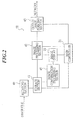
- FIG. 2 is a block diagram showing an internal structure of the image management apparatus 50.
- Image management apparatus 50 Information handled by the image management apparatus 50 can be classified as follows.
- the condition information is necessary for receiving image files and for outputting the received image files to outer apparatuses such as the image storage apparatus 54 or the like as processed image files.
- the condition information includes the following:
- the image processing information concerns gradation processing or frequency processing at the image processing section 42.
- the output apparatus information concerns outer apparatuses such as the image storage apparatus 54 or the like that reproduce and output image data. It specifies an output area, an enlargement/reduction ratio, output format (multi-format, split radiographing format), overlay, whether to conduct gradation processing and frequency processing or the like.
- the overlay information concerns whether to overlay AP/PA, R/L, comments or the like, a position of the overlay or the like.
- Information of postponement To postpone the transmission.
- the information of postponement is specified when it is necessary to review and transmit the image later.
- Information of priority (emergency): The information of priority is specified when preferential output such as emergency radiographing is required. In this case, the radiographing is registered at the top of a queue.
- the patient information concerns a patient.
- the patient ID information includes an ID number, a full name, a sex, a date of birth of a patient or the like.
- the order information is necessary for a doctor to request radiographing.
- the order information includes information concerning patient conditions, instructions of a date and a method for the requested examination or the like.
- the implementation information concerns a result of receiving and image processing.
- the implementation system information includes a part of system information as follows such as a system structure at the time when radiographing is conducted or the like.
- a file handled by the image management apparatus 50 is stored in the storage section 41, and is classified as follows.
- a condition key is a key for setting image process conditions and output conditions on image files in advance, and a condition file corresponding to each condition key is prepared.
- the condition file is composed of the above-mentioned condition information.
- the condition file is classified in terms of radiographic regions (lung, abdomen, head or the like), radiographing postures (erect posture, supine posture or the like), radiographing directions (front, side or the like), characteristics of a patient (sex, age, physique or the like), a disease name, a radiologist or the like, and a name and radiographing information corresponding to each of the above-mentioned classifying factors are set in advance.
- the main controller 21 sets a condition file group corresponding to each of the plurality of classifications, then sets a plurality of condition files for every set condition file group, and stores the plurality of condition files in the storage section 41. Then, the optimum condition is selected at the time of receiving images.
- an image header file is prepared.
- the image header file is composed of a reservation file of the radiographing (namely, radiographing information and patient information) and implementation information.
- radiographing namely, radiographing information and patient information
- implementation information when a user refers to the photographing information, the patient information or the implementation information for changing, the user refers to the image header file.
- the reduced image file is obtained by reducing image data at a certain reduction ratio.
- the reduced image data for display is used as data displayed on the image display 22 in FIG. 2.
- the reduced image data for image processing is used for calculating a parameter in order to conduct image processing. Its reduction ratio is determined by conforming length of one pixel after the reduction to predetermined length. As a result, after the reduction, it is possible to compensate the difference of reading pixel sizes.
- the calculation of a parameter for image processing is not conducted on original image data but conducted on the reduced image data for image processing.
- the output image file is a file of output image data on which predetermined processing among frequency processing, gradation processing, overlay processing, rotation processing and enlargement/reduction processing has been applied.
- the system file is a file of the above-mentioned system information.
- Received images are displayed in thumbnail mode.
- Image files can be displayed as a list
- a screen and operating procedures can be customized to each user.
- FIG. 1 and FIG. 2 operation and a structure of the image management apparatus 50 shown in FIG. 1 and FIG. 2 will be explained with reference to figures, the operation and the structure capable of receiving the image information adjusted or corrected by any one of the medical image generating apparatuses 51, 56, 57 and 59, in a case that quality of a medical image received from any one of the medical image generating apparatuses 51, 56, 57 and 59 is bad or a case that accompanying information regarding the received medical image is not sufficient.
- FIG. 3 is a block diagram showing a control system for controlling the image management apparatus 50 shown in FIG. 1 and FIG. 2 to receive images adjusted or corrected by any one of the medical image generating apparatuses 51, 56, 57 and 59.
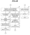
- FIG. 4A and FIG. 4B are flow charts for explaining each step of operation of the image management apparatus 50 shown in FIG. 1 and FIG. 2.
- the image management apparatus 50 shown in FIG. 1 and FIG. 2 comprises a re-generating request section 21a for requesting any one of the medical image generating apparatuses 51, 56, 57, 59 and the HIS/RIS 58 to have any one of the medical image generating apparatuses 51, 56, 57 and 59 re-generate the medical image in a case that it is not possible to apply image processing suitable for at least one of image-forming and image-storing on image information inputted from the storage section 41 for storing the image file received by the receiving section 40 and various types of accompanying information together.
- the image management apparatus 50 can receive image processing information as additional information to have any one of the medical image generating apparatuses 51, 56, 57 and 59 re-generate a medical image through the operation section 21b and send the image processing information along with request information to have any one of the medical image generating apparatuses 51, 56, 57 and 59 re-generate the medical image from the re-generating request section 21a to any one of the medical image generating apparatuses 51, 56, 57, 59 and the HIS/RIS 58.
- FIG. 1 operation of the image management apparatus 50 shown in FIG. 1, FIG. 2 and FIG. 3 will be explained with reference to flow charts in FIG. 4A and FIG. 4B.
- the HIS/RIS 58 issues a radiographing order according to instruction of a doctor or the like to any one of the medical image generating apparatuses 51, 56, 57 and 59 (S1). Based on the radiographing order, for example, the medical image generating apparatus 51 radiographs a medical image and performs necessary image processing (S2). Then, the medical image generating apparatus 51 sends the medical image to the image management apparatus 50 (S3) and the image management apparatus 50 displays the received medical image along with accompanying information thereof on the screen of the image display 22 (S4). Then, the image management apparatus 50 applies image processing on the displayed medical image according to user operation (S5).
- the image management apparatus 50 determines that optimum medical image and sufficient accompanying information are obtained with the above-mentioned image processing and are suitable to be stored in the image storage apparatus 54 (S6;YES), the image management apparatus 50 applies optimum image processing on the optimum medical image (S7) and outputs the image-processed medical image from an output section 43a (FIG. 3) of the output image forming section 43 to the image storage apparatus 54 (S8).
- the image management apparatus 50 when the image management apparatus 50 does not obtain optimum medical image or sufficient accompanying information (S6;NO), the image management apparatus 50 requests any one of the HIS/RIS 58 and the medical image generating apparatus 51 to have the medical image generating apparatus 51 re-generate the image information (S9). In this case, the image management apparatus 50 adds image processing information for re-generating or re-radiographing so as to obtain an optimum medical image and sufficient accompanying information, and modifies the image processing information that already has been obtained as necessary.
- a user confirms that the accompanying information is sufficient on the screen.
- the accompanying information can be corrected after the image management apparatus 50 corrects the accompanying information according to operation at the operation section 21b, the user confirms whether or not the accompanying information is sufficient.
- DICOM which is a medical image standard network protocol, is used for communication to request the HIS/RIS 58 to have any one of the medical image generating apparatuses 51, 56, 57 and 59 re-generate the image information.
- the image management apparatus 50 may request the medical image generating apparatus 51 by sending additional information for instructing to send a combination of an unprocessed medical image and contents of image processing performed by the medical image generating apparatus 51 in Step S9, instead of instructing to re-generate the medical image.
- the image management apparatus 50 applies image processing on the unprocessed medical image in comparison with the contents of image processing performed by the medical image generating apparatus 51.
- the medical image in the image management apparatus 50 may be either deleted or stored for awhile to be deleted later after the request to re-generate the medical image.
- Step S9 in a case that the image management apparatus 50 sends a request to the HIS/RIS 58 to have any one of the medical image generating apparatuses 51, 56, 57 and 59 re-generate the medical image (S10), based on the request of re-generation, the HIS/RIS 58 automatically generates a re-radiographing order and issues it to the medical image generating apparatus 51 (S11). Based on the re-radiographing order, the medical image generating apparatus 51 radiographs a medical image and performs necessary image processing (S12). Then, the medical image generating apparatus 51 sends the medical image along with accompanying information thereof to the image management apparatus 50 (S13).
- the medical image generating apparatus 51 corrects the medical image such as re-performs image processing or the like (S15) and sends the corrected medical image to the image management apparatus 50 (S16).
- Step S3 the image management apparatus 50 obtains optimum medical image and sufficient accompanying information at last by returning to Step S3 as mentioned above in which the image management apparatus 50 receives the medical image along with accompanying information thereof sent in Step S13 or S16 as mentioned above and then performing the steps following Step S3 (S6)
- the image management apparatus 50 applies optimum image processing on the optimum medical image (S7) and outputs the image-processed medical image to the image storage apparatus 54 (S8).
- Step S9 the request to re-generate may always be sent to the HIS/RIS 58 in order for any one of the medical image generating apparatuses 51, 56, 57 and 59 to correct or re-generate the medical image according to need.
- Step S8 the medical image may be outputted to the image forming apparatus 55.
- the image management apparatus 50 confirms whether or not the medical image and accompanying information thereof are suitable for image-forming.
- the image management apparatus 50 in a case that when performing image processing on a received medical image, the image management apparatus 50 cannot obtain a medical image suitable for at least one of image-forming and image-storing, the image management apparatus 50 can automatically request any one of the medical image generating apparatuses 51, 56, 57, 59 and the HIS/RIS 58 to have any one of the medical image generating apparatuses 51, 56, 57 and 59 re-generate and re-send the medical image.
- the image management apparatus 50 can automatically receive image information such as a medical image re-generated with correction or adjustment by re-radiographing, accompanying information thereof or the like from any one of the medical image generating apparatuses 51, 56, 57 and 59. As a result, the image management apparatus 50 can obtain the image information having a quality medical image and sufficient accompanying information and output the image information suitable for image-forming or image-storing to the image forming apparatus 55 or the image storage apparatus 54. Further, conveniently, it is possible that the image management apparatus 50 automatically receives re-generated image information without having a user travel to the HIS/RIS 58 that is located externally.
- the storage section 41 of the image management apparatus 50 shown in FIG. 2 and FIG. 3 stores programs for executing each step in the flow charts in FIG. 4A and FIG. 4B as mentioned above, and the main controller 21 can load and execute the programs according to need.
- one of the medical image generating apparatuses 51, 56, 57 and 59 may be a modality other than the above-mentioned CR and CT.
- it may be a medical image generating apparatus for any modality of MRI (Magnetic Resonance Imaging), DR (Digital Radiography), US (Ultrasound) and so on, of course.
- each of these medical image generating apparatuses may be connected to the medical network system of the present invention.
- a protocol such as PPS, Detach or the like may be used for communication for requesting the HIS/RIS 58 to have any one of the medical image generating apparatus 51, 56, 57 and 59 re-generate the medical image.
- FIG. 4A and FIG. 4B the case that optimum medical image and sufficient accompanying information are obtained with image processing applied by the image management apparatus 50 and judged suitable to be stored in the image storage apparatus 54 (S6) has been explained.
- any one of the medical image generating apparatuses 51, 56, 57, 59, the image forming apparatus 55 and the image storage apparatus 54 performs image processing according to user operation.
- the image management apparatus 50 regards it as not suitable in Step S6 and requests to have any one of the medical image generating apparatuses 51, 56, 57 and 59 re-generate the medical image.
Landscapes
- Health & Medical Sciences (AREA)
- Medical Informatics (AREA)
- Radiology & Medical Imaging (AREA)
- Engineering & Computer Science (AREA)
- Epidemiology (AREA)
- General Health & Medical Sciences (AREA)
- Nuclear Medicine, Radiotherapy & Molecular Imaging (AREA)
- Primary Health Care (AREA)
- Public Health (AREA)
- Measuring And Recording Apparatus For Diagnosis (AREA)
- Medical Treatment And Welfare Office Work (AREA)
- Processing Or Creating Images (AREA)
- Apparatus For Radiation Diagnosis (AREA)
- Television Signal Processing For Recording (AREA)
Applications Claiming Priority (2)
| Application Number | Priority Date | Filing Date | Title |
|---|---|---|---|
| JP2002266221A JP2004097652A (ja) | 2002-09-12 | 2002-09-12 | 画像管理装置及び画像管理装置のためのプログラム |
| JP2002266221 | 2002-09-12 |
Publications (2)
| Publication Number | Publication Date |
|---|---|
| EP1406202A2 true EP1406202A2 (de) | 2004-04-07 |
| EP1406202A3 EP1406202A3 (de) | 2004-10-20 |
Family
ID=31986629
Family Applications (1)
| Application Number | Title | Priority Date | Filing Date |
|---|---|---|---|
| EP03255577A Withdrawn EP1406202A3 (de) | 2002-09-12 | 2003-09-08 | Medizinisches Bildverwaltungssystem und Vorrichtung |
Country Status (3)
| Country | Link |
|---|---|
| US (1) | US20040071369A1 (de) |
| EP (1) | EP1406202A3 (de) |
| JP (1) | JP2004097652A (de) |
Cited By (3)
| Publication number | Priority date | Publication date | Assignee | Title |
|---|---|---|---|---|
| EP1953660A1 (de) * | 2006-12-11 | 2008-08-06 | Konica Minolta Medical & Graphic, Inc. | Radiographiesystem und Gerät zur Erkennung von Strahlungsbildern |
| CN101199426B (zh) * | 2006-12-11 | 2012-08-29 | 柯尼卡美能达医疗印刷器材株式会社 | 放射线摄影系统以及放射线图像检测器 |
| CN107341342A (zh) * | 2017-06-15 | 2017-11-10 | 重庆柚瓣家科技有限公司 | 用于养老机器人的药品管理系统 |
Families Citing this family (13)
| Publication number | Priority date | Publication date | Assignee | Title |
|---|---|---|---|---|
| US6954802B2 (en) * | 1998-09-29 | 2005-10-11 | Tdk Electronics Corporation | Removable media recording station for the medical industry |
| US20050240445A1 (en) * | 1998-09-29 | 2005-10-27 | Michael Sutherland | Medical archive library and method |
| US7269303B2 (en) * | 2001-06-04 | 2007-09-11 | Hewlett-Packard Development Company, L.P. | Remote digital image enhancement system and method |
| US20090226065A1 (en) * | 2004-10-09 | 2009-09-10 | Dongqing Chen | Sampling medical images for virtual histology |
| JP5121154B2 (ja) * | 2005-04-08 | 2013-01-16 | キヤノン株式会社 | 画像管理システム及び画像管理方法並びにプログラム |
| US8849068B2 (en) | 2005-04-08 | 2014-09-30 | Canon Kabushiki Kaisha | Image management system, image management method, and program |
| JP2010246862A (ja) * | 2009-03-24 | 2010-11-04 | Japan Health Science Foundation | 医用画像作成装置及びプログラム |
| JP5331044B2 (ja) * | 2010-03-31 | 2013-10-30 | 富士フイルム株式会社 | 放射線画像システム |
| JP2012130369A (ja) * | 2010-12-20 | 2012-07-12 | Konica Minolta Medical & Graphic Inc | 検像装置及び検像システム |
| JP2013045159A (ja) * | 2011-08-22 | 2013-03-04 | Infocom Corp | 警告通知システム、検像システム、データサーバ及びプログラム |
| JP5809015B2 (ja) * | 2011-10-17 | 2015-11-10 | インフォコム株式会社 | 再撮指示情報登録が可能な診療画像撮影業務にかかわるシステム及び再撮指示登録方法 |
| JP5947658B2 (ja) * | 2012-08-06 | 2016-07-06 | キヤノン株式会社 | 制御装置、制御方法、及びプログラム |
| CN104243771A (zh) * | 2014-09-10 | 2014-12-24 | 北京淳中视讯科技有限公司 | 图像拼接处理器及其ip管理方法 |
Family Cites Families (9)
| Publication number | Priority date | Publication date | Assignee | Title |
|---|---|---|---|---|
| US5469353A (en) * | 1993-11-26 | 1995-11-21 | Access Radiology Corp. | Radiological image interpretation apparatus and method |
| US5883985A (en) * | 1996-12-10 | 1999-03-16 | General Electric Company | Method for compensating image data to adjust for characteristics of a network output device |
| IL122361A0 (en) * | 1997-11-29 | 1998-04-05 | Algotec Systems Ltd | Image compression method |
| JPH11239165A (ja) * | 1998-02-20 | 1999-08-31 | Fuji Photo Film Co Ltd | メディカルネットワークシステム |
| JPH11282937A (ja) * | 1998-03-31 | 1999-10-15 | Fuji Photo Film Co Ltd | メディカルネットワークシステム |
| JP4408555B2 (ja) * | 2000-11-21 | 2010-02-03 | 富士フイルム株式会社 | 画像および情報処理装置 |
| DE10105585A1 (de) * | 2001-02-07 | 2003-07-10 | Siemens Ag | Verfahren zum Betrieb eines Magnetresonanzgeräts |
| JP3704492B2 (ja) * | 2001-09-11 | 2005-10-12 | テラリコン・インコーポレイテッド | ネットワーク環境におけるレポートシステム |
| JP2003284692A (ja) * | 2002-03-28 | 2003-10-07 | Konica Corp | 医用画像処理装置、医用画像処理方法、プログラム、及び記録媒体 |
-
2002
- 2002-09-12 JP JP2002266221A patent/JP2004097652A/ja active Pending
-
2003
- 2003-09-08 EP EP03255577A patent/EP1406202A3/de not_active Withdrawn
- 2003-09-08 US US10/657,381 patent/US20040071369A1/en not_active Abandoned
Cited By (4)
| Publication number | Priority date | Publication date | Assignee | Title |
|---|---|---|---|---|
| EP1953660A1 (de) * | 2006-12-11 | 2008-08-06 | Konica Minolta Medical & Graphic, Inc. | Radiographiesystem und Gerät zur Erkennung von Strahlungsbildern |
| US7508915B2 (en) | 2006-12-11 | 2009-03-24 | Konica Minolta Medical & Graphic Inc. | Radiographying system and radiation image detecting device |
| CN101199426B (zh) * | 2006-12-11 | 2012-08-29 | 柯尼卡美能达医疗印刷器材株式会社 | 放射线摄影系统以及放射线图像检测器 |
| CN107341342A (zh) * | 2017-06-15 | 2017-11-10 | 重庆柚瓣家科技有限公司 | 用于养老机器人的药品管理系统 |
Also Published As
| Publication number | Publication date |
|---|---|
| EP1406202A3 (de) | 2004-10-20 |
| US20040071369A1 (en) | 2004-04-15 |
| JP2004097652A (ja) | 2004-04-02 |
Similar Documents
| Publication | Publication Date | Title |
|---|---|---|
| US20040186371A1 (en) | Medical image processing apparatus and medical network system | |
| EP1406202A2 (de) | Medizinisches Bildverwaltungssystem und Vorrichtung | |
| EP1434157A2 (de) | Bildinformationsverarbeitungsgerät und medizinisches Netzwerksystem | |
| US7127097B2 (en) | Image processing apparatus, image processing method, program for executing image processing method, and storage medium that stores program for executing image processing method | |
| US20100171682A1 (en) | Medical image display apparatus and computer readable medium | |
| JP2004097651A (ja) | 画像情報処理装置、医用ネットワークシステム及び画像情報処理装置のためのプログラム | |
| EP1484707A2 (de) | Medizinisches Bildsystem und Verfahren zur Verarbeitung medizinischer Bilder | |
| US20020131628A1 (en) | Medical image generating apparatus, medical image processing apparatus and medical network system | |
| JP2006280717A (ja) | 医用画像システム | |
| JP3530210B2 (ja) | 画像出力制御装置 | |
| US7171612B2 (en) | Medical image processing apparatus and medical network system | |
| JP4337133B2 (ja) | 医用画像処理装置、医用ネットワークシステム及び医用画像処理装置のためのプログラム | |
| JP4288693B2 (ja) | 医用画像処理装置、医用ネットワークシステム及び医用画像処理装置のためのプログラム | |
| US20020131627A1 (en) | Medical image generating apparatus, medical image processing apparatus and medical network system | |
| US7439996B2 (en) | Medical image recording system and medical image recording apparatus | |
| JP2003180636A (ja) | 画像処理装置、医用ネットワークシステム、画像処理方法、画像処理方法の実行のためのプログラム及びプログラムを格納した記憶媒体 | |
| US20050201608A1 (en) | Medical image reproducing system and medical image reproducing method | |
| JP4626165B2 (ja) | 医用画像処理装置 | |
| JP3965684B2 (ja) | 医用画像処理装置、医用ネットワークシステム及び医用画像処理装置のためのプログラム | |
| JP2002279395A (ja) | 医用画像生成装置、医用画像処理装置及び医用ネットワークシステム | |
| JP2004073341A (ja) | 医用画像処理装置、医用ネットワークシステム及び医用画像処理装置のためのプログラム | |
| JP4311057B2 (ja) | 医用画像処理装置、医用ネットワークシステム及び医用画像処理装置のためのプログラム | |
| JP2005224473A (ja) | 医用画像システム、医用画像検像システム及びプログラム | |
| JP4576979B2 (ja) | 乳房画像生成システム | |
| JP2005224472A (ja) | 医用画像システム、医用画像検像システム及びプログラム |
Legal Events
| Date | Code | Title | Description |
|---|---|---|---|
| PUAI | Public reference made under article 153(3) epc to a published international application that has entered the european phase |
Free format text: ORIGINAL CODE: 0009012 |
|
| AK | Designated contracting states |
Kind code of ref document: A2 Designated state(s): AT BE BG CH CY CZ DE DK EE ES FI FR GB GR HU IE IT LI LU MC NL PT RO SE SI SK TR |
|
| AX | Request for extension of the european patent |
Extension state: AL LT LV MK |
|
| PUAL | Search report despatched |
Free format text: ORIGINAL CODE: 0009013 |
|
| AK | Designated contracting states |
Kind code of ref document: A3 Designated state(s): AT BE BG CH CY CZ DE DK EE ES FI FR GB GR HU IE IT LI LU MC NL PT RO SE SI SK TR |
|
| AX | Request for extension of the european patent |
Extension state: AL LT LV MK |
|
| AKX | Designation fees paid | ||
| REG | Reference to a national code |
Ref country code: DE Ref legal event code: 8566 |
|
| STAA | Information on the status of an ep patent application or granted ep patent |
Free format text: STATUS: THE APPLICATION IS DEEMED TO BE WITHDRAWN |
|
| 18D | Application deemed to be withdrawn |
Effective date: 20050421 |