EP1406203A2 - Planung für die orthopädische Chirurgie - Google Patents
Planung für die orthopädische Chirurgie Download PDFInfo
- Publication number
- EP1406203A2 EP1406203A2 EP03255895A EP03255895A EP1406203A2 EP 1406203 A2 EP1406203 A2 EP 1406203A2 EP 03255895 A EP03255895 A EP 03255895A EP 03255895 A EP03255895 A EP 03255895A EP 1406203 A2 EP1406203 A2 EP 1406203A2
- Authority
- EP
- European Patent Office
- Prior art keywords
- geometric parameters
- user
- patient
- anatomical features
- patient image
- Prior art date
- Legal status (The legal status is an assumption and is not a legal conclusion. Google has not performed a legal analysis and makes no representation as to the accuracy of the status listed.)
- Granted
Links
- 238000012829 orthopaedic surgery Methods 0.000 title claims abstract description 33
- 238000000034 method Methods 0.000 claims abstract description 68
- 210000003127 knee Anatomy 0.000 claims abstract description 18
- 210000000689 upper leg Anatomy 0.000 claims description 26
- 210000001624 hip Anatomy 0.000 claims description 20
- 238000004590 computer program Methods 0.000 claims description 18
- 239000007943 implant Substances 0.000 claims description 9
- 210000002303 tibia Anatomy 0.000 claims description 9
- 210000000629 knee joint Anatomy 0.000 claims description 6
- 210000004197 pelvis Anatomy 0.000 claims description 6
- 238000004891 communication Methods 0.000 claims description 5
- 230000005540 biological transmission Effects 0.000 claims description 3
- 238000001356 surgical procedure Methods 0.000 abstract description 20
- 238000011540 hip replacement Methods 0.000 abstract description 11
- 210000000988 bone and bone Anatomy 0.000 description 19
- 238000013150 knee replacement Methods 0.000 description 6
- 238000003384 imaging method Methods 0.000 description 5
- 239000002184 metal Substances 0.000 description 5
- 210000004394 hip joint Anatomy 0.000 description 4
- 210000001503 joint Anatomy 0.000 description 4
- 238000005259 measurement Methods 0.000 description 4
- QTBSBXVTEAMEQO-UHFFFAOYSA-M Acetate Chemical compound CC([O-])=O QTBSBXVTEAMEQO-UHFFFAOYSA-M 0.000 description 3
- 230000008901 benefit Effects 0.000 description 3
- 238000013479 data entry Methods 0.000 description 3
- 238000002601 radiography Methods 0.000 description 3
- 241001227561 Valgus Species 0.000 description 2
- 210000000588 acetabulum Anatomy 0.000 description 2
- 238000013459 approach Methods 0.000 description 2
- 230000009286 beneficial effect Effects 0.000 description 2
- 238000002059 diagnostic imaging Methods 0.000 description 2
- 238000010586 diagram Methods 0.000 description 2
- 238000002595 magnetic resonance imaging Methods 0.000 description 2
- 239000000463 material Substances 0.000 description 2
- 230000003287 optical effect Effects 0.000 description 2
- 239000004033 plastic Substances 0.000 description 2
- 208000035965 Postoperative Complications Diseases 0.000 description 1
- 230000003190 augmentative effect Effects 0.000 description 1
- 239000002775 capsule Substances 0.000 description 1
- 239000004568 cement Substances 0.000 description 1
- 239000000919 ceramic Substances 0.000 description 1
- 230000001010 compromised effect Effects 0.000 description 1
- 238000002591 computed tomography Methods 0.000 description 1
- 238000013500 data storage Methods 0.000 description 1
- 238000006073 displacement reaction Methods 0.000 description 1
- 230000000694 effects Effects 0.000 description 1
- 238000005516 engineering process Methods 0.000 description 1
- 230000007613 environmental effect Effects 0.000 description 1
- 238000002513 implantation Methods 0.000 description 1
- 238000007689 inspection Methods 0.000 description 1
- 238000002372 labelling Methods 0.000 description 1
- 210000002414 leg Anatomy 0.000 description 1
- 238000007726 management method Methods 0.000 description 1
- 238000013507 mapping Methods 0.000 description 1
- 239000003550 marker Substances 0.000 description 1
- 210000004417 patella Anatomy 0.000 description 1
- 238000006467 substitution reaction Methods 0.000 description 1
- 238000003325 tomography Methods 0.000 description 1
- 238000012546 transfer Methods 0.000 description 1
- 210000001364 upper extremity Anatomy 0.000 description 1
- 230000000007 visual effect Effects 0.000 description 1
Images



Classifications
-
- A—HUMAN NECESSITIES
- A61—MEDICAL OR VETERINARY SCIENCE; HYGIENE
- A61F—FILTERS IMPLANTABLE INTO BLOOD VESSELS; PROSTHESES; DEVICES PROVIDING PATENCY TO, OR PREVENTING COLLAPSING OF, TUBULAR STRUCTURES OF THE BODY, e.g. STENTS; ORTHOPAEDIC, NURSING OR CONTRACEPTIVE DEVICES; FOMENTATION; TREATMENT OR PROTECTION OF EYES OR EARS; BANDAGES, DRESSINGS OR ABSORBENT PADS; FIRST-AID KITS
- A61F2/00—Filters implantable into blood vessels; Prostheses, i.e. artificial substitutes or replacements for parts of the body; Appliances for connecting them with the body; Devices providing patency to, or preventing collapsing of, tubular structures of the body, e.g. stents
- A61F2/02—Prostheses implantable into the body
- A61F2/30—Joints
- A61F2/3094—Designing or manufacturing processes
- A61F2/30942—Designing or manufacturing processes for designing or making customized prostheses, e.g. using templates, CT or NMR scans, finite-element analysis or CAD-CAM techniques
-
- A—HUMAN NECESSITIES
- A61—MEDICAL OR VETERINARY SCIENCE; HYGIENE
- A61B—DIAGNOSIS; SURGERY; IDENTIFICATION
- A61B90/00—Instruments, implements or accessories specially adapted for surgery or diagnosis and not covered by any of the groups A61B1/00 - A61B50/00, e.g. for luxation treatment or for protecting wound edges
- A61B90/36—Image-producing devices or illumination devices not otherwise provided for
-
- G—PHYSICS
- G16—INFORMATION AND COMMUNICATION TECHNOLOGY [ICT] SPECIALLY ADAPTED FOR SPECIFIC APPLICATION FIELDS
- G16H—HEALTHCARE INFORMATICS, i.e. INFORMATION AND COMMUNICATION TECHNOLOGY [ICT] SPECIALLY ADAPTED FOR THE HANDLING OR PROCESSING OF MEDICAL OR HEALTHCARE DATA
- G16H20/00—ICT specially adapted for therapies or health-improving plans, e.g. for handling prescriptions, for steering therapy or for monitoring patient compliance
- G16H20/40—ICT specially adapted for therapies or health-improving plans, e.g. for handling prescriptions, for steering therapy or for monitoring patient compliance relating to mechanical, radiation or invasive therapies, e.g. surgery, laser therapy, dialysis or acupuncture
-
- G—PHYSICS
- G16—INFORMATION AND COMMUNICATION TECHNOLOGY [ICT] SPECIALLY ADAPTED FOR SPECIFIC APPLICATION FIELDS
- G16H—HEALTHCARE INFORMATICS, i.e. INFORMATION AND COMMUNICATION TECHNOLOGY [ICT] SPECIALLY ADAPTED FOR THE HANDLING OR PROCESSING OF MEDICAL OR HEALTHCARE DATA
- G16H30/00—ICT specially adapted for the handling or processing of medical images
- G16H30/40—ICT specially adapted for the handling or processing of medical images for processing medical images, e.g. editing
-
- G—PHYSICS
- G16—INFORMATION AND COMMUNICATION TECHNOLOGY [ICT] SPECIALLY ADAPTED FOR SPECIFIC APPLICATION FIELDS
- G16H—HEALTHCARE INFORMATICS, i.e. INFORMATION AND COMMUNICATION TECHNOLOGY [ICT] SPECIALLY ADAPTED FOR THE HANDLING OR PROCESSING OF MEDICAL OR HEALTHCARE DATA
- G16H40/00—ICT specially adapted for the management or administration of healthcare resources or facilities; ICT specially adapted for the management or operation of medical equipment or devices
- G16H40/60—ICT specially adapted for the management or administration of healthcare resources or facilities; ICT specially adapted for the management or operation of medical equipment or devices for the operation of medical equipment or devices
- G16H40/67—ICT specially adapted for the management or administration of healthcare resources or facilities; ICT specially adapted for the management or operation of medical equipment or devices for the operation of medical equipment or devices for remote operation
-
- G—PHYSICS
- G16—INFORMATION AND COMMUNICATION TECHNOLOGY [ICT] SPECIALLY ADAPTED FOR SPECIFIC APPLICATION FIELDS
- G16H—HEALTHCARE INFORMATICS, i.e. INFORMATION AND COMMUNICATION TECHNOLOGY [ICT] SPECIALLY ADAPTED FOR THE HANDLING OR PROCESSING OF MEDICAL OR HEALTHCARE DATA
- G16H50/00—ICT specially adapted for medical diagnosis, medical simulation or medical data mining; ICT specially adapted for detecting, monitoring or modelling epidemics or pandemics
- G16H50/50—ICT specially adapted for medical diagnosis, medical simulation or medical data mining; ICT specially adapted for detecting, monitoring or modelling epidemics or pandemics for simulation or modelling of medical disorders
-
- G—PHYSICS
- G16—INFORMATION AND COMMUNICATION TECHNOLOGY [ICT] SPECIALLY ADAPTED FOR SPECIFIC APPLICATION FIELDS
- G16H—HEALTHCARE INFORMATICS, i.e. INFORMATION AND COMMUNICATION TECHNOLOGY [ICT] SPECIALLY ADAPTED FOR THE HANDLING OR PROCESSING OF MEDICAL OR HEALTHCARE DATA
- G16H70/00—ICT specially adapted for the handling or processing of medical references
- G16H70/20—ICT specially adapted for the handling or processing of medical references relating to practices or guidelines
-
- A—HUMAN NECESSITIES
- A61—MEDICAL OR VETERINARY SCIENCE; HYGIENE
- A61B—DIAGNOSIS; SURGERY; IDENTIFICATION
- A61B17/00—Surgical instruments, devices or methods
- A61B17/14—Surgical saws
- A61B17/15—Guides therefor
-
- A—HUMAN NECESSITIES
- A61—MEDICAL OR VETERINARY SCIENCE; HYGIENE
- A61B—DIAGNOSIS; SURGERY; IDENTIFICATION
- A61B17/00—Surgical instruments, devices or methods
- A61B17/14—Surgical saws
- A61B17/15—Guides therefor
- A61B17/154—Guides therefor for preparing bone for knee prosthesis
-
- A—HUMAN NECESSITIES
- A61—MEDICAL OR VETERINARY SCIENCE; HYGIENE
- A61B—DIAGNOSIS; SURGERY; IDENTIFICATION
- A61B90/00—Instruments, implements or accessories specially adapted for surgery or diagnosis and not covered by any of the groups A61B1/00 - A61B50/00, e.g. for luxation treatment or for protecting wound edges
- A61B90/36—Image-producing devices or illumination devices not otherwise provided for
- A61B2090/364—Correlation of different images or relation of image positions in respect to the body
-
- A—HUMAN NECESSITIES
- A61—MEDICAL OR VETERINARY SCIENCE; HYGIENE
- A61B—DIAGNOSIS; SURGERY; IDENTIFICATION
- A61B90/00—Instruments, implements or accessories specially adapted for surgery or diagnosis and not covered by any of the groups A61B1/00 - A61B50/00, e.g. for luxation treatment or for protecting wound edges
- A61B90/36—Image-producing devices or illumination devices not otherwise provided for
- A61B90/37—Surgical systems with images on a monitor during operation
- A61B2090/376—Surgical systems with images on a monitor during operation using X-rays, e.g. fluoroscopy
-
- A—HUMAN NECESSITIES
- A61—MEDICAL OR VETERINARY SCIENCE; HYGIENE
- A61B—DIAGNOSIS; SURGERY; IDENTIFICATION
- A61B34/00—Computer-aided surgery; Manipulators or robots specially adapted for use in surgery
- A61B34/10—Computer-aided planning, simulation or modelling of surgical operations
-
- A—HUMAN NECESSITIES
- A61—MEDICAL OR VETERINARY SCIENCE; HYGIENE
- A61F—FILTERS IMPLANTABLE INTO BLOOD VESSELS; PROSTHESES; DEVICES PROVIDING PATENCY TO, OR PREVENTING COLLAPSING OF, TUBULAR STRUCTURES OF THE BODY, e.g. STENTS; ORTHOPAEDIC, NURSING OR CONTRACEPTIVE DEVICES; FOMENTATION; TREATMENT OR PROTECTION OF EYES OR EARS; BANDAGES, DRESSINGS OR ABSORBENT PADS; FIRST-AID KITS
- A61F2/00—Filters implantable into blood vessels; Prostheses, i.e. artificial substitutes or replacements for parts of the body; Appliances for connecting them with the body; Devices providing patency to, or preventing collapsing of, tubular structures of the body, e.g. stents
- A61F2/02—Prostheses implantable into the body
- A61F2/30—Joints
- A61F2/32—Joints for the hip
-
- A—HUMAN NECESSITIES
- A61—MEDICAL OR VETERINARY SCIENCE; HYGIENE
- A61F—FILTERS IMPLANTABLE INTO BLOOD VESSELS; PROSTHESES; DEVICES PROVIDING PATENCY TO, OR PREVENTING COLLAPSING OF, TUBULAR STRUCTURES OF THE BODY, e.g. STENTS; ORTHOPAEDIC, NURSING OR CONTRACEPTIVE DEVICES; FOMENTATION; TREATMENT OR PROTECTION OF EYES OR EARS; BANDAGES, DRESSINGS OR ABSORBENT PADS; FIRST-AID KITS
- A61F2/00—Filters implantable into blood vessels; Prostheses, i.e. artificial substitutes or replacements for parts of the body; Appliances for connecting them with the body; Devices providing patency to, or preventing collapsing of, tubular structures of the body, e.g. stents
- A61F2/02—Prostheses implantable into the body
- A61F2/30—Joints
- A61F2/32—Joints for the hip
- A61F2/34—Acetabular cups
-
- A—HUMAN NECESSITIES
- A61—MEDICAL OR VETERINARY SCIENCE; HYGIENE
- A61F—FILTERS IMPLANTABLE INTO BLOOD VESSELS; PROSTHESES; DEVICES PROVIDING PATENCY TO, OR PREVENTING COLLAPSING OF, TUBULAR STRUCTURES OF THE BODY, e.g. STENTS; ORTHOPAEDIC, NURSING OR CONTRACEPTIVE DEVICES; FOMENTATION; TREATMENT OR PROTECTION OF EYES OR EARS; BANDAGES, DRESSINGS OR ABSORBENT PADS; FIRST-AID KITS
- A61F2/00—Filters implantable into blood vessels; Prostheses, i.e. artificial substitutes or replacements for parts of the body; Appliances for connecting them with the body; Devices providing patency to, or preventing collapsing of, tubular structures of the body, e.g. stents
- A61F2/02—Prostheses implantable into the body
- A61F2/30—Joints
- A61F2/32—Joints for the hip
- A61F2/36—Femoral heads ; Femoral endoprostheses
-
- A—HUMAN NECESSITIES
- A61—MEDICAL OR VETERINARY SCIENCE; HYGIENE
- A61F—FILTERS IMPLANTABLE INTO BLOOD VESSELS; PROSTHESES; DEVICES PROVIDING PATENCY TO, OR PREVENTING COLLAPSING OF, TUBULAR STRUCTURES OF THE BODY, e.g. STENTS; ORTHOPAEDIC, NURSING OR CONTRACEPTIVE DEVICES; FOMENTATION; TREATMENT OR PROTECTION OF EYES OR EARS; BANDAGES, DRESSINGS OR ABSORBENT PADS; FIRST-AID KITS
- A61F2/00—Filters implantable into blood vessels; Prostheses, i.e. artificial substitutes or replacements for parts of the body; Appliances for connecting them with the body; Devices providing patency to, or preventing collapsing of, tubular structures of the body, e.g. stents
- A61F2/02—Prostheses implantable into the body
- A61F2/30—Joints
- A61F2/32—Joints for the hip
- A61F2/36—Femoral heads ; Femoral endoprostheses
- A61F2/3662—Femoral shafts
- A61F2/367—Proximal or metaphyseal parts of shafts
-
- A—HUMAN NECESSITIES
- A61—MEDICAL OR VETERINARY SCIENCE; HYGIENE
- A61F—FILTERS IMPLANTABLE INTO BLOOD VESSELS; PROSTHESES; DEVICES PROVIDING PATENCY TO, OR PREVENTING COLLAPSING OF, TUBULAR STRUCTURES OF THE BODY, e.g. STENTS; ORTHOPAEDIC, NURSING OR CONTRACEPTIVE DEVICES; FOMENTATION; TREATMENT OR PROTECTION OF EYES OR EARS; BANDAGES, DRESSINGS OR ABSORBENT PADS; FIRST-AID KITS
- A61F2/00—Filters implantable into blood vessels; Prostheses, i.e. artificial substitutes or replacements for parts of the body; Appliances for connecting them with the body; Devices providing patency to, or preventing collapsing of, tubular structures of the body, e.g. stents
- A61F2/02—Prostheses implantable into the body
- A61F2/30—Joints
- A61F2/38—Joints for elbows or knees
-
- A—HUMAN NECESSITIES
- A61—MEDICAL OR VETERINARY SCIENCE; HYGIENE
- A61F—FILTERS IMPLANTABLE INTO BLOOD VESSELS; PROSTHESES; DEVICES PROVIDING PATENCY TO, OR PREVENTING COLLAPSING OF, TUBULAR STRUCTURES OF THE BODY, e.g. STENTS; ORTHOPAEDIC, NURSING OR CONTRACEPTIVE DEVICES; FOMENTATION; TREATMENT OR PROTECTION OF EYES OR EARS; BANDAGES, DRESSINGS OR ABSORBENT PADS; FIRST-AID KITS
- A61F2/00—Filters implantable into blood vessels; Prostheses, i.e. artificial substitutes or replacements for parts of the body; Appliances for connecting them with the body; Devices providing patency to, or preventing collapsing of, tubular structures of the body, e.g. stents
- A61F2/02—Prostheses implantable into the body
- A61F2/30—Joints
- A61F2/38—Joints for elbows or knees
- A61F2/3859—Femoral components
-
- A—HUMAN NECESSITIES
- A61—MEDICAL OR VETERINARY SCIENCE; HYGIENE
- A61F—FILTERS IMPLANTABLE INTO BLOOD VESSELS; PROSTHESES; DEVICES PROVIDING PATENCY TO, OR PREVENTING COLLAPSING OF, TUBULAR STRUCTURES OF THE BODY, e.g. STENTS; ORTHOPAEDIC, NURSING OR CONTRACEPTIVE DEVICES; FOMENTATION; TREATMENT OR PROTECTION OF EYES OR EARS; BANDAGES, DRESSINGS OR ABSORBENT PADS; FIRST-AID KITS
- A61F2/00—Filters implantable into blood vessels; Prostheses, i.e. artificial substitutes or replacements for parts of the body; Appliances for connecting them with the body; Devices providing patency to, or preventing collapsing of, tubular structures of the body, e.g. stents
- A61F2/02—Prostheses implantable into the body
- A61F2/30—Joints
- A61F2/38—Joints for elbows or knees
- A61F2/389—Tibial components
-
- A—HUMAN NECESSITIES
- A61—MEDICAL OR VETERINARY SCIENCE; HYGIENE
- A61F—FILTERS IMPLANTABLE INTO BLOOD VESSELS; PROSTHESES; DEVICES PROVIDING PATENCY TO, OR PREVENTING COLLAPSING OF, TUBULAR STRUCTURES OF THE BODY, e.g. STENTS; ORTHOPAEDIC, NURSING OR CONTRACEPTIVE DEVICES; FOMENTATION; TREATMENT OR PROTECTION OF EYES OR EARS; BANDAGES, DRESSINGS OR ABSORBENT PADS; FIRST-AID KITS
- A61F2/00—Filters implantable into blood vessels; Prostheses, i.e. artificial substitutes or replacements for parts of the body; Appliances for connecting them with the body; Devices providing patency to, or preventing collapsing of, tubular structures of the body, e.g. stents
- A61F2/02—Prostheses implantable into the body
- A61F2/30—Joints
- A61F2002/30001—Additional features of subject-matter classified in A61F2/28, A61F2/30 and subgroups thereof
- A61F2002/30003—Material related properties of the prosthesis or of a coating on the prosthesis
- A61F2002/3006—Properties of materials and coating materials
- A61F2002/3008—Properties of materials and coating materials radio-opaque, e.g. radio-opaque markers
-
- A—HUMAN NECESSITIES
- A61—MEDICAL OR VETERINARY SCIENCE; HYGIENE
- A61F—FILTERS IMPLANTABLE INTO BLOOD VESSELS; PROSTHESES; DEVICES PROVIDING PATENCY TO, OR PREVENTING COLLAPSING OF, TUBULAR STRUCTURES OF THE BODY, e.g. STENTS; ORTHOPAEDIC, NURSING OR CONTRACEPTIVE DEVICES; FOMENTATION; TREATMENT OR PROTECTION OF EYES OR EARS; BANDAGES, DRESSINGS OR ABSORBENT PADS; FIRST-AID KITS
- A61F2/00—Filters implantable into blood vessels; Prostheses, i.e. artificial substitutes or replacements for parts of the body; Appliances for connecting them with the body; Devices providing patency to, or preventing collapsing of, tubular structures of the body, e.g. stents
- A61F2/02—Prostheses implantable into the body
- A61F2/30—Joints
- A61F2002/30001—Additional features of subject-matter classified in A61F2/28, A61F2/30 and subgroups thereof
- A61F2002/30316—The prosthesis having different structural features at different locations within the same prosthesis; Connections between prosthetic parts; Special structural features of bone or joint prostheses not otherwise provided for
- A61F2002/30535—Special structural features of bone or joint prostheses not otherwise provided for
- A61F2002/30604—Special structural features of bone or joint prostheses not otherwise provided for modular
- A61F2002/30616—Sets comprising a plurality of prosthetic parts of different sizes or orientations
-
- A—HUMAN NECESSITIES
- A61—MEDICAL OR VETERINARY SCIENCE; HYGIENE
- A61F—FILTERS IMPLANTABLE INTO BLOOD VESSELS; PROSTHESES; DEVICES PROVIDING PATENCY TO, OR PREVENTING COLLAPSING OF, TUBULAR STRUCTURES OF THE BODY, e.g. STENTS; ORTHOPAEDIC, NURSING OR CONTRACEPTIVE DEVICES; FOMENTATION; TREATMENT OR PROTECTION OF EYES OR EARS; BANDAGES, DRESSINGS OR ABSORBENT PADS; FIRST-AID KITS
- A61F2/00—Filters implantable into blood vessels; Prostheses, i.e. artificial substitutes or replacements for parts of the body; Appliances for connecting them with the body; Devices providing patency to, or preventing collapsing of, tubular structures of the body, e.g. stents
- A61F2/02—Prostheses implantable into the body
- A61F2/30—Joints
- A61F2/3094—Designing or manufacturing processes
- A61F2/30942—Designing or manufacturing processes for designing or making customized prostheses, e.g. using templates, CT or NMR scans, finite-element analysis or CAD-CAM techniques
- A61F2002/30948—Designing or manufacturing processes for designing or making customized prostheses, e.g. using templates, CT or NMR scans, finite-element analysis or CAD-CAM techniques using computerized tomography, i.e. CT scans
-
- A—HUMAN NECESSITIES
- A61—MEDICAL OR VETERINARY SCIENCE; HYGIENE
- A61F—FILTERS IMPLANTABLE INTO BLOOD VESSELS; PROSTHESES; DEVICES PROVIDING PATENCY TO, OR PREVENTING COLLAPSING OF, TUBULAR STRUCTURES OF THE BODY, e.g. STENTS; ORTHOPAEDIC, NURSING OR CONTRACEPTIVE DEVICES; FOMENTATION; TREATMENT OR PROTECTION OF EYES OR EARS; BANDAGES, DRESSINGS OR ABSORBENT PADS; FIRST-AID KITS
- A61F2/00—Filters implantable into blood vessels; Prostheses, i.e. artificial substitutes or replacements for parts of the body; Appliances for connecting them with the body; Devices providing patency to, or preventing collapsing of, tubular structures of the body, e.g. stents
- A61F2/02—Prostheses implantable into the body
- A61F2/30—Joints
- A61F2/3094—Designing or manufacturing processes
- A61F2/30942—Designing or manufacturing processes for designing or making customized prostheses, e.g. using templates, CT or NMR scans, finite-element analysis or CAD-CAM techniques
- A61F2002/30953—Designing or manufacturing processes for designing or making customized prostheses, e.g. using templates, CT or NMR scans, finite-element analysis or CAD-CAM techniques using a remote computer network, e.g. Internet
-
- A—HUMAN NECESSITIES
- A61—MEDICAL OR VETERINARY SCIENCE; HYGIENE
- A61F—FILTERS IMPLANTABLE INTO BLOOD VESSELS; PROSTHESES; DEVICES PROVIDING PATENCY TO, OR PREVENTING COLLAPSING OF, TUBULAR STRUCTURES OF THE BODY, e.g. STENTS; ORTHOPAEDIC, NURSING OR CONTRACEPTIVE DEVICES; FOMENTATION; TREATMENT OR PROTECTION OF EYES OR EARS; BANDAGES, DRESSINGS OR ABSORBENT PADS; FIRST-AID KITS
- A61F2/00—Filters implantable into blood vessels; Prostheses, i.e. artificial substitutes or replacements for parts of the body; Appliances for connecting them with the body; Devices providing patency to, or preventing collapsing of, tubular structures of the body, e.g. stents
- A61F2/02—Prostheses implantable into the body
- A61F2/30—Joints
- A61F2/3094—Designing or manufacturing processes
- A61F2/30942—Designing or manufacturing processes for designing or making customized prostheses, e.g. using templates, CT or NMR scans, finite-element analysis or CAD-CAM techniques
- A61F2002/30963—Designing or manufacturing processes for designing or making customized prostheses, e.g. using templates, CT or NMR scans, finite-element analysis or CAD-CAM techniques using templates, e.g. grid charts
-
- A—HUMAN NECESSITIES
- A61—MEDICAL OR VETERINARY SCIENCE; HYGIENE
- A61F—FILTERS IMPLANTABLE INTO BLOOD VESSELS; PROSTHESES; DEVICES PROVIDING PATENCY TO, OR PREVENTING COLLAPSING OF, TUBULAR STRUCTURES OF THE BODY, e.g. STENTS; ORTHOPAEDIC, NURSING OR CONTRACEPTIVE DEVICES; FOMENTATION; TREATMENT OR PROTECTION OF EYES OR EARS; BANDAGES, DRESSINGS OR ABSORBENT PADS; FIRST-AID KITS
- A61F2/00—Filters implantable into blood vessels; Prostheses, i.e. artificial substitutes or replacements for parts of the body; Appliances for connecting them with the body; Devices providing patency to, or preventing collapsing of, tubular structures of the body, e.g. stents
- A61F2/02—Prostheses implantable into the body
- A61F2/30—Joints
- A61F2/32—Joints for the hip
- A61F2/36—Femoral heads ; Femoral endoprostheses
- A61F2/3609—Femoral heads or necks; Connections of endoprosthetic heads or necks to endoprosthetic femoral shafts
- A61F2002/3611—Heads or epiphyseal parts of femur
-
- A—HUMAN NECESSITIES
- A61—MEDICAL OR VETERINARY SCIENCE; HYGIENE
- A61F—FILTERS IMPLANTABLE INTO BLOOD VESSELS; PROSTHESES; DEVICES PROVIDING PATENCY TO, OR PREVENTING COLLAPSING OF, TUBULAR STRUCTURES OF THE BODY, e.g. STENTS; ORTHOPAEDIC, NURSING OR CONTRACEPTIVE DEVICES; FOMENTATION; TREATMENT OR PROTECTION OF EYES OR EARS; BANDAGES, DRESSINGS OR ABSORBENT PADS; FIRST-AID KITS
- A61F2250/00—Special features of prostheses classified in groups A61F2/00 - A61F2/26 or A61F2/82 or A61F9/00 or A61F11/00 or subgroups thereof
- A61F2250/0058—Additional features; Implant or prostheses properties not otherwise provided for
- A61F2250/0096—Markers and sensors for detecting a position or changes of a position of an implant, e.g. RF sensors, ultrasound markers
- A61F2250/0098—Markers and sensors for detecting a position or changes of a position of an implant, e.g. RF sensors, ultrasound markers radio-opaque, e.g. radio-opaque markers
-
- A—HUMAN NECESSITIES
- A61—MEDICAL OR VETERINARY SCIENCE; HYGIENE
- A61F—FILTERS IMPLANTABLE INTO BLOOD VESSELS; PROSTHESES; DEVICES PROVIDING PATENCY TO, OR PREVENTING COLLAPSING OF, TUBULAR STRUCTURES OF THE BODY, e.g. STENTS; ORTHOPAEDIC, NURSING OR CONTRACEPTIVE DEVICES; FOMENTATION; TREATMENT OR PROTECTION OF EYES OR EARS; BANDAGES, DRESSINGS OR ABSORBENT PADS; FIRST-AID KITS
- A61F2310/00—Prostheses classified in A61F2/28 or A61F2/30 - A61F2/44 being constructed from or coated with a particular material
- A61F2310/00005—The prosthesis being constructed from a particular material
- A61F2310/00011—Metals or alloys
-
- A—HUMAN NECESSITIES
- A61—MEDICAL OR VETERINARY SCIENCE; HYGIENE
- A61F—FILTERS IMPLANTABLE INTO BLOOD VESSELS; PROSTHESES; DEVICES PROVIDING PATENCY TO, OR PREVENTING COLLAPSING OF, TUBULAR STRUCTURES OF THE BODY, e.g. STENTS; ORTHOPAEDIC, NURSING OR CONTRACEPTIVE DEVICES; FOMENTATION; TREATMENT OR PROTECTION OF EYES OR EARS; BANDAGES, DRESSINGS OR ABSORBENT PADS; FIRST-AID KITS
- A61F2310/00—Prostheses classified in A61F2/28 or A61F2/30 - A61F2/44 being constructed from or coated with a particular material
- A61F2310/00005—The prosthesis being constructed from a particular material
- A61F2310/00179—Ceramics or ceramic-like structures
Definitions
- the invention relates to a computer-implemented method of planning orthopaedic surgery, in particular surgery for the replacement of hip and knee joints, and also to a computer program product for use in the planning of orthopaedic surgery.
- Orthopaedic surgery includes the replacement of damaged or worn joints with prostheses or implants. It is desirable to perform some pre-operative planning in order to select a prosthesis which is an appropriate size for the patient. Typically, this is done by using templates representing a two-dimensional projection of a three-dimensional prosthesis.
- the templates are in the form of pre-printed acetate sheets which are overlaid on X-ray images of the joint which is to be replaced. This technique allows a surgeon to choose a suitable prosthesis from those available, and also to plan surgical cut lines and determine the required orientation of the prosthesis.
- the X-ray image with its overlaid template is referred to by the surgeon during the operation.
- a first aspect of the present invention is directed to computer-implemented method of planning orthopaedic surgery, comprising:
- the benefits of a computer-implemented method of planning orthopaedic surgery are substantial.
- digital medical imaging techniques including digital radiography.
- the resulting digital images can be stored in a central archive in a hospital and accessed either from a computer workstation positioned on a local computer network within the hospital or from a more remote machine by making use of the Internet.
- they can readily and quickly be transferred between hospitals if patients are moved.
- Such features are advantageous over corresponding ways of working with conventional images such as X-ray films, which exist only as a single version; are costly to produce, time-consuming to transfer, for example via internal and external postal networks; and are prone to loss or damage.
- the images themselves can be of better quality than those obtained by conventional imaging methods, and in the case of radiography, lower X-ray doses can be used, which is clearly beneficial to patient health.
- PES Picture Archiving and Communications Systems
- These are systems which can electronically acquire, manage, store and deliver digital images for a whole hospital. Images can be stored directly to a central archive from digital imaging apparatus used to obtain them. Each image is stored together with associated patient information, and can be accessed from any workstation connected to the archive, or possibly remotely over the Internet or a large area network.
- the patient images may be stored in a variety of file formats, such as .jpeg, .tiff, or .gif. Use of an image format that is compatible with the DICOM standard is preferred, as this is commonly used in hospitals for handling digital images.
- the present invention allows any magnification of the X-ray image to be accurately taken into account and corrected for. This helps to avoid selection and subsequent implantation of an incorrectly sized prosthesis, thus increasing the chance that the operation will be successful.
- a library which can be stored on the computer or elsewhere on an associated network.
- the library can readily be kept up to date and can carry a wide range of templates.
- a hospital will stock prostheses from only one or two manufacturers, so the library is preferably arranged so only a subset of the included prostheses is accessible by a user in that hospital, corresponding to those used by (and in many cases stocked by) the hospital.
- the use of digital images in conjunction with digital templates removes difficulties arising from the magnification of the images, such as the need to provide a wide selection of differently sized acetate templates to cover a range of magnifications.
- an adjustable geometrical construct introduces a high level of flexibility into the planning method, by allowing the surgeon to fit the construct accurately in all directions. This assist the surgeon to specify a precise mapping of the patient's bone structure, giving a better match to the available prostheses.
- the geometrical construct may be specific to and customised to a brand of orthopaedic prosthesis previously selected by the user.
- the patient image is an X-ray image.
- surgeons will use X-ray images to plan orthopaedic surgery.
- the present invention is not limited to the use of X-ray image.
- the patient image may alternatively by any suitable medical image showing the anatomical features of interest. These may include images obtained by computer tomography or magnetic resonance imaging.
- the geometric parameters may include lengths and/or angles. This allows anatomical features to be mapped in a way that corresponds with characteristics of the prostheses.
- the method may further comprise, before the selecting:
- Some orthopaedic surgical procedures can usefully be planned by considering two alternative images of the anatomical features of interest.
- Both the prostheses and the bone structure are three-dimensional, so that consideration of the bone structure from more than one view can lead to the choice of a better fitting prosthesis, because geometric parameters in three dimensions can be used in the template selection.
- the patient image may be an anterior-posterior view and the further patient image can be a medio-lateral image.
- three or more patient images can be used in the planning method.
- the geometric parameters can be adjusted according to anatomical features of a femur so as to allow selection of a template representing a femoral component of a hip prosthesis and/or a template representing an acetabular component of a hip prosthesis.
- the method is well-suited to the planning of hip replacement surgery, which can be performed relatively simply using a single X-ray image of the patient's pelvic area, typically in an anterior-posterior view.
- hip revision surgery in which a failed prosthesis is replaced, may require consideration of two patient images.
- the geometric parameters and further geometric parameters are adjusted according to anatomical features of a knee joint so as to allow selection of templates representing femoral and tibial components of a knee prosthesis.
- the planning of knee replacement surgery typically requires two orthogonal images of the knee joint to be considered, to provide sufficient parameters for a good match of prosthesis to be obtained.
- a second aspect of the present invention is directed to a computer-implemented method of planning orthopaedic surgery, comprising:
- the method according to the second aspect is well-suited to complex planning, such as for a knee replacement operation.
- the surgeon normally considers more than one patient image, taken from different directions, which is laborious when using conventional acetate templates.
- two images can readily be considered by providing a geometrical construct for each image, and using the geometrical parameters from each to select the template.
- the second aspect achieves the same result by the alternative approach of providing a single three-dimensional geometrical construct which is viewed in each image direction as a two-dimensional projection in that direction.
- the patient's bone structure can hence be mapped in all relevant directions at once, with no mismatch between templating of the two images.
- the first patient image is an anterior-posterior view and the second patient image is a medio-lateral view.
- the geometric parameters can be adjusted according to anatomical features of a femur and/or a tibia so as to allow selection of a template representing a femoral component of a knee prosthesis and/or a template representing a tibial component of a knee prosthesis.
- the geometric parameters can be adjusted according to anatomical features of a femur so as to allow selection of a template representing a femoral component of a hip prosthesis.
- a computer program product carrying machine-readable instructions for implementing the method of the first aspect of the invention.
- a computer program product carrying machine-readable instructions for implementing the method of the second aspect of the invention.
- the machine-readable instructions may be stored in a recording medium, such as volatile or non-volatile computer memory, magnetic or optical storage media.
- the machine-readable instructions may also be conveyed on a transmission medium, such as a wireless transmission medium, a conducting cable or an optical cable.
- a computer system for implementing a method of planning orthopaedic surgery comprising:
- the display device is further operable to display a further one of the patient images; and the processor is further operable to:
- a computer system for implementing a method of planning orthopaedic surgery comprising:
- the library of templates is stored such that it can be accessed by the processor via the Internet.
- This arrangement gives a central library that can be administered by a single provider and accessed by users in any hospital with Internet access. In this way, library can conveniently be kept fully up to date for all users.
- the patient images are stored in an archive comprised within a Picture Archiving and Communication System.
- the first embodiment of the invention relates to computer-implemented planning for hip replacement surgery.
- the head of the femur is replaced with a metal implant having a stem which is inserted into the canal within the femur (femoral component), and/or the acetabulum in the pelvis is replaced with a metal, ceramic or plastic cup-shaped implant (acetabular component).
- the planning is based on a previously obtained X-ray image of a patient's pelvic region, although images obtained by alternative imaging techniques may also be used.
- Figure 1 shows the basic steps of the planning procedure.
- Step S1 an input step, an X-ray image is loaded into a computer system and displayed to a user.
- the computer system contains software operable to execute the various steps of the planning procedure.
- the X-ray image is an anterior-posterior (from the front) view of the patient showing anatomical features that are relevant for the hip replacement surgery being planned, namely the pelvis and upper parts of the left and right femurs.
- Step S2 a scaling step, the X-ray image is scaled according to user input so that dimensions of the relevant bones can be accurately compared with assorted sizes of prosthesis.
- the scaling step is performed in a scaling window displayed by the computer system.
- Step S3 a planning step, a geometrical construct defined by a plurality of interrelated geometric parameters is displayed over the X-ray image.
- the user configures the geometrical construct by adjusting its geometric parameters according to the anatomical features of the underlying pelvic image.
- the planning step is performed in a planning window displayed to the user by the computer system.
- Step S4 a templating step, templates of the prosthetic components are displayed over the X-ray image.
- the templates are scaled representations of femoral and acetabular components that have been selected automatically according to a prosthesis selection algorithm.
- This algorithm selects the templates from a template library or database in accordance with the geometric parameters set by the user in the planning step.
- the templating step is performed in a templating window displayed to the user by the computer system. There is also the facility to allow the user to return to the planning window from the templating window and readjust the geometrical construct, for example if the displayed template represents an apparently unsatisfactory prosthesis.
- the prosthesis selection algorithm can then be reapplied to update the templating window. This iterative possibility is indicated by the feedback arrow leading from Step S4 to Step S3 in Figure 1. It is also possible for the user to position and rotate the template directly.
- Step S5 an output step, the surgery planning is complete and the result of the planning procedure is output from the computer system.
- the result is the specification of a prosthesis from those determined by the prosthesis selection algorithm as being suitable for the patient.
- Step S 1 Input
- the input step involves obtaining a digital version of the desired X-ray image and displaying it on a workstation for viewing by the user.
- the workstation has access to a Picture Archiving and Communication System (PACS).
- PACS Picture Archiving and Communication System
- This is a hospital-based computerised system which can store diagnostic images of different types (including X-ray images, computerised tomography (CT) images, magnetic resonance imaging (MRI) images) in a digital format organised in a single central archive. Each image has associated patient information such as the name and date of birth of the patient also stored in the archive.
- the archive is connected to a computer network provided with a number of workstations, so that users all around the hospital site can access and view any image as needed. Additionally, users remote from the site may be permitted to access the archive over the Internet.
- the input step is achieved by the user accessing the PACS archive and selecting the appropriate X-ray image.
- the data file for the image is then transferred over the network to the user's workstation, and the image is displayed on the workstation's monitor.
- the digital X-ray image may be provided on a CD-ROM or other computer readable storage device, or downloaded directly from a digital radiography imager, which takes X-ray images in a digital format. If it is desired to use an X-ray image which has been recorded on film in the conventional manner, it will be necessary to convert the film image to a digital image file using a film digitiser. The digital image can then be stored on a computer readable storage device or entered onto a PACS archive, for future access.
- the database may be located on the workstation on which the user is performing the planning, or it may be on a separate computer system connected to the workstation via a computer network, such as a local area network, or via the Internet.
- the database may be downloaded and/or updated from a remote location to the workstation via such a network.
- the database may be updated by adding or removing data. It is important that the database be kept up to date, so that the planning process does not result in the selection of a template which no longer accurately represents the corresponding prosthesis or which contains labelling or other information that no longer correctly reflects the corresponding prosthesis or which has been withdrawn by the manufacturer.
- Templates may be provided with expiration dates to facilitate this.
- the database is advantageously compiled, administered and updated by an external provider.
- the database contains details of all prostheses currently on the market, and is adapted for use in individual hospitals by being configured so that a user can only access details of prostheses actually stocked by the relevant hospital.
- Each hospital can be supplied with one or more copies of the database.
- a single central database is made available and maintained by the external provider and accessed by the user over the Internet. Access to templates may thereby be restricted to legitimate users.
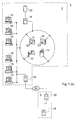
- FIG. 1(a) shows an example computer network which can be used to implement an embodiment of the method of the present invention.
- the network 1 comprises a local area network in a hospital 2.
- the hospital 2 is equipped with a number of workstations 16 which each have access, via the local area network, to a hospital computer server 18 having an associated storage device 20.
- a PACS archive is stored on the storage device 20 so that images in the archive can be viewed at any of the workstations 16.
- a number of medical imaging devices 8, 10, 12, 14 are connected to the hospital computer server 18. Images recorded with the devices 8, 10, 12, 14 can be stored directly into the PACS archive on the storage device 20.
- the local area network is connected to the Internet 24 by a hospital Internet server 22.
- An external database provider 4 maintains a database of prostheses for use in the method of the present invention.
- the database is stored on a provider server 28, which is connected to the Internet 24 by an Internet provider server 30. Hence workstation users in the hospital 2 can access the database of prostheses via the Internet 24.
- the database includes an entry, or data file, for each available separable component of a prosthesis which could be chosen for use as a result of using the planning method.
- the data file is implemented as an object.
- Various fields are provided for each data file, so that all the information which is needed for selection of the most appropriate prosthesis is available. This typically includes a wide range of measurements and angles describing the shape of the prosthetic component, its intended relationships to the bones, and possibly the materials from which the prosthesis is made.
- each entry should include user-friendly information which uniquely identifies each prosthesis (for example by manufacturer name and serial number) and is comprehensible when returned to the user at the end of planning, to allow procurement of the required prosthesis.
- an alternative identifier may be included, such as a unique code number, for internal computer network use, for example, communication with a stock control program.
- Each data file in the database is structured so as to have a header part, containing prosthetic system identification data, and a body part containing two-subdivisions.
- a prosthestic system includes the prosthesis components and may include other material such as measuring devices, and represents a particular product range from a manufacturer.
- Each prosthetic system may be embodied in a single data file in the database.
- One subdivision of the body part contains field definitions defining the relevant characteristics of the prosthesis (such as "femoral size", "offset angle"), and the other subdivision contains records or values for each field definition.
- the data file may also contain graphical banners for display to the user, and an expiration date.
- the data file also contains data relating to the X-ray view and patient side (left/right) for which the data file is appropriate.
- the user is required to enter information relating to the planning which is going to be performed.
- information relating to the planning which is going to be performed.
- details of whether the operation is to be performed on the right or left side of the patient's body, and the type of operation being planned, i.e. what prosthesis is to be implanted is required.
- This enables appropriate geometrical constructs and templates to be displayed during the planning procedure.
- the user is required to indicate a range of prostheses which are to be considered during planning, for example, prostheses or prosthetic systems from a particular manufacturer. This facility is provided because many surgeons prefer to work with only one or two brands of prosthesis, so it is advantageous to limit the range appropriately before planning begins. All this information is entered using a standard data information box displayed on the screen, which prompts the user for the necessary data.
- the X-ray image is displayed, it is scaled or calibrated in a scaling step, so that the physical dimensions of the patient's bone structure can be determined and used to choose an appropriately sized prosthesis.
- the bones shown in X-ray images contain a degree of magnification, which may or may not be known. This arises from the fact that the beam of X-rays used to record the image is not collimated, and is passing through a patient with a given thickness so that the bones themselves are never in direct contact with the imaging surface. If the exact distances between the imaging surface, the bones and the X-ray source are not known, the degree of magnification will also be unknown.
- the X-ray image should include a reference object of known size in the correct plane, i.e. the plane of the hip joint.
- the reference object can be any convenient feature that is opaque to X-rays and which has a precisely known size.
- a precisely machined disc of metal or other reference marker may have been placed in the X-ray field of view in the correct plane at the time the X-ray image was taken.
- the femoral head of a hip replacement already performed on the same patient can be used as the reference object, if the specification of the femoral component is known.
- Scaling is performed using a display window called the scaling window, which displays the X-ray image together with controls to allow the user to implement the scaling step.
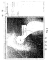
- Figure 2 is a computer screen shot of the scaling window.
- the reference object is a previously implanted hip prosthesis with a spherical femoral head 67.
- the user selects one of several scaling tools from a tool bar 68.
- the tool displays an adjustable shape 66 over the X-ray image 72, which can be manipulated by the user (using a mouse or other computer input device) to match the size and shape of the X-ray opaque object.
- the user has selected a tool 70 which gives a circular shape 66.
- the user opens a data entry box 76.
- the user then types the actual known size of the opaque object into the data entry box 76, in this case, the diameter of the spherical femoral head 67. This information is used to scale the image 72 appropriately, so that any dimension subsequently defined with reference to the image can be calculated.
- scaling tools include a linear measure which can be positioned along a known width or length of the opaque object, and a rectangle shape which can be used to outline a rectangular opaque object. Other shapes may be provided as appropriate.
- magnification must be estimated by the user using knowledge of the protocol used in the hospital radiology department when capturing the image. However, this technique is less accurate and more error-prone than using an opaque object for scaling the image.
- the magnification of the X-ray image may be known, so that it is not necessary to use one of the adjustable scaling tools from the toolbar 68, or to estimate the magnification.
- the user can enter the magnification as a percentage figure into a data entry box (not shown), and this information is used to scale the image. A percentage value of 100 can be entered if the input image is already scaled.
- the user is required to confirm that the information provided for scaling (size or percentage) is correct before the remainder of the planning procedure can be performed.
- Step S3 Planning
- Figure 3 shows a screen shot of a planning window which is displayed to the user once scaling is complete.
- the planning window includes the X-ray image 72, a user choice panel 79 for user choice of different planning tools, and information panels 77 showing details of prostheses in available prosthetic systems. Banners showing the available prostheses may be displayed to the user in the information panel 77. The user may select alternative prosthetic systems at this stage.
- the user selects from the user choice panel 79 a planning tool corresponding to the operation being planned.
- a planning tool for planning a hip replacement operation has been chosen.
- the planning tool overlays a geometrical construct 80 on the X-ray image 72.
- the geometrical construct 80 comprises a plurality of shapes and lines which are defined by geometric parameters corresponding to spatial information which is required to select suitable templates. If the geometrical construct is correctly positioned over the X-ray image, the dimensions and layout of the patient's bone structure can be calculated from the size of the construct and the known scale of the X-ray image.
- Figure 3(a) shows the geometrical construct 80 in more detail.
- various geometrical constructs described herein will be referred to as "wizards".
- the geometrical construct is therefore termed the hip wizard, and the example shown in Figures 3 and 3(a) corresponds to a right hip joint.
- a femur 50 and an acetabulum 52 are shown in outline in Figure 3(a), with the wizard 80 overlaid in an approximate position.
- the hip wizard 80 comprises a circle 88 having three manipulation handles 90A, 90B and 90C.
- the centre of the circle 88 is connected by a first line 91 to a second line 86, the first line 91 meeting the second line 86 at fixed angle A, which corresponds to a characteristic angle in the range of prostheses indicated for consideration in the input step S1.
- the second line 86 has an associated box 82 in the shape of a quadrangle and having a manipulation handle 84A, 84B, 84C and 84D at each comer.
- the box 82 overlies the second line 86 such that the second line 86 bisects the lines forming short edges of the box 82 between manipulation handles 84A and 84B, and between 84C and 84D.
- the wizard 80 is displayed over the X-ray image 72.
- the wizard 80 is automatically positioned vertically and horizontally in an initial position which is close to the correct position. This is achieved using prior knowledge and experience of the typical field of view of an X-ray of the pelvic area taken for the purpose of planning hip replacement surgery.
- the user can drag the hip wizard 80 across the image 72 and resize it by use of the manipulation handles, by using a mouse or similar device.
- the user manipulates the box 82 by dragging each of the comer handles 84A-D so that the box 82 outlines the femoral canal, within the femur.
- the purpose of positioning the box 82 is to locate the second line 86 (which moves with the box 82 such that it always bisects the box edges 84A-84B and 84C-84D) correctly along the length of the femur such that it follows the longitudinal axis of the bone. This is achieved by positioning the upper part of the box 82 at the point at which the femur is flared, and the lower end of the box far enough down the canal so that the second line 86 is satisfactorily aligned with the axis of the femur.
- a selection algorithm is executed, which selects from the database one or more data files representing suitable prostheses.
- geometrical parameters are taken from the manipulated wizard 80. Each of these geometrical parameters corresponds to parameters which describe the available prostheses.
- the length of long edge 84A-84D of the box 82 corresponds to the shaft size (length) of the prosthesis to be implanted; the size of the circle 88 corresponds to the size of the acetabular component; and the distance between the line 86 and the centre of the circle 88 is used to measure the patient's physiological offset (shortest distance between the axis of rotation of the femoral head and the axis of the femoral shaft) and corresponds to the offset size of the prosthesis.
- Each prosthesis has an associated representative to-scale template which shows a two-dimensional outline of the prosthesis.
- the parameters of the prostheses are stored in the database linked in the input step.
- Each prosthesis has a data file which holds pertinent details relating to it, including all relevant dimensions, and manufacturer's details and part numbers, plus data representing the corresponding template.
- the selection algorithm compares each of the wizard geometrical parameters in turn with the equivalent parameter for each of the accessible data files representing prosthetic components in the database, and selects one or more prosthetic components which best match the wizard parameters.
- the algorithm may include specified tolerance levels for particular parameters, so that it can return a range of prostheses of varying suitability. For example, it may be that the particular patient dimensions mean that one prosthesis is a good fit in the femoral component and less good in the acetabular component whereas an alternative prosthesis is a good fit in the acetabular component but less good in the femoral component.
- tolerances means that the algorithm will identify both prostheses.
- the panels include lists of a range of values of each prosthesis parameter.
- the parameter values of the prostheses selected by the selection algorithm are highlighted in the lists, for example by shading of the text.
- Further manipulation of the wizard 80 causes re-execution of the selection algorithm, and the information panels 77 are then updated to indicate the results of this re-execution. In this way, the user can monitor the selection process and is provided with an immediate visual indication of how any given manipulation of the wizard 80 affects prosthesis selection.
- the user can then click on an on-screen control button 92 to progress to the templating step of the planning procedure.
- Figure 4 shows a screen shot of a templating window which is displayed to the user in the templating step.
- the X-ray image 72 is shown, upon which a template of a prosthesis is superimposed.
- the templating window includes a series of prosthesis chooser panels 98, which are similar to the information panels 77 of the planning window, in that the prosthesis chooser panels 98 list a range of values for each prosthesis parameter.
- the prostheses selected by the selection algorithm in the planning step are presented in the chooser panels 98 as a series of recommendations, identified by shaded areas 100A and 100B overlying the relevant values. Darker shading indicates the best available match for that parameter, while paler shading indicates less good matches which nonetheless fall within the algorithm tolerance levels and are hence regarded as usable. Unshaded values are not recommended for use but may still be chosen by the user using clinical judgement.
- the template data from the relevant data file and the orientation of the hip wizard from the planning step are used to depict the corresponding template in position over the femur and pelvis in the X-ray image 72.
- the template consists of outlined parts representing the femoral component of the prosthesis 94, also known as a stem, and the acetabular component 96, also known as an acetabular cup.
- the user can then consider the template and make a decision as to whether the prosthesis it represents appears to be suitable, based on how well the template aligns with the patient's bone structure as shown on the X-ray image 72.
- the user can make alternative choices from the chooser panels 98, to display other templates for the purposes of comparison.
- the template femoral and acetabular components 94 and 96 may be repositioned by clicking and dragging, to aid in the decision making process.
- the user can return to the planning window by clicking on a control included in the template window.
- the hip wizard can then be further manipulated, and the selection algorithm will be reexecuted using the new wizard parameters, with the results shown in the information panels 77.
- the shaded values in the chooser panels 98 are updated accordingly to show the modified recommendations as selected by the selection algorithm. Choice of prostheses and display of corresponding templates then continues as before.
- the values in the chooser panels 98 lists are displayed in bold or standard text.
- Standard text is used to represent values which are not available in conjunction with already chosen values of other parameters, owing to the range of prostheses available. The use of standard or bold text is therefore updated each time a value is selected.
- Step S5 Output
- a record of the choice of prosthesis can be output in one or more ways, depending on the system to which the workstation is connected.
- a window with checkboxes can be provided on the display for the user to indicate which outputs are required at that time.
- the output step generates an output data file which records the choice of prosthesis together with the patient's details.
- the prosthesis will typically be identified by manufacturer name and a serial number, although a particular hospital could use an internal identification system. Additional information can usefully be included in the output data file, such as user's (surgeon's) name, date of planning, and proposed or actual date of operation.
- the output step then delivers the output data file in the way indicated by the user or according to pre-set configurations.
- the file should be recorded in the archive, with a link to the relevant X-ray image so that the two files can be retrieved together if necessary.
- the file can be stored in a separate archive or database unconnected with the X-ray image file which may be on the hard disk of the workstation used or on an alternative data storage medium, or it may be transferred to a different computer, for example, by email.
- the workstation is connected to a printer, either locally or over a network, the data file can be printed out as a hard copy for inclusion in the patient's paper records or for use as a memo to other hospital staff.
- the data file may usefully be sent over a network to a stock control department of the hospital where it can be compared, either manually, or preferably by computer, with records of existing prosthesis stock. In this way, a particular prosthesis can be earmarked for a particular operation, or a stock order can be generated if the prosthesis is not in stock.
- the second embodiment of the invention relates to computer-implemented planning for knee replacement surgery.
- the knee capsule is opened, and the ends of the femur and tibia are trimmed and augmented with implants which may or may not be held in place with cement.
- the femoral component is a metal shell
- the tibial component is a metal and plastic trough.
- This embodiment has the same basic steps as the first embodiment, and the planning procedure is performed in a similar way.
- the two embodiments differ in that the knee replacement planning is usually based on two previously obtained X-ray images of a patient's knee, one from the side (medio-lateral (ML) view) and one from the front (anterior-posterior (AP) view).
- ML latitude-lateral
- AP anterior-posterior
- two different views of the knee are used to ensure that the selected prosthesis is a sufficiently good fit.
- the ML view is normally used to select and choose the femoral component
- the AP view is normally used to select and choose the tibial component.
- a planning window is displayed, which includes a view of one of the X-ray images, and a planning tool selector to permit selection of a geometrical construct, or wizard.
- the planning is performed in each of the X-ray image views by using two geometrical constructs or wizards which are independently manipulatable. These are referred to as the femoral wizard and the tibial wizard and respectively correspond to the femoral component and the tibial component of the prosthesis.
- the wizards are referred to with reference to the ML and AP views. Within each view, the femoral and tibial wizards are independent of one another. However, the femoral wizard in the ML view is interrelated with the femoral wizard in the AP view, and similarly for the tibial wizard in each of the two views. The nature of this inter-relation will be discussed in more detail later.
- Figure 5 shows a screen shot of the planning window, with a ML X-ray image 200 displayed.
- Both the femoral wizard and the tibial wizard have been selected using a planning tool 106, so that the ML view femoral wizard 102A and the ML view tibial wizard 104A displayed overlaid on the knee bones in the X-ray image 200.
- Figure 5(a) shows the ML view femoral and tibial wizards 102A, 104A in more detail.
- An underlying image of a femur 54 and tibia 56 is shown in outline, with the wizards 102A, 104A in an approximate position.
- the ML view femoral wizard 102A comprises five straight lines. Four of these lines 202, 204, 205, and 206 are connected to form a quadrangular box, with the line 202 extending beyond the box.
- the fifth line 203 extends across the box parallel to the line 204.
- the relative lengths of the lines and the angles between them can be altered by dragging on suitably provided drag handles 106, 110 and 112.
- the ML view femoral wizard 102A is positioned by using the handle 106 to move the whole wizard 102A so that the extending line 202 is placed on the anterior edge of the femur.
- the handle 106 itself is located along the line 202 such that if the handle 106 is positioned just above the start of the patella groove on the femur, the femoral wizard is approximately correctly placed.
- This line 202 is then orientated by dragging with the handle 110 so that the line 202 is moved onto the anterior femoral shaft proximal to the handle 106.
- the comer 112 between the lines 204 and 206 is manipulated so that the line 206 just contacts the distal and anterior margins of the condyles.
- the box formed by lines 202, 204, 205 and 206 then outlines the end of the femur.
- the line 203 represents the predicted surgical cut line and the parallel line 204 lies along the physiological bearing surface of the joint.
- Line 205 which is also parallel to line 203, is a measurement line that provides a geometrical parameter which is used to recommend an appropriately sized femoral component.
- the ML view tibial wizard 104A comprises a tibial shaft axis definition line 118A having a drag handle 116 at each end, and an elongate rectangular box 120A also having a drag handles 121A and 121B at each end.
- the tibial shaft axis definition line 118A is at an angle to the long sides of the box 120A which is defined by a characteristic angle of the range of prostheses selected for consideration in the input step, known as the posterior slope angle.
- the ML view tibial wizard 104A is positioned by using the handles 116 to move the tibial shaft axis definition line 118A such that it is positioned along the axis of the tibia, by aligning it with the straight part of the posterior edge of the tibia. Then the handles 121A and 121B are used to drag the box 120A into position such that the line forming the upper long side of the box approximates to the knee joint line.
- Figure 5 shows the ML view wizards 102A, 104A suitably positioned.
- the user now refers to the AP X-ray image by clicking on a suitably-labelled button in the planning window to bring up this image in place of the ML X-ray image 200.
- the planning window may display both images at once.
- Figure 6 shows a screen shot of the planning window with an AP X-ray image 210 displayed. Both the femoral and tibial wizards have been selected using the planning tool 106, so the AP view femoral wizard 102B and the AP view tibial wizard 104B are shown superimposed over the X-ray image 210.
- Figure 6(a) shows the AP view femoral wizard 102B and tibial wizard 104B in more detail.
- An underlying image of a femur 54 and tibia 56 is shown in outline, with the wizards 102B, 104B in an approximate position.
- the AP view femoral wizard 102B comprises a quadrangular box 210 having a drag handle 130 at each comer, and a substantially horizontal elongate box 134 positioned below the box 210.
- the wizard 102B is positioned by dragging the handles 130 so as to outline the femoral shaft, so as to establish the axis of the femoral shaft, as shown in Figure 6.
- the box 134 is then positioned by the user so that its upper horizontal edge corresponds to the desired position of the distal femoral surgical cut line to be used in the forthcoming operation. Additionally, the user is required to enter into a dialogue box the valgus angle of the patient, which is determined in advance by physical examination of the patient (The valgus angle is the angle of displacement of the femur from the weight-bearing axis down the leg). This establishes the final parameter required for this wizard.
- the AP view tibial wizard 104B comprises an elongate box 120B and a tibial shaft axis definition line 118B.
- the angle between the box 120B and the line 118B is defined by the posterior slope angle of prostheses indicated for consideration in the input step.
- the user positions the handles at either end of the elongate rectangular box 120B on the medial and lateral margins of the tibial plateau so that upper horizontal line 122 of the elongate rectangular box 120B is aligned with the physiological bearing surface line of the joint, or the lower horizontal line 123 of the elongate rectangular box 120B is aligned with the predicted surgical cut line.
- Clinical judgement and personal preference is used to determine which of these two lines 122 or 123 is used to appropriately position the AP view tibial wizard 104B.
- the distance between the two lines 122 and 123 is fixed and determined by the prosthetic system chosen by the user, because each system requires a surgical cut line to be made appropriately relative to the bearing surface of the joint.
- Figure 6 shows the AP view femoral and tibial wizards 102B, 104B correctly positioned.
- the user can move between the AP and ML images or can view both simultaneously to adjust the wizards until they are suitably positioned.
- a selection algorithm similar to that used in the first embodiment is executed to select suitable prostheses from a database, using parameters derived from the dimensions and positions of the manipulated wizards.
- the database is similar to that described for hip replacement prostheses, with the additional feature that each data file includes image data for two templates, one showing the ML outline of the prosthesis and one showing the AP outline.
- the results of the selection algorithm appear in information panels 135 in the planning window.
- the planning process can move onto the templating step, which is activated, as before, by the user clicking on a suitably labelled button in the planning window to move to a templating window.
- either of the two X-ray images can be displayed (or, alternatively, both can be displayed at one time).
- Figure 7 shows a templating window displaying an ML image
- Figure 8 shows a templating window displaying an AP image.
- the user is provided with chooser panels 128 listing values of parameters relevant to the two components of knee prostheses, with the results returned by the algorithm recommended by shaded areas.
- Choice of a parameter from each panel by the user allows the corresponding templates to be superimposed on the X-ray image for inspection by the user.
- the templates show the femoral component 124 and the tibial component 126. These can be dragged across and rotated on the image independently so that the user can check the size and positioning before making a final choice of prosthesis.
- this can be implemented in a various ways, and is based on the fact that the knee prosthesis components are three-dimensional, and bone measurements in three dimensions need to be considered to choose an appropriate prosthesis. Two possible implementations will now be described.
- the femoral wizard and the tibial wizard are each three-dimensional geometrical constructs, unrelated to each other.
- the AP and ML views of each wizard are two-dimensional projections of these constructs, so that in each view, each wizard appears as a two-dimensional collection of lines and shapes which are interconnected. Manipulation in one view hence moves corresponding parts of the three-dimensional construct so that the other projection in the other view is correspondingly altered.
- the wizards can be manipulated in three dimensions and accurately matched to the imaged bone structure.
- a second implementation uses two two-dimensional geometrical constructs for each of the tibial and femoral wizards, one construct for each view.
- the wizards can be manipulated, but there is no corresponding adjustment of that wizard in the other view.
- Each view therefore results in the determination of a different set of geometric parameters.
- the inter-relation is provided at the level of the database, where the data file for each prosthesis contains parameters describing that prosthesis. These parameters correspond to the geometric parameters determined from each of the two-dimensional wizards, so that each database entry is linked to both views of the wizard. Therefore, manipulation of the wizards in either view causes the selection algorithm to compare the geometric parameters with those in the database and possibly select and recommend a different prosthesis.
- the present invention has been described in detail with respect to hip and total knee replacement surgery, it is not limited thereto. It can also be used for planning surgery for the replacement of other joints, including small joints and joints in the spine and upper limb, if a suitable database of prostheses is provided, and the geometrical constructs are modified so as to map the relevant measurements for the joint in question.
- the first and second embodiments described herein use one and two patient images respectively, there is no limit to the amount of views which can be accommodated.
- the use of one or two images to plan orthopaedic surgery is typical, with three being used occasionally.
- a corresponding number of views of wizards must be provided (be they various two-dimensional projections of a single three-dimensional geometrical construct, or separate wizards linked via the parameters in the database), together with templates representing each of the appropriate outlines of the prostheses.
- the present invention may be used for planning hip replacement surgery with ML patient images, if the database includes ML templates of the femoral and acetabular components, and a ML view hip wizard is provided.
Landscapes
- Health & Medical Sciences (AREA)
- Engineering & Computer Science (AREA)
- Public Health (AREA)
- Medical Informatics (AREA)
- General Health & Medical Sciences (AREA)
- Epidemiology (AREA)
- Primary Health Care (AREA)
- Biomedical Technology (AREA)
- Nuclear Medicine, Radiotherapy & Molecular Imaging (AREA)
- Life Sciences & Earth Sciences (AREA)
- Surgery (AREA)
- Oral & Maxillofacial Surgery (AREA)
- Veterinary Medicine (AREA)
- Heart & Thoracic Surgery (AREA)
- Animal Behavior & Ethology (AREA)
- Pathology (AREA)
- Radiology & Medical Imaging (AREA)
- Bioethics (AREA)
- Physics & Mathematics (AREA)
- Transplantation (AREA)
- Cardiology (AREA)
- Orthopedic Medicine & Surgery (AREA)
- Business, Economics & Management (AREA)
- General Business, Economics & Management (AREA)
- Data Mining & Analysis (AREA)
- Databases & Information Systems (AREA)
- Manufacturing & Machinery (AREA)
- Vascular Medicine (AREA)
- Geometry (AREA)
- Molecular Biology (AREA)
- Urology & Nephrology (AREA)
- Processing Or Creating Images (AREA)
- Prostheses (AREA)
- Transplanting Machines (AREA)
- Buildings Adapted To Withstand Abnormal External Influences (AREA)
- Image Processing (AREA)
- Feedback Control In General (AREA)
- Paper (AREA)
Applications Claiming Priority (2)
| Application Number | Priority Date | Filing Date | Title |
|---|---|---|---|
| GB0222414A GB2393625C (en) | 2002-09-26 | 2002-09-26 | Orthopaedic surgery planning |
| GB0222414 | 2002-09-26 |
Publications (3)
| Publication Number | Publication Date |
|---|---|
| EP1406203A2 true EP1406203A2 (de) | 2004-04-07 |
| EP1406203A3 EP1406203A3 (de) | 2004-11-24 |
| EP1406203B1 EP1406203B1 (de) | 2006-08-02 |
Family
ID=9944843
Family Applications (1)
| Application Number | Title | Priority Date | Filing Date |
|---|---|---|---|
| EP03255895A Expired - Lifetime EP1406203B1 (de) | 2002-09-26 | 2003-09-19 | Planung für die orthopädische Chirurgie |
Country Status (5)
| Country | Link |
|---|---|
| US (1) | US7388972B2 (de) |
| EP (1) | EP1406203B1 (de) |
| AT (1) | ATE335244T1 (de) |
| DE (1) | DE60307194T2 (de) |
| GB (1) | GB2393625C (de) |
Cited By (29)
| Publication number | Priority date | Publication date | Assignee | Title |
|---|---|---|---|---|
| DE102004039680A1 (de) * | 2004-08-16 | 2005-11-10 | Siemens Ag | Verfahren zur Durchführung von Messungen mit einem bildgebenden medizinischen Untersuchungssystem und bildgebendes medizinisches Untersuchungssystem |
| WO2006109022A3 (en) * | 2005-04-09 | 2007-01-18 | Depuy Int Ltd | Acetabular cup positioning |
| WO2007017642A1 (en) * | 2005-08-05 | 2007-02-15 | Depuy Orthopädie Gmbh | Computer assisted surgery system |
| WO2007109467A1 (en) * | 2006-03-17 | 2007-09-27 | Zimmer, Inc. | Methods of predetermining the contour of a resected bone surface and assessing the fit of a prosthesis on the bone |
| DE102006048451A1 (de) * | 2006-10-11 | 2008-04-17 | Siemens Ag | Verfahren zur virtuellen Anpassung eines Objekts an ein Körperteil eines Patienten |
| FR2913592A1 (fr) * | 2007-03-15 | 2008-09-19 | Vincent Costalat | Procede et dispositif pour determiner la dimension d'un implant |
| DE102007046704A1 (de) * | 2007-09-28 | 2009-04-23 | Siemens Ag | Verfahren und Vorrichtung zur Unterstützung der Auswertung von medizinischen Bilddaten |
| WO2010122145A1 (en) * | 2009-04-25 | 2010-10-28 | Siemens Aktiengesellschaft | A method and a system for assessing the relative pose of an implant and a bone of a creature |
| EP2175418A3 (de) * | 2008-10-08 | 2011-05-11 | Fujifilm Medical Systems U.S.A. Inc. | Verfahren und Anordnung zur chirurgischen Modellierung |
| EP2175419A3 (de) * | 2008-10-08 | 2011-05-18 | Fujifilm Medical Systems U.S.A. Inc. | Verfahren und Anordnung zur chirurgischen Planung |
| EP2336927A1 (de) * | 2008-09-18 | 2011-06-22 | Nemaris LLC | Wissenschaftliches Bildverarbeitungssystem |
| US7983777B2 (en) | 2005-08-19 | 2011-07-19 | Mark Melton | System for biomedical implant creation and procurement |
| US8331634B2 (en) | 2006-09-26 | 2012-12-11 | Siemens Aktiengesellschaft | Method for virtual adaptation of an implant to a body part of a patient |
| WO2013012561A1 (en) * | 2011-07-15 | 2013-01-24 | Murphy Stephen B | Surgical planning system and method |
| US9017337B2 (en) | 2008-02-28 | 2015-04-28 | DuPuy Orthopadie GmbH | Acetabular alignment guide |
| US9129054B2 (en) | 2012-09-17 | 2015-09-08 | DePuy Synthes Products, Inc. | Systems and methods for surgical and interventional planning, support, post-operative follow-up, and, functional recovery tracking |
| US9179983B2 (en) | 2007-08-14 | 2015-11-10 | Zimmer, Inc. | Method of determining a contour of an anatomical structure and selecting an orthopaedic implant to replicate the anatomical structure |
| US9990720B2 (en) | 2015-12-18 | 2018-06-05 | Episurf Ip-Management Ab | System and method for creating a decision support material indicating damage to an anatomical joint |
| EP3329881A1 (de) * | 2011-02-24 | 2018-06-06 | DePuy Ireland Unlimited Company | Aufrechterhaltung einer korrekten mechanik in der hüftarthroplastie |
| EP3113710B1 (de) | 2014-02-25 | 2018-10-10 | Jointpoint, Inc. | Systeme und verfahren zur intraoperativen bildanalyse |
| WO2020162860A3 (en) * | 2019-02-08 | 2020-09-10 | Imed Surgical Teknoloji A.S. | A system providing special joint and bone structure |
| US10987176B2 (en) | 2018-06-19 | 2021-04-27 | Tornier, Inc. | Virtual guidance for orthopedic surgical procedures |
| US11250561B2 (en) | 2017-06-16 | 2022-02-15 | Episurf Ip-Management Ab | Determination and visualization of damage to an anatomical joint |
| US11526988B2 (en) | 2015-12-18 | 2022-12-13 | Episurf Ip-Management Ab | System and method for creating a decision support material indicating damage to an anatomical joint |
| US11645749B2 (en) | 2018-12-14 | 2023-05-09 | Episurf Ip-Management Ab | Determination and visualization of damage to an anatomical joint |
| US11642174B2 (en) | 2014-02-25 | 2023-05-09 | DePuy Synthes Products, Inc. | Systems and methods for intra-operative image analysis |
| US11887306B2 (en) | 2021-08-11 | 2024-01-30 | DePuy Synthes Products, Inc. | System and method for intraoperatively determining image alignment |
| US12295772B2 (en) | 2014-02-25 | 2025-05-13 | DePuy Synthes Products, Inc. | Systems and methods for intra-operative image analysis |
| US12465374B2 (en) | 2019-12-18 | 2025-11-11 | Howmedica Osteonics Corp. | Surgical guidance for surgical tools |
Families Citing this family (185)
| Publication number | Priority date | Publication date | Assignee | Title |
|---|---|---|---|---|
| US7547307B2 (en) | 2001-02-27 | 2009-06-16 | Smith & Nephew, Inc. | Computer assisted knee arthroplasty instrumentation, systems, and processes |
| EP1372516B1 (de) * | 2001-02-27 | 2009-05-13 | Smith & Nephew, Inc. | Chirurgisches navigationssystem zur teilweisen kniegelenkrekonstruktion |
| EP1418013B1 (de) | 2002-11-08 | 2005-01-19 | Howmedica Osteonics Corp. | Lasererzeugte poröse Oberfläche |
| US20060147332A1 (en) | 2004-12-30 | 2006-07-06 | Howmedica Osteonics Corp. | Laser-produced porous structure |
| US7862570B2 (en) | 2003-10-03 | 2011-01-04 | Smith & Nephew, Inc. | Surgical positioners |
| US7764985B2 (en) | 2003-10-20 | 2010-07-27 | Smith & Nephew, Inc. | Surgical navigation system component fault interfaces and related processes |
| WO2005048851A1 (en) | 2003-11-14 | 2005-06-02 | Smith & Nephew, Inc. | Adjustable surgical cutting systems |
| CA2561493A1 (en) | 2004-03-31 | 2005-10-20 | Smith & Nephew, Inc. | Methods and apparatuses for providing a reference array input device |
| US8109942B2 (en) | 2004-04-21 | 2012-02-07 | Smith & Nephew, Inc. | Computer-aided methods, systems, and apparatuses for shoulder arthroplasty |
| US8260591B2 (en) * | 2004-04-29 | 2012-09-04 | Align Technology, Inc. | Dynamically specifying a view |
| US20060239577A1 (en) * | 2005-03-10 | 2006-10-26 | Piatt Joseph H | Process of using computer modeling, reconstructive modeling and simulation modeling for image guided reconstructive surgery |
| EP1861821A2 (de) * | 2005-03-24 | 2007-12-05 | Image Metrics Limited | Verfahren und system zur beschreibung der kniegelenksmorphologie |
| US8728387B2 (en) | 2005-12-06 | 2014-05-20 | Howmedica Osteonics Corp. | Laser-produced porous surface |
| US9808262B2 (en) | 2006-02-15 | 2017-11-07 | Howmedica Osteonics Corporation | Arthroplasty devices and related methods |
| CA2642615A1 (en) | 2006-02-15 | 2007-08-30 | Otismed Corp | Arthroplasty jigs and related methods |
| US9113971B2 (en) | 2006-02-27 | 2015-08-25 | Biomet Manufacturing, Llc | Femoral acetabular impingement guide |
| US8568487B2 (en) | 2006-02-27 | 2013-10-29 | Biomet Manufacturing, Llc | Patient-specific hip joint devices |
| US8092465B2 (en) | 2006-06-09 | 2012-01-10 | Biomet Manufacturing Corp. | Patient specific knee alignment guide and associated method |
| US8241293B2 (en) | 2006-02-27 | 2012-08-14 | Biomet Manufacturing Corp. | Patient specific high tibia osteotomy |
| US8298237B2 (en) | 2006-06-09 | 2012-10-30 | Biomet Manufacturing Corp. | Patient-specific alignment guide for multiple incisions |
| US20080257363A1 (en) * | 2007-04-17 | 2008-10-23 | Biomet Manufacturing Corp. | Method And Apparatus For Manufacturing An Implant |
| US8603180B2 (en) | 2006-02-27 | 2013-12-10 | Biomet Manufacturing, Llc | Patient-specific acetabular alignment guides |
| US8473305B2 (en) | 2007-04-17 | 2013-06-25 | Biomet Manufacturing Corp. | Method and apparatus for manufacturing an implant |
| US7967868B2 (en) * | 2007-04-17 | 2011-06-28 | Biomet Manufacturing Corp. | Patient-modified implant and associated method |
| US9289253B2 (en) | 2006-02-27 | 2016-03-22 | Biomet Manufacturing, Llc | Patient-specific shoulder guide |
| US8535387B2 (en) | 2006-02-27 | 2013-09-17 | Biomet Manufacturing, Llc | Patient-specific tools and implants |
| US10278711B2 (en) | 2006-02-27 | 2019-05-07 | Biomet Manufacturing, Llc | Patient-specific femoral guide |
| US8407067B2 (en) | 2007-04-17 | 2013-03-26 | Biomet Manufacturing Corp. | Method and apparatus for manufacturing an implant |
| US8377066B2 (en) | 2006-02-27 | 2013-02-19 | Biomet Manufacturing Corp. | Patient-specific elbow guides and associated methods |
| US8864769B2 (en) | 2006-02-27 | 2014-10-21 | Biomet Manufacturing, Llc | Alignment guides with patient-specific anchoring elements |
| US8608749B2 (en) | 2006-02-27 | 2013-12-17 | Biomet Manufacturing, Llc | Patient-specific acetabular guides and associated instruments |
| US8591516B2 (en) | 2006-02-27 | 2013-11-26 | Biomet Manufacturing, Llc | Patient-specific orthopedic instruments |
| US20150335438A1 (en) | 2006-02-27 | 2015-11-26 | Biomet Manufacturing, Llc. | Patient-specific augments |
| US8133234B2 (en) | 2006-02-27 | 2012-03-13 | Biomet Manufacturing Corp. | Patient specific acetabular guide and method |
| US8608748B2 (en) | 2006-02-27 | 2013-12-17 | Biomet Manufacturing, Llc | Patient specific guides |
| US8858561B2 (en) | 2006-06-09 | 2014-10-14 | Blomet Manufacturing, LLC | Patient-specific alignment guide |
| US9339278B2 (en) | 2006-02-27 | 2016-05-17 | Biomet Manufacturing, Llc | Patient-specific acetabular guides and associated instruments |
| US9918740B2 (en) | 2006-02-27 | 2018-03-20 | Biomet Manufacturing, Llc | Backup surgical instrument system and method |
| US9173661B2 (en) | 2006-02-27 | 2015-11-03 | Biomet Manufacturing, Llc | Patient specific alignment guide with cutting surface and laser indicator |
| US8282646B2 (en) | 2006-02-27 | 2012-10-09 | Biomet Manufacturing Corp. | Patient specific knee alignment guide and associated method |
| US8070752B2 (en) | 2006-02-27 | 2011-12-06 | Biomet Manufacturing Corp. | Patient specific alignment guide and inter-operative adjustment |
| US9907659B2 (en) * | 2007-04-17 | 2018-03-06 | Biomet Manufacturing, Llc | Method and apparatus for manufacturing an implant |
| US9345548B2 (en) | 2006-02-27 | 2016-05-24 | Biomet Manufacturing, Llc | Patient-specific pre-operative planning |
| US8112292B2 (en) | 2006-04-21 | 2012-02-07 | Medtronic Navigation, Inc. | Method and apparatus for optimizing a therapy |
| WO2008115188A2 (en) | 2006-05-08 | 2008-09-25 | C. R. Bard, Inc. | User interface and methods for sonographic display device |
| EP1862151B1 (de) * | 2006-05-31 | 2010-10-06 | BrainLAB AG | Verfahren zur Auswahl eines Oberschenkelimplantats |
| US9795399B2 (en) | 2006-06-09 | 2017-10-24 | Biomet Manufacturing, Llc | Patient-specific knee alignment guide and associated method |
| US7953612B1 (en) * | 2006-07-17 | 2011-05-31 | Ecomglobalmedical Research & Development, Inc | System and method for providing a searchable database of surgical information |
| US20080021299A1 (en) * | 2006-07-18 | 2008-01-24 | Meulink Steven L | Method for selecting modular implant components |
| US8660635B2 (en) | 2006-09-29 | 2014-02-25 | Medtronic, Inc. | Method and apparatus for optimizing a computer assisted surgical procedure |
| WO2008045807A2 (en) | 2006-10-09 | 2008-04-17 | Active Implants Corporation | Meniscus prosthetic device |
| US20080120262A1 (en) * | 2006-11-16 | 2008-05-22 | Koninklijke Philips Electronics, N.V. | What-if planning for medical procedures |
| US7831096B2 (en) * | 2006-11-17 | 2010-11-09 | General Electric Company | Medical navigation system with tool and/or implant integration into fluoroscopic image projections and method of use |
| US8460302B2 (en) | 2006-12-18 | 2013-06-11 | Otismed Corporation | Arthroplasty devices and related methods |
| WO2008087556A2 (en) * | 2007-01-16 | 2008-07-24 | Optasia Medical, Limited | Image processing systems and methods |
| CA2683717C (en) * | 2007-04-19 | 2016-10-11 | Mako Surgical Corp. | Implant planning using captured joint motion information |
| EP2139433B1 (de) * | 2007-04-28 | 2014-11-12 | DePuy International Limited | Bestimmung des versatzes des kopfteils einer orthopädischen gelenkprothesenschaft-komponente |
| US20110092859A1 (en) | 2007-06-25 | 2011-04-21 | Neubardt Seth L | System for determining and placing spinal implants or prostheses |
| US8265949B2 (en) | 2007-09-27 | 2012-09-11 | Depuy Products, Inc. | Customized patient surgical plan |
| US9786022B2 (en) * | 2007-09-30 | 2017-10-10 | DePuy Synthes Products, Inc. | Customized patient-specific bone cutting blocks |
| US8398645B2 (en) | 2007-09-30 | 2013-03-19 | DePuy Synthes Products, LLC | Femoral tibial customized patient-specific orthopaedic surgical instrumentation |
| US8357111B2 (en) | 2007-09-30 | 2013-01-22 | Depuy Products, Inc. | Method and system for designing patient-specific orthopaedic surgical instruments |
| US8460303B2 (en) | 2007-10-25 | 2013-06-11 | Otismed Corporation | Arthroplasty systems and devices, and related methods |
| USD642263S1 (en) | 2007-10-25 | 2011-07-26 | Otismed Corporation | Arthroplasty jig blank |
| US8065166B2 (en) | 2007-10-30 | 2011-11-22 | Onemednet Corporation | Methods, systems, and devices for managing medical images and records |
| US9171344B2 (en) | 2007-10-30 | 2015-10-27 | Onemednet Corporation | Methods, systems, and devices for managing medical images and records |
| US10582934B2 (en) | 2007-11-27 | 2020-03-10 | Howmedica Osteonics Corporation | Generating MRI images usable for the creation of 3D bone models employed to make customized arthroplasty jigs |
| US8480679B2 (en) | 2008-04-29 | 2013-07-09 | Otismed Corporation | Generation of a computerized bone model representative of a pre-degenerated state and useable in the design and manufacture of arthroplasty devices |
| US8311306B2 (en) | 2008-04-30 | 2012-11-13 | Otismed Corporation | System and method for image segmentation in generating computer models of a joint to undergo arthroplasty |
| US8617171B2 (en) | 2007-12-18 | 2013-12-31 | Otismed Corporation | Preoperatively planning an arthroplasty procedure and generating a corresponding patient specific arthroplasty resection guide |
| US8715291B2 (en) | 2007-12-18 | 2014-05-06 | Otismed Corporation | Arthroplasty system and related methods |
| US8545509B2 (en) | 2007-12-18 | 2013-10-01 | Otismed Corporation | Arthroplasty system and related methods |
| US8777875B2 (en) | 2008-07-23 | 2014-07-15 | Otismed Corporation | System and method for manufacturing arthroplasty jigs having improved mating accuracy |
| US8160345B2 (en) | 2008-04-30 | 2012-04-17 | Otismed Corporation | System and method for image segmentation in generating computer models of a joint to undergo arthroplasty |
| US8221430B2 (en) | 2007-12-18 | 2012-07-17 | Otismed Corporation | System and method for manufacturing arthroplasty jigs |
| US8737700B2 (en) | 2007-12-18 | 2014-05-27 | Otismed Corporation | Preoperatively planning an arthroplasty procedure and generating a corresponding patient specific arthroplasty resection guide |
| EP2242453B1 (de) | 2008-02-20 | 2018-11-28 | Mako Surgical Corp. | Implantierungsplanung mithilfe korrigierter informationen zu erfassten gelenkbewegungen |
| US8734455B2 (en) | 2008-02-29 | 2014-05-27 | Otismed Corporation | Hip resurfacing surgical guide tool |
| US8549888B2 (en) | 2008-04-04 | 2013-10-08 | Nuvasive, Inc. | System and device for designing and forming a surgical implant |
| US7991599B2 (en) | 2008-04-09 | 2011-08-02 | Active Implants Corporation | Meniscus prosthetic device selection and implantation methods |
| US8016884B2 (en) * | 2008-04-09 | 2011-09-13 | Active Implants Corporation | Tensioned meniscus prosthetic devices and associated methods |
| US8361147B2 (en) * | 2008-04-09 | 2013-01-29 | Active Implants Corporation | Meniscus prosthetic devices with anti-migration features |
| US8617175B2 (en) | 2008-12-16 | 2013-12-31 | Otismed Corporation | Unicompartmental customized arthroplasty cutting jigs and methods of making the same |
| US8078440B2 (en) | 2008-09-19 | 2011-12-13 | Smith & Nephew, Inc. | Operatively tuning implants for increased performance |
| US8165658B2 (en) | 2008-09-26 | 2012-04-24 | Medtronic, Inc. | Method and apparatus for positioning a guide relative to a base |
| US8249318B2 (en) * | 2008-09-26 | 2012-08-21 | OsteoWare, Inc. | Method for identifying implanted reconstructive prosthetic devices |
| US9364291B2 (en) * | 2008-12-11 | 2016-06-14 | Mako Surgical Corp. | Implant planning using areas representing cartilage |
| US8170641B2 (en) | 2009-02-20 | 2012-05-01 | Biomet Manufacturing Corp. | Method of imaging an extremity of a patient |
| WO2010126797A1 (en) | 2009-04-29 | 2010-11-04 | Onemednet Corporation | Methods, systems, and devices for managing medical images and records |
| DE102009028503B4 (de) | 2009-08-13 | 2013-11-14 | Biomet Manufacturing Corp. | Resektionsschablone zur Resektion von Knochen, Verfahren zur Herstellung einer solchen Resektionsschablone und Operationsset zur Durchführung von Kniegelenk-Operationen |
| US8632547B2 (en) | 2010-02-26 | 2014-01-21 | Biomet Sports Medicine, Llc | Patient-specific osteotomy devices and methods |
| US9066727B2 (en) | 2010-03-04 | 2015-06-30 | Materialise Nv | Patient-specific computed tomography guides |
| DE102010020783B4 (de) * | 2010-05-18 | 2022-05-12 | Siemens Healthcare Gmbh | Verfahren und System zum Ermitteln von 3D-Geometriedaten von Objekten |
| GB201008281D0 (en) | 2010-05-19 | 2010-06-30 | Nikonovas Arkadijus | Indirect analysis and manipulation of objects |
| US8908937B2 (en) | 2010-07-08 | 2014-12-09 | Biomet Manufacturing, Llc | Method and device for digital image templating |
| US9262802B2 (en) * | 2010-07-21 | 2016-02-16 | Arthromeda, Inc. | Independent digital templating software, and methods and systems using same |
| US9271744B2 (en) | 2010-09-29 | 2016-03-01 | Biomet Manufacturing, Llc | Patient-specific guide for partial acetabular socket replacement |
| US9968376B2 (en) | 2010-11-29 | 2018-05-15 | Biomet Manufacturing, Llc | Patient-specific orthopedic instruments |
| US8917290B2 (en) * | 2011-01-31 | 2014-12-23 | Biomet Manufacturing, Llc | Digital image templating |
| US9241745B2 (en) | 2011-03-07 | 2016-01-26 | Biomet Manufacturing, Llc | Patient-specific femoral version guide |
| US8715289B2 (en) | 2011-04-15 | 2014-05-06 | Biomet Manufacturing, Llc | Patient-specific numerically controlled instrument |
| US9675400B2 (en) | 2011-04-19 | 2017-06-13 | Biomet Manufacturing, Llc | Patient-specific fracture fixation instrumentation and method |
| US8668700B2 (en) | 2011-04-29 | 2014-03-11 | Biomet Manufacturing, Llc | Patient-specific convertible guides |
| US8956364B2 (en) | 2011-04-29 | 2015-02-17 | Biomet Manufacturing, Llc | Patient-specific partial knee guides and other instruments |
| US8532807B2 (en) | 2011-06-06 | 2013-09-10 | Biomet Manufacturing, Llc | Pre-operative planning and manufacturing method for orthopedic procedure |
| US9084618B2 (en) | 2011-06-13 | 2015-07-21 | Biomet Manufacturing, Llc | Drill guides for confirming alignment of patient-specific alignment guides |
| US8764760B2 (en) | 2011-07-01 | 2014-07-01 | Biomet Manufacturing, Llc | Patient-specific bone-cutting guidance instruments and methods |
| US20130001121A1 (en) | 2011-07-01 | 2013-01-03 | Biomet Manufacturing Corp. | Backup kit for a patient-specific arthroplasty kit assembly |
| US8597365B2 (en) | 2011-08-04 | 2013-12-03 | Biomet Manufacturing, Llc | Patient-specific pelvic implants for acetabular reconstruction |
| US9066734B2 (en) | 2011-08-31 | 2015-06-30 | Biomet Manufacturing, Llc | Patient-specific sacroiliac guides and associated methods |
| US9295497B2 (en) | 2011-08-31 | 2016-03-29 | Biomet Manufacturing, Llc | Patient-specific sacroiliac and pedicle guides |
| US9386993B2 (en) | 2011-09-29 | 2016-07-12 | Biomet Manufacturing, Llc | Patient-specific femoroacetabular impingement instruments and methods |
| US9554910B2 (en) | 2011-10-27 | 2017-01-31 | Biomet Manufacturing, Llc | Patient-specific glenoid guide and implants |
| KR20130046337A (ko) | 2011-10-27 | 2013-05-07 | 삼성전자주식회사 | 멀티뷰 디바이스 및 그 제어방법과, 디스플레이장치 및 그 제어방법과, 디스플레이 시스템 |
| WO2013062848A1 (en) | 2011-10-27 | 2013-05-02 | Biomet Manufacturing Corporation | Patient-specific glenoid guides |
| US9451973B2 (en) | 2011-10-27 | 2016-09-27 | Biomet Manufacturing, Llc | Patient specific glenoid guide |
| US9301812B2 (en) | 2011-10-27 | 2016-04-05 | Biomet Manufacturing, Llc | Methods for patient-specific shoulder arthroplasty |
| EP3451298A3 (de) | 2011-12-14 | 2019-06-05 | Stryker European Holdings I, LLC | Verfahren zur erzeugung eines knochenplattenentwurfs |
| US9237950B2 (en) | 2012-02-02 | 2016-01-19 | Biomet Manufacturing, Llc | Implant with patient-specific porous structure |
| WO2013116812A1 (en) | 2012-02-03 | 2013-08-08 | Orthohub, Inc. | External fixator deformity correction systems and methods |
| US11207132B2 (en) | 2012-03-12 | 2021-12-28 | Nuvasive, Inc. | Systems and methods for performing spinal surgery |
| US9180010B2 (en) | 2012-04-06 | 2015-11-10 | Howmedica Osteonics Corp. | Surface modified unit cell lattice structures for optimized secure freeform fabrication |
| JP5705788B2 (ja) * | 2012-06-05 | 2015-04-22 | 株式会社日立製作所 | アセンブリモデル類似構造検索システム、及びアセンブリモデル類似構造検索方法 |
| US9402637B2 (en) | 2012-10-11 | 2016-08-02 | Howmedica Osteonics Corporation | Customized arthroplasty cutting guides and surgical methods using the same |
| SE536759C2 (sv) | 2012-10-18 | 2014-07-15 | Ortoma Ab | Metod och system för planering av position för implantatkomponent |
| US9050134B2 (en) * | 2012-11-30 | 2015-06-09 | Zimmer Gmbh | Device and method for verifying compatability between cooperating parts of a joint prosthesis |
| US9060788B2 (en) | 2012-12-11 | 2015-06-23 | Biomet Manufacturing, Llc | Patient-specific acetabular guide for anterior approach |
| US9204977B2 (en) | 2012-12-11 | 2015-12-08 | Biomet Manufacturing, Llc | Patient-specific acetabular guide for anterior approach |
| US20140218397A1 (en) * | 2013-02-04 | 2014-08-07 | Mckesson Financial Holdings | Method and apparatus for providing virtual device planning |
| US9204937B2 (en) * | 2013-02-19 | 2015-12-08 | Stryker Trauma Gmbh | Software for use with deformity correction |
| US9839438B2 (en) | 2013-03-11 | 2017-12-12 | Biomet Manufacturing, Llc | Patient-specific glenoid guide with a reusable guide holder |
| US9579107B2 (en) | 2013-03-12 | 2017-02-28 | Biomet Manufacturing, Llc | Multi-point fit for patient specific guide |
| US9826981B2 (en) | 2013-03-13 | 2017-11-28 | Biomet Manufacturing, Llc | Tangential fit of patient-specific guides |
| JP6382293B2 (ja) | 2013-03-13 | 2018-08-29 | デピュイ・シンセス・プロダクツ・インコーポレイテッド | 外部骨固定装置 |
| US9498233B2 (en) | 2013-03-13 | 2016-11-22 | Biomet Manufacturing, Llc. | Universal acetabular guide and associated hardware |
| US8864763B2 (en) | 2013-03-13 | 2014-10-21 | DePuy Synthes Products, LLC | External bone fixation device |
| US9039706B2 (en) | 2013-03-13 | 2015-05-26 | DePuy Synthes Products, Inc. | External bone fixation device |
| US9299138B2 (en) * | 2013-03-14 | 2016-03-29 | DePuy Synthes Products, Inc. | Generating a patient-specific orthopaedic surgical plan from medical image data |
| US10452238B2 (en) | 2013-03-15 | 2019-10-22 | Blue Belt Technologies, Inc. | Systems and methods for determining a position for placing of a joint prosthesis |
| US20140259629A1 (en) * | 2013-03-15 | 2014-09-18 | Conformis, Inc. | Devices, methods and systems for forming implant components |
| US9517145B2 (en) | 2013-03-15 | 2016-12-13 | Biomet Manufacturing, Llc | Guide alignment system and method |
| US9968408B1 (en) | 2013-03-15 | 2018-05-15 | Nuvasive, Inc. | Spinal balance assessment |
| US10467752B2 (en) * | 2013-06-11 | 2019-11-05 | Atsushi Tanji | Bone cutting support system, information processing apparatus, image processing method, and image processing program |
| CN105611884B (zh) | 2013-10-09 | 2019-06-28 | 纽文思公司 | 在手术脊椎程序期间术中设计并评定脊椎畸形矫正的系统 |
| US9848922B2 (en) | 2013-10-09 | 2017-12-26 | Nuvasive, Inc. | Systems and methods for performing spine surgery |
| US20150112349A1 (en) | 2013-10-21 | 2015-04-23 | Biomet Manufacturing, Llc | Ligament Guide Registration |
| US10282488B2 (en) | 2014-04-25 | 2019-05-07 | Biomet Manufacturing, Llc | HTO guide with optional guided ACL/PCL tunnels |
| US9408616B2 (en) | 2014-05-12 | 2016-08-09 | Biomet Manufacturing, Llc | Humeral cut guide |
| US9561040B2 (en) | 2014-06-03 | 2017-02-07 | Biomet Manufacturing, Llc | Patient-specific glenoid depth control |
| US9839436B2 (en) | 2014-06-03 | 2017-12-12 | Biomet Manufacturing, Llc | Patient-specific glenoid depth control |
| AU2015277134B2 (en) | 2014-06-17 | 2019-02-28 | Nuvasive, Inc. | Systems and methods for planning, performing, and assessing spinal correction during surgery |
| US9826994B2 (en) | 2014-09-29 | 2017-11-28 | Biomet Manufacturing, Llc | Adjustable glenoid pin insertion guide |
| US9833245B2 (en) | 2014-09-29 | 2017-12-05 | Biomet Sports Medicine, Llc | Tibial tubercule osteotomy |
| US9913669B1 (en) | 2014-10-17 | 2018-03-13 | Nuvasive, Inc. | Systems and methods for performing spine surgery |
| EP3247300B1 (de) | 2015-01-09 | 2020-07-15 | Azevedo Da Silva, Sara Isabel | Planungssystem für orthopädische chirurgie |
| US9931168B2 (en) | 2015-01-12 | 2018-04-03 | Biomet Manufacuturing. LLC | Plan implementation |
| US20160262800A1 (en) | 2015-02-13 | 2016-09-15 | Nuvasive, Inc. | Systems and methods for planning, performing, and assessing spinal correction during surgery |
| US9820868B2 (en) | 2015-03-30 | 2017-11-21 | Biomet Manufacturing, Llc | Method and apparatus for a pin apparatus |
| US10226262B2 (en) | 2015-06-25 | 2019-03-12 | Biomet Manufacturing, Llc | Patient-specific humeral guide designs |
| US10568647B2 (en) | 2015-06-25 | 2020-02-25 | Biomet Manufacturing, Llc | Patient-specific humeral guide designs |
| US9532845B1 (en) * | 2015-08-11 | 2017-01-03 | ITKR Software LLC | Methods for facilitating individualized kinematically aligned total knee replacements and devices thereof |
| US10082384B1 (en) | 2015-09-10 | 2018-09-25 | Stryker European Holdings I, Llc | Systems and methods for detecting fixation frame parameters |
| US10010372B1 (en) | 2016-01-06 | 2018-07-03 | Paul Beck | Marker Positioning Apparatus |
| US10004564B1 (en) | 2016-01-06 | 2018-06-26 | Paul Beck | Accurate radiographic calibration using multiple images |
| AU2017225796B2 (en) | 2016-03-02 | 2021-03-25 | Nuvasive, Inc. | Systems and methods for spinal correction surgical planning |
| US10010346B2 (en) | 2016-04-20 | 2018-07-03 | Stryker European Holdings I, Llc | Ring hole planning for external fixation frames |
| US10251705B2 (en) | 2016-06-02 | 2019-04-09 | Stryker European Holdings I, Llc | Software for use with deformity correction |
| US10010350B2 (en) | 2016-06-14 | 2018-07-03 | Stryker European Holdings I, Llc | Gear mechanisms for fixation frame struts |
| US10835318B2 (en) | 2016-08-25 | 2020-11-17 | DePuy Synthes Products, Inc. | Orthopedic fixation control and manipulation |
| WO2018106182A1 (en) | 2016-12-06 | 2018-06-14 | National University Of Singapore | Methods of reconstructing skulls |
| US10499997B2 (en) | 2017-01-03 | 2019-12-10 | Mako Surgical Corp. | Systems and methods for surgical navigation |
| US10874433B2 (en) | 2017-01-30 | 2020-12-29 | Stryker European Holdings I, Llc | Strut attachments for external fixation frame |
| US11158415B2 (en) | 2017-02-16 | 2021-10-26 | Mako Surgical Corporation | Surgical procedure planning system with multiple feedback loops |
| US10722310B2 (en) | 2017-03-13 | 2020-07-28 | Zimmer Biomet CMF and Thoracic, LLC | Virtual surgery planning system and method |
| AU2018203479B2 (en) | 2017-05-18 | 2024-04-18 | Howmedica Osteonics Corp. | High fatigue strength porous structure |
| US11051829B2 (en) | 2018-06-26 | 2021-07-06 | DePuy Synthes Products, Inc. | Customized patient-specific orthopaedic surgical instrument |
| US11259872B2 (en) * | 2018-07-25 | 2022-03-01 | Think Surgical Inc. | Intraoperative adjustment of a pre-operatively planned implant cavity to improve implant fit |
| US11439436B2 (en) | 2019-03-18 | 2022-09-13 | Synthes Gmbh | Orthopedic fixation strut swapping |
| US11304757B2 (en) | 2019-03-28 | 2022-04-19 | Synthes Gmbh | Orthopedic fixation control and visualization |
| US20220351847A1 (en) * | 2019-09-16 | 2022-11-03 | Centre Hospitalier Universitaire De Nice | A device and a method for determining a path of a patient in a hospital |
| SE543797C2 (en) * | 2019-10-29 | 2021-07-27 | Ortoma Ab | Method for Planning an Orthopedic Procedure |
| US11334997B2 (en) | 2020-04-03 | 2022-05-17 | Synthes Gmbh | Hinge detection for orthopedic fixation |
| US11107586B1 (en) | 2020-06-24 | 2021-08-31 | Cuptimize, Inc. | System and method for analyzing acetabular cup position |
| US12611254B2 (en) | 2020-12-31 | 2026-04-28 | Depuy Ireland Unlimited Company | Apparatus, system, and method for determining a position of a hip prosthesis in a bone of a patient |
| CN113674841A (zh) * | 2021-08-23 | 2021-11-19 | 东成西就教育科技有限公司 | 一种用于术前影像的模板测量系统 |
Family Cites Families (18)
| Publication number | Priority date | Publication date | Assignee | Title |
|---|---|---|---|---|
| DE3626549A1 (de) * | 1986-08-06 | 1988-02-11 | Mecron Med Prod Gmbh | Verfahren zur herstellung einer endoprothese mit individueller anpassung |
| GB9014057D0 (en) * | 1990-06-25 | 1990-08-15 | Thackray C F Ltd | Prosthetic implant manufacture |
| US5360446A (en) * | 1992-12-18 | 1994-11-01 | Zimmer, Inc. | Interactive prosthesis design system for implantable prosthesis |
| DE4341367C1 (de) * | 1993-12-04 | 1995-06-14 | Harald Dr Med Dr Med Eufinger | Verfahren zur Herstellung von Endoprothesen |
| US5977979A (en) * | 1995-10-31 | 1999-11-02 | International Business Machines Corporation | Simulated three-dimensional display using bit-mapped information |
| US5682886A (en) * | 1995-12-26 | 1997-11-04 | Musculographics Inc | Computer-assisted surgical system |
| US5880976A (en) * | 1997-02-21 | 1999-03-09 | Carnegie Mellon University | Apparatus and method for facilitating the implantation of artificial components in joints |
| EP1079756B1 (de) * | 1998-05-28 | 2004-08-04 | Orthosoft, Inc. | Interaktives und rechnerunterstüztes chirurgisches system |
| DE69922317D1 (de) * | 1998-09-29 | 2005-01-05 | Koninkl Philips Electronics Nv | Bildverarbeitungsverfahren für medizinische Ultraschall-Abbildungen der Knochenstruktur, und ein Gerät für rechnerunterstützte Chirurgie |
| KR20020003362A (ko) * | 1999-01-15 | 2002-01-12 | 추후제출 | 좌표화된 투시법을 이용하여 해부학적 대상물을 측정하는장치 및 방법 |
| US6424332B1 (en) * | 1999-01-29 | 2002-07-23 | Hunter Innovations, Inc. | Image comparison apparatus and method |
| DE10003533A1 (de) * | 2000-01-27 | 2001-08-09 | Fraunhofer Ges Forschung | Vorrichtung und Verfahren zur Bestimmung von Daten für das Einpassen einer Knochen- oder Gelenk-Prothese |
| US6772026B2 (en) * | 2000-04-05 | 2004-08-03 | Therics, Inc. | System and method for rapidly customizing design, manufacture and/or selection of biomedical devices |
| US6711432B1 (en) * | 2000-10-23 | 2004-03-23 | Carnegie Mellon University | Computer-aided orthopedic surgery |
| US6701174B1 (en) * | 2000-04-07 | 2004-03-02 | Carnegie Mellon University | Computer-aided bone distraction |
| DE60109541T2 (de) * | 2000-09-18 | 2006-02-16 | Fuji Photo Film Co., Ltd., Minami-Ashigara | System zum Auswählen, Anzeigen und Speichern von Kunstknochenschablonen und Aufzeichnungsträger dafür |
| US6584339B2 (en) * | 2001-06-27 | 2003-06-24 | Vanderbilt University | Method and apparatus for collecting and processing physical space data for use while performing image-guided surgery |
| JP2003271749A (ja) * | 2002-03-18 | 2003-09-26 | Fuji Photo Film Co Ltd | 手術支援システム |
-
2002
- 2002-09-26 GB GB0222414A patent/GB2393625C/en not_active Expired - Fee Related
-
2003
- 2003-09-19 DE DE60307194T patent/DE60307194T2/de not_active Expired - Lifetime
- 2003-09-19 AT AT03255895T patent/ATE335244T1/de not_active IP Right Cessation
- 2003-09-19 EP EP03255895A patent/EP1406203B1/de not_active Expired - Lifetime
- 2003-09-24 US US10/670,640 patent/US7388972B2/en not_active Expired - Lifetime
Non-Patent Citations (4)
| Title |
|---|
| HECTEC GMBH: "mediCAD in der othopaedischen Chirurgie", PROSPEKT DER FIRMA HECTEC, 1999, pages 6 PAGES, XP002989706 |
| JUERGEN WAHRBURG, DR.-ING. (PROJEKTLEITUNG): "Interaktives Chirurgie-Robotersystem", PROSPEKT DER UNIVERSITAET SIEGEN (INSTITUT FEUR REGELUNGS- UND STEUERUNGSTECHNIK), 2002, pages 4 PAGES, XP002989709 |
| LINK: "PPs - praeoperatived CAD Planungssystem", PROSPEKT DER FIRMA LINK, 1999, pages 8 PAGES, XP002989707 |
| WAHRBURG J., GROSS I.: "Computer-assisted planning for total hip replacement procedures based on multiple 2D images", WISSENSCHAFTLICHER BEITRAG ZUR KONFERENZ 'CAOS 2001', 6 July 2001 (2001-07-06) - 8 July 2001 (2001-07-08), PITTSBURGH, PENNSYLVANIA, USA, pages 3 PAGES, XP002989708 |
Cited By (73)
| Publication number | Priority date | Publication date | Assignee | Title |
|---|---|---|---|---|
| DE102004039680A1 (de) * | 2004-08-16 | 2005-11-10 | Siemens Ag | Verfahren zur Durchführung von Messungen mit einem bildgebenden medizinischen Untersuchungssystem und bildgebendes medizinisches Untersuchungssystem |
| JP4943419B2 (ja) * | 2005-04-09 | 2012-05-30 | デピュー インターナショナル リミテッド | 寛骨臼カップの位置付け |
| WO2006109022A3 (en) * | 2005-04-09 | 2007-01-18 | Depuy Int Ltd | Acetabular cup positioning |
| JP2008534208A (ja) * | 2005-04-09 | 2008-08-28 | デピュー インターナショナル リミテッド | 寛骨臼カップの位置付け |
| US8206405B2 (en) | 2005-04-09 | 2012-06-26 | Depuy International Limited | Acetabular cup positioning |
| WO2007017642A1 (en) * | 2005-08-05 | 2007-02-15 | Depuy Orthopädie Gmbh | Computer assisted surgery system |
| US7983777B2 (en) | 2005-08-19 | 2011-07-19 | Mark Melton | System for biomedical implant creation and procurement |
| WO2007109467A1 (en) * | 2006-03-17 | 2007-09-27 | Zimmer, Inc. | Methods of predetermining the contour of a resected bone surface and assessing the fit of a prosthesis on the bone |
| US9504579B2 (en) | 2006-03-17 | 2016-11-29 | Zimmer, Inc. | Methods of predetermining the contour of a resected bone surface and assessing the fit of a prosthesis on the bone |
| AU2007227129B2 (en) * | 2006-03-17 | 2012-06-14 | Mohamed Mahfouz | Methods of predetermining the contour of a resected bone surface and assessing the fit of a prosthesis on the bone |
| US8331634B2 (en) | 2006-09-26 | 2012-12-11 | Siemens Aktiengesellschaft | Method for virtual adaptation of an implant to a body part of a patient |
| DE102006048451A1 (de) * | 2006-10-11 | 2008-04-17 | Siemens Ag | Verfahren zur virtuellen Anpassung eines Objekts an ein Körperteil eines Patienten |
| FR2913592A1 (fr) * | 2007-03-15 | 2008-09-19 | Vincent Costalat | Procede et dispositif pour determiner la dimension d'un implant |
| US10881462B2 (en) | 2007-08-14 | 2021-01-05 | Zimmer, Inc. | Method of determining a contour of an anatomical structure and selecting an orthopaedic implant to replicate the anatomical structure |
| US9179983B2 (en) | 2007-08-14 | 2015-11-10 | Zimmer, Inc. | Method of determining a contour of an anatomical structure and selecting an orthopaedic implant to replicate the anatomical structure |
| US8316294B2 (en) | 2007-09-28 | 2012-11-20 | Siemens Aktiengesellschaft | Method and apparatus for assisting the evaluation of medical image data |
| DE102007046704B4 (de) * | 2007-09-28 | 2018-02-08 | Siemens Healthcare Gmbh | Verfahren und Vorrichtung zur Unterstützung der Auswertung von medizinischen Bilddaten |
| DE102007046704A1 (de) * | 2007-09-28 | 2009-04-23 | Siemens Ag | Verfahren und Vorrichtung zur Unterstützung der Auswertung von medizinischen Bilddaten |
| US9017337B2 (en) | 2008-02-28 | 2015-04-28 | DuPuy Orthopadie GmbH | Acetabular alignment guide |
| EP2336927A1 (de) * | 2008-09-18 | 2011-06-22 | Nemaris LLC | Wissenschaftliches Bildverarbeitungssystem |
| US8750583B2 (en) | 2008-10-08 | 2014-06-10 | Fujifilm Medical Systems Usa, Inc. | Method and system for surgical modeling |
| US8160325B2 (en) | 2008-10-08 | 2012-04-17 | Fujifilm Medical Systems Usa, Inc. | Method and system for surgical planning |
| US8634618B2 (en) | 2008-10-08 | 2014-01-21 | Fujifilm Medical Systems Usa, Inc. | Method and system for surgical planning |
| EP2175418A3 (de) * | 2008-10-08 | 2011-05-11 | Fujifilm Medical Systems U.S.A. Inc. | Verfahren und Anordnung zur chirurgischen Modellierung |
| EP2175419A3 (de) * | 2008-10-08 | 2011-05-18 | Fujifilm Medical Systems U.S.A. Inc. | Verfahren und Anordnung zur chirurgischen Planung |
| US8160326B2 (en) | 2008-10-08 | 2012-04-17 | Fujifilm Medical Systems Usa, Inc. | Method and system for surgical modeling |
| JP2012524577A (ja) * | 2009-04-25 | 2012-10-18 | シーメンス アクチエンゲゼルシヤフト | インプラントと生物の骨との相対位置を評価する方法およびシステム |
| US8660331B2 (en) | 2009-04-25 | 2014-02-25 | Siemens Aktiengesellschaft | Method and a system for assessing the relative pose of an implant and a bone of a creature |
| WO2010122145A1 (en) * | 2009-04-25 | 2010-10-28 | Siemens Aktiengesellschaft | A method and a system for assessing the relative pose of an implant and a bone of a creature |
| EP3329881A1 (de) * | 2011-02-24 | 2018-06-06 | DePuy Ireland Unlimited Company | Aufrechterhaltung einer korrekten mechanik in der hüftarthroplastie |
| US10064729B2 (en) | 2011-02-24 | 2018-09-04 | Depuy Ireland Unlimited Company | Methods for maintaining proper mechanics THA |
| US11482316B2 (en) | 2011-07-15 | 2022-10-25 | Surgical Planning Associates Inc. | Surgical planning system and method |
| WO2013012561A1 (en) * | 2011-07-15 | 2013-01-24 | Murphy Stephen B | Surgical planning system and method |
| US10540479B2 (en) | 2011-07-15 | 2020-01-21 | Stephen B. Murphy | Surgical planning system and method |
| US10166019B2 (en) | 2012-09-17 | 2019-01-01 | DePuy Synthes Products, Inc. | Systems and methods for surgical and interventional planning, support, post-operative follow-up, and, functional recovery tracking |
| US11798676B2 (en) | 2012-09-17 | 2023-10-24 | DePuy Synthes Products, Inc. | Systems and methods for surgical and interventional planning, support, post-operative follow-up, and functional recovery tracking |
| US9129054B2 (en) | 2012-09-17 | 2015-09-08 | DePuy Synthes Products, Inc. | Systems and methods for surgical and interventional planning, support, post-operative follow-up, and, functional recovery tracking |
| US10595844B2 (en) | 2012-09-17 | 2020-03-24 | DePuy Synthes Products, Inc. | Systems and methods for surgical and interventional planning, support, post-operative follow-up, and functional recovery tracking |
| US11749396B2 (en) | 2012-09-17 | 2023-09-05 | DePuy Synthes Products, Inc. | Systems and methods for surgical and interventional planning, support, post-operative follow-up, and, functional recovery tracking |
| US11923068B2 (en) | 2012-09-17 | 2024-03-05 | DePuy Synthes Products, Inc. | Systems and methods for surgical and interventional planning, support, post-operative follow-up, and functional recovery tracking |
| US9700292B2 (en) | 2012-09-17 | 2017-07-11 | DePuy Synthes Products, Inc. | Systems and methods for surgical and interventional planning, support, post-operative follow-up, and functional recovery tracking |
| US12295772B2 (en) | 2014-02-25 | 2025-05-13 | DePuy Synthes Products, Inc. | Systems and methods for intra-operative image analysis |
| US11642174B2 (en) | 2014-02-25 | 2023-05-09 | DePuy Synthes Products, Inc. | Systems and methods for intra-operative image analysis |
| US11534127B2 (en) | 2014-02-25 | 2022-12-27 | DePuy Synthes Products, Inc. | Systems and methods for intra-operative image analysis |
| EP3113710B1 (de) | 2014-02-25 | 2018-10-10 | Jointpoint, Inc. | Systeme und verfahren zur intraoperativen bildanalyse |
| US11282195B2 (en) | 2015-12-18 | 2022-03-22 | Episurf Ip-Management Ab | System and method for creating a decision support material indicating damage to an anatomical joint |
| US10672124B2 (en) | 2015-12-18 | 2020-06-02 | Episurf Ip-Management Ab | System and method for creating a decision support material indicating damage to an anatomical joint |
| US9990720B2 (en) | 2015-12-18 | 2018-06-05 | Episurf Ip-Management Ab | System and method for creating a decision support material indicating damage to an anatomical joint |
| US11526988B2 (en) | 2015-12-18 | 2022-12-13 | Episurf Ip-Management Ab | System and method for creating a decision support material indicating damage to an anatomical joint |
| US11250561B2 (en) | 2017-06-16 | 2022-02-15 | Episurf Ip-Management Ab | Determination and visualization of damage to an anatomical joint |
| US11657287B2 (en) | 2018-06-19 | 2023-05-23 | Howmedica Osteonics Corp. | Virtual guidance for ankle surgery procedures |
| US12237066B2 (en) | 2018-06-19 | 2025-02-25 | Howmedica Osteonics Corp. | Multi-user collaboration and workflow techniques for orthopedic surgical procedures using mixed reality |
| US12380986B2 (en) | 2018-06-19 | 2025-08-05 | Howmedica Osteonics Corp. | Virtual guidance for orthopedic surgical procedures |
| US11571263B2 (en) | 2018-06-19 | 2023-02-07 | Howmedica Osteonics Corp. | Mixed-reality surgical system with physical markers for registration of virtual models |
| US11478310B2 (en) | 2018-06-19 | 2022-10-25 | Howmedica Osteonics Corp. | Virtual guidance for ankle surgery procedures |
| US11439469B2 (en) | 2018-06-19 | 2022-09-13 | Howmedica Osteonics Corp. | Virtual guidance for orthopedic surgical procedures |
| US12362057B2 (en) | 2018-06-19 | 2025-07-15 | Howmedica Osteonics Corp. | Virtual guidance for orthopedic surgical procedures |
| US10987176B2 (en) | 2018-06-19 | 2021-04-27 | Tornier, Inc. | Virtual guidance for orthopedic surgical procedures |
| US12020801B2 (en) | 2018-06-19 | 2024-06-25 | Howmedica Osteonics Corp. | Virtual guidance for orthopedic surgical procedures |
| US12046349B2 (en) | 2018-06-19 | 2024-07-23 | Howmedica Osteonics Corp. | Visualization of intraoperatively modified surgical plans |
| US12050999B2 (en) | 2018-06-19 | 2024-07-30 | Howmedica Osteonics Corp. | Virtual guidance for orthopedic surgical procedures |
| US12112843B2 (en) | 2018-06-19 | 2024-10-08 | Howmedica Osteonics Corp. | Mixed reality-aided education related to orthopedic surgical procedures |
| US12112269B2 (en) | 2018-06-19 | 2024-10-08 | Howmedica Osteonics Corp. | Mixed reality-aided surgical assistance in orthopedic surgical procedures |
| US12125577B2 (en) | 2018-06-19 | 2024-10-22 | Howmedica Osteonics Corp. | Mixed reality-aided education using virtual models or virtual representations for orthopedic surgical procedures |
| US12148518B2 (en) | 2018-06-19 | 2024-11-19 | Howmedica Osteonics Corp. | Neural network for recommendation of shoulder surgery type |
| US12170139B2 (en) | 2018-06-19 | 2024-12-17 | Howmedica Osteonics Corp. | Virtual checklists for orthopedic surgery |
| US11645531B2 (en) | 2018-06-19 | 2023-05-09 | Howmedica Osteonics Corp. | Mixed-reality surgical system with physical markers for registration of virtual models |
| US12266440B2 (en) | 2018-06-19 | 2025-04-01 | Howmedica Osteonics Corp. | Automated instrument or component assistance using mixed reality in orthopedic surgical procedures |
| US12347545B2 (en) | 2018-06-19 | 2025-07-01 | Howmedica Osteonics Corp. | Automated instrument or component assistance using externally controlled light sources in orthopedic surgical procedures |
| US11645749B2 (en) | 2018-12-14 | 2023-05-09 | Episurf Ip-Management Ab | Determination and visualization of damage to an anatomical joint |
| WO2020162860A3 (en) * | 2019-02-08 | 2020-09-10 | Imed Surgical Teknoloji A.S. | A system providing special joint and bone structure |
| US12465374B2 (en) | 2019-12-18 | 2025-11-11 | Howmedica Osteonics Corp. | Surgical guidance for surgical tools |
| US11887306B2 (en) | 2021-08-11 | 2024-01-30 | DePuy Synthes Products, Inc. | System and method for intraoperatively determining image alignment |
Also Published As
| Publication number | Publication date |
|---|---|
| GB2393625B (en) | 2004-08-18 |
| GB2393625A (en) | 2004-03-31 |
| DE60307194D1 (de) | 2006-09-14 |
| US7388972B2 (en) | 2008-06-17 |
| US20050054917A1 (en) | 2005-03-10 |
| ATE335244T1 (de) | 2006-08-15 |
| GB2393625C (en) | 2004-08-18 |
| EP1406203B1 (de) | 2006-08-02 |
| GB0222414D0 (en) | 2002-11-06 |
| EP1406203A3 (de) | 2004-11-24 |
| DE60307194T2 (de) | 2007-06-21 |
Similar Documents
| Publication | Publication Date | Title |
|---|---|---|
| EP1406203B1 (de) | Planung für die orthopädische Chirurgie | |
| US11534313B2 (en) | Patient-specific pre-operative planning | |
| US10893876B2 (en) | Method and apparatus for manufacturing an implant | |
| US8634618B2 (en) | Method and system for surgical planning | |
| US20190070010A1 (en) | Systems and methods for facilitating surgical procedures involving custom medical implants | |
| US11554019B2 (en) | Method and apparatus for manufacturing an implant | |
| EP2083758B1 (de) | Systeme und verfahren für den entwurf, zur analyse und zur verwendung von orthopädischen vorrichtungen | |
| US8484001B2 (en) | Pre-operative medical planning system and method for use thereof | |
| US9179983B2 (en) | Method of determining a contour of an anatomical structure and selecting an orthopaedic implant to replicate the anatomical structure | |
| US20160117817A1 (en) | Method of planning, preparing, supporting, monitoring and/or subsequently checking a surgical intervention in the human or animal body, apparatus for carrying out such an intervention and use of the apparatus | |
| EP2778998A1 (de) | Erzeugung eines chirurgischen Plans von medizinischen Bilddaten | |
| US20030176860A1 (en) | Operation aid system | |
| EP3247300B1 (de) | Planungssystem für orthopädische chirurgie | |
| US11607233B2 (en) | Acetabular guide | |
| EP3815630B1 (de) | Einstellbare osteotomieschiene für den oberschenkelhals | |
| Joskowicz et al. | Computer integrated revision total hip replacement surgery: Preliminary report | |
| CN111616725A (zh) | 一种髋关节假体的尺寸匹配方法 | |
| WO2025068054A1 (en) | Apparatus, system, and method for determining a position of a hip prosthesis in a bone of a patient | |
| Gu et al. | Computer-assisted orthopedic diagnosis and preoperative planning system based on the integrated electronic patient record |
Legal Events
| Date | Code | Title | Description |
|---|---|---|---|
| PUAI | Public reference made under article 153(3) epc to a published international application that has entered the european phase |
Free format text: ORIGINAL CODE: 0009012 |
|
| 17P | Request for examination filed |
Effective date: 20031007 |
|
| AK | Designated contracting states |
Kind code of ref document: A2 Designated state(s): AT BE BG CH CY CZ DE DK EE ES FI FR GB GR HU IE IT LI LU MC NL PT RO SE SI SK TR |
|
| AX | Request for extension of the european patent |
Extension state: AL LT LV MK |
|
| PUAL | Search report despatched |
Free format text: ORIGINAL CODE: 0009013 |
|
| AK | Designated contracting states |
Kind code of ref document: A3 Designated state(s): AT BE BG CH CY CZ DE DK EE ES FI FR GB GR HU IE IT LI LU MC NL PT RO SE SI SK TR |
|
| AX | Request for extension of the european patent |
Extension state: AL LT LV MK |
|
| 17Q | First examination report despatched |
Effective date: 20050119 |
|
| TPAC | Observations filed by third parties |
Free format text: ORIGINAL CODE: EPIDOSNTIPA |
|
| AKX | Designation fees paid |
Designated state(s): AT BE BG CH CY CZ DE DK EE ES FI FR GB GR HU IE IT LI LU MC NL PT RO SE SI SK TR |
|
| GRAP | Despatch of communication of intention to grant a patent |
Free format text: ORIGINAL CODE: EPIDOSNIGR1 |
|
| GRAS | Grant fee paid |
Free format text: ORIGINAL CODE: EPIDOSNIGR3 |
|
| GRAA | (expected) grant |
Free format text: ORIGINAL CODE: 0009210 |
|
| AK | Designated contracting states |
Kind code of ref document: B1 Designated state(s): AT BE BG CH CY CZ DE DK EE ES FI FR GR HU IE IT LI LU MC NL PT RO SE SI SK TR |
|
| PG25 | Lapsed in a contracting state [announced via postgrant information from national office to epo] |
Ref country code: BE Free format text: LAPSE BECAUSE OF FAILURE TO SUBMIT A TRANSLATION OF THE DESCRIPTION OR TO PAY THE FEE WITHIN THE PRESCRIBED TIME-LIMIT Effective date: 20060802 Ref country code: RO Free format text: LAPSE BECAUSE OF FAILURE TO SUBMIT A TRANSLATION OF THE DESCRIPTION OR TO PAY THE FEE WITHIN THE PRESCRIBED TIME-LIMIT Effective date: 20060802 Ref country code: AT Free format text: LAPSE BECAUSE OF FAILURE TO SUBMIT A TRANSLATION OF THE DESCRIPTION OR TO PAY THE FEE WITHIN THE PRESCRIBED TIME-LIMIT Effective date: 20060802 Ref country code: NL Free format text: LAPSE BECAUSE OF FAILURE TO SUBMIT A TRANSLATION OF THE DESCRIPTION OR TO PAY THE FEE WITHIN THE PRESCRIBED TIME-LIMIT Effective date: 20060802 Ref country code: FI Free format text: LAPSE BECAUSE OF FAILURE TO SUBMIT A TRANSLATION OF THE DESCRIPTION OR TO PAY THE FEE WITHIN THE PRESCRIBED TIME-LIMIT Effective date: 20060802 Ref country code: CZ Free format text: LAPSE BECAUSE OF FAILURE TO SUBMIT A TRANSLATION OF THE DESCRIPTION OR TO PAY THE FEE WITHIN THE PRESCRIBED TIME-LIMIT Effective date: 20060802 Ref country code: SK Free format text: LAPSE BECAUSE OF FAILURE TO SUBMIT A TRANSLATION OF THE DESCRIPTION OR TO PAY THE FEE WITHIN THE PRESCRIBED TIME-LIMIT Effective date: 20060802 Ref country code: SI Free format text: LAPSE BECAUSE OF FAILURE TO SUBMIT A TRANSLATION OF THE DESCRIPTION OR TO PAY THE FEE WITHIN THE PRESCRIBED TIME-LIMIT Effective date: 20060802 Ref country code: CH Free format text: LAPSE BECAUSE OF FAILURE TO SUBMIT A TRANSLATION OF THE DESCRIPTION OR TO PAY THE FEE WITHIN THE PRESCRIBED TIME-LIMIT Effective date: 20060802 Ref country code: LI Free format text: LAPSE BECAUSE OF FAILURE TO SUBMIT A TRANSLATION OF THE DESCRIPTION OR TO PAY THE FEE WITHIN THE PRESCRIBED TIME-LIMIT Effective date: 20060802 |
|
| RIN1 | Information on inventor provided before grant (corrected) |
Inventor name: KITSON, DAVID |
|
| REG | Reference to a national code |
Ref country code: CH Ref legal event code: EP |
|
| REG | Reference to a national code |
Ref country code: IE Ref legal event code: FG4D |
|
| REF | Corresponds to: |
Ref document number: 60307194 Country of ref document: DE Date of ref document: 20060914 Kind code of ref document: P |
|
| PG25 | Lapsed in a contracting state [announced via postgrant information from national office to epo] |
Ref country code: MC Free format text: LAPSE BECAUSE OF NON-PAYMENT OF DUE FEES Effective date: 20060930 |
|
| PG25 | Lapsed in a contracting state [announced via postgrant information from national office to epo] |
Ref country code: BG Free format text: LAPSE BECAUSE OF FAILURE TO SUBMIT A TRANSLATION OF THE DESCRIPTION OR TO PAY THE FEE WITHIN THE PRESCRIBED TIME-LIMIT Effective date: 20061102 Ref country code: DK Free format text: LAPSE BECAUSE OF FAILURE TO SUBMIT A TRANSLATION OF THE DESCRIPTION OR TO PAY THE FEE WITHIN THE PRESCRIBED TIME-LIMIT Effective date: 20061102 Ref country code: SE Free format text: LAPSE BECAUSE OF FAILURE TO SUBMIT A TRANSLATION OF THE DESCRIPTION OR TO PAY THE FEE WITHIN THE PRESCRIBED TIME-LIMIT Effective date: 20061102 |
|
| PG25 | Lapsed in a contracting state [announced via postgrant information from national office to epo] |
Ref country code: ES Free format text: LAPSE BECAUSE OF FAILURE TO SUBMIT A TRANSLATION OF THE DESCRIPTION OR TO PAY THE FEE WITHIN THE PRESCRIBED TIME-LIMIT Effective date: 20061113 |
|
| NLV1 | Nl: lapsed or annulled due to failure to fulfill the requirements of art. 29p and 29m of the patents act | ||
| PG25 | Lapsed in a contracting state [announced via postgrant information from national office to epo] |
Ref country code: PT Free format text: LAPSE BECAUSE OF FAILURE TO SUBMIT A TRANSLATION OF THE DESCRIPTION OR TO PAY THE FEE WITHIN THE PRESCRIBED TIME-LIMIT Effective date: 20070102 |
|
| PGFP | Annual fee paid to national office [announced via postgrant information from national office to epo] |
Ref country code: IE Payment date: 20070109 Year of fee payment: 4 |
|
| REG | Reference to a national code |
Ref country code: CH Ref legal event code: PL |
|
| ET | Fr: translation filed | ||
| PLBI | Opposition filed |
Free format text: ORIGINAL CODE: 0009260 |
|
| PLAX | Notice of opposition and request to file observation + time limit sent |
Free format text: ORIGINAL CODE: EPIDOSNOBS2 |
|
| 26 | Opposition filed |
Opponent name: HECTEC GMBH Effective date: 20070502 |
|
| PLBB | Reply of patent proprietor to notice(s) of opposition received |
Free format text: ORIGINAL CODE: EPIDOSNOBS3 |
|
| PG25 | Lapsed in a contracting state [announced via postgrant information from national office to epo] |
Ref country code: GR Free format text: LAPSE BECAUSE OF FAILURE TO SUBMIT A TRANSLATION OF THE DESCRIPTION OR TO PAY THE FEE WITHIN THE PRESCRIBED TIME-LIMIT Effective date: 20061103 |
|
| PG25 | Lapsed in a contracting state [announced via postgrant information from national office to epo] |
Ref country code: EE Free format text: LAPSE BECAUSE OF FAILURE TO SUBMIT A TRANSLATION OF THE DESCRIPTION OR TO PAY THE FEE WITHIN THE PRESCRIBED TIME-LIMIT Effective date: 20060802 |
|
| PG25 | Lapsed in a contracting state [announced via postgrant information from national office to epo] |
Ref country code: HU Free format text: LAPSE BECAUSE OF FAILURE TO SUBMIT A TRANSLATION OF THE DESCRIPTION OR TO PAY THE FEE WITHIN THE PRESCRIBED TIME-LIMIT Effective date: 20070203 Ref country code: TR Free format text: LAPSE BECAUSE OF FAILURE TO SUBMIT A TRANSLATION OF THE DESCRIPTION OR TO PAY THE FEE WITHIN THE PRESCRIBED TIME-LIMIT Effective date: 20060802 Ref country code: LU Free format text: LAPSE BECAUSE OF NON-PAYMENT OF DUE FEES Effective date: 20060919 |
|
| PG25 | Lapsed in a contracting state [announced via postgrant information from national office to epo] |
Ref country code: IE Free format text: LAPSE BECAUSE OF NON-PAYMENT OF DUE FEES Effective date: 20070919 |
|
| PG25 | Lapsed in a contracting state [announced via postgrant information from national office to epo] |
Ref country code: CY Free format text: LAPSE BECAUSE OF FAILURE TO SUBMIT A TRANSLATION OF THE DESCRIPTION OR TO PAY THE FEE WITHIN THE PRESCRIBED TIME-LIMIT Effective date: 20060802 |
|
| PLCK | Communication despatched that opposition was rejected |
Free format text: ORIGINAL CODE: EPIDOSNREJ1 |
|
| PLBN | Opposition rejected |
Free format text: ORIGINAL CODE: 0009273 |
|
| STAA | Information on the status of an ep patent application or granted ep patent |
Free format text: STATUS: OPPOSITION REJECTED |
|
| 27O | Opposition rejected |
Effective date: 20090603 |
|
| REG | Reference to a national code |
Ref country code: FR Ref legal event code: PLFP Year of fee payment: 13 |
|
| PGFP | Annual fee paid to national office [announced via postgrant information from national office to epo] |
Ref country code: DE Payment date: 20150916 Year of fee payment: 13 |
|
| PGFP | Annual fee paid to national office [announced via postgrant information from national office to epo] |
Ref country code: FR Payment date: 20150629 Year of fee payment: 13 |
|
| PGFP | Annual fee paid to national office [announced via postgrant information from national office to epo] |
Ref country code: IT Payment date: 20150925 Year of fee payment: 13 |
|
| REG | Reference to a national code |
Ref country code: DE Ref legal event code: R119 Ref document number: 60307194 Country of ref document: DE |
|
| REG | Reference to a national code |
Ref country code: FR Ref legal event code: ST Effective date: 20170531 |
|
| PG25 | Lapsed in a contracting state [announced via postgrant information from national office to epo] |
Ref country code: FR Free format text: LAPSE BECAUSE OF NON-PAYMENT OF DUE FEES Effective date: 20160930 Ref country code: DE Free format text: LAPSE BECAUSE OF NON-PAYMENT OF DUE FEES Effective date: 20170401 |
|
| PG25 | Lapsed in a contracting state [announced via postgrant information from national office to epo] |
Ref country code: IT Free format text: LAPSE BECAUSE OF NON-PAYMENT OF DUE FEES Effective date: 20160919 |