EP1408340A2 - Procédé de traitement d'information d'images obtenues par imagerie par résonance magnétique - Google Patents
Procédé de traitement d'information d'images obtenues par imagerie par résonance magnétique Download PDFInfo
- Publication number
- EP1408340A2 EP1408340A2 EP03022748A EP03022748A EP1408340A2 EP 1408340 A2 EP1408340 A2 EP 1408340A2 EP 03022748 A EP03022748 A EP 03022748A EP 03022748 A EP03022748 A EP 03022748A EP 1408340 A2 EP1408340 A2 EP 1408340A2
- Authority
- EP
- European Patent Office
- Prior art keywords
- image information
- magnetic resonance
- resonance imaging
- imaging image
- spectral intensity
- Prior art date
- Legal status (The legal status is an assumption and is not a legal conclusion. Google has not performed a legal analysis and makes no representation as to the accuracy of the status listed.)
- Withdrawn
Links
- 238000002595 magnetic resonance imaging Methods 0.000 title claims abstract description 120
- 238000000034 method Methods 0.000 title claims abstract description 37
- 238000012545 processing Methods 0.000 title claims abstract description 28
- 230000003595 spectral effect Effects 0.000 claims abstract description 66
- 238000004364 calculation method Methods 0.000 claims abstract description 26
- 238000005259 measurement Methods 0.000 claims description 53
- 210000000988 bone and bone Anatomy 0.000 claims description 15
- 238000002591 computed tomography Methods 0.000 claims description 8
- 238000001739 density measurement Methods 0.000 claims description 6
- 125000004435 hydrogen atom Chemical class [H]* 0.000 claims description 3
- 239000011159 matrix material Substances 0.000 description 11
- 238000007689 inspection Methods 0.000 description 10
- 150000002431 hydrogen Chemical class 0.000 description 8
- 230000006870 function Effects 0.000 description 4
- XLYOFNOQVPJJNP-UHFFFAOYSA-N water Substances O XLYOFNOQVPJJNP-UHFFFAOYSA-N 0.000 description 4
- 230000001066 destructive effect Effects 0.000 description 2
- 238000010586 diagram Methods 0.000 description 2
- 230000001678 irradiating effect Effects 0.000 description 2
- 230000005855 radiation Effects 0.000 description 2
- 210000003625 skull Anatomy 0.000 description 2
- 238000013459 approach Methods 0.000 description 1
- 210000004556 brain Anatomy 0.000 description 1
- 230000003925 brain function Effects 0.000 description 1
- 150000001721 carbon Chemical class 0.000 description 1
- 230000001413 cellular effect Effects 0.000 description 1
- 150000002829 nitrogen Chemical class 0.000 description 1
- 230000003287 optical effect Effects 0.000 description 1
- 230000002285 radioactive effect Effects 0.000 description 1
- 238000011160 research Methods 0.000 description 1
- 230000035945 sensitivity Effects 0.000 description 1
- 238000001228 spectrum Methods 0.000 description 1
- 238000002834 transmittance Methods 0.000 description 1
Images


Classifications
-
- G—PHYSICS
- G01—MEASURING; TESTING
- G01R—MEASURING ELECTRIC VARIABLES; MEASURING MAGNETIC VARIABLES
- G01R33/00—Arrangements or instruments for measuring magnetic variables
- G01R33/20—Arrangements or instruments for measuring magnetic variables involving magnetic resonance
- G01R33/44—Arrangements or instruments for measuring magnetic variables involving magnetic resonance using nuclear magnetic resonance [NMR]
- G01R33/48—NMR imaging systems
- G01R33/54—Signal processing systems, e.g. using pulse sequences ; Generation or control of pulse sequences; Operator console
- G01R33/56—Image enhancement or correction, e.g. subtraction or averaging techniques, e.g. improvement of signal-to-noise ratio and resolution
- G01R33/5608—Data processing and visualization specially adapted for MR, e.g. for feature analysis and pattern recognition on the basis of measured MR data, segmentation of measured MR data, edge contour detection on the basis of measured MR data, for enhancing measured MR data in terms of signal-to-noise ratio by means of noise filtering or apodization, for enhancing measured MR data in terms of resolution by means for deblurring, windowing, zero filling, or generation of gray-scaled images, colour-coded images or images displaying vectors instead of pixels
Definitions
- This invention relates to a method for processing magnetic resonance imaging image information that is preferably used for a nondestructive inspection of an internal of a three-dimensional object such as a human body and also relates to a magnetic resonance imaging system used in the method.
- X-ray photography has been widely used for inspecting an internal of a human body.
- the X-ray irradiating a human body transmits the human body, however, transmittance becomes low in bones. Then fracture of the bone can be diagnosed with an X-ray photograph.
- Non-patent document 1 "Research of Structural Image Process for Optical Brain Function Measurement" by Masahiko Matuo, Hirofumi Hamada, Naohiro Fujikawa, Hideaki Ninomiya, Hideo Eda and Satoru Miyauchi, p55 of the Proceedings of Japan Soc. ME & BE Conference (May, 2002)
- the present claimed invention intends to provide a new method for a nondestructive inspection on an internal of three-dimensional object without harmful electromagnetic waves such as radioactive rays.
- the method for processing magnetic resonance imaging image information in accordance with the present claimed invention is characterized by that a magnetic resonance spectral intensity value is measured at each of a plurality of measuring points that are arranged at predetermined intervals along a lengthwise direction, a crosswise direction and a height direction on an object to be measured and several kinds of magnetic resonance imaging image information as a set of the magnetic resonance spectral intensity values measured at the measuring point are obtained by a plurality of different spectral intensity measuring methods with respect to the object to be measured, a magnetic resonance spectral intensity value at the predetermined position is obtained directly or indirectly from a measured results of the magnetic resonance spectral intensity values that is included in the magnetic resonance imaging image information and the predetermined position is set to be identical for all of the several varieties of magnetic resonance imaging image information with respect to each of the magnetic resonance imaging image information, and new image information at the predetermined position is derived by linear calculation between the spectral intensity values.
- a magnetic longitudinal relaxation velocity of hydrogen nucleus in a water molecule is low and a magnetic transverse relaxation velocity thereof is high, it is possible to inspect inside of an object to be measured in a nondestructive manner without irradiating harmful X-rays such as to derive an image of a bone structure by eliminating a spectrum originating hydrogen nucleus in a water molecule by obtaining a magnetic resonance imaging image of a magnetic longitudinal relaxation measurement and a magnetic resonance imaging image of a magnetic transverse relaxation measurement conducted on a living organism.
- the new image information is information showing a bone structure.
- magnetic resonance imaging image information by a magnetic longitudinal relaxation measurement and magnetic resonance imaging image information by a magnetic transverse relaxation measurement are obtained.
- magnetic resonance imaging image information by a nuclear density measurement is further obtained in addition to the magnetic resonance imaging image information by the magnetic longitudinal relaxation measurement and the magnetic resonance imaging image information by the magnetic transverse relaxation measurement.
- a measuring position is often set at different positions according to a kind of measurement. Then in order to obtain spectral intensity values at an identical predetermined position by several different measurements, it is preferable that with respect to at least one kind of the magnetic resonance imaging image information, a magnetic resonance spectral intensity value at the predetermined position is obtained by interpolation of the measured results of the magnetic resonance spectral intensity value that is included in the magnetic resonance imaging image information.
- a measuring point of a kind of magnetic resonance imaging image information is set as a predetermined position and other magnetic resonance imaging image information is obtained from magnetic resonance spectral intensity of the magnetic resonance imaging image information at the predetermined position by interpolation of magnetic resonance spectral intensity values of the other magnetic resonance imaging image information at the predetermined position, which makes it possible to obtain new image information with ease.
- the magnetic resonance spectral intensity value is a hydrogen nucleus magnetic resonance spectral intensity value. This is because that a lot of hydrogen atoms are included in a living body and sensitivity of nucleus magnetic resonance of hydrogen atom is high compared with most of other nuclei.
- a comparison is further made between new image information obtained by a linear calculation of the spectral intensity values at the predetermined position and image information obtained by an X-ray computed tomography. It is possible to obtain an image data showing a position and/or a condition of a bone directly if the X-ray computed tomography is uses.
- a comparison is further made between new image information obtained by a linear calculation of the spectral intensity values at the predetermined position and image information obtained by an X-ray computed tomography is a concept including that the image information is output simultaneously on a same display and the image information is output to a printing media such as a paper so as to make the image information visible based on the image information and a linear calculation is made between a spectral intensity values of the image information at the predetermined position so as to derive further new information.
- the system further functions at least as an information obtaining portion that obtains magnetic resonance imaging image information, a first obtained image information storing portion that stores magnetic resonance imaging image information obtained by a predetermined method, a second obtained image information storing portion that stores magnetic resonance imaging image information obtained by a method different from the predetermined method, a linear calculation portion that conducts a linear calculation based on the magnetic resonance imaging image information stored in the first obtained image information storing portion and the magnetic resonance imaging image information stored in the second obtained image information storing portion, a calculated result image information storing portion that stores new image information as a calculated result of the linear calculation portion and an image output portion that outputs an image based on the image information stored in the calculated result image information storing portion.
- the system also functions as an interpolating calculation portion that three-dimensionally aligns the magnetic resonance imaging image information stored in the first obtained image information storing portion with the magnetic resonance imaging image information stored in the second obtained image information storing portion and a spectral intensity value at the predetermined position set identical to other measuring point is obtained by interpolation of the magnetic resonance imaging image information stored in either one of the first and the second obtained image information storing portions, it is possible to obtain a spectral intensity value at the same predetermined position and to make a linear calculation even though a measuring point varies between the magnetic resonance imaging image information stored in the first obtaining image information storing portion and that in the second obtaining image information storing portion.
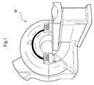
- a method for processing magnetic resonance imaging image information in accordance with the embodiment uses a magnetic resonance imaging system (hereinafter called as MRI system) M shown in Fig. 1.
- Magnetic resonance imaging image information by a magnetic longitudinal relaxation measurement (hereinafter called as T 1 measurement)
- magnetic resonance imaging image information by a magnetic transverse relaxation measurement (hereinafter called as T 2 measurement) are obtained with the MRI system M .
- the MRI system M has the same arrangement as that of a universally known and widely used system for a medical checkup and acts the same so as to obtain the magnetic resonance imaging image information by the T 1 measurement and the magnetic resonance imaging image information by the T 2 measurement.
- an x axis is set along a horizontal direction of a human body
- a y axis is set along a cross direction thereof
- a z axis is set along a vertical direction thereof
- an x-y plane is a sliced image plane on which a matrix is set.
- Measuring points are set at a same pitch along the horizontal direction and the cross direction of the body of a subject as an object to be measured, namely, along the x axis and the y axis with a same matrix score.
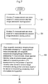
- the MRI system M functions as an information obtaining portion 1 that obtains the magnetic resonance imaging image information, a first obtained image information storing portion 2 that stores the magnetic resonance imaging image information obtained by the T 1 measurement, a second obtained image information storing portion 3 that stores the magnetic resonance imaging image information obtained by the T 2 measurement, and an image output portion 8 that outputs an image.
- the first obtained image information storing portion 2 and the second obtained image information storing portion 3 are arranged in a memory space of an internal memory of the MRI system M .
- the obtained magnetic resonance imaging image information is, as shown in Fig. 3, stored in the first obtained image information storing portion 2 and the second obtained image information storing portion 3.
- a spectral intensity value at each point is indicated on a basis of 16-bit (65536) with the minimum of 0 and the maximum of 65535 and a spectral intensity value at a matrix point ( x,y ) on a sliced image plane of the z th piece is stored in the x th row and the y th column on the z th piece.
- the spectral intensity value of water is low by the T 1 measurement and high by the T 2 measurement.
- the spectral intensity value of a bone is low by both the T 1 measurement and the T 2 measurement.
- the spectral intensity value of a brain is middle by both the T 1 measurement and the T 2 measurement, however, highish by the T 1 measurement.
- the spectral intensity value of skin is middle by the T 1 measurement and highish by the T 2 measurement.
- the above-mentioned tendency is shown in Fig. 4.
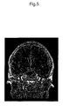
- An example of the image obtained by the T 1 measurement is shown in Fig. 5 and an example of the image obtained by the T 2 measurement is shown in Fig. 6 respectively.
- the spectral intensity value of the magnetic resonance imaging image information obtained by the T 1 measurement and the spectral intensity value of the magnetic resonance imaging image information obtained by the T 2 measurement at the same position are linear-calculated so as to obtain image information showing a bone structure of a head portion.
- the MRI system M further functions as an interpolating processing portion 4, an interpolating processing result information storing portion 5, a linear calculating portion 6 and a calculated result image information storing portion 7.
- the interpolating processing result information storing portion 5 and the calculated result image information storing portion 7 are arranged in the memory space.
- the interpolating processing portion 4 three-dimensionally aligns the magnetic resonance imaging image information obtained by the T 1 measurement and the magnetic resonance imaging image information obtained by the T 2 measurement and a spectral intensity value at the same point as the measured point used for the T 1 measurement is obtained by interpolation of the magnetic resonance imaging image information obtained by the T 2 measurement.
- the interpolating processing result information storing portion 5 stores a calculated result by the interpolating processing portion 4.
- the linear calculating portion 6 inverts each bit of the spectral intensity value stored in the first obtained image information storing portion 2 and also calculates difference between the inverted result and the spectral intensity value stored in the interpolating processing result information storing portion 5 multiplied by a constant number a.
- the constant number a is so set that a calculated result of a spectral intensity value of water is zero.
- the calculated result image information storing portion 7 stores calculated result image information as a set of the calculated results.
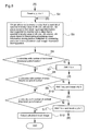
- the information obtaining portion 1 conducts the T 1 measurement and a result of the T 1 measurement is stored in the first obtained image information storing portion 2.
- the information obtaining portion 1 conducts the T 2 measurement and a result of the T 2 measurement is stored in the first obtained image information storing portion 2.
- an x direction of the matrix namely a variable x showing a row in Fig. 3
- a y direction of the matrix namely a variable y showing a column in Fig. 3
- a variable z showing a number of a sliced image plane are reset to 1.
- magnetic resonance imaging image information by a hydrogen nucleus density measurement may be used.
- spectral intensity values of three kinds of image information namely magnetic resonance imaging image information by the hydrogen nucleus density measurement, magnetic resonance imaging image information by the magnetic longitudinal relaxation measurement and the magnetic resonance imaging image information by the magnetic transverse relaxation measurement at a predetermined position may be linearly calculated so as to obtain new image information.
- the spectral intensity values at each position by the hydrogen nucleus density measurement show a tendency different from the spectral intensity values at each position by the magnetic longitudinal relaxation measurement and by the magnetic transverse relaxation measurement, as shown in Fig. 4. Then if the spectral intensity value at the predetermined position by the hydrogen nucleus density measurement is also used as a variable of the linear calculation, further new information can be obtained.
- the above-mentioned method for processing magnetic resonance imaging image information may be used to obtain information on a portion other than a bone structure of human, further information on other than human.
- an algorithm of the linear calculation may be set arbitrarily tailored to a kind of information to be obtained.
- the predetermined position is not determined based on a measuring point of one kind of magnetic resonance imaging image information but may be determined independently from the measuring point of the magnetic resonance imaging image information obtained by the MRI system and the spectral intensity value at the predetermined position of all of the obtained magnetic resonance imaging image information may be obtained by interpolation.
- an algorithm of interpolation is not the linear interpolation used in the above-described embodiment and may be other algorithm.
- the nucleus magnetic resonance spectral may use other atomic nucleus such as a carbon nucleus or a nitrogen nucleus in addition to a hydrogen nucleus.
- comparison may be made between image information obtained by the magnetic resonance imaging image information and image information obtained by an X-ray computed tomography. More concretely, the image information obtained by the magnetic resonance imaging image information and the image information obtained by the X-ray computed tomography are output simultaneously on a same display and both of the image information are output to a printing media such as a paper so as to make the image information visible or a linear calculation is made between a spectral intensity value of the image information obtained by the magnetic resonance imaging image information at the predetermined position and a spectral intensity value of the image information obtained by the X-ray computed tomography at the predetermined position so as to derive further new information.
- a state of a bone can be obtained more accurately by comparison of the information directly showing a position of the bone by the X-ray computed tomography.
- the present claimed invention derives new image information showing an internal state of an object to be measured such as image information showing a bone structure by linear calculation of a plurality of magnetic resonance imaging image information, there is no need of nuclear radiation such as an X-ray that is harmful to a human body when a non-destructive inspection is conducted on an inside of the object to be measured, thereby to improve safety for a non-destructive inspection like this.
Landscapes
- Physics & Mathematics (AREA)
- Radiology & Medical Imaging (AREA)
- Nuclear Medicine, Radiotherapy & Molecular Imaging (AREA)
- Health & Medical Sciences (AREA)
- Engineering & Computer Science (AREA)
- Signal Processing (AREA)
- General Health & Medical Sciences (AREA)
- High Energy & Nuclear Physics (AREA)
- Condensed Matter Physics & Semiconductors (AREA)
- General Physics & Mathematics (AREA)
- Magnetic Resonance Imaging Apparatus (AREA)
- Image Processing (AREA)
- Image Analysis (AREA)
Applications Claiming Priority (2)
| Application Number | Priority Date | Filing Date | Title |
|---|---|---|---|
| JP2002298642A JP3757276B2 (ja) | 2002-10-11 | 2002-10-11 | 磁気共鳴イメージング装置の画像情報処理方法、及び磁気共鳴イメージング装置 |
| JP2002298642 | 2002-10-11 |
Publications (2)
| Publication Number | Publication Date |
|---|---|
| EP1408340A2 true EP1408340A2 (fr) | 2004-04-14 |
| EP1408340A3 EP1408340A3 (fr) | 2005-10-05 |
Family
ID=32025583
Family Applications (1)
| Application Number | Title | Priority Date | Filing Date |
|---|---|---|---|
| EP03022748A Withdrawn EP1408340A3 (fr) | 2002-10-11 | 2003-10-09 | Procédé de traitement d'information d'images obtenues par imagerie par résonance magnétique |
Country Status (3)
| Country | Link |
|---|---|
| US (1) | US6943549B2 (fr) |
| EP (1) | EP1408340A3 (fr) |
| JP (1) | JP3757276B2 (fr) |
Families Citing this family (6)
| Publication number | Priority date | Publication date | Assignee | Title |
|---|---|---|---|---|
| JP3757276B2 (ja) * | 2002-10-11 | 2006-03-22 | 独立行政法人情報通信研究機構 | 磁気共鳴イメージング装置の画像情報処理方法、及び磁気共鳴イメージング装置 |
| JP2008073228A (ja) * | 2006-09-21 | 2008-04-03 | Ge Medical Systems Global Technology Co Llc | 画像診断システムおよびその操作装置 |
| WO2009120635A2 (fr) | 2008-03-23 | 2009-10-01 | Scott Rosa | Procédé d’imagerie diagnostique |
| US8923948B2 (en) * | 2010-07-30 | 2014-12-30 | Vanderbilt University | System and method for determining mechanical properties of bone structures |
| US8538115B2 (en) * | 2011-08-17 | 2013-09-17 | The Board Of Trustees Of The Leland Stanford Junior University | Coil compression for three dimensional autocalibrating parallel imaging with cartesian sampling |
| US9733293B1 (en) * | 2012-09-21 | 2017-08-15 | Qualcomm Incorporated | Differential pixel test for capacitive touch screens |
Family Cites Families (7)
| Publication number | Priority date | Publication date | Assignee | Title |
|---|---|---|---|---|
| JPH04241839A (ja) * | 1991-01-08 | 1992-08-28 | Fuji Electric Co Ltd | Mri画像処理方法 |
| WO1995015537A1 (fr) * | 1993-11-30 | 1995-06-08 | Arch Development Corporation | Procede et systeme automatises destines a l'alignement et a la correlation d'images obtenues selon deux techniques d'imagerie differentes |
| US6285901B1 (en) * | 1999-08-25 | 2001-09-04 | Echo Medical Systems, L.L.C. | Quantitative magnetic resonance method and apparatus for bone analysis |
| US6278891B1 (en) * | 1999-08-25 | 2001-08-21 | Echo Medical Systems, Llc | Nuclear magnetic resonance method and apparatus for bone analysis and imaging |
| EP1319217B1 (fr) * | 2000-09-14 | 2008-11-12 | The Board Of Trustees Of The Leland Stanford Junior University | Technique servant a manipuler des images medicales |
| US6775405B1 (en) * | 2000-09-29 | 2004-08-10 | Koninklijke Philips Electronics, N.V. | Image registration system and method using cross-entropy optimization |
| JP3757276B2 (ja) * | 2002-10-11 | 2006-03-22 | 独立行政法人情報通信研究機構 | 磁気共鳴イメージング装置の画像情報処理方法、及び磁気共鳴イメージング装置 |
-
2002
- 2002-10-11 JP JP2002298642A patent/JP3757276B2/ja not_active Expired - Lifetime
-
2003
- 2003-10-07 US US10/679,343 patent/US6943549B2/en not_active Expired - Fee Related
- 2003-10-09 EP EP03022748A patent/EP1408340A3/fr not_active Withdrawn
Also Published As
| Publication number | Publication date |
|---|---|
| EP1408340A3 (fr) | 2005-10-05 |
| JP3757276B2 (ja) | 2006-03-22 |
| US6943549B2 (en) | 2005-09-13 |
| US20040150399A1 (en) | 2004-08-05 |
| JP2004129911A (ja) | 2004-04-30 |
Similar Documents
| Publication | Publication Date | Title |
|---|---|---|
| Banhart | Advanced tomographic methods in materials research and engineering | |
| Depuydt et al. | A quantitative evaluation of IMRT dose distributions: refinement and clinical assessment of the gamma evaluation | |
| Leclerc et al. | Voxel-scale digital volume correlation | |
| Tanner et al. | Radiotherapy planning of the pelvis using distortion corrected MR images: the removal of system distortions | |
| JP4309062B2 (ja) | 画像化方法 | |
| Renversade et al. | Comparison between diffraction contrast tomography and high-energy diffraction microscopy on a slightly deformed aluminium alloy | |
| JP3364507B2 (ja) | 生体内の電流源分布を推定して表示する方法及びシステム | |
| US9851456B2 (en) | TOF-PET tomograph and a method of imaging using a TOF-PET tomograph, based on a probability of production and lifetime of a positronium | |
| US20130112874A1 (en) | Imaging modality using penetrating radiations | |
| Guizar-Sicairos et al. | Validation study of small-angle X-ray scattering tensor tomography | |
| Dudak et al. | X-ray micro-CT scanner for small animal imaging based on Timepix detector technology | |
| CN115980104A (zh) | 多角度扫描编码孔x射线衍射断层成像系统及成像方法 | |
| US6943549B2 (en) | Method for processing magnetic resonance imaging image information and magnetic resonance imaging system | |
| US7433086B2 (en) | Edge detection and correcting system and method | |
| CN109310383A (zh) | 用于对x射线成像设备进行校准的测试对象 | |
| JPH09103419A (ja) | Mr画像の形成方法および形成装置 | |
| Braga | Non‐invasive imaging techniques | |
| CN103340644B (zh) | 一种图像位置补偿方法及装置 | |
| CN110215223A (zh) | 散射校正方法、系统、可读存储介质和设备 | |
| US7186984B2 (en) | Method for quantifying the radioactivity of living structures of small dimensions by employing emission tomography | |
| CN114391857A (zh) | 一种基于移动最小二乘算法的双能x射线骨密度检测方法 | |
| Chen et al. | Rapid diagnosis and continuous monitoring of intracerebral hemorrhage with magnetic induction tomography based on stacked autoencoder | |
| Bureev et al. | Digital X-ray tomography | |
| Thomas et al. | A dual modality approach to quantitative quality control in emission tomography | |
| CN111089869A (zh) | 多能探测器x射线相衬成像方法及系统、储存介质及设备 |
Legal Events
| Date | Code | Title | Description |
|---|---|---|---|
| PUAI | Public reference made under article 153(3) epc to a published international application that has entered the european phase |
Free format text: ORIGINAL CODE: 0009012 |
|
| AK | Designated contracting states |
Kind code of ref document: A2 Designated state(s): AT BE BG CH CY CZ DE DK EE ES FI FR GB GR HU IE IT LI LU MC NL PT RO SE SI SK TR |
|
| AX | Request for extension of the european patent |
Extension state: AL LT LV MK |
|
| RAP1 | Party data changed (applicant data changed or rights of an application transferred) |
Owner name: NATIONAL INSTITUTE OF INFORMATION AND COMMUNICATIO |
|
| PUAL | Search report despatched |
Free format text: ORIGINAL CODE: 0009013 |
|
| RIC1 | Information provided on ipc code assigned before grant |
Ipc: 7G 06T 7/00 B Ipc: 7G 01R 33/56 A |
|
| AK | Designated contracting states |
Kind code of ref document: A3 Designated state(s): AT BE BG CH CY CZ DE DK EE ES FI FR GB GR HU IE IT LI LU MC NL PT RO SE SI SK TR |
|
| AX | Request for extension of the european patent |
Extension state: AL LT LV MK |
|
| AKX | Designation fees paid | ||
| STAA | Information on the status of an ep patent application or granted ep patent |
Free format text: STATUS: THE APPLICATION IS DEEMED TO BE WITHDRAWN |
|
| 18D | Application deemed to be withdrawn |
Effective date: 20060406 |
|
| REG | Reference to a national code |
Ref country code: DE Ref legal event code: 8566 |