EP1415005B1 - Hochregulierte endogene retroviren in prostatakrebs - Google Patents
Hochregulierte endogene retroviren in prostatakrebs Download PDFInfo
- Publication number
- EP1415005B1 EP1415005B1 EP20010996222 EP01996222A EP1415005B1 EP 1415005 B1 EP1415005 B1 EP 1415005B1 EP 20010996222 EP20010996222 EP 20010996222 EP 01996222 A EP01996222 A EP 01996222A EP 1415005 B1 EP1415005 B1 EP 1415005B1
- Authority
- EP
- European Patent Office
- Prior art keywords
- seq
- herv
- sequence
- polypeptide
- sequences
- Prior art date
- Legal status (The legal status is an assumption and is not a legal conclusion. Google has not performed a legal analysis and makes no representation as to the accuracy of the status listed.)
- Expired - Lifetime
Links
Images


Classifications
-
- C—CHEMISTRY; METALLURGY
- C07—ORGANIC CHEMISTRY
- C07K—PEPTIDES
- C07K14/00—Peptides having more than 20 amino acids; Gastrins; Somatostatins; Melanotropins; Derivatives thereof
- C07K14/005—Peptides having more than 20 amino acids; Gastrins; Somatostatins; Melanotropins; Derivatives thereof from viruses
-
- A—HUMAN NECESSITIES
- A61—MEDICAL OR VETERINARY SCIENCE; HYGIENE
- A61P—SPECIFIC THERAPEUTIC ACTIVITY OF CHEMICAL COMPOUNDS OR MEDICINAL PREPARATIONS
- A61P13/00—Drugs for disorders of the urinary system
- A61P13/08—Drugs for disorders of the urinary system of the prostate
-
- A—HUMAN NECESSITIES
- A61—MEDICAL OR VETERINARY SCIENCE; HYGIENE
- A61P—SPECIFIC THERAPEUTIC ACTIVITY OF CHEMICAL COMPOUNDS OR MEDICINAL PREPARATIONS
- A61P15/00—Drugs for genital or sexual disorders; Contraceptives
-
- A—HUMAN NECESSITIES
- A61—MEDICAL OR VETERINARY SCIENCE; HYGIENE
- A61P—SPECIFIC THERAPEUTIC ACTIVITY OF CHEMICAL COMPOUNDS OR MEDICINAL PREPARATIONS
- A61P25/00—Drugs for disorders of the nervous system
-
- A—HUMAN NECESSITIES
- A61—MEDICAL OR VETERINARY SCIENCE; HYGIENE
- A61P—SPECIFIC THERAPEUTIC ACTIVITY OF CHEMICAL COMPOUNDS OR MEDICINAL PREPARATIONS
- A61P3/00—Drugs for disorders of the metabolism
- A61P3/08—Drugs for disorders of the metabolism for glucose homeostasis
- A61P3/10—Drugs for disorders of the metabolism for glucose homeostasis for hyperglycaemia, e.g. antidiabetics
-
- A—HUMAN NECESSITIES
- A61—MEDICAL OR VETERINARY SCIENCE; HYGIENE
- A61P—SPECIFIC THERAPEUTIC ACTIVITY OF CHEMICAL COMPOUNDS OR MEDICINAL PREPARATIONS
- A61P31/00—Antiinfectives, i.e. antibiotics, antiseptics, chemotherapeutics
- A61P31/12—Antivirals
- A61P31/14—Antivirals for RNA viruses
-
- A—HUMAN NECESSITIES
- A61—MEDICAL OR VETERINARY SCIENCE; HYGIENE
- A61P—SPECIFIC THERAPEUTIC ACTIVITY OF CHEMICAL COMPOUNDS OR MEDICINAL PREPARATIONS
- A61P35/00—Antineoplastic agents
-
- A—HUMAN NECESSITIES
- A61—MEDICAL OR VETERINARY SCIENCE; HYGIENE
- A61P—SPECIFIC THERAPEUTIC ACTIVITY OF CHEMICAL COMPOUNDS OR MEDICINAL PREPARATIONS
- A61P37/00—Drugs for immunological or allergic disorders
- A61P37/02—Immunomodulators
- A61P37/04—Immunostimulants
-
- C—CHEMISTRY; METALLURGY
- C12—BIOCHEMISTRY; BEER; SPIRITS; WINE; VINEGAR; MICROBIOLOGY; ENZYMOLOGY; MUTATION OR GENETIC ENGINEERING
- C12N—MICROORGANISMS OR ENZYMES; COMPOSITIONS THEREOF; PROPAGATING, PRESERVING, OR MAINTAINING MICROORGANISMS; MUTATION OR GENETIC ENGINEERING; CULTURE MEDIA
- C12N7/00—Viruses; Bacteriophages; Compositions thereof; Preparation or purification thereof
-
- C—CHEMISTRY; METALLURGY
- C12—BIOCHEMISTRY; BEER; SPIRITS; WINE; VINEGAR; MICROBIOLOGY; ENZYMOLOGY; MUTATION OR GENETIC ENGINEERING
- C12Q—MEASURING OR TESTING PROCESSES INVOLVING ENZYMES, NUCLEIC ACIDS OR MICROORGANISMS; COMPOSITIONS OR TEST PAPERS THEREFOR; PROCESSES OF PREPARING SUCH COMPOSITIONS; CONDITION-RESPONSIVE CONTROL IN MICROBIOLOGICAL OR ENZYMOLOGICAL PROCESSES
- C12Q1/00—Measuring or testing processes involving enzymes, nucleic acids or microorganisms; Compositions therefor; Processes of preparing such compositions
- C12Q1/68—Measuring or testing processes involving enzymes, nucleic acids or microorganisms; Compositions therefor; Processes of preparing such compositions involving nucleic acids
- C12Q1/6876—Nucleic acid products used in the analysis of nucleic acids, e.g. primers or probes
- C12Q1/6883—Nucleic acid products used in the analysis of nucleic acids, e.g. primers or probes for diseases caused by alterations of genetic material
- C12Q1/6886—Nucleic acid products used in the analysis of nucleic acids, e.g. primers or probes for diseases caused by alterations of genetic material for cancer
-
- G—PHYSICS
- G01—MEASURING; TESTING
- G01N—INVESTIGATING OR ANALYSING MATERIALS BY DETERMINING THEIR CHEMICAL OR PHYSICAL PROPERTIES
- G01N33/00—Investigating or analysing materials by specific methods not covered by groups G01N1/00 - G01N31/00
- G01N33/48—Biological material, e.g. blood, urine; Haemocytometers
- G01N33/50—Chemical analysis of biological material, e.g. blood, urine; Testing involving biospecific ligand binding methods; Immunological testing
- G01N33/53—Immunoassay; Biospecific binding assay; Materials therefor
- G01N33/575—Immunoassay; Biospecific binding assay; Materials therefor for cancer
- G01N33/57555—Immunoassay; Biospecific binding assay; Materials therefor for cancer of the prostate
-
- A—HUMAN NECESSITIES
- A61—MEDICAL OR VETERINARY SCIENCE; HYGIENE
- A61K—PREPARATIONS FOR MEDICAL, DENTAL OR TOILETRY PURPOSES
- A61K39/00—Medicinal preparations containing antigens or antibodies
- A61K2039/505—Medicinal preparations containing antigens or antibodies comprising antibodies
-
- A—HUMAN NECESSITIES
- A61—MEDICAL OR VETERINARY SCIENCE; HYGIENE
- A61K—PREPARATIONS FOR MEDICAL, DENTAL OR TOILETRY PURPOSES
- A61K39/00—Medicinal preparations containing antigens or antibodies
- A61K2039/51—Medicinal preparations containing antigens or antibodies comprising whole cells, viruses or DNA/RNA
- A61K2039/53—DNA (RNA) vaccination
-
- A—HUMAN NECESSITIES
- A61—MEDICAL OR VETERINARY SCIENCE; HYGIENE
- A61K—PREPARATIONS FOR MEDICAL, DENTAL OR TOILETRY PURPOSES
- A61K39/00—Medicinal preparations containing antigens or antibodies
-
- C—CHEMISTRY; METALLURGY
- C12—BIOCHEMISTRY; BEER; SPIRITS; WINE; VINEGAR; MICROBIOLOGY; ENZYMOLOGY; MUTATION OR GENETIC ENGINEERING
- C12N—MICROORGANISMS OR ENZYMES; COMPOSITIONS THEREOF; PROPAGATING, PRESERVING, OR MAINTAINING MICROORGANISMS; MUTATION OR GENETIC ENGINEERING; CULTURE MEDIA
- C12N2740/00—Reverse transcribing RNA viruses
- C12N2740/00011—Details
- C12N2740/10011—Retroviridae
- C12N2740/10021—Viruses as such, e.g. new isolates, mutants or their genomic sequences
-
- C—CHEMISTRY; METALLURGY
- C12—BIOCHEMISTRY; BEER; SPIRITS; WINE; VINEGAR; MICROBIOLOGY; ENZYMOLOGY; MUTATION OR GENETIC ENGINEERING
- C12N—MICROORGANISMS OR ENZYMES; COMPOSITIONS THEREOF; PROPAGATING, PRESERVING, OR MAINTAINING MICROORGANISMS; MUTATION OR GENETIC ENGINEERING; CULTURE MEDIA
- C12N2740/00—Reverse transcribing RNA viruses
- C12N2740/00011—Details
- C12N2740/10011—Retroviridae
- C12N2740/10022—New viral proteins or individual genes, new structural or functional aspects of known viral proteins or genes
-
- C—CHEMISTRY; METALLURGY
- C12—BIOCHEMISTRY; BEER; SPIRITS; WINE; VINEGAR; MICROBIOLOGY; ENZYMOLOGY; MUTATION OR GENETIC ENGINEERING
- C12Q—MEASURING OR TESTING PROCESSES INVOLVING ENZYMES, NUCLEIC ACIDS OR MICROORGANISMS; COMPOSITIONS OR TEST PAPERS THEREFOR; PROCESSES OF PREPARING SUCH COMPOSITIONS; CONDITION-RESPONSIVE CONTROL IN MICROBIOLOGICAL OR ENZYMOLOGICAL PROCESSES
- C12Q1/00—Measuring or testing processes involving enzymes, nucleic acids or microorganisms; Compositions therefor; Processes of preparing such compositions
- C12Q1/70—Measuring or testing processes involving enzymes, nucleic acids or microorganisms; Compositions therefor; Processes of preparing such compositions involving virus or bacteriophage
- C12Q1/701—Specific hybridization probes
- C12Q1/702—Specific hybridization probes for retroviruses
-
- C—CHEMISTRY; METALLURGY
- C12—BIOCHEMISTRY; BEER; SPIRITS; WINE; VINEGAR; MICROBIOLOGY; ENZYMOLOGY; MUTATION OR GENETIC ENGINEERING
- C12Q—MEASURING OR TESTING PROCESSES INVOLVING ENZYMES, NUCLEIC ACIDS OR MICROORGANISMS; COMPOSITIONS OR TEST PAPERS THEREFOR; PROCESSES OF PREPARING SUCH COMPOSITIONS; CONDITION-RESPONSIVE CONTROL IN MICROBIOLOGICAL OR ENZYMOLOGICAL PROCESSES
- C12Q2600/00—Oligonucleotides characterized by their use
- C12Q2600/158—Expression markers
-
- G—PHYSICS
- G01—MEASURING; TESTING
- G01N—INVESTIGATING OR ANALYSING MATERIALS BY DETERMINING THEIR CHEMICAL OR PHYSICAL PROPERTIES
- G01N2333/00—Assays involving biological materials from specific organisms or of a specific nature
- G01N2333/005—Assays involving biological materials from specific organisms or of a specific nature from viruses
- G01N2333/08—RNA viruses
- G01N2333/15—Retroviridae, e.g. bovine leukaemia virus, feline leukaemia virus, feline leukaemia virus, human T-cell leukaemia-lymphoma virus
Definitions
- the present invention relates to the diagnosis of cancer, particularly prostate cancer.
- it relates to a subgroup of human endogenous retroviruses (HERVs) which show up-regulated expression in tumors, particularly prostate tumors.
- HERVs human endogenous retroviruses
- Prostate cancer is the most common type of cancer in men in the USA.
- Benign prostatic hyperplasia (BPH) is the abnormal growth of benign prostate cells in which the prostate grows and pushes against the urethra and bladder, blocking the normal flow of urine. More than half of the men in the USA between the ages of 60 and 70 and as many as 90 percent between the ages of 70 and 90 have symptoms of BPH. Although this condition is seldom a threat to life, it may require treatment to relieve symptoms.
- Prostate cancer may remain in the prostate gland, or it may spread to nearby lymph nodes and may also spread to the bones, bladder, rectum, and other organs.
- Prostate cancer is diagnosed by measuring the levels of pmstate-specific antigen (PSA) and prostatic acid phosphatase (PAP) in the blood.
- PSA pmstate-specific antigen
- PAP prostatic acid phosphatase
- the level of PSA in blood may rise in men who have prostate cancer, BPH, or an infection in the prostate.
- the level of PAP rises above normal in many prostate cancer patients, especially if the cancer has spread beyond the prostate.
- PSA or PAP levels may also indicate other, non-cancerous problems.
- HERVs human endogenous retroviruses
- the invention provides a method for diagnosing cancer, especially prostate cancer, the method comprising the step of detecting the presence or absence of an expression product of a HML-2 endogenous retrovirus in a patient sample, wherein the patient sample contains prostate cells and for wherein the patient is suspected of having prostate cancer wherein up-regulation of expression of at least 150% relative to a negative control is indicative of prostate cancer.
- the HML-2 expression product which is detected is either a mRNA transcript or a polypeptide translated from such a transcript
- These expression products may be detected directly or indirectly.
- a direct test uses an assay which detects HML-2 RNA or polypeptide in a patient sample.
- An indirect test uses an assay which detects biomolecules which are not directly expressed in vivo from HML-2 e.g. an assay to detect cDNA which has been reverse-transcribed from a HML-2 mRNA, or an assay to detect an antibody which has been raised in response to a HML-2 polypeptide.
- the patient sample will generally comprise cells, preferably, prostate cells. These may be present in a sample of tissue, preferably, prostate tissue, or may be cells, preferably, prostate cells which have escaped into circulation ( e.g. during metastasis). Instead of or as well as comprising prostate cells, the sample may comprise virions which contain mRNA from HML-2.
- the patient sample may comprise cells, preferably, prostate cells and/or virions (as described above for mRNA), or may comprise antibodies which recognize HML-2 polypeptides. Such antibodies will typically be present in circulation.
- the patient sample is tissue sample (e.g. a biopsy), preferably, a prostate sample (e.g . a biopsy) or a blood sample.
- tissue sample e.g. a biopsy
- prostate sample e.g. a biopsy
- blood sample e.g. a blood sample
- the patient is generally a human, preferably human male, and more preferably an adult human male.
- Expression products may be detected in the patient sample itself, or it may be detected in material derived from the sample (e.g . the supernatant of a cell lysate, or a RNA extract, or cDNA generated from a RNA extract, or polypeptides translated from a RNA extract, or cells derived from culture of cells extracted from a patient etc. ). These are still considered to be "patient samples" within the meaning of the invention.
- Methods of the invention can be conducted in vitro or in vivo.
- patient samples include isolated cells, whole tissues, or bodily fluids (e.g. blood, plasma, serum, urine, pleural effusions, cerebro-spinal fluid, etc .)
- bodily fluids e.g. blood, plasma, serum, urine, pleural effusions, cerebro-spinal fluid, etc .
- the diagnostic method of the invention is based on mRNA detection, it typically involves detecting a RNA comprising six basic regions. From 5' to 3', these are:
- PCA-mRNA molecules prostate cancer associated mRNA
- PCAVs prostate cancer associated viruses
- PCA-mRNAs include all six of these regions, most HERVs are defective in that they have accumulated multiple stop codons, frameshifts, or larger deletions etc. This means that many PCA-mRNAs do not include all six regions. As all PCA-mRNAs are transcribed under the control of the same group of LTRs, however, transcription of all PCA-mRNAs is up-regulated in prostate tumors even though the mRNA may not encode functional polypeptides.
- the mRNA to be detected has the formula N 1 -N 2 -N 3 -N 4 -N 5 -polyA, wherein:
- N 1 , N 2 , N 3 , N 4 or N 5 needs to be present, it is preferred that two, three, four or five of these regions are present. It is preferred that at least one of N 1 and/or N 5 is present.
- N 1 is preferably present in the mRNA to be detected ( i.e . the invention is preferably based on the detection of mRNA driven by a 5' LTR). More preferably, at least N 1 -N 2 is present.
- N 1 is present, it is preferably at the 5' end of the mRNA (i.e . 5'- N 1 -).
- N 5 is present, it is preferably immediately before a 3' polyA tail ( i.e .... -N 5 -polyA-3').
- N 4 preferably comprises a polypeptide-coding sequence (e.g . encoding a HML-2 polypeptide). Examples of HML-2 polypeptide-coding sequences are described below.
- the RNA will generally have a 5' cap.
- the method of the invention preferably comprises an initial step of: (a) extracting RNA (e.g . mRNA) from a patient sample; (b) removing DNA from a patient sample without removing mRNA; and/or (c) removing or disrupting DNA which comprises SEQ ID 4, but not RNA which comprises SEQ ID 4, from a patient sample.
- RNA e.g . mRNA
- the method of the invention preferably comprises an initial step of: (a) extracting RNA (e.g . mRNA) from a patient sample; (b) removing DNA from a patient sample without removing mRNA; and/or (c) removing or disrupting DNA which comprises SEQ ID 4, but not RNA which comprises SEQ ID 4, from a patient sample.
- RNA-specific assay can be used which is not affected by the presence of homologous DNA.
- RNA may be enriched e.g . using oligo-dT techniques.
- Methods for removing DNA from biological samples without removing mRNA are well known [ e.g . appendix C of ref. 2] and include DNase digestion.
- Methods for removing DNA, but not RNA, comprising PCA-mRNA sequences will use a reagent which is specific to a sequence within a PCA-mRNA e.g . a restriction enzyme which recognizes a DNA sequence within SEQ ID 4, but which does not cleave the corresponding RNA sequence.
- a reagent which is specific to a sequence within a PCA-mRNA e.g . a restriction enzyme which recognizes a DNA sequence within SEQ ID 4, but which does not cleave the corresponding RNA sequence.
- Methods for specifically purifying PCA-mRNAs from a sample may also be used.
- One such method uses an affinity support which binds to PCA-mRNAs.
- the affinity support may include a polypeptide sequence which binds to the PCAV-mRNA e.g . the cORF polypeptide, which binds to the LTR of HERV-K mRNAs in a sequence-specific manner, or HIV Rev protein, which has been shown to recognize the HERV-K LTR [3].
- RNA-specific detection technique Detecting the presence or absence of a particular RNA sequence in a sample [ e.g . refs. 2 & 8]. If a sample contains genomic PCAV DNA, the detection technique will generally be RNA-specific; if the sample contains no PCAV DNA, the detection technique may or may not be RNA-specific.
- Hybridization-based detection techniques may be used, in which a polynucleotide probe complementary to a region of PCA-mRNA is contacted with a RNA-containing sample under hybridizing conditions. Detection of hybridization indicates that nucleic acid complementary to the probe is present.
- Hybridization techniques for use with RNA include Northern blots, in situ hybridization and arrays.
- Sequencing may also be used, in which the sequence(s) of RNA molecules in a sample are obtained. These techniques reveal directly whether a sequence of interest is present in a sample. Sequence determination of the 5' end of a RNA corresponding to N 1 will generally be adequate.
- Amplification-based techniques may also be used. These include PCR, SDA, SSSR, LCR, TMA, NASBA, T7 amplification etc.
- the technique preferably gives exponential amplification.
- a preferred technique for use with RNA is RT-PCR [ e.g . see chapter 15 of ref. 2]. RT-PCR of mRNA from prostate cells is reported in references 4, 5, 6 & 7.
- RNA RNA which are derived from RNA
- a typical indirect method of detecting mRNA is to prepare cDNA by reverse transcription and then to directly detect the cDNA.
- Direct detection of cDNA will generally use the same techniques as described above for direct detection of RNA (but it will be appreciated that methods such as RT-PCR are not suitable for DNA detection and that cDNA is doublo-stranded, so detection techniques can be based on a sequence, on its complement, or on the double-stranded molecule).
- the method will involve detecting expression of a polypeptide encoded by a PCAV-mRNA. This will typically involve detecting one or more of the following HML-2 polypeptides: gag, prt, pol, env, cORF. Although some PCA-mRNAs encode all of these polypeptides (e.g. ERVK6 [1]), the polypeptide-coding regions of most HERVs (including PCAVs) contain mutations which mean that one or more coding-regions in the mRNA transcript are either mutated or absent. Thus not all PCAVs have the ability to encode all HML-2 polypeptides.
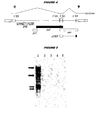
- transcripts which encode HML-2 polypeptides are generated by alternative splicing of the full-length mRNA copy of the endogenous genome [ e.g. Figure 4 of ref. 143].
- HML-2 gag polypeptide is encoded by the first long ORF in a complete HML-2 genome [140]. Full-length gag polypeptide is proteolytically cleaved.
- gag nucleotide sequences are: SEQ IDs 7, 8, 9 & 11 [HERV-K(CH)]; SEQ ID 85 [HERV-K108]; SEQ ID 91 [HERV-K(C7)]; SEQ ID 97 [BFRV-K(II)]; SEQ ID 102 [HERV-K10].
- gag polypeptide sequences are: SEQ IDs 46, 47, 48, 49, 56 & 57 [HERV-K(CH)]; SEQ ID 92 [HERV-K(C7)]; SEQ ID 98 [HERV-K(II)]; SEQ IDs 103 & 104 [HERV-K10]; SEQ ID 146 [ ⁇ ERVK66'].
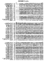
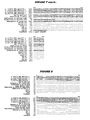
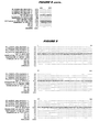
- gag polypeptide sequences An alignment of gag polypeptide sequences is shown in Figure 7 .
- HML-2 prt polypeptide is encoded by the second long ORF in a complete HML-2 genome. It is translated as a gag-prt fusion polypeptide. The fusion polypeptide is proteolytically cleaved to give a protease.
- prt nucleotide sequences are: SEQ ID 86 [HERV-K(108)]; SEQ ID 99 [HERV-K(II)]; SEQ ID 105 [HERV-K10].
- prt polypeptide sequences are: SEQ ID 106 [HERV-K10]; SEQ ID 147 ['ERVK6'].
- HML-2 pol polypeptide is encoded by the third long ORF in a complete HML-2 genome. It is translated as a gag-prt-pol fusion polypeptide. The fusion polypeptide is proteolytically cleaved to give three pol products - reverse transcriptase, endonuclease and integrase [14].
- pol nucleotide sequences are: SEQ ID 87 [HERV-K(108)]; SEQ ID 93 [HERV-K(C7)]; SEQ ID 100 [HERV-K(II)]; SEQ ID 107 [HERV-K10].
- polypeptide sequences are: SEQ ID 94 [HERV-K(C7)]; SEQ ID 108 [HERV-K10]; SEQ ID 148 ['ERVK6'].
- HML-2 env polypeptide is encoded by the fourth long ORF in a complete HML-2 genome.
- the translated polypeptide is proteolytically cleaved.
- env nucleotide sequences are: SEQ ID 88 [HERV-K(108)]; SEQ ID 95 [HERV-K(C7)]; SEQ ID 101 [HERV-K(II)]; SEQ ID 107 [HERV-K10].
- env polypeptide sequences are: SEQ ID 96 [HERV-K(C7)]; SEQ ID 108 [HERV-K10] ; SEQ ID 149 ['ERVK6'].
- HML-2 cORP polypeptide is encoded by an ORF which shares the same 5' region and start codon as env. After amino acid 87, a splicing event removes env-coding sequences and the cORF-coding sequence continues in the reading frame +1 relative to that of env [15,16; see below]. cORF has also been called Rec [17].
- cORF nucleotide sequences are: SEQ ID 89 and SEQ ID 90 [HERV-K(108)]
- Examples of cORF polypeptide sequences are SEQ ID 109.
- Suitable techniques include standard immunohistological methods, immunoprecipitation, immunofluorescence, ELISA, RIA, FIA, etc.
- the invention provides a method for detecting the presence of and/or measuring a level of a polypeptide in a biological sample, wherein the method uses an antibody specific for the polypeptide.
- the method generally comprises the steps of: a) contacting the sample with an antibody specific for the polypeptide; and b) detecting binding between the antibody and polypeptides in the sample.
- Polypeptides can also be detected by functional assays e.g . assays to detect binding activity or enzymatic activity.
- functional assays e.g . assays to detect binding activity or enzymatic activity.
- a functional assay for cORF is disclosed in references 16,129 & 130.
- a functional assay for the protease is disclosed in reference 140.
- polypeptides can be separated using 2D-PAGE and polypeptide spots can be sequenced ( e.g . by mass spectroscopy) in order to identify if a sequence is present in a target polypeptide.
- Detection methods may be adapted for use in vivo (e.g. to locate or identify sites where cancer cells are present).
- an antibody specific for a target polypeptide is administered to an individual ( e.g . by injection) and the antibody is located using standard imaging techniques (e.g. magnetic resonance imaging, computed tomography scanning, etc .). Appropriate labels (e.g. spin labels etc .) will be used. Using these techniques, cancer cells are differentially labeled.
- An immunofluorescence assay can be easily performed on cells without the need for purification of the target polypeptide.
- the cells are first fixed onto a solid support, such as a microscope slide or microtiter well.
- the membranes of the cells are then permeablized in order to permit entry of polypeptide-specific antibody (NB: fixing and permeabilization can be achieved together).
- NB fixing and permeabilization can be achieved together.
- the fixed cells are exposed to an antibody which is specific for the encoded polypeptide and which is fluorescently labeled.
- the presence of this label e.g. visualized under a microscope
- polypeptides may be preferred to detect molecules which are produced by the body in response to a polypeptide (i.e . indirect detection of a polypeptide). This will typically involve the detection of antibodies, so the patient sample will generally be a blood sample. Antibodies can be detected by conventional immunoassay techniques e.g. using PCAV polypeptides which will typically be immobilized.
- Antibodies against HERV-K polypeptides have been detected in humans [143].
- references to a percentage sequence identity between two amino acid sequences means that, when aligned, that percentage of amino acids are the same in comparing the two sequences.
- This alignment and the percent homology or sequence identity can be determined using software programs known in the art, for example those described in section 7.7.18 of reference 11.
- a preferred alignment is determined by the Smith-Waterman homology search algorithm using an affine gap search with a gap open penalty of 12 and a gap extension penalty of 2, BLOSUM matrix of 62.
- the Smith-Waterman homology search algorithm is taught in reference 32.
- polypeptide refers to amino acid polymers of any length.
- the polymer may be linear or branched, it may comprise modified amino acids, and it may be interrupted by non-amino acids.
- the terms also encompass an amino acid polymer that has been modified naturally or by intervention; for example, disulfide bond formation, glycosylation, lipidation, acetylation, phosphorylation, or any other manipulation or modification, such as conjugation with a labeling component
- polypeptides containing one or more analogs of an amino acid including, for example, unnatural amino acids, etc .
- Polypeptides can occur as single chains or associated chains.
- Polypeptides of the invention can be naturally or non-naturally glycosylated (i.e. the polypeptide has a glycosylation pattern that differs from the glycosylation pattern found in the corresponding naturally occurring polypeptide).
- Mutants can include amino acid substitutions, additions or deletions.
- the amino acid substitutions can be conservative amino acid substitutions or substitutions to eliminate non-essential amino acids, such as to alter a glycosylation site, a phosphorylation site or an acetylation site, or to minimize misfolding by substitution or deletion of one or more cysteine residues that are not necessary for function.
- Conservative amino acid substitutions are those that preserve the general charge, hydrophobicity/hydrophilicity, and/or steric bulk of the amino acid substituted.
- Variants can be designed so as to retain or have enhanced biological activity of a particular region of the polypeptide ( e.g.
- amino acid alterations for production of variants can be based upon the accessibility (interior vs. exterior) of the amino acid (e.g . ref. 33), the thermostability of the variant polypeptide (e.g. ref. 34), desired glycosylation sites (e.g . ref. 35), desired disulfide bridges ( e.g . refs. 36 & 37), desired metal binding sites ( e.g . refs.38 & 39), and desired substitutions with in proline loops ( e.g . ref. 40). Cysteine-depleted muteins can be produced as disclosed in reference 41.
- Antibodies be polyclonal or monoclonal and may be produced by any suitable means ( e.g . by recombinant expression).
- Antibodies may include a label.
- the label may be detectable directly, such as a radioactive or fluorescent label.
- the label may be detectable indirectly, such as an enzyme whose products are detectable (e.g . luciferase, ß-galactosidase, peroxidase etc .).
- Antibodies may be attached to a solid support.
- Antibodies be prepared by administering (e.g. injecting) a polypeptide of the invention to an appropriate animal (e.g. a rabbit, hamster, mouse or other rodent).
- an appropriate animal e.g. a rabbit, hamster, mouse or other rodent.
- Antigen-binding fragments of antibodies include Fv, scFv, Fc, Fab, F(ab') 2 etc.
- the antibodies may be chimeric or humanized [ e.g. refs. 42 & 43], or fully human antibodies may be used. Because humanized antibodies are far less immunogenic in humans than the original non-human monoclonal antibodies, they can be used for the treatment of humans with far less risk of anaphylaxis. Thus, these antibodies may be preferred in therapeutic applications that involve in vivo administation to a human such as, use as radiation sensitizers for the treatment of neoplastic disease or use in methods to reduce the side effects of cancer therapy.
- Humanized antibodies may be achieved by a variety of methods including, for example:
- CDRs are amino acid sequences which together define the binding affinity and specificity of a Fv region of a native immunoglobulin binding site [ e.g. refs. 51 & 52].
- constant region refers to the portion of the antibody molecule that confers effector functions.
- mouse constant regions are substituted by human constant regions.
- the constant regions of humanized antibodies are derived from human immunoglobulins.
- the heavy chain constant region can be selected from any of the 5 isotypes: alpha, delta, epsilon, gamma or mu.
- One method of humanizing antibodies comprises aligning the heavy and light chain sequences of a non-human antibody to human heavy and light chain sequences, replacing the non-human framework residues with human framework residues based on such alignment, molecular modeling of the conformation of the humanized sequence in comparison to the conformation of the non-human parent antibody, and repeated back mutation of residues in the framework region which disturb the structure of the non-human CDRs until the predicted conformation of the CDRs in the humanized sequence model closely approximates the conformation of the non-human CDRs of the parent non-human antibody.
- Such humanized antibodies may be further derivatized to facilitate uptake and clearance e.g, via Ashwell receptors. [refs. 53 & 54]
- Humanized or fully-human antibodies can also be produced using transgenic animals that are engineered to contain human immunoglobulin loci.
- ref. 55 discloses transgenic animals having a human Ig locus wherein the animals do not produce functional endogenous immunoglobulins due to the inactivation of endogenous heavy and light chain loci.
- Ref. 56 also discloses transgenic non-primate mammalian hosts capable of mounting an immune response to an immunogen, wherein the antibodies have primate constant and/or variable regions, and wherein the endogenous immunoglobulin-encoding loci are substituted or inactivated.
- Ref. 57 discloses the use of the Cre/Lox system to modify the immunoglobulin locus in a mammal, such as to replace all or a portion of the constant or variable region to form a modified antibody molecule.
- Ref. 58 discloses non-human mammalian hosts having inactivated endogenous Ig loci and functional human Ig loci.
- Ref. 59 discloses methods of making transgenic mice in which the mice lack endogenous heavy claims, and express an exogenous immunoglobulin locus comprising one or more xenogeneic constant regions.
- an immune response can be produced to a PCAV polypeptide, and antibody-producing cells can be removed from the animal and used to produce hybridomas that secrete human monoclonal antibodies.
- Immunization protocols, adjuvants, and the like are known in the art, and are used in immunization of, for example, a transgenic mouse as described in ref. 60.
- the monoclonal antibodies can be tested for the ability to inhibit or neutralize the biological activity or physiological effect of the corresponding polypeptide.
- HML-2 transcripts are up-regulated in tumors, including prostate tumors.
- a reference point is needed i.e. a control.
- Analysis of the control sample gives a standard level of RNA and/or protein expression against which a patient sample can be compared.
- a negative control gives a background or basal level of expression against which a patient sample can be compared.
- Higher levels of expression product relative to a negative control indicate that the patient from whom the sample was taken has, for example, prostate cancer.
- negative controls would include lifetime baseline levels of expression or the expression level observed in pooled normals.
- equivalent levels of expression product indicate that the patient does not have a HML-2-related cancer such as prostate cancer.
- a positive control gives a level of expression against which a patient sample can be compared. Equivalent or higher levels of expression product relative to a positive control indicate that the patient from whom the sample was taken has cancer such as prostate cancer. Conversely, lower levels of expression product indicate that the patient does not have a HML-2 related cancer such as prostate cancer.
- a negative control will generally comprise cells which are not from a tumor cell, e.g. a prostate tumor cell.
- a negative control will generally be a blood sample from a patient who does not have a prostate tumor.
- the negative control could be a sample from the same patient as the patient sample, but from a tissue in which HML-2 expression is not up-regulated e.g. a non-tumor non-prostate cell.
- the negative control could be a prostate cell from the same patient as the patient sample, but taken at an earlier stage in the patient's life.
- the negative control could be a cell from a patient without a prostate tumor. This cell may or may not be a prostate cell.
- the negative control cell could be a prostate cell from a patient with BPH.
- a positive control will generally comprise cells from a tumor cell e.g . a prostate tumor.
- a negative control will generally be a blood sample from a patient who has a prostate tumor.
- the positive control could be a prostate tumor cell from the same patient as the patient sample, but taken at an earlier stage in the patient's life ( e.g . to monitor remission).
- the positive control could be a cell from another patient with a prostate tumor.
- the positive control could be a prostate cell line.
- HML-2 expression in the control can be assessed at the same time as expression in the patient sample. Alternatively, HML-2 expression in the control can be assessed separately (earlier or later).
- control may be an absolute value i.e . a level of expression which has been empirically determined from samples taken from prostate tumor patients ( e.g . under standard conditions).
- the up-regulation relative to the control (100%) will usually be at least 150% ( e.g. 200%, 250%, 300%, 400%, 500%, 600% or more).
- the invention provides a method for diagnosing prostate cancer. It will be appreciated that "diagnosis" according to the invention can range from a definite clinical diagnosis of disease to an indication that the patient should undergo further testing which may lead to a definite diagnosis.
- diagnosis can range from a definite clinical diagnosis of disease to an indication that the patient should undergo further testing which may lead to a definite diagnosis.
- the method of the invention can be used as part of a screening process, with positive samples being subjected to further analysis.
- diagnosis includes monitoring the progress of cancer in a patient already known to have the cancer.
- Cancer can also be staged by the methods of the invention.
- the cancer is prostate cancer.
- the efficacy of a treatment regimen (therametrics) of a cancer associated can also monitored by the method of the invention e.g. to determine its efficacy.
- Susceptibility to a cancer can also be detected e.g. where up-regulation of expression has occurred, but before cancer has developed. Prognostic methods are also encompassed.
- Endogenous retroviruses were identified in human genomic DNA by their homology to retroviruses of other vertebrates [131, 132]. It is believed that the human genome probably contains numerous copies of endogenous proviral DNAs, but little is known about their function. Most HERV families have relatively few members (1-50) but one family (HERV-H) consists of ⁇ 1000 copies per haploid genome distributed on all chromosomes. The large numbers and general transcriptional activity of HERVs in embryonic and tumor cell lines suggest that they could act as disease-causing insertional mutagens or affect adj acent gene expression in a neutral or beneficial way.
- the K family of human endogenous retroviruses is well known [133]. It is related to the mouse mammary tumor virus (MMTV) and is present in the genomes of humans, apes and old world monkeys, but several human HERV-K proviruses are unique to humans [134].
- the HERV-K family is present at 30-50 full-length copies per haploid human genome and possesses long open reading frames that potentially are translated into viral proteins [135, 136].
- Two types of proviral genomes are known, which differ by the presence (type 2) or absence (type 1) of a stretch of 292 nucleotides in the overlapping boundary of the pol and env genes [137].
- HERV-K10 has been shown to encode a full-length gag homologous 73 kDa protein and a functional protease [140].
- Gag proteins released in form of particles from HERV-K have been identified in the cell culture supernatant of the teratocarcinoma derived cell line Tera 1. These retrovirus-like particles (termed “human teratocarcinoma derived virus” or HTDV) have been shown to have a 90% sequence homology to the HERV-K10 genome [138, 143].
- retroviral proviruses While the HERV-K family is present in the genome of every human cell, a high level of expression of mRNAs, proteins and particles is observed only in human teratocarcinoma cell lines [144]. In other tissues and cell lines, only a basal level of expression of mRNA has been demonstrated even using very sensitive methods.
- the expression of retroviral proviruses is generally regulated by elements of the 5' long terminal repeat (LTR). Furthermore, the activation of expression of an endogenous retrovirus may trigger the expression of a downstream gene that triggers a neoplastic effect.
- HML-2 is a subgroup of the HERV-K family [146].
- HERV isolates which are members of the HML-2 subgroup include HERV-K10 [137,142], the 27 HML-2 viruses shown in Figure 4 of reference 147, HERV-K(C7) [148], HERV-K(II) [145], HERV-K(CH)
- the invention is based on the finding that HML-2 mRNA expression is up-regulated in prostate tumors. Because HML-2 is a well-recognized family, the skilled person will be able to determine without difficulty whether any particular endogenous retroviruses is or is not a HML-2.
- Preferred members of the HML-2 family for use in accordance with the present invention are those whose proviral genome has an LTR which has at least 75% sequence identity to SEQ ID 150 (the LTR sequence from HML-2.HOM [1]).
- Example LTRs include SEQ IDs 151-154.
- the present invention is based on the discovery of elevated levels of multiple HML-2 polynucleotides in prostate tumor samples as compared to normal prostate tissue.
- One particular HML-2 whose mRNA was found to be up-regulated is designated herein as ⁇ HERV-K(CH)'.
- HERV-K(CH) Sequences from HERV-K(CH) are shown in SEQ IDs 14-39 and have been deposited with the ATCC (see Table 7). The skilled person will be able to classify any further HERV as HERV-K(CH) or not based on sequence identity to these HERV-K(CH) polynucleotides. Preferably such a comparison is to one or more, or all, of the polynucleotide sequences disclosed herein or of the polynucleotide inserts in the ATCC-deposited isolates.
- sequence identity based on a comparison to any one or more, or all, of the sequences in SEQ IDs 7-10 and SEQ IDs 14-39 taking into consideration the spontaneous mutation rate associated with retroviral replication.
- the differences in the sequences are consistent with a HERV-K(CH) isolate or consistent with another HERV.
- HERV-K(CH) is therefore a specific member of the HML-2 subgroup which can be used in the invention as described above. It can also be used in methods previously described in relation to HERV-K e.g . the diagnosis of testicular cancer [142], autoimmune diseases, multiple sclerosis [149], insulin-dependent diabetes mellitus (IDDM) [150] etc.
- the disclosure provides an isolated polynucleotide comprising: (a) the nucleotide sequence of any of SEQ IDs 7-10; (b) the nucleotide sequence of any of SEQ IDs 27-39; (c) the complement of a nucleotide sequence of any of SEQ IDs 7-10; or (d) the complement of the nucleotide sequence of any of SEQ IDs 27-39.
- the disclosure also provides an isolated polynucleotide comprising a fragment of: (a) a nucleotide sequence shown in SEQ IDs 7-10; (b) the nucleotide sequence shown in any of SEQ IDs 27-39; (c) the complement of a nucleotide sequence shown in SEQ IDs 7-10; or (d) the complement of the nucleotide sequence shown in any of SEQ IDs 27-39.
- the fragment is preferably at least x nucleotides in length, wherein x is at least 7 ( e.g. at least 8, 9, 10,11, 12, 13, 14,15, 16,17, 18, 19, 20, 21, 22, 23, 24, 25, 30, 35, 40, 45, 50, 60, 70, 75, 80, 90,100 etc .).
- the value of x may be between about 150 and about 200 or be between about 250 and about 300.
- the value of x may be about 350, about 400, about 450, about 500, about 550, about 600, about 650, about 700, or about 750.
- the value of x may be less than 2000 ( e.g . less than 1000, 500, 100, or 50).
- the fragment is preferably neither one of the following sequences nor a fragment of one of the following sequences: (i) the nucleotide sequence shown in SEQ ID 42; (ii) the nucleotide sequence shown in SEQ ID 43; (iii) the nucleotide sequence shown in SEQ ID 44; (iv) the nucleotide sequence shown in SEQ ID 45; (v) a known polynucleotide; or (vi) a polynucleotide known as of 7th December 2000 (e.g . a polynucleotide available in a public database such as GenBank of GeneSeq before 7th December 2000).
- the fragment is preferably a contiguous sequence of one of polynucleotides of (a), (b), (c) or (d) that remains unmasked following application of a masking program for masking low complexity (e.g . XBLAST) to the sequence ( i.e . one would select an unmasked region, as indicated by the polynucleotides outside the poly-n stretches of the masked sequence produced by the masking program).
- a masking program for masking low complexity e.g . XBLAST
- probes are particularly useful as probes.
- Probes can be used, for example, to determine the presence or absence of a polynucleotide of the invention (or variants thereof) in a sample.
- probes particularly labeled probes of DNA sequences, one can isolate homologous or related genes.
- the source of homologous genes can be any species e.g. primate species, Particularly human; rodents, such as rats and mice; canines; felines; bovines; ovines; equines; yeast; nematodes; etc .
- Probes from more than one polynucleotide sequence of the disclosure can hybridize with the same nucleic acid if the nucleic acid from which they were derived corresponds to a single sequence ( e.g . more than one can hybridize to a single cDNA derived from the same mRNA).
- Preferred fragments e.g . for the identification of HERV-K(CH) polynucleotides associated with cancer
- SEQ ID 59 from gag region
- SEQ IDs 60-70 from pol region
- SEQ IDs 71-82 from 3' pol region
- Preferred fragments e.g . for the simultaneous identification of HERV-K(CH) polynucleotides, HERV-KII polynucleotides and/or HERV-K10 polynucleotides
- SEQ IDs 83 & 84 from gag region
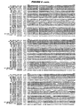
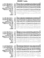
- Polynucleotide probes unique to HERV-K(CH), HERV-KII and HERV-K10 gag regions are provided in Table 1; polynucleotide probes unique to HERV-K(CH), HERV-KII, and HERV-K10 protease 3' and polymerase 5' regions are provided in Table 2; polynucleotide probes unique to HERV-K(CH), HERV-KII, and HERV-K10 3' pol only regions are provided in Table 3.
- the disclosure also provides an isolated polynucleotide comprising (a) a segment that is a fragment of the sequence shown in SEQ IDs 7-10 or SEQ IDs 27-39, wherein (i) said fragment is at least 10 nucleotides in length and (ii) corresponds identically in its entirety to a portion of SEQ ID 44 and/or 45; and, optionally, (b) one or more segments flanking the segment defined in (a), wherein the presence of said optional segment(s) causes said polynucleotide to not correspond identically to any portion of a sequence shown in SEQ IDs 7-10 or SEQ IDs 27-39.
- the optional flanking segments share less than 40% sequence identity to the nucleic acid sequences shown in SEQ IDs 7-10, SEQ ID 44 and/or SEQ ID 45. In other embodiments, the optional flanking segments have no contiguous sequence of 10, 12, 15 or 20 nucleotides in common with SEQ IDs 7-10, SEQ ID 44 and/or SEQ ID 45. In yet other embodiments, the optional flanking segment is not present. In further embodiments, a fragment of the polynucleotide sequence is up to at least 30,40,50,60,70,80,90,100,200,300,400, 500, 1000, or 1500 nucleotide in length.
- the disclosure also provides an isolated polynucleotide having formula 5'-A-B-C-3', wherein: A is a nucleotide sequence consisting of ⁇ nucleotides; B is a nucleotide sequence consisting of a fragment of b nucleotides from (i) the nucleotide sequence shown in SEQ IDs 7-10, (ii) the nucleotide sequence shown in any of SEQ IDs 27-39, (iii) the complement of the nucleotide sequence shown in SEQ IDs 7-10, or (iv) the complement of the nucleotide sequence shown in any of SEQ IDs 27-39; C is a nucleotide sequence consisting of c nucleotides; and wherein said polynucleotide is not a fragment of (i) the nucleotide sequence shown in SEQ IDs 7-10, (ii) the nucleotide sequence shown in any of SEQ IDs 27-39, (iii) the
- a+c is at least 1 (e.g . at least 2, 3, 4, 5, 6, 7, 8, 9,10, 11, 12, 13, 14, 15, 16, 17, 18, 19, 20, 21, 22, 23, 24, 25, 26, 27, 28, 29, 30, 35, 40, 45, 50, 60, 70, 80, 90, 100 etc.) and b is at least 7 ( e.g. at least 8, 9, 10, 11, 12,13, 14, 15, 16,17, 18,19, 20, 21, 22, 23, 24, 25, 30, 35, 40, 45, 50, 60, 70, 80, 90, 100 etc.). It is preferred that the value of a + b + c is at least 9 ( e.g .
- a + b + c is at most 200 ( e.g . at most 190, 180, 170, 160, 150, 140, 130, 120, 110, 100, 90, 80, 70, 60, 50, 40, 30, 25, 20, 19, 18, 17, 16, 15, 14, 13, 12, 11, 10, 9).
- a and/or C may comprise a promoter sequence (or its complement).
- a polynucleotide having at least s % identity to: (a) SEQ IDs 7-10; (b) a fragment ofx nucleotides of SEQ IDs 7-10; (c) SEQ IDs 11-13; (b) a fragment of x nucleotides of SEQ IDs 11-13.
- the value of s is at least 50 (e.g. at least 55, 60, 65, 70, 75, 80, 85, 90, 91, 92, 93, 94, 9S, 96, 97, 98, 99, 99.5, 99.9 etc.).
- the value ofx is at least 7 (e.g. 8, 9, 10,11, 12,13, 14,15, 16, 17, 18, 19, 20, 21, 22, 23, 24, 25, 30, 35, 40, 45, 50, 60, 70, 80, 90, 100 etc.).
- polynucleotides include naturally-occurring variants (e.g . degenerate variants, allelic variants, etc .), homologs, orthologs, and functional mutants.
- Variants can be identified by hybridization of putative variants with the polynucleotide sequences disclosed in SEQ IDs 14-39 herein, preferably by hybridization under stringent conditions. For example, by using appropriate wash conditions, variants can be identified where the allelic variant exhibits at most about 25-30% base pair (bp) mismatches relative to the selected polynucleotide probe. In general, allelic variants contain 15-25% bp mismatches, and can contain as little as even 5-15%, or 2-5%, or 1-2% bp mismatches, as well as a single bp mismatch.
- bp base pair
- the disclosure also encompasses homologs corresponding to any one of the polynucleotide sequences provided herein, where the source of homologous genes can be any mammalian species (e.g . primate species, particularly human; rodents, such as rats, etc.). Between mammalian species ( e.g . human and primate), homologs generally have substantial sequence similarity (e.g . at least 75% sequence identity, usually at least 90%, more usually at least 95%) between nucleotide sequences. Sequence similarity is calculated based on a reference sequence, which may be a subset of a larger sequence, such as a conserved motif, coding region, flanking region, domain, etc . A reference sequence will usually be at least about 18 contiguous nt long, more usually at least about 30 nt long, and may extend to the complete sequence that is being compared. Algorithms for sequence analysis are known in the art.
- a preferred HERV-K(CH) isolate is an isolate sequence which is shown in SEQ IDs 7-10.
- Another typical classification of the relationship of retroviruses is based on the amino acid sequence similarities in the reverse transcriptase protein.
- an even more preferred class of HERV-K(CH) isolates are those having an amino acid sequence identity of at least 90%, more preferably 95% to the 5' polymerase region encoded by the nucleotide sequence shown in SEQ ID 12, as determined by the Smith-Waterman homology search algorithm using an affine gap search with a gap open penalty of 12 and a gap extension penalty of 2, BLOSUM matrix of 62.
- these prostate cancer-associated polynucleotide sequences define a class of human endogenous retroviruses, designated herein as HERV-K(CH), whose members comprise variations which, without wanted to be bound by theory, may be due to the presence of polymorphisms or allelic variations.
- an isolated polynucleotide comprising a polynucleotide that selectively hybridizes, relative to a known polynucleotide, to: (a) the nucleotide sequence shown in SEQ IDs 7-10; (b) the nucleotide sequence shown in any of SEQ IDs 27-39; (c) the complement of the nucleotide sequence shown in SEQ IDs 7-10; (d) the complement of the nucleotide sequence shown in any of SEQ IDs 27-39; (e) a fragment of the nucleotide sequence shown in SEQ IDs 7-10; (f) a fragment of the nucleotide sequence shown in any of SEQ IDs 27-39; (g) the complement of a fragment of the nucleotide sequence shown in SEQ IDs 7-10; (h) the complement of a fragment of the nucleotide sequence shown in any of SEQ IDs 27-39; (j) a nucleotide sequence shown in SEQ IDs
- the fragment of (e), (f), (g) or (h) is preferably at least x nucleotides in length, wherein x is as defined in H.1.2 above, and is preferably not one of the sequences (i), (ii), (iii), (iv), (v) or (vi) as defined H.1.2 above.
- Hybridization reactions can be performed under conditions of different "stringency", as described in B.4 above.
- the polynucleotide hybridizes under low stringency conditions; in other embodiments it hybridizes under intermediate stringency conditions; in other embodiments, it hybridizes under high stringency conditions.
- an isolated polynucleotide comprising: (a) a HERV-K(CH) cDNA insert as deposited at the ATCC and having an ATCC accession number given in Table 7; (b) a HERV-K(CH) sequence as shown in any one of SEQ IDs 14-26; (c) a HERV-K(CH) sequence as shown in any one of SEQ IDs 27-39; or (d) a fragment of (a), (b) or (c).
- the fragment of (d) is preferably at least x nucleotides in length, wherein x is at least 7 (e.g. at least 8, 9, 10, 11, 12, 13, 14, 15, 16, 17, 18, 19, 20, 21, 22, 23, 24, 25, 30, 35, 40, 45, 50, 60, 70, 80, 90, 100 etc.).
- Preferred polynucleotides are those having a sequence set forth in any one of the polynucleotide sequences SEQ IDs 7-10 and SEQ IDs 14-39 provided herein; polynucleotides obtained from the biological materials described herein, in particular, polynucleotide sequences present in the isolates deposited with the ATCC and having ATCC accession numbers given in Table 7 or other biological sources (particularly human sources) or by hybridization to the above mentioned sequences under stringent conditions (particularly conditions of high stringency); genes corresponding to the provided polynucleotides; variants of the provided polynucleotides and their corresponding genes particularly those variants that retain a biological activity of the encoded gene product ( e.g .
- the isolated polynucleotides preferably comprise a polynucleotide having a HERV-K(CH) sequence.
- a polynucleotide can encode all or a part of a polypeptide, such as the gag region, 5' pol region or 3' pol region of a human endogenous retrovirus. Double or single stranded fragments can be obtained from the DNA sequence by chemically synthesizing oligonucleotides in accordance with conventional methods, by restriction enzyme digestion, by PCR amplification, etc .
- Polynucleotides can be cDNAs or genomic DNAs, as well as fragments thereof, particularly fragments that encode a biologically active gene product and/or are useful in the methods disclosed herein ( e.g . in diagnosis, as a unique identifier of a differentially expressed gene of interest, etc ).
- cDNA as used herein is intended to include all nucleic acids that share the arrangement of sequence elements found in native mature mRNA species, where sequence elements are exons and 3' and 5' non-coding regions. Normally mRNA species have contiguous exons, with the intervening introns, when present, being removed by nuclear RNA splicing, to create a continuous open reading frame encoding a polypeptide.
- mRNA species can also exist with both exons and introns, where the introns may be removed by alternative splicing. Furthermore it should be noted that different species of mRNAs encoded by the same genomic sequence can exist at varying levels in a cell, and detection of these various levels of mRNA species can be indicative of differential expression of the encoded gene product in the cell.
- a genomic sequence of interest comprises the nucleic acid present between the initiation codon and the stop codon, as defined in the listed sequences, including all of the introns that are normally present in a native chromosome. It can further include the 3' and 5' untranslated regions found in the mature mRNA. It can further include specific transcriptional and translational regulatory sequences, such as promoters, enhancers, etc ., including about 1 kb, but possibly more, of flanking genomic DNA at either the 5' and 3' end of the transcribed region.
- the genomic DNA can be isolated as a fragment of 100 kbp or smaller; and substantially free of flanking chromosomal sequence.
- the genomic DNA flanking the coding region, either 3' and 5', or internal regulatory sequences as sometimes found in introns contains sequences required for proper tissue, stage-specific, or disease-state specific expression.
- Polynucleotides can be provided as linear molecules or within circular molecules, and can be provided within autonomously replicating molecules (vectors) or within molecules without replication sequences. Expression of the polynucleotides can be regulated by their own or by other regulatory sequences known in the art.
- the polynucleotides can be introduced into suitable host cells using a variety of techniques available in the art, such as transferrin polycation-mediated DNA transfer, transfection with naked or encapsulated nucleic acids, liposome-mediated DNA transfer, intracellular transportation of DNA-coated latex beads, protoplast fusion, viral infection, electroporation, gene gun, calcium phosphate-mediated transfection, and the like.
- a polynucleotide sequence that is "shown in” or “depicted in” a SEQ ID NO or Figure means that the sequence is present as an identical contiguous sequence in the SEQ ID NO or Figure.
- the term encompasses portions, or regions of the SEQ ID NO or Figure as well as the entire sequence contained within the SEQ ID NO or Figure.
- Deduced polypeptides encoded by the HERV-K(CH) polynucleotides include the gag translations shown in SEQ IDS 46-49 and the 3' pol translations shown in SEQ IDs 50-55.
- a polypeptide sequence encoded by the polynucleotide having the sequence shown in SEQ ID 15 is provided in SEQ ID 56; a polypeptide sequence encoded by the polynucleotide having the sequence shown in SEQ ID 14, is shown in SEQ ID 57.
- a consensus 3' pol polypeptide sequence encoded by the polynucleotides having the sequence shown in SEQ IDs 21-27, inclusive, is provided in SEQ ID 58.
- polypeptides encompassed by the present disclosure include those encoded by polynucleotides of the disclosure e.g. SEQ IDs 7-10 and SEQ IDs 14-39, as well as polynucleotides deposited with the ATCC as disclosed herein, as well as nucleic acids that, by virtue of the degeneracy of the genetic code, are not identical in sequence to the disclosed polynucleotides and encode the polypeptides.
- the disclosure includes within its scope a polypeptide encoded by a polynucleotide having the sequence of any one of the polynucleotide sequences provided herein, or a variant thereof.
- a single large gag polypeptide is synthesized (e.g . a 73 kDa gag protein in HERV-K10) which is subsequently cleaved into multiple functional peptides by a functional protease encoded by the pol or protease region of the genome.
- a functional protease encoded by the pol or protease region of the genome Overexpression of sequences corresponding to both gag and pol domains of the HERV-K(CH) suggest such a mechanism. Sequences corresponding to the env and the nuclear RNA transport protein cORF region of the HERV-K(CH) genome may also be overexpressed.
- the polypeptides encoded by the open reading frames within the over-expressed polynucleotide sequences may play a significant role in the progression of prostate tumors.
- an isolated polypeptide comprising a fragment of: (a) a polypeptide sequence encoded within a HERV-K(CH) open reading frame; (b) a polypeptide sequence encoded by a polynucleotide shown in SEQ ID 11, 12 or 13; or (c) an amino acid sequence as shown in any one of SEQ IDs 46-49, 50-55, 56-57 or 58.
- the fragment is preferably at least x amino acids in length, wherein x is at least 5 ( e.g . at least 6,7, 8, 9, 10, 11, 12, 13, 14, 15, 16, 17, 18, 19, 20, 21, 22, 23, 24, 25, 30, 35, 40, 45, 50, 60, 70, 75, 80, 90, 100, 125, 150, 200, 300, 400, 500 or more etc .).
- x is at least 5 ( e.g . at least 6,7, 8, 9, 10, 11, 12, 13, 14, 15, 16, 17, 18, 19, 20, 21, 22, 23, 24, 25, 30, 35, 40, 45, 50, 60, 70, 75, 80, 90, 100, 125, 150, 200, 300, 400, 500 or more etc .).
- the value of x will typically not exceed 1000.
- the fragment may include an epitope e.g . an epitope of the amino acid sequence shown in SEQ IDs 56, 57 or 58.
- SEQ IDs 46-49 provide a translation of the HERV-K(CH) polynucleotides having a sequence shown in SEQ IDs 14, 15, 16 and 40 (the sequence of SEQ ID 40 is from a polynucleotide found in a normal prostate library) corresponding to polynucleotides encoding the gag region.
- SEQ IDs 50-55 provide a translation of the HERV-K(CH) polynucleotides having a sequence shown in SEQ IDs 21-26, inclusive, corresponding to the 3' region of pol.
- SEQ IDs 56 & 57 provide translations of the HERV-K(CH) polynucleotide of SEQ ID 15 and SEQ ID 14, respectively.
- SEQ ID 58 provides a consensus translation of the polynucleotide from the 3' pol region (SEQ IDs 21-26, inclusive).
- polypeptide fragments such as, epitopes, of at least 5 amino acids, at least 6 amino acids, at least 8 amino acids, at least 10 amino acids, at least 11 amino acids, at least 12 amino acids, at least 13 amino acids, at least 14 amino acids and at least 15 amino acids of the translations shown in SEQ IDs 46-49 and 50-55.
- the HERV-K(CH) epitopes of the amino acid sequence as shown in SEQ IDs 56-58 were determined by the Jameson-Wolf antigenic index
- 3' pol SEQ ID 58
- Jameson-Wolf algorithm amino acids: 1-10; 15-35; 45-55; 60-85; 100-115; 125-140; 170-190; 195-215; 230-268.
- Additional epitope-containing fragments include amino acids 1-8; 2-10; 1-15; 5-15; 7-15; 10-20; 12-20; 15-23; 20-28; 28-35; 15-30; 15-40; 20-30; 45-52; 48-55; 60-68; 60-70; 65-73; 70-78; 75-83; 70-80; 65-75; 68-75; 75-85; 78-85; 65-85; 60-75; 100-108; 103-110; 105-113; 108-115; 125-133; 128-135; 132-140; 170-178; 175-182; 180-187; 182-190; 195-202; 200-208; 205-212; 208-215; 230-237; 235-242; 240-247; 245-252; 250-257; 255-262; 260-268; 230-250; 235-255; 240-260; 245-268; 230-245; 235-245; 235-250; 240-255; 245-260; 250-2
- gag The following regions in gag (SEQ ID 56) were determined to be antigenic by Jameson-Wolf algorithm: amino acids: 1-40; 45-60; 80-105; 130-145; 147-183; 186-220; 245-253; 255-288. Additional epitope-containing fragments include amino acids 1-8; 2-10; 1-15; 5-15; 7-15; 10-20; 12-20; 15-23; 20-28; 28-35; 30-37; 33-40; 1-20; 20-40; 1-15; 15-30; 15-40; 45-52; 50-57; 55-62; 50-60; 1-60; 80-87; 85-92; 80-90; 90-97; 95-102; 98-105; 85-100; 90-105; 80-100; 85-105; 130-137; 135-142; 140-147; 145-152; 150-157; 155-162; 160-167; 165-172; 170-177; 175-183; 180-187; 185-192; 190-197; 195-202; 200-207;
- gag The following regions in gag (SEQ ID 57) were determined to be antigenic by Jameson-Wolf algorithm: amino acids: 1-40; 80-105; 145-180; 185-225; 240-335. Additional epitope-containing fragments include amino acids 1-8; 2-10; 1-15; 5-15; 7-15; 10-20; 12-20; 15-23; 20-28; 28-35; 30-37; 33-40; 1-20; 20-40; 1-15; 15-30; 15-40; 80-87; 85-92; 80-90; 90-97; 95-102; 98-105; 85-100; 90-105; 80-100; 85-105; 145-152; 150-157; 155-162; 160-167; 165-172; 170-177; 175-182; 180-187; 185-192; 190-197; 195-202; 200-207; 205-212; 210-217; 215-212; 218-225; 145-160; 150-165; 155-170; 160-175; 170-185;
- isolated polypeptide having formula 5'-A-B-C-3', wherein: A is an amino acid sequence consisting of a amino acids; B is an amino acid sequence consisting of a fragment of b amino acids from (i) the amino acid sequence encoded by a polynucleotide shown in SEQ ID 11, 12 or 13; (ii) any one of SEQ IDs 46-49, 50-55, 56-57 or 58; C is an amino acid sequence consisting of c amino acids; and wherein said polypeptide is not a fragment of the amino acid sequence defined in (i) or (ii).
- a+c is at least 1 (e.g . at least 2, 3, 4, 5, 6, 7, 8, 9,10,11, 12,13,14, 15, 16,17, 18, 19, 20, 21, 22, 23, 24, 25, 26, 27, 28, 29, 30, 35, 40, 45, 50, 60, 70, 80, 90, 100 etc.) and b is at least 7 ( e.g . at least 8, 9, 10, 11, 12, 13, 14, 15, 16, 17, 18, 19, 20, 21, 22, 23, 24, 25, 30, 35, 40, 45, 50, 60, 70, 80, 90, 100 etc .). It is preferred that the value of a + b + c is at least 9 ( e.g .
- a + b + c is at most 200 ( e.g . at most 190, 180, 170, 160,150, 140, 130, 120, 110, 100, 90, 80, 70, 60, 50, 40, 30, 25, 20, 19, 18, 17, 16, 15, 14, 13, 12, 11, 10, 9).
- polypeptide having at least s% identity to: (a) the polypeptide sequences encoded by SEQ IDs 7-45; (b) a fragment of x amino acids of the polypeptide sequences encoded by SEQ IDs 7-45; (c) the polypeptide sequences SEQ IDs 46-58; (d) a fragment of x amino acids of the polypeptide sequences SEQ IDs 46-58.
- the value of s is at least 35 ( e.g .
- the value ofx is at least 7 ( e.g. 8, 9,10,11,12,13, 14, 15, 16,17,18, 19, 20, 21, 22, 23, 24, 25, 30, 35, 40, 45, 50, 60, 70, 80, 90,100.
- polypeptides include naturally-occurring variants (e.g . allelic variants, etc .), homologs, orthologs, and functional mutants.
- Variants of the naturally-occurring polypeptides wherein such variants are homologous or substantially similar to the naturally occurring polypeptide, can be of an origin of the same or different species as the naturally occurring polypeptide (e.g . human, murine, or some other species that naturally expresses the recited polypeptide, usually a mammalian species).
- These polypeptide variants are encoded by polynucleotides, and the genetic code can be used to select appropriate codons to construct the corresponding variants.
- Polypeptides such as those shown in SEQ IDs 46-58, encoded by HERV-K(CH) polynucleotides are differentially expressed in prostate cancer cells. Such polypeptides are referred to herein as "polypeptides associated with prostate cancer" or "HERV-K(CH) polypeptides".
- the polypeptides can be used to generate antibodies specific for a polypeptide associated with prostate cancer, which antibodies are in turn useful in diagnostic methods, prognostic methods, therametric methods, and the like as discussed in more detail herein. Polypeptides are also useful as targets for therapeutic intervention, as discussed in more detail herein.
- the isolated polypeptides preferably comprise a polypeptide having a HERV-K(CH) sequence.
- Polypeptides such as polypeptides of the gag regions or polypeptides of the pol regions, encoded by the polynucleotides disclosed herein, such as polynucleotides having the sequences as shown in SEQ IDs 7-10 and SEQ IDs 14-39, and in isolates deposited with the ATCC and having ATCC accession numbers given in Table 7 and/or their corresponding full length genes, can be used to screen peptide libraries to identify binding partners, such as receptors, from among the encoded polypeptides.
- Peptide libraries can be synthesized according to methods known in the art ( e.g . see refs. 151 & 152).
- polypeptide refers to both the full length polypeptide encoded by the recited polynucleotide, the polypeptide encoded by the gene represented by the recited polynucleotide, as well as portions or fragments thereof.
- a polypeptide sequence that is "shown in” or “depicted in” a SEQ ID NO or Figure means that the sequence is present as an identical contiguous sequence in the SEQ ID NO or Figure.
- the term encompasses portions, or regions of the SEQ ID NO or Figure as well as the entire sequence contained within the SEQ ID NO or Figure.
- the present disclosure also provides isolated antibodies or antigen binding fragments thereof, that bind to a polypeptide disclosed herein.
- the present disclosure also provides isolated antibodies or antigen binding fragments thereof, that bind to a polypeptide encoded by a polynucleotide disclosure herein.
- the present disclosure also provides isolated antibodies that bind to a polypeptide, or antigen binding fragment thereof, encoded by a polynucleotide made by the method comprising the following steps i) immunizing a host animal with a composition comprising said polypeptide, or antigen binding fragment thereof, and ii) collecting cells from said host expressing antibodies against the antigen or antigen binding fragment thereof.
- antibodies that bind to a polypeptide, or antigen binding fragment thereof, encoded by a polynucleotide disclosed herein made by the method comprising the following steps: providing a cell line producing an antibody, wherein said antibody binds to a polypeptide, or antigen binding fragment thereof, encoded by a polynucleotide disclosed herein and culturing said cell line under conditions wherein said antibodies are produced.
- the antibodies are collected and monoclonal antibodies are produced using the collected host cells or genetic material derived from the collected host cells.
- the antibody is a polyclonal antibody.
- the antibody is attached to a solid surface or further comprises a detectable label.
- Antibodies may be isolated antibodies, that bind a polypeptide encoded by a polynucleotide described herein. Antibodies can be provided in a composition comprising the antibody and a buffer and/or a pharmaceutically acceptable excipient. Antibodies specific for a polypeptide associated with cancer are useful in a variety of diagnostic and therapeutic methods, as discussed in detail herein.
- Expression products of a polynucleotide described herein, as well as the corresponding mRNA (particularly mRNAs having distinct secondary and/or tertiary structures), cDNA, or complete gene, or fragments of said expression products can be prepared and used for raising antibodies for experimental, diagnostic, and therapeutic purposes.
- a polynucleotide described herein, as well as the corresponding mRNA (particularly mRNAs having distinct secondary and/or tertiary structures), cDNA, or complete gene, or fragments of said expression products can be prepared and used for raising antibodies for experimental, diagnostic, and therapeutic purposes.
- polynucleotides to which a corresponding gene has not been assigned this provides an additional method of identifying the corresponding gene.
- the polynucleotide or related cDNA is expressed as described above, and antibodies are prepared.
- These antibodies are specific to an epitope on the polypeptide encoded by the polynucleotide, and can precipitate or bind to the corresponding native polypeptide in a cell or tissue preparation or in a cell-free extract of an in vitro expression system.
- Polyclonal or monoclonal antibodies to the HERV -K(CH) polypeptides or an epitope thereof can be made for use in immunoassays by any of a number of methods know in the art.
- epitope reference is made to an antigenic determinant of a polypeptide.
- the presence of an epitope is demonstrated by the ability of an antibody to bind a polypeptide with specificity. Two antibodies are considered to be directed to the same epitope if they cross block each others binding to the same polypeptide.
- One approach for preparing antibodies to a polypeptide is the selection and preparation of an amino acid sequence of all or part of the polypeptide, chemically synthesizing the sequence and injecting it into an appropriate animal, typically a rabbit, hamster or a mouse.
- Oligopeptides can be selected as candidates for the production of an antibody to the HERV-K(CH) polypeptide based upon the oligopeptides lying in hydrophilic regions, which are thus likely to be exposed in the mature polypeptide. Additional oligopeptides can be determined using, for example, the Antigenicity Index [30].
- humanized monoclonal antibodies are provided, wherein the antibodies are specific for HERV-K(CH) polypeptides and do not appreciably bind other HERV polypeptides.
- the phrase "humanized antibody” refers to an antibody derived from a non-human antibody, typically a mouse monoclonal antibody.
- a humanized antibody may be derived from a chimeric antibody that retains or substantially retains the antigen-binding properties of the parental, non-human, antibody but which exhibits diminished immunogenicity in humans as compared to the parental antibody.
- chimeric antibody refers to an antibody containing sequence derived from two different antibodies (see, e.g. ref. 153) which typically originate from different species. Most typically, chimeric antibodies comprise human and murine antibody fragments, generally human constant and mouse variable regions.
- Methods for preparation of the human or primate HERV-K(CH) or an epitope thereof include, but are not limited to chemical synthesis, recombinant DNA techniques or isolation from biological samples. Chemical synthesis of a peptide can be performed, for example, by the classical Merrifeld method of solid phase peptide synthesis [154] or the FMOC strategy on a Rapid Automated Multiple Peptide Synthesis system (E.I. du Pont de Nemours Company, Wilmington, DE) [155].
- Polyclonal antibodies can be prepared by immunizing rabbits or other animals by injecting antigen followed by subsequent boosts at appropriate intervals. The animals are bled and sera assayed against purified HERV-K(CH) usually by ELISA or by bioassay based upon the ability to block the action of HERV-K(CH). When using avian species, e.g. chicken, turkey and the like, the antibody can be isolated from the yolk of the egg. Monoclonal antibodies can be prepared after the method of Milstein and Kohler by fusing splenocytes from immunized mice with continuously replicating tumor cells such as myeloma or lymphoma cells. [156, 157, 158]. The hybridoma cells so formed are then cloned by limiting dilution methods and supernates assayed for antibody production by ELISA, RIA or bioassay.
- antibodies either polyclonal or monoclonal, to the HERV-K(CH) polypeptides can be produced by any suitable method known in the art as discussed above.
- murine or human monoclonal antibodies can be produced by hybridoma technology or, alternatively, the HERV-K(CH) polypeptides, or an immunologically active fragment thereof, or an anti-idiotypic antibody, or fragment thereof can be administered to an animal to elicit the production of antibodies capable of recognizing and binding to the HERV-K(CH) polypeptides.
- Such antibodies can be from any class of antibodies including, but not limited to IgG, IgA, IgM, IgD, and IgE or in the case of avian species, IgY and from any subclass of antibodies.
- the invention provides methods for diagnosing the presence of prostate cancer in a test sample associated with expression of a polynucleotide in a test cell sample, comprising the steps of: i) detecting a level of expression of at least one polynucleotide of the invention, or a fragment thereof, or at least one polynucleotide found in an isolate selected from the group consisting of ATCC accession numbers given in Table 7, or a fragment thereof; and ii) comparing said level of expression of the polynucleotide in the test sample with a level of expression of polynucleotide in the control cell sample, wherein differential expression of the polynucleotide in the test cell sample relative to the level of polynucleotide expression in the control cell sample is indicative of the presence of cancer in the test cell sample.
- the detecting is measuring the level of an RNA transcript; measuring the level of a polynucleotide; or measuring by a method including PCR, TMA, bDNA, NAT or Nasba.
- the polynucleotide is attached to a solid support.
- compositions comprising a test cell sample and an isolated polynucleotide disclosed herein.
- the present invention further provides methods for detecting prostate cancer associated with expression of a polypeptide in a test cell sample, comprising the steps of: i) detecting a level of expression of at least one polypeptide disclosed herein, or a fragment thereof and ii) comparing said level of expression of the polypeptide in the test sample with a level of expression of polypeptide in the control cell sample, wherein an altered level of expression of the polypeptide in the test cell sample relative to the level of expression of the polypeptide in the control cell sample is indicative of the presence of cancer in the test cell sample.
- the present disclosure also provides methods for detecting prostate cancer associated with the presence of an antibody in a test cell sample, comprising the steps of: i) detecting a level of an antibody and ii) comparing said level of said antibody in the test sample with a level of said antibody in the control cell sample, wherein an altered level of antibody in said test cell sample relative to the level of antibody in the control cell sample is indicative of the presence of cancer in the test cell sample.
- This disclosure also provides methods for detecting prostate cancer associated with elevated levels of HERV-K(CH) polynucleotides, by means of (i) detecting polynucleotides having at least 65%, at least 70%, at least 75%, at least 80%, at least 85%, at least 90% at least 91%, at least 92%, at least 93%, at least 94%, at least 95%, at least 96%, at least 97%, at least 98%, at least 99% or at least 100% identity to the polynucleotide shown in SEQ IDs 7-10 or to polynucleotides in isolates deposited with the ATCC and having ATCC deposit accession numbers PTA-2561, PTA-2572, PTA-2566, PTA-2571, PTA-2562, PTA-2573, PTA-2560, PTA-2565, PTA-2568, PTA-2564, PTA-2569, PTA-2567, PTA-2559, PTA-2563, PTA-2570, as measured by the alignment program GCG Gap (Suite
- the treatment regimen of a prostate cancer associated with elevated levels of HERV-K(CH) polynucleotides may also monitored by detecting levels of the polynucleotides and polypeptides in order to assess the staging of the cancer and/or efficacy of particular cancer therapies.
- the present invention provides methods of using the polynucleotides described herein for detecting prostate cancer cells, facilitating diagnosis of prostate cancer and the severity of a cancer (e.g . tumor grade, tumor burden, and the like) in a subject, facilitating a determination of the prognosis of a subject, and assessing the responsiveness of the subject to therapy (e.g . by providing a measure of therapeutic effect through, for example, assessing tumor burden during or following a chemotherapeutic regimen).
- Detection can be based on detection of a polynucleotide that is differentially expressed in a prostate cancer cell and/or detection of a polypeptide encoded by a polynucleotide that is differentially expressed in a prostate cancer cell.
- the detection methods of the invention can be conducted in vitro or in vivo, on isolated cells, or in whole tissues or a bodily fluid e.g . blood, plasma, serum, urine, and the like).
- kits for detecting the presence and/or a level of a polynucleotide that is differentially expressed in a cancer cell e.g . by detection of an mRNA encoded by the differentially expressed gene of interest
- a polypeptide encoded thereby in a biological sample.
- Procedures using these kits can be performed by clinical laboratories, experimental laboratories, medical practitioners, or private individuals.
- the kits of the disclosure for detecting a polypeptide encoded by a polynucleotide that is differentially expressed in a prostate cancer cell may comprise a moiety that specifically binds the polypeptide, which may be an antibody that binds the polypeptide or fragment thereof.
- kits of the disclosure used for detecting a polynucleotide that is differentially expressed in a prostate cancer cell may comprise a moiety that specifically hybridizes to such a polynucleotide.
- the kit may optionally provide additional components that are useful in the procedure, including, but not limited to, buffers, developing reagents, labels, reacting surfaces, means for detection, control samples, standards, instructions, and interpretive information.
- kits for detecting prostate cancer comprising at least one of polynucleotides having the sequence as shown in SEQ IDs 7-10, SEQ IDs 14-39, or fragments thereof, or having the sequence found in an isolate deposited with the ATCC and having ATCC accession numbers PTA-2561, PTA-2572, PTA-2566, PTA-2571, PTA-2562, PTA-2573, PTA-2560, PTA-2565, PTA-2568, PTA-2564, PTA-2569, PTA-2567, PTA-2559, PTA-2563, PTA-2570 or fragments thereof.
- methods are provided for detecting a polypeptide encoded by a gene differentially expressed in a prostate cancer cell.
- Any of a variety of known methods can be used for detection, including, but not limited to, immunoassay, using antibody that binds the polypeptide, e.g. by enzyme-linked immunosorbent assay (ELISA), radioimmunoassay (RIA), and the like; and functional assays for the encoded polypeptide, e.g. binding activity or enzymatic activity.
- ELISA enzyme-linked immunosorbent assay
- RIA radioimmunoassay
- the detection methods and other methods described herein can be readily varied. Such variations are within the intended scope of the invention.
- the probe for use in detection can be immobilized on a solid support, and the test sample contacted with the immobilized probe. Binding of the test sample to the probe can then be detected in a variety of ways, e.g . by detecting a detectable label bound to the test sample to facilitate detected of test sample-immobilized probe complexes.
- the present invention further provides methods for detecting the presence of and/or measuring a level of a polypeptide in a biological sample, which polypeptide is encoded by a polynucleotide that is differentially expressed in a prostate cancer cell, using an antibody specific for the encoded polypeptide.
- the methods generally comprise: a) contacting the sample with an antibody specific for a polypeptide encoded by a polynucleotide that is differentially expressed in a prostate cancer cell; and b) detecting binding between the antibody and molecules of the sample.
- Detection of specific binding of the antibody specific for the encoded prostate cancer-associated polypeptide, when compared to a suitable control is an indication that encoded polypeptide is present in the sample.
- Suitable controls include a sample known not to contain the encoded polypeptide or known not to contain elevated levels of the polypeptide; such as normal prostate tissue, and a sample contacted with an antibody not specific for the encoded polypeptide, e.g. an anti-idiotype antibody.
- a variety of methods to detect specific antibody-antigen interactions are known in the art and can be used in the method, including, but not limited to, standard immunohistological methods, immunoprecipitation, an enzyme immunoassay, and a radioimmunoassay.
- the specific antibody will be detectably labeled, either directly or indirectly.
- Direct labels include radioisotopes; enzymes whose products are detectable (e.g. luciferase, ß-galactosidase, and the like); fluorescent labels (e.g. fluorescein isothiocyanate, rhodamine, phycoerythrin, and the like); fluorescence emitting metals, e.g. 152 Eu, or others of the lanthanide series, attached to the antibody through metal chelating groups such as EDTA; chemiluminescent compounds, e.g. luminol, isoluminol, acridinium salts, and the like; bioluminescent compounds, e.g.
- luciferin aequorin (green fluorescent protein), and the like.
- the antibody may be attached (coupled) to an insoluble support, such as a polystyrene plate or a bead.
- Indirect labels include second antibodies specific for antibodies specific for the encoded polypeptide ("first specific antibody"), wherein the second antibody is labeled as described above; and members of specific binding pairs, e.g. biotin-avidin, and the like.
- the biological sample may be brought into contact with and immobilized on a solid support or carrier, such as nitrocellulose, that is capable of immobilizing cells, cell particles, or soluble proteins.
- the support may then be washed with suitable buffers, followed by contacting with a detectably-labeled first specific antibody. Detection methods are known in the art and will be chosen as appropriate to the signal emitted by the detectable label. Detection is generally accomplished in comparison to suitable controls, and to appropriate standards.
- the methods are adapted for use in vivo, e.g. to locate or identify sites where cancer cells, such as prostate cancer cells, are present.
- methods are provided for detecting a cancer cell by detecting expression in the cell of a transcript that is differentially expressed in a cancer cell.
- Any of a variety of known methods can be used for detection, including, but not limited to, detection of a transcript by hybridization with a polynucleotide that hybridizes to a polynucleotide that is differentially expressed in a prostate cancer cell; detection of a transcript by a polymerase chain reaction using specific oligonucleotide primers; in situ hybridization of a cell using as a probe a polynucleotide that hybridizes to a gene that is differentially expressed in a prostate cancer cell.
- the methods can be used to detect and/or measure mRNA levels of a gene that is differentially expressed in a prostate cancer cell.
- the methods comprise: a) contacting a sample with a polynucleotide that corresponds to a differentially expressed gene described herein under conditions that allow hybridization; and b) detecting hybridization, if any.
- Detection of differential hybridization when compared to a suitable control, is an indication of the presence in the sample of a polynucleotide that is differentially expressed in a cancer cell.
- Appropriate controls include, for example, a sample which is known not to contain a polynucleotide that is differentially expressed in a cancer cell, and use of a labeled polynucleotide of the same "sense" as the polynucleotide that is differentially expressed in the cancer cell.
- the cancer cell is a prostate cancer cell. Conditions that allow hybridization are known in the art, and have been described in more detail above.
- Detection can also be accomplished by any known method, including, but not limited to, in situ hybridization, PCR (polymerase chain reaction), RT-PCR (reverse transcription-PCR), TMA, bDNA, and Nasba and "Northern” or RNA blotting, or combinations of such techniques, using a suitably labeled polynucleotide.
- PCR polymerase chain reaction
- RT-PCR reverse transcription-PCR
- TMA reverse transcription-PCR
- bDNA reverse transcription-PCR
- Nasba and "Northern” or RNA blotting or combinations of such techniques, using a suitably labeled polynucleotide.
- a variety of labels and labeling methods for polynucleotides are known in the art and can be used in the assay methods of the invention. Specific hybridization can be determined by comparison to appropriate controls.
- Polynucleotide generally comprising at least 10 nt, at least 12nt or at least 15 contiguous nucleotides of a polynucleotide provided herein, such as, for example, those having the sequence as depicted in SEQ IDs 7-10, and 3-28, are used for a variety of purposes, such as probes for detection of and/or measurement of, transcription levels of a polynucleotide that is differentially expressed in a prostate cancer cell.
- a probe that hybridizes specifically to a polynucleotide disclosed herein should provide a detection signal at least 5-, 10-, or 20-fold higher than the background hybridization provided with other unrelated sequences.
- probe as used herein is meant to refer to a polynucleotide sequence used to detect a differentially expressed gene product in a test sample.
- the probe can be detectably labeled and contacted with, for example, an array comprising immobilized polynucleotides obtained from a test sample (e.g . mRNA).
- the probe can be immobilized on an array and the test sample detectably labeled.
- Nucleotide probes are used to detect expression of a gene corresponding to the provided polynucleotide.
- Northern blots mRNA is separated electrophoretically and contacted with a probe. A probe is detected as hybridizing to an mRNA species of a particular size. The amount of hybridization can be quantitated to determine relative amounts of expression, for example under a particular condition.
- Probes are used for in situ hybridization to cells to detect expression. Probes can also be used in vivo for diagnostic detection of hybridizing sequences. Probes are typically labeled with a radioactive isotope. Other types of detectable labels can be used such as chromophores, fluorophores, and enzymes. Other examples of nucleotide hybridization assays are described in refs. 185 and 186.
- PCR is another means for detecting small amounts of target nucleic acids (see, e.g . refs. 187, 188 & 189).
- Two primer polynucleotides nucleotides that hybridize with the target nucleic acids are used to prime the reaction.
- the primers can be composed of sequence within or 3' and 5' to the HERV-K(CH) polynucleotides disclosed herein. Alternatively, if the primers are 3' and 5' to these polynucleotides, they need not hybridize to them or the complements.
- the amplified target nucleic acids can be detected by methods known in the art (e.g . Southern blot).
- mRNA or cDNA can also be detected by traditional blotting techniques (e.g . Southern blot, Northern blot, etc.) described in ref. 8 ( e.g . without PCR amplification).
- traditional blotting techniques e.g . Southern blot, Northern blot, etc.
- mRNA or cDNA generated from mRNA using a polymerase enzyme can be purified and separated using gel electrophoresis, and transferred to a solid support, such as nitrocellulose. The solid support is exposed to a labeled probe, washed to remove any unhybridized probe, and duplexes containing the labeled probe are detected.
- Methods using PCR amplification can be performed on the DNA from a single cell, although it is convenient to use at least about 10 5 cells.
- the use of the polymerase chain reaction is described in ref. 190, and a review of techniques may be found in pages 14.2 to 14.33 of reference 8.
- a detectable label may be included in the amplification reaction. Suitable detectable labels include fluorochromes,( e.g .
- fluorescein isothiocyanate FITC
- rhodamine Texas Red
- phycoerythrin allophycocyanin
- 6-carboxyfluorescein (6-FAM)
- 6-carboxy-X-rhodamine ROX
- 2',7'-dimethoxy-4',5'-dichloro-6-carboxyfluorescein 5-carboxyfluorescein
- TAMRA N,N,N',N'-tetramethyl-6-carboxyrhodamine
- BEX 6-carboxy-2',4',7',4,7-hexachlorofluorescein
- the label may be a two stage system, where the polynucleotides is conjugated to biotin, haptens, etc . having a high affinity binding partner, e.g . avidin, specific antibodies, etc ., where the binding partner is conjugated to a detectable label.
- the label may be conjugated to one or both of the primers.
- the pool of nucleotides used in the amplification is labeled, so as to incorporate the label into the amplification product.
- the present invention further relates to methods of detecting/diagnosing a neoplastic or preneoplastic condition in a mammal (for example, a human).
- Examples of conditions that can be detected/diagnosed in accordance with these methods include, but are not limited to prostate cancers. Polynucleotides corresponding to genes that exhibit the appropriate expression pattern can be used to detect prostate cancer in a subject Reference 191 reviews markers of cancer.
- One detection/diagnostic method comprises: (a) obtaining from a mammal (eg a human) a biological sample, (b) detecting the presence in the sample of a HERV-K(CH) polypeptide and (c) comparing the amount of product present with that in a control sample.
- a mammal eg a human
- detecting the presence in the sample of a HERV-K(CH) polypeptide and comparing the amount of product present with that in a control sample.
- the presence in the sample of elevated levels of a HERV-K(CH) gene product indicates that the subject has a neoplastic or preneoplastic condition.
- the compound is preferably a binding protein, e.g. an antibody, polyclonal or monoclonal, or antigen binding fragment thereof, which can be labeled with a detectable marker (eg fluorophore, chromophore or isotope, etc).
- a detectable marker eg fluorophore, chromophore or isotope, etc.
- the compound can be attached to a solid support. Determination of formation of the complex can be effected by contacting the complex with a further compound (eg an antibody) that specifically binds to the first compound (or complex). Like the first compound, the further compound can be attached to a solid support and/or can be labeled with a detectable marker.
- HERV-K(CH) polypeptide in accordance with the present invention makes possible the identification of subjects (patients) that are likely to benefit from adjuvant therapy.
- a biological sample from a post-primary therapy subject e.g. subject having undergone surgery
- tissue from the cut site of a surgically removed tumor can be examined (e.g . by immunofluorescence), the presence of elevated levels of product (relative to the surrounding tissue) being indicative of incomplete removal of the tumor.
- Subjects undergoing non-surgical therapy can also be monitored, the presence in samples from such subjects of elevated levels of HERV-K(CH) polypeptide being indicative of the need for continued treatment
- Staging of the disease can also be effected, for example, by prostate biopsy e.g . with antibody specific for a HERV-K(CH) polypeptide.
- a kit can be used in the detection of a HERV-K(CH) polypeptide.
- the kit can comprise a compound that specifically binds a HERV-K(CH) polypeptide, such as, for example, binding proteins including antibodies or binding fragments thereof (e.g. F(ab') 2 fragments) disposed within a container means.
- the kit can further comprise ancillary reagents, for processing the binding assay.
- composition comprising X may consist exclusively of X or may include something additional e.g . X + Y.
- Neoplastic cells refer to cells which exhibit relatively autonomous growth, so that they exhibit an aberrant growth phenotype characterized by a significant loss of control of cell proliferation (i.e. de-regulated cell division).
- Neoplastic cells can be malignant or benign and include prostate cancer derived tissue.
- Candidate polynucleotides that may represent genes differentially expressed in cancer were obtained from both publicly-available sources and from cDNA libraries generated from selected cell lines and patient tissues.
- a normalized cDNA library was prepared from one patient tumor tissue and cloned polynucleotides for spotting on microarrays were isolated from the library. Normal and tumor tissues from 13 patients were processed to generate T7 RNA polymerase transcribed polynucleotides, which were, in turn, assessed for expression in the microarrays.
- the tissues that served as sources for these libraries and polynucleotides are summarized in Table 4.
- Normalization The objective of normalization is to generate a cDNA library in which all transcripts expressed in a particular cell type or tissue are equally represented [refs. 192 & 193], and therefore isolation of as few as 30,000 recombinant clones in an optimally normalized library may represent the entire gene expression repertoire of a cell, estimated to number 10,000 per cell.
- the source materials for generating the normalized prostate libraries were cryopreserved prostate tumor tissue from a patient with Gleason grade 3+3 adenocarcinoma and normal prostate biopsies from a pool of at-risk subjects under medical surveillance.
- Prostate epithelia were harvested directly from frozen sections of tissue by laser capture microdissection (LCM, Arcturus Engineering Inc., Mountain View, CA), carried out according to methods well known in the art (e.g . ref. 194), to provide substantially homogenous cell samples.
- LCM laser capture microdissection
- Single-stranded cDNA (“normalized” cDNA) was purified by hydroxyapatite chromatography (#130-0520, BioRad, Hercules, CA) following the manufacturer's recommended procedures, amplified and converted to double-stranded cDNA by three cycles of PCR amplification, and cloned into plasmid vectors using standard procedures (ref 8). All primers/adaptors used in the normalization and cloning process are provided by the manufacturer in the SMARTTM PCR cDNA synthesis kit (ClonTech, Palo Alto, CA). Supercompetent cells (XL-2 Blue Ultracompetent Cells, Stratagene, California) were transfected with the normalized cDNA libraries, plated on plated on solid media and grown overnight at 36°C.
- masking does not influence the final search results, except to eliminate sequences of relative little interest due to their low complexity, and to eliminate multiple "hits" based on similarity to repetitive regions common to multiple sequences e.g . Alu repeats.
- the remaining sequences were then used in a BLASTN vs . GenBank search.
- the sequences were also used as query sequence in a BLASTX vs . NRP (non-redundant proteins) database search.
- sequence identity analysis The number of times a sequence is represented in a library is determined by performing sequence identity analysis on cloned cDNA sequences and assigning transcript identity to each isolated clone.
- each sequence was checked to see if it was a mitochondrial, bacterial or ribosomal contaminant. Such sequences were excluded from the subsequent analysis.
- sequence artifacts e.g. vector and repetitive elements
- sequences were compared via BLAST [198] to GenBank and EST databases for gene identification and were compared with each other via FastA [199] to calculate the frequency of cDNA appearance in the normalized cDNA library.
- the sequences were also searched against the GenBank and GeneSeq nucleotide databases using the BLASTN program (BLASTN 1.3MP [198]).
- the sequences were analyzed against a non-redundant protein (NRP) database with the BLASTX program (BLASTX 1.3MP [198]).
- NRP non-redundant protein
- This protein database is a combination of the Swiss-Prot, PIR, and NCBI GenPept protein databases.
- the BLASTX program was run using the default BLOSUM-62 substitution matrix with the filter parameter: "xnu+seg". The score cutoff utilized was 75.
- cDNA sequences representing a variety of candidate genes to be screened for differential expression in prostate cancer were assayed by hybridization on polynucleotide arrays.
- the cDNA sequences included cDNA clones isolated from cell lines or tissues as described above.
- the cDNA sequences analyzed also included polynucleotides comprising sequence overlap with sequences in the Unigene database, and which encode a variety gene products of various origins, functionality, and levels of characterization.
- cDNAs were spotted onto reflective slides (Amersham) according to methods well known in the art at a density of 9,216 spots per slide representing 4608 sequences (including controls) spotted in duplicate, with approximately 0.8 ⁇ l of an approximately 200ng/ ⁇ l solution of cDNA.
- PCR products of selected cDNA clones corresponding to the gene products of interest were prepared in a 50% DMSO solution. These PCR products were spotted onto Amersham aluminum microarray slides at a density of 9216 clones per array using a Molecular Dynamics Generation III spotting robot. Clones were spotted in duplicate, for a total of 4608 different sequences per chip.
- cDNA probes were prepared from total RNA obtained by laser capture microdissection (LCM, Arcturus Enginering Inc., Mountain View, CA) of tumor tissue samples and normal tissue samples isolated from the patients described above.
- RNA was first reverse transcribed into cDNA using a primer containing a T7 RNA polymerase promoter, followed by second strand DNA synthesis.
- cDNA was then transcribed in vitro to produce antisense RNA using the T7 promoter-mediated expression (e.g . ref. 200), and the antisense RNA was then converted into cDNA.
- the second set of cDNAs were again transcribed in vitro, using the T7 promoter, to provide antisense RNA.
- This antisense RNA was then fluorescently labeled, or the RNA was again converted into cDNA, allowing for third round of T7-mediated amplification to produce more antisense RNA.
- Probes were labeled by making fluorescently labeled cDNA from the RNA starting material. Fluorescently-labeled cDNAs prepared from the tumor RNA sample were compared to fluorescently labeled cDNAs prepared from normal cell RNA sample. For example, the cDNA probes from the normal cells were labeled with Cy3 fluorescent dye (green) and cDNA probes prepared from the tumor cells were labeled with Cy5 fluorescent dye (red).
- the differential expression assay was performed by mixing equal amounts of probes from tumor cells and normal cells of the same patient.
- the arrays were pre-hybridized by incubation for about 2 hrs at 60°C in 5X SSC/0.2% SDS/1 mM EDTA, and then washed three times in water and twice in isopropanol.
- the probe mixture was then hybridized to the array under conditions of high stringency (overnight at 42°C in 50% formamide, 5X SSC, and 0.2% SDS. After hybridization, the array was washed at 55°C three times as follows: 1) first wash in 1X SSC/0.2% SDS; 2) second wash in 0.1X SSC/0.2% SDS; and 3) third wash in 0.1X SSC.
- the arrays were then scanned for green and red fluorescence using a Molecular Dynamics Generation III dual color laser-scanner/detector.
- the images were processed using BioDiscovery Autogene software, and the data from each scan set normalized. The experiment was repeated, this time labeling the two probes with the opposite color in order to perform the assay in both "color directions.” Each experiment was sometimes repeated with two more slides (one in each color direction).
- the data from each scan was normalized, and the level fluorescence for each sequence on the array expressed as a ratio of the geometric mean of 8 replicate spots/genes from the four arrays or 4 replicate spots/gene from 2 arrays or some other permutation.
- Table 6 summarizes the results for gene products differentially expressed in the prostate tumor samples relative to normal cells.
- the ratio of differential expression is expressed as the normalized hybridization signal associated with the tumor probe divided by the normalized hybridization signal with the normal probe; thus, a ratio greater than 1 indicates that the gene product is increased in expression in cancerous cells relative to normal cells, while a ratio of less than 1 indicates the opposite.
- the results from each patient are identified by "Pat” with the corresponding patient identification number.
- “Concordance” indicates the % of patients in which differential expression of the selected gene product in tumor cells was at least a two-fold different from normal cells.
- Table 6 provides those gene products that were differentially expressed and were classified as gag, 5'-pol (reverse transcriptase) and 3'-pol (integrase) related sequences. It may be possible to examine the function of these gene products in development of cancer and metastasis through use of small molecule inhibitors known to affect the activity of such enzymes.
- PCR products of 16 different clones from one prostate tumor patient were sequenced. PCR products from these and other clones from the same library were spotted on DNA microarrays. RNA from 13 prostate tumor patients were assayed on the microarrays and then the full inserts of some of the 16 clones were sequenced (Table 6).
- the 16 isolates were initially determined in a first pass sequencing reaction to have the sequences as shown in SEQ IDs 27-39, inclusive.
- the isolate from the normal prostate tissue was initially determined in a first pass sequencing reaction to have the sequence as shown in SEQ ID 41.
- a first pass sequencing reaction refers to a high-throughput process, where PCR reactions generate the sequencing template then sequencing is performed with one of the PCR primers, in a single direction.
- a search of public databases revealed that these 16 isolates have some degree of identity to regions of the human endogenous retrovirus HERV-K(II) sequence disclosed in Genbank accession number AB047240 and shown in SEQ ID 44, and also to HERV-K(10), but are nonetheless unique.
- the isolates were subjected to a second round of nucleic acid sequencing and were found to have the sequences as shown in SEQ IDs 14-26, inclusive.
- the isolate from the normal prostate tissue was subjected to a second round of nucleic acid sequencing and found to have the sequence as shown in SEQ ID 40.
- This second round of sequencing is a customized process, where sequencing is performed on purified dsDNA template in a DNA vector. Sequencing is done from both ends of the template, forward and reverse, with primers designed from the flanking regions of the vector, and new primers are synthesized for every additional reaction needed to span the entire insert.
- Genbank disclosure of HERV-K(II) provides only an incomplete characterization of its genetic features and no association with any disease.
- the Genbank disclosure characterizes HERV-KII as having a gag gene located at nucleotide 2113-4116 and an env gene located at nucleotide 7437-8174.
- Detailed analysis of the reported HERV-K(II) sequence indicates that the HERV-K(II) genome includes regions related to gag, protease, 5'-end of pol (reverse transcriptase) and 3'-end of pol (integrase) domains of a retrovirus.
- the location of the protease gene is from about nucleotide 3917 to about 4920 and the location of the polymerase domain is from about nucleotide 4797 to about 7468.
- SEQ IDs 7, 8, 9 and 10 Composite HERV-K(CH) polynucleotide sequences are shown in SEQ IDs 7, 8, 9 and 10 and Figure 1 provides a schematic illustration of a human endogenous retrovirus and the HERV-K(CH) species within the schematic illustration.
- SEQ ID 7 is a composite sequence of the polynucleotides SEQ IDs 14-16, inclusive, and has a consensus sequence as shown in SEQ ID 11. This region corresponds to the gag region of a human endogenous retrovirus.
- SEQ IDs 8 and 9 are composites sequence of the polynucleotides having a sequence as shown in SEQ IDs 17-20, inclusive, and has a consensus sequence as shown in SEQ ID 12. This region corresponds to the 5' pol region of a human endogenous retrovirus.
- SEQ ID 10 is a composite sequence of the polynucleotides having a sequence as shown in SEQ IDs 21-26, inclusive, and has a consensus sequence as shown in SEQ ID 13. This region corresponds to the 3' pol region of a human endogenous retrovirus
- Homology to HERV-K(II) gag region varied from 87% to 99%.
- Homology to HERV-K(II) 5'-pol (reverse transcriptase) region varied from 87% to 97%.
- Homology to HERV-K(II) 3'-pol (integrase) region was approximately 89%.
- the homology of the gag region clones was approximately 79%, the 5'-pol region between 81 % and 89% and the 3'-pol region was approximately 89%.
- Table 5 illustrates the homology of the sequences of the individual clones with the corresponding HERV-K(II) and HERV-K(10) regions.
- Consensus polynucleotide sequences SEQ IDs 11-13 were generated with Multiple Sequence Alignment (MSA), a web implementation of the GCG Pileup and Pretty programs.
- MSA Multiple Sequence Alignment
- the program uses a clustering algorithm similar to the Clustal program described in reference 201.
- the default values for the alignments and consensus extraction were 8 for gap open and 2 for gap extension.
- the poling plurality or minimum number of like sequences specified to assign a residue to the consensus sequence was 2.
- the "N" represents where there is no qualifying minimum representative base. i.e. at least two sequences with the same base at that site.
- HERV-K(CH) is a member of the HML-2 subgroup of the HERV-K family.
- HERV-K(H) and HERV-K(10) are also members of this sub-group.
- HML-2 endogenous retroviruses are up-regulated in prostate tumors.
- Tumor samples taken from patients with breast and colon cancer were investigated for up-regulation of HML-2 and HML-6 HERV-K viruses using the microarray techniques described above.
- HML-2 viruses are up-regulated in tissue from prostate tumors, but not from colon or breast tumors.
- HML-6 expression is not up-regulated in any of the tumors.
- HERV-K(CH) sequences in human prostate cancer cells and tissues.
- DNA from prostate cancer tissue and other human cancer tissues, human colon, normal human tissues including non-cancerous prostate, and from other human cell lines are extracted following the procedure of ref. 202.
- the DNA is re-suspended in a solution containing 0.05 M Tris HCl buffer, pH 7.8, and 0.1 mM EDTA, and the amount of DNA recovered is determined by microfluorometry using Hoechst 33258 dye [ref. 203].
- PCR Polymerase chain reaction
- Thermocycling is performed in a DNA cycler by denaturation at 94° C. for 3 min. followed by either 35 or 50 cycles of 94° C. for 1.5 min., 50° C. for 2 min. and 72° C. for 3 min.
- the ability of the PCR to amplify the selected regions of the HERV-K(CH) gene is tested by using a cloned HERV-K(CH) polynucleotide(s) as a positive template(s).
- Optimal Mg 2+ , primer concentrations and requirements for the different cycling temperatures are determined with these templates.
- the master mix recommended by the manufacturer is used. To detect possible contamination of the master mix components, reactions without template are routinely tested.
- Southern blotting and hybridization are performed as described in reference 204, using the cloned sequences labeled by the random primer procedure [205]. Prehybridization and hybridization are performed in a solution containing 6xSSPE, 5% Denhardt's, 0.5% SDS, 50% formamide, 100 ⁇ g/ml denaturated salmon testis DNA, incubated for 18 hrs at 42° C., followed by washings with 2xSSC and 0.5% SDS at room temperature and at 37° C. and finally in 0.1xSSC with 0.5% SDS at 68° C. for 30 min (ref. 8). For paraffin-embedded tissue sections the conditions described in ref. 206 are followed using primers designed to detect a 250 bp sequence.
- restriction fragments from the HERV-K(CH) cDNA are cloned into the expression vector pMT2 (pages 16.17-16.22 of ref. 8) and transfected into COS cells grown in DMEM supplemented with 10% FCS. Transfections are performed employing calcium phosphate techniques (pages 16.32-16.40 of ref. 8) and cell lysates are prepared forty-eight hours after transfection from both transfected and untransfected COS cells. Lysates are subjected to analysis by immunoblotting using anti-peptide antibody.
- Polypeptides unique to HERV-K(CH) are synthesized or isolated from bacterial or other (e.g. yeast, baculovirus) expression systems and conjugated to rabbit serum albumin (RSA) with m-maleimido benzoic acid N-hydroxysuccinimide ester (MBS) (Pierce, Rockford, Ill.). Immunization protocols with these peptides are performed according to standard methods. Initially, a pre-bleed of the rabbits is performed prior to immunization. The first immunization includes Freund's complete adjuvant and 500 ⁇ g conjugated peptide or 100 ⁇ g purified peptide. All subsequent immunizations, performed four weeks after the previous injection, include Freund's incomplete adjuvant with the same amount of protein. Bleeds are conducted seven to ten days after the immunizations.
- RSA rabbit serum albumin
- MFS m-maleimido benzoic acid N-hydroxysuccinimide ester
- the corresponding HERV-K(CH) polypeptide is conjugated to RSA with MBS, and coupled to CNBr-activated Sepharose (Pharmacia, Sweden).
- Antiserum is diluted 10-fold in 10 mM Tris-HCl, pH 7.5, and incubated overnight with the affinity matrix. After washing, bound antibodies are eluted from the resin with 100 mM glycine, pH 2.5.
- the following procedure is employed. After the recombinant HERV-K(CH) pol or gag or env related polypeptides are purified, the recombinant polypeptide is diluted in PBS to a concentration of 5 ⁇ g/ml (500 ng/100 ⁇ l). 100 microliters of the diluted antigen solution is added to each well of a 96-well Immulon 1 plate (Dynatech Laboratories, Chantilly, Va.), and the plate is then incubated for 1 hour at room temperature, or overnight at 4° C., and washed 3 times with 0.05% Tween 20 in PBS.
- Blocking to reduce nonspecific binding of antibodies is accomplished by adding to each well 200 ⁇ l of a 1% solution of bovine serum albumin in PBS/Tween 20 and incubation for 1 hour. After aspiration of the blocking solution, 100 ⁇ l of the primary antibody solution (anticoagulated whole blood, plasma, or serum), diluted in the range of 1/16 to 1/2048 in blocking solution, is added and incubated for 1 hour at room temperature or overnight at 4° C.
- the primary antibody solution anticoagulated whole blood, plasma, or serum
- the wells are then washed 3 times, and 100 ⁇ l goat anti-human IgG antibody conjugated to horseradish peroxidase (organon Teknika, Durham, N.C.), diluted 1/500 or 1/1000 in PBS/Tween 20, 100 ⁇ l of o-phenylenediamine dihydrochloride (OPD, Sigma) solution is added to each well and incubated for 5-15 minutes.
- OPD solution is prepared by dissolving a 5 mg OPD tablet in 50 ml 1% methanol in H 2 O and adding 50 ⁇ l 30% H 2 O 2 immediately before use. The reaction is stopped by adding 25 1 of 4M H 2 SO 4 Absorbance are read at 490 nm in a microplate reader (Bio-Rad).
- the present invention also relates to a method of stimulating an immune response against cells that express HERV-K(CH) polypeptides in a patient using HERV-K(CH) gag, and/or pol polypeptides of the invention that acts as an antigen produced by or associated with a malignant cell.
- This aspect of the invention provides a method of stimulating an immune response in a human against prostate cells or cells that express a HERV-K(CH) pol or gag polynucleotides and polypeptides.
- the method comprises the step of administering to a human an immunogenic amount of a polypeptide comprising: (a) the amino acid sequence of a human endogenous retrovirus HERV-K(CH) polypeptide or (b) a mutein or variant of a polypeptide comprising the amino acid sequence of a human endogenous retrovirus HERV-K(CH) polypeptide.
- HERV-K(CH) nucleic acids are used to generate genetically modified non-human animals, or site specific gene modifications thereof, in cell lines, for the study of function or regulation of prostate tumor-related genes, or to create animal models of diseases, including prostate cancer.
- the term "transgenic” is intended to encompass genetically modified animals having an exogenous HERV-K(CH) gene(s) that is stably transmitted in the host cells where the gene(s) may be altered in sequence to produce a modified polypeptide, or having an exogenous HERV-K(CH) LTR promoter operably linked to a reporter gene.
- Transgenic animals may be made through a nucleic acid construct randomly integrated into the genome.
- Vectors for stable integration include plasmids, retroviruses and other animal viruses, YACs, and the like.
- transgenic mammals e.g . cows, pigs, goats, horses, etc.
- rodents e.g . rats, mice, etc.
- the modified cells or animals are useful in the study of HERV-K(CH) gene function and regulation.
- a series of small deletions and/or substitutions may be made in the HERV-K(CH) gene to determine the role of different domains in prostate tumorigenesis.
- Specific constructs of interest include, but are not limited to, anti-sense constructs to block HERV-K(CH) gene expression, expression of dominant negative HERV-K(CH) gene mutations, and over-expression of a HERV-K(CH) gene.
- Expression of a HERV-K(CH) gene or variants thereof in cells or tissues where it is not normally expressed or at abnormal times of development is provided.
- polypeptides derived from HERV-K(CH) in cells in which it is otherwise not normally produced, changes in cellular behavior can be induced.
- DNA constructs for random integration need not include regions of homology to mediate recombination. Conveniently, markers for positive and negative selection are included. For various techniques for transfecting mammalian cells, see ref. 207.
- ES cells For embryonic stem (ES) cells, an ES cell line is employed, or embryonic cells is obtained freshly from a host, e.g. mouse, rat, guinea pig, etc. Such cells are grown on an appropriate fibroblast-feeder layer or grown in the presence of appropriate growth factors, such as leukemia inhibiting factor (LIF).
- LIF leukemia inhibiting factor
- ES cells When ES cells are transformed, they may be used to produce transgenic animals. After transformation, the cells are plated onto a feeder layer in an appropriate medium. Cells containing the construct may be detected by employing a selective medium. After sufficient time for colonies to grow, they are picked and analyzed for the occurrence of integration of the construct. Those colonies that are positive may then be used for embryo manipulation and blastocyst injection.
- LIF leukemia inhibiting factor
- Blastocysts are obtained from 4 to 6 week old superovulated females.
- the ES cells are trypsinized, and the modified cells are injected into the blastocoel of the blastocyst. After injection, the blastocysts are returned to each uterine horn of pseudopregnant females. Females are then allowed to go to term and the resulting chimeric animals screened for cells bearing the construct.
- chimeric progeny can be readily detected.
- the chimeric animals are screened for the presence of the modified gene and males and females having the modification are mated to produce homozygous progeny. If the gene alterations cause lethality at some point in development, tissues or organs are maintained as allogeneic or congenic grafts or transplants, or in in vitro culture.
- the transgenic animals may be any non-human mammal, such as laboratory animals, domestic animals, etc.
- the transgenic animals are used in functional studies, drug screening, etc., e.g. to determine the effect of a candidate drug on prostate cancer, to test potential therapeutics or treatment regimens, etc.
- the present invention encompasses the use of antibodies to HERV-K(CH) polypeptides to accurately stage prostate cancer patients at initial presentation and for early detection of metastatic spread of prostate cancer. Radioimmunoscintigraphy using monoclonal antibodies specific for HERV-K(CH) gag or HERV-K(CH) pol or portions thereof or other HERV-K(CH) polypeptides can provide an additional tumor-specific diagnostic test.
- the monoclonal antibodies of the instant invention are used for histopathological diagnosis of prostate carcinomas.
- Subcutaneous human xenografts of prostate cancer cells in nude mice is used to test whether a technetium-99m ( 99m Tc)-labeled monoclonal antibody of the invention can successfully image the xenografted prostate cancer by external gamma scintography as described for seminoma cells in ref. 208.
- a technetium-99m ( 99m Tc)-labeled monoclonal antibody of the invention can successfully image the xenografted prostate cancer by external gamma scintography as described for seminoma cells in ref. 208.
- Each monoclonal antibody specific for a HERV-K(CH) polypeptide is purified from ascitic fluid of BALB/c mice bearing hybridoma tumors by affinity chromatography on polypeptide A-Sepharose. Purified antibodies, including control monoclonal antibodies such as an avidin-specific monoclonal antibody [209] are labeled with 99m Tc following reduction, using the methods of refs.
- Nude mice bearing human prostate cancer cells are injected intraperitoneally with 200-500 ⁇ Ci of 99m Tc-labeled antibody. Twenty-four hours after injection, images of the mice are obtained using a Siemens ZLC3700 gamma camera equipped with a 6 mm pinhole collimator set approximately 8 cm from the animal. To determine monoclonal antibody biodistribution following imaging, the normal organs and tumors are removed, weighed, and the radioactivity of the tissues and a sample of the injectate are measured. Additionally, HERV-K(CH) -specific antibodies conjugated to antitumor compounds are used as prostate cancer-specific chemotherapy.
- GenBank ID HERV HML Subgroup Result AB047240 K HML-2 65 AF164611 K HML-2 63 AF164612 K HML-2 63 AF079797 K HML-6 3 BC005351 H - 0 XM_054932 H - 0 TABLE 10 - Expression of HERV-K viruses in colon and breast tumors
- the "Result" columns give the % of patient samples which showed up-regulation of the GenBank sequence given in the first column in tumor tissue relative to non-tumor tissue.
Landscapes
- Health & Medical Sciences (AREA)
- Life Sciences & Earth Sciences (AREA)
- Chemical & Material Sciences (AREA)
- Organic Chemistry (AREA)
- Engineering & Computer Science (AREA)
- Immunology (AREA)
- General Health & Medical Sciences (AREA)
- Medicinal Chemistry (AREA)
- Bioinformatics & Cheminformatics (AREA)
- Genetics & Genomics (AREA)
- Molecular Biology (AREA)
- Proteomics, Peptides & Aminoacids (AREA)
- Biochemistry (AREA)
- Zoology (AREA)
- Wood Science & Technology (AREA)
- Nuclear Medicine, Radiotherapy & Molecular Imaging (AREA)
- Veterinary Medicine (AREA)
- Public Health (AREA)
- Animal Behavior & Ethology (AREA)
- Pharmacology & Pharmacy (AREA)
- General Chemical & Material Sciences (AREA)
- Chemical Kinetics & Catalysis (AREA)
- Biomedical Technology (AREA)
- Pathology (AREA)
- Analytical Chemistry (AREA)
- Biotechnology (AREA)
- Virology (AREA)
- Microbiology (AREA)
- Hematology (AREA)
- Urology & Nephrology (AREA)
- General Engineering & Computer Science (AREA)
- Physics & Mathematics (AREA)
- Biophysics (AREA)
- Diabetes (AREA)
- Oncology (AREA)
- Food Science & Technology (AREA)
- Cell Biology (AREA)
- General Physics & Mathematics (AREA)
- Gastroenterology & Hepatology (AREA)
- Hospice & Palliative Care (AREA)
Claims (13)
- Verfahren zum Diagnostizieren von Prostatakrebs, wobei das Verfahren den Schritt des Nachweisens des Vorhandenseins oder Fehlens eines Expressionsprodukts eines endogenen HML-2-Retrovirus in einer Patientenprobe umfasst, wobei die Patientenprobe Prostatazellen enthält und/oder wobei der Verdacht besteht, dass der Patient Prostatakrebs aufweist, wobei eine Hochregulation der Expression von wenigstens 150 % bezogen auf eine Negativkontrolle indikativ für Prostatakrebs ist.
- Verfahren gemäß Anspruch 1, wobei das Expressionsprodukt eine RNA oder ein Polypeptid ist.
- Verfahren gemäß einem der vorstehenden Ansprüche, wobei die Patientenprobe eine Prostataprobe oder eine Blutprobe ist.
- Verfahren gemäß einem der vorstehenden Ansprüche, wobei das Expressionsprodukt eine RNA mit der folgenden Formel ist: N1-N2-N3-N4-N5-polyA, wobei: N1 wenigstens 75 % Sequenzidentität mit SEQ ID 155 aufweist; N2 wenigstens 75 % Sequenzidentität mit SEQ ID 156 aufweist; N3 wenigstens 75 % Sequenzidentität mit SEQ ID 6 aufweist; N4 eine beliebige RNA-Sequenz umfasst; N5 wenigstens 75 % Sequenzidentität mit SEQ ID 5 aufweist; und N1 und/oder N5 vorhanden sind, während N2, N3, N4 und polyA optional sind.
- Verfahren gemäß Anspruch 4, wobei die RNA N1 umfasst.
- Verfahren gemäß Anspruch 5, wobei N1 an dem 5'-Ende der RNA angeordnet ist.
- Verfahren gemäß Anspruch 4, wobei N4 eine Polypeptid-codierende Sequenz umfasst.
- Verfahren gemäß Anspruch 2, wobei das Polypeptid von einer mRNA, die folgende Formel gemäß Anspruch 4 aufweist : N1-N2-N3-N4-N5-polyA, codiert wird.
- Verfahren gemäß Anspruch 8, wobei die mRNA ein oder mehrere der folgenden HML-2-Polypeptide codiert: gag, prt, pol, env, cORF, tat.
- Verfahren gemäß Anspruch 8 oder Anspruch 9, wobei das Polypeptid unter Verwendung eines Antikörpers nachgewiesen wird.
- Verfahren gemäß einem der Ansprüche 1 bis 6, wobei dem Schritt ein Schritt des Anreicherns von RNA in der Patientenprobe vorausgeht.
- Verfahren gemäß einem der Ansprüche 1 bis 6 oder 11, wobei das Expressionsprodukt unter Verwendung von PCR, SDA, SSSR, LCR, TMA oder NASBA nachgewiesen wird.
- Verfahren gemäß Anspruch 12, wobei die PCR RT-PCR ist.
Priority Applications (2)
| Application Number | Priority Date | Filing Date | Title |
|---|---|---|---|
| EP20100176402 EP2336368A1 (de) | 2000-12-07 | 2001-12-07 | Positive eingestellte endogene Retroviren in Prostatakrebs |
| EP20100176900 EP2339035A1 (de) | 2000-12-07 | 2001-12-07 | Endogene Retroviren, die in Prostatkrebs hochreguliert sind |
Applications Claiming Priority (5)
| Application Number | Priority Date | Filing Date | Title |
|---|---|---|---|
| US25183000P | 2000-12-07 | 2000-12-07 | |
| US251830P | 2000-12-07 | ||
| US16604 | 2001-12-07 | ||
| PCT/US2001/047824 WO2002046477A2 (en) | 2000-12-07 | 2001-12-07 | Endogenous retroviruses up-regulated in prostate cancer |
| US10/016,604 US7776523B2 (en) | 2000-12-07 | 2001-12-07 | Endogenous retroviruses up-regulated in prostate cancer |
Related Child Applications (2)
| Application Number | Title | Priority Date | Filing Date |
|---|---|---|---|
| EP10176402.5 Division-Into | 2010-09-13 | ||
| EP10176900.8 Division-Into | 2010-09-15 |
Publications (2)
| Publication Number | Publication Date |
|---|---|
| EP1415005A2 EP1415005A2 (de) | 2004-05-06 |
| EP1415005B1 true EP1415005B1 (de) | 2012-11-21 |
Family
ID=26688828
Family Applications (3)
| Application Number | Title | Priority Date | Filing Date |
|---|---|---|---|
| EP20010996222 Expired - Lifetime EP1415005B1 (de) | 2000-12-07 | 2001-12-07 | Hochregulierte endogene retroviren in prostatakrebs |
| EP20100176900 Withdrawn EP2339035A1 (de) | 2000-12-07 | 2001-12-07 | Endogene Retroviren, die in Prostatkrebs hochreguliert sind |
| EP20100176402 Withdrawn EP2336368A1 (de) | 2000-12-07 | 2001-12-07 | Positive eingestellte endogene Retroviren in Prostatakrebs |
Family Applications After (2)
| Application Number | Title | Priority Date | Filing Date |
|---|---|---|---|
| EP20100176900 Withdrawn EP2339035A1 (de) | 2000-12-07 | 2001-12-07 | Endogene Retroviren, die in Prostatkrebs hochreguliert sind |
| EP20100176402 Withdrawn EP2336368A1 (de) | 2000-12-07 | 2001-12-07 | Positive eingestellte endogene Retroviren in Prostatakrebs |
Country Status (7)
| Country | Link |
|---|---|
| US (2) | US7776523B2 (de) |
| EP (3) | EP1415005B1 (de) |
| JP (4) | JP2004535765A (de) |
| AU (1) | AU2002227365A1 (de) |
| CA (1) | CA2430379A1 (de) |
| ES (1) | ES2397627T3 (de) |
| WO (1) | WO2002046477A2 (de) |
Families Citing this family (48)
| Publication number | Priority date | Publication date | Assignee | Title |
|---|---|---|---|---|
| EP1415005B1 (de) * | 2000-12-07 | 2012-11-21 | Novartis Vaccines and Diagnostics, Inc. | Hochregulierte endogene retroviren in prostatakrebs |
| AT411262B (de) * | 2001-09-27 | 2003-11-25 | Wolff Klaus Dr | Humanes endogenes retrovirus |
| ATE458003T1 (de) * | 2001-12-07 | 2010-03-15 | Novartis Vaccines & Diagnostic | Bei prostatakrebs hochreguliertes endogenes retrovirus |
| US20060275747A1 (en) * | 2001-12-07 | 2006-12-07 | Hardy Stephen F | Endogenous retrovirus up-regulated in prostate cancer |
| WO2003050258A2 (en) | 2001-12-07 | 2003-06-19 | Chiron Corporation | Endogenous retrovirus polypeptides linked to oncogenic transformation |
| ATE545651T1 (de) | 2002-06-13 | 2012-03-15 | Novartis Vaccines & Diagnostic | Vektoren zur expression von hml-2-polypeptiden |
| EP1613263A4 (de) * | 2003-01-31 | 2007-11-28 | Pathologica Llc | Überwachung und behandlung von amyotropher lateralsklerose |
| EP1606387A4 (de) * | 2003-03-04 | 2008-04-23 | Alexion Pharma Inc | Zur erzeugung konstanter hybridbereiche verwendete vektoren |
| CA2501301A1 (en) * | 2005-03-18 | 2006-09-18 | Her Majesty The Queen In Right Of Canada As Represented By The Minister Of Health | Human endogenous foamy retrovirus and uses thereof |
| AT502292B1 (de) | 2005-05-11 | 2010-04-15 | Avir Green Hills Biotechnology | Melanomdiagnose |
| US20100105085A1 (en) * | 2006-08-29 | 2010-04-29 | Nir Dotan | Method of diagnosing and stratifying anti-phospholipid syndrome |
| US10260106B2 (en) | 2007-01-10 | 2019-04-16 | Henry Ford Health System | Methods and compositions for identification of prostate cancer markers |
| US20110045464A1 (en) * | 2007-01-10 | 2011-02-24 | Henry Ford Health System | Methods and compositions for identification of prostate cancer markers |
| US20110053157A1 (en) * | 2008-02-01 | 2011-03-03 | The General Hospital Corporation | Use of microvesicles in diagnosis, prognosis and treatment of medical diseases and conditions |
| EP3461912B1 (de) | 2009-09-09 | 2022-07-13 | The General Hospital Corporation | Verwendung von mikrovesikeln bei der analyse von nukleinsäureprofilen |
| WO2011031892A1 (en) | 2009-09-09 | 2011-03-17 | The General Hospital Corporation | Use of microvesicles in analyzing kras mutations |
| CN105004864B (zh) * | 2009-09-18 | 2017-11-03 | 阿斯图特医药公司 | 用于肾损伤和肾衰竭的诊断及预后方法和组合物 |
| CA2807552A1 (en) | 2010-08-06 | 2012-02-09 | Moderna Therapeutics, Inc. | Engineered nucleic acids and methods of use thereof |
| WO2012031008A2 (en) | 2010-08-31 | 2012-03-08 | The General Hospital Corporation | Cancer-related biological materials in microvesicles |
| LT4108671T (lt) | 2010-10-01 | 2025-01-10 | Modernatx, Inc. | Modifikuoti nukleozidai, nukleotidai, nukleorūgštys bei jų panaudojimas |
| CA2817111C (en) | 2010-11-10 | 2019-03-05 | Exosome Diagnostics, Inc. | Method for isolation of nucleic acid containing particles and extraction of nucleic acids therefrom |
| US8710200B2 (en) | 2011-03-31 | 2014-04-29 | Moderna Therapeutics, Inc. | Engineered nucleic acids encoding a modified erythropoietin and their expression |
| US9464124B2 (en) | 2011-09-12 | 2016-10-11 | Moderna Therapeutics, Inc. | Engineered nucleic acids and methods of use thereof |
| SMT202200229T1 (it) | 2011-10-03 | 2022-07-21 | Modernatx Inc | Nucleosidi, nucleotidi e acidi nucleici modificati e loro usi |
| RU2649364C2 (ru) | 2011-12-16 | 2018-04-02 | Модерна Терапьютикс, Инк. | Составы на основе модифицированного нуклеозида, нуклеотида и нуклеиновой кислоты |
| FR2984359B1 (fr) * | 2011-12-20 | 2017-11-24 | Biomerieux Sa | Procede pour le diagnostic ou le pronostic, in vitro, du cancer de la prostate |
| FR2984361B1 (fr) * | 2011-12-20 | 2017-11-24 | Biomerieux Sa | Procede pour le diagnostic ou le pronostic in vitro du cancer du testicule |
| FR2984362B1 (fr) * | 2011-12-20 | 2017-11-24 | Biomerieux Sa | Procede pour le diagnostic ou le pronostic in vitro du cancer du poumon |
| FR2984363B1 (fr) * | 2011-12-20 | 2017-11-24 | Biomerieux Sa | Procede pour le diagnostic ou le pronostic in vitro du cancer du sein |
| FR2984364A1 (fr) * | 2011-12-20 | 2013-06-21 | Biomerieux Sa | Procede pour le diagnostic ou le pronostic in vitro du cancer de l'ovaire |
| FR2984360B1 (fr) * | 2011-12-20 | 2017-11-24 | Biomerieux Sa | Procede pour le diagnostic ou le pronostic in vitro du cancer du colon |
| US9572897B2 (en) | 2012-04-02 | 2017-02-21 | Modernatx, Inc. | Modified polynucleotides for the production of cytoplasmic and cytoskeletal proteins |
| AU2013243954A1 (en) | 2012-04-02 | 2014-10-30 | Moderna Therapeutics, Inc. | Modified polynucleotides for the production of cosmetic proteins and peptides |
| US10501512B2 (en) | 2012-04-02 | 2019-12-10 | Modernatx, Inc. | Modified polynucleotides |
| US9283287B2 (en) | 2012-04-02 | 2016-03-15 | Moderna Therapeutics, Inc. | Modified polynucleotides for the production of nuclear proteins |
| WO2014004385A2 (en) * | 2012-06-25 | 2014-01-03 | Board Of Regents, The University Of Texas System | Anti-cancer vaccines |
| HRP20220607T1 (hr) | 2012-11-26 | 2022-06-24 | Modernatx, Inc. | Terminalno modificirana rna |
| US8980864B2 (en) | 2013-03-15 | 2015-03-17 | Moderna Therapeutics, Inc. | Compositions and methods of altering cholesterol levels |
| US10023626B2 (en) | 2013-09-30 | 2018-07-17 | Modernatx, Inc. | Polynucleotides encoding immune modulating polypeptides |
| BR112016007255A2 (pt) | 2013-10-03 | 2017-09-12 | Moderna Therapeutics Inc | polinucleotídeos que codificam receptor de lipoproteína de baixa densidade |
| DK4023249T3 (da) | 2014-04-23 | 2025-01-13 | Modernatx Inc | Nukleinsyrevacciner |
| JP6830660B2 (ja) * | 2015-04-03 | 2021-02-17 | 国立大学法人京都大学 | がんの治療薬のスクリーニング方法 |
| CA3016914C (en) * | 2016-03-15 | 2019-08-27 | Laboratory Corporation Of America Holdings | Methods of assessing protein interactions between cells |
| US20180057890A1 (en) * | 2016-05-19 | 2018-03-01 | Llew Keltner | Methods of diagnosis and therapeutic targeting of clinically intractable malignant tumors |
| WO2019075562A1 (en) * | 2017-10-17 | 2019-04-25 | University Of Manitoba | ENDOGENIC-K RETROVIRUS (ERVK) ENCODING AN ALTERNATIVE ENVELOPE PROTEIN |
| WO2019090355A1 (en) * | 2017-11-06 | 2019-05-09 | Children's National Medical Center | Cells expressing antibodies and methods of treatment using the same |
| CN112867502A (zh) * | 2018-09-06 | 2021-05-28 | 里昂贝拉尔德中心 | Herv-k衍生抗原作为共有肿瘤抗原用于抗癌疫苗 |
| WO2024262558A1 (ja) * | 2023-06-19 | 2024-12-26 | 北海道公立大学法人 札幌医科大学 | ヒト内在性レトロウイルス遺伝子によってコードされる腫瘍抗原ペプチド |
Family Cites Families (96)
| Publication number | Priority date | Publication date | Assignee | Title |
|---|---|---|---|---|
| US4816567A (en) | 1983-04-08 | 1989-03-28 | Genentech, Inc. | Recombinant immunoglobin preparations |
| US4959314A (en) | 1984-11-09 | 1990-09-25 | Cetus Corporation | Cysteine-depleted muteins of biologically active proteins |
| US4683195A (en) | 1986-01-30 | 1987-07-28 | Cetus Corporation | Process for amplifying, detecting, and/or-cloning nucleic acid sequences |
| US4683202A (en) | 1985-03-28 | 1987-07-28 | Cetus Corporation | Process for amplifying nucleic acid sequences |
| US4777127A (en) | 1985-09-30 | 1988-10-11 | Labsystems Oy | Human retrovirus-related products and methods of diagnosing and treating conditions associated with said retrovirus |
| GB8702816D0 (en) | 1987-02-07 | 1987-03-11 | Al Sumidaie A M K | Obtaining retrovirus-containing fraction |
| US5219740A (en) | 1987-02-13 | 1993-06-15 | Fred Hutchinson Cancer Research Center | Retroviral gene transfer into diploid fibroblasts for gene therapy |
| US5124246A (en) | 1987-10-15 | 1992-06-23 | Chiron Corporation | Nucleic acid multimers and amplified nucleic acid hybridization assays using same |
| US5206152A (en) | 1988-04-08 | 1993-04-27 | Arch Development Corporation | Cloning and expression of early growth regulatory protein genes |
| US5010175A (en) | 1988-05-02 | 1991-04-23 | The Regents Of The University Of California | General method for producing and selecting peptides with specific properties |
| US5422120A (en) | 1988-05-30 | 1995-06-06 | Depotech Corporation | Heterovesicular liposomes |
| AP129A (en) | 1988-06-03 | 1991-04-17 | Smithkline Biologicals S A | Expression of retrovirus gag protein eukaryotic cells |
| GB8815795D0 (en) | 1988-07-02 | 1988-08-10 | Bkl Extrusions Ltd | Glazing bead |
| US5530101A (en) | 1988-12-28 | 1996-06-25 | Protein Design Labs, Inc. | Humanized immunoglobulins |
| WO1990007936A1 (en) | 1989-01-23 | 1990-07-26 | Chiron Corporation | Recombinant therapies for infection and hyperproliferative disorders |
| EP0465529B1 (de) | 1989-03-21 | 1998-04-29 | Vical, Inc. | Expression von exogenen polynukleotidsequenzen in wirbeltieren |
| US5703055A (en) | 1989-03-21 | 1997-12-30 | Wisconsin Alumni Research Foundation | Generation of antibodies through lipid mediated DNA delivery |
| JPH0832638B2 (ja) | 1989-05-25 | 1996-03-29 | カイロン コーポレイション | サブミクロン油滴乳剤を含んで成るアジュバント製剤 |
| US5800992A (en) | 1989-06-07 | 1998-09-01 | Fodor; Stephen P.A. | Method of detecting nucleic acids |
| US5143854A (en) | 1989-06-07 | 1992-09-01 | Affymax Technologies N.V. | Large scale photolithographic solid phase synthesis of polypeptides and receptor binding screening thereof |
| EP0845537A1 (de) | 1989-08-18 | 1998-06-03 | Chiron Corporation | Rekombinante Retroviren, die einen Prodrug exprimieren, zur Behandlung von GVhD |
| US5585362A (en) | 1989-08-22 | 1996-12-17 | The Regents Of The University Of Michigan | Adenovirus vectors for gene therapy |
| JP3068180B2 (ja) | 1990-01-12 | 2000-07-24 | アブジェニックス インコーポレイテッド | 異種抗体の生成 |
| ZA911974B (en) | 1990-03-21 | 1994-08-22 | Res Dev Foundation | Heterovesicular liposomes |
| US5252296A (en) | 1990-05-15 | 1993-10-12 | Chiron Corporation | Method and apparatus for biopolymer synthesis |
| US5149655A (en) | 1990-06-21 | 1992-09-22 | Agracetus, Inc. | Apparatus for genetic transformation |
| JP2641322B2 (ja) | 1990-07-27 | 1997-08-13 | カイロン コーポレイション | 大きな櫛型分枝状ポリヌクレオチド |
| ATE190354T1 (de) | 1990-12-20 | 2000-03-15 | Arch Dev Corp The University O | Kontrolle der genexpression durch ionisierende strahlung |
| DE59108506D1 (de) | 1991-04-16 | 1997-03-06 | Siemens Ag | Ausgangpufferverstärker |
| DK0648271T3 (da) | 1991-08-20 | 2003-07-21 | Us Gov Health & Human Serv | Adenovirusmedieret overførsel af gener til mave-/tarmkanal |
| WO1993010218A1 (en) | 1991-11-14 | 1993-05-27 | The United States Government As Represented By The Secretary Of The Department Of Health And Human Services | Vectors including foreign genes and negative selective markers |
| GB9125623D0 (en) | 1991-12-02 | 1992-01-29 | Dynal As | Cell modification |
| IT1253009B (it) | 1991-12-31 | 1995-07-10 | Sclavo Ricerca S R L | Mutanti immunogenici detossificati della tossina colerica e della tossina lt, loro preparazione ed uso per la preparazione di vaccini |
| WO1993014778A1 (en) | 1992-01-23 | 1993-08-05 | Vical, Inc. | Ex vivo gene transfer |
| FR2688514A1 (fr) | 1992-03-16 | 1993-09-17 | Centre Nat Rech Scient | Adenovirus recombinants defectifs exprimant des cytokines et medicaments antitumoraux les contenant. |
| EP0650370A4 (de) | 1992-06-08 | 1995-11-22 | Univ California | Auf spezifische gewebe abzielende verfahren und zusammensetzungen. |
| WO1993025698A1 (en) | 1992-06-10 | 1993-12-23 | The United States Government As Represented By The | Vector particles resistant to inactivation by human serum |
| ATE188613T1 (de) | 1992-06-25 | 2000-01-15 | Smithkline Beecham Biolog | Adjuvantien enthaltende impfstoffzusammensetzung |
| NZ255101A (en) | 1992-07-24 | 1997-08-22 | Cell Genesys Inc | A yeast artificial chromosome (yac) vector containing an hprt minigene expressible in murine stem cells and genetically modified rodent therefor |
| GB2269175A (en) | 1992-07-31 | 1994-02-02 | Imperial College | Retroviral vectors |
| CA2145641C (en) | 1992-12-03 | 2008-05-27 | Richard J. Gregory | Pseudo-adenovirus vectors |
| JP4028593B2 (ja) | 1993-03-23 | 2007-12-26 | グラクソスミスクライン・バイオロジカルス・ソシエテ・アノニム | 3−o脱アシル化モノホスホリルリピドa含有ワクチン組成物 |
| DK0695169T3 (da) | 1993-04-22 | 2003-03-17 | Skyepharma Inc | Multivesikulære liposomer med indkapslet cyclodextrin og farmakologisk aktive forbindelser samt fremgangsmåder til anvendelse af disse |
| CA2166118C (en) | 1993-06-24 | 2007-04-17 | Frank L. Graham | Adenovirus vectors for gene therapy |
| US5858659A (en) | 1995-11-29 | 1999-01-12 | Affymetrix, Inc. | Polymorphism detection |
| US6015686A (en) | 1993-09-15 | 2000-01-18 | Chiron Viagene, Inc. | Eukaryotic layered vector initiation systems |
| WO1995007994A2 (en) | 1993-09-15 | 1995-03-23 | Viagene, Inc. | Recombinant alphavirus vectors |
| ES2256842T4 (es) | 1993-10-25 | 2007-02-01 | Canji, Inc. | Vector de adenovirus recombinante y metodos de uso. |
| CN1099868C (zh) | 1993-11-16 | 2003-01-29 | 斯卡法玛公司 | 具有控制释放活性成分作用的囊 |
| GB9326253D0 (en) | 1993-12-23 | 1994-02-23 | Smithkline Beecham Biolog | Vaccines |
| US5578832A (en) | 1994-09-02 | 1996-11-26 | Affymetrix, Inc. | Method and apparatus for imaging a sample on a device |
| US5631734A (en) | 1994-02-10 | 1997-05-20 | Affymetrix, Inc. | Method and apparatus for detection of fluorescently labeled materials |
| EP0772689B1 (de) | 1994-05-09 | 2007-12-19 | Oxford Biomedica (UK) Limited | Retrovirale vektoren mit verminderter rekombinationsrate |
| US5571639A (en) | 1994-05-24 | 1996-11-05 | Affymax Technologies N.V. | Computer-aided engineering system for design of sequence arrays and lithographic masks |
| US5807522A (en) | 1994-06-17 | 1998-09-15 | The Board Of Trustees Of The Leland Stanford Junior University | Methods for fabricating microarrays of biological samples |
| US6974666B1 (en) | 1994-10-21 | 2005-12-13 | Appymetric, Inc. | Methods of enzymatic discrimination enhancement and surface-bound double-stranded DNA |
| US5556752A (en) | 1994-10-24 | 1996-09-17 | Affymetrix, Inc. | Surface-bound, unimolecular, double-stranded DNA |
| WO1996017072A2 (en) | 1994-11-30 | 1996-06-06 | Chiron Viagene, Inc. | Recombinant alphavirus vectors |
| US5599695A (en) | 1995-02-27 | 1997-02-04 | Affymetrix, Inc. | Printing molecular library arrays using deprotection agents solely in the vapor phase |
| US6130364A (en) | 1995-03-29 | 2000-10-10 | Abgenix, Inc. | Production of antibodies using Cre-mediated site-specific recombination |
| DE69637481T2 (de) | 1995-04-27 | 2009-04-09 | Amgen Fremont Inc. | Aus immunisierten Xenomäusen stammende menschliche Antikörper gegen IL-8 |
| GB9513261D0 (en) | 1995-06-29 | 1995-09-06 | Smithkline Beecham Biolog | Vaccines |
| US5856174A (en) | 1995-06-29 | 1999-01-05 | Affymetrix, Inc. | Integrated nucleic acid diagnostic device |
| US5707829A (en) | 1995-08-11 | 1998-01-13 | Genetics Institute, Inc. | DNA sequences and secreted proteins encoded thereby |
| US5858723A (en) * | 1995-12-05 | 1999-01-12 | Behringwerke Aktiengesellschaft | Polypeptides and antibodies for diagnosing and treating seminoma |
| WO1997025431A1 (en) | 1996-01-10 | 1997-07-17 | Corixa Corporation | Compositions and methods for the treatment and diagnosis of cancer |
| AU2253397A (en) | 1996-01-23 | 1997-08-20 | Affymetrix, Inc. | Nucleic acid analysis techniques |
| AU2189397A (en) | 1996-02-08 | 1997-08-28 | Affymetrix, Inc. | Chip-based speciation and phenotypic characterization of microorganisms |
| US6458530B1 (en) | 1996-04-04 | 2002-10-01 | Affymetrix Inc. | Selecting tag nucleic acids |
| EP0953052B1 (de) | 1996-05-06 | 2009-03-04 | Oxford BioMedica (UK) Limited | Rekombinationsunfähige retrovirale vektoren |
| JP4215172B2 (ja) | 1996-12-03 | 2009-01-28 | アムジェン フレモント インク. | 複数のV▲下H▼およびV▲下κ▼領域を含むヒトIg遺伝子座を有するトランスジェニック哺乳動物、ならびにそれから産生される抗体 |
| GB9712347D0 (en) | 1997-06-14 | 1997-08-13 | Smithkline Beecham Biolog | Vaccine |
| DE69838992T2 (de) | 1997-09-05 | 2008-12-24 | Glaxosmithkline Biologicals S.A., Rixensart | Öl-in-Wasser Emulsionen mit Saponinen |
| US6747137B1 (en) * | 1998-02-13 | 2004-06-08 | Genome Therapeutics Corporation | Nucleic acid sequences relating to Candida albicans for diagnostics and therapeutics |
| CA2325939A1 (en) | 1998-04-09 | 1999-10-21 | Martin Friede | Vaccine |
| GB9817052D0 (en) | 1998-08-05 | 1998-09-30 | Smithkline Beecham Biolog | Vaccine |
| EP1588714A2 (de) | 1998-10-16 | 2005-10-26 | GlaxoSmithKline Biologicals S.A. | Adjuvanzsysteme und Impfstoffe |
| EP2322210A1 (de) | 1999-04-19 | 2011-05-18 | GlaxoSmithKline Biologicals S.A. | Adjuvans Zusammensetzung enthaltend Saponin und ein Immunstimulatorisches Oligonukleotid |
| EP1259812A2 (de) | 1999-05-28 | 2002-11-27 | Ludwig Institute For Cancer Research | Mit brust-, magen- und prostatakrebs in verbindung stehende antigene und deren verwendung |
| EP1074617A3 (de) | 1999-07-29 | 2004-04-21 | Research Association for Biotechnology | Primers für Synthese von ganzen-Länge cDNS und deren Anwendung |
| JP2003509452A (ja) | 1999-09-24 | 2003-03-11 | スミスクライン ビーチャム バイオロジカルズ ソシエテ アノニム | ポリオキシエチレンアルキルエーテル又はエステル及び少なくとも一つのノニオン界面活性剤を含有するアジュバント |
| KR20020038770A (ko) | 1999-09-24 | 2002-05-23 | 장 스테판느 | 애쥬번트로서의 폴리옥시에틸렌 소르비탄 에스테르와옥톡시놀의 조합물의 용도 및 백신과 관련된 이의 용도 |
| AU2074201A (en) | 1999-12-08 | 2001-06-18 | Millennium Pharmaceuticals, Inc. | Novel genes, compositions, kits, and methods for identification, assessment, prevention, and therapy of cervical cancer |
| NO314091B1 (no) | 2000-01-12 | 2003-01-27 | Biotec Pharmacon Asa | Varmelabil uracil-DNA-glykosylase, DNA-sekvens som koder for enzymet, mikroorganisme som inneholder DNA-sekvensen, samt anvendelse av enzymet |
| WO2001055327A2 (en) | 2000-01-31 | 2001-08-02 | Human Genome Sciences, Inc. | Nucleic acids, proteins, and antibodies |
| US20020081590A1 (en) | 2000-02-04 | 2002-06-27 | Aeomica, Inc. | Methods and apparatus for predicting, confirming, and displaying functional information derived from genomic sequence |
| AU2001241541A1 (en) * | 2000-02-17 | 2001-08-27 | Millennium Predictive Medicine, Inc. | Novel genes, compositions, kits, and methods for identification, assessment, prevention, and therapy of human prostate cancer |
| JP2004502406A (ja) * | 2000-03-09 | 2004-01-29 | カイロン コーポレイション | ヒト遺伝子およびヒト遺伝子発現産物 |
| JP2004504808A (ja) | 2000-03-27 | 2004-02-19 | コリクサ コーポレイション | 前立腺癌の治療及び診断のための組成物及び方法 |
| US6436703B1 (en) * | 2000-03-31 | 2002-08-20 | Hyseq, Inc. | Nucleic acids and polypeptides |
| EP1415005B1 (de) * | 2000-12-07 | 2012-11-21 | Novartis Vaccines and Diagnostics, Inc. | Hochregulierte endogene retroviren in prostatakrebs |
| US20060275747A1 (en) * | 2001-12-07 | 2006-12-07 | Hardy Stephen F | Endogenous retrovirus up-regulated in prostate cancer |
| WO2003050258A2 (en) * | 2001-12-07 | 2003-06-19 | Chiron Corporation | Endogenous retrovirus polypeptides linked to oncogenic transformation |
| ATE545651T1 (de) * | 2002-06-13 | 2012-03-15 | Novartis Vaccines & Diagnostic | Vektoren zur expression von hml-2-polypeptiden |
| US20080300147A1 (en) * | 2004-03-26 | 2008-12-04 | Nasser Chegini | Detection and Treatment of Fibrotic Disorders |
| US20090231609A1 (en) | 2005-06-29 | 2009-09-17 | Jan Chipchase | Smarter Printing |
-
2001
- 2001-12-07 EP EP20010996222 patent/EP1415005B1/de not_active Expired - Lifetime
- 2001-12-07 US US10/016,604 patent/US7776523B2/en not_active Expired - Fee Related
- 2001-12-07 JP JP2002548193A patent/JP2004535765A/ja active Pending
- 2001-12-07 EP EP20100176900 patent/EP2339035A1/de not_active Withdrawn
- 2001-12-07 WO PCT/US2001/047824 patent/WO2002046477A2/en not_active Ceased
- 2001-12-07 EP EP20100176402 patent/EP2336368A1/de not_active Withdrawn
- 2001-12-07 CA CA 2430379 patent/CA2430379A1/en not_active Abandoned
- 2001-12-07 AU AU2002227365A patent/AU2002227365A1/en not_active Abandoned
- 2001-12-07 ES ES01996222T patent/ES2397627T3/es not_active Expired - Lifetime
-
2008
- 2008-02-28 JP JP2008048814A patent/JP2008212152A/ja not_active Withdrawn
-
2010
- 2010-06-21 US US12/819,648 patent/US20110020352A1/en not_active Abandoned
-
2012
- 2012-03-30 JP JP2012080484A patent/JP2012125260A/ja not_active Withdrawn
-
2013
- 2013-03-28 JP JP2013068150A patent/JP2013165722A/ja active Pending
Also Published As
| Publication number | Publication date |
|---|---|
| WO2002046477A2 (en) | 2002-06-13 |
| EP2339035A1 (de) | 2011-06-29 |
| WO2002046477A3 (en) | 2004-02-19 |
| EP2336368A1 (de) | 2011-06-22 |
| ES2397627T3 (es) | 2013-03-08 |
| CA2430379A1 (en) | 2002-06-13 |
| JP2012125260A (ja) | 2012-07-05 |
| JP2013165722A (ja) | 2013-08-29 |
| US20100136522A1 (en) | 2010-06-03 |
| AU2002227365A1 (en) | 2002-06-18 |
| JP2008212152A (ja) | 2008-09-18 |
| JP2004535765A (ja) | 2004-12-02 |
| US7776523B2 (en) | 2010-08-17 |
| EP1415005A2 (de) | 2004-05-06 |
| US20110020352A1 (en) | 2011-01-27 |
Similar Documents
| Publication | Publication Date | Title |
|---|---|---|
| EP1415005B1 (de) | Hochregulierte endogene retroviren in prostatakrebs | |
| EP1578919B1 (de) | Bei prostatakrebs hochreguliertes endogenes retrovirus | |
| EP1521594B1 (de) | Mit onkogener transformation in verbindung gebrachte endogene retroviruspolypeptide | |
| EP1876241A2 (de) | Zusammensetzungen und Verfahren zur Behandlung und Diagnose von Brustkrebs | |
| US20060275747A1 (en) | Endogenous retrovirus up-regulated in prostate cancer | |
| JP4270523B2 (ja) | 乳癌の処置および診断のための組成物および方法 | |
| WO1997025431A1 (en) | Compositions and methods for the treatment and diagnosis of cancer | |
| JP2005520543A (ja) | 癌における新規組成物および方法 | |
| AU2009200751A1 (en) | Novel compositions and methods in cancer | |
| JP2005508154A (ja) | 外套細胞リンパ腫の外套組織球から単離されたレトロウイルス | |
| JP2008507281A (ja) | 癌における新規の治療標的 | |
| HK1037880A (en) | Compositions and methods for the treatment and diagnosis of breast cancer | |
| JP2006068004A (ja) | 新規ヒト内在性レトロウイルスHC2のenv遺伝子 |
Legal Events
| Date | Code | Title | Description |
|---|---|---|---|
| PUAI | Public reference made under article 153(3) epc to a published international application that has entered the european phase |
Free format text: ORIGINAL CODE: 0009012 |
|
| 17P | Request for examination filed |
Effective date: 20030618 |
|
| AK | Designated contracting states |
Kind code of ref document: A2 Designated state(s): AT BE CH CY DE DK ES FI FR GB GR IE IT LI LU MC NL PT SE TR |
|
| AX | Request for extension of the european patent |
Extension state: AL LT LV MK RO SI |
|
| RAP1 | Party data changed (applicant data changed or rights of an application transferred) |
Owner name: NOVARTIS VACCINES AND DIAGNOSTICS, INC. |
|
| REG | Reference to a national code |
Ref country code: DE Ref legal event code: R079 Ref document number: 60147413 Country of ref document: DE Free format text: PREVIOUS MAIN CLASS: C12Q0001700000 Ipc: C07K0014150000 |
|
| RIC1 | Information provided on ipc code assigned before grant |
Ipc: C07K 14/15 20060101AFI20120330BHEP Ipc: C12Q 1/68 20060101ALI20120330BHEP Ipc: C12N 7/00 20060101ALI20120330BHEP Ipc: G01N 33/574 20060101ALI20120330BHEP |
|
| GRAP | Despatch of communication of intention to grant a patent |
Free format text: ORIGINAL CODE: EPIDOSNIGR1 |
|
| DAX | Request for extension of the european patent (deleted) | ||
| GRAS | Grant fee paid |
Free format text: ORIGINAL CODE: EPIDOSNIGR3 |
|
| GRAA | (expected) grant |
Free format text: ORIGINAL CODE: 0009210 |
|
| AK | Designated contracting states |
Kind code of ref document: B1 Designated state(s): AT BE CH CY DE DK ES FI FR GB GR IE IT LI LU MC NL PT SE TR |
|
| REG | Reference to a national code |
Ref country code: GB Ref legal event code: FG4D |
|
| REG | Reference to a national code |
Ref country code: CH Ref legal event code: NV Representative=s name: E. BLUM AND CO. AG PATENT- UND MARKENANWAELTE , CH Ref country code: CH Ref legal event code: EP |
|
| REG | Reference to a national code |
Ref country code: AT Ref legal event code: REF Ref document number: 585038 Country of ref document: AT Kind code of ref document: T Effective date: 20121215 |
|
| REG | Reference to a national code |
Ref country code: IE Ref legal event code: FG4D |
|
| REG | Reference to a national code |
Ref country code: DE Ref legal event code: R096 Ref document number: 60147413 Country of ref document: DE Effective date: 20130117 |
|
| REG | Reference to a national code |
Ref country code: NL Ref legal event code: T3 |
|
| REG | Reference to a national code |
Ref country code: SE Ref legal event code: TRGR |
|
| REG | Reference to a national code |
Ref country code: ES Ref legal event code: FG2A Ref document number: 2397627 Country of ref document: ES Kind code of ref document: T3 Effective date: 20130308 |
|
| REG | Reference to a national code |
Ref country code: AT Ref legal event code: MK05 Ref document number: 585038 Country of ref document: AT Kind code of ref document: T Effective date: 20121121 |
|
| PG25 | Lapsed in a contracting state [announced via postgrant information from national office to epo] |
Ref country code: FI Free format text: LAPSE BECAUSE OF FAILURE TO SUBMIT A TRANSLATION OF THE DESCRIPTION OR TO PAY THE FEE WITHIN THE PRESCRIBED TIME-LIMIT Effective date: 20121121 |
|
| PG25 | Lapsed in a contracting state [announced via postgrant information from national office to epo] |
Ref country code: GR Free format text: LAPSE BECAUSE OF FAILURE TO SUBMIT A TRANSLATION OF THE DESCRIPTION OR TO PAY THE FEE WITHIN THE PRESCRIBED TIME-LIMIT Effective date: 20130222 Ref country code: BE Free format text: LAPSE BECAUSE OF FAILURE TO SUBMIT A TRANSLATION OF THE DESCRIPTION OR TO PAY THE FEE WITHIN THE PRESCRIBED TIME-LIMIT Effective date: 20121121 Ref country code: PT Free format text: LAPSE BECAUSE OF FAILURE TO SUBMIT A TRANSLATION OF THE DESCRIPTION OR TO PAY THE FEE WITHIN THE PRESCRIBED TIME-LIMIT Effective date: 20130321 |
|
| PGFP | Annual fee paid to national office [announced via postgrant information from national office to epo] |
Ref country code: NL Payment date: 20121225 Year of fee payment: 12 |
|
| PG25 | Lapsed in a contracting state [announced via postgrant information from national office to epo] |
Ref country code: AT Free format text: LAPSE BECAUSE OF FAILURE TO SUBMIT A TRANSLATION OF THE DESCRIPTION OR TO PAY THE FEE WITHIN THE PRESCRIBED TIME-LIMIT Effective date: 20121121 |
|
| PG25 | Lapsed in a contracting state [announced via postgrant information from national office to epo] |
Ref country code: DK Free format text: LAPSE BECAUSE OF FAILURE TO SUBMIT A TRANSLATION OF THE DESCRIPTION OR TO PAY THE FEE WITHIN THE PRESCRIBED TIME-LIMIT Effective date: 20121121 Ref country code: MC Free format text: LAPSE BECAUSE OF NON-PAYMENT OF DUE FEES Effective date: 20121231 |
|
| REG | Reference to a national code |
Ref country code: IE Ref legal event code: MM4A |
|
| PLBE | No opposition filed within time limit |
Free format text: ORIGINAL CODE: 0009261 |
|
| STAA | Information on the status of an ep patent application or granted ep patent |
Free format text: STATUS: NO OPPOSITION FILED WITHIN TIME LIMIT |
|
| 26N | No opposition filed |
Effective date: 20130822 |
|
| PG25 | Lapsed in a contracting state [announced via postgrant information from national office to epo] |
Ref country code: IE Free format text: LAPSE BECAUSE OF NON-PAYMENT OF DUE FEES Effective date: 20121207 |
|
| REG | Reference to a national code |
Ref country code: DE Ref legal event code: R097 Ref document number: 60147413 Country of ref document: DE Effective date: 20130822 |
|
| PG25 | Lapsed in a contracting state [announced via postgrant information from national office to epo] |
Ref country code: TR Free format text: LAPSE BECAUSE OF FAILURE TO SUBMIT A TRANSLATION OF THE DESCRIPTION OR TO PAY THE FEE WITHIN THE PRESCRIBED TIME-LIMIT Effective date: 20121121 |
|
| PGFP | Annual fee paid to national office [announced via postgrant information from national office to epo] |
Ref country code: SE Payment date: 20121227 Year of fee payment: 12 |
|
| PG25 | Lapsed in a contracting state [announced via postgrant information from national office to epo] |
Ref country code: LU Free format text: LAPSE BECAUSE OF NON-PAYMENT OF DUE FEES Effective date: 20121207 Ref country code: CY Free format text: LAPSE BECAUSE OF FAILURE TO SUBMIT A TRANSLATION OF THE DESCRIPTION OR TO PAY THE FEE WITHIN THE PRESCRIBED TIME-LIMIT Effective date: 20121121 |
|
| REG | Reference to a national code |
Ref country code: NL Ref legal event code: V1 Effective date: 20140701 |
|
| REG | Reference to a national code |
Ref country code: SE Ref legal event code: EUG |
|
| PG25 | Lapsed in a contracting state [announced via postgrant information from national office to epo] |
Ref country code: SE Free format text: LAPSE BECAUSE OF NON-PAYMENT OF DUE FEES Effective date: 20131208 |
|
| PG25 | Lapsed in a contracting state [announced via postgrant information from national office to epo] |
Ref country code: NL Free format text: LAPSE BECAUSE OF NON-PAYMENT OF DUE FEES Effective date: 20140701 |
|
| PGFP | Annual fee paid to national office [announced via postgrant information from national office to epo] |
Ref country code: CH Payment date: 20141229 Year of fee payment: 14 Ref country code: GB Payment date: 20141229 Year of fee payment: 14 |
|
| PGFP | Annual fee paid to national office [announced via postgrant information from national office to epo] |
Ref country code: IT Payment date: 20141230 Year of fee payment: 14 |
|
| PGFP | Annual fee paid to national office [announced via postgrant information from national office to epo] |
Ref country code: DE Payment date: 20141230 Year of fee payment: 14 Ref country code: ES Payment date: 20141231 Year of fee payment: 14 |
|
| PGFP | Annual fee paid to national office [announced via postgrant information from national office to epo] |
Ref country code: FR Payment date: 20141230 Year of fee payment: 14 |
|
| REG | Reference to a national code |
Ref country code: DE Ref legal event code: R119 Ref document number: 60147413 Country of ref document: DE |
|
| REG | Reference to a national code |
Ref country code: CH Ref legal event code: PL |
|
| GBPC | Gb: european patent ceased through non-payment of renewal fee |
Effective date: 20151207 |
|
| REG | Reference to a national code |
Ref country code: FR Ref legal event code: ST Effective date: 20160831 |
|
| PG25 | Lapsed in a contracting state [announced via postgrant information from national office to epo] |
Ref country code: LI Free format text: LAPSE BECAUSE OF NON-PAYMENT OF DUE FEES Effective date: 20151231 Ref country code: DE Free format text: LAPSE BECAUSE OF NON-PAYMENT OF DUE FEES Effective date: 20160701 Ref country code: GB Free format text: LAPSE BECAUSE OF NON-PAYMENT OF DUE FEES Effective date: 20151207 Ref country code: CH Free format text: LAPSE BECAUSE OF NON-PAYMENT OF DUE FEES Effective date: 20151231 |
|
| PG25 | Lapsed in a contracting state [announced via postgrant information from national office to epo] |
Ref country code: FR Free format text: LAPSE BECAUSE OF NON-PAYMENT OF DUE FEES Effective date: 20151231 |
|
| PG25 | Lapsed in a contracting state [announced via postgrant information from national office to epo] |
Ref country code: IT Free format text: LAPSE BECAUSE OF NON-PAYMENT OF DUE FEES Effective date: 20151207 |
|
| PG25 | Lapsed in a contracting state [announced via postgrant information from national office to epo] |
Ref country code: ES Free format text: LAPSE BECAUSE OF NON-PAYMENT OF DUE FEES Effective date: 20151208 |
|
| REG | Reference to a national code |
Ref country code: ES Ref legal event code: FD2A Effective date: 20180710 |